The anatomy ofthe sinus ofValsalva - Thorax · Thorax(1970), 25, 79. Theanatomyofthe sinus...
7
Thorax (1970), 25, 79. The anatomy of the sinus of Valsalva KENNETH REID Nuffield Department of Surgery, The Radcliffe Infirmary, Oxford The anatomy of the aortic root has been studied in different species, and common structural features of importance have been detailed. The findings confirm the universal presence of a well- marked ridge limiting the distal extent of the aortic sinuses. Redefining the sinuses in this way shows that normally the coronary ostia lie within the sinus proximal to the ridge and it appears that their position relative to the ridge is related to an animal's fleetness. Aortic cusps in the fresh state have no recoil tendency to bring them into apposition but lie in a collapsed open position. It is suggested that prosthetic outflow valves should imitate the normal valve in this important respect. The need for accurate knowledge of the aortic root is imperative not only as replacement of diseased aortic valves becomes established surgical practice but also as many of the hitherto accepted ideas concerning coronary blood flow have to be discarded. The early optimism which attended the use of valve prostheses and homo- and hetero- graft valve substitutes has been to a major extent tempered by later experience, and, although the outlook remains encouraging, it is clear that all valve replacements currently being used fall far short of the ideal. More precise knowledge is essential if further progress is to be made, and this applies especially to the design of future valve prostheses both for replacement and for heart substitute pumps. Additional information about aortic root fluid dynamic events will give an important insight into the way in which coronary blood flow is maintained when severe demands are made on the heart during strenuous exercise and how it may be diminished by a variety of disease processes. This anatomical study was undertaken as a critical and complementary part of a flow study project designed to elucidate the function of the sinus of Valsalva and of the aortic valve closure mechanism (Reid and Bellhouse, 1968a). HISTORICAL The earliest documented interest in the sinus of Valsalva dates from the Renaissance with the description and drawings of Leonardo da Vinci (1513). However, whilst his notes indicate intense theoretical interest in the possible role of the aortic sinus in the closure mechanism of the valve, his drawings of the sinus remain diagrammatic and he chooses to ignore the position of the coronary arteries (Fig. 1). In passing, it is of interest that Leonardo outlines a method for the construction of a glass model of the aortic root so that the movements of fluid within the sinuses can be closely observed using dye studies. In 1740 was published the first anatomical account of the aortic sinuses (Valsalva, 1740). Valsalva was struck by the uniform presence of sinuses in a variety of birds and mammals and concluded that they must serve a common purpose in all these creatures. He suggested that their main function was to dis- sipate the violence of systolic contraction by allowing blood to enter the sinuses during systole. His account goes on to relate the sinuses to coronary artery filling, suggesting that this takes place in diastole. While Valsalva gives a detailed description of the valve cusps, somewhat sur- prisingly, a detailed account of the configuration of a sinus is omitted. Since 1740, surgical interest in the sinus has been limited to the relatively rare condition of aneurysmal dilatation, most commonly affecting the right and non-coronary sinuses (Hudson, 1965; Burchell and Edwards, 1951). Anatomical descriptions stemming from this give good detailed accounts of the topography of the sinuses in man, although again omitting details of the sinuses themselves (Edwards and Burchell, 1957). A recent study gives a careful histological picture of the aortic root in the dog and also compares the size and position of the right and left coronary ostia (Boucek, Takashita, and Fojaco, 1964). However, because the sinuses themselves lack precise ana- tomical definition in standard and specialized texts, being invariably described as 'slight dilatations of 79 on March 31, 2020 by guest. Protected by copyright. http://thorax.bmj.com/ Thorax: first published as 10.1136/thx.25.1.79 on 1 January 1970. Downloaded from
Transcript of The anatomy ofthe sinus ofValsalva - Thorax · Thorax(1970), 25, 79. Theanatomyofthe sinus...

Thorax (1970), 25, 79.
The anatomy of the sinus of ValsalvaKENNETH REID
Nuffield Department of Surgery, The Radcliffe Infirmary, Oxford
The anatomy of the aortic root has been studied in different species, and common structuralfeatures of importance have been detailed. The findings confirm the universal presence of a well-marked ridge limiting the distal extent of the aortic sinuses. Redefining the sinuses in this way
shows that normally the coronary ostia lie within the sinus proximal to the ridge and it appears
that their position relative to the ridge is related to an animal's fleetness. Aortic cusps in the freshstate have no recoil tendency to bring them into apposition but lie in a collapsed open position.It is suggested that prosthetic outflow valves should imitate the normal valve in this importantrespect.
The need for accurate knowledge of the aorticroot is imperative not only as replacement ofdiseased aortic valves becomes established surgicalpractice but also as many of the hitherto acceptedideas concerning coronary blood flow have to bediscarded. The early optimism which attended theuse of valve prostheses and homo- and hetero-graft valve substitutes has been to a major extenttempered by later experience, and, although theoutlook remains encouraging, it is clear that allvalve replacements currently being used fall farshort of the ideal. More precise knowledge isessential if further progress is to be made, and thisapplies especially to the design of future valveprostheses both for replacement and for heartsubstitute pumps. Additional information aboutaortic root fluid dynamic events will give animportant insight into the way in which coronaryblood flow is maintained when severe demands aremade on the heart during strenuous exercise andhow it may be diminished by a variety of diseaseprocesses. This anatomical study was undertakenas a critical and complementary part of a flowstudy project designed to elucidate the function ofthe sinus of Valsalva and of the aortic valveclosure mechanism (Reid and Bellhouse, 1968a).
HISTORICAL
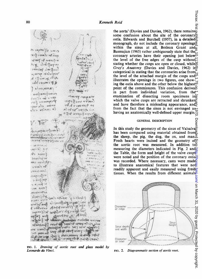
The earliest documented interest in the sinus ofValsalva dates from the Renaissance with thedescription and drawings of Leonardo da Vinci(1513). However, whilst his notes indicate intensetheoretical interest in the possible role of theaortic sinus in the closure mechanism of the valve,his drawings of the sinus remain diagrammatic
and he chooses to ignore the position of thecoronary arteries (Fig. 1). In passing, it is ofinterest that Leonardo outlines a method for theconstruction of a glass model of the aortic rootso that the movements of fluid within the sinusescan be closely observed using dye studies. In 1740was published the first anatomical account of theaortic sinuses (Valsalva, 1740). Valsalva was struckby the uniform presence of sinuses in a variety ofbirds and mammals and concluded that they mustserve a common purpose in all these creatures.He suggested that their main function was to dis-sipate the violence of systolic contraction byallowing blood to enter the sinuses during systole.His account goes on to relate the sinuses tocoronary artery filling, suggesting that this takesplace in diastole. While Valsalva gives a detaileddescription of the valve cusps, somewhat sur-prisingly, a detailed account of the configurationof a sinus is omitted.
Since 1740, surgical interest in the sinus hasbeen limited to the relatively rare condition ofaneurysmal dilatation, most commonly affectingthe right and non-coronary sinuses (Hudson,1965; Burchell and Edwards, 1951). Anatomicaldescriptions stemming from this give good detailedaccounts of the topography of the sinuses in man,although again omitting details of the sinusesthemselves (Edwards and Burchell, 1957). A recentstudy gives a careful histological picture of theaortic root in the dog and also compares the sizeand position of the right and left coronary ostia(Boucek, Takashita, and Fojaco, 1964). However,because the sinuses themselves lack precise ana-tomical definition in standard and specialized texts,being invariably described as 'slight dilatations of
79
on March 31, 2020 by guest. P
rotected by copyright.http://thorax.bm
j.com/
Thorax: first published as 10.1136/thx.25.1.79 on 1 January 1970. D
ownloaded from
Kenneth Reid
-tA-e +^
C? .aVe, -v
* 'F "' ." U.'t..*; ..
a''?, r-4 a
4 .-.V' aWaMt'iP
S .'rra¶44fW r"-il ' ^t:
:4'e# t
'4' '4P
...
'5rn'4-, *w' .' 'f2
*wtl( 4'
the aorta' (Davies and Davies, 1962), there remainssome confusion about the site of the coronaryostia. Edwards and Burchell (1957), in a detailedmonograph, do not include the coronary openingswithin the sinus at all, Boileau Grant andBasmajian (1965) rather ambiguously state that thecoronary arteries have their opening just belowthe level of the free edges of the cusp withoutstating whether the cusps are open or closed, whileGray's Anatomy (Davies and- Davies, 1962) iscategorical in stating that the coronaries arise fromthe level of the attached margin of the cusps andillustrates the openings in two figures, one show-ing the ostia above and the other below the highestpoint of the commissures. This confusion derivesin part from individual variation, from theexamination of dissecting room specimens inwhich the valve cusps are retracted and shrunkenand have therefore a misleading appearance, andfrom the fact that the sinus is not envisaged ashaving an anatomically well-defined upper margin.
GENERAL DESCRIPTION
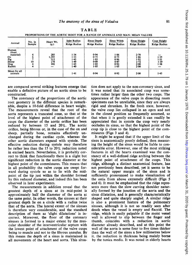
In this study the geometry of the sinus of Valsalvahas been compared using material obtained fromthe sheep, the pig, the dog, the ox, and man.Fresh hearts were incised and the geometry ofthe aortic root was measured. In addition tomeasuring the diameters indicated in Fig. 2 andthe Table, the form and height of the valve cuspswere noted and the position of the coronary ostiawas recorded. Where necessary, casts were madeto illustrate anatomical features that were notreadily apparent and easily measured using freshtissues. When the results from different animals
'4 a&t
"'V
.4
4,4+ v 4 g *jt iFx njlr gje~~~~~~~~~~~~A~1, .....;;2...a:6e9>+
r~}iQ .'s4 +_ja 8w'tr t4 t r^
eJtif " t4wwa A . J Wtnft wi tSl'.4In' tSY X e#
'V *wwwt 4s
FIG. 1. Drawing of aortic root and glass model byLeonardo da Vinci. FIG. 2. Diagrammatic section of aortic root.
80
on March 31, 2020 by guest. P
rotected by copyright.http://thorax.bm
j.com/
Thorax: first published as 10.1136/thx.25.1.79 on 1 January 1970. D
ownloaded from
The anatomy of the sinus of Valsalva
TABLEPROPORTIONS OF THE AORTIC ROOT FOR A RANGE OF ANIMALS AND MAN: MEAN VALUES
No. in Heart Wt. Inlet Radius Sinus Depth Sinus Width Sinus Height Cusp HeightAnimal Sample (g.) Ridge Radius Ridge Radius Ridge Radius Ridge Radius Ridge Radius
Human(fixedmaterial) 6 285 1 34 0 94 1-06 2-70 -
Ox 6 1,295 1-33 0 75 0 75 1-86 1-35Pig 7 212 1-45 1-03 0-96 1-92 1 51Sheep 10 174 1-49 0-89 0 87 1X72 1-39Dog 4 102 1-45 1-05 0-92 2 00 1-38Rabbit 4 - 1-40 1-05 1-05 2 00 -
Mean for allanimals 141 094 092 200 145
are compared several striking features emerge thatenable a definitive picture of an aortic sinus to beconstructed.The constancy of the proportions of the aortic
root geometry in the different species is remark-able, despite a 10-fold difference in heart weight.The measurements reveal that the root of theaorta represents a truncated cone, so that at thelevel of the highest point of attachment of thecusps the diameter of the aortic orifice has beenreduced by between 15 and 20%. The aorticorifice, being fibrous or, in the case of the ox andsheep, partially bone, remains effectively un-changed during the cardiac cycle, whereas theother aortic diameters expand with systole. Theeffective reduction during systole may thereforebe rather less than the 15 to 20% reduction notedin excised hearts. Nevertheless, it is probably cor-rect to think that functionally there is a slight butsignificant reduction in the aortic diameter at thehighest point of the commissures. This means thatin all probability the valve cusps are swept for-ward during systole so as to lie with the mid-point of the tip just within the shoulder formedby this reduced diameter, and indeed this has beenobserved in later experiments.The measurements in addition reveal that the
greatest depth of a sinus at its mid-point isapproximately half the diameter of the aorta atthe same point. In other words, the sinuses at theirgreatest depth lie on a circle with a radius twicethat of the aorta. The lateral extent of the sinusesis therefore very considerable, and the standarddescription of them as 'slight dilatations' is in-correct. Moreover, the floor of the coronarysinuses is formed to a major extent by the mus-cular shoulder of the interventricular septum withthe lowest point of attachment of the valve cuspsbeing to muscle and not to the fibrous annulus. Bythis means the depth of a sinus is maintained inall movements of the heart and aorta. This situa-
tion does not apply to the non-coronary sinus, andit was noted that its associated cusp was some-times rather larger than the other two cusps. Theappearance of the valve cusps in dissecting roomspecimens can be unreliable, since they are alwaysrigid and shrunken. In the fresh state, however,the valve cusp lies collapsed in an open and notin the closed position so frequently assumed, sothat when it is gently extended it can readily beappreciated that in systole the cusp very nearlyoccludes its sinus, so that the highest point of thecusp tip is close to the highest point of the com-missures (Figs 3 and 4).
It might be argued that if the upper limit of thesinus is anatomically poorly defined, then measur-ing the height of the sinus would be liable to con-siderable error. However, one of the most strikingfeatures in all the hearts examined was the con-stancy of a well-defined ridge arching between thehighest point of attachment of the cusps. Thisridge, although a distinct anatomical feature, hasnot previously been described, yet it seems to bethe natural upper margin of the sinus and issufficiently pronounced to make visualization ofthe ostia from above extremely difficult (Figs 5and 6). It must be emphasized that the ridge repre-sents more than the slow curving shoulder natur-ally formed by the junction of the aorta and thesinus dilatation, and is generally characteristicallyshaped and quite sharply angled. A similar ridgeis also a prominent feature of the pulmonarysinuses, although it is not so readily seen afterdeath when the vessel is empty and flaccid. Theridge, which is easily palpable if the moist vesselwall is allowed to slip between the finger andtbumb, coincides with a reduction in aorticdiameter already described, and at this point thewall of the aorta is some four to five times thickerthan the wall of the sinus a few millimetres belowit, the reduction in thickness being mainly borneby the tunica media. It was noted in elderly hearts
81
on March 31, 2020 by guest. P
rotected by copyright.http://thorax.bm
j.com/
Thorax: first published as 10.1136/thx.25.1.79 on 1 January 1970. D
ownloaded from
Kenneth Reid
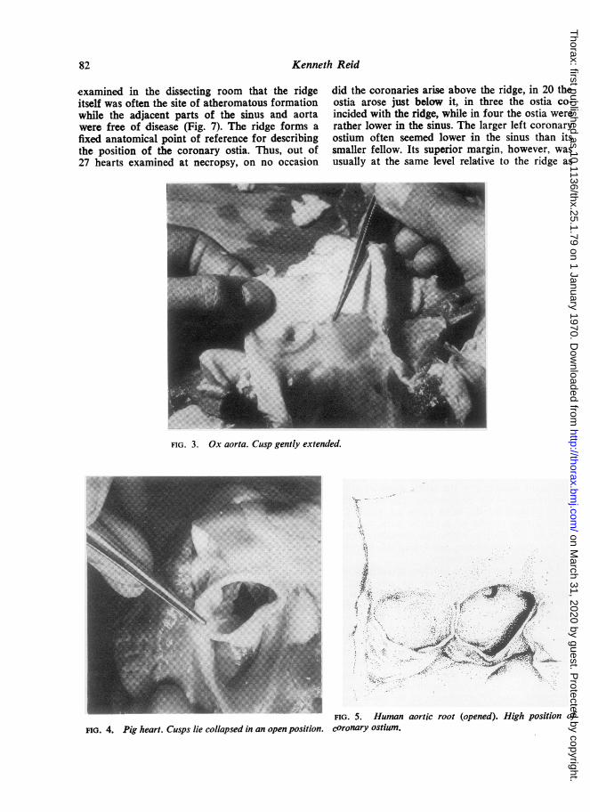
examined in the dissecting room that the ridgeitself was often the site of atheromatous formationwhile the adjacent parts of the sinus and aortawere free of disease (Fig. 7). The ridge forms afixed anatomical point of reference for describingthe position of the coronary ostia. Thus, out of27 hearts examined at necropsy, on no occasion
did the coronaries arise above the ridge, in 20 theostia arose just below it, in three the ostia co-incided with the ridge, while in four the ostia wererather lower in the sinus. The larger left coronaryostium often seemed lower in the sinus than itssmaller fellow. Its superior margin, however, wasusually at the same level relative to the ridge as
FIG. 3. Ox aorta. Cusp gently extended.
.4
.1
.x-.I ,
;.
FIG. 5. Human
FIG. 4. Pig heart. Cusps lie collapsed in an open position. coronary ostium.aortic root (opened). High position of
82
4t...
,f
on March 31, 2020 by guest. P
rotected by copyright.http://thorax.bm
j.com/
Thorax: first published as 10.1136/thx.25.1.79 on 1 January 1970. D
ownloaded from

The anatomy of the sinus of Valsalva
Vo.-
..
1 -
I1
/. I
I I
/ i.
7
1. .-,
V.
b
FIG. 6. (a) Ox aortic root. Low position of ostium. (b) Human sinus ridge in profile.
FIG. 7. Excised portion of 66-year-old male aortic rootshowing atheroma at sinus ridge (human).
that of the right coronary artery. The direction ofthe coronary arteries as they leave the sinuses isalways towards the apex of the heart (Fig. 8). Thismeans that the inferior or proximal margin of thecoronary ostium presents in its turn a promontoryagainst which fluid circulating in the sinus canabut. It was of great interest that in the group
FIG. 8. Aortic root of dog. Note e.tcnt ci sirus floorformed by interventricular septum and high position ofcoronary ostium.
examined the fleetest of the animals, namely thedog and man, demonstrated the highest positionof the coronary ostia (Fig. 9).While no systematic study of the pulmonary
root has yet been undertaken, the pulmonaryartery was frequently incised at the same time asmeasurements of the aorta were being made. Thepresence of a ridge was confirmed and the generalconfiguration appeared to parallel closely the
a
k6".4 -
.-F
rl.
,II(t
I.. ..,
on March 31, 2020 by guest. P
rotected by copyright.http://thorax.bm
j.com/
Thorax: first published as 10.1136/thx.25.1.79 on 1 January 1970. D
ownloaded from

Kenneth Reid
FIG. 9. Cast of aortic root (dog). FIG. 10. Posterior aspect of ox cusp showing strandedarrangement ofreinforcing fibres.
aortic situation. However, the pulmonary rootafter death collapses and its shape is lost. Thedepth of the sinuses was therefore observed in theanaesthetized dog at operation, when they appearedto be of the same order relative to the pulmonaryartery as the aortic sinuses to the aorta.The commissures were also examined in the
group of animals studied. Cleland (1964), discuss-ing the surgery of the aortic valve, states that thevalve cusps are attached to each other for a dis-tance of 1 to 2 mm. at the base (the highest pointof attachment of the valve cusp) where the twocusps run together. In this series, while a very fewanimals and man occasionally demonstratedmacroscopic interlacing of fibres between thecusps, this was not the general arrangement. Theinter-digitating collagenous fibres of adjacentcusps and the aortic wall combine to produce aslight but palpable ridge just below the highestpoint of the commissures dense enough to give agritty sensa-tion when incised with a scalpel.Despite this, the cusp flaps themselves remainquite separate from one another. The point atwhich the cusps were most intimately associatedis not at the base, the point of insertion of therelatively non-fibrous lunule, but below this, wherethe denser contact surfaces of the cusps insert. Itwas often noted that at this same point, but onthe sinus surface of the valve cusp, one or two ofthe principal fibre bundles forming the supporting
tissue of the cusp were frequently quite separatefrom the cusp although they always inserted intothe cusp before reaching the node (Fig. 10).
DISCUSSION AND CONCLUSIONS
The more or less identical geometry of the aorticroot together with so many other commonanatomical species strongly suggests that thehaemodynamics of the region in differentspecies may also be closely parallel. Moreover,the form and features of the pulmonary rootmake it seem likely that similar root fluidevents occur in this situation. Direct observationsof aortic roots in the fresh state confirm theuniversal presence of a sinus ridge limiting thedistal extent of the sinus of Valsalva and indicatethat the valve cusps have no significant elasticrecoil tending to bring them into apposition butlie collapsed in an 'open' position. It follows fromthis that some other mechanism in life operatesso as to achieve valve closure. It seems likely thatmany of the complications that have attended theuse of aortic valve prostheses that utilize a tri-leaflet principle, notably fatigue and embolic prob-lems, stem from an inherent design fault wherebythe leaflets have been made in such a way as tospring back into a closed position (Hufnagel,1969; Hittmann, 1966). All these prosthetic valvesare associated with pressure gradients across them
84
on March 31, 2020 by guest. P
rotected by copyright.http://thorax.bm
j.com/
Thorax: first published as 10.1136/thx.25.1.79 on 1 January 1970. D
ownloaded from

The anatomy of the sinus of Valsalva
and marked degrees of turbulence, both indicativeof valvular stenosis (Reid and Bellhouse, 1968b).Likewise, the presence and importance of thesinus ridge as inherent in aortic valve geometryhas not been fully appreciated. The coincidentalobservation that the coronary ostia always liewithin the sinus of Valsalva as redefined suggeststhat systolic coronary blood flow is more impor-tant than is generally assumed. Especially signi-ficant is the different location of the ostia relativeto the sinus ridge in fleet and grazing animals. Thesuggestion is that the fleetest animals have ostiamost favourably placed near the ridge so as toexploit the 'ram' effect of blood leaving the ven-tricle in systole. It seems reasonable to assumethat the aortic sinuses are therefore a means ofensuring that the coronary arteries are kept primedwith blood during systole (this is borne out byobservation of cine-angiograms), regulate coronaryflow responses during exercise (Van Citters andFranklin, 1969; Gregg, 1963), and act as pressurerecovery chambers initiating valve closure duringthe latter part of systole. It follows that disruptionof aortic root geometry by either disease or sur-gery would be accompanied by reduction ofsystolic coronary blood flow and defective valveclosure, effects that would be more marked withexercise.
It is suggested that outlet non-return checkvalves as prostheses should be designed so as toreproduce the essential anatomical features of the
aortic root, namely cusps that at rest lie in thefully open position and are in important relationto a sinus ridge.
REFERENCESBoucek, R. J., Takashita, R., and Fojaco, R. (1964). Functional
anatomy of the ascending aorta and the coronary ostia (dog).Amer. J. Anat., 114, 273.
Burchell, H. B., and Edwards, J. (1951). Aortic sinus aneurysm withcommnunications into the right ventricle and associated ventri-cular septal defect. Proc. Mayo Clin., 26, 336..
Cleland, W. P. (1964). The surgery of the aortic valve. In RecentAdvances in Surgery, 6th ed., Ch. 10, p. 186. Ed. Taylor, S.Churchill, London.
Davies, D. V., and Davies, F., ed. (1962). Gray's Anatomy, Descriptiveand Applied, 33rd ed. Longmans, London.
Edwards, J. E., and Burchell, H. B. (1957). The pathological anatomyof deficiencies between the aortic root and the heart, includingaortic sinus aneurysms. Thorax, 12, 125.
Grant, J. C. Boileau, and Basmajian, J. V. (1965). Heart and pericar-dium. In Grant's Method of Anatomy, 7th ed., p. 500. Williamsand Wilkins, Baltimore.
Gregg, D. E. (1963). Physiology of the coronary circulation. Circula-tion, 27, 1128.
Hittman Associates Inc., Baltimore, Maryland (1966). AnalyticalReport on Six Studies Basic to Consideration of Artificial HeartProgramme, Vol. 2-Technology survey for artificial heartdevices 3-41, Section E. Valves.
Hudson, R. E. B. (1965). The aortic valve. Cardiovascular Pathology,Vol. 2, p. 1969. Arnold, London.
Hufnagel, C. (1969). New Hufnagel tri-leaflet aortic valve. Heyer-Schulte Corporation Medical Products, California.
Leonardo da Vinci (1513). Aortic pulmonary valves. Leonardo daVinci on the Human Body (1952). By O'Malley, C. D., andSaunders, J. B. de C., pp. 258-274. Henry Schuman, New York.
Reid, K. G., and Bellhouse, B. J. (1968a). The mechanism of aorticvalve closure and the function of the sinus of Valsalva. Proceed-ings of the European Congress of Cardiovascular Surgeons, RoyalCollege of Surgeons, London. Minerva Med., Milan.
-- (1968b). A Bio-engineering Study of the Aortic Root.University of Oxford, Department ofEngineering Science Report.
Valsalva, A. M.(1740). Arteriae Magnae Sinus In Opera. Ed. Morgagni,J.B.,Vol.1,p.129. Venice.
Van Citters, R. L., and Franklin, D. L. (1969). Cardiovascularperformance of Alaska sled dogs during exercise. Circulat. Res.,24,33.
85
on March 31, 2020 by guest. P
rotected by copyright.http://thorax.bm
j.com/
Thorax: first published as 10.1136/thx.25.1.79 on 1 January 1970. D
ownloaded from