THE ANATOMY OF THE BRACHIAL PLEXUS AS … strated with MRI the vascular low-signal intensity...
10
THE ANATOMY OF THE BRACHIAL PLEXUS AS DISPLAYED BY MAGNETIC RESONANCE IMAGING: TECHNIQUE AND APPLICATION James D. Collins, MD, Anthony C. Disher, MD, and Theodore Q. Miller, MD Los Angeles, California Full field of view coronal chest magnetic resonance imaging (MRI) routinely displays bilateral images of the brachial plexus, surface anatomy, and anatomic structures. Eighty pa- tients had chest radiographs correlated with surgery for thoracic outlet syndrome. The PA chest film findings correlated with the surgical findings: smaller thoracic inlet on the concave side of the cervicothoracic spine scoliosis, shorter distance between the dorsal spine of the second or third thoracic vertebral body to the concavity of the first ribs, asymmetric clavicles and coracoid processes, synchon- drosis of the first and second ribs, and muscle atrophy on the side of the clinical complaints. More than 235 patients were imaged. One hundred sixty-five of these were imaged with a 1.5-T unit and 3-D reconstruction MRI. Coronal, transverse (axial), oblique transverse, and sag- ittal plane Ti-weighted, selected T2-weighted, and fast spine echo pulse sequences were obtained, 4- to 5-mm slice thickness, 40 to 45 cm full field of view, 512x256 matrix and 2 NEX. Two-dimensional time of flight (2D TOF), From the Department of Radiological Sciences, UCLA School of Medicine, and the Department of Radiology, Charles R. Drew Postgraduate School of Medicine, Los Angeles, California. Presented at the 10th Annual Meeting of the American Association of Clinical Anatomists, June 17, 1993, Seattle, Washington; the 98th Annual Convention and Scientific Assem- bly of the National Medical Association, August 9, 1993, San Antonio, Texas; and the Sunderland Society, September 22, 1993, Seattle, Washington. Address reprint requests to Dr James D. Collins, UCLA School of Medicine, Dept of Radiologi- cal Sciences, 10833 Le Conte Ave, Los Angeles, CA 90024- 1721. magnetic resonance angiography (MRA) se- quences were obtained in selected patients. Coronal, transverse, and sagittal sequences were reformatted for evaluation. Saline water bags were placed between the neck and thorax to enhance the signal-to-noise ratio. Compro- mising abnormalities of the brachial plexus were confirmed at surgery. Compromise of the neurovascular supply seemed to be one etiol- ogy that could be demonstrated. The clinical history, technique, and anatomic bilateral bra- chial plexus imaging is stressed to improve patient care. The cervical rib is one of the compromising brachial plexopathies selected for this presentation. (J Nat! Med Assoc. 1 995;87:489-498.) Key words * anatomy * brachial plexopathy * nerve model * MRI * pathology * 3-D color reconstruction Conventional roentgenography, linear tomography, computerized tomography (CT), and magnetic reso- nance imaging (MRI) are radiographic modalities used to image the thorax, shoulder girdle, and soft tissues. I Vascular structures, surface anatomy, and the soft tissues are incompletely imaged by conven- tional radiographic techniques. Computerized to- mography extends the capabilities of radiographic imaging to obtain detailed transverse (axial) ana- tomic sections but CT does not definitively separate tumors from vascular and neurovascular structures. Computed tomography reconstructed imaging of soft-tissue anatomy is not satisfactory, and detailed peripheral nerve imaging is not possible. Magnetic JOURNAL OF THE NATIONAL MEDICAL ASSOCIATION, VOL. 87, NO. 7 489
Transcript of THE ANATOMY OF THE BRACHIAL PLEXUS AS … strated with MRI the vascular low-signal intensity...

THE ANATOMY OF THE BRACHIALPLEXUS AS DISPLAYED BYMAGNETIC RESONANCE IMAGING:TECHNIQUE AND APPLICATIONJames D. Collins, MD, Anthony C. Disher, MD, and Theodore Q. Miller, MDLos Angeles, California
Full field of view coronal chest magneticresonance imaging (MRI) routinely displaysbilateral images of the brachial plexus, surfaceanatomy, and anatomic structures. Eighty pa-tients had chest radiographs correlated withsurgery for thoracic outlet syndrome. The PAchest film findings correlated with the surgicalfindings: smaller thoracic inlet on the concaveside of the cervicothoracic spine scoliosis,shorter distance between the dorsal spine ofthe second or third thoracic vertebral body tothe concavity of the first ribs, asymmetricclavicles and coracoid processes, synchon-drosis of the first and second ribs, and muscleatrophy on the side of the clinical complaints.
More than 235 patients were imaged. Onehundred sixty-five of these were imaged with a1.5-T unit and 3-D reconstruction MRI. Coronal,transverse (axial), oblique transverse, and sag-ittal plane Ti-weighted, selected T2-weighted,and fast spine echo pulse sequences wereobtained, 4- to 5-mm slice thickness, 40 to 45cm full field of view, 512x256 matrix and 2NEX. Two-dimensional time of flight (2D TOF),
From the Department of Radiological Sciences, UCLA Schoolof Medicine, and the Department of Radiology, Charles R. DrewPostgraduate School of Medicine, Los Angeles, California.Presented at the 10th Annual Meeting of the AmericanAssociation of Clinical Anatomists, June 17, 1993, Seattle,Washington; the 98th Annual Convention and Scientific Assem-bly of the National Medical Association, August 9, 1993, SanAntonio, Texas; and the Sunderland Society, September 22,1993, Seattle, Washington. Address reprint requests to DrJames D. Collins, UCLA School of Medicine, Dept of Radiologi-cal Sciences, 10833 Le Conte Ave, Los Angeles, CA 90024-1721.
magnetic resonance angiography (MRA) se-quences were obtained in selected patients.Coronal, transverse, and sagittal sequenceswere reformatted for evaluation. Saline waterbags were placed between the neck and thoraxto enhance the signal-to-noise ratio. Compro-mising abnormalities of the brachial plexuswere confirmed at surgery. Compromise of theneurovascular supply seemed to be one etiol-ogy that could be demonstrated. The clinicalhistory, technique, and anatomic bilateral bra-chial plexus imaging is stressed to improvepatient care. The cervical rib is one of thecompromising brachial plexopathies selectedfor this presentation. (J Nat! Med Assoc.1 995;87:489-498.)
Key words * anatomy * brachial plexopathy* nerve model * MRI * pathology
* 3-D color reconstruction
Conventional roentgenography, linear tomography,computerized tomography (CT), and magnetic reso-nance imaging (MRI) are radiographic modalitiesused to image the thorax, shoulder girdle, and softtissues. I Vascular structures, surface anatomy, andthe soft tissues are incompletely imaged by conven-tional radiographic techniques. Computerized to-mography extends the capabilities of radiographicimaging to obtain detailed transverse (axial) ana-tomic sections but CT does not definitively separatetumors from vascular and neurovascular structures.Computed tomography reconstructed imaging ofsoft-tissue anatomy is not satisfactory, and detailedperipheral nerve imaging is not possible. Magnetic
JOURNAL OF THE NATIONAL MEDICAL ASSOCIATION, VOL. 87, NO. 7 489

BRACHIAL PLEXUS
me
Figure 1. Cadaver dissection demonstrating the "M" or "W" ofthe left brachial plexus. Legend: lateral cord (2 bar whitearrows), brachial artery (BrA), median nerve (Me), musculo-cutaneous nerve (Mu), ulnar nerve (Ul), medial branch of thelateral cord of Mu (black single bar arrow), and lateral branch ofthe medial cord forming the median nerve (Me).
resonance imaging separates proton densities withinorgan systems and does not require reconstruction orrepositioning of the patient. The multi-planer MRIdisplays the anatomy of the brachial plexus andperipheral nerves for investigation by sequentialimaging of landmark anatomy according to protondensity distribution.2
The anatomy of the brachial plexus and peripheralnerves are graphically displayed in classical text-books.3'4 Anatomic and clinicopathologic nerve studiesallow investigative radiological studies using MRI to gobeyond textbook descriptions.5'6 Bilateral brachialplexus and peripheral nerve 3-D reconstruction imagingmakes possible demonstration of the relationship ofnerves to their surrounding landmark anatomy (Figure1). The knowledge of peripheral nerve and brachialplexus anatomic imaging offers new avenues forteaching medical students, residents, and health profes-sionals. I
The brachial plexus lies within the fascial planes ofthe neck and axilla, but is routinely displayed on MRI ofthe thorax and shoulder girdle.' Magnetic resonancemultiplane imaging in the supine position allowsbilateral sequential imaging of the thorax and thebrachial plexus. This is the feature that gave us theopportunity to image the brachial plexus anatomy.construct 3-D color images, and present the anatomyand compromising abnormalities of the brachial plexusas displayed by MRI.2'7
Compromising abnormalities of the brachial plexusand brachial plexopathies result from disorders of thecervicothoracic levels of the vertebral column, the firstrib, vascular supply, and soft tissues.8,9 The anterior andmiddle scalene muscles, through which the brachialplexus nerve roots pass and adhere to the subclavianartery, arise from the cervicothoracic levels of thevertebral column. The muscles insert and, in part,support the first ribs. The first ribs are curved, flat, andslope obliquely to form most of the thoracic inlet. Theslope that gives an anterosuperior directed uppersurface changes with respiration and cervicothoracicspine scoliosis, and affects structures crossing the rib.The anterior scalene muscle and the posterior insertionof middle scalene muscle and the first ribs form thescalene triangle. The change in the slope of the first ribaffects the interscalene triangle contents.2'5 The scalenemuscles are an important visual anatomic landmark inthe understanding of brachial plexopathy displayed byMRI.The upper nerve roots of the brachial plexus descend
along the margin of the middle scalene muscle and jointhe lower nerve roots to envelope the subclavian artery.The nerve roots bind to the artery within the scalenetriangle to form a neurovascular bundle.'0 This bundlepasses with sympathetic fibers unprotected over thefirst rib through the scalene triangle, beneath theclavicle and subclavius muscle, inferior to the coracoidprocess and posterior to the tendon of the pectoralisminor muscle into the axilla.
The subclavian artery, similar to the artery within thefemoral triangle, is unprotected4'8 but is not routinelycannulated for vascular studies. Different from thefemoral artery, a muscle (the anterior scalene muscle)separates the subclavian vein from the subclavianartery. This makes the anterior scalene muscle animportant anatomic landmark in determining compres-sion (effacement) of the scalene triangle contents.Enlargements of the anterior scalene muscle or atrophyof the anterior scalene muscle increases or decreases thedistance between the subclavian artery and the sub-
490 JOURNAL OF THE NATIONAL MEDICAL ASSOCIATION, VOL. 87, NO. 7
BRACHIAL PLEXUS
clavian vein as demonstrated on transverse MRIsequences.2 This distance may be measured on the firstimage or raw data of the magnetic resonance angiogra-phy (MRA) 2-D time of flight (TOF) of the brachialplexus. 'A thin layer of deep fascia envelopes the neurovascu-
lar bundle of the upper limb. Four spaces are formedfrom the root of the neck to the lower part of the axilla.The spaces that may have clinical problems thatcompromise the brachial plexus8 include the interverte-bral foramina, scalene triangle, costoclavicular, costo-coracoid, and the axilla. In most individuals, the fascialplane spacing between soft tissues and osseous struc-tures is adequate to perform routine functions withoutcompromising the nerves and the artery that combine toform the neurovascular bundle. Pathology involvingperipheral nerves alters fascial planes. Acute or perma-nent changes may alter the adjacent tissues, therebycompromising the vascular supply that nourishes theperipheral nerves.2 5 6 This results in patients presentingwith clinical symptoms. These changes also may occurfrom articular motion of the shoulder girdle, autoim-mune disease, hypertrophic enlargement of the scalenetriangle muscles, congenital rib anomalies, pressure byligaments, instances of abnormal insertion of thescalene (anomalous) muscles, fractures, primary andsecondary lymphedema and tumors, crushing injuriesof the thorax, and arteriosclerotic disease that results invascular thrombosis and ruptured silicone breast im-plants.2'5'6'8 Patients' complaints may stem from degen-erative changes of their osseous structures. Aging andlaxity of their muscular structures may result incomplaints from congenital compromising abnormali-ties that were not obvious problems at a youngerage.2'5'8
Magnetic resonance imaging demonstrates soft-tissue detail by proton distribution and provides highresolution imaging of nerves, vascular structures, andthe lymphatics.",2"2"3 The phospholipids found in themyelin of nerves are high-intensity signals whenvisualized in vitro and intermediate- to high-intensitysignals when demonstrated in vivo. Positive modeT 1-weighted images show blood flow as low-signalintensities (black) and subcutaneous fat as high-signalintensities (white). Diminished blood flow may appearas an intermediate-signal intensity.'4 The T1-weightedsequence displays the deep fascial layer of tissuecontaining the neurovascular compartments. Fast spineecho (FSE) with nonfat and nonwater suppressiontechnique) displays the long axis of peripheral nerveswith high-signal intensities.2
The signal-to-noise ratio decreases with FSE imag-ing, causing decreased contrast and resolution of theimages. The low-signal intensities of flowing blood andthe high-signal intensities of myelin combine to varythe high signal intensity of nerves. The signal intensityof nerves then may depend on the imaging axis of thatnerve.' The combination of a rich blood supply to thespinal cord and cerebral spinal fluid results in theintermediate-signal intensity of the spinal cord. Nerveroots may have high-signal intensities after they exitfrom the subarachnoid space. The low-signal intensityvertical bands of nutrient vessels may interrupt thehigh-signal intensity of nerves.2,10,14.15 Compressioninjuries of nerves involve an ischemic component andalter blood supply. Ischemia initiates inflammatorychanges causing edema and fibrosis. Inflammation andedema diminishes the signal intensity of nerves andalters their architecture when compromised in autoim-mune disease.12
Bilateral imaging of the brachial plexus is based on thefact that the brachial plexus envelopes and adheres to amajor artery forming a neurovascular bundle and thatdisplaying the artery ensures imaging the nerves.5,10,15 In1985, we presented our findings to David Maxwell of theUCLA anatomy department to supplement instruction inanatomy. He prepared a template list of anatomicalstructures for teaching the first-year medical studentcourse in gross anatomy to determine if our team coulddemonstrate the anatomy of the thorax and shouldergirdle with MRI. We imaged the prepared list, includingbilateral imaging of the brachial plexus. We presented ourresults for teaching medical students, residents, and otherhealth professionals.'
Compromising abnormalities of the brachial plexushad not been imaged by MRI. Therefore, we alsopresented our findings to the UCLA Department ofSurgery. A list was prepared of 80 patients who hadsurgery for brachial plexus compression to determine ifthere were any consistent radiographic findings. The plainchest radiographs revealed several variations in alignmentof the osseous structures which correlated with the MRI.2We demonstrated our findings and suggested MRI as theimaging modality that might contribute answers prior tosurgical management. The UCLA Department of Surgerythen requested assistance with selected patients forbilateral brachial plexus imaging. As a result of ourpresentation, more than 235 patients have undergonebilateral brachial plexus MRI since our presentation.
In 1945, Sunderland's anatomic nerve model demon-strated the blood supply of nerves,10 and in 1989, thegross anatomic nerve model of Collins et al'4 demon-
JOURNAL OF THE NATIONAL MEDICAL ASSOCIATION, VOL. 87, NO. 7 491
BRACHIAL PLEXUS
strated with MRI the vascular low-signal intensitymarginating the nerves that contribute to the separation ofnerves from surrounding tissues.1014 This article de-scribes a new technique that uses the knowledge ofanatomy to analyze the various etiologies of thoracicoutlet syndrome and to demonstrate effacement of nervescorrelated with patients' clinical complaints.We are unable to display entire sequential images for
the plane imaged. The images included in this article wereselected because they supported each other as agroup.28"15 Each image cannot be studied withoutconsidering the other images. The grainy nature of figures(noise) results because of the high magnification used, thediminished signal intensity due to the small amount of fatand increased muscle mass as in body builders,2'5 and the512 X 256 matrix that increases the resolution butdecreases contrast, resulting in a grainy appearance. Theenlarged and windowed (contrasted) cervical rib imagesdemonstrate the anatomy of the brachial plexus bygray-scale MRI and 3-D color reconstruction.
MRI IMAGING OF THE BRACHIAL PLEXUSTechnique
Plain chest radiographs (PA and lateral) are obtainedand reviewed prior to the MR examination. Theprocedure is discussed and the patient examined.Respiratory gating is applied throughout the procedureto minimize motion artifact and maximize the contrastof the soft-tissue signal intensities. The patient ispositioned supine in the body coil, arms down to theside (adduction and internal rotation), and imaging ismonitored at the MRI station.A body coil is used because it offers optimal full field
of view for bilateral imaging of the brachial plexus andprovides uniform signal-to-noise ratio across the imag-ing field that is necessary for 3-D reconstruction.Surface coils are limited to depth and field of view andare not adequate for bilateral imaging of the brachialplexus. Intravenous contrast agents are not admini-stered. A water bag (500 mL normal saline) is placed onthe right and the left sides of the neck above theshoulder girdle to increase signal-to-noise ratio forhigher resolution imaging. A full field of view (40 to 48cm) of the neck and the thorax is used to image bothsupraclavicular fossae. Contiguous (4 to 5 mm) coronal,transverse (axial), oblique transverse, and sagittalTl-weighted images are obtained. If there is clinicalevidence of scarring, tumor, or lymphatic obstruction,T2-weighted images or FSE pulse sequences areobtained. Four imaging sequences are acquired: co-ronal, transverse, oblique transverse, and sagittal.
Coronal abduction and external rotation of the upperextremities are used to determine if the change in armposition mimics the clinical complaint.
The coronal sequence is first to be imaged. Thebrachial plexus envelopes the artery (forming a neu-rovascular bundle), and the nerves are best imagedwhen the cursors are aligned to the arterial bloodsupply. The axillary artery margins vary in each patientand the cursors must be adjusted for each MRI brachialplexus examination. The cursors are positioned fromthe skin surface of the posterior chest wall to the skinsurface of the anterior chest wall for symmetry and 3-Dreconstruction and also to detect abnormalities that maymimic brachial plexopathies. The superior landmark isset at the base of the skull and the inferior landmark isset at the level of the kidneys. The image that bestdemonstrates the arterial blood flow to the upperextremities is selected as the baseline image for theremaining sequences. The transverse sequence is setfrom the baseline coronal image at the superior aspectof the third cervical vertebral body to the carina. Thelateral margins of the shoulder girdle are imaged toensure bilateral, simultaneous display of the brachialplexus. The oblique transverse sequence is set byaligning the cursors to the arterial blood supply of eachupper extremity using the baseline coronal sequence.The cursors are centered to the plane of the axillaryartery 2 cm below the inferior cord of the brachialplexus to the superior margin of the coracoid process.This sequence is necessary to detect signal intensity,architecture, and effacement of the long axis of thenerves, arteries, and veins.
The sagittal sequence is obtained by aligning thecursors lateral to the coracoid process and medially tothe insertion of the anterior scalene muscle on the firstrib. The sagittal plane is necessary to detect effacementof the neurovascular bundle by the coracoid process,pectoralis minor muscle, clavicle and subclavius mus-cle, axillary masses, and abnormalities of the scalenetriangle. When an image sequence is completed, it isimmediately transferred to another screen at an inde-pendent workstation for review and 3-D reformatdisplay. The software for this 3-D reconstruction isalready prepared in the 1.5-T MRI unit (Signa, GeneralElectric Medical Systems, Milwaukee, Wisconsin). Theimages are stored on CT tape (GE 9800) format and onoptical disks for 3-D color reconstruction at an ISGconsole (ISG Technologies Inc, Mississauga, Ontario,Canada). The entire study is monitored by the radiolo-gist and requires 11/2 hours. Selected color, black andwhite laser prints, and transparencies are obtained for
492 JOURNAL OF THE NATIONAL MEDICAL ASSOCIATION, VOL. 87, NO. 7

BRACHIAL PLEXUS
lectures and poster presentations; annotated images arepreserved on VHS, archived digital tapes, and opticaldisks.
EquipmentMagnetic resonance images are obtained on the 1.5-T
MR scanner. The 3-D reformatted images are videotapedon a separate work console at the monitoring station, andcomputerized color is applied to the images using the ISGconsole. A 512 X 256 matrix format is used.
Case ReportsCase 1: Cervical Rib. An 83-year-old, 50 kg
female presented with a long history of hypertensiontreated with diazide, arthritis in the hands and knees,exploratory laparotomy, and total hysterectomy withbilateral salpingo-oophorectomy in 1988; she under-went exploratory laparotomy with adhesions lysis ofsmall bowel obstruction in 1990. In addition, she givesa 5-year history of progressive atrophy and weakness inthe right hand and ulnar distribution. She described athrobbing sensation in the upper forearm that wouldextend into the palm of the hand, especially the ulnarside. When holding objects, she would develop a tremorin her right hand.
In 1989, a neurologic evaluation indicated progres-sive atrophy and weakness in the right hand and ulnardistribution with likely median nerve involvement. Shereceived physical therapy with protective elbow padsand hand splint. A repeat electromyogram and nerveconduction study in November 1993 indicated descrip-tions of a true right neurogenic thoracic outlet syn-drome: marked thenar atrophy with normal mediansensory nerve action potential, poor ulnar sensorypotential, and chronic denervation/reinnervationchanges in all C8/T1 muscles. Nerve conduction studiesdemonstrated no ulnar compression at the wrist orelbow, and ruled out an all-ulnar hand. The left upperextremity conduction studies were normal. The neuro-logical diagnosis was right neurovascular compressionof the brachial plexus. Radiographs for a cervical ribwere suggested. The radiographic reports made nomention of cervical ribs. A bilateral MRI of the brachialplexus was suggested to rule out compression at thescalene triangle. A PA and lateral chest radiograph wereobtained the day of the MRI examination.
The PA chest radiograph (Figure 2) demonstratedbilateral asymmetrical cervical ribs and narrowing ofthe intercostal spaces on the concaved side of thescoliosis. Measurements between the dorsal spine of thethird thoracic vertebral body to the concavity of the first
ribs revealed a smaller right thoracic inlet. Cervicotho-racic spine scoliosis, decreased soft-tissue muscle massover the right cervical region, degenerative compres-sion of the cervical vertebral bodies (C4-C6), degenera-tive changes of the cervicothoracic junction (C7-T1),and asymmetrical insertions of the first ribs at thesternum were present. The cervical ribs were small,asymmetric, posterior, and faintly displayed. The rightcervical rib was larger and angled anteromedially to itslower insertion on the first rib.The bilateral brachial plexus MRI examination
demonstrated asymmetrical cervical vertebral spinepillars, cervicothoracic spine scoliosis (Figure 3), anddegenerative compression of the cervicothoracic verte-bra (Figure 4) and the bilateral cervical ribs. The rightcervical rib compressed the C8 and T 1 nerve rootproximal to the formation of the displaced inferior trunk(Figure 4). The left cervical rib caused minimal superioreffacement of the left 8th cervical nerve root and theadjacent sympathetic nerves.
The coronal images were the best sequence of theseries to demonstrate the asymmetric degenerativechanges of the cervical spine pillars (higher on the rightthan left) and compression of the cervical vertebralbodies (C4-C6) (Figures 3 and 4). The nerve rootsfollowed the asymmetry of the cervical spine. Degener-ative changes of the acromioclavicular joint accentu-ated the inferior angulation of the right clavicle andsubclavius muscle. The subclavian arteries formed highloops over the first ribs, with the right subclavian arteryhigher than the left (Figure 3). The right cervical ribeffaced the subclavian artery, the 8th cervical nerve, andthe 1st thoracic nerve proximal to formation of theinferior nerve trunk, displaced anteriorly by the de-scending loop of the right subclavian artery (Figure 4).The left cervical rib did not appear to cause significanteffacement of the nerve roots.
The transverse sequence demonstrated the enlargedhead of the right clavicle, which was bowed anteriorlycompared with the left clavicle, the asymmetriccervicothoracic spine, and atrophy of the right scalenemuscles. The right nerve roots, divisions, and terminalbranches appeared smaller. The cervical rib effaced theright C8-TI nerve roots, sympathetic nerves, and thesubclavian artery as the nerve roots crossed the apex ofthe right lung (Figure 5), and the spinal cord wastethered to the right. Blood flow decreased in the leftinternal jugular vein. The bilateral oblique transverseimaging sequence confirmed the above findings anddisplayed mild effacement of the C8 nerve root by thesmaller left cervical rib.
JOURNAL OF THE NATIONAL MEDICAL ASSOCIATION, VOL. 87, NO. 7 493

BRACHIAL PLEXUS
*. w,:
1 ;'X.Z~~~~~~~~~~~~~~~~~~~~~~~~7gMM! 9
Figure 2. PA chest radi-ograph demonstrating anapparent rotated appear-ance secondary to scoli-osis. The larger right andleft ribs are marginatedby small arrows. Legend:aorta (A), superior marginright first (1 A) and left firstribs (2A), cervical verte-bral bodies (C6 & C7)transverse process of theright and left Ti vertebra(arrow heads), ends ofcervical ribs (single bararrow), and right (RL) andleft lungs (LL). Figure 3. Image 24 of the coronal series demonstrates asymmetry and splaying by the right cervical rib (outlined arrow).Legend: lateral pillars of the cervical spine (C3-C6), spinal cord (21), ventral ramus of the 2nd spinal nerve (2), C5 spinal nerve roots (5),C7 spinal nerve roots (7), right spinal accessories nerve (SA), right transverse process (Tp) of first (T1) and second (T2) thoracicvertebra, right lung (RL), sympathetic nerve (S and arrowheads), right first rib (small arrows), and right middle scalene muscle (MS).Figure 4. Image 27 of the coronal series demonstrates slight narrowing of the right subclavian artery (4 small arrows) and compressed(C4-C6) cervical vertebral bodies. Legend: right cervical rib (CR), C8 spinal nerve root (8), vertebral artery (VA), C4 & C5 spinal nerveroots (4 & 5), external jugular vein (ExJ), spinal accessories nerve (SA), brachiocephalic artery (BA), first rib (R), stellate ganglia (SG),middle cervical ganglia (MC), inferior (I) and middle nerve trunks (10), sympathetic nerve and right first rib (small arrows), first thoracicnerve root (1T), right and left vagus nerves (V), first thoracic vertebra (T1), right (RL) and left (LL) lungs, recurrent laryngeal nerves (33),left subclavian artery (S), trachea (T), esophagus (E), and aorta (A). Figure 5. Transverse image 18 demonstrates effacement of nervesby the right cervical rib. Legend: ansa subclavia (3), left normal C8 and right effaced C8 nerve roots (8), effaced first thoracic nerve root(4 small arrows) joining Ti nerve root (4 arrowheads), ascending (S) and descending (Sd) loops of subclavian artery, external (EJ) andinternal jugular (RJ & LJ) veins, anterior scalene muscles (RAS & LAS), common carotid arteries (RC & LC), trachea (T), esophagus (E),vertebral arteries (VA), dorsal spine (Sp) and spinal cord (21), multifidus muscle (Mul), spinal accessories nerve (SA), trapezius muscles(RTr & LTr), transverse process (38), C7 nerve root (7), middle scalene muscle (MS), left first rib (37 and 4 large arrows), vertebralarteries (VA), lamina of vertebral body (39), and sternocleidomastoid muscle (ST). Figure 6. First right sagittal image demonstrating thecervical rib (2 bar arrows) effacing C8 (8) and the first thoracic nerve root (1 T). Legend: mandible (Ma), internal jugular vein (RJ), anteriorscalene muscle (AS), vagus (V), recurrent laryngeal and superior cardiac nerves (33), trapezius muscle (Tr), brachiocephalic artery(BA), sternocleidomastoid muscle (ST), common carotid artery (CC), aorta (A), supraclavicular nerves (Sc), clavicle (C), manubriumsternum (M), neck subcutaneous fat (4 black arrows), and right lung (RL).
Figure 6 was the first image of the right sagittalsequence that demonstrated the cervical rib effacing theC8-T 1 nerve roots complementing the coronal andtransverse sequences. The recurrent laryngeal nerveparalleled to its origin from the vagus nerve and theadjacent superior cardiac nerves. The left sagittalsequence was normal.The 3-D color generated images (Figures 7, 8, 9, and
10) were cross referenced to the gray scale sequential
images to demonstrate the in vivo anatomic relationshipbetween the neurovascular bundles, osseous structures,and adjacent landmark anatomy. Figure 7 was rotatedinto a posterior oblique display to accentuate thedegenerative compression of C4-C6 cervical vertebralbodies, the asymmetric cervicothoracic spine scoliosis,and the bilateral cervical ribs. The thoracic spine wasconvexed to the right at the T5-T6 level. Figure 8demonstrated the nerve roots, nerve divisions, and
494 JOURNAL OF THE NATIONAL MEDICAL ASSOCIATION, VOL. 87, NO. 7

BRACHIAL PLEXUS
terminal branches of the brachial plexus. The rightcervical rib position and the C8-T 1 (TI darken) next tothe right long thoracic nerve crossed the first rib. Thelateral position of the radial nerve and medial positionof the right median and the ulnar nerves reflected theinternal rotation adduction of the arms with the handson the chest wall. The left brachial plexus appearedsmaller.The nerves and vascular structures were combined
into one image (Figure 9). The image displayedasymmetry and the loops of the subclavian arteries. Theleft cervical nerve root joined the sympathetic chain.The right inferior nerve trunk and the median nerveroots (lateral to the inferior trunk) arched anteriorly andover the right subclavian artery. Figure 10 combined theosseous, nerves, and vascular structures into one imageto demonstrate bilateral anatomic landmark relation-ships. The right subclavian artery arched high over thefirst rib and the cervical rib effaced the C8-T1 nerveroots. The smaller left cervical rib effaced the superiormargin of the C8 nerve root and sympathetic nervebranches. The manubrium sterni asymmetric articula-tion with the clavicles complemented the PA chestradiograph (Figure 2)
The 3-D images confirmed the gray scale imagingseries and displayed the compression changes of thecervical vertebral spine. The cervical rib compressionand effacement was displayed. The patient was in-formed of our findings. Because of her age, surgery wasnot recommended. Her physician elected to continueobservation and physical therapy as tolerated.Case 2: Neurolemmoma of the Median
Nerve. A 28-year-old male presented with a painfulpalpable mass on the medial aspect of the right upperarm. The neurological examination determined themass involved the median nerve. A bilateral brachialplexus MRI was requested to rule out possiblemultiple tumors prior to surgery. The radiologistrecorded a brief history and modified physicalexamination to confirm the location of the mass. Abilateral brachila plexus imaging was performed withcoronal and transverse sequences to ensure theproximal bilateral brachial plexus was normal andwithout any ohter tumor present.The coronal and transverse sequences were within
normal limits except for the mass involving the mediannerve of the right upper arm. Saline water bags wereplaced along the long axis of the right upper extremity.Oblique sagittal TI (Figure 11) and oblique sagittal(Figure 12) sequences were obtained of the right upperarm. The tumor was demonstrated and surgically
proven to be a neurolemmoma. The patient tolerated thesurgery without sequelae.
DISCUSSIONBilateral imaging of the brachial plexus requires
knowledge of landmark anatomy and the blood supplyof the normal brachial plexus that allows the radiologistto use computerized imaging to explain the clinicalmanifestations of the pathology. The clinical historymust be correlated with the anatomy to furtherunderstand the compromising brachial plexopathiesdisplayed by MRI.2,15
Radiology strives to increase the quality of imagingby subtracting the background and increasing the signalto noise ratio. The simple placement of saline waterbags next to the neck and next to the long axis of theextremities provides increased signal-to-noise ratio andincreased definition of peripheral nerve and soft-tissueimaging.'4"16 Respiratory compensation (gating) dimin-ishes artifacts caused by the uncontrolled movements ofbreathing (Figure 11) and increases artifacts in FSEpulse sequence imaging (Figure 12). Cursor linesindicate the position of each image and are required forcross reference between sequences. The absence of thenumbered cursor lines impedes interpretation.
Surface coils are used exclusively by some institu-tions. Their studies are designed to include surface coilsto image the brachial plexus. Although our institutionhas surface coils and the phased-array torso coils, theseare not routinely used. Furthermore, the phased-arraytorso coils cost $250 000 or more. The surfacepenetration does not give uniform imaging that thebody coil provides. We have been using saline waterbags to enhance MRI at our institution for more than 9years. Most health professionals and clinical radiologic-anatomists would not be able to afford the phased-arraytorso coils; the contiguous anatomic structures to thoseimportant in these cases would not be imaged. We mayhave found use for the phased-array torso coil, but thistechnique does not allow recognition of landmarkanatomic structures in sequential imaging. The addedfeature would not allow us to use the different planes ofimaging.
The authors stress enlarging selected images todemonstrate nerve effacement that we demonstrated inour cervical rib presentation (Figures 2 through 10).Cervical ribs are congenital abnormalities of thecervicothoracic spine. They may be present in patientsand not cause clinical complaints until an injury ofsome sort occurs.5.8 Often they go undetected and donot present a problem. When chest radiographs are
JOURNAL OF THE NATIONAL MEDICAL ASSOCIATION, VOL. 87, NO. 7 495
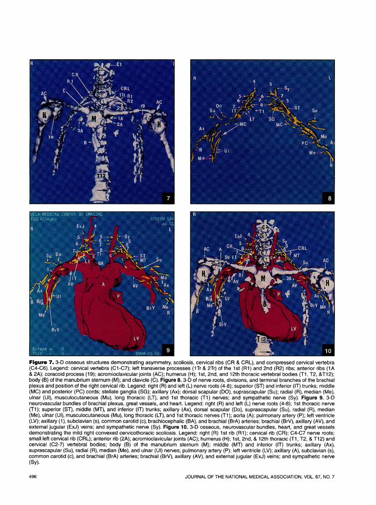
Figure 7. 3-D osseous structures demonstrating asymmetry, scoliosis, cervical ribs (CR & CRL), and compressed cervical vertebra(C4-C6). Legend: cervical vertebra (C1-C7); left transverse processes (1Tr & 2Tr) of the 1st (Ri) and 2nd (R2) ribs; anterior ribs (1A& 2A); coracoid process (19); acromioclavicular joints (AC); humerus (H); 1 st, 2nd, and 12th thoracic vertebral bodies (Ti, T2, &T1 2);body (B) of the manubrium sternum (M); and clavicle (C). Figure 8. 3-D of nerve roots, divisions, and terminal branches of the brachialplexus and position of the right cervical rib. Legend: right (R) and left (L) nerve roots (4-8); superior (ST) and inferior (IT) trunks; middle(MC) and posterior (PC) cords; stellate ganglia (SG); axillary (Ax); dorsal scapular (DO), suprascapular (Su); radial (R), median (Me),ulnar (Ul), musculocutaneous (Mu), long thoracic (LT), and 1st thoracic (T1) nerves; and sympathetic nerve (Sy). Figure 9. 3-Dneurovascular bundles of brachial plexus, great vessels, and heart. Legend: right (R) and left (L) nerve roots (4-8); 1 st thoracic nerve(Ti); superior (ST), middle (MT), and inferior (IT) trunks; axillary (Ax), dorsal scapular (Do), suprascapular (Su), radial (R), median(Me), ulnar (Ul), musculocutaneous (Mu), long thoracic (LT), and 1 st thoracic nerves (Ti); aorta (A); pulmonary artery (P); left ventricle(LV); axillary (1), subclavian (s), common carotid (c), brachiocephalic (BA), and brachial (BrA) arteries; brachial (BrV), axillary (AV), andexternal jugular (ExJ) veins; and sympathetic nerve (Sy). Figure 10. 3-D osseous, neurovascular bundles, heart, and great vesselsdemonstrating the mild right convexed cervicothoracic scoliosis. Legend: right (R) 1st rib (Ri); cervical rib (CR); C4-C7 nerve roots;small left cervical rib (CRL); anterior rib (2A); acromioclavicular joints (AC); humerus (H); 1st, 2nd, & 12th thoracic (Ti, T2, & T12) andcervical (C2-7) vertebral bodies; body (B) of the manubrium sternum (M); middle (MT) and inferior (IT) trunks; axillary (Ax),suprascapular (Su), radial (R), median (Me), and ulnar (Ul) nerves; pulmonary artery (P); left ventricle (LV); axillary (A), subclavian (s),common carotid (c), and brachial (BrA) arteries; brachial (BrV), axillary (AV), and external jugular (ExJ) veins; and sympathetic nerve(Sy).
496 JOURNAL OF THE NATIONAL MEDICAL ASSOCIATION, VOL. 87, NO. 7

BRACHIAL PLEXUS
Figure I11 Ti -weighted sagittal oblique image of the rightupper arm. The ulnar (Ul) nerve is effaced (open arrow) by theneurolemmoma. Legend: humerus (H), axillary (Ax), median(Me) and ulnar (Ul) nerves; brachial artery (BrA) and brachial(BrV); anterior deltoid (D) and posterior deltoid (1 1); short headof the biceps (79); teres major (17) and teres minor (18) andcoracobrachialis (81) muscles; musculocutaneus (Mu) nerve;and nutrient blood supply to the ulnar nerve (X).
reviewed, the cervical ribs are routinely listed as anincidental finding. Seldom does the radiologist receivea follow-up communication of their detection.Our patient was 79 years of age before her complaints
were related to a physician. The cervical ribs were notidentified. Her surgical admissions were emergencyclinical situations that did not permit quality chestimages. The ribs were suboptimally visualized on theportable chest radiographs. The consulting neurologistsuggested a bilateral brachial plexus MRI examinationbecause he was aware of our procedure. The MRIconfirmed the clinical neurologic examination. The
Figure 12 FSE of Figure 11 demonstrating decreasedcontrast, motion artifact, and tumor (open arrow). Legend:humerus (H), axillary (Ax), radial (R), median (Me), and ulnar(Ul) nerves; brachial (BrA) artery and brachial (BrV); anterior (D)and posterior (11) deltoid; short head of the biceps (79); teresmajor (17) and minor (18); coracobrachialis (81), flexor carpiulnaris (Fcu), and long head of the triceps (20) muscles; andmusculocutaneus (Mu) nerve.
bilateral cervical ribs, muscle laxity, and degenerativechanges of the osseous structures all seem to havecontributed to the diagnosis of a right thoracic outletsyndrome.5'8"15The authors are aware of alternative diagnoses that
may have caused her complaints. We are also aware thatour narrative presentation included statements withoutall of the images displayed. We did present a moreextensive report for the patient's file supported by theseselected images. Unlike radiographic images, a diagno-sis is not made from a single plain filin.'7"8 Multipleimages are provided in an MRI sequence from which a
JOURNAL OF THE NATIONAL MEDICAL ASSOCIATION, VOL. 87, NO. 7 497

BRACHIAL PLEXUS
selected group may best demonstrate the abnormality.Since it is not possible to present all of the images, thoseselected best demonstrated the pathology.
CONCLUSION3-D reconstruction is a useful tool to confirm gray
scale MRI examinations. Tailored anatomic bilateralMRI of the brachial plexus correlated with the clinicalhistory allows the study of brachial plexopathies.15 Weplan to continue reporting compromising abnormalitiesof the brachial plexus from our MRI experience.
Literature Cited1. Collins JD, Batra P, Brown K, Shaver ML. Anatomy of
the thorax and shoulder girdle as displayed by magneticresonance imaging. AnatRec. 1986;214:24A.
2. Collins JD, Shaver ML, Disher A, Miller TQ. Compromis-ing abnormalities of the brachial plexus as displayed bymagnetic resonance imaging. Anat Rec. 1 993;236(suppl1 ):44A.
3. Clemente CD. Gray's Anatomy. 31st ed. Philadelphia,Pa: Lea and Febiger; 1985.
4. Clemente CD. Anatomy: A RegionalAtlas of The HumanBody. 3rd ed. Baltimore, Md: Urban and Schwarzenberg; 1987.
5. Sunderland S. Nerves and Nerve Injuries. Baltimore,Md: Williams & Wilkins; 1968.
6. Dyck PK, Barnes J, Lais L. Pathological Alterations ofthe Peripheral Nervous System of Humans: Peripheral Neu-ropathy. 2nd ed. Philadelphia, Pa: WB Saunders Co; 1984.
7. Collins JD, Batra P, Brown K, Shaver ML. Anatomy ofthe thorax and shoulder girdle as displayed by magneticresonance imaging. J Natl Med Assoc. 1991;83:46-52.
8. Lord JW, Rosati LM. Thoracic-outlet syndromes. ClinicalSymposia. Newark, New Jersey. Ciba Pharmaceutical Com-pany. 1971;23:1-32.
9. Lord, JW. Critical reappraisal of diagnostic and thera-peutic modalities for thoracic outlet syndromes. Surg GynecolObstet. 1989;1 68:337-340.
10. Sunderland S. Blood supply of the nerves to the upperlimb in man. Archives of Neurology and Psychiatry. 1945;53:91 -115.
1 1. Collins JD, Shaver ML, Disher A, Miller TQ. The vascularsupply of the brachial plexus as displayed by magneticresonance imaging: magnetic resonance angiography (MRA).Presented at the Eleventh Annual Scientific Program of theAmerican Association of Clinical Anatomists; June 16, 1994;Galveston, Texas.
12. Collins JD, Batra P, Brown K, Shaver ML. Imaging thethoracic lymphatics: experimental studies of swine. ClinicalAnatomy. 1991;4:1-14.
13. Collins JD, Batra P, Brown K, Shaver ML. Magneticresonance imaging of chest wall lesions. J Natl Med Assoc.1991;83:352-360.
14. Collins JD, Shaver ML, Batra P, Brown K. Nerves onmagnetic resonance imaging. J Natl Med Assoc. 1989;81:129-134.
15. Collins JD, Shaver ML, Disher AC, Miller TQ. Compro-mising abnormalities of the brachial plexus as displayed bymagnetic resonance imaging. Clinical Anatomy. 1995;8:1-16.
16. Collins JD, Kovacs, BJ, Shaver M, Hall T, Brown K, BatraP, Sinha S. Enhancement of magnetic resonance images usingwater bags. J Natl Med Assoc. 1990;82:197-199.
17. Rigler LG. Roentgen diagnosis of small pleural effu-sions. JAMA. 1931 ;96:104-108.
18. Rigler LG. A half century of radiology. Semin Roent-genol. 1976;11:1-11.
498 JOURNAL OF THE NATIONAL MEDICAL ASSOCIATION, VOL. 87, NO. 7