THE ANATOMY AND HISTOLOGY OF THE ALIMENTARY CANAL … · 2019-06-03 · Alimentary Canal of a...
10
THE ANATOMY AND HISTOLOGY OF THE ALIMENTARY CANAL OF A CARNIVOROUS FISH MEGALOPS CYPRINOIDES (BROUSS)* BY S. M. KAMAL PASHA (Department o/Zoology, Presidency College, Madras) Received March 23, 1964 (Communicated by Dr. S. Krishnaswamy, F.A.SC.) THIS paper, third in the series, deals with the structure of the alimentary canal of a carnivorous fish Megalops cyprinoides. Similar accounts of an omni- vorous fish Mystus gulio and a herbivorous fish Tilapia mossambica were given previously (Pasha, 1964 a and 1964 b). Megalops c),prinoides, commonly known as ox-eye herring, is a favourite fish with anglers. It inhabits the seas, estuaries and tanks. It is able to adapt itself well and grows to a large size. The body of the fish has large scales. Eyes are very conspicuous as they are comparatively large. The material for the present investigation was collected from Coovum River, Madras. Standard fixatives and staining techniques were employed throughout the investigation. FOOD, FEEDING HABITS AND GROSS ANATOMY The stomach contents of about 30 fish were examined. The contents consisted chiefly of crustacea. A few insect larvae and rotifers were also noticed. In the laboratory they were fed on earthworms. When bits of earthworm were thrown to the fish which had been starved, the bits were snapped and swallowed while they were descending the water column. But the fish rarely tried to take the material after they had reached the bottom. These observations are indicative that the fish is a surface feeder or column feeder and that it is carnivorous. The alimentary canal consists of the mouth, buccal cavity pharynx, oesophagus, stomach, pyloric caeca, intestine and rectum (Text-Fig. 1). The gape measures about 2"5 cm. in a fish measuring 24.3 cm. The mouth * Formed part of the thesis approved by the University of Madras. 107
Transcript of THE ANATOMY AND HISTOLOGY OF THE ALIMENTARY CANAL … · 2019-06-03 · Alimentary Canal of a...

T H E A N A T O M Y AND HISTOLOGY OF T H E A L I M E N T A R Y CANAL OF A CARNIVOROUS FISH
MEGALOPS CYPRINOIDES (BROUSS)*
BY S. M. KAMAL PASHA (Department o/Zoology, Presidency College, Madras)
Received March 23, 1964
(Communicated by Dr. S. Krishnaswamy, F.A.SC.)
THIS paper, third in the series, deals with the structure of the alimentary canal of a carnivorous fish Megalops cyprinoides. Similar accounts of an omni- vorous fish Mystus gulio and a herbivorous fish Tilapia mossambica were given previously (Pasha, 1964 a and 1964 b).
Megalops c),prinoides, commonly known as ox-eye herring, is a favourite fish with anglers. It inhabits the seas, estuaries and tanks. It is able to adapt itself well and grows to a large size. The body of the fish has large scales. Eyes are very conspicuous as they are comparatively large.
The material for the present investigation was collected from Coovum River, Madras. Standard fixatives and staining techniques were employed throughout the investigation.
FOOD, FEEDING HABITS AND GROSS ANATOMY
The stomach contents of about 30 fish were examined. The contents consisted chiefly of crustacea. A few insect larvae and rotifers were also noticed. In the laboratory they were fed on earthworms. When bits of earthworm were thrown to the fish which had been starved, the bits were snapped and swallowed while they were descending the water column. But the fish rarely tried to take the material after they had reached the bottom. These observations are indicative that the fish is a surface feeder or column feeder and that it is carnivorous.
The alimentary canal consists of the mouth, buccal cavity pharynx, oesophagus, stomach, pyloric caeca, intestine and rectum (Text-Fig. 1). The gape measures about 2"5 cm. in a fish measuring 24.3 cm. The mouth
* Formed part of the thesis approved by the University of Madras.
107

108 S.M. KAMAL PASHA
; - ,
TEx"r-Fm. 1
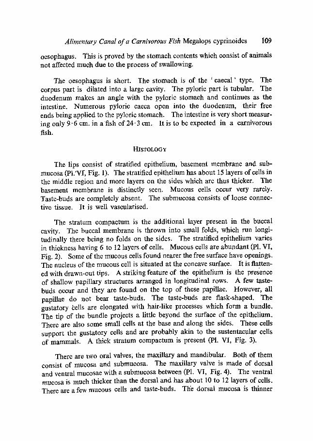
is bounded by lips, of which the lower is thicker. The lower jaw is slightly prolonged and hence the mouth faces upwards. This is suited to the feeding habit of the fish. The upper jaw is shorter and the lip is rather thin. The i jaw consists of premaxilla and maxilla on each side. The premaxillae are united with each other in front. On the sides they are attached to the skull. But the maxilla on each side is movably articulated. About three-fourths of the maxilla is attached to the skull with a membrane and the hind one- third is free. At about two-thirds from the anterior end, it is also attached to the lower jaw with a membrane. So whenever the lower jaw goes down while opening the mouth, the hinder part of the maxilla is pulled down on each side. This arrangement provides for the large gape of the fish. Teeth are present on both the jaws. They are incurved. The mucous membrane of the buccal cavity is thrown into folds in the roof, not in other regions. There is a well-developed tongue which is free anteriorly, the rest of it being attached to the floor of the buccal cavity. On the roof of the buccal cavity, a little behind the tip of the upper jaw, there are vomerine teeth forming two patches. Besides, there are small pointed teeth scattered on the entire roof of the buccal cavity and the dorsal surface of the tongue. The maxillary and mandibular valves are present, of which the maxillary valve is broader. The buccal cavity is not very spacious. It measures only 2.5 cm. from lips to pharynx in a fish measuring 24"3 cm.
The buccal cavity continues into the pharynx which has four gill arches; of these the first is the longest. Each gill arch has only a single row of gill takers, these are closely arranged. Thegill rakers of the first arch are longer than those of the other arches. The presence of long gill rakers is an adapta- tion for the filtering mode of feeding of the fish. A similar feature was noticed by Al-Hussaini (1947) in Atherina. Both the floor and the roof of the pharynx have teeth similar to those found on the jaws. These probably help the fish in quieting down the prey and pressing it before it enters the
Alimentary Canal of a Carnivorous Fish Megalops cyprinoides 109
oesophagus. This is proved by the stomach contents which consist of animals not affected much due to the process of swallowing.
The oesophagus is short. The stomach is of the ' caecal ' type. The corpus part is dilated into a large cavity. The pyloric part is tubular. The duodenum makes an angle with the pyloric stomach and continues as the intestine. Numerous pyloric caeca open into the duodenum, their free ends being applied to the pyloric stomach. The intestine is very short measur- ing only 9.6 cm. in a fish of 24.3 cm. It is to be expected in a carnivorous fish.
HISTOLOGY
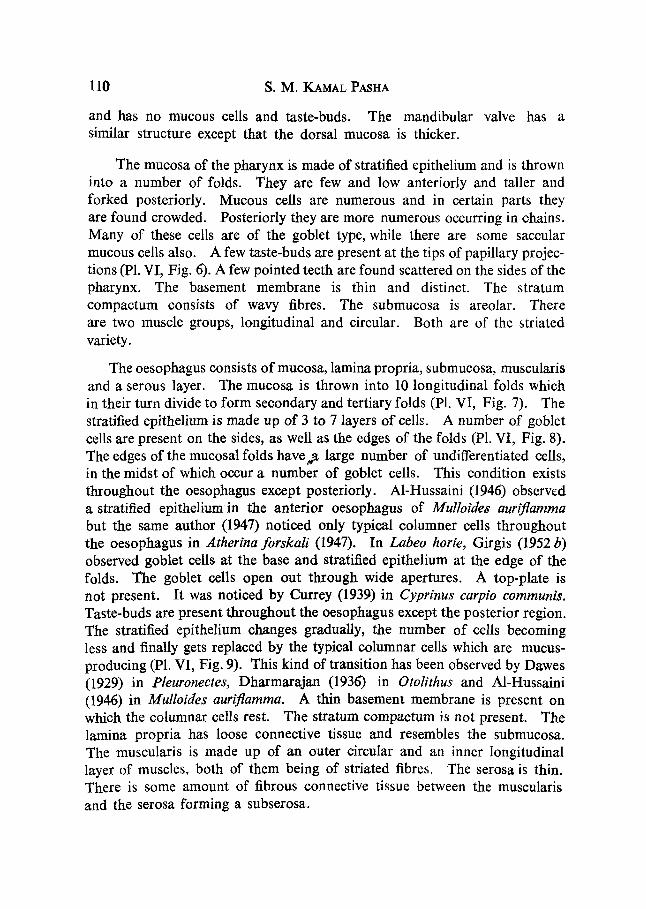
The lips consist of stratified epithelium, basement membrane and sub- mucosa (P1.'Vr, Fig. 1). The stratified epithelium has about 15 layers of cells in the middle region and more layers on the sides which are thus thicker. The basement membrane is distinctly seen. Mucous cells occur very rarely. Taste-buds are completely absent. The submucosa consists of loose connec- tive tissue. It is well vascularised.
The stratum compactum is the additional layer present in the buceal cavity. The buccal membrane is thrown into small folds, which run longi- tudinally there being no folds on the sides. The stratified epithelium varies in thiokness having 6 to 12 layers o f cells. Mucous cells are abundant (P1. VI, Fig. 2). Some of the mucous cells found nearer the free surface have openings. The nucleus of the mucous cell is situated at the concave surface. It is flatten- ed with drawn-out tips. A striking feature of the epithelium is the presence of shallow papillary structures arranged in longitudinal rows. A few taste- buds occur and they are found on the top of these papillae. However, all papillae do not bear taste-buds. The taste-buds are flask-shaped. The gustatory ~ells are elongated with hair-like processes which form a bundle. The tip of the bundle projects a little beyond the surface of the epithelium. There are also some small cells at the base and along the sides. These cells support the gustatory ceils and are probably akin to the sustentacular cells of mammals. A thick stratum compactum is present (P1. VI, Fig. 3).
There are two oral valves; the maxillary and mandibular. Both of them consist of mucosa and submucosa. The maxillary valve is made of dorsal and ventral mucosae with a submucosa between (P1. VI, Fig. 4). The ventral mucosa is much thicker than the dorsal and has about 10 to 12 layers of cells. There are a few mucous cells and taste-buds. The dorsal mucosa is thinner
110 S.M. KAMAL PASHA
and has no mucous cells and taste-buds. The mandibular valve has a similar structure except that the dorsal mucosa is thicker.
The mucosa of the pharynx is made of stratified epithelium and is thrown into a number of folds. They are few and low anteriorly and taller and forked posteriorly. Mucous cells are numerous and in certain parts they are found crowded. Posteriorly they are more numerous occurring in chains. Many of these cells are of the goblet type, while there are some saccular mucous cells also. A few taste-buds are present at the tips of papillary projec- tions (P1. VI, Fig. 6). A few pointed teeth are found scattered on the sides of the pharynx. The basement membrane is thin and distinct. The stratum compactum consists of wavy fibres. The submucosa is areolar. There are two muscle groups, longitudinal and circular. Both are of the striated variety.
The oesophagus consists of mucosa, lamina propria, submucosa, muscularis and a serous layer. The mucosa is thrown into 10 longitudinal folds which in their turn divide to form secondary and tertiary folds (P1. VI, Fig. 7). The stratified epithelium is made up of 3 to 7 layers of cells. A number of goblet cells are present on the sides, as well as the edges of the folds (P1. VI, Fig. 8). The edges of the mucosal folds have#n large number of undifferentiated cells, in the midst of which occur a number of goblet cells. This condition exists throughout the oesophagus except posteriorly. A1-Hussaini (1946) observed a stratified epithelium in the anterior oesophagus of Mulloides auriflamma but the same author (1947) noticed only typical columner cells throughout the oesophagus in Atherina forskali (1947). In Labeo horle, Girgis (1952 b) observed goblet cells at the base and stratified epithelium at the edge of the folds. The goblet cells open out through wide apertures. A top-plate is not present. It was noticed by Currey (1939) in Cyprinus carpio communis. Taste-buds are present throughout the oesophagus except the posterior region. The stratified epithelium changes gradually, the number of cells becoming less and finally gets replaced by the typical columnar cells which are mucus- producing (P1. VI, Fig. 9). This kind of transition has been observed by Dawes (1929) in Pleuronectes, Dharmarajan (193{;) in Otolithus and A1-Hussaini (1946) in Mulloides auriflamma. A thin basement membrane is present on which the columnar ceils rest. The stratum compactum is not present. The lamina propria has loose connective tissue and resembles the submucosa. The musculaxis is made up of an outer circular and an inner longitudinal layer of muscles, both of them being of striated fibres. The serosa is thin. There is some amount of fibrous connective tissue between the muscularis and the serosa forming a subserosa.

Alimentary Canal of a Carnivorous Fish Megalops cyprinoides 111
The anterior part of the stomach next to the oesophagus is an oesogaster, a transitional region with a combination of features of the stomach and oesophagus. This transitional region has the same features as found in Mystus gulio (Pasha, 1964 a).
The stomach has 7-8 gastric folds. These are not as arborising as those of the oesophagus. In the anterior part the gastric glands are few and poste- riorly they are numerous, consequently the gastric epithelium is thinner anteriorly and thicker posteriorly. The corpus and the pytoric parts of the stomach are morphologically and histologically distinguishable. Both of them, however, eonsist of mucosa, submucosa, muscularis and serosa (P1. VI, Fig. 10).
The mucosa of the corpus part of the stomach can be divided into two layers, the superficial layer consisting of the columnar cells and the glandular layer having glandular cells. However, the two layers are closely associated. The superficial layer is made up of columnar cells which are very compactly arranged. Mucus-secreting or goblet cells are entirely absent unlike in the posterior region of the oesophagus. The columnar cells perform the func- tion of absorption according to Greene (1912), Dawes (1929) and Blake (1936) while Al-Hussaini (1946) is of the opinion that these cells have the function of mucus secretion. In Megalops cyprinoides, they stain red with thionin which is characteristic of mucus and therefore they are mucus-secreting in function.
The glandular epithelium is made up of the gastric glands which occur next to the superficial epithelium. The gastric glands are of simple type, very compactly arranged. The lamina propria extends as narrow strands of tissue between the glands binding the adjacent glands together. The glandular cells are polygonal or rhomboidal in shape, each with a spherical nucleus at the centre. The cells are so disposed as to form tubular structures (P1. VI, Fig. 11). The cytoplasm of these cells gets stained deeper and shows the presence of numerous zymogen granules which are uniformly distributed. All the glandular cells are similar, there being no differentiation into peptic and oxyntic cells as found in the stomach of mammals. The glands open in groups of two or three into the crypts. Where they open the cells change from glandular to columnar. The opening is constricted and narrow. The neck cells observed by Al-Hussaini (1946) in Mulloides at the opening of the gland are not present here. The mucus-producing cells in the neck of the glands, as noted by Dawes (1929), are also absent.
The lamina propria is found next to the glandular epithelium and con- sists of strands of connective tissue. This extends between the glands and

112 S. M. KaMAL PASHA
holds the glands in position. It is highly vascular. The submueosa is found between the lamina propria and the circular muscles. It is areolar occurring as a network with large meshes. The muscularis consists of an inner circular layer and an outer longitudinal layer. Both are made of unstriated fibres. The longitudinal layer is much thicker than the circular layer at the beginning of the stomach. This condition is reversed a little posteriorly. The serosa is made up of one or two layers of flattened cells.
The pyloric region of the stomach is characterised by the presence of much thicker musculature and the absence of gastric glands. The circular layer measures about 200/z in the thinner region and about 500/~ in the thicker region (P1. VI, Fig. 12). This difference in thickness is seen in the wall of the pyloric region as a whole. The circular layer thickens considerably in front of the pyloric valve, forming the pyloric sphincter. The longitudinal layer and the serosa are thin. At the pylorie orifice the epithelium projects into the lumen forming the pyloric valve (PI. VI, Fig. 13). The valve occurs as a ledge of the epithelium along with the lamina propria and submucosa. The muscles do not enter it and consequently the closure of the passage from the pylorus to the duodenum must be by the sphincter, since the valve cannot be tightened due to the absence of muscles in it.
The duodenum, intestine, pylorie caeca and rectum form the rest of the intestine. All of them have almost the same structure varying only in minor details. The wall of the intestine is composed of mucosa, lamina propria, submucosa, muscularis and serosa.
The mucosa is thrown into folds most of which run longitudinally, a few transverse folds also being present. There are about .25 folds which vary in their shapes, though all of them are uniformly low. The columnar and the goblet cells are the only two types of ceils that form the mucosa (P1. VI, Fig. 14). The columnar cells are long and slender. The free border shows a top-plate. This covers the epithelium throughout except in places when the goblet ceils open into the lumen. The mucus-secreting cells which are typically goblet-shaped form the second type of cells noticed in the epithelium. They occur mostly on the sides of the folds, only a few being present in the crests. They do not occur in regular sequence but are found scattered. Generally, the goblet cells are of two shapes; some are funnel-like, while others are vase-shaped. In both the varieties the nucleus occurs basally which is at a lower level compared to the position of the nuclei of the columnar cells. The goblet cells open out by wide apertures.
The lamina propria consists of highly .vascular loose connective tissue. The submucosa is not distinguishable from the lamina propria. The muscu-

Alimentary Canal of a Carnivorous Fish Megalops cyprinoides 113
laris consists of the circular and the longitudinal layers. The circular layer is thicker than the longitudinal layer. The serosa is very thin.
There is no intestino-rectal valve to demarcate the rectum from the intestine. However, the posterior part having slightly thicker musculature may be distinguished as the rectum. The rectal mucosa has the same two types of cells, the columnar and the goblet cells. The columnar cells are just like those of the intestine while the goblet cells are larger and occur in abundance. In certain regions they are arranged in a regular sequence.
The presence of the pyloric caeca, according to Barrington (1957) is a complicating factor in many fishes. They are particularly characteristic of the Actinopterygii. They are said to vary from one to over a thousand (Rahimullah, 1945). The pyloric caeca do not differ much from the intestine. They have the same structure with only a few minor differences. The mucosal folds are more slender than those of the intestine (P1. VI, Fig. 15). The columnar cells are like those of the intestine. The goblet cells are larger and occur in large numbers. The lamina propria and the submucosa resemble those of the intestine. The circular muscle layer is slightly thicker than the longi- tudinal. The serosa is very thin. Many of the caeca are surrounded by the pancreatic tissue.
SUMMARY
The anatomy and histology of the alimentary canal of Megalops cypri- noides, a carnivorous fish, has been described. The mouth is upturned and there are no pharyngeal teeth patches. The gill rakers are well developed. Taste-buds are absent from the lips. The buccal membrane has only a few taste-buds. The pharynx is not divisible into distinct regions; taste-buds and mucous cells attain the maximum development in the pharynx. The oesophagus has deep folds; they are much branched; Taste-buds are present.
A transitional region, termed the oesogaster, is present. The stomach is of the ' caecal ' type ; Gastric glands are present only in the corpus region. The glandular ceils do not show differentiation into oxyntic and peptic cells. The pyloric stomach has no gastric glands and generally its musculature is thicker than that of the corpus region.
A pyloric valve is present. The intestine is very short and the intestinal mucosa has only two kinds of cells, columnar and goblet cells.
Pyloric caeca are present; histologically they resemble the intestine. B3

114 S.M. KAMAL PASHA
An ileo-rectal valve is not present. But the rectum has thicker muscu- lature and a large number of goblet ceils.
ACKNOWLEDGEMENTS
I am grateful to Prof. P. K. Menon, former Professor and Head of the Department of Zoology, Presidency College, Madras, for suggesting the problem, guidance and instruction. I am also grateful to Dr. S. Krishna- swamy, Professor of Zoology, University of Madras, Madurai Centre, for kindly forwarding my papers for publication and for a number of suggestions.
AI-Hussaini, A. H.
Barrington, E. J. W.
Blake, I. H.
Currey, E.
Dawes, B.
Dharmarajan, M.
Girgis, S.
Green, C. W.
Pasha, S. M. Kamal
Rahimullah, M.
REFERENCES
.. "The anatomy and histology of the alimentary tract of the bottom-feeder Malloides auriflamma," J. Morph., 1946, 78, 121-54.
.. "The anatomy and histology of the plankton feeder Atherina ]brskali," Ibid., 1947, "80, 251-86.
.. "The alimentary canal and digestion," Physiology of Fishes, Ed., by Brown, M. F., Academic Press, Inc., New York, 1957.
.. "Studies on the comparative histology of the digestive tube of certain teleost fishes. A bottom feeding fish, the .sea robin, Prionotus carolinus," J. Morph., 1936, 60, 77-102.
. . "The histology of the digestive tube of the carp Cyprinus carpio commtmis," Ibid., 1939, 65, 53-78.
. . "The histology of the alimentary canal of the plaice, Pleuro- nectes platessa," Quart. Jour. Micr. ScL, 1929, 73, 243-47.
. . "The anatomy and histology of the alimentary system of Otolithus tuber--Part II ," M.Sc. Thesis of the Madras University, 1936.
.. "On the anatomy and histology of the alimentary tract of a herbivorous bottom feeding cyprinoid fish Labeo horie,'" J. Morph., 1952b, 90, 317-62.
. . "Anatomy and histology of alimentary tract of the king salmon," Bull. U.S. Bureau Fish., 1912, 32, 73-100.
.. "The anatomy and histology of the alimentary canal of an omnivorous fish Mystus gulio," Proc. Ind. Acad. Sci., 1964 a, 59 (4), 211-21.
. . "The anatomy and histology of the alimentary canal of a herbivorous fish Tilapia mossambica," Ibid., 1964'b, 59 (6), 340-49.
. . " A comparative .study of the morphology, histology and probable functions of the pyloric caeca in Indian fishes together with a discussion on their homology," Ibid., 1945, 21 B, 1-37.

S. M. Kamal Pasha Proc. Ind. Acad. Sci., B, Vol. LX, Pl. VI
+i ~ i ~ , ~ ~ ~ • , ~ ~ ~ ~ I ~ ~ ~
FiGs. 1-15

A l i m e n t a r y C a n a l o f a C a r n i v o r o u s F i sh M e g a l o p s c y p r i n o i d e s 115
EXPLANATION OF PLATE V I
FIG. 1. T.S. of lip. FIG. 2. T.S. of buccal membrane. FIG. 3. T.S. of buccal membrane (part enlarged). FI~. 4. T.S. of maxillary oral valve. FIG. 5. T.S. of pharynx. FIG. 6. T.S. of pharynx (part enlarged). F16. 7. T.S. of anterior oesophagus. FIG. 8. T.S. of anterior oesophagus (part enlarged) FIG. 9. T.S. of post-oesophagus. FIG. 10. T.S. of stomach. FIG. 11. T.S. of stomach showing gastric glands. FIG. 12. T.S. of pyloric stomach. Fro. 13. L.S. of pylorus. FIG. 14. T.S. of intestine (part enlarged). FIG. 15. T.S. of pyloric caecum (part enlarged).
ABBREVIATIONS
b.m., basement membrane; c.c., columnar cell; c.m., circular muscle layer; c.st., corpus of the stomach; c.t., connective tissue; d.m., dorsal mucosa; du., duodenum; g.c., goblet cell; g.cr., gastric crypt; g.g., gastric gland; g.g.c., gastric gland cell; int., intestine; L, lumen; le., leucocytes; Lm., longitudinal muscle layer; l.p., lamina propria; m., mucus; m.c., mucous cell; m.f , mucosal fold; nu., nucleus; oe., oesophagus; p., pylorus; p.e., pyloric stomach; p.p., papillary projection ; p.v., pyloric valve; rec., rectum; s., serosa; s.m., submucosa; at.c, stratum compactum; st.e., stratified epithelium; t.b., taste-bud; t.p., top-plate.