The Acute Red Eye - fmhs.auckland.ac.nz · Case Scenario Links The Acute Red Eye • Acute chronic...
71
The Acute Red Eye Dr Shenton Chew Dr Jennifer Craig Professor Charles McGhee
Transcript of The Acute Red Eye - fmhs.auckland.ac.nz · Case Scenario Links The Acute Red Eye • Acute chronic...
The Acute Red Eye
Dr Shenton Chew Dr Jennifer Craig Professor Charles McGhee
Case Scenario Links The Acute Red Eye • Acute or chronic red eye (Oph01)
• Acute trauma to the eye (Oph02)
• Child with red swelling around one eye (Oph10)
2
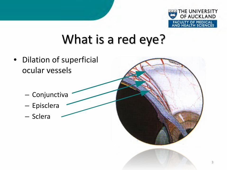
What is a red eye? • Dilation of superficial
ocular vessels
– Conjunctiva – Episclera – Sclera
3
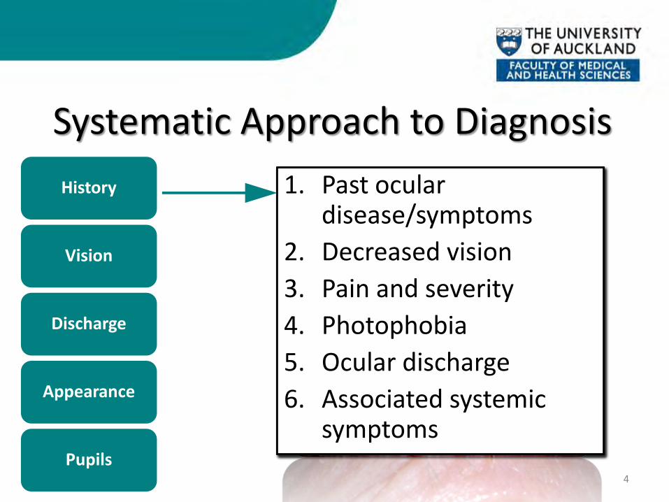
1. Past ocular disease/symptoms
2. Decreased vision 3. Pain and severity 4. Photophobia 5. Ocular discharge 6. Associated systemic
symptoms
1. Snellen visual acuity 2. Pinhole
Systematic Approach to Diagnosis
4
History
Vision
Discharge
Appearance
Pupils
1. Serous/Watery 2. Mucoid 3. Purulent 4. Mucopurulent
1. Past ocular disease/symptoms
2. Decreased vision 3. Pain and severity 4. Photophobia 5. Ocular discharge 6. Associated systemic
symptoms

1. Past ocular disease/symptoms
2. Decreased vision 3. Pain and severity 4. Photophobia 5. Ocular discharge 6. Associated systemic
symptoms
1. Snellen visual acuity 2. Pinhole
Systematic Approach to Diagnosis
5
History
Vision
Discharge
Appearance
Pupils
1. Serous/Watery 2. Mucoid 3. Purulent 4. Mucopurulent
1. Snellen visual acuity 2. Pinhole
1. Past ocular disease/symptoms
2. Decreased vision 3. Pain and severity 4. Photophobia 5. Ocular discharge 6. Associated systemic
symptoms
1. Snellen visual acuity 2. Pinhole
Systematic Approach to Diagnosis
6
History
Vision
Discharge
Appearance
Pupils
1. Serous/Watery 2. Mucoid 3. Purulent 4. Mucopurulent
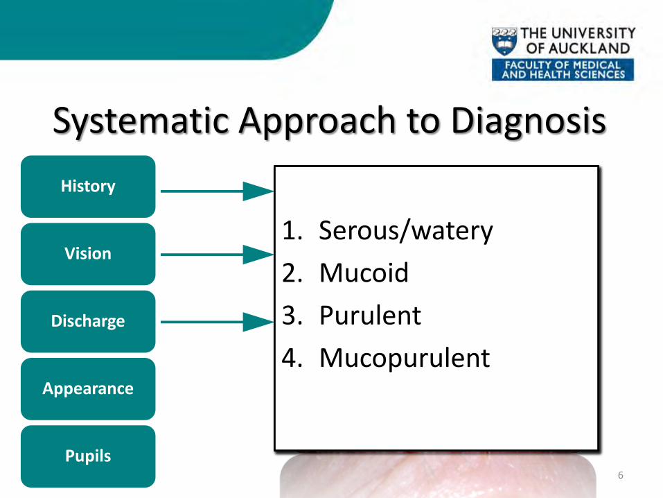
1. Serous/watery 2. Mucoid 3. Purulent 4. Mucopurulent
1. Past ocular disease/symptoms
2. Decreased vision 3. Pain and severity 4. Photophobia 5. Ocular discharge 6. Associated systemic
symptoms
1. Snellen visual acuity 2. Pinhole
Systematic Approach to Diagnosis
7
History
Vision
Discharge
Appearance
Pupils
1. Serous/Watery 2. Mucoid 3. Purulent 4. Mucopurulent
1. Past ocular disease/symptoms
2. Decreased vision 3. Pain and severity 4. Photophobia 5. Ocular discharge 6. Associated systemic
symptoms
1. Snellen visual acuity 2. Pinhole
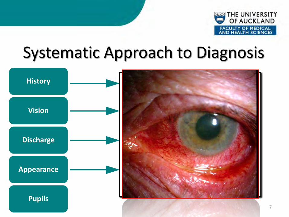
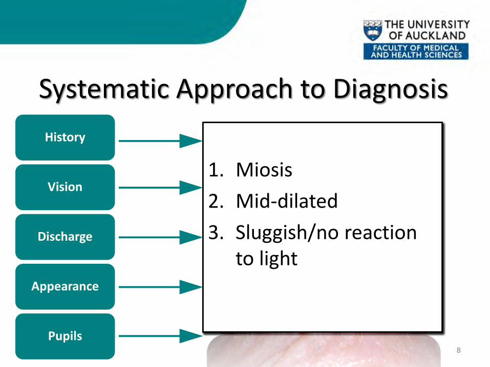
Systematic Approach to Diagnosis
8
History
Vision
Discharge
Appearance
Pupils
1. Serous/Watery 2. Mucoid 3. Purulent 4. Mucopurulent
1. Miosis 2. Mid-dilated 3. Sluggish/no reaction
to light
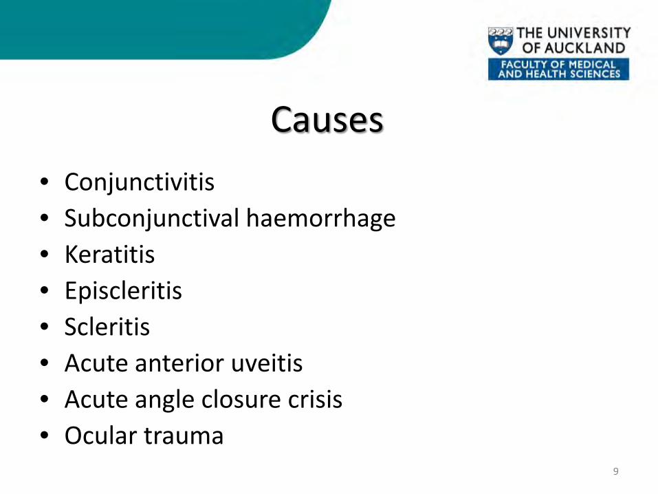
Causes • Conjunctivitis • Subconjunctival haemorrhage • Keratitis • Episcleritis • Scleritis • Acute anterior uveitis • Acute angle closure crisis • Ocular trauma
9

CONJUNCTIVITIS
10
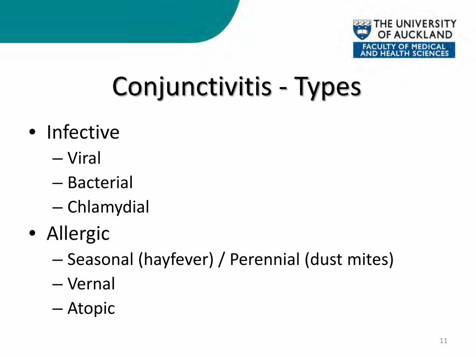
Conjunctivitis - Types • Infective
– Viral – Bacterial – Chlamydial
• Allergic – Seasonal (hayfever) / Perennial (dust mites) – Vernal – Atopic
11
Conjunctivitis
12
History
Vision
Discharge
Appearance
Pupils
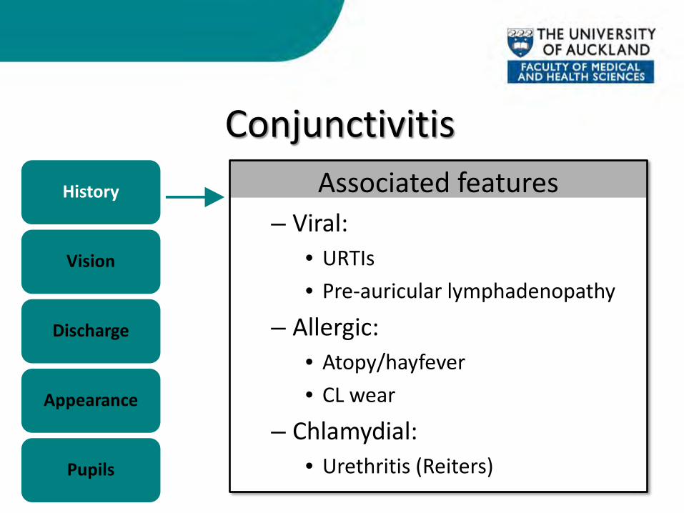
Associated features – Viral:
• URTIs • Pre-auricular lymphadenopathy
– Allergic: • Atopy/hayfever • CL wear
– Chlamydial: • Urethritis (Reiters)
Conjunctivitis
13
History
Vision
Discharge
Appearance
Pupils
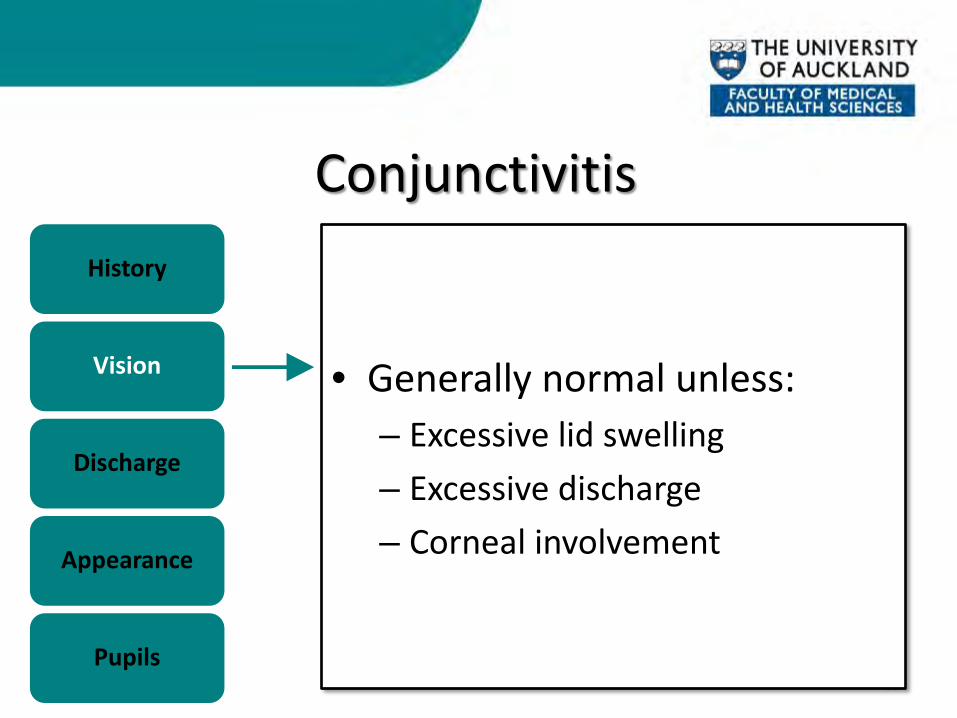
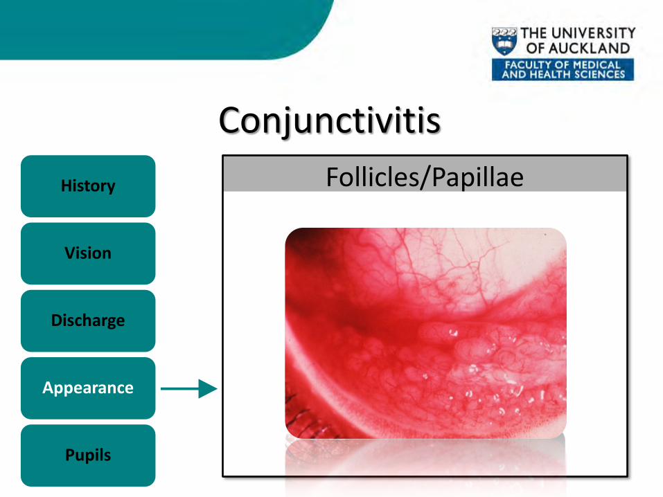
• Generally normal unless: – Excessive lid swelling – Excessive discharge – Corneal involvement
Conjunctivitis
14
History
Vision
Discharge
Appearance
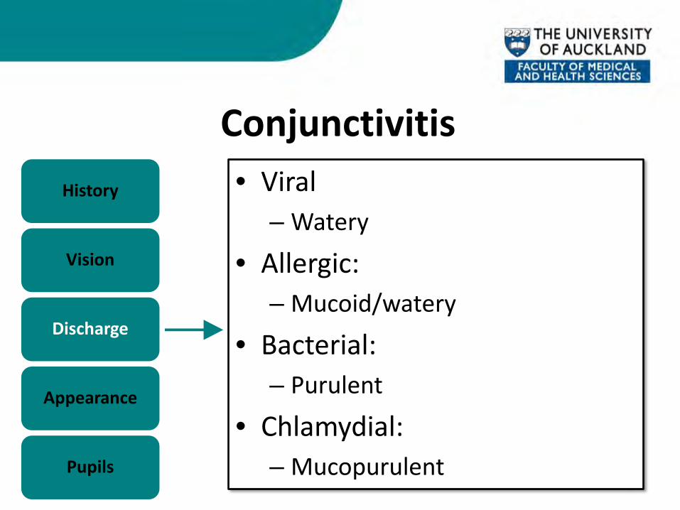
Pupils
• Viral – Watery
• Allergic: – Mucoid/watery
• Bacterial: – Purulent
• Chlamydial: – Mucopurulent
Conjunctivitis
15
History
Vision
Discharge
Appearance
Pupils
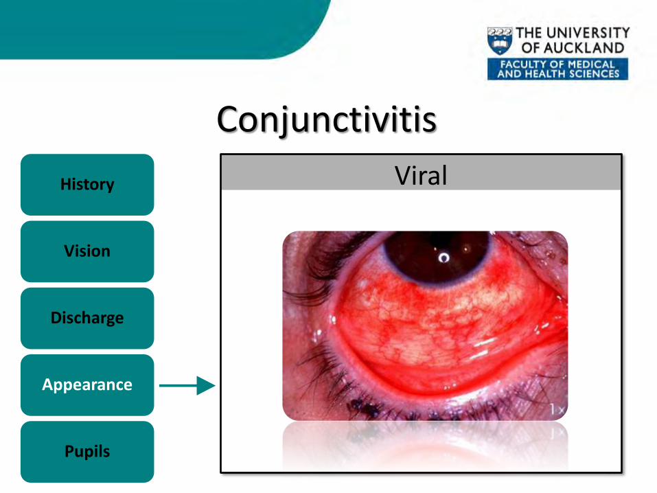
Viral
Conjunctivitis
16
History
Vision
Discharge
Appearance
Pupils
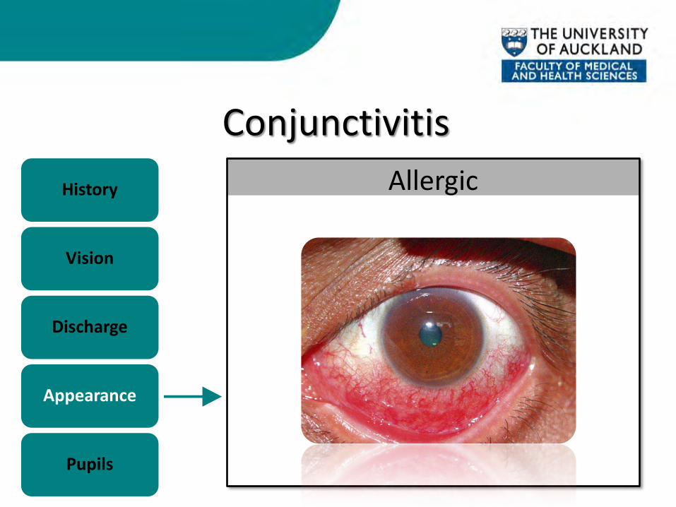
Allergic
Conjunctivitis
17
History
Vision
Discharge
Appearance
Pupils
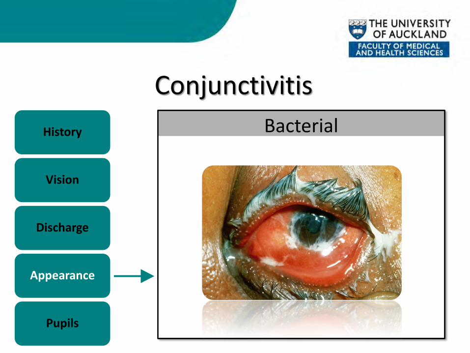
Bacterial
Conjunctivitis
18
History
Vision
Discharge
Appearance
Pupils
Follicles/Papillae
Conjunctivitis
19
History
Vision
Discharge
Appearance
Pupils
Normal

Conjunctivitis - Management • Swab and isolate responsible organism
• Bacterial = Topical Chloramphenicol • Viral = Supportive Rx (compresses, lubricants) • Chlamydia = Oral Azithromycin/doxycycline
20

KERATITIS
21
Keratitis
22
History
Vision
Discharge
Appearance
Pupils
– Painful (foreign body sensation) – Photophobic – Tearing – Hx of CL wear/trauma
Keratitis
23
History
Vision
Discharge
Appearance
Pupils
• Decreased
– Especially if involves visual axis
Keratitis
24
History
Vision
Discharge
Appearance
Pupils
• Watery, purulent – (depends on cause)
Keratitis
25
History
Vision
Discharge
Appearance
Pupils
– Circumcorneal injection – Corneal infiltrate/hazy cornea – Overlying epithelial defect
Keratitis
26
History
Vision
Discharge
Appearance
Pupils
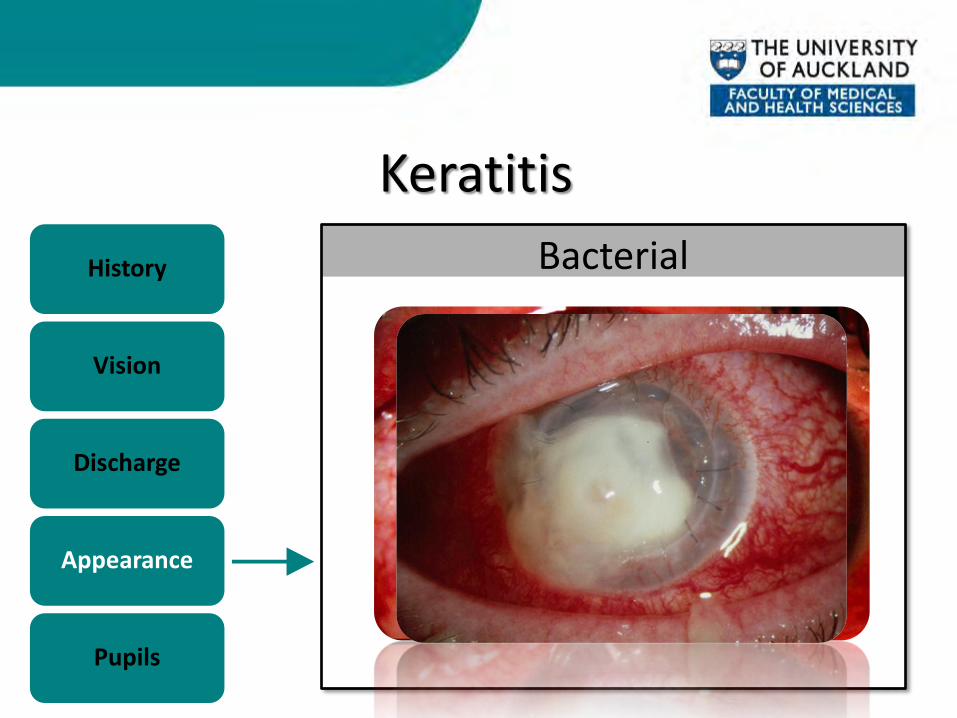
Bacterial

Keratitis
27
History
Vision
Discharge
Appearance
Pupils
Fungal
Keratitis
28
History
Vision
Discharge
Appearance
Pupils
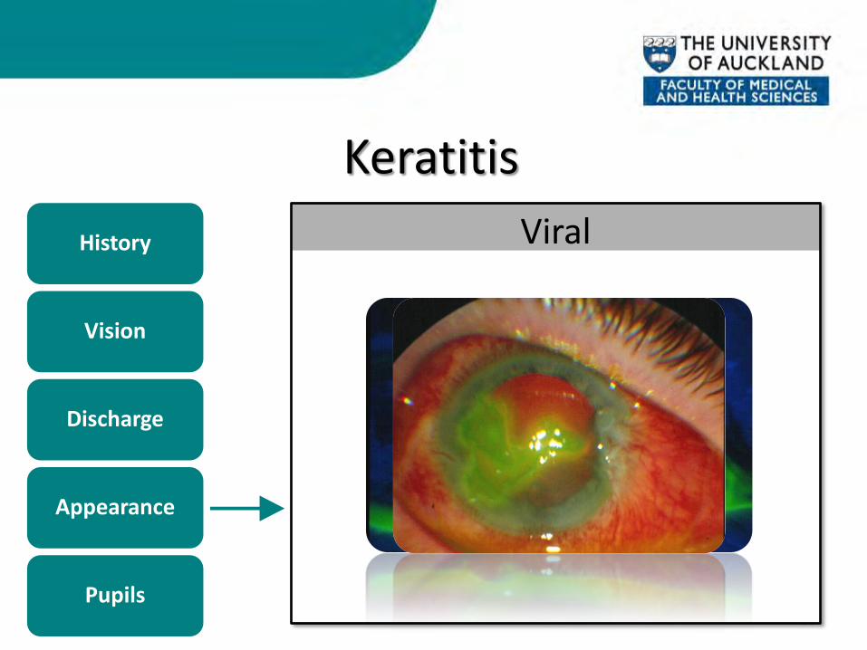
Viral

Keratitis
29
History
Vision
Discharge
Appearance
Pupils
Normal
Keratitis- Management • Isolate responsible organism
– Corneal scrape
• Intensive treatment and close followup
30

ACUTE ANGLE CLOSURE CRISIS
31
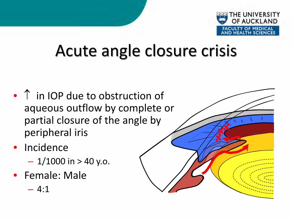
• ↑ in IOP due to obstruction of aqueous outflow by complete or partial closure of the angle by peripheral iris
• Incidence – 1/1000 in > 40 y.o.
• Female: Male – 4:1
Acute angle closure crisis
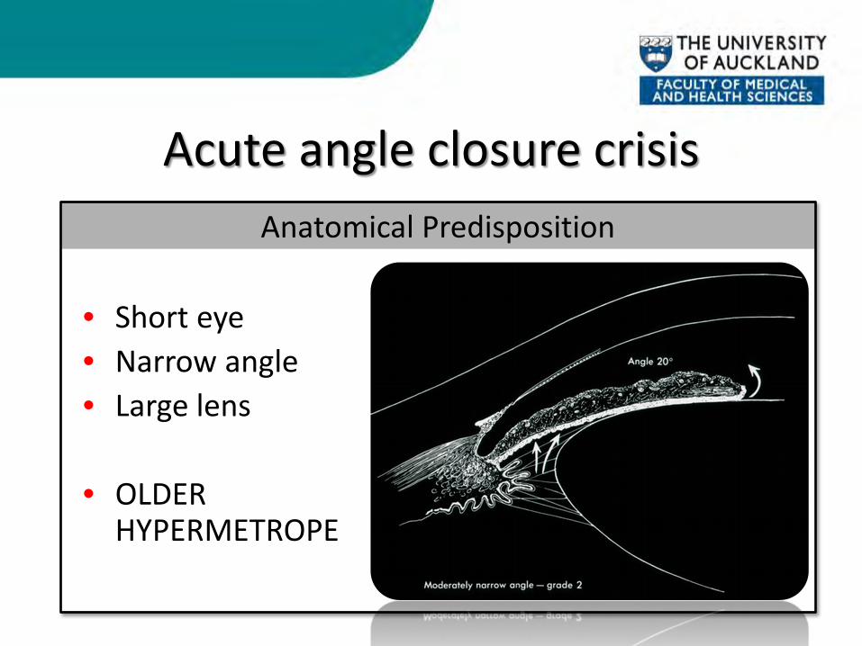
Acute angle closure crisis Anatomical Predisposition
• Short eye • Narrow angle • Large lens
• OLDER
HYPERMETROPE

Acute angle closure crisis
34
History
Vision
Discharge
Appearance
Pupils
– Intense ocular pain & headache – Nausea & vomiting – Photophobia – Premonitory symptoms – Hypermetrope
Acute angle closure crisis
35
History
Vision
Discharge
Appearance
Pupils
• Very blurred
– Secondary to corneal oedema
Acute angle closure crisis
36
History
Vision
Discharge
Appearance
Pupils
• None

Acute angle closure crisis
37
History
Vision
Discharge
Appearance
Pupils
– Circumcorneal injection – Cloudy cornea – Optic nerve head swelling
• If prolonged attack
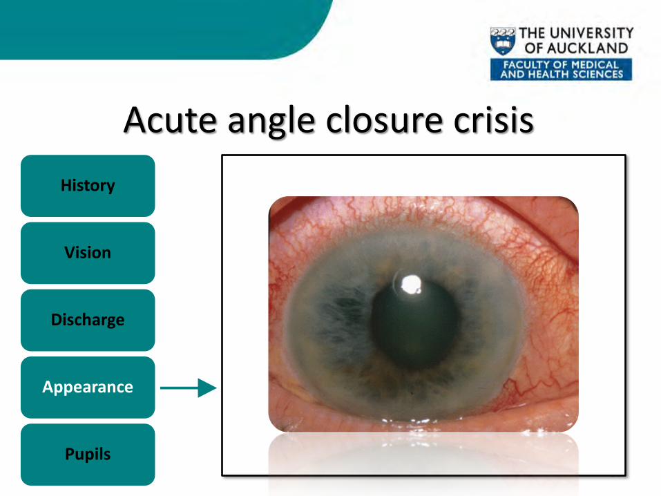
Acute angle closure crisis
38
History
Vision
Discharge
Appearance
Pupils

Acute angle closure crisis
39
History
Vision
Discharge
Appearance
Pupils

Acute angle closure crisis
40
History
Vision
Discharge
Appearance
Pupils
Acute angle closure crisis
41
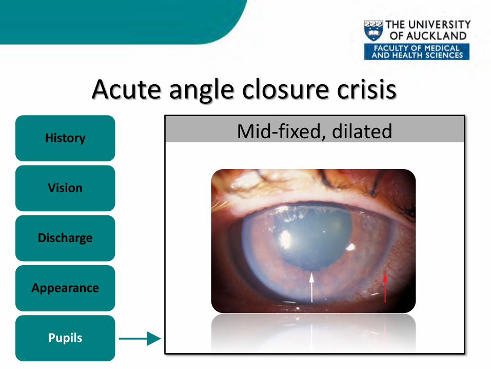
History
Vision
Discharge
Appearance
Pupils
Mid-fixed, dilated
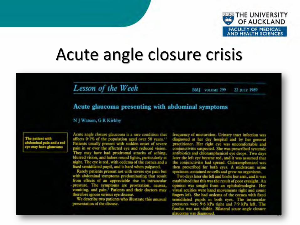
Acute angle closure crisis • Be on the lookout! • Acute closed angle glaucoma masquerading as
systemic illness – Dayan M, Turner B, McGhee CNJ – Br Med J 1996; 313:413-5
42

AACC - Management • Reduce IOP (often starts > 50 mmHg)
– Medical • Topical:
– Alpha-agonist, Beta-blockers, Mitotics (Pilocarpine) • Systemic:
– Carbonic anhydrase inhibitors (Diamox), Osmotics (Mannitol)
– Surgical • Peripheral iridotomy • Clear lens extraction/trabeculectomy
43
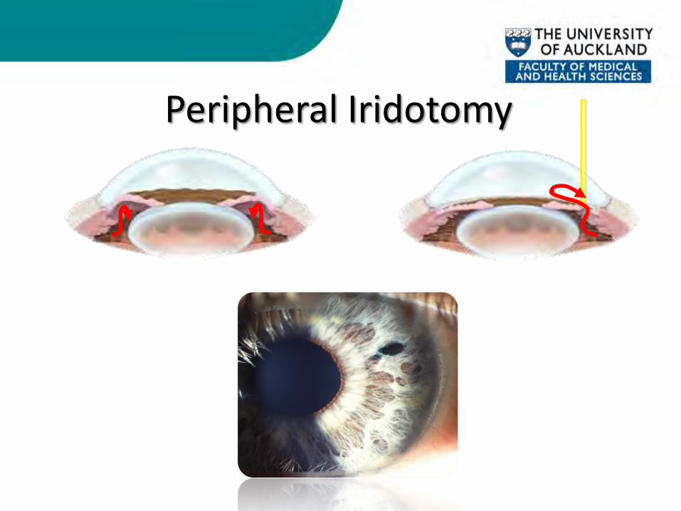
Peripheral Iridotomy
ACUTE ANTERIOR UVEITIS/IRITIS
45
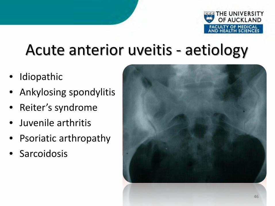
Acute anterior uveitis - aetiology • Idiopathic • Ankylosing spondylitis • Reiter’s syndrome • Juvenile arthritis • Psoriatic arthropathy • Sarcoidosis
46
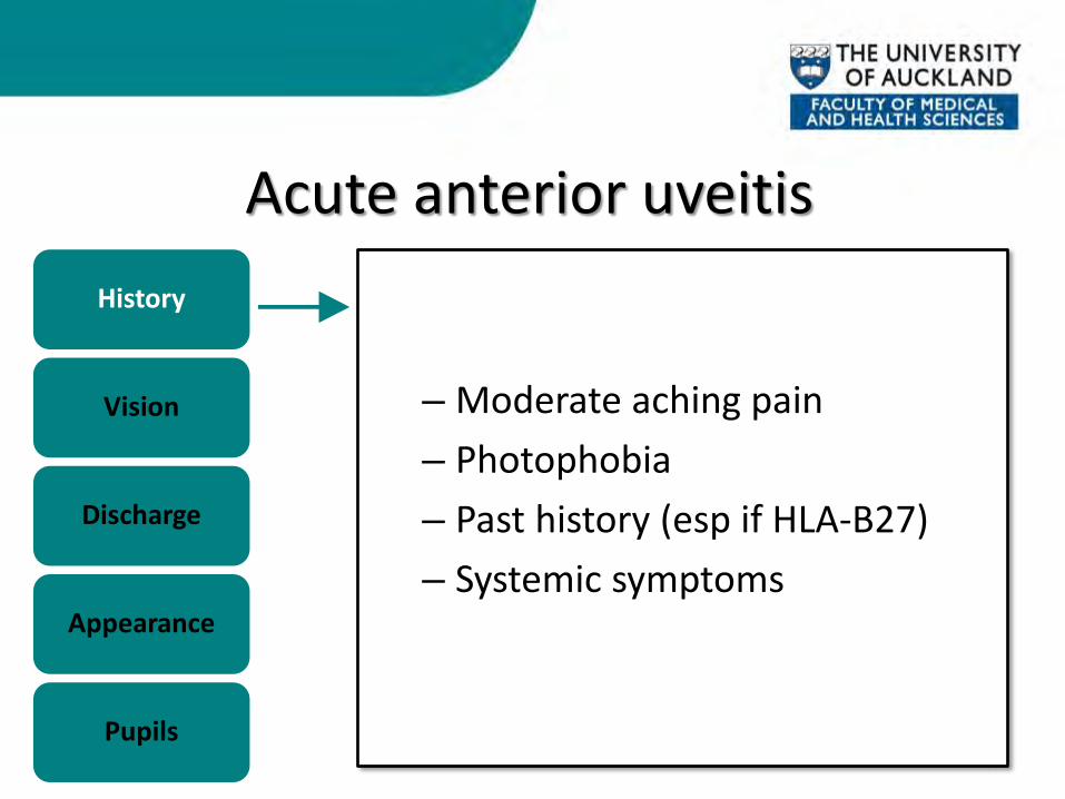
Acute anterior uveitis
47
History
Vision
Discharge
Appearance
Pupils
– Moderate aching pain – Photophobia – Past history (esp if HLA-B27) – Systemic symptoms
Acute anterior uveitis
48
History
Vision
Discharge
Appearance
Pupils
• Blurred

Acute anterior uveitis
49
History
Vision
Discharge
Appearance
Pupils
• None
Acute anterior uveitis
50
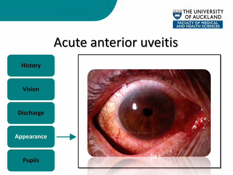
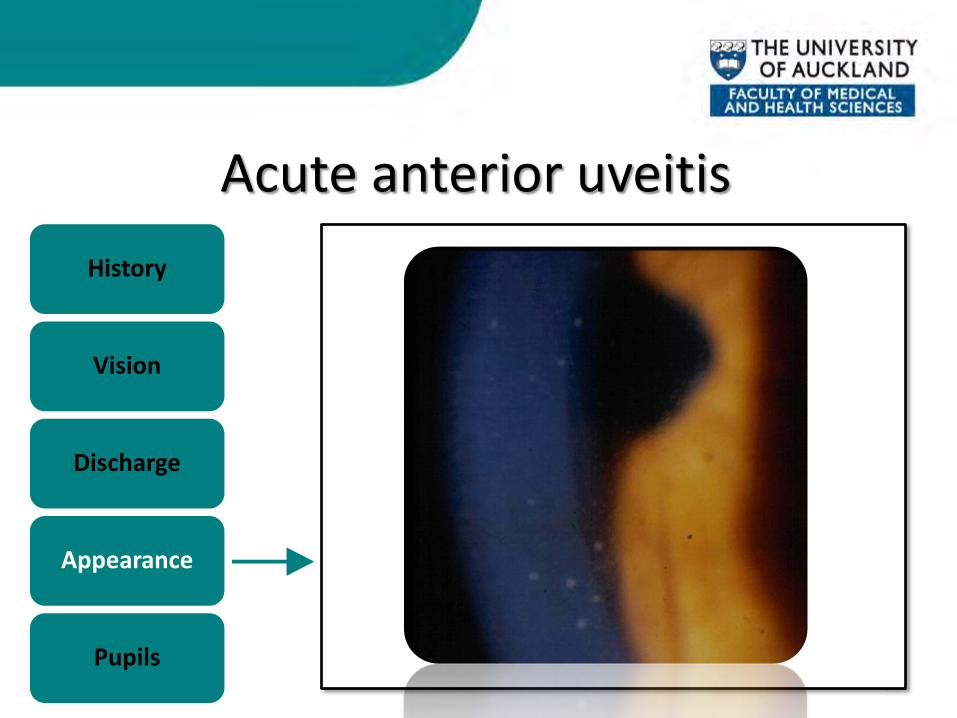
History
Vision
Discharge
Appearance
Pupils
– Circumcorneal injection – Clear cornea
Acute anterior uveitis
51
History
Vision
Discharge
Appearance
Pupils
Acute anterior uveitis
52
History
Vision
Discharge
Appearance
Pupils
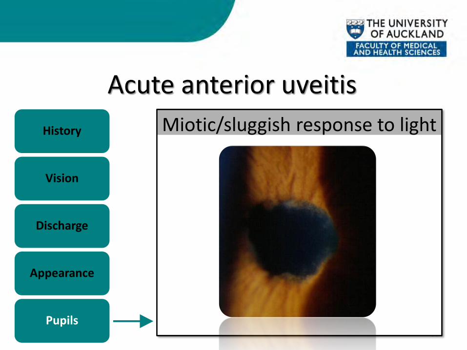
Acute anterior uveitis
53
History
Vision
Discharge
Appearance
Pupils
Miotic/sluggish response to light
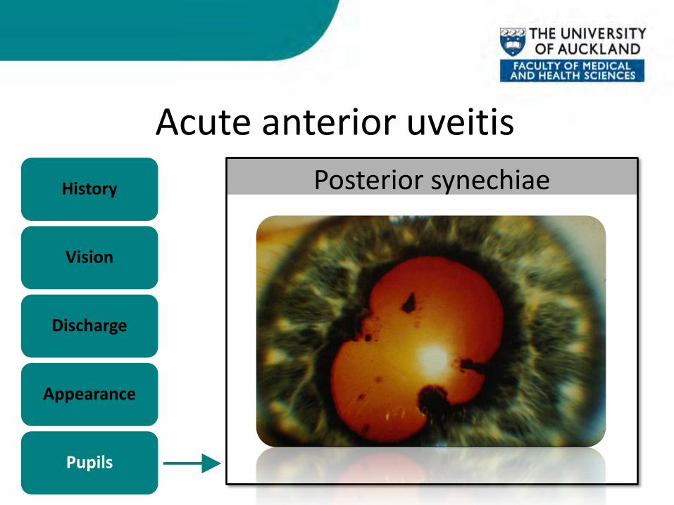
Acute anterior uveitis
54
History
Vision
Discharge
Appearance
Pupils
Posterior synechiae
AAU - Management • Subdue inflammation
– Topical corticosteroids (g.predforte)
• Prevent posterior synechiae – Mydriatics (g.cyclopentolate)
• Watch for elevated IOP – Topical ocular hypotensives (g.timolol)
55

SCLERITIS/EPISCLERITIS
56

Scleritis • Relatively uncommon
Episcleritis • Relatively common
57

Scleritis • Relatively uncommon • Severe boring pain
– “wakes from sleep”
Episcleritis • Relatively common • Mild ocular discomfort
58
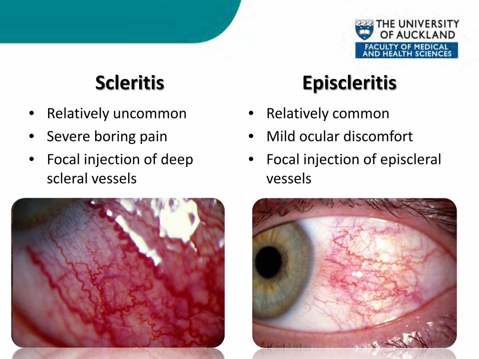
Scleritis • Relatively uncommon • Severe boring pain • Focal injection of deep
scleral vessels
Episcleritis • Relatively common • Mild ocular discomfort • Focal injection of episcleral
vessels
59
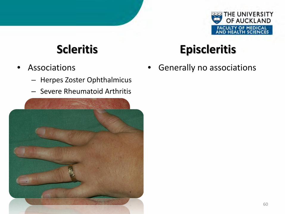
Scleritis • Associations
– Herpes Zoster Ophthalmicus – Severe Rheumatoid Arthritis
Episcleritis • Generally no associations
60
Scleritis • Associations
– Herpes Zoster Ophthalmicus – Severe Rheumatoid Arthritis
• Can lead to blindness if untreated – po.prednisone
Episcleritis • Generally no associations
• Usually requires no treatment – g.lubricants/g.voltaren
61

SUBCONJUNCTIVAL HAEMORRHAGE
62
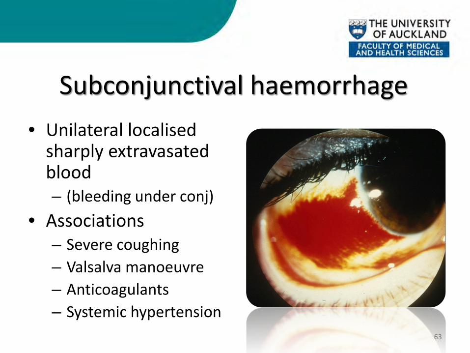
Subconjunctival haemorrhage • Unilateral localised
sharply extravasated blood – (bleeding under conj)
• Associations – Severe coughing – Valsalva manoeuvre – Anticoagulants – Systemic hypertension
63
OCULAR TRAUMA
64
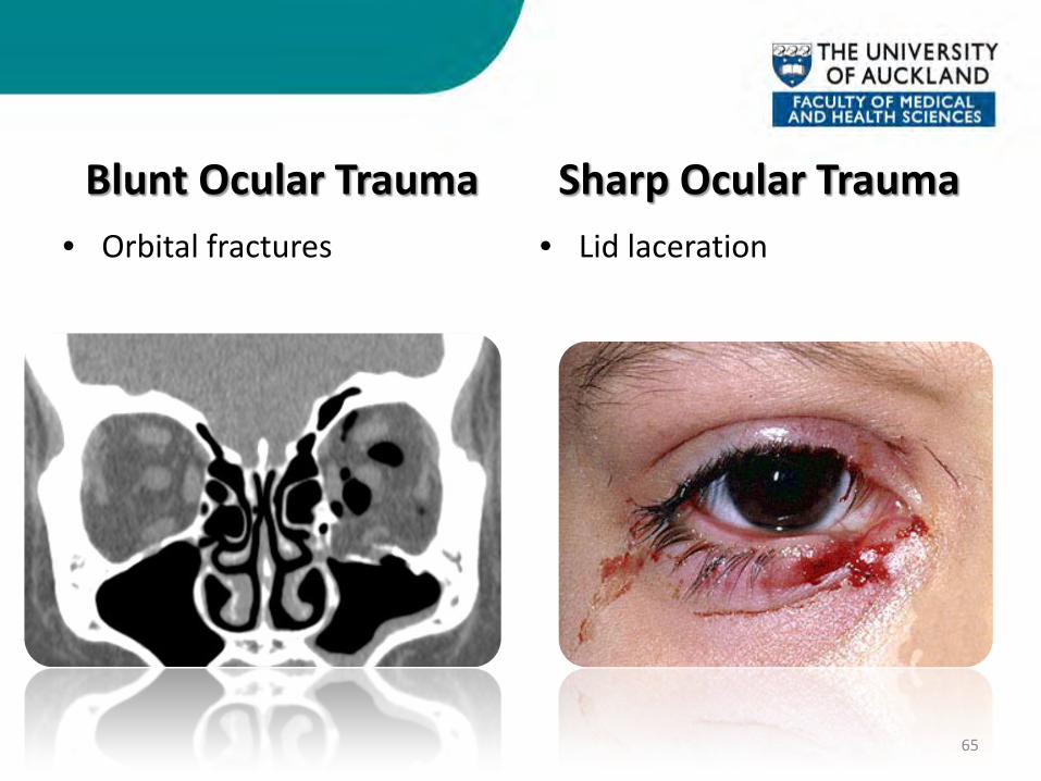
Blunt Ocular Trauma • Orbital fractures
Sharp Ocular Trauma • Lid laceration
65
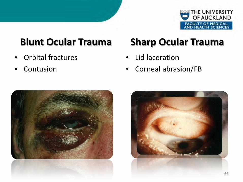
Blunt Ocular Trauma • Orbital fractures • Contusion
Sharp Ocular Trauma • Lid laceration • Corneal abrasion/FB
66
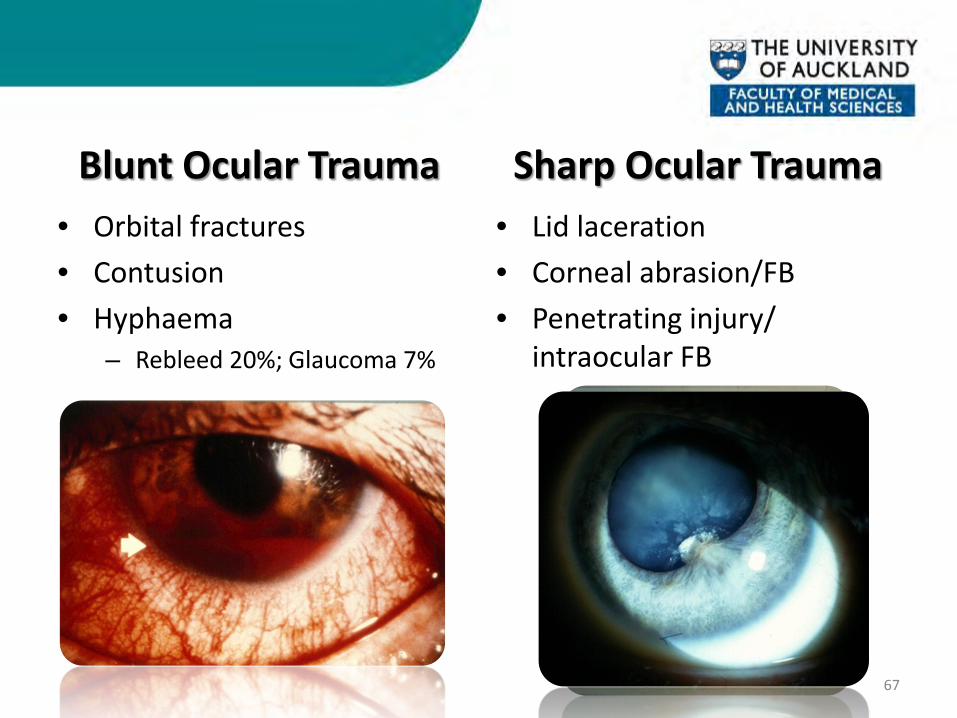
Blunt Ocular Trauma • Orbital fractures • Contusion • Hyphaema
– Rebleed 20%; Glaucoma 7%
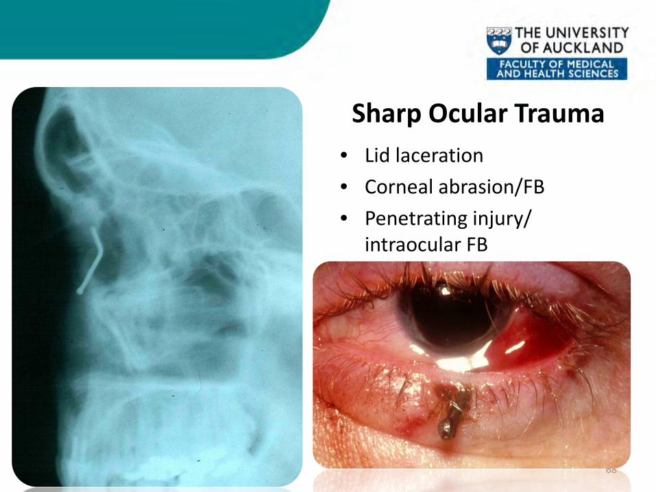
Sharp Ocular Trauma • Lid laceration • Corneal abrasion/FB • Penetrating injury/
intraocular FB
67
Blunt Ocular Trauma • Orbital fractures • Contusion • Hyphaema • Iris damage • Lens damage • Vitreous haemorrhage • Retinal haemorrhage • Retinal detachment • Globe rupture
Sharp Ocular Trauma • Lid laceration • Corneal abrasion/FB • Penetrating injury/
intraocular FB
68


70
Conclusions
• Systematic Approach • History • Vision • Discharge • Appearance • Pupils
• BE AWARE OF SIGHT THREATENING CONDITIONS!

The End All material contained in this presentation is copyright of
The University of Auckland, Department of Ophthalmology and should not be reproduced without written permission