The accuracy of a 3D printing surgical guide determined by ... · The digital image of the soft...
7
The Journal of Advanced Prosthodontics 279 The accuracy of a 3D printing surgical guide determined by CBCT and model analysis Boyoung Ma 1 , Taeseok Park 2 , Inkon Chun 2 , Kwidug Yun 1 * 1 Department of Prosthodontics, School of Dentistry, Chonnam National University, Gwangju, Republic of Korea 2 DMAX Co. Ltd., Gwangju, Republic of Korea PURPOSE. The aim of this clinical study was to assess the accuracy of the implants placed using a universal digital surgical guide. MATERIALS AND METHODS. Among 17 patients, 28 posterior implants were included in this study. The digital image of the soft tissue acquired from cast scan and hard tissue from CBCT have been superimposed and planned the location, length, diameter of the implant fixture. Then digital surgical guides were created using 3D printer. Each of angle deviations, coronal, apical, depth deviations of planned and actually placed implants were calculated using CBCT scans and casts. To compare implant positioning errors by CBCT scans and plaster casts, data were analyzed with independent samples t-test. RESULTS. The results of the implant positioning errors calculated by CBCT and casts were as follows. The means for CBCT analyses were: angle deviation: 4.74 ± 2.06°, coronal deviation: 1.37 ± 0.80 mm, and apical deviation: 1.77 ± 0.86 mm. The means for cast analyses were: angle deviation: 2.43 ± 1.13°, coronal deviation: 0.82 ± 0.44 mm, apical deviation: 1.19 ± 0.46 mm, and depth deviation: 0.03 ± 0.65 mm. There were statistically significant differences between the deviations of CBCT scans and cast. CONCLUSION. The model analysis showed lower deviation value comparing the CBCT analysis. The angle and length deviation value of the universal digital guide stent were accepted clinically. [ J Adv Prosthodont 2018;10:279-85] KEYWORDS: Stents; Computer-Assisted Surgery; Computer-Assisted Radiotherapy Planning https://doi.org/10.4047/jap.2018.10.4.279 https://jap.or.kr J Adv Prosthodont 2018;10:279-85 INTRODUCTION The traditional method of placing an implant is to construct a radiological guiding stents and then converting it to a surgi- cal guiding device after Cone-beam computed tomography (CBCT) were taken. 1 However, this traditional surgical guid- ing stent has complicated and inaccurate lab procedure, and difficult in placing the implant fixture as planned. 2 The digital surgical guide was introduced to compensate this. Scanned image of intraoral cavity and CBCT image are used to plan the placement of implant considering bone, mucosa, and tooth. 3 Using digital surgical guide, drilling and placing implant at a preset position is possible, which makes less error compare to the traditional method, but only when sur- gical guide is maintained accurate and stable. 4 The accuracy of implant placement with a digital surgical guide is evaluated by the deviation in the planned implant and the placed implant. 5 A previous study showed coronal deviation of 1.09 mm, apical deviation of 1.28 mm, and axis angle deviation of 3.9°. The deviation may vary in different studies. 6 Most previous studies were conducted with the full edentulous ridges. There are little studies about the accuracy test in the partial edentulous ridges. Furthermore, the accu- racy studies were conducted with the digital guide stent only for the only one company. Also, many studies were con- ducted in the laboratory study, there is no clinical study. In previous studies, the accuracy of implant was assessed by overlapping CBCT before and after surgery. Analysis of implant error is not accurate due to resolution and distortion of CBCT, and error in superimposing two CBCT images. In addition, resolution is decreased due to the metal artifact when there are many metal structures. 7,8 Corresponding author: Kwidug Yun Department of Prosthodontics, School of Dentistry, Chonnam National University, 33 Youngbongro, Bukgu, Gwangju 61186, Republic of Korea Tel. +82625305631: e-mail, [email protected] Received October 24, 2017 / Last Revision February 1, 2018 / Accepted February 27, 2018 © 2018 The Korean Academy of Prosthodontics This is an Open Access article distributed under the terms of the Creative Commons Attribution Non-Commercial License (http://creativecommons. org/licenses/by-nc/3.0) which permits unrestricted non-commercial use, distribution, and reproduction in any medium, provided the original work is properly cited. pISSN 2005-7806, eISSN 2005-7814 This research was financially supported by the Ministry of Trade, Industry, and Energy (MOTIE), Korea, under the “Regional industry based organization support program”(reference number R0004032) supervised by the Korea Institute for Advancement of Technology (KIAT).
Transcript of The accuracy of a 3D printing surgical guide determined by ... · The digital image of the soft...

The Journal of Advanced Prosthodontics 279
The accuracy of a 3D printing surgical guide determined by CBCT and model analysis
Boyoung Ma1, Taeseok Park2, Inkon Chun2, Kwidug Yun1*1Department of Prosthodontics, School of Dentistry, Chonnam National University, Gwangju, Republic of Korea2DMAX Co. Ltd., Gwangju, Republic of Korea
PURPOSE. The aim of this clinical study was to assess the accuracy of the implants placed using a universal digital surgical guide. MATERIALS AND METHODS. Among 17 patients, 28 posterior implants were included in this study. The digital image of the soft tissue acquired from cast scan and hard tissue from CBCT have been superimposed and planned the location, length, diameter of the implant fixture. Then digital surgical guides were created using 3D printer. Each of angle deviations, coronal, apical, depth deviations of planned and actually placed implants were calculated using CBCT scans and casts. To compare implant positioning errors by CBCT scans and plaster casts, data were analyzed with independent samples t-test. RESULTS. The results of the implant positioning errors calculated by CBCT and casts were as follows. The means for CBCT analyses were: angle deviation: 4.74 ± 2.06°, coronal deviation: 1.37 ± 0.80 mm, and apical deviation: 1.77 ± 0.86 mm. The means for cast analyses were: angle deviation: 2.43 ± 1.13°, coronal deviation: 0.82 ± 0.44 mm, apical deviation: 1.19 ± 0.46 mm, and depth deviation: 0.03 ± 0.65 mm. There were statistically significant differences between the deviations of CBCT scans and cast. CONCLUSION. The model analysis showed lower deviation value comparing the CBCT analysis. The angle and length deviation value of the universal digital guide stent were accepted clinically. [ J Adv Prosthodont 2018;10:279-85]
KEYWORDS: Stents; Computer-Assisted Surgery; Computer-Assisted Radiotherapy Planning
https://doi.org/10.4047/jap.2018.10.4.279https://jap.or.kr J Adv Prosthodont 2018;10:279-85
INTRODUCTION
The traditional method of placing an implant is to construct a radiological guiding stents and then converting it to a surgi-cal guiding device after Cone-beam computed tomography (CBCT) were taken.1 However, this traditional surgical guid-ing stent has complicated and inaccurate lab procedure, and difficult in placing the implant fixture as planned.2 The digital surgical guide was introduced to compensate this. Scanned
image of intraoral cavity and CBCT image are used to plan the placement of implant considering bone, mucosa, and tooth.3 Using digital surgical guide, drilling and placing implant at a preset position is possible, which makes less error compare to the traditional method, but only when sur-gical guide is maintained accurate and stable.4
The accuracy of implant placement with a digital surgical guide is evaluated by the deviation in the planned implant and the placed implant.5 A previous study showed coronal deviation of 1.09 mm, apical deviation of 1.28 mm, and axis angle deviation of 3.9°. The deviation may vary in different studies.6 Most previous studies were conducted with the full edentulous ridges. There are little studies about the accuracy test in the partial edentulous ridges. Furthermore, the accu-racy studies were conducted with the digital guide stent only for the only one company. Also, many studies were con-ducted in the laboratory study, there is no clinical study. In previous studies, the accuracy of implant was assessed by overlapping CBCT before and after surgery. Analysis of implant error is not accurate due to resolution and distortion of CBCT, and error in superimposing two CBCT images. In addition, resolution is decreased due to the metal artifact when there are many metal structures.7,8
Corresponding author: Kwidug YunDepartment of Prosthodontics, School of Dentistry, Chonnam National University, 33 Youngbongro, Bukgu, Gwangju 61186, Republic of KoreaTel. +82625305631: e-mail, [email protected] October 24, 2017 / Last Revision February 1, 2018 / Accepted February 27, 2018
© 2018 The Korean Academy of ProsthodonticsThis is an Open Access article distributed under the terms of the Creative Commons Attribution Non-Commercial License (http://creativecommons.org/licenses/by-nc/3.0) which permits unrestricted non-commercial use, distribution, and reproduction in any medium, provided the original work is properly cited.
pISSN 2005-7806, eISSN 2005-7814
This research was financially supported by the Ministry of Trade, Industry, and Energy (MOTIE), Korea, under the “Regional industry based organization support program”(reference number R0004032) supervised by the Korea Institute for Advancement of Technology (KIAT).

280
The primary purpose of this study is to assess the implant placement error by using CBCT and plaster cast after plac-ing implant in the posterior tooth with universal digital sur-gical guide and kit.
MATERIALS AND METHODS
This study was approved by the Institutional Review Board, Chonnam National University Dental Hospital (IRB No. CNUDH-2016-007). To calculate the number of subjects required for this study, in vitro experiment9 was performed using a partial edentulous epoxy model (M. Tech, Seoul, Korea). The number of the subjects is 26, which is calculat-ed using G*power 3.1 program (Heinrich Heine University, Düsseldorf, Germany). 28 implants were selected in this study considering 10% failure rate. The following criteria
were used to recruit 28 implants placement (Table 1). Before placing an implant, patient’s preliminary impres-
sion was taken and the diagnostic model was fabricated using hard plaster. 3D model scanner (Freedom HD, Degree of Freedom, Seoul, Korea) was used to scan the diagnostic model and the information of patient’s intraoral soft tissue surface was saved as Surface Tesselation Language (STL) file. Patient’s hard tissue information was obtained by taking CBCT (Alphard-3030, ASAHI Rogentgen, Kyoto, Japan) and saved as Digital imaging and communications in medicine (DICOM) file. After superimposing the STL and DICOM files on the remaining natural teeth using In2guide (Cybermed, Seoul, Korea) software, the surgical guide was fabricated using 3D printer considering the diameter, length, and position of implant (Fig. 1). 4 weeks after placing the implant with digital surgical guide, CBCT was taken to eval-
Table 1. Patient selection criteria for inclusion and exclusion
Inclusion criteria Exclusion criteria
① Missing premolar or molar for more than 3 months ① Acute periodontitis
② Wish to replace the missing tooth with an implant ② Heavy smoking
③ Willing to sign for informed consent ③ Physical or psychological disorders prohibiting implant treatment
④ Sufficient bone for implant placement ④ Previous therapeutic radiation of the head-neck region
⑤ Sufficient attached mucosa present ⑤ Younger than 18 years
⑥ Mouth opening ≥ 40 mm
⑦ Good general health
Fig. 1. Pre-surgical procedure. (A) Diagnostic cast, (B) STL data scanned by 3D model scanner, (C) Pre-surgical CBCT image, (D) Planning on position and angulation of the implant, (E) Surgical guide fabricated by stereolithography.
A B C
D
E
J Adv Prosthodont 2018;10:279-85
The Journal of Advanced Prosthodontics 281
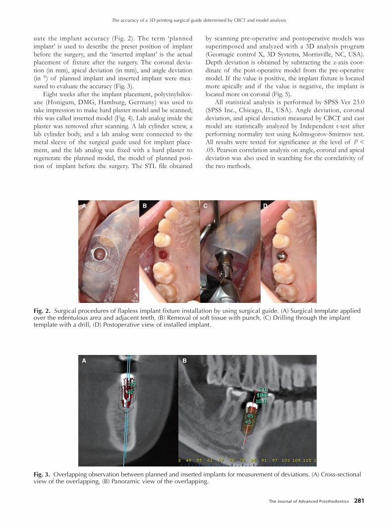
uate the implant accuracy (Fig. 2). The term ‘planned implant’ is used to describe the preset position of implant before the surgery, and the ‘inserted implant’ is the actual placement of fixture after the surgery. The coronal devia-tion (in mm), apical deviation (in mm), and angle deviation (in °) of planned implant and inserted implant were mea-sured to evaluate the accuracy (Fig. 3).
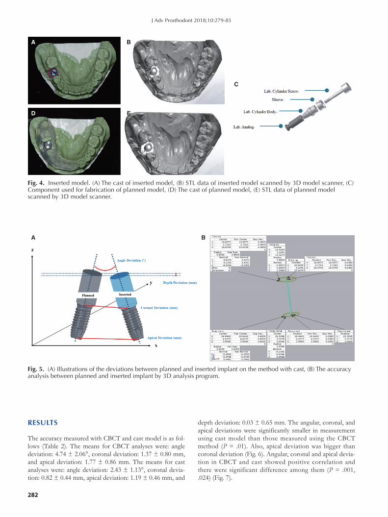
Eight weeks after the implant placement, polyvinylsilox-ane (Honigum, DMG, Hamburg, Germany) was used to take impression to make hard plaster model and be scanned; this was called inserted model (Fig. 4). Lab analog inside the plaster was removed after scanning. A lab cylinder screw, a lab cylinder body, and a lab analog were connected to the metal sleeve of the surgical guide used for implant place-ment, and the lab analog was fixed with a hard plaster to regenerate the planned model, the model of planned posi-tion of implant before the surgery. The STL file obtained
by scanning pre-operative and postoperative models was superimposed and analyzed with a 3D analysis program (Geomagic control X, 3D Systems, Morrisville, NC, USA). Depth deviation is obtained by subtracting the z-axis coor-dinate of the post-operative model from the pre-operative model. If the value is positive, the implant fixture is located more apically and if the value is negative, the implant is located more on coronal (Fig. 5).
All statistical analysis is performed by SPSS Ver 23.0 (SPSS Inc., Chicago, IL, USA). Angle deviation, coronal deviation, and apical deviation measured by CBCT and cast model are statistically analyzed by Independent t-test after performing normality test using Kolmogorov-Smirnov test. All results were tested for significance at the level of P < .05. Pearson correlation analysis on angle, coronal and apical deviation was also used in searching for the correlativity of the two methods.
Fig. 2. Surgical procedures of flapless implant fixture installation by using surgical guide. (A) Surgical template applied over the edentulous area and adjacent teeth, (B) Removal of soft tissue with punch, (C) Drilling through the implant template with a drill, (D) Postoperative view of installed implant.
A B C D
Fig. 3. Overlapping observation between planned and inserted implants for measurement of deviations. (A) Cross-sectional view of the overlapping, (B) Panoramic view of the overlapping.
A B
The accuracy of a 3D printing surgical guide determined by CBCT and model analysis
282
RESULTS
The accuracy measured with CBCT and cast model is as fol-lows (Table 2). The means for CBCT analyses were: angle deviation: 4.74 ± 2.06°, coronal deviation: 1.37 ± 0.80 mm, and apical deviation: 1.77 ± 0.86 mm. The means for cast analyses were: angle deviation: 2.43 ± 1.13°, coronal devia-tion: 0.82 ± 0.44 mm, apical deviation: 1.19 ± 0.46 mm, and
depth deviation: 0.03 ± 0.65 mm. The angular, coronal, and apical deviations were significantly smaller in measurement using cast model than those measured using the CBCT method (P= .01).Also, apical deviationwas bigger thancoronal deviation (Fig. 6). Angular, coronal and apical devia-tion in CBCT and cast showed positive correlation and there were significant difference among them (P= .001,.024) (Fig. 7).
Fig. 4. Inserted model. (A) The cast of inserted model, (B) STL data of inserted model scanned by 3D model scanner, (C) Component used for fabrication of planned model, (D) The cast of planned model, (E) STL data of planned model scanned by 3D model scanner.
A B
C
D E
Fig. 5. (A) Illustrations of the deviations between planned and inserted implant on the method with cast, (B) The accuracy analysis between planned and inserted implant by 3D analysis program.
A B
J Adv Prosthodont 2018;10:279-85

The Journal of Advanced Prosthodontics 283
Table 2. The angle, coronal, apical and depth deviation between CBCT and cast
Method Mean ± SD Max Min P value
Angle deviation (°) CBCT 4.74 ± 2.06a 8.86 0.00 0.001
Cast 2.43 ± 1.13b 5.70 1.15
Coronal deviation (mm) CBCT 1.37 ± 0.80a 3.76 0.18 0.002
Cast 0.82 ± 0.44b 1.85 0.13
Apical deviation (mm) CBCT 1.77 ± 0.86a 3.76 0.45 0.003
Cast 1.19 ± 0.46b 2.51 0.37
Depth deviation (mm) Cast -0.03 ± 0.65 1.58 -2.12
Different superscript letters indicate significant differences.
Fig. 6. The angle and length deviation between CBCT and cast. Results show mean ± SD angle deviation. * indicates statistical differences (P < .05).
A B
8
6
4
2
0CBCT Cast
*P = .001
Dev
iatio
n (°
)
32.5
21.5
10.5
0-0.5
CBCT Cast
*P = .001
Dev
iatio
n (m
m)
*P = .003
Coronal deviation Apical deviation
A B
C
Fig. 7. Scatter plot for the Pearson correlation between CBCT analysis and cast analysis. (A) The angle deviation (P = .001, R = 0.635), (B) The coronal deviation (P = .054, R = 0.368), (C) The apical deviation (P = .024, R = 0.426).
6.00
5.00
4.00
3.00
2.00
1.00
3.00
2.50
2.00
1.50
1.00
.50
.00
VAR
000
04VA
R 0
0006
.00 2.00 4.00 6.00 8.00 10.00VAR 00001
.00 1.00 2.00 3.00 4.00VAR 00003
2.00
1.50
1.00
.50
.00
VAR
000
05
.00 1.00 2.00 3.00 4.00VAR 00002
The accuracy of a 3D printing surgical guide determined by CBCT and model analysis

284
DISCUSSION
While digital surgical guide makes it possible to drill and place an implant in a preset position, the procedure has to include accurate position and accurate analysis before the surgery. Effort has been made to improve the accuracy of digital surgical guide as well as maintenance of surgical guide and errors occurring during the manufacturing process.
During implant procedure, 1 angle deviation makes 0.34 mm length deviation in the 10-mm fixture apical area. 5° angle deviation makes 1.7 mm length deviation. If the space between implant and tooth root were set to 1.5 mm during implant planning, 5° angle error will impair the tooth root. Thus the angle deviation should be no more than 3° to implant installed safely without the tooth damaged.10 If the important anatomical structure such as inferior alveolar nerve is close by, acceptable surgical guide’s maximum angle deviation is less than 3° and maximum vertical error is less than -1.5 mm.10 Less loosening of implant and passive fit is possible when the angle between the hex of fixture and hexagonal freedom of abutment is less than 5° and the dis-tance is 150 µm.11,12 This study shows that angular, coronal, and apical deviation are accurate enough to avoid the dam-age of major anatomical structure during the procedure, but less accurate to connect directly to pre-manufactured hexag-onal implant prosthesis. Therefore, it is necessary to manu-facture prosthesis by taking an impression after the implant placement, or to use non-hexagonl implant fixture.1
In this study, deviation measured by CBCT is similar to that of other studies but angle deviation is somewhat high-er. The reason for this is that the previous studies used sur-gical kit and implant fixture of the subsidiary company that makes surgical guide, while universal surgical guide kit is used in this study. Also, compared the previous studies, the more rearmost molars are included in this study. Reference marker was not used when taking CBCT, so higher error occurred during overlapping preoperative and postoperative CBCT.
The angle, coronal, and apical deviation were statistically significantly smaller than those of CBCT in this study (P < .05). Not only there is no error occurred while superimpos-ing pre-operative and postoperative CBCTs, but also can additional radiation be decreased by using cast model to analyze accuracy. However, errors can occur when taking impression or making plaster cast model. There was more error in overlapping CBCTs because of the absence of marker in CBCT analysis. Error can be decreased by using only one cast model to reproduce preoperative and postop-erative cast model. Further studies in comparing these two methods are required by adding marker to improve accuracy of superimposition. In cast model analysis, depth deviation can be measured by setting axis of the implant as z-axis and obtaining difference in the z-axis. Average of depth devia-tion is -0.03 ± 0.65 mm, which is more on the coronal side than planned position. This result is similar to that of the previous study, especially when those who have less experi-ence with digital guided surgery tend to have less reliability
in the accuracy of the surgical guide.13
This study shows that the deviation obtained by the plaster cast is significantly smaller, which can be useful in evaluation of implant placement accuracy. In addition, the angular deviation may become larger toward the farthest tooth in implant placement.
CONCLUSION
The present study results showed significantly smaller devia-tion values using cast model analysis than those measured using the CBCT superimposition method. The angle and length deviation value of the universal digital guide stent were clinically acceptable.
ORCID
Boyoung Ma https://orcid.org/0000-0002-8147-056XTaeweok Park https://orcid.org/0000-0003-0180-4409Inkon Chun https://orcid.org/0000-0003-0539-8313Kwidug Yun https://orcid.org/0000-0002-2965-3967
REFERENCES
1. Misch CE. Dental implant prosthetics. Elsevier Health Sciences; MO, USA, 2014.
2. Nickenig HJ, Eitner S. Reliability of implant placement after virtual planning of implant positions using cone beam CT da-ta and surgical (guide) templates. J Craniomaxillofac Surg 2007;35:207-11.
3. Ganz SD. Three-dimensional imaging and guided surgery for dental implants. Dent Clin North Am 2015;59:265-90.
4. Shim JS, Kim NH, Kim JE. A procedure for the computer-guided implant planning: A narrative review. J Korean Dent Assoc 2016;54:108-22.
5. Van Assche N, Vercruyssen M, Coucke W, Teughels W, Jacobs R, Quirynen M. Accuracy of computer-aided implant place-ment. Clin Oral Implants Res 2012;23:112-23.
6. Sicilia A, Botticelli D; Working Group 3. Computer-guided implant therapy and soft- and hard-tissue aspects. The third EAO consensus conference 2012. Clin Oral Implants Res 2012;23:157-61.
7. Martorelli M. A new approach in CT artifact removal: three cases study in maxillofacial surgery. Int J Interact Des Manuf 2013;7:115-24.
8. Komiyama A, Pettersson A, Hultin M, Näsström K, Klinge B. Virtually planned and template-guided implant surgery: an ex-perimental model matching approach. Clin Oral Implants Res 2011;22:308-13.
9. Yoon JH. The accuracy estimate of surgical stents fabricated by digital methods in installing implants on dental models. Chonnam National University. Master Degree Thesis. 2017.
10. Choi B, Jeong S. Digital flapless implantology. Seoul; Ji-Sung Publishing Co.; 2015. p. 32-51.
11. Al Quran FA, Rashdan BA, Zomar AA, Weiner S. Passive fit and accuracy of three dental implant impression techniques. Quintessence Int 2012;43:119-25.
J Adv Prosthodont 2018;10:279-85

The Journal of Advanced Prosthodontics 285
12. Binon PP. The effect of implant/abutment hexagonal misfit on screw joint stability. Int J Prosthodont 1996;9:149-60.
13. Pozzi A, Polizzi G, Moy PK. Guided surgery with tooth-sup-ported templates for single missing teeth: A critical review. Eur J Oral Implantol 2016;9:S135-53.
14. Verhamme LM, Meijer GJ, Boumans T, de Haan AF, Bergé SJ, Maal TJ. A clinically relevant accuracy study of computer-planned implant placement in the edentulous maxilla using mucosa-supported surgical templates. Clin Implant Dent Relat Res 2015;17:343-52.
15. Ersoy AE, Turkyilmaz I, Ozan O, McGlumphy EA. Reliability of implant placement with stereolithographic surgical guides generated from computed tomography: clinical data from 94 implants. J Periodontol 2008;79:1339-45.
16. Behneke A, Burwinkel M, Behneke N. Factors influencing transfer accuracy of cone beam CT-derived template-based implant placement. Clin Oral Implants Res 2012;23:416-23.
17. Kang BG, Kim HJ, Chung CH. Accuracy of the CT guided implant template by using an intraoral scanner according to the edentulous distance. J Korean Acad Prosthodont 2017;55: 1-8.
18. Pettersson A, Kero T, Gillot L, Cannas B, Fäldt J, Söderberg R, Näsström K. Accuracy of CAD/CAM-guided surgical tem-plate implant surgery on human cadavers: Part I. J Prosthet Dent 2010;103:334-42.
19. Cassetta M, Di Mambro A, Giansanti M, Stefanelli LV, Cavallini C. The intrinsic error of a stereolithographic surgical template in implant guided surgery. Int J Oral Maxillofac Surg 2013;42:264-75.
20. Ozan O, Turkyilmaz I, Ersoy AE, McGlumphy EA, Rosenstiel SF. Clinical accuracy of 3 different types of computed to-mography-derived stereolithographic surgical guides in im-plant placement. J Oral Maxillofac Surg 2009;67:394-401.
21. Valente F, Schiroli G, Sbrenna A. Accuracy of computer-aid-ed oral implant surgery: a clinical and radiographic study. Int J Oral Maxillofac Implants 2009;24:234-42.
22. Vasak C, Watzak G, Gahleitner A, Strbac G, Schemper M, Zechner W. Computed tomography-based evaluation of tem-plate (NobelGuide™)-guided implant positions: a prospective radiological study. Clin Oral Implants Res 2011;22:1157-63.
23. Park C, Raigrodski AJ, Rosen J, Spiekerman C, London RM. Accuracy of implant placement using precision surgical guides with varying occlusogingival heights: an in vitro study. J Prosthet Dent 2009;101:372-81.
The accuracy of a 3D printing surgical guide determined by CBCT and model analysis