TG and VLDL Physiology
6
Triglyceride Transport Thomas Dayspring MD, FACP Triglyceride (TG) or more accurately triacylglycerol is simply a compound of three acyl groups (fatty acids) attached to a glycerol molecule. They are easily measured in the laboratory and reported as serum TG level: it actually refers to all of the TG that are being trafficked within all of the lipoproteins that exist in a deciliter of plasma. A physiologic fasting TG concentration is on average 30 mg/dL with one standard deviation rangi ng from 10 to 70 mg/dL. The two major lipoproteins that transport TG are chylomicrons and very low density lipoprotein (VLDL). Since chylomicrons are basically postprandial lipoproteins with very short half life’s (and hour or so) most of the TG reported are in VLDLs (4-6 hour half life). The purpose of the triglyceride-rich lipoproteins (TGRLP), VLDL and chylomicrons are to transport energy in the form of fatty a cids (FA) trafficked as triglycerides (TG), to the energy-requiring tissues (muscles) or the energy- storing tissues, the adipocytes. If the muscles do not require the fuel, the FA in the TG will then be delivered for storage in adipocytes. Normally the liver (hepatocytes) packages TG, cholesterol and phospholipids with a single, molecule of apolipoprotein B100 (apoB) and the enterocytes package the lipids with a truncated version of apoB; termed apolipoprotein B48 (has 48% of the molecular weight of hepatically produced apoB100). A normally composed VLDL particle has five times more TG than cholesterol (a 5:1 ratio) and since most TG are presumed to be in VLDLs; the VLDL-C is calculated as TG/5. If 150 mg/dL is a normal TG, then a normal VLDL-C is < 30 mg/dL. A high VLDL-C implies a high TG level. In conditions of elevated TG, many of the VLDL will be very large and TG-rich. If hepatic TG levels are not elevated the liver produces fewer as well as smaller VLDL particles (VLDL-C is reduced). Very L ow Densi ty Lipopr otein (VLDL) Very Low Densi ty Lip opr otein (VLDL) The primary TG The primary TG transporting lipoprotein transporting lipoprotein Size, depending on TG Size, depending on TG content varies from 350 content varies from 350 Å Å (35 nm) to 700 (35 nm) to 700 Å Å (70 nm) (70 nm) Normal particle Normal particle composition is 80% TG composition is 80% TG and 20% cholesterol and 20% cholesterol VLDL (or a 5 to 1 ratio) (or a 5 to 1 ratio) VLDL- - Phospolipids Phospolipids C ~ TG/5 C ~ TG/5 TG and Cholesteryl TG and Cholesteryl ester core ester core Free cholesterol Free cholesterol VLDL particles, as they exit the liver rapidly acquire several other apolipoproteins (some in the space of Disse and others transferred from circulating HDL particles). These include apoC-I, apoC-II, apoC-III, apoE, apoA-II, apoA-V and others. These apolipoproteins are integral to the proper trafficking of lipids and efficient catabolism of the VLDL and chylomicron. If any of the aforementioned apolipoproteins are absent or present in abnormal concentrations or defective, the VLDL particle will have altered catabolism. Typically when a VLDL (or chylomicron) enters vascular beds rich in the triglyceridase called lipoprotein lipase (LPL) and VLDL receptors, notably skeletal muscle, adipose tissue, and myocardium, rapid hydrolysis of the TG to
-
Upload
jodyann-likrisha-mcleod -
Category
Documents
-
view
220 -
download
0
Transcript of TG and VLDL Physiology
7/27/2019 TG and VLDL Physiology
http://slidepdf.com/reader/full/tg-and-vldl-physiology 1/6
Triglyceride TransportThomas Dayspring MD, FACP
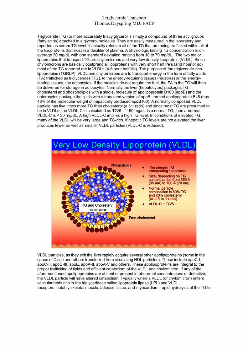
Triglyceride (TG) or more accurately triacylglycerol is simply a compound of three acyl groups(fatty acids) attached to a glycerol molecule. They are easily measured in the laboratory andreported as serum TG level: it actually refers to all of the TG that are being trafficked within all ofthe lipoproteins that exist in a deciliter of plasma. A physiologic fasting TG concentration is onaverage 30 mg/dL with one standard deviation ranging from 10 to 70 mg/dL. The two majorlipoproteins that transport TG are chylomicrons and very low density lipoprotein (VLDL). Sincechylomicrons are basically postprandial lipoproteins with very short half life’s (and hour or so)most of the TG reported are in VLDLs (4-6 hour half life). The purpose of the triglyceride-richlipoproteins (TGRLP), VLDL and chylomicrons are to transport energy in the form of fatty acids(FA) trafficked as triglycerides (TG), to the energy-requiring tissues (muscles) or the energy-storing tissues, the adipocytes. If the muscles do not require the fuel, the FA in the TG will thenbe delivered for storage in adipocytes. Normally the liver (hepatocytes) packages TG,cholesterol and phospholipids with a single, molecule of apolipoprotein B100 (apoB) and theenterocytes package the lipids with a truncated version of apoB; termed apolipoprotein B48 (has48% of the molecular weight of hepatically produced apoB100). A normally composed VLDLparticle has five times more TG than cholesterol (a 5:1 ratio) and since most TG are presumed tobe in VLDLs; the VLDL-C is calculated as TG/5. If 150 mg/dL is a normal TG, then a normalVLDL-C is < 30 mg/dL. A high VLDL-C implies a high TG level. In conditions of elevated TG,many of the VLDL will be very large and TG-rich. If hepatic TG levels are not elevated the liver
produces fewer as well as smaller VLDL particles (VLDL-C is reduced).
Very Low Density Lipoprotein (VLDL)Very Low Density Lipoprotein (VLDL)
The primary TGThe primary TGtransporting lipoproteintransporting lipoprotein
Size, depending on TGSize, depending on TGcontent varies from 350content varies from 350 Å
Å(35 nm) to 700(35 nm) to 700 Å Å
(70 nm)(70 nm)
Normal particleNormal particlecomposition is 80% TGcomposition is 80% TGand 20% cholesteroland 20% cholesterol
VLDL
(or a 5 to 1 ratio)(or a 5 to 1 ratio)
VLDL--
PhospolipidsPhospolipids
C ~ TG/5C ~ TG/5TG and CholesterylTG and Cholesterylester coreester core
Free cholesterolFree cholesterol
VLDL particles, as they exit the liver rapidly acquire several other apolipoproteins (some in thespace of Disse and others transferred from circulating HDL particles). These include apoC-I,apoC-II, apoC-III, apoE, apoA-II, apoA-V and others. These apolipoproteins are integral to theproper trafficking of lipids and efficient catabolism of the VLDL and chylomicron. If any of theaforementioned apolipoproteins are absent or present in abnormal concentrations or defective,the VLDL particle will have altered catabolism. Typically when a VLDL (or chylomicron) entersvascular beds rich in the triglyceridase called lipoprotein lipase (LPL) and VLDLreceptors, notably skeletal muscle, adipose tissue, and myocardium, rapid hydrolysis of the TG to
7/27/2019 TG and VLDL Physiology
http://slidepdf.com/reader/full/tg-and-vldl-physiology 2/6
Triglyceride TransportThomas Dayspring MD, FACP
fatty acids occurs (this process is termed lipolysis). The free (unesterified) fatty acids (FFA orNEFA) enter the nearby cell with by simple diffusion or cell membrane fatty acid transportproteins or also attach to circulating albumin and are dispersed in plasma for delivery to othercells. It is VLDL surface apoC-II which is the ligand for LPL. The VLDL or chylomicron docks asfollows: surface apoA-V attaches to endothelial proteoglycans in areas rich in LPL and VLDLreceptors. The VLDL receptor docks the particle and apoC-II interacts with LPL initiating lipolysis.If one has absent or defective apoC-II, apoA-V or lacks LPL, effective lipolysis will not occur andsuch patients have variable degrees of hypertriglyceridemia (fasting and especially postprandial).Unlike LDL receptors, VLDL receptors do not endocytose VLDLs or chylomicrons but rather helpdock the particle so LPL-mediated lipolysis can occur and then facilitate FA entry into the cell.
Excess ApoC-III has the ability to block the attachment of C-II to LPL thus modulating the rate ofVLDL lipolysis. Too much apoC-III (often seen in insulin resistant patients) markedly delays thecatabolism of TG-rich lipoproteins like VLDL and chylomicrons. It is also thought that too muchapoC-III can displace apoE off of the VLDL or hinder CII/LPL interaction. This is associated withboth fasting and especially postprandial hypertriglyceridemia. When you see a Type 2 diabetic ormetabolic syndrome patients with TG > 150 mg/dL, you should suspect elevated apoCIII (anindependent risk factor for CHD). The apoC-III/apoC-II ratio is an indicator of the rate of VLDL &chylomicron lipolysis: high ratios indicate delayed lipolysis.
ApoA5 or apoA-V is becoming better understood as a regulator of TG. It is thought that apoA-Von the VLDL surface locates or “parks” the lipoprotein by attaching to surface proteoglycans inthe vicinity of LPL, where surface apoC-II can interact with the LPL. ApoC-III and apoA-V andlikely other apolipoproteins are regulated by PPAR-alpha and hepatic nuclear factor 4-alpha(inhibits CIII and induces A-V) and may be amenable to pharmacologic modulation.
The several molecules of apoE on the TG-rich lipoprotein particle serves (as does apoB) as aligand for the LDL receptor and the LDL receptor related protein and VLDL receptor. If there isabnormal or absent apoE on VLDL, this will also result in delayed LDL receptor (LDLr) or LDLReceptor Related protein (LRP) associated hepatic clearance of VLDL, VLDL remnants (definedbelow) and IDL (elevating apoB and TG levels). This is seen in the Type III FredricksonHyperlipidemia patients. ApoC-I can also interfere with apoE binding to VLDL and LDL receptorsand thereby delay clearance of remnant VLDL particles. ApoC-I is also an inhibitor of cholesterol
ester transfer protein (CETP) discussed below.
As the VLDL looses its TG core, it reduces in size, and looses surface phospholipids (which arepicked up by phospholipid transfer factor), as well as surface apolipoproteins (which are pickedup by HDL). The VLDL gets smaller and smaller (called a remnant) and ultimately becomes anapoB particle termed an intermediate density lipoprotein (IDL). The apoB is a nontransferableapolipoprotein (stays with the particle from birth to death), so the IDL particle still has the samesingle molecule of apoB with some of the remaining apoE. Most of the VLDL remnants and IDLsand their cholesterol content are rapidly cleared by liver LDL receptors which bind to the surfaceapoE/apoB: this process is termed indirect reverse cholesterol transport. A few are subjected tofurther lipolysis and become LDLs.
Depending on their TG content, VLDL particles can vary tremendously in size from 35-40 to
several hundred nanometers (nm). Keep in mind that for an apoB particle to enter the wall of theartery it has to be less than 70 nm in diameter. On an NMR LipoProfile, the smaller and mediumVLDL particles are identified as V1-V4. The very large VLDLs and chylomicrons (V5 and V6)cannot enter the arterial wall (too large) and are thus do not deliver sterols to plaque. That is whysome people with very serious hypertriglyceridemia are not at risk for CHD. They simply have afew large particles carrying lots of TG (the apoB or LDL-P is normal).
7/27/2019 TG and VLDL Physiology
http://slidepdf.com/reader/full/tg-and-vldl-physiology 3/6
Triglyceride TransportThomas Dayspring MD, FACP
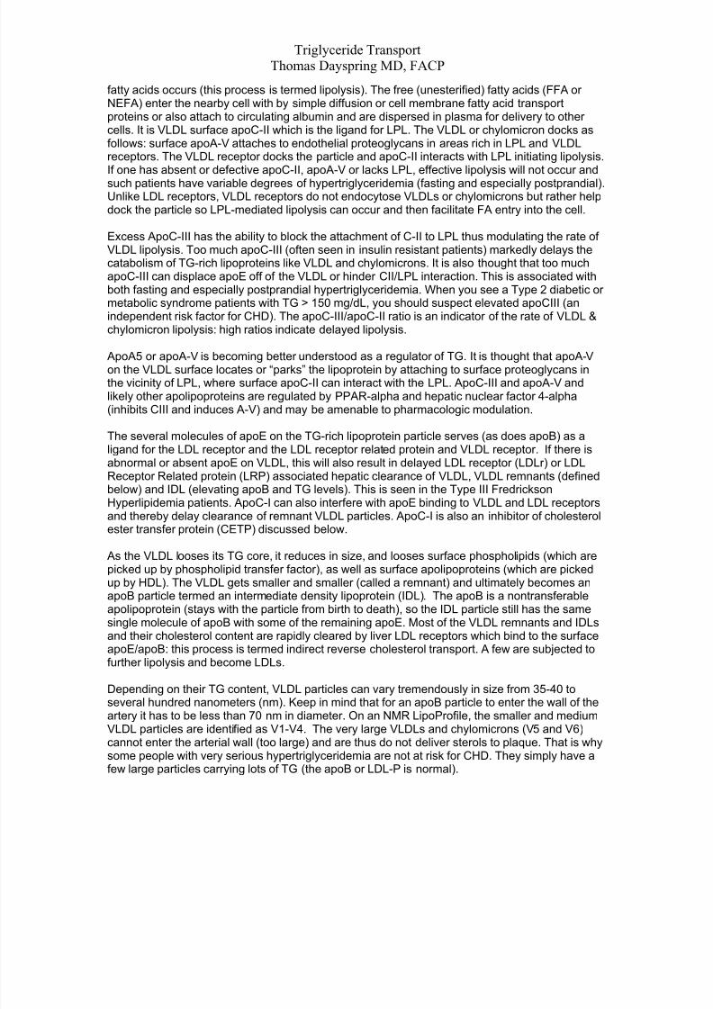
TG-rich Lipoprotein Apolipoproteins
Apo B(100)
Apo CII Ligand forLipoprotein Lipase
Particle Stability
& Ligand forLDL Receptor
Ligand for VLDL & LDLReceptor relatedprotein (VLDLr
and LRP)
Apo CIII Apo E
Blocks Apo B,CII and E
Apo AV
Enhancesparticle lipolysis
Lipoprotein Abnormalitiesin TG/HDL Axis Disorders
TG-richVLDL
Triglyceride Cholesteryl ester
apoCIII
apoB
TG
↑apoCIII is an independent riskfactor for CHD↑ apoCIII blocks or displacesapoCII and apoE, which delaysVLDL lipolysis
↑apoCIII is associated withfasting and postprandialhypertriglyceridemia
↑apoCIII is common in insulinresistant patients
7/27/2019 TG and VLDL Physiology
http://slidepdf.com/reader/full/tg-and-vldl-physiology 4/6
Triglyceride TransportThomas Dayspring MD, FACP
The IDL particle, which may still be trafficking residual TG is a substrate for hepatic lipase (HL)which has both triglyceridase and phospholipase activity. The IDL particle, as it looses TG(lipolysis) shrinks and becomes an LDL. The IDL particle because of its apoE and apoB contentcan also internalized by hepatic LDL receptors. The physiologic half life of a VLDL becoming anLDL is about 4-6 hours. The half life of an LDL is 2-3 days. LDL particles, if present in physiologicconcentrations are ultimately removed by the liver LDL receptors in (indirect reverse cholesteroltransport). If present in excess quantities, smaller VLDL, IDL or LDL particles enter the arterialwall resulting in atherogenesis.
If a patient has a physiologic TG of 30 mg/dL (10-70), they make only enough VLDLparticles needed to transport the TGs for cellular energy. That is, they have physiologic levels ofnormally sized VLDL particles. After efficient lipolysis they would have physiologic amounts of
LDL particles (an LDL-P well under 1000 nmol/L) and the LDL-C would be 20-40 mg/dL(considered a physiologic LDL-C).
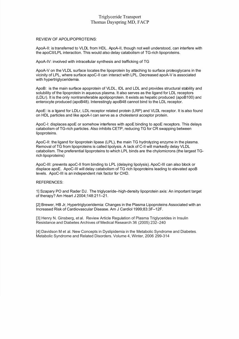
It is not unusual to see low HDL-C levels in patients with high TG. VLDL particles that do notundergo rapid catabolism, linger in the plasma, elevating apoB levels and increasing CV risk.What also happens is the TG and cholesteryl ester (CE) are exchanged between HDL, LDL andVLDL, via CETP. The longer the half-life of VLDL, the more time they have to swap TG for CEfrom HDL using CETP. The HDL particles become TG-rich and cholesterol-poor, therebyreducing HDL-C levels. Hepatic lipase then hydrolyses the remaining TG and surfacephospholipids creating very small, cholesterol deficient, small HDL particles (~7 nm in diameter)which are easily excreted by the glomerulus, lowering apoA-I or HDL-P concentrations. A similarCETP-mediated exchange of TG for cholesteryl ester occurs with LDL particles enhancing theformation of small LDL particles through a similar process. We now call such patients, TG/HDL
axis disorders and of course this is most commonly seen in metabolic syndrome patients andType 2 diabetics. In this scenario the VLDL, which has transferred its TG, becomes CE rich(increased VDL-C). As lipolysis proceeds the VLDLs loose further TG and become smaller.These CE-rich, TG poor VLDLs are called remnants and have been associated with increasedCV risk. However, the vastly preponderant particle present in TG/HDL axis disorders is the LDL.
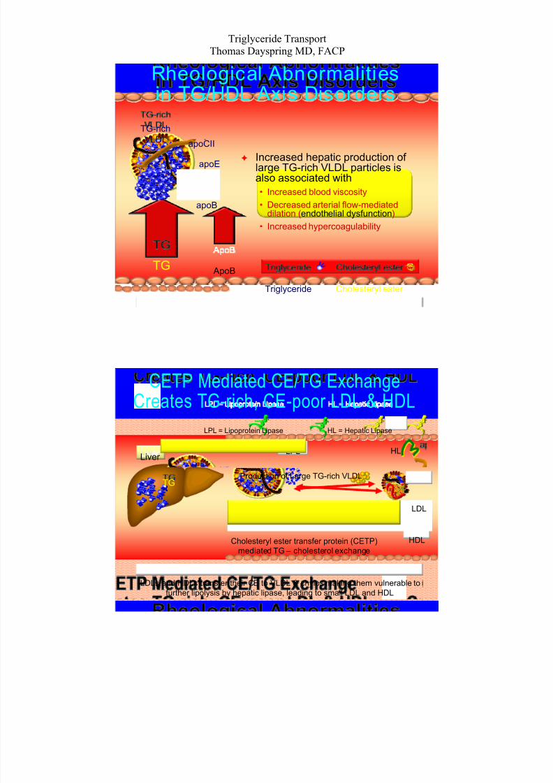
Much of the risk associated with elevated TG is related to its association with increased apoB.Too many VLDLs cause increased levels of IDLs and especially LDLs. ApoB and LDL-P starts toelevate with TG levels >100 mg /dL and by a value of 200 mg/dL 80% of patients will have a highapoB or LDL-P. However when there are increased amounts of large TG-rich VLDLs and/orchylomicrons, the excess fat increases blood viscosity, down regulates NO and causesendothelia dysfunction, increase coagulation markers (PAI-1 and fibrinogen) and induces(probably through increased apoC-III) multiple inflammatory markers.
7/27/2019 TG and VLDL Physiology
http://slidepdf.com/reader/full/tg-and-vldl-physiology 5/6
Triglyceride TransportThomas Dayspring MD, FACP
Rheological Abnormalitiesin TG/HDL Axis Disorders
TG-rich
VLDL
Triglyceride Cholesteryl ester
apoB
TG ApoB
apoCII
•
•
•
Increased hepatic production oflarge TG-rich VLDL particles isalso associated with
elial dysfunction
apoE
Increased blood viscosity
Decreased arterial flow-mediateddilation (endoth )
Increased hypercoagulability
Liver
TG
LPL
HDL
CETP Mediated CE/TG Exchange
Creates TG-rich, CE-poor LDL & HDLLPL = Lipoprotein Lipase HL = Hepatic Lipase
HL
LDL
Production of Large TG-rich VLDL
Cholesteryl ester transfer protein (CETP)
mediated TG – cholesterol exchange
LDLs and HDLs transfer their CE to VLDL & chylos making them vulnerable tofurther lipolysis by hepatic lipase, leading to small LDL and HDL
7/27/2019 TG and VLDL Physiology
http://slidepdf.com/reader/full/tg-and-vldl-physiology 6/6
Triglyceride TransportThomas Dayspring MD, FACP
REVIEW OF APOLIPOPROTEINS:
ApoA-II: is transferred to VLDL from HDL. ApoA-II, though not well understood, can interfere withthe apoCII/LPL interaction. This would also delay catabolism of TG-rich lipoproteins.
ApoA-IV: involved with intracellular synthesis and trafficking of TG
ApoA-V on the VLDL surface locates the lipoprotein by attaching to surface proteoglycans in thevicinity of LPL, where surface apoC-II can interact with LPL. Decreased apoA-V is associatedwith hypertriglyceridemia.
ApoB: is the main surface apoprotein of VLDL, IDL and LDL and provides structural stability andsolubility of the lipoprotein in aqueous plasma. It also serves as the ligand for LDL receptors(LDLr). It is the only nontransferable apolipoprotein. It exists as hepatic produced (apoB100) andenterocyte produced (apoB48). Interestingly apoB48 cannot bind to the LDL receptor.
ApoE: is a ligand for LDLr, LDL receptor related protein (LRP) and VLDL receptor. It is also foundon HDL particles and like apoA-I can serve as a cholesterol acceptor protein.
ApoC-I: displaces apoE or somehow interferes with apoE binding to apoE receptors. This delayscatabolism of TG-rich particles. Also inhibits CETP, reducing TG for CR swapping betweenlipoproteins.
ApoC-II: the ligand for lipoprotein lipase (LPL), the main TG hydrolyzing enzyme in the plasma.Removal of TG from lipoproteins is called lipolysis. A lack of C-II will markedly delay VLDLcatabolism. The preferential lipoproteins to which LPL binds are the chylomicrons (the largest TG-rich lipoproteins)
ApoC-III: prevents apoC-II from binding to LPL (delaying lipolysis). ApoC-III can also block ordisplace apoE. ApoC-III will delay catabolism of TG rich lipoproteins leading to elevated apoBlevels. ApoC-III is an independent risk factor for CHD.
REFERENCES:
1] Szapary PO and Rader DJ. The triglyceride–high-density lipoprotein axis: An important targetof therapy? Am Heart J 2004;148:211–21.
[2] Brewer, HB Jr, Hypertriglyceridemia: Changes in the Plasma Lipoproteins Associated with anIncreased Risk of Cardiovascular Disease. Am J Cardiol 1999;83:3F–12F.
[3] Henry N. Ginsberg, et al. Review Article Regulation of Plasma Triglycerides in InsulinResistance and Diabetes Archives of Medical Research 36 (2005) 232–240
[4] Davidson M et al. New Concepts in Dyslipidemia in the Metabolic Syndrome and Diabetes.
Metabolic Syndrome and Related Disorders. Volume 4, Winter, 2006 299-314