Tetanus Toxin and Botulinum Toxin A Utilize Unique ... · Tetanus Toxin and Botulinum Toxin A...
8
Tetanus Toxin and Botulinum Toxin A Utilize Unique Mechanisms To Enter Neurons of the Central Nervous System Faith C. Blum, Chen Chen,* Abby R. Kroken, and Joseph T. Barbieri Medical College of Wisconsin, Microbiology and Molecular Genetics, Milwaukee, Wisconsin, USA Botulinum neurotoxins (BoNTs) and tetanus neurotoxin (TeNT) are the most toxic proteins for humans. While BoNTs cause flaccid paralysis, TeNT causes spastic paralysis. Characterized BoNT serotypes enter neurons upon binding dual receptors, a ganglioside and a neuron-specific protein, either synaptic vesicle protein 2 (SV2) or synaptotagmin, while TeNT enters upon binding gangliosides as dual receptors. Recently, TeNT was reported to enter central nervous system (CNS) neurons upon synap- tic vesicle cycling that was mediated by the direct binding to SV2, implying that TeNT and BoNT utilize common mechanisms to enter CNS neurons. This prompted an assessment of TeNT entry into CNS neurons, using the prototypic BoNT serotype A as a reference for SV2-mediated entry into synaptic vesicles, analyzing the heavy-chain receptor binding domain (HCR) of each toxin. Synaptic vesicle cycling stimulated the entry of HCR/A into neurons, while HCR/T entered neurons with similar levels of efficiency in depolarized and nondepolarized neurons. ImageJ analysis identified two populations of cell-associated HCR/T in synaptic vesicle cycling neurons, a major population which segregated from HCR/A and a minor population which colocalized with HCR/A. HCR/T did not inhibit HCR/A entry into neurons in competition experiments and did not bind SV2, the protein receptor for BoNT/A. Intoxication experiments showed that TeNT efficiently cleaved VAMP2 in depolarized neurons and neu- rons blocked for synaptic vesicle cycling. These experiments demonstrate that TeNT enters neurons by two pathways, one inde- pendent of stimulated synaptic vesicle cycling and one by synaptic vesicles independent of SV2, showing that TeNT and BoNT/A enter neurons by unique mechanisms. T he clostridial neurotoxins (CNTs) are the most toxic proteins for humans and include category A agents, the botulinum neurotoxins (BoNTs) (19). CNT neuronal toxicity occurs by specific binding to neuronal receptors and cleavage of neuron- specific soluble N-ethylmaleimide-sensitive factor attachment protein receptor (SNARE) proteins. CNTs are synthesized as sin- gle-chain A-B toxins that are nicked to an N-terminal light-chain protease domain (LC) which remains associated with the C-ter- minal heavy chain (HC) by a disulfide bond (33). The HC com- prises a translocation domain (HCT) and a receptor binding do- main (HCR). The HCR is composed of an N-terminal subdomain (HCRN) and a C-terminal subdomain (HCRC). HCRN has not been assigned a function, while HCRC contains dual, host neu- ron-specific receptor binding pockets (27, 28, 35). CNTs also include tetanus neurotoxin (TeNT), which shares 35% amino acid identity and a conserved tertiary structure with BoNTs (24). Although BoNTs and TeNT cleave SNARE proteins, BoNTs elicit flaccid paralysis and TeNT elicits spastic paralysis due to differential trafficking within -motor neurons. BoNTs are classified into seven serotypes (A to G). BoNT/A and BoNT/E cleave synaptosomal-associated protein of 25 kDa (SNAP25) (3, 4, 27, 36, 38); BoNT/B, BoNT/D, BoNT/F, BoNT/G, and TeNT cleave vesicle-associated membrane protein 2 (VAMP2, also re- ferred to as synaptobrevin 2); and BoNT/C cleaves SNAP25 and syntaxin 1A (5, 17, 27, 29, 30, 35). The observation that TeNT and BoNT/B cleave VAMP2 at the same residue underscores the im- portance of intracellular trafficking as being responsible for their unique pathologies (12, 37). Membrane depolarization stimulates the opening of Ca 2 channels and entry of extracellular Ca 2 into neurons, which drives synaptic vesicle (SV) exocytosis (42). Synaptotagmin binds intracellular Ca 2 and initiates migration of the SV to the plasma membrane, where fusion releases neurotransmitters into the syn- apse (42). The mechanism for SV cycling from the plasma mem- brane is complex: in addition to direct SV cycling, a rapid endo- some pathway retrieves SV proteins from the plasma membrane (22). BoNT/A entry into neurons follows the initial binding to gan- glioside on the surface of -motor neurons (26, 27). Upon fusion of SVs to the plasma membrane, BoNT/A binds a luminal domain of synaptic vesicle protein 2 (SV2) and is internalized upon SV cycling off the membrane (13). Acidification of the lumen of the SV stimulates HCT insertion into the SV membrane, which facil- itates translocation of the LC into the cytosol (16). On -motor neurons, TeNT binds receptors concentrated in lipid microdo- mains (21), which include polysialogangliosides. TeNT is endocy- tosed via a clathrin- and Rab5-mediated pathway and retrograde traffics within the lumen of neutral endosomes that are marked with Rab7 (20). TeNT is transcytosed to an interneuronal synapse by a mechanism that remains undefined, where TeNT binds and intoxicates neurons of the central nervous system (CNS), blocking the release of inhibitory neurotransmitters and eliciting spastic paralysis (39, 40). Recent studies reported that TeNT entered neu- rons of the CNS upon membrane depolarization (25, 43), through Received 17 January 2012 Returned for modification 31 January 2012 Accepted 28 February 2012 Published ahead of print 5 March 2012 Editor: J. B. Bliska Address correspondence to Joseph T. Barbieri, [email protected]. * Present address: University of Chicago, Microbiology, Chicago, Illinois, USA. Supplemental material for this article may be found at http://iai.asm.org/. Copyright © 2012, American Society for Microbiology. All Rights Reserved. doi:10.1128/IAI.00057-12 1662 iai.asm.org 0019-9567/12/$12.00 Infection and Immunity p. 1662–1669 on March 5, 2019 by guest http://iai.asm.org/ Downloaded from
-
Upload
nguyenthien -
Category
Documents
-
view
237 -
download
0
Transcript of Tetanus Toxin and Botulinum Toxin A Utilize Unique ... · Tetanus Toxin and Botulinum Toxin A...
Tetanus Toxin and Botulinum Toxin A Utilize Unique MechanismsTo Enter Neurons of the Central Nervous System
Faith C. Blum, Chen Chen,* Abby R. Kroken, and Joseph T. Barbieri
Medical College of Wisconsin, Microbiology and Molecular Genetics, Milwaukee, Wisconsin, USA
Botulinum neurotoxins (BoNTs) and tetanus neurotoxin (TeNT) are the most toxic proteins for humans. While BoNTs causeflaccid paralysis, TeNT causes spastic paralysis. Characterized BoNT serotypes enter neurons upon binding dual receptors, aganglioside and a neuron-specific protein, either synaptic vesicle protein 2 (SV2) or synaptotagmin, while TeNT enters uponbinding gangliosides as dual receptors. Recently, TeNT was reported to enter central nervous system (CNS) neurons upon synap-tic vesicle cycling that was mediated by the direct binding to SV2, implying that TeNT and BoNT utilize common mechanisms toenter CNS neurons. This prompted an assessment of TeNT entry into CNS neurons, using the prototypic BoNT serotype A as areference for SV2-mediated entry into synaptic vesicles, analyzing the heavy-chain receptor binding domain (HCR) of eachtoxin. Synaptic vesicle cycling stimulated the entry of HCR/A into neurons, while HCR/T entered neurons with similar levels ofefficiency in depolarized and nondepolarized neurons. ImageJ analysis identified two populations of cell-associated HCR/T insynaptic vesicle cycling neurons, a major population which segregated from HCR/A and a minor population which colocalizedwith HCR/A. HCR/T did not inhibit HCR/A entry into neurons in competition experiments and did not bind SV2, the proteinreceptor for BoNT/A. Intoxication experiments showed that TeNT efficiently cleaved VAMP2 in depolarized neurons and neu-rons blocked for synaptic vesicle cycling. These experiments demonstrate that TeNT enters neurons by two pathways, one inde-pendent of stimulated synaptic vesicle cycling and one by synaptic vesicles independent of SV2, showing that TeNT and BoNT/Aenter neurons by unique mechanisms.
The clostridial neurotoxins (CNTs) are the most toxic proteinsfor humans and include category A agents, the botulinum
neurotoxins (BoNTs) (19). CNT neuronal toxicity occurs byspecific binding to neuronal receptors and cleavage of neuron-specific soluble N-ethylmaleimide-sensitive factor attachmentprotein receptor (SNARE) proteins. CNTs are synthesized as sin-gle-chain A-B toxins that are nicked to an N-terminal light-chainprotease domain (LC) which remains associated with the C-ter-minal heavy chain (HC) by a disulfide bond (33). The HC com-prises a translocation domain (HCT) and a receptor binding do-main (HCR). The HCR is composed of an N-terminal subdomain(HCRN) and a C-terminal subdomain (HCRC). HCRN has notbeen assigned a function, while HCRC contains dual, host neu-ron-specific receptor binding pockets (27, 28, 35).
CNTs also include tetanus neurotoxin (TeNT), which shares�35% amino acid identity and a conserved tertiary structure withBoNTs (24). Although BoNTs and TeNT cleave SNARE proteins,BoNTs elicit flaccid paralysis and TeNT elicits spastic paralysisdue to differential trafficking within �-motor neurons. BoNTs areclassified into seven serotypes (A to G). BoNT/A and BoNT/Ecleave synaptosomal-associated protein of 25 kDa (SNAP25) (3, 4,27, 36, 38); BoNT/B, BoNT/D, BoNT/F, BoNT/G, and TeNTcleave vesicle-associated membrane protein 2 (VAMP2, also re-ferred to as synaptobrevin 2); and BoNT/C cleaves SNAP25 andsyntaxin 1A (5, 17, 27, 29, 30, 35). The observation that TeNT andBoNT/B cleave VAMP2 at the same residue underscores the im-portance of intracellular trafficking as being responsible for theirunique pathologies (12, 37).
Membrane depolarization stimulates the opening of Ca2�
channels and entry of extracellular Ca2� into neurons, whichdrives synaptic vesicle (SV) exocytosis (42). Synaptotagmin bindsintracellular Ca2� and initiates migration of the SV to the plasmamembrane, where fusion releases neurotransmitters into the syn-
apse (42). The mechanism for SV cycling from the plasma mem-brane is complex: in addition to direct SV cycling, a rapid endo-some pathway retrieves SV proteins from the plasma membrane(22).
BoNT/A entry into neurons follows the initial binding to gan-glioside on the surface of �-motor neurons (26, 27). Upon fusionof SVs to the plasma membrane, BoNT/A binds a luminal domainof synaptic vesicle protein 2 (SV2) and is internalized upon SVcycling off the membrane (13). Acidification of the lumen of theSV stimulates HCT insertion into the SV membrane, which facil-itates translocation of the LC into the cytosol (16). On �-motorneurons, TeNT binds receptors concentrated in lipid microdo-mains (21), which include polysialogangliosides. TeNT is endocy-tosed via a clathrin- and Rab5-mediated pathway and retrogradetraffics within the lumen of neutral endosomes that are markedwith Rab7 (20). TeNT is transcytosed to an interneuronal synapseby a mechanism that remains undefined, where TeNT binds andintoxicates neurons of the central nervous system (CNS), blockingthe release of inhibitory neurotransmitters and eliciting spasticparalysis (39, 40). Recent studies reported that TeNT entered neu-rons of the CNS upon membrane depolarization (25, 43), through
Received 17 January 2012 Returned for modification 31 January 2012Accepted 28 February 2012
Published ahead of print 5 March 2012
Editor: J. B. Bliska
Address correspondence to Joseph T. Barbieri, [email protected].
* Present address: University of Chicago, Microbiology, Chicago, Illinois, USA.
Supplemental material for this article may be found at http://iai.asm.org/.
Copyright © 2012, American Society for Microbiology. All Rights Reserved.
doi:10.1128/IAI.00057-12
1662 iai.asm.org 0019-9567/12/$12.00 Infection and Immunity p. 1662–1669
on March 5, 2019 by guest
http://iai.asm.org/
Dow
nloaded from
a direct interaction with SV2 similar to that seen with BoNT/A(43). This prompted the current study to establish how TeNTenters CNS neurons, which identified two entry pathways utilizedby TeNT. One pathway is independent of stimulated SV cycling,and the second pathway is mediated by SVs but is independent ofa direct interaction with SV2, making it distinct from that ofBoNT/A.
MATERIALS AND METHODSProduction and purification of BoNT/A and TeNT. TeNT was pur-chased from List Biological (CA). Production and purification of the re-ceptor binding domain (FLAG-HCR/A) of BoNT/A (residues 870 to1296) and FLAG-tagged or hemagglutinin (HA)-tagged HCR/T of TeNT(residues 865 to 1315) were performed as previously described (1, 9).
Rat cortical neuron cultures. Glass-bottomed 24-well plates(MatTek) or 12-well plates (Falcon) were incubated overnight at roomtemperature (RT) with poly-D-lysine (P0899; Sigma) (50 �g/ml of water,0.5 ml/well for 24-well plates and 1 ml/well for 12-well plates). Plates werewashed with cell-culture-grade water twice and incubated in completeneurobasal medium for at least 1 h at 37°C (0.5 ml for 24-well plates and1 ml for 12-well plates) before neurons were plated. Rat E18 cortical neu-rons or rat E18 hippocampal neurons (BrainBits LLC) were cultured inneurobasal medium (catalog no. 21103; Invitrogen) supplemented with0.5 mM Glutamax-I (catalog no. 35050; Invitrogen), 2% B27 supplement(catalog no. 17504; Invitrogen), and Primocin (InvivoGen). After 7 days,half of the medium was replenished. Neurons were used at day 10 to 14postplating.
HCR/A and HCR/T binding to rat cortical neurons. Cortical neuronswere incubated with 40 nM FLAG-HCR/T or FLAG-HCR/A for 1 h at 4°Cin low potassium buffer (low K buffer: 15 mM HEPES, 145 mM NaCl, 5.6mM KCl, 2.2 mM CaCl, 0.5 mM MgCl2, pH 7.4) or under membranedepolarizing conditions with calcium (high K buffer: 15 mM HEPES, 95mM NaCl, 56 mM KCl, 2.2 mM CaCl, 0.5 mM MgCl2, pH 7.4) or withoutcalcium (high K/EGTA buffer: 15 mM HEPES, 95 mM NaCl, 56 mM KCl,0.5 mM MgCl2, 0.2 mM EGTA, pH 7.4) and then washed with Dulbecco’sphosphate-buffered saline (DPBS). Cells were collected in radioimmuno-precipitation assay (RIPA) buffer with mammalian protease inhibitors(Sigma), incubated for 5 min on ice, and centrifuged (16,000 � g, 3 min),and soluble material was subjected to SDS-PAGE and Western blotting.HCRs contained an N-terminal 3�FLAG epitope and were detected withhorseradish peroxidase (HRP)-conjugated mouse anti-FLAG M2 anti-body (final dilution of 1:20,000; Sigma). Alternatively, cells were pro-cessed for immunofluorescence (IF).
Immunofluorescence. Cells were fixed with 4% (wt/vol) paraformal-dehyde in DPBS for 15 min at RT, washed twice with DPBS, and subjectedto IF. Cells were incubated with Alexa 594-wheat germ agglutinin (WGA)(1:500 dilution, catalog no. I34406; Invitrogen) in PBS for 15 min at RT,washed, and fixed again with 4% (wt/vol) paraformaldehyde in DPBS for15 min at RT. Cells were then permeabilized with 0.1% Triton X-100 in4% formaldehyde in DPBS for 15 min at RT, incubated with 150 mMglycine in DPBS for 10 min at RT, washed with DPBS twice, and incubatedin blocking solution (10% [vol/vol] normal goat serum, 2.5% [wt/vol]cold fish skin gelatin [Sigma], 0.1% Triton X-100, 0.05% Tween 20 inDPBS) for 1 h at RT. Following this incubation, cells were treated withprimary antibody in antibody incubation solution (5% [vol/vol] normalgoat serum, 1% [wt/vol] cold fish skin gelatin, 0.1% Triton X-100, 0.05%Tween 20 in DPBS) overnight at 4°C. Cells were washed 5 times withDPBS and incubated with goat or rat IgG Alexa-labeled secondary anti-bodies (Molecular Probes) in antibody incubation solution for 1 h at RT.Cells were washed 3 times, fixed with 4% paraformaldehyde in DPBS for15 min at RT, and washed with DPBS. Mounting reagent Citifluor AF-3(Electron Microscopy Sciences) was added, and images were capturedwith a Nikon TE2000 total internal reflection fluorescence (TIRF) micro-scope equipped with a CFI Plan Apo VC 100� oil, numerical-aperture(NA) 1.49 type objective using a Photometrics CoolSnap HQ2 camera.
Image analyses were performed using MetaMorph version 7.0, and figureswere compiled using Photoshop CS (Adobe).
Entry and colocalization of HCR/A and HCR/T with intracellularmarker proteins. Cortical neurons were incubated with 40 nM FLAG-HCR/A or FLAG-HCR/T or transferrin-Alexa 488 (5 �g/ml) for 5 min at37°C in low K buffer or high K buffer and fixed as described above. FLAGor HA epitope-tagged HCRs were analyzed in this study. HCRs wereprobed with mouse anti-FLAG M2 antibody (1:15,000 dilution; Sigma) orrat anti-HA antibody (1:2,000 dilution; Roche), and marker proteins wereprobed with rabbit anti-Rab5 antibody (1:1,000 dilution; Abcam) andguinea pig or mouse anti-synaptophysin 1 antibody (1:2,000 dilution[SYSY 101002] or 1:1,000 dilution [SYSY 101011]). Bound primary anti-bodies were detected with goat anti-mouse IgG, donkey anti-rat IgG, goatanti-rabbit IgG, or goat anti-guinea pig IgG conjugated to Alexa 488, 561,or 633 (1:250 to 1:1,000 dilution; Invitrogen). Images were captured byTIRF microscopy. Intracellular colocalization measurements, determinedas percentages, were performed using Metamorph version 7.0. Back-ground (anti-FLAG and anti-HA antibody staining without added HCRsor Rab5 and synaptophysin staining without primary antibodies) wascalculated using a median filter of 32 by 32 using Process: Median Filter.Background was subtracted from test images using Process: Arithmetic.For colocalization analysis, thresholds were set for test and backgroundimages and measured using Process: Measure Colocalization. Percent co-localization was defined as the area of colocalization between two markersdivided by the area of one marker, multiplied by 100. Correlation coeffi-cients were measured using WCIF-ImageJ version 1.37c. Pearson’s (cor-relation) coefficients were calculated using the Manders Coefficientsplugin. We have observed that segregated staining is demonstrated by acorrelation coefficient below 0.3; dependent staining is a correlation co-efficient above 0.6. Values between 0.3 and 0.6 have complex staining. Ratcortical neurons were also incubated with anti-synaptotagmin 1 Oyster650 antibody, which recognizes a luminal epitope of synaptotagmin (1:400 dilution; SYSY 105311C5), in low K buffer, high K buffer, or highK/EGTA buffer for 5 min at 37°C. Cells were washed, fixed, and imagedfor internalized antibody by TIRF microscopy as described above.
CNT cleavage of VAMP2 in rat cortical neurons. Rat cortical neuronswere washed twice with DPBS and incubated with 40 nM TeNT in low Kbuffer, high K buffer, or high K/EGTA buffer for 20 min at 37°C. Cellswere washed with DPBS and incubated in conditioned neurobasal me-dium for an additional 18 h. Cells were washed with DPBS prior to fixa-tion and processed for immunofluorescence as described above. VAMPcleavage was assayed with mouse anti-VAMP2/synaptobrevin antibody(1:500 dilution; SYSY clone 69.1), which recognizes only intact VAMP2/synaptobrevin. Alternatively, rat cortical neurons were incubated with 1nM BoNT/D in neurobasal medium for 18 h. Intoxicated and controlneurons were washed twice with DPBS and incubated with HCR/A,HCR/T, or antisynaptotagmin, with time and buffer as indicated in thefigure legends. Cells were washed with DPBS, fixed, and processed forimmunofluorescence.
Immunoprecipitation. Synaptic vesicle lysate was prepared from ratbrains (Pel-Freez Corp.) as described previously (1) and solubilized in0.5% CHAPS {3-[(3-cholamidopropyl)-dimethylammonio]-1-propane-sulfonate} or 0.5% Triton X-100 in 10 mM HEPES, pH 7.4, with mam-malian protease inhibitors (Sigma-Aldrich). Five �g of HCR was added to1 ml of buffer containing 10 �g synaptic vesicle lysate, and the mixture wasincubated for 30 min at 4°C. Forty �l of M2 FLAG-agarose beads (Sigma-Aldrich) was blocked in 1% bovine serum albumin (BSA) and added tothe HCR and synaptic vesicle mixture, which was incubated for 2 h at 4°Cwith gentle rotation. Beads were recovered by centrifugation and washedthree times with 900 �l of HEPES buffer with detergent and proteaseinhibitors. Specific FLAG interactions were eluted with 150 �g FLAGpeptide (Sigma-Aldrich) for 30 min. Soluble material was removed andanalyzed by SDS-PAGE and Western blotting. Immunoprecipitationswere probed for SV2A (mouse, 1:5,000 dilution), SV2B (rabbit, 1:500dilution; SYSY 119102), SV2C (rabbit, 1:500 dilution; SYSY 119202), and
Entry of TeNT into CNS Neurons
May 2012 Volume 80 Number 5 iai.asm.org 1663
on March 5, 2019 by guest
http://iai.asm.org/
Dow
nloaded from
FLAG. The SV2A monoclonal antibody developed by K. M. Buckley wasobtained from the Developmental Studies Hybridoma Bank developed bythe National Institute of Child Health and Human Development andmaintained by the Department of Biological Sciences, University of Iowa(Iowa City, IA). Densitometry was performed with Alpha Imager soft-ware.
RESULTSEntry of HCR/T and HCR/A into primary cortical neurons.Binding and entry into cortical neurons were monitored by totalinternal reflection fluorescence (TIRF) microscopy. TIRF micros-copy facilitates measurements near the plasma membrane that aredifficult to quantify by conventional microscopy due to the den-sity of axons formed by cultured primary neurons. TIRF micros-copy detects fluorescence within approximately 100 nm of thecoverslip, largely eliminating signal from the cell body and focus-ing our study on axons and dendrites of the cultured neurons. SV
cycling was stimulated by incubating primary cortical, hippocam-pal, or spinal cord neurons in a high concentration of potassiumwith extracellular calcium (56 mM KCl with 2.2 mM CaCl). Toxinentry was measured by analyzing the epitope-tagged receptorbinding domains of BoNT/A (HCR/A) and TeNT (HCR/T),which are nontoxic but retain the structure-function propertiesand compete with CNTs for entry into cells (9, 10, 33). The HCRswere tagged with FLAG or HA epitopes, which exhibit the samebinding properties for all assays performed. BoNT/A enters neu-rons specifically through SVs, due to interactions with SV2 (13);we used the uptake of HCR/A as a control for SV cycling and as amarker for SVs which have fused to the plasma membrane.
At 4°C, HCR/T binding to primary neurons was readily de-tected, while HCR/A binding was near background levels (see Fig.S1 in the supplemental material). This indicated a difference ininitial association of TeNT and BoNT/A with neurons. At 37°C,
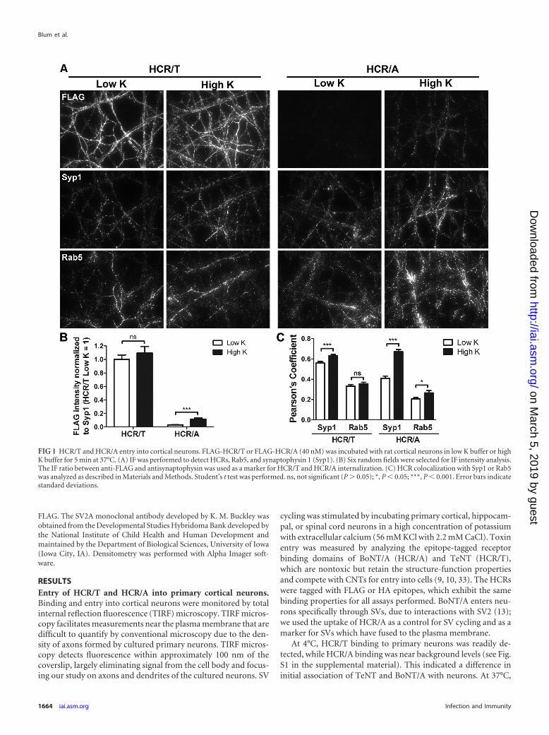
FIG 1 HCR/T and HCR/A entry into cortical neurons. FLAG-HCR/T or FLAG-HCR/A (40 nM) was incubated with rat cortical neurons in low K buffer or highK buffer for 5 min at 37°C. (A) IF was performed to detect HCRs, Rab5, and synaptophysin 1 (Syp1). (B) Six random fields were selected for IF intensity analysis.The IF ratio between anti-FLAG and antisynaptophysin was used as a marker for HCR/T and HCR/A internalization. (C) HCR colocalization with Syp1 or Rab5was analyzed as described in Materials and Methods. Student’s t test was performed. ns, not significant (P � 0.05); *, P � 0.05; ***, P � 0.001. Error bars indicatestandard deviations.
Blum et al.
1664 iai.asm.org Infection and Immunity
on March 5, 2019 by guest
http://iai.asm.org/
Dow
nloaded from
HCR/T bound depolarized and nondepolarized neurons (high orlow extracellular potassium) with similar levels of intensity. Incontrast, HCR/A binding to nondepolarized neurons was nearbackground levels and was detectable in depolarized neurons (Fig.1). HCR/T colocalized with synaptophysin (SV protein marker) inhigh K buffer and low K buffer, while the colocalization of HCR/Awith synaptophysin was greater in high K buffer than in low Kbuffer. There was limited colocalization of HCR/T or HCR/A withRab5, an endosome marker protein. Thus, HCR/T and HCR/Aassociate uniquely with neurons, but both do so through SV pro-tein-specific entry pathways. Quantitation showed that in depo-larized neurons there was �20-fold more HCR/T cell bound thanHCR/A and that the amount of HCR/T bound was dose depen-dent (see Fig. S2 in the supplemental material). Additional exper-iments indicated that HCR/A and HCR/T entered hippocampaland spinal cord neurons similarly to cortical neurons (data notshown). As cortical neurons are more abundant than either hip-pocampal or spinal cord neurons, experiments were continuedwith cortical neurons.
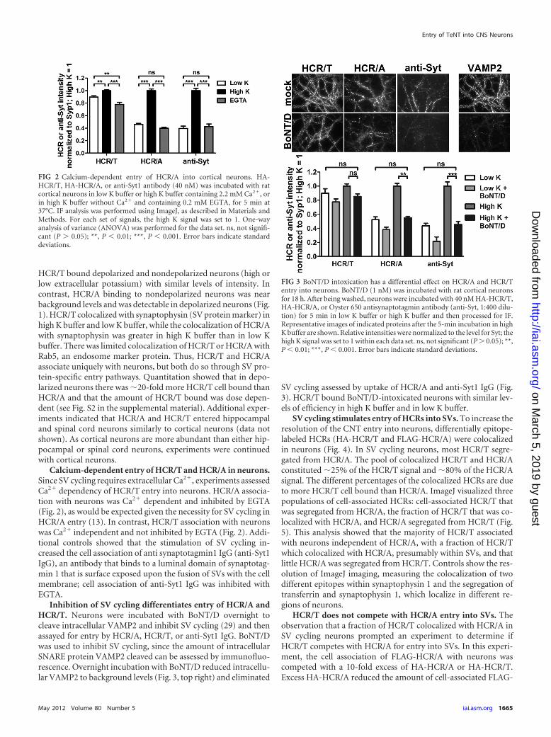
Calcium-dependent entry of HCR/T and HCR/A in neurons.Since SV cycling requires extracellular Ca2�, experiments assessedCa2� dependency of HCR/T entry into neurons. HCR/A associa-tion with neurons was Ca2� dependent and inhibited by EGTA(Fig. 2), as would be expected given the necessity for SV cycling inHCR/A entry (13). In contrast, HCR/T association with neuronswas Ca2� independent and not inhibited by EGTA (Fig. 2). Addi-tional controls showed that the stimulation of SV cycling in-creased the cell association of anti synaptotagmin1 IgG (anti-Syt1IgG), an antibody that binds to a luminal domain of synaptotag-min 1 that is surface exposed upon the fusion of SVs with the cellmembrane; cell association of anti-Syt1 IgG was inhibited withEGTA.
Inhibition of SV cycling differentiates entry of HCR/A andHCR/T. Neurons were incubated with BoNT/D overnight tocleave intracellular VAMP2 and inhibit SV cycling (29) and thenassayed for entry by HCR/A, HCR/T, or anti-Syt1 IgG. BoNT/Dwas used to inhibit SV cycling, since the amount of intracellularSNARE protein VAMP2 cleaved can be assessed by immunofluo-rescence. Overnight incubation with BoNT/D reduced intracellu-lar VAMP2 to background levels (Fig. 3, top right) and eliminated
SV cycling assessed by uptake of HCR/A and anti-Syt1 IgG (Fig.3). HCR/T bound BoNT/D-intoxicated neurons with similar lev-els of efficiency in high K buffer and in low K buffer.
SV cycling stimulates entry of HCRs into SVs. To increase theresolution of the CNT entry into neurons, differentially epitope-labeled HCRs (HA-HCR/T and FLAG-HCR/A) were colocalizedin neurons (Fig. 4). In SV cycling neurons, most HCR/T segre-gated from HCR/A. The pool of colocalized HCR/T and HCR/Aconstituted �25% of the HCR/T signal and �80% of the HCR/Asignal. The different percentages of the colocalized HCRs are dueto more HCR/T cell bound than HCR/A. ImageJ visualized threepopulations of cell-associated HCRs: cell-associated HCR/T thatwas segregated from HCR/A, the fraction of HCR/T that was co-localized with HCR/A, and HCR/A segregated from HCR/T (Fig.5). This analysis showed that the majority of HCR/T associatedwith neurons independent of HCR/A, with a fraction of HCR/Twhich colocalized with HCR/A, presumably within SVs, and thatlittle HCR/A was segregated from HCR/T. Controls show the res-olution of ImageJ imaging, measuring the colocalization of twodifferent epitopes within synaptophysin 1 and the segregation oftransferrin and synaptophysin 1, which localize in different re-gions of neurons.
HCR/T does not compete with HCR/A entry into SVs. Theobservation that a fraction of HCR/T colocalized with HCR/A inSV cycling neurons prompted an experiment to determine ifHCR/T competes with HCR/A for entry into SVs. In this experi-ment, the cell association of FLAG-HCR/A with neurons wascompeted with a 10-fold excess of HA-HCR/A or HA-HCR/T.Excess HA-HCR/A reduced the amount of cell-associated FLAG-
FIG 2 Calcium-dependent entry of HCR/A into cortical neurons. HA-HCR/T, HA-HCR/A, or anti-Syt1 antibody (40 nM) was incubated with ratcortical neurons in low K buffer or high K buffer containing 2.2 mM Ca2�, orin high K buffer without Ca2� and containing 0.2 mM EGTA, for 5 min at37°C. IF analysis was performed using ImageJ, as described in Materials andMethods. For each set of signals, the high K signal was set to 1. One-wayanalysis of variance (ANOVA) was performed for the data set. ns, not signifi-cant (P � 0.05); **, P � 0.01; ***, P � 0.001. Error bars indicate standarddeviations.
FIG 3 BoNT/D intoxication has a differential effect on HCR/A and HCR/Tentry into neurons. BoNT/D (1 nM) was incubated with rat cortical neuronsfor 18 h. After being washed, neurons were incubated with 40 nM HA-HCR/T,HA-HCR/A, or Oyster 650 antisynaptotagmin antibody (anti-Syt, 1:400 dilu-tion) for 5 min in low K buffer or high K buffer and then processed for IF.Representative images of indicated proteins after the 5-min incubation in highK buffer are shown. Relative intensities were normalized to the level for Syt; thehigh K signal was set to 1 within each data set. ns, not significant (P � 0.05); **,P � 0.01; ***, P � 0.001. Error bars indicate standard deviations.
Entry of TeNT into CNS Neurons
May 2012 Volume 80 Number 5 iai.asm.org 1665
on March 5, 2019 by guest
http://iai.asm.org/
Dow
nloaded from
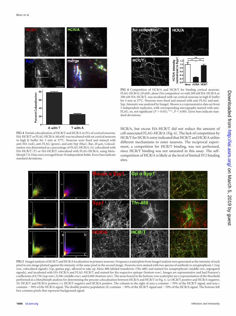
HCR/A, but excess HA-HCR/T did not reduce the amount ofcell-associated FLAG-HCR/A (Fig. 6). The lack of competition byHCR/T for HCR/A entry indicated that HCR/T and HCR/A utilizedifferent mechanisms to enter neurons. The reciprocal experi-ment, a competition for HCR/T binding, was not performed,since HCR/T binding was not saturated in this assay. The self-competition of HCR/A is likely at the level of limited SV2 bindingsites.
FIG 4 Partial colocalization of HCR/T and HCR/A in SVs of cortical neurons.HA-HCR/T or FLAG-HCR/A (40 nM) was incubated with rat cortical neuronsin high K buffer for 5 min at 37°C. Neurons were fixed and stained withanti-HA (red), anti-FLAG (green), and anti-Syp (blue). Bar, 20 �m. Colocal-ization was determined as a percentage of FLAG-HCR/A (A) colocalized withHA-HCR/T (T) or HA-HCR/T colocalized with FLAG-HCR/A, using Meta-Morph 7.0. Data were averaged from 10 independent fields. Error bars indicatestandard deviations.
FIG 5 ImageJ analysis of HCR/T and HCR/A localization in primary neurons. Frequency scatterplots from ImageJ analysis were generated as the intensity of eachpixel in one image plotted against the intensity of the same pixel in the second image. Neurons were stained with two species of antibody to synaptophysin 1 (toprow, colocalized signals) (Gp, guinea pig), allowed to take up Alexa 488-labeled transferrin (Tfn-488) and stained for synaptophysin (middle row, segregatedsignals), and incubated with HA-HCR/A and FLAG-HCR/T and stained for the respective epitope (bottom row). Images are representative and had Pearson’scoefficients of 0.756 (top row), 0.206 (middle row), and 0.600 (bottom row). The areas boxed in the bottom-row scatterplot are a representation of the thresholdperformed in a MetaMorph analysis for determining the percent colocalization between HCR/A and HCR/T in Fig. 4. (a) HCR/T positive and HCR/A negative;(b) HCR/T and HCR/A positive; (c) HCR/T negative and HCR/A positive. The column to the right of area a contains �70% of the HCR/T signal, and area ccontains �30% of the HCR/A signal. The double positive population (b) contains �30% of the HCR/T signal and �70% of the HCR/A signal. The bottom-leftbox contains pixels that represent background signal.
FIG 6 Competition of HCR/A and HCR/T for binding cortical neurons.FLAG-HCR/A (20 nM), alone (No competitor) or with 200 nM HA-HCR/A or200 nM HA-HCR/T, was incubated with rat cortical neurons in high K bufferfor 5 min at 37°C. Neurons were fixed and stained with anti-FLAG and anti-Syp. Intensity was analyzed by ImageJ. Shown is a representative data set from3 independent replicates, with corresponding micrographs stained with anti-FLAG. ns, not significant (P � 0.05); ***, P � 0.001. Error bars indicate stan-dard deviations.
Blum et al.
1666 iai.asm.org Infection and Immunity
on March 5, 2019 by guest
http://iai.asm.org/
Dow
nloaded from
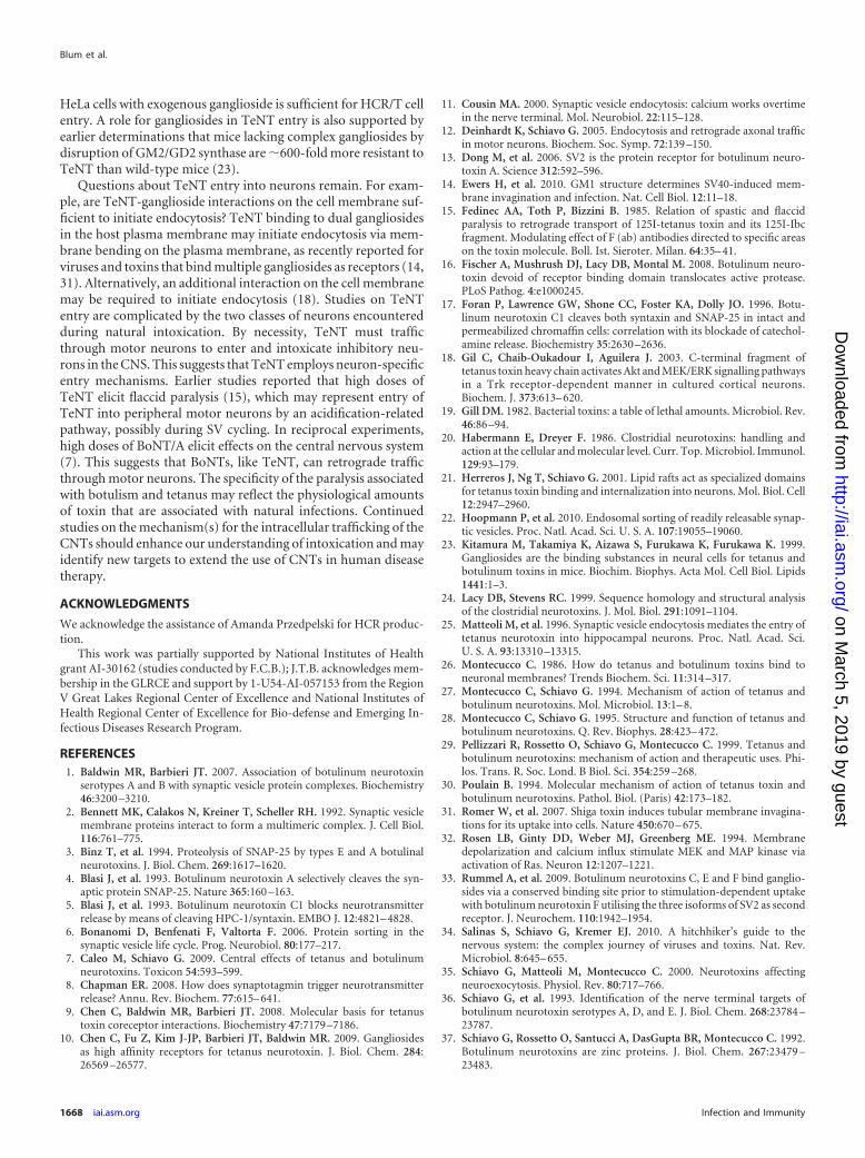
HCR/T does not bind SV2. Synaptic vesicle protein 2 (SV2) isthe protein receptor that targets BoNT/A to SVs (13). Experi-ments measured the direct binding of SV2 to FLAG-HCR/A andFLAG-HCR/T by immunoprecipitation. HCR-SV2 binding wasmeasured in detergent-extracted SV lysates. HCR/A bound allthree SV2 isotypes in CHAPS-solubilized lysates and bound SV2Aand SV2B in Triton X-100-solubilized lysates. HCR/T did notbind detectable amounts of the SV2 isotypes in either detergent(Fig. 7). These experiments show that, within the limits of detec-tion, HCR/T does not directly bind SV2.
Intoxication of neurons by TeNT. TeNT intoxication of de-polarized, nondepolarized, and EGTA-treated neurons was alsomeasured. TeNT efficiently cleaved VAMP2 under all conditions(Fig. 8). There was a small, but statistically significant, increase inVAMP2 cleavage in depolarized neurons relative to that in restingneurons, supporting entry of a subpopulation of TeNT into SVsupon membrane depolarization.
DISCUSSION
The current study shows that TeNT utilizes two entry pathwaysinto neurons of the CNS. HCR/T entered neurons independent ofstimulated synaptic vesicle cycling as well as via synaptic vesiclecycling. Colocalization analysis showed that most cell-associatedHCR/T is segregated from HCR/A, with a subpopulation ofHCR/T colocalized with HCR/A. Since HCR/T did not competewith HCR/A for entry into neurons and did not bind SV2, theprotein receptor for BoNT/A, TeNT and BoNT/A enter SVs byindependent mechanisms.
Endocytosis is essential for delivery of cargo between theplasma membrane and intracellular organelles (41). In neurons,intracellular trafficking is segregated, where SVs are present in theneuronal axon and transferrin receptor localizes in the soma anddendrites (reviewed in reference 6). Neurons utilize specializedendocytosis to rapidly cycle SV proteins on and off the plasmamembrane. This involves multiple pathways to replenish SVs forneuronal signaling (8, 11, 32). In addition to direct cycling of SVproteins from the plasma membrane (42), an endosome-medi-ated pathway appears to recycle SV proteins from the plasmamembrane (22). HCR/T entered neurons that were blocked for SVcycling, supporting the use of an endosome-mediated SV proteinrecycling pathway. An endosomal TeNT entry pathway has beendescribed in motor neurons (12), and this pathway retrograde
traffics neurotrophin receptors TrkB and p75NTR, as well as po-liovirus and canine adenovirus (reviewed in reference 34).
The organization of SV proteins into complexes was first de-scribed by Bennett and colleagues (2); in that study, SV proteincomplexes were detected following detergent solubilization.CHAPS solubilization preserved a large SV-protein complex com-posed of SV2, synaptophysin, synaptotagmin, VAMP2, and theproton vATPase, while Triton X-100 solubilization preserved onlyinteractions between SV2-synaptotagmin and synaptophysin-VAMP2. Although the physiological function of these SV proteincomplexes remains unclear, interactions with SV complexes andBoNT HCRs have been observed (1). In the current study, HCR/Abound SV2 isoforms in detergent-solubilized SV lysates, whileHCR/T did not bind any isoform of SV2, indicating that HCR/Tdoes not bind SV2 directly or that the affinity was too low to detectunder the assay conditions. An earlier study that reported thedirect binding of TeNT to SV2 (43) did not normalize for immu-noprecipitation efficiency, which may account for the differencesin result. The current study supports entry of TeNT into neuronsthrough the direct binding to dual gangliosides, rather than viaSV2 binding (10), where TeNT binding and entry into ganglio-sides are necessary and sufficient in neurons and nonneuronalcells. Chen et al. (10) demonstrated that treatment of ganglioside-loaded PC12 cells, a pheochromocytoma cell line, with proteinaseK does not change the cell association of HCR/T and that loading
FIG 7 Differential binding of HCR/A and HCR/T to SV2. FLAG-HCR/A orFLAG-HCR/T (5 �g) was incubated with 10 �g of an SV cell lysate. BoundHCR was extracted with anti-FLAG agarose beads. HCRs were eluted withFLAG peptide, and eluted material was analyzed by Western blotting. Quan-titation showed that in the CHAPS cell lysate SV2B bound to HCR/T was �2%of SV2B bound to HCR/A.
FIG 8 TeNT cleavage of VAMP2 in rat cortical neurons. Rat cortical neuronswere incubated alone (mock) or with 20 nM TeNT in high K buffer (High K),low K buffer (Low K), or high K/EGTA buffer (EGTA) for 20 min at 37°C. Cellswere washed and incubated in conditioned neurobasal medium for an addi-tional 18 h. Cells were fixed and processed for VAMP2 content by IF. VAMP2cleavage was assayed with mouse anti-VAMP2 antibody (1:500 dilution; SYSYclone 69.1), which recognizes only intact VAMP2. Data are representative of 5random fields from two independent experiments. *, P � 0.05; ***, P � 0.001.Error bars indicate standard deviations.
Entry of TeNT into CNS Neurons
May 2012 Volume 80 Number 5 iai.asm.org 1667
on March 5, 2019 by guest
http://iai.asm.org/
Dow
nloaded from
HeLa cells with exogenous ganglioside is sufficient for HCR/T cellentry. A role for gangliosides in TeNT entry is also supported byearlier determinations that mice lacking complex gangliosides bydisruption of GM2/GD2 synthase are �600-fold more resistant toTeNT than wild-type mice (23).
Questions about TeNT entry into neurons remain. For exam-ple, are TeNT-ganglioside interactions on the cell membrane suf-ficient to initiate endocytosis? TeNT binding to dual gangliosidesin the host plasma membrane may initiate endocytosis via mem-brane bending on the plasma membrane, as recently reported forviruses and toxins that bind multiple gangliosides as receptors (14,31). Alternatively, an additional interaction on the cell membranemay be required to initiate endocytosis (18). Studies on TeNTentry are complicated by the two classes of neurons encounteredduring natural intoxication. By necessity, TeNT must trafficthrough motor neurons to enter and intoxicate inhibitory neu-rons in the CNS. This suggests that TeNT employs neuron-specificentry mechanisms. Earlier studies reported that high doses ofTeNT elicit flaccid paralysis (15), which may represent entry ofTeNT into peripheral motor neurons by an acidification-relatedpathway, possibly during SV cycling. In reciprocal experiments,high doses of BoNT/A elicit effects on the central nervous system(7). This suggests that BoNTs, like TeNT, can retrograde trafficthrough motor neurons. The specificity of the paralysis associatedwith botulism and tetanus may reflect the physiological amountsof toxin that are associated with natural infections. Continuedstudies on the mechanism(s) for the intracellular trafficking of theCNTs should enhance our understanding of intoxication and mayidentify new targets to extend the use of CNTs in human diseasetherapy.
ACKNOWLEDGMENTS
We acknowledge the assistance of Amanda Przedpelski for HCR produc-tion.
This work was partially supported by National Institutes of Healthgrant AI-30162 (studies conducted by F.C.B.); J.T.B. acknowledges mem-bership in the GLRCE and support by 1-U54-AI-057153 from the RegionV Great Lakes Regional Center of Excellence and National Institutes ofHealth Regional Center of Excellence for Bio-defense and Emerging In-fectious Diseases Research Program.
REFERENCES1. Baldwin MR, Barbieri JT. 2007. Association of botulinum neurotoxin
serotypes A and B with synaptic vesicle protein complexes. Biochemistry46:3200 –3210.
2. Bennett MK, Calakos N, Kreiner T, Scheller RH. 1992. Synaptic vesiclemembrane proteins interact to form a multimeric complex. J. Cell Biol.116:761–775.
3. Binz T, et al. 1994. Proteolysis of SNAP-25 by types E and A botulinalneurotoxins. J. Biol. Chem. 269:1617–1620.
4. Blasi J, et al. 1993. Botulinum neurotoxin A selectively cleaves the syn-aptic protein SNAP-25. Nature 365:160 –163.
5. Blasi J, et al. 1993. Botulinum neurotoxin C1 blocks neurotransmitterrelease by means of cleaving HPC-1/syntaxin. EMBO J. 12:4821– 4828.
6. Bonanomi D, Benfenati F, Valtorta F. 2006. Protein sorting in thesynaptic vesicle life cycle. Prog. Neurobiol. 80:177–217.
7. Caleo M, Schiavo G. 2009. Central effects of tetanus and botulinumneurotoxins. Toxicon 54:593–599.
8. Chapman ER. 2008. How does synaptotagmin trigger neurotransmitterrelease? Annu. Rev. Biochem. 77:615– 641.
9. Chen C, Baldwin MR, Barbieri JT. 2008. Molecular basis for tetanustoxin coreceptor interactions. Biochemistry 47:7179 –7186.
10. Chen C, Fu Z, Kim J-JP, Barbieri JT, Baldwin MR. 2009. Gangliosidesas high affinity receptors for tetanus neurotoxin. J. Biol. Chem. 284:26569 –26577.
11. Cousin MA. 2000. Synaptic vesicle endocytosis: calcium works overtimein the nerve terminal. Mol. Neurobiol. 22:115–128.
12. Deinhardt K, Schiavo G. 2005. Endocytosis and retrograde axonal trafficin motor neurons. Biochem. Soc. Symp. 72:139 –150.
13. Dong M, et al. 2006. SV2 is the protein receptor for botulinum neuro-toxin A. Science 312:592–596.
14. Ewers H, et al. 2010. GM1 structure determines SV40-induced mem-brane invagination and infection. Nat. Cell Biol. 12:11–18.
15. Fedinec AA, Toth P, Bizzini B. 1985. Relation of spastic and flaccidparalysis to retrograde transport of 125I-tetanus toxin and its 125I-Ibcfragment. Modulating effect of F (ab) antibodies directed to specific areason the toxin molecule. Boll. Ist. Sieroter. Milan. 64:35– 41.
16. Fischer A, Mushrush DJ, Lacy DB, Montal M. 2008. Botulinum neuro-toxin devoid of receptor binding domain translocates active protease.PLoS Pathog. 4:e1000245.
17. Foran P, Lawrence GW, Shone CC, Foster KA, Dolly JO. 1996. Botu-linum neurotoxin C1 cleaves both syntaxin and SNAP-25 in intact andpermeabilized chromaffin cells: correlation with its blockade of catechol-amine release. Biochemistry 35:2630 –2636.
18. Gil C, Chaib-Oukadour I, Aguilera J. 2003. C-terminal fragment oftetanus toxin heavy chain activates Akt and MEK/ERK signalling pathwaysin a Trk receptor-dependent manner in cultured cortical neurons.Biochem. J. 373:613– 620.
19. Gill DM. 1982. Bacterial toxins: a table of lethal amounts. Microbiol. Rev.46:86 –94.
20. Habermann E, Dreyer F. 1986. Clostridial neurotoxins: handling andaction at the cellular and molecular level. Curr. Top. Microbiol. Immunol.129:93–179.
21. Herreros J, Ng T, Schiavo G. 2001. Lipid rafts act as specialized domainsfor tetanus toxin binding and internalization into neurons. Mol. Biol. Cell12:2947–2960.
22. Hoopmann P, et al. 2010. Endosomal sorting of readily releasable synap-tic vesicles. Proc. Natl. Acad. Sci. U. S. A. 107:19055–19060.
23. Kitamura M, Takamiya K, Aizawa S, Furukawa K, Furukawa K. 1999.Gangliosides are the binding substances in neural cells for tetanus andbotulinum toxins in mice. Biochim. Biophys. Acta Mol. Cell Biol. Lipids1441:1–3.
24. Lacy DB, Stevens RC. 1999. Sequence homology and structural analysisof the clostridial neurotoxins. J. Mol. Biol. 291:1091–1104.
25. Matteoli M, et al. 1996. Synaptic vesicle endocytosis mediates the entry oftetanus neurotoxin into hippocampal neurons. Proc. Natl. Acad. Sci.U. S. A. 93:13310 –13315.
26. Montecucco C. 1986. How do tetanus and botulinum toxins bind toneuronal membranes? Trends Biochem. Sci. 11:314 –317.
27. Montecucco C, Schiavo G. 1994. Mechanism of action of tetanus andbotulinum neurotoxins. Mol. Microbiol. 13:1– 8.
28. Montecucco C, Schiavo G. 1995. Structure and function of tetanus andbotulinum neurotoxins. Q. Rev. Biophys. 28:423– 472.
29. Pellizzari R, Rossetto O, Schiavo G, Montecucco C. 1999. Tetanus andbotulinum neurotoxins: mechanism of action and therapeutic uses. Phi-los. Trans. R. Soc. Lond. B Biol. Sci. 354:259 –268.
30. Poulain B. 1994. Molecular mechanism of action of tetanus toxin andbotulinum neurotoxins. Pathol. Biol. (Paris) 42:173–182.
31. Romer W, et al. 2007. Shiga toxin induces tubular membrane invagina-tions for its uptake into cells. Nature 450:670 – 675.
32. Rosen LB, Ginty DD, Weber MJ, Greenberg ME. 1994. Membranedepolarization and calcium influx stimulate MEK and MAP kinase viaactivation of Ras. Neuron 12:1207–1221.
33. Rummel A, et al. 2009. Botulinum neurotoxins C, E and F bind ganglio-sides via a conserved binding site prior to stimulation-dependent uptakewith botulinum neurotoxin F utilising the three isoforms of SV2 as secondreceptor. J. Neurochem. 110:1942–1954.
34. Salinas S, Schiavo G, Kremer EJ. 2010. A hitchhiker’s guide to thenervous system: the complex journey of viruses and toxins. Nat. Rev.Microbiol. 8:645– 655.
35. Schiavo G, Matteoli M, Montecucco C. 2000. Neurotoxins affectingneuroexocytosis. Physiol. Rev. 80:717–766.
36. Schiavo G, et al. 1993. Identification of the nerve terminal targets ofbotulinum neurotoxin serotypes A, D, and E. J. Biol. Chem. 268:23784 –23787.
37. Schiavo G, Rossetto O, Santucci A, DasGupta BR, Montecucco C. 1992.Botulinum neurotoxins are zinc proteins. J. Biol. Chem. 267:23479 –23483.
Blum et al.
1668 iai.asm.org Infection and Immunity
on March 5, 2019 by guest
http://iai.asm.org/
Dow
nloaded from
38. Schiavo G, et al. 1993. Botulinum neurotoxins serotypes A and E cleaveSNAP-25 at distinct COOH-terminal peptide bonds. FEBS Lett. 335:99 –103.
39. Schwab M, Thoenen H. 1977. Selective trans-synaptic migration of teta-nus toxin after retrograde axonal transport in peripheral sympatheticnerves: a comparison with nerve growth factor. Brain Res. 122:459 – 474.
40. Schwab ME, Thoenen H. 1978. Selective binding, uptake, and retrograde
transport of tetanus toxin by nerve terminals in the rat iris. An electronmicroscope study using colloidal gold as a tracer. J. Cell Biol. 77:1–13.
41. Scita G, Di Fiore PP. 2010. The endocytic matrix. Nature 463:464 – 473.42. Sudhof TC. 2004. The synaptic vesicle cycle. Annu. Rev. Neurosci. 27:
509 –547.43. Yeh FL, et al. 2010. SV2 mediates entry of tetanus neurotoxin into central
neurons. PLoS Pathog. 6:e1001207.
Entry of TeNT into CNS Neurons
May 2012 Volume 80 Number 5 iai.asm.org 1669
on March 5, 2019 by guest
http://iai.asm.org/
Dow
nloaded from