Testicular and epididymal ultrasonography in Santa Inês lambs … · Andrade et al. Testicular and...
9
Anim. Reprod., v.11, n.2, p.110-118, Apr./Jun. 2014 _________________________________________ 4 Corresponding author: [email protected] Phone: +55(81)3320-6412; Fax: +55(81)3320-6057 Received: May 12, 2013 Accepted: April 8, 2014 Testicular and epididymal ultrasonography in Santa Inês lambs raised in Brazil A.K.G. Andrade 1 , A.T. Soares 1,2 , F.F. Freitas 2 , S.V. Silva 1 , C.E. Peña-Alfaro 3 , A.M. Batista 1 , M.M.P. Guerra 1,4 1 Andrology Laboratory (ANDROLAB), Veterinary Medicine Department, UFRPE, Recife, PE, Brazil. 2 State Company for Agricultural Research in Paraiba S/A, EMEPA, João Pessoa, PB, Brazil. 3 Federal University of Campina Grande, Patos, PB, Brazil. Abstract The objective of this work was to study, through ultrasonographic evaluation, changes in testes and epididymides of clinically healthy, peripubertal and pubertal Santa Inês lambs raised in Brazil. Periodic evaluations of weight, biometric characteristics (scrotal circumference, width and length) and ultrasound examinations of the testes and epididymides of 20 lambs were performed between 84 and 280 days old at intervals of 28 days. Scans were performed in the sagittal, transverse, frontal and oblique planes to evaluate the echotexture of the testicular parenchyma and mediastinum and the tail epididymis as well as the thickness and width of the mediastinum testis. The testicular parenchyma demonstrated a homogeneous echogenicity pattern ranging from low to moderate. The echogenicity of testicular parenchyma increased in direct proportion to animal age, being higher in pubertal lambs when compared to prepubertal at the same age. The mediastinum testis was observed in 100% of the evaluated animals, regardless of the scan plane used, and was classified as diffuse or moderately or highly echogenic. Echogenicity and the thickness of the mediastinum testis increased in direct proportion to animal age. The epididymal tail was presented in hypoechoic relation to the testicular parenchyma. Based on these results, it was concluded that ultrasound is useful tool for selection and morphophysiological evaluation of Santa Inês lambs on peripubertal and pubertal phases, when used in combination with other methods such as semen evaluation. Keywords: epididymis, ram lambs, testis, ultrasonography. Introduction Sheep farming is of great importance for the Brazilian agribusiness, especially in the northeast and south regions where the largest producers are located. In this context, the Santa Inês breed emerged as an excellent alternative for producers seeking productive animals that were well adapted to Brazilian climatic conditions. This breed also has high prolificacy, early maturity and good maternal ability (Sousa et al., 2003), which are essential for productivity and reproductive performance. The use of adequate biotechnology has allowed genetic improvements in livestock and a consequent increase in productivity. Among these biotechnologies, ultrasound imaging has been widely used in reproductive clinical examinations. Ultrasound (US) is a rapid and noninvasive andrology technique that, coupled with data from clinical examinations, may lead to early diagnosis of disorders of the testes and related structures (Pechman and Eilts, 1987). Because it is easily accessible and does not cause deleterious effects, US is the diagnostic method of choice in the initial evaluation of the scrotum and its contents (Saito and Cerri, 1999). The main functions of US are to evaluate anatomical structures and determine the echogenicity of testicular parenchyma and mediastinum (Chandolia et al., 1997; Clark et al., 2003). It can also be useful in monitoring progressive changes that occur in testis at different stages of maturation (Ahmad and Noakes, 1995), as some authors observed an increased echogenicity of testicular parenchyma in direct proportion with age (Hamm and Fobbe, 1995; Chandolia et al., 1997; Brito et al., 2004). According to Tapping and Cast (2008), testes of prepubertal animals had low to medium echogenicity, whereas testes of postpubertal animals demonstrated medium homogeneous echogenicity. Some studies involving US assessment of normal and pathological genitals of males have been conducted in bulls (Pechman and Eilts, 1987; Chandolia et al., 1997), rams (Cartee et al., 1990; Ahmad et al., 1991; Ahmad and Noakes, 1995; Gouletsou et al., 2003; Andrade Moura et al., 2008; Jucá et al., 2009), goats (Ahmad et al., 1991; Ahmad and Noakes, 1995) and other mammals (Pugh et al., 1990; Love, 1992; Clark and Althouse, 2002; Pozor, 2005; Ball, 2008). However, despite the advantages of this diagnostic method, there is little information about testis aspects by ultrasound image in domestic animals. Given the importance of US as a complementary diagnostic method in andrological examination and in monitoring progressive physiological changes that occur in the genitalia of young males and because of the lack of information
Transcript of Testicular and epididymal ultrasonography in Santa Inês lambs … · Andrade et al. Testicular and...

Anim. Reprod., v.11, n.2, p.110-118, Apr./Jun. 2014
_________________________________________
4Corresponding author: [email protected] Phone: +55(81)3320-6412; Fax: +55(81)3320-6057 Received: May 12, 2013 Accepted: April 8, 2014
Testicular and epididymal ultrasonography in Santa Inês lambs raised in Brazil
A.K.G. Andrade1, A.T. Soares1,2, F.F. Freitas2, S.V. Silva1, C.E. Peña-Alfaro3, A.M. Batista1, M.M.P. Guerra1,4
1Andrology Laboratory (ANDROLAB), Veterinary Medicine Department, UFRPE, Recife, PE, Brazil.
2State Company for Agricultural Research in Paraiba S/A, EMEPA, João Pessoa, PB, Brazil. 3Federal University of Campina Grande, Patos, PB, Brazil.
Abstract
The objective of this work was to study, through ultrasonographic evaluation, changes in testes and epididymides of clinically healthy, peripubertal and pubertal Santa Inês lambs raised in Brazil. Periodic evaluations of weight, biometric characteristics (scrotal circumference, width and length) and ultrasound examinations of the testes and epididymides of 20 lambs were performed between 84 and 280 days old at intervals of 28 days. Scans were performed in the sagittal, transverse, frontal and oblique planes to evaluate the echotexture of the testicular parenchyma and mediastinum and the tail epididymis as well as the thickness and width of the mediastinum testis. The testicular parenchyma demonstrated a homogeneous echogenicity pattern ranging from low to moderate. The echogenicity of testicular parenchyma increased in direct proportion to animal age, being higher in pubertal lambs when compared to prepubertal at the same age. The mediastinum testis was observed in 100% of the evaluated animals, regardless of the scan plane used, and was classified as diffuse or moderately or highly echogenic. Echogenicity and the thickness of the mediastinum testis increased in direct proportion to animal age. The epididymal tail was presented in hypoechoic relation to the testicular parenchyma. Based on these results, it was concluded that ultrasound is useful tool for selection and morphophysiological evaluation of Santa Inês lambs on peripubertal and pubertal phases, when used in combination with other methods such as semen evaluation. Keywords: epididymis, ram lambs, testis, ultrasonography.
Introduction
Sheep farming is of great importance for the Brazilian agribusiness, especially in the northeast and south regions where the largest producers are located. In this context, the Santa Inês breed emerged as an excellent alternative for producers seeking productive animals that were well adapted to Brazilian climatic conditions. This breed also has high prolificacy, early maturity and good maternal ability (Sousa et al., 2003),
which are essential for productivity and reproductive performance.
The use of adequate biotechnology has allowed genetic improvements in livestock and a consequent increase in productivity. Among these biotechnologies, ultrasound imaging has been widely used in reproductive clinical examinations. Ultrasound (US) is a rapid and noninvasive andrology technique that, coupled with data from clinical examinations, may lead to early diagnosis of disorders of the testes and related structures (Pechman and Eilts, 1987). Because it is easily accessible and does not cause deleterious effects, US is the diagnostic method of choice in the initial evaluation of the scrotum and its contents (Saito and Cerri, 1999).
The main functions of US are to evaluate anatomical structures and determine the echogenicity of testicular parenchyma and mediastinum (Chandolia et al., 1997; Clark et al., 2003). It can also be useful in monitoring progressive changes that occur in testis at different stages of maturation (Ahmad and Noakes, 1995), as some authors observed an increased echogenicity of testicular parenchyma in direct proportion with age (Hamm and Fobbe, 1995; Chandolia et al., 1997; Brito et al., 2004). According to Tapping and Cast (2008), testes of prepubertal animals had low to medium echogenicity, whereas testes of postpubertal animals demonstrated medium homogeneous echogenicity.
Some studies involving US assessment of normal and pathological genitals of males have been conducted in bulls (Pechman and Eilts, 1987; Chandolia et al., 1997), rams (Cartee et al., 1990; Ahmad et al., 1991; Ahmad and Noakes, 1995; Gouletsou et al., 2003; Andrade Moura et al., 2008; Jucá et al., 2009), goats (Ahmad et al., 1991; Ahmad and Noakes, 1995) and other mammals (Pugh et al., 1990; Love, 1992; Clark and Althouse, 2002; Pozor, 2005; Ball, 2008). However, despite the advantages of this diagnostic method, there is little information about testis aspects by ultrasound image in domestic animals.
Given the importance of US as a complementary diagnostic method in andrological examination and in monitoring progressive physiological changes that occur in the genitalia of young males and because of the lack of information

Andrade et al. Testicular and epididymal ultrasonography in ram.
Anim. Reprod., v.11, n.2, p.110-118, Apr./Jun. 2014 111
regarding its use in the ovine species, this work aimed to study changes in testes and epididymides of clinically healthy, peripubertal and pubertal Santa Inês lambs raised in Brazil by means ultrasonographic evaluation.
Materials and Methods
The experiment was carried out at the
Benjamin Maranhão Experimental Station in Tacima City, Paraíba State, Brazil (06°29’18’’S and 35°38’14’’W) at an altitude of 168 m, with a tropical, semiarid climate and a mean annual precipitation of 431.8 mm3. We used 20 clinically healthy Santa Inês lambs raised under semi-intensive management with access to native pasture, silage corn and sorghum, commercial concentrate [maize based concentrate (52.5%), wheat bran (30%), soybean (15%), ammonium chloride (0.5%), limestone (1%) and mineral salt (1%)] and water and mineral salt ad libitum.
During selection and prior to each evaluation point in this study, animals were subjected to andrological exams in order to detect possible diseases that could interfere with their reproductive potential. The examination included a general clinical evaluation (inspection of body condition and presence of hereditary defects and functionality of the nervous, respiratory, circulatory, digestive and musculoskeletal systems) and morphological evaluation of the genitalia (inspection and palpation of the scrotum, testis, epididymis, spermatic cords, prepuce and penis).
Periodic evaluations of weight development, measurements of biometric characteristics of the testis (scrotal circumference, width and length), ultrasound examinations of the testis and epididymis and semen collections were performed by the same technician from weaning (approximately 84 days old) at intervals of 28 days until all lambs had reached puberty which was defined according to Wolf et al. (1965), when the ejaculate showed sperm motility rate above 10% with at least 50 x 106 cells/ml. Semen collections and analyses were performed using an electro-ejaculation method and ejaculates were evaluated by macroscopic (appearance, color and odor) and microscopic (wave motion,
motility, sperm vigor, concentration and sperm cell morphology) characteristics, according to methods by Mies Filho (1987).
To estimate weight development, the animals were weighed individually on a mechanical scale at each evaluation. Scrotal circumference (SC) was measured in centimeters (cm) using a millimeter tape on the portion of greatest diameter of the scrotum. Testicular length (TL) and width (TW) were measured with a caliper, and the results are expressed in centimeters. To measure the TL, testes were measured, excluding the epididymal tail in dorsal-ventral direction. TW was measured in the middle portion of each testis in the lateral-medial direction. Testicular volume (TV) was determined using the mathematical equation of the spheroid prolate proposed by Bailey et al. (1998): TV = 2 x [4/3 x π x (TL/2)² x (TW/2)]; TV was expressed in cm3 and represented the average of both testes.
Ultrasonographic evaluations of the testis were carried out using a Falco Vet 100 (Pie Medical; Holland) connected to a linear array transducer with a frequency of 8.0 MHz and a printer (Sony, São Paulo, Brazil) for image documentation. All evaluations were performed by a single professional, and the ultrasound device was adjusted in a straightforward manner at the beginning of each working day to minimize the variables that could interfere in the study. Rams were physically restrained by two assistants while the testes were immobilized without pressure, and each testis was scanned in the sagittal, transverse, frontal and oblique planes (Fig. 1) to evaluate the testicular parenchyma echotexture, the mediastinum testis (MT) and epididymal tail. The oblique scan plane was used only for evaluation of the epididymal tail. Evaluations of epididymal body and head were not performed because the use of a linear array transducer hindered the contact between the transducer and these structures. Measurements of the mediastinum thickness (frontal plane) and testicular width (transverse plane) were performed by ultrasound in both testes. Measurements of the testis width performed with ultrasound were compared with the measurements done with a caliper. Both were measured in centimeters.
Figure 1. Image demonstrating ultrasound examination in the frontal (A), sagittal (B) and transverse planes (C) in the testis of Santa Inês lamb.

Andrade et al. Testicular and epididymal ultrasonography in ram.
112 Anim. Reprod., v.11, n.2, p.110-118, Apr./Jun. 2014
Descriptive statistics were performed on various aggregations of data collected on the echogenicity of the testis and related structures. Statistical Analysis System, version 8.0, 2000, was used for the statistical analysis of semen characteristics and testicular data. An analysis of variance (ANOVA) was performed, and differences between means were compared using the Tukey-Kramer multiple comparison test at a 5% significance level. The measurements of the testis width obtained with caliper were compared with those obtained in the ultrasound images scanned in transverse plane, and the means were analyzed using a Z test at a 1% significance level. The thickness of the mediastinum testis (cm), scrotal circumference (cm) and testicular volume (cm3) were analyzed using linear regressions of these variables as a function of age.
Results
General clinical examination and morphological
evaluation of the external genitalia of the lambs (by inspection and palpation), performed at selection and prior to each time point of the experiment, did not detect any abnormalities. The testes were symmetrical with homogeneous consistency, had free mobility within the scrotum and were absent of a pain reaction. The epididymides and spermatic cords were easily palpated, and like other structures, had no apparent clinical changes.
The average age at puberty was 194.6 ± 44 days, with a mean body weight of 23.24 ± 2.51 kg and average scrotal circumference of 20.5 ± 1.8 cm. The semen evaluation from pubertal animals showed a progressive increase of the microscopic characteristics of ejaculates with progressive decrease of sperm morphological defects, showing significant differences (P < 0.05) between 140 and 280 days of age for wave motion, progressive motility, concentration and total defects. All parameters were highly variable between animals and between ejaculates from the same animal (Table 1).
Table 1. Semen parameters (mean ± SD) of pubertal lambs of Santa Inês breed at different ages.
Age (days)
Wave motion (0-5)
PM (%)
Vigor (0-5)
Concentration (x109)
Total defects (%)
140 0.7 ± 0.6a 16.7 ± 11.5ab 1.3 ± 0.6 0.2 ± 0.1ab 18.67 ± 2.51a
168 1.0 ± 0.8a 23.0 ± 13.4a 2.1 ± 0.6 0.4 ± 0.2a 16.60 ± 3.63a
196 1.4 ± 1.3a 35.0 ± 20.3ab 2.3 ± 1.0 0.9 ± 0.8ab 16.07 ± 3.41a
224 1.7 ± 1.2ab 47.5 ± 21.1bc 2.6 ± 1.1 1.0 ± 0.8ab 14.88 ± 5.44a
252 2.3 ± 1.4ab 50.0 ± 21.1bc 2.9 ± 0.9 1.4 ± 1.1ab 13.06 ± 5.30ab
280 2.7 ± 0.9b 54.0 ± 19.0c 2.9 ± 0.8 1.7 ± 1.1b 10.15 ± 4.04b
PM = progressive motility. Different superscripts in the same column indicate statistical difference (P < 0.05).
At ultrasonography, the testicular parenchyma (TP, Tables 2 and 3) appeared homogeneous, ranging from low (Fig. 2A and 2B) to moderate echogenicity (Fig. 2C and 2D), regardless of the testis (right or left) and the scan plane used, increasing in direct proportion to the animal age. Furthermore, when comparing the testes of prepubertal and pubertal lambs of the same age, there was predominance of images with low echogenicity in prepubertal animals and moderate echogenicity in those that had reached puberty (Table 3).
The mediastinum testis (MT; Table 2) was visualized in 100% of evaluated animals, presenting as a hyperechoic line of variable thickness in the center of the testicular parenchyma when scanned in the frontal plane and as a point in the center of hyperechogenic parenchyma when scanned in the transverse plane. The MT was classified as having moderate (Fig. 2E and 2H), high (Fig. 2F and 2I) or diffuse (Fig. 2G) echogenicity, showing increased echogenicity and thickness in direct proportion with the animal age. Although it appears less frequently than moderately and highly echogenic MT, diffuse-type MT was found in all ages. It should be highlighted that in two animals (10%), this diffuse MT appeared hypoechoic when compared to TP, but earlier in one animal (at 140, 168 and 196 days of age) than in the other (168, 196 and 224 days old).
The epididymal tail (ET; Table 2), also clearly visualized in 100% of animals, had reduced echogenicity when compared to TP in most images analyzed (Fig. 3A). However, in some animals aged between 84 and 168 days, it was isoechoic regarding TP.
The scrotal septum was seen as a hyperechoic line between the right and left testes (Fig. 3B). Testicular tunics were identified close to the scrotum (Fig. 3C).
The pampiniform plexus (Fig. 3D) was clearly visualized in the sagittal and transverse planes, especially with increasing age when the testes become more pendulous, being less echogenic than the testicular parenchyma and composed of numerous anechoic tubular structures.
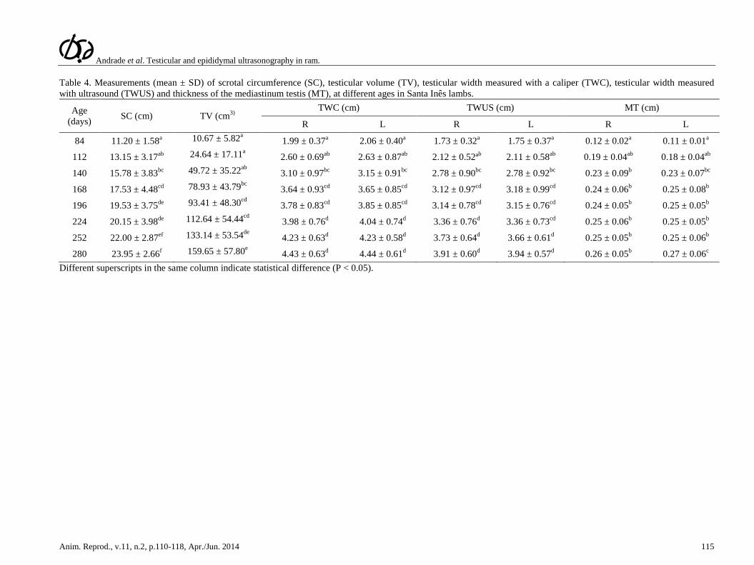
The mean values of scrotal circumference (SC), testicular volume (TV), testicular width with calipers (TWC), testicular width in ultrasound (WTUS) and thickness of MT at each evaluation are presented in Table 4. There was no significant difference (P > 0.05) between the right and left testes for TWC, WTUS and MT.
The mean testicular width obtained with calipers (WTC) was 3.49 cm while the average on the ultrasound (WTUS) was 3.03 cm, differing significantly (α = 1%) according to a Z test comparing the two means.
Scrotal circumference (SC; Fig. 4A), testicular volume (TV; Fig. 4B) and thickness of MT (Fig. 4C)

Andrade et al. Testicular and epididymal ultrasonography in ram.
Anim. Reprod., v.11, n.2, p.110-118, Apr./Jun. 2014 113
increased proportionally with animal age. Such correlations can be made based on the following linear
regression equations, respectively: y = 6.51 + 0.06 x; y = -57.23 + 0.77 x; and y = 0.12 + 0.0006 x.
Table 2. Frequency of echogenicity of testicular parenchyma and mediastinum (frontal plane) and epididymis cauda of Santa Inês lambs at peripubertal and pubertal phases.
Age (days)
Testicular parenchyma (%)
Mediastinum testis (%)
Epidydimal tail (%)
Right Left Right Left Right Left 84 100.0 LE 100.0 LE 80.0 ME
0.0 AE 80.0 ME 0.0 AE
50.0 HIP 50.0 HIP
0.0 ME 0.0 ME 20.0 DIF 20.0 DIF 50.0 ISO 50.0 ISO
112 90.0 LE 90.0 LE 80.0 ME 80.0 ME 65.0 HIP 65.0 HIP 10.0 ME 10.0 ME 0.0 HE 0.0 HE 35.0 ISO 35.0 ISO 20.0 DIF 20.0 DIF
140 70.0 LE 70.0 LE 65.0 ME 65.0 ME 85.0 HIP 85.0 HIP 30.0 ME 30.0 ME 15.0 HE 15.0 HE 15.0 ISO 15.0 ISO 20.0 DIF 20.0 DIF
168 50.0 LE 50.0 LE 55.0 ME 55.0 ME 95.0 HIP 95.0 HIP 50.0 ME 50.0 ME 25.0 HE 25.0 HE 5.0 ISO 5.0 ISO 20.0 DIF 20.0 DIF
196 40.0 LE 40.0 LE 55.0 ME 55.0 ME 100.0 HIP 100.0 HIP 60.0 ME 60.0 ME 25.0 HE 30.0 HE 0.0 ISO 0.0 ISO 20.0 DIF 15.0 DIF
224 40.0 LE 40.0 LE 50.0 ME 50.0 ME 100.0 HIP 100.0 HIP 60.0 ME 60.0 ME 35.0 HE 35.0 HE 0.0 ISO 0.0 ISO 15.0 DIF 15.0 DIF
252 20.0 LE 20.0 LE 35.0 ME 35.0 ME 100.0 HIP 100.0 HIP 80.0 ME 80.0 ME 50.0 HE 50.0 HE 0.0 ISO 0.0 ISO 15.0 DIF 15.0 DIF
280 10.0 LE 10.0 LE 35.0 ME 35.0 ME 100.0 HIP 100.0 HIP 90.0 ME 90.0 ME 55.0 HE 55.0 HE 0.0 ISO 0.0 ISO 10.0 DIF 10.0 DIF
LE = low echogenicity; ME = moderate echogenicity; HE = high echogenicity; DIF = diffuse, HIP = hypoechoic; ISO = isoechoic.
Table 3. Number of prepubertal and pubertal Santa Inês lambs and their respective echogenicity of testicular parenchyma (TP) frequencies at different ages.
Age (days) Prepubertal Pubertal (n) PT Echogenicity (%) (n) PT Echogenicity (%)
140 17 76.5 LE 3 33.3 LE 23.5 ME 66.7 ME 168 10 70.0 LE 10 30.0 LE 30.0 ME 70.0 ME 196 6 66.7 LE 14 28.6 LE 33.3 ME 71.4 ME 224 4 100.0 LE 16 25.0 LE 0.0 ME 75.0 ME 252 2 100.0 LE 18 11.1 LE 0.0 ME 88.9 ME 280 0 0.0 LE 20 10.0 LE 0.0 ME 90.0 ME
LE = low echogenicity; ME = moderate echogenicity.

Andrade et al. Testicular and epididymal ultrasonography in ram.
114 Anim. Reprod., v.11, n.2, p.110-118, Apr./Jun. 2014
Figure 2. Ultrasound images of the testis of clinically normal Santa Inês lambs, showing: low echogenicity of the left (LTP) and right (RTP) testicular parenchyma, at 112 days of age - at sagittal (A) and transverse (B) plane; moderate echogenicity of the LTP at 196 days of age – at sagittal (C) and transverse (D) planes; mediastinum testicular (MT) moderately echogenic (E), highly echogenic (F) and diffuse (G), at 224, 252 and 196 days of age, respectively; mediastinum testicular (MT) moderately echogenic (H) and highly echogenic (I), at 252 and 224 days of age, respectively - in the transverse plane.
Figure 3. Ultrasound images of the testis and epididymis of clinically normal Santa Inês lambs, showing: testicular parenchymal (TP) and epididymal tail (ET), at 196 days of age (A); scrotal septum (SS), at 84 days of age - at frontal plane (B); testicular tunic (T) and scrotum (S), at 168 days of age - at transverse plane (C); pampiniform plexus (PP), at 196 days of old - at sagittal plane (D).
Andrade et al. Testicular and epididymal ultrasonography in ram.
Anim. Reprod., v.11, n.2, p.110-118, Apr./Jun. 2014 115
Table 4. Measurements (mean ± SD) of scrotal circumference (SC), testicular volume (TV), testicular width measured with a caliper (TWC), testicular width measured with ultrasound (TWUS) and thickness of the mediastinum testis (MT), at different ages in Santa Inês lambs.
Age (days) SC (cm) TV (cm3) TWC (cm) TWUS (cm) MT (cm)
R L R L R L
84 11.20 ± 1.58a 10.67 ± 5.82a 1.99 ± 0.37a 2.06 ± 0.40a 1.73 ± 0.32a 1.75 ± 0.37a 0.12 ± 0.02a 0.11 ± 0.01a
112 13.15 ± 3.17ab 24.64 ± 17.11a 2.60 ± 0.69ab 2.63 ± 0.87ab 2.12 ± 0.52ªb 2.11 ± 0.58ab 0.19 ± 0.04ab 0.18 ± 0.04ab
140 15.78 ± 3.83bc 49.72 ± 35.22ab 3.10 ± 0.97bc 3.15 ± 0.91bc 2.78 ± 0.90bc 2.78 ± 0.92bc 0.23 ± 0.09b 0.23 ± 0.07bc
168 17.53 ± 4.48cd 78.93 ± 43.79bc 3.64 ± 0.93cd 3.65 ± 0.85cd 3.12 ± 0.97cd 3.18 ± 0.99cd 0.24 ± 0.06b 0.25 ± 0.08b
196 19.53 ± 3.75de 93.41 ± 48.30cd 3.78 ± 0.83cd 3.85 ± 0.85cd 3.14 ± 0.78cd 3.15 ± 0.76cd 0.24 ± 0.05b 0.25 ± 0.05b
224 20.15 ± 3.98de 112.64 ± 54.44cd 3.98 ± 0.76d 4.04 ± 0.74d 3.36 ± 0.76d 3.36 ± 0.73cd 0.25 ± 0.06b 0.25 ± 0.05b
252 22.00 ± 2.87ef 133.14 ± 53.54de 4.23 ± 0.63d 4.23 ± 0.58d 3.73 ± 0.64d 3.66 ± 0.61d 0.25 ± 0.05b 0.25 ± 0.06b
280 23.95 ± 2.66f 159.65 ± 57.80e 4.43 ± 0.63d 4.44 ± 0.61d 3.91 ± 0.60d 3.94 ± 0.57d 0.26 ± 0.05b 0.27 ± 0.06c
Different superscripts in the same column indicate statistical difference (P < 0.05).

Andrade et al. Testicular and epididymal ultrasonography in ram.
116 Anim. Reprod., v.11, n.2, p.110-118, Apr./Jun. 2014
Figure 4. Linear regression of scrotal circumference (cm; A), testicular volume (cm³; B) and mediastinum testis thickness (cm; C) of Santa Inês lambs (at pubertal and peripubertal phases), depending on the age (days). Linear regression equation obtained: y (SP) = 6.51 + 0.06 x (days); determining coefficient = 0.98 (A); y (TV) = -57.23 + 0.77 x (days); determination coefficient = 0.99 (B); y (MT) = 0.12 + 0.0006 x (days); determining coefficient = 0.75 (C).
Discussion Although some authors have studied the
ultrasonographic appearance of the testes and epididymes of clinically healthy sheep (Cartee et al., 1990; Ahmad et al., 1991; Gouletsou et al., 2003; Andrade Moura et al., 2008; Jucá et al., 2009), there are few reports on Santa Inês sheep, especially in relation to lambs in pubertal and peripubertal phases of reproductive development. Andrade Moura et al. (2008) and Jucá et al. (2009) evaluated testes and sexual glands by ultrasonography of 49 Santa Inês rams, aiming to use it as a diagnostic method for morphophysiological evaluation of the males. However, these authors did not study the testicular development in relation to the age of young animals as was done in this experiment.
In the studies used as a basis for this research, there were variations in the nomenclature used to describe the ultrasonographic appearance of testicular parenchyma. There is a need for standardization to facilitate further studies and to establish normal parameters. In this experiment, the testicular parenchyma (TP) in the lambs demonstrated homogeneity, with echogenicity ranging from low to moderate in both testes, regardless of scan plane used,
corroborating findings by Gouletsou et al. (2003). According to Cartee et al. (1990), sheep testicular parenchyma is predominantly homogeneous and hypoechoic. Pechman and Eilts (1987), Pugh et al. (1990) and Andrade Moura et al. (2008), in a study that aimed to describe the testicular parenchyma echotexture of Santa Inês sheep at different ages, found a variation of hypoechoic images of low and high intensity in all groups, with predominance of hypoechoic images of low intensity.
The mediastinum testis was visualized in 100% of the evaluated animals, presenting as a hyperechoic line of variable thickness at the center of the testicular parenchyma when visualized in the frontal plane and as a hyperechoic point in the center of this parenchyma when visualized in the transverse plane, confirming previous studies (Ahmad et al., 1991; Gouletsou et al., 2003; Andrade Moura et al., 2008; Jucá et al., 2009).
Gouletsou et al. (2003) visualized MT in 87% of the animals assessed and reported that the MT presence and shape determine different echogenic impressions in TP. The MT was classified as moderately echogenic, highly echogenic or diffuse, with higher echogenicity and increased thickness in direct proportion to animal age as evidenced in this study.

Andrade et al. Testicular and epididymal ultrasonography in ram.
Anim. Reprod., v.11, n.2, p.110-118, Apr./Jun. 2014 117
Moreover, Pechman and Eilts (1987) and Andrade Moura et al. (2008), using cattle and sheep, respectively, concluded that the mediastinum thickness and echogenicity increased with age. According to Dyce et al. (1990), the seminiferous tubules form a network in the MT. Thus, this increase in the MT thickness may be explained by the important and considerable anatomical changes in the seminiferous tubules, which develop with age as they become longer, increase in diameter and "twist" and as the lumen forms and the basement membrane thickens (Hamm and Fobbe, 1995).
The diffuse type of MT was found in all ages, noting that the diffuse MT was present in two animals (10%) with hypoechoic appearance in relation to the TP at 140, 168 and 196 days of age in one and at 168, 196 and 224 days in the other animal. According to Tapping and Cast (2008), this structure can often be seen as a hypoechoic area with striated appearance.
In this study, the epididymal tail (ET) was clearly visualized in 100% of the animals with reduced echogenicity when compared to most of the TP images analyzed, confirming Pechman and Eilts (1987), Ahmad et al., (1991) and Gouletsou et al., (2003). However, in younger animals, the ET was isoechoic when compared to TP, which can be explained by a low echogenicity of these animals, which is closer of the pattern described for ET.
Determination of the animal puberty age varies largely between different methods and criteria. Wolf et al. (1965) considered puberty in cattle as the age at which the ejaculate has a minimum of 50 x 106 cells/ml with at least 10% progressive motility. Other authors such as Mukasa-Mugerwa and Ezaz, (1992) and Wheaton and Godfrey (2003) used the same parameters to characterize puberty in lambs. According to this characterization, three lambs (15.0%) in this study showed semen consistent with puberty at 140 days of age, and all animals (100%) reached puberty by 280 days of age. The average age at puberty observed in this study was 194.6 ± 44 days, mean body weight was 23.24 ± 2.51 kg and average scrotal circumference was 20.5 ± 1.8 cm. Souza et al. (2000) found an average age at puberty of 159.5 days for the same breed of sheep. The difference in age could be attributed to the fact that they used the total detachment of the penis from the prepuce as the evaluation parameter, a phenomenon that preceded the release of the first mobile spermatozoa in the ejaculate.
Semen evaluation of animals revealed a gradual increase in physical and microscopic characteristics of ejaculates with progressive decrease of sperm morphological defects in direct proportion with age, demonstrating differences from 140 to 280 days for the mean progressive motility, concentration and total defects. Large variations of these parameters resulted in a high standard deviation, which can be explained by different stages of sexual maturity in the animals, even among animals of the same age group. According to
Skinner et al. (1968), puberty and sexual maturity in sheep are more closely related to body weight than the age of the animal.
Ultrasound examination may also be useful in monitoring the progressive changes that occur in the testes (Ahmad and Noakes, 1995). Differences in the echogenicity of the TP were observed between prepubertal and pubertal animals at the same age, with a predominance of images of low echogenicity in prepubertal lamb and moderate echogenicity in pubertal lamb, confirming Tapping and Cast (2008).
Biometric analysis of testicular development is extremely important, as it is significantly correlated with the reproductive activity of the animal (Mukasa-Mugerwa and Ezaz, 1992). The US is a viable technique for measuring testis (length, width and height) in situ, in dogs, sheep and cattle (Eilts et al., 1993). Louvandini et al. (2008), evaluating biometric features of testes of Santa Inês lambs, observed that the testis shape directly affected its volume, reporting that the formula for prolate spheroid is a more reliable predictor of TV than the cylinder formula because it best approximates the shape of oval cylindrical testes of Santa Inês sheep; the prolate spheroid formula was therefore used to calculate the TV of animals in this study.
A difference was found between testicular width measurements obtained with calipers and ultrasound, as the first was larger than the latter. In a study that determined the relationship between in vivo measurements of bovine testes obtained with ultrasound and calipers, Bailey et al., (1998) observed that the width at ultrasound showed a higher correlation with the real width than that obtained with the calipers because only the testicular parenchyma was measured in the ultrasound, while caliper measurements also included the body of epididymis, scrotum and testicular tunics.
However, regarding the measurement of TV, according to these authors, the volume obtained with calipers had higher correlation with the real volume when compared to volume measurements obtained with ultrasound. This statement can be explained by difficulties in assessing the testicular length by ultrasound in older animals because the transducer is sometimes smaller than the testis, as was demonstrated in this study.
In conclusion, the US is useful tool for selection and morphophysiological evaluation of Santa Inês lambs at peripubertal and pubertal phases, when used in combination with other methods such as semen evaluation. This technique may be included in breeding soundness examination of breeding rams.
Acknowledgments
Authors are grateful to the following Brazilian
fostering agencies: Coordenação de Aperfeiçoamento de Pessoal de Nível Superior (CAPES) and Conselho Nacional de Desenvolvimento Científico e Tecnológico (CNPq) for funding; Fundação de Amparo à Ciência e

Andrade et al. Testicular and epididymal ultrasonography in ram.
118 Anim. Reprod., v.11, n.2, p.110-118, Apr./Jun. 2014
Tecnologia do Estado de Pernambuco (FACEPE) for a scholarship for the masters study; and the State Company for Agricultural Research in the Paraiba State (EMEPA/PB), Campo de Santana Campus, for authorizing the use of the males.
References Ahmad N, Noakes DE, Subandrio AL. 1991. B-mode real time ultrasonographic imaging of the testis and epididymis of sheep and goats. Vet Rec, 12:491-496. Ahmad N, Noakes DE. 1995. A clinical and ultrasonographic study of induced testicular and epididymal lesions in goats and a ram. Anim Reprod Sci, 39:35-48. Andrade Moura JC, Jucá AF, Gusmão AL, Pinho TG, Bittencourt TCB, Barbosa CMP. 2008. Ecotextura testicular do carneiro Santa Inês. Hora Vet, 162:19-22. Bailey TL, Hudso RS, Powe TA, Riddell MG, Wolfe DF, Carson RL. 1998. Caliper ultrasonographic measurements of bovine testicles and a mathematical formula for determining testicular volume and weight in vivo. Theriogenology, 49:581-594. Ball AB. 2008. Diagnostic methods for evaluation of stallion subfertility: a review. J Equine Vet Sci, 28:650-664. Brito LFC, Silva AEDF, Unanian MM, Dode MAN, Barbosa RT, Kastelic JP. 2004. Sexual development in early late maturing Bos indicus and Bos indicus x Bos taurus crossbred bulls in Brasil. Theriogenology, 62:1198-1217. Cartee RE, Rumph PF, Abuzaid S, Carson R. 1990. Ultrasonographic examination and measurement of ram testicles. Theriogenology, 33:867-875. Chandolia RK, Bartlewski PM, Omeke BC, Beard AP, Rawlings NC, Pierson RA. 1997. Ultrasonography of the developing reproductive tract in ram lambs effects of a GnRH agonist. Theriogenology, 48:99-117. Clark SG, Althouse GC. 2002. B-mode ultrassonographic examination of the accessory sex glands of boars. Theriogenology, 57:2003-2013. Clark SG, Schaeffer DJ, Althouse GC. 2003. B-mode ultrasonographic evaluation of paired testicular diameter of mature boars in relation to average total of sperm numbers. Theriogenology, 60: 1011-1023. Dyce KM, Sack WO, Wensing CJG. 1990. Tratado de Anatomia Veterinária. Rio de Janeiro: Guanabara Koogan. Eilts BE, Williams DB, Moser EB. 1993. Ultrasonic measurement of canine testis. Theriogenology, 40:819-828. Gouletsou PG, Amiridis GS, Cripps PJ, Lainas T, Deligianis K, Saratsis P, Fthenakis GC. 2003. Ultrasonographic appearance of clinically healthy testicles and epididymes of rams. Theriogenology,
59:1959-1972. Hamm B, Fobbe F. 1995. Maturation of the testis: ultrasound evaluation. Ultrasound Med Biol, 21:143-147. Jucá AF, Andrade Moura JC, Gusmão AL, Bittencourt TC, Nascimento MC, Barbosa CMP. 2009. Avaliação ultrassonográfica dos testículos e das glândulas sexuais anexas de carneiros Santa Inês. Cienc Anim Bras, 10:650-659. Louvandini H, McManus C, Martins RD, Lucci CM, Corrêa PS. 2008. Características biométricas testiculares em carneiros Santa Inês submetidos a diferentes regimes de suplementação protéica e tratamentos anti-helmínticos. Cienc Anim Bras, 9:638-647. Love CC. 1992. Ultrasonographic evaluation of the testis, epididymis, and spermatic cord of the stallion. Vet Clin North Am Equine Pract, 8:167-182. Mies Filho A. 1987. Inseminação Artificial. 6th ed. Porto Alegre: Sulina. Mukasa-Mugerwa E, Ezaz Z. 1992. Relationship of testicular growth and size to age, body weight and onset of puberty in Menz ram lambs. Theriogenology, 38:979-988. Pechman RD, Eilts BE. 1987. B-mode ultrasonography of the bull testicle. Theriogenology, 27:431-441. Pozor M. 2005. Diagnostic applications of ultrasonography to stallion´s reproductive tract. Theriogenology, 64:505-509. Pugh CR, Konde LG, Park RD. 1990. Testicular ultrasound in the normal dog. Vet Radiol, 31:195-199. Saito OC, Cerri GG. 1999. Ultra-sonografia: pequenas partes. São Paulo: Sarvier. Skinner JD, Booth WD, Rowson LEA, Karg H. 1968. The post-natal developments of the reproductive tract of the Suffolk ram, and changes in the gonadotrophin content of the pituitary. J Reprod Fertil, 16:463-477. Sousa WH, Lôbo RNB, Morais OR. 2003. Ovinos Santa Inês: estado de arte e perspectivas. In: Anais do II Simpósio Internacional sobre Caprinos e Ovinos de Corte, 2003, João Pessoa. João Pessoa, PB: EMEPA. pp. 501-522. Souza CEA, Moura AAA, Lima ACB, Ciríaco LT. 2000. Desenvolvimento testicular, idade à puberdade e características seminais em carneiros da raça Santa Inês no estado do Ceará. In: Anais da XXXVI Reunião Anual da Sociedade Brasileira de Zootecnia, 2000, Viçosa, MG. Viçosa: SBZ. CD-ROM. Tapping CR, Cast JE. 2008. Scrotal ultrasound: a pictorial review. Ultrasound, 16:226-233. Wheaton JE, Godfrey RW. 2003. Plasma LH, FSH, testosterone, and age at puberty in ram lambs actively immunized against an inhibin a-subunit peptide. Theriogenology, 60:933-941. Wolf FR, Almquist JO, Hale EB. 1965. Prepuberal behaviour and puberal characteristics of beef bulls on high nutrient allowance. J Anim Sci, 24:761-765.
![Isolated Testicular Tuberculosis Mimicking Testicular ... involvement, but testicular involvement is an unusual clinical condition [3]. In this report, a case with isolated testicular](https://static.fdocuments.in/doc/165x107/5f3d57bf74280d66ef795ba2/isolated-testicular-tuberculosis-mimicking-testicular-involvement-but-testicular.jpg)