Ten Tantalizers: Microscopes that Continue to Fascinate
28
1 Ten Tantalizers Ten Tantalizers: Microscopes that Continue to Fascinate R. Jordan Kreindler (USA) Manuel del Cerro (USA) ___________________________________________________________________ As with the history of living organisms, the history of the microscope shows an evolutionary trend. In both cases, these followed the principle of 'survival of the fittest'. For animals there were examples that were notable in their time, sometimes spectacular, but also sometimes transitory terminal paths in the process of evolution. In the evolution of living organisms, e.g., mastodons and dinosaurs made a long and, some would say, spectacular, albeit temporary impact, while roaches are still here, over 300 million years after they first evolved. Today although they are long gone, we still discuss dinosaurs, with interest and fascination. There are books, toys, TV shows, and movies depicting them. Many museums, e.g., the American Museum of Natural History in NY, USA, have large areas dedicated to their display. That is, although transitory in the evolution of living organisms they are still of significant interest, as are some smaller living organisms such as the volvox and tardigrade. In the case of microscopes, their history is much shorter. While some microscopes, like living organisms, served as ancestors for those that followed, others served only as examples of evolutionary paths that would be terminated. However, like dinosaurs and tardigrades there are microscopes that continue to fascinate, whether they were terminal stops on the path to modern microscopes, or precursors to what followed. Although much has been written about historical landmark microscopes, this brief paper takes a slightly different tact. It discusses microscopes that, for various reasons, have caught our interests. These microscopes run the range from relatively small to large, simple to complex, inexpensive to more costly. They were made in Asia, Continental Europe, the UK, or the US, and produced, and constructed of brass, wood, aluminum, or alloys. Some are "landmarks", and some represent, at least in its construction, terminal paths. However, regardless of their place in the history of microscopy all are tantalizing examples, and we’re pleased to share our fascination.
Transcript of Ten Tantalizers: Microscopes that Continue to Fascinate
1
Ten Tantalizers
Ten Tantalizers:
Microscopes that Continue to Fascinate R. Jordan Kreindler (USA)
Manuel del Cerro (USA) ___________________________________________________________________
As with the history of living organisms, the history of the microscope shows an evolutionary trend. In both cases, these followed the principle of 'survival of the fittest'. For animals there were examples that were notable in their time, sometimes spectacular, but also sometimes transitory terminal paths in the process of evolution.
In the evolution of living organisms, e.g., mastodons and dinosaurs made a long and, some would say, spectacular, albeit temporary impact, while roaches are still here, over 300 million years after they first evolved. Today although they are long gone, we still discuss dinosaurs, with interest and fascination. There are books, toys, TV shows, and movies depicting them. Many museums, e.g., the American Museum of Natural History in NY, USA, have large areas dedicated to their display. That is, although transitory in the evolution of living organisms they are still of significant interest, as are some smaller living organisms such as the volvox and tardigrade.
In the case of microscopes, their history is much shorter. While some microscopes, like living organisms, served as ancestors for those that followed, others served only as examples of evolutionary paths that would be terminated.
However, like dinosaurs and tardigrades there are microscopes that continue to fascinate, whether they were terminal stops on the path to modern microscopes, or precursors to what followed.
Although much has been written about historical landmark microscopes, this brief paper takes a slightly different tact. It discusses microscopes that, for various reasons, have caught our interests. These microscopes run the range from relatively small to large, simple to complex, inexpensive to more costly. They were made in Asia, Continental Europe, the UK, or the US, and produced, and constructed of brass, wood, aluminum, or alloys. Some are "landmarks", and some represent, at least in its construction, terminal paths. However, regardless of their place in the history of microscopy all are tantalizing examples, and we’re pleased to share our fascination.
2
Ten Tantalizers
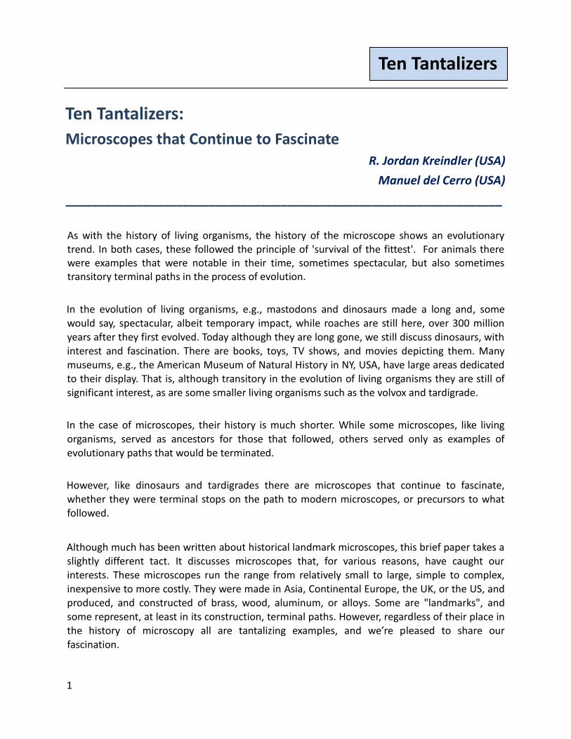
1. Aluminum Swift Portable Histological (Quad-pod)
We begin our discussion with a terminal path microscope that represents one of the truly inappropriate avenues of microscope development.
During the late 1880s and 1890s aluminum could finally be obtained at a price more suitable for use in consumer products. Before this, aluminum had been more expensive than platinum. The previous high cost of aluminum and its then current lower cost lead to its being the "wonder metal" of the time. Many products previously made of brass or other metals now could be, and were, produced in aluminum, regardless of whether this was a wise decision or not.
In 1894, the top of the Washington Monument was capped with a sheet of pure aluminum, which was then as expensive as aluminum bronze plated with platinum. The aluminum cap was considerably more difficult to manufacture than planned, and its final cost was over 300% of that budgeted. At the time aluminum, although recent scientific advances made its extraction from ore less expensive, was still relatively costly to shape. The choice of aluminum for the cap was given front page headlines. This publicity, along with the concurrent publicity discussing the advances in reducing costs for aluminum extraction, gained aluminum public recognition as a rare, but desirable metal.
Figure 1. Swift Portable Histological quad-pod folding microscope, aluminum version. c. 1895
3
Ten Tantalizers
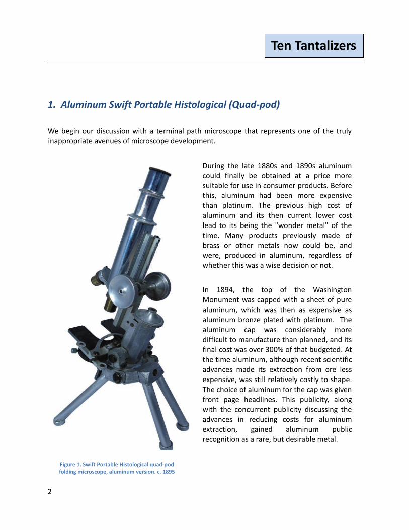
At first glance, aluminum would seem an ideal choice for a field portable microscope. It's lightweight, it's a metal, and it's reasonably robust when subject to weathering. However, its collateral characteristics made it probably one of the worst metals that could have been chosen. Pure, unalloyed aluminum is relatively fragile and soft. So, microscopes constructed from it had a very short in-use lifespan.
This example is, as might be expected, quite delicate. It has required repair numerous times, including one repair
for a split in its mechanical stage. However, its light weight of 3 pounds 6 ounces, is truly amazing compared to its brass counterpart weighing 7 pounds 15 ounces. Similarly configured, the aluminum version weights about one-half of the brass version, Fig. 2, noting that for both versions the optical components use brass, not aluminum. The fragility of this pure aluminum microscope is also notable. Because of this fragility, all pure aluminum microscopes from the 19th century are relatively rare, many were disposed of when they broke, which was frequently. All remaining 19th century pure aluminum microscopes are rare, as is the Swift example shown in Fig. 1.
Figure 2. Swift Portable Histological quad-pod folding microscope, brass version, c. 1900
4
Ten Tantalizers
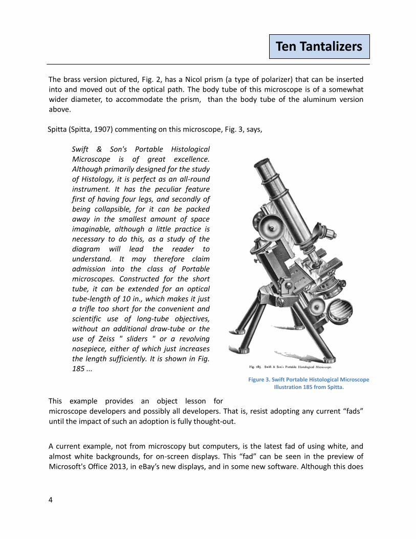
The brass version pictured, Fig. 2, has a Nicol prism (a type of polarizer) that can be inserted into and moved out of the optical path. The body tube of this microscope is of a somewhat wider diameter, to accommodate the prism, than the body tube of the aluminum version above. Spitta (Spitta, 1907) commenting on this microscope, Fig. 3, says,
Swift & Son's Portable Histological Microscope is of great excellence. Although primarily designed for the study of Histology, it is perfect as an all-round instrument. It has the peculiar feature first of having four legs, and secondly of being collapsible, for it can be packed away in the smallest amount of space imaginable, although a little practice is necessary to do this, as a study of the diagram will lead the reader to understand. It may therefore claim admission into the class of Portable microscopes. Constructed for the short tube, it can be extended for an optical tube-length of 10 in., which makes it just a trifle too short for the convenient and scientific use of long-tube objectives, without an additional draw-tube or the use of Zeiss " sliders " or a revolving nosepiece, either of which just increases the length sufficiently. It is shown in Fig. 185 ...
This example provides an object lesson for microscope developers and possibly all developers. That is, resist adopting any current “fads” until the impact of such an adoption is fully thought-out.
A current example, not from microscopy but computers, is the latest fad of using white, and almost white backgrounds, for on-screen displays. This “fad” can be seen in the preview of Microsoft's Office 2013, in eBay’s new displays, and in some new software. Although this does
Figure 3. Swift Portable Histological Microscope Illustration 185 from Spitta.
5
Ten Tantalizers
provide a distinctive façade, it’s usually too bright to read easily. It’s the authors' hope this "fad" will be shorter-lived than pure aluminum microscopes.
2. R. & J. Beck Folding Wenham Binocular with "Lunch Box" Travel Case
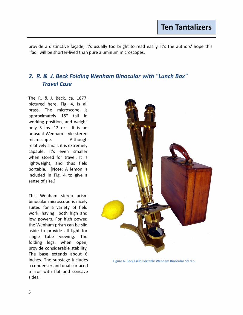
The R. & J. Beck, ca. 1877, pictured here, Fig. 4, is all brass. The microscope is approximately 15" tall in working position, and weighs only 3 lbs. 12 oz. It is an unusual Wenham-style stereo microscope. Although relatively small, it is extremely capable. It's even smaller when stored for travel. It is lightweight, and thus field portable. [Note: A lemon is included in Fig. 4 to give a sense of size.]
This Wenham stereo prism binocular microscope is nicely suited for a variety of field work, having both high and low powers. For high power, the Wenham prism can be slid aside to provide all light for single tube viewing. The folding legs, when open, provide considerable stability, The base extends about 6 inches. The substage includes a condenser and dual surfaced mirror with flat and concave sides.
Figure 4. Beck Field Portable Wenham Binocular Stereo
6
Ten Tantalizers
This Wenham binocular has the unique ability to fold into a remarkably compact lunch box-style 9- 1/2" x 3" x 7" case, for storage and travel. This storage/carrying case has a solid handle attached to its top that contains a unique lock and securing mechanism. The handle is divided into two sections. The top section has two small metal dowels that coincide with opening for in the bottom section..
Moving the top and bottom sections of the handle together snaps the case shut so nothing can fall out. The case is further secured by a keyed lock, in the center of the handle, and the
microscope and case were clearly designed to be carried safely into the field.
As Hartley noted (Hartley, 1993), it was the British love for the Wenham binocular microscope, with its attractive long body tubes that allowed comfortable image convergence using the Wenham prism, that likely led to the center of microscope development shifting from the UK to Europe at the turn of the 19th century.
The Wenham design was well suited for the production of ever larger and more spectacular benchtop microscopes. However, these were built at ever increasing costs, primarily for wealthy amateurs, rather than scientific needs. Meanwhile Continental makers were designing new short tube, relatively less expensive, instruments to meet the requirements of scientists. It was these 'Continental' microscopes that took the lead from the British designed long tube instruments and were pre-eminent as microscope development moved into the 20th century. The production of British long tube microscopes effectively ended about the first quarter of the 20th century, with some holdouts, to be replaced by the Continental-style short tube microscope.
7
Ten Tantalizers
3. Moginie Folding Tripod Microscope Signed by Moginie
Relatively little is known about William Moginie (Moginie, undated), as he was not a major manufacturer. Although not a large volume maker, he was an innovator at a time when most makers were not, as manufacturers often copied ideas from each other.
Some of what has been published about Moginie is unfortunately inaccurate, as new information has surfaced. Many of these inaccuracies are still repeated today, as most have come from the same original sources or derivative works.
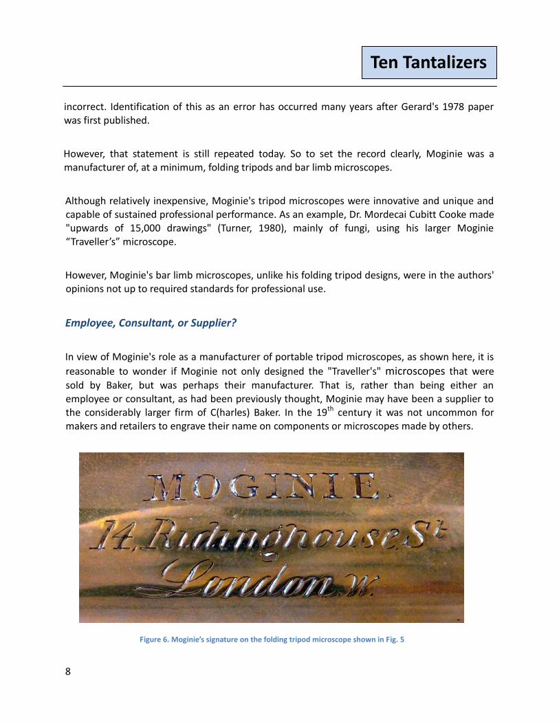
For example, in his excellent paper, Dr. M. Cooke's Microscope designed by W. Moginie (Turner, 1980), Gerard Turner commenting on Dr. Cooke's microscope states, "The constructor is named as Moginie, a name not found on instruments as a maker or retailer".
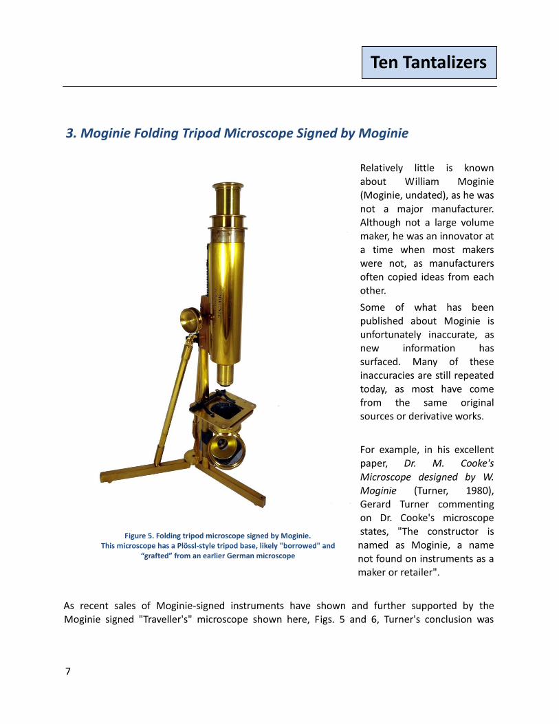
As recent sales of Moginie-signed instruments have shown and further supported by the Moginie signed "Traveller's" microscope shown here, Figs. 5 and 6, Turner's conclusion was
Figure 5. Folding tripod microscope signed by Moginie. This microscope has a Plössl-style tripod base, likely "borrowed" and
“grafted” from an earlier German microscope
8
Ten Tantalizers
incorrect. Identification of this as an error has occurred many years after Gerard's 1978 paper was first published.
However, that statement is still repeated today. So to set the record clearly, Moginie was a manufacturer of, at a minimum, folding tripods and bar limb microscopes.
Although relatively inexpensive, Moginie's tripod microscopes were innovative and unique and capable of sustained professional performance. As an example, Dr. Mordecai Cubitt Cooke made "upwards of 15,000 drawings" (Turner, 1980), mainly of fungi, using his larger Moginie “Traveller’s” microscope.
However, Moginie's bar limb microscopes, unlike his folding tripod designs, were in the authors' opinions not up to required standards for professional use.
Employee, Consultant, or Supplier?
In view of Moginie's role as a manufacturer of portable tripod microscopes, as shown here, it is
reasonable to wonder if Moginie not only designed the "Traveller's" microscopes that were
sold by Baker, but was perhaps their manufacturer. That is, rather than being either an employee or consultant, as had been previously thought, Moginie may have been a supplier to the considerably larger firm of C(harles) Baker. In the 19th century it was not uncommon for makers and retailers to engrave their name on components or microscopes made by others.
Figure 6. Moginie’s signature on the folding tripod microscope shown in Fig. 5
9
Ten Tantalizers
Inside the bell jar with Dr. Cooke's microscope, stored at the Royal Horticultural Society (originally The Horticultural Society of London), was a note stating " Students Portable Microscope especially constructed to order by Moginie with large body and rack movement". Turner had seen a similarly designed, but smaller, tripod microscope described in Jabez Hogg's 6th edition of The Microscope (Hogg, 1867) and in the 4th edition of Beale's How to Work with the Microscope (Beale, 1870). The microscope's "invention", described in Hogg, is credited to Mr. Moginie, of Mr. Baker's establishment.
This citation and the note, with Dr. Cooke's microscope, referencing Moginie as the source were the circumstantial evidence that led Mr. Turner to conclude that Dr. Cooke's microscope was made by C. Baker of High Holborn, although designed by Moginie. However, as Sherlock Holmes said in the Boscome Valley Mystery, "Circumstantial evidence is a very tricky thing ... It may seem to point very straight to one thing, but if you shift your point of view a little, you may find it pointing in an equally uncompromising manner to something entirely different". (Doyle, 1892)
In 1978, Gerard Turner had no way to know later research would confirm Moginie was a manufacturer. In his conclusion, Gerard did not account for the differences between Baker's version and Dr. Cooke's Moginie, including the larger size, coarse, and fine focusing on Dr. Cooke's instrument. However, that instrument was unsigned.
Cooke's Moginie microscope is now owned by the Royal Botanic Gardens (RBG), Kew. It was gifted to them, along with some of Dr. Cooke's drawings and fungi specimens, by the RHS. Unfortunately, sometime during the transfer to the RBG, Kew the base of Dr. Cooke's Moginie was misplaced or lost.
Brian Bracegirdle has Moginie manufacturing in the 1860s at 14 Ridinghouse Street, London and in the 1870s at 26 Litchfield Grove, Finchley, London (Bracegirdle, 1996). The proceedings of the "Microscopical Society of London" (MSL), later the Royal Microscopical Society (RMS), record Moginie's election to the society in 1866. His residence at the time of his election to the MSL is shown as 35, Queen Square. This would place his residence and manufacturing location at Ridinghouse Street less than 2 miles from Baker's address.
10
Ten Tantalizers
The Moginie signature, Fig. 6, includes the geographic postcode designator "W". London
geographic postal district designators were introduced in the late 1850s. Thus, the 1860s date of
Moginie's location is in keeping with Bracegirdle's identification. Baker was at his High Holborn
Street location before London's geographic postcodes were implemented, so it's not surprising
to see Baker continued the tradition of signing his instruments "London" without a geographic
suffix. The Baker "Traveller's" microscope was introduced and sold by C. Baker in 1867.
Bracegirdle's dates combined with Moginie's signature on the portable field tripod microscope,
as shown in Figs 3 and 4, would add support to identifying Moginie as the maker of the
Traveller's portable, and thus possibly Baker's supplier for these microscopes. Moginie
Traveller's microscopes, sold by Baker, are discussed in (Kreindler and Goren, November 2011).
As his obituarist noted on Moginie's death, Microscopy had "lost a prominent and valued member".
11
Ten Tantalizers
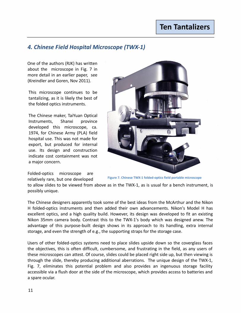
4. Chinese Field Hospital Microscope (TWX-1)
One of the authors (RJK) has written about the microscope in Fig. 7 in more detail in an earlier paper, see (Kreindler and Goren, Nov 2011). This microscope continues to be tantalizing, as it is likely the best of the folded optics instruments. The Chinese maker, TaiYuan Optical Instruments, Shanxi province developed this microscope, ca. 1974, for Chinese Army (PLA) field hospital use. This was not made for export, but produced for internal use. Its design and construction indicate cost containment was not a major concern. Folded-optics microscope are relatively rare, but one developed to allow slides to be viewed from above as in the TWX-1, as is usual for a bench instrument, is possibly unique. The Chinese designers apparently took some of the best ideas from the McArthur and the Nikon H folded-optics instruments and then added their own advancements. Nikon's Model H has excellent optics, and a high quality build. However, its design was developed to fit an existing Nikon 35mm camera body. Contrast this to the TWX-1's body which was designed anew. The advantage of this purpose-built design shows in its approach to its handling, extra internal storage, and even the strength of e.g., the supporting straps for the storage case. Users of other folded-optics systems need to place slides upside down so the coverglass faces the objectives, this is often difficult, cumbersome, and frustrating in the field, as any users of these microscopes can attest. Of course, slides could be placed right side up, but then viewing is through the slide, thereby producing additional aberrations. The unique design of the TWX-1, Fig. 7, eliminates this potential problem and also provides an ingenuous storage facility accessible via a flush door at the side of the microscope, which provides access to batteries and a spare ocular.
Figure 7. Chinese TWX-1 folded-optics field portable microscope
12
Ten Tantalizers
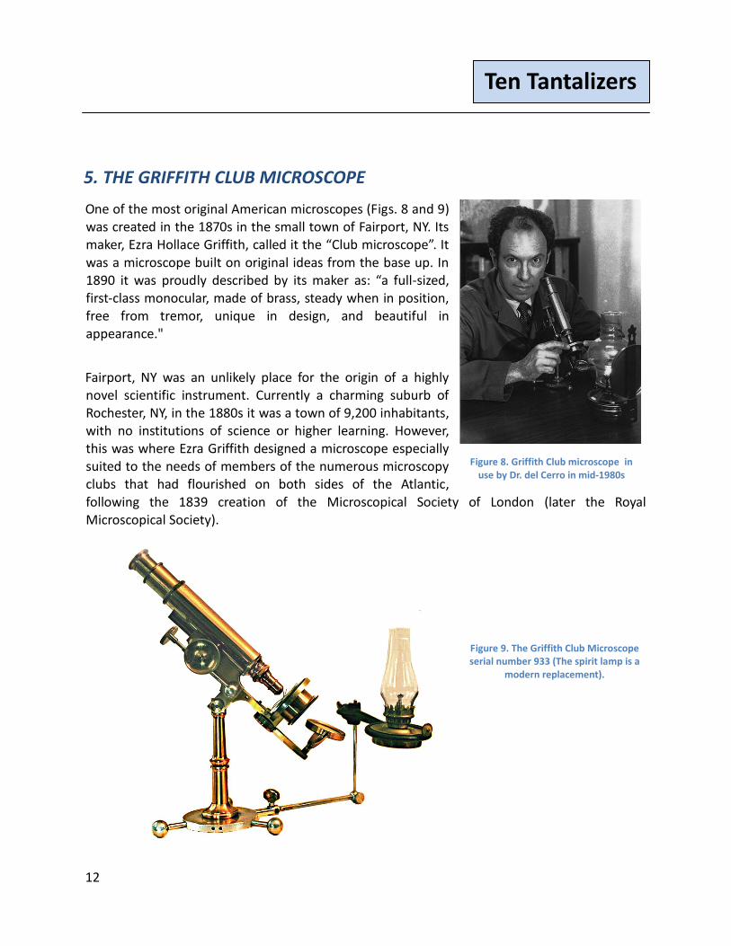
5. THE GRIFFITH CLUB MICROSCOPE
One of the most original American microscopes (Figs. 8 and 9) was created in the 1870s in the small town of Fairport, NY. Its maker, Ezra Hollace Griffith, called it the “Club microscope”. It was a microscope built on original ideas from the base up. In 1890 it was proudly described by its maker as: “a full-sized, first-class monocular, made of brass, steady when in position, free from tremor, unique in design, and beautiful in appearance."
Fairport, NY was an unlikely place for the origin of a highly novel scientific instrument. Currently a charming suburb of Rochester, NY, in the 1880s it was a town of 9,200 inhabitants, with no institutions of science or higher learning. However, this was where Ezra Griffith designed a microscope especially suited to the needs of members of the numerous microscopy clubs that had flourished on both sides of the Atlantic, following the 1839 creation of the Microscopical Society of London (later the Royal Microscopical Society).
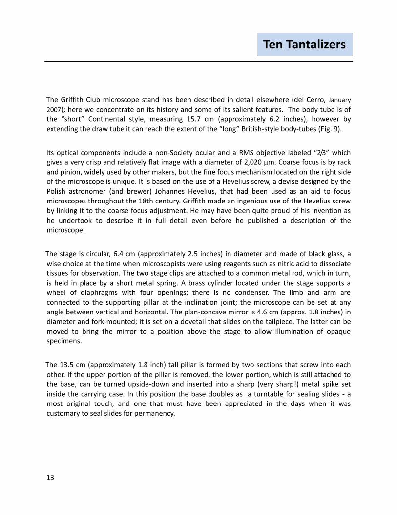
Figure 9. The Griffith Club Microscope serial number 933 (The spirit lamp is a
modern replacement).
Figure 8. Griffith Club microscope in use by Dr. del Cerro in mid-1980s
13
Ten Tantalizers
The Griffith Club microscope stand has been described in detail elsewhere (del Cerro, January
2007); here we concentrate on its history and some of its salient features. The body tube is of the “short” Continental style, measuring 15.7 cm (approximately 6.2 inches), however by extending the draw tube it can reach the extent of the “long” British-style body-tubes (Fig. 9).
Its optical components include a non-Society ocular and a RMS objective labeled “2/3” which gives a very crisp and relatively flat image with a diameter of 2,020 µm. Coarse focus is by rack and pinion, widely used by other makers, but the fine focus mechanism located on the right side of the microscope is unique. It is based on the use of a Hevelius screw, a devise designed by the Polish astronomer (and brewer) Johannes Hevelius, that had been used as an aid to focus microscopes throughout the 18th century. Griffith made an ingenious use of the Hevelius screw by linking it to the coarse focus adjustment. He may have been quite proud of his invention as he undertook to describe it in full detail even before he published a description of the microscope.
The stage is circular, 6.4 cm (approximately 2.5 inches) in diameter and made of black glass, a wise choice at the time when microscopists were using reagents such as nitric acid to dissociate tissues for observation. The two stage clips are attached to a common metal rod, which in turn, is held in place by a short metal spring. A brass cylinder located under the stage supports a wheel of diaphragms with four openings; there is no condenser. The limb and arm are connected to the supporting pillar at the inclination joint; the microscope can be set at any angle between vertical and horizontal. The plan-concave mirror is 4.6 cm (approx. 1.8 inches) in diameter and fork-mounted; it is set on a dovetail that slides on the tailpiece. The latter can be moved to bring the mirror to a position above the stage to allow illumination of opaque specimens.
The 13.5 cm (approximately 1.8 inch) tall pillar is formed by two sections that screw into each other. If the upper portion of the pillar is removed, the lower portion, which is still attached to the base, can be turned upside-down and inserted into a sharp (very sharp!) metal spike set inside the carrying case. In this position the base doubles as a turntable for sealing slides - a most original touch, and one that must have been appreciated in the days when it was customary to seal slides for permanency.
14
Ten Tantalizers
The metal base is circular; 8.5 cm in diameter and 8 mm thick (approximately 3.3 by 0.3 inches), with three distinctive ball-ended metal projections that provide extra support to the microscope and facilitate the use of the base as a turntable. The upper side of the base carries the inscription: “E.H.Griffith. Pat.Dec.14,86 Fairport.N.Y. 933.”
A distinctive feature of the "Club microscope” is a "built in" illumination system provided by an articulated metal arm that is secured to the base by means of two prongs that can be inserted into corresponding holes drilled into the thickness of the base. The distal end of the arm holds a spirit lamp.
The wooden carrying case is an important complement to the microscope; it accommodates the partially disassembled stand for transportation to the field or to club sessions, and doubles as the support of the turn table. In the case of the Griffith microscope discussed here, the case retains its original red-velvet lining and leather. The case is inscribed with the number 888 in gold, and it has a double gold rim all around the border of both the case and the lid. A sharp metal spike is fixed to the bottom of the case which serves to accommodate the base to be used as a turntable as described above. A brass canister lodged inside the case is signed E. H. Griffith and contains a 1/5" objective with the serial number 148.
We have found no sale records for the Griffith Club microscope, but it must have been a highly regarded instrument. It is known that in 1893 First Lady Frances Folsom Cleveland, had one of them in the White House. That same year, at the Chicago World’s Columbian Exhibition, the Griffith microscope was awarded a prize by the Exhibition's jury in, Substantial recognition for this queen of grace and utility amongst microscopes. Recognized abroad as well, Ezra Griffith was elected a Fellow of the Royal Microscopical Society.
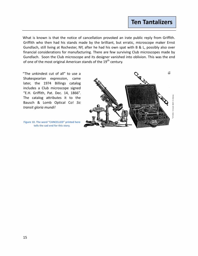
Alas, glory and commercial success were not to march together long! Ezra Griffith designed the optics and the stand of his microscope, but he depended on others for the actual manufacturing of them. Early stages were apparently made by John Field of Birmingham, England. Later, the microscopes were made, advertised, and marketed by Bausch & Lomb of Rochester, NY. This may have been a very successful move given the industrial and financial power of this company. Unfortunately, the association ended on a bitter note. A B & L catalog proclaimed the demise of the Club Microscope with “Cancelled”, Fig. 10. We don't know what brought this about. It might have been Griffith’s habit of continuously introducing minor variations to his stands (every surviving Club microscope differs from each other in some detail. This may have run counter to B & L’s mass production approach, that accounted so much for the success of the company), or some other reason.
15
Ten Tantalizers
What is known is that the notice of cancellation provoked an irate public reply from Griffith. Griffith who then had his stands made by the brilliant, but erratic, microscope maker Ernst Gundlach, still living at Rochester, NY, after he had his own spat with B & L, possibly also over financial considerations for manufacturing. There are few surviving Club microscopes made by Gundlach. Soon the Club microscope and its designer vanished into oblivion. This was the end of one of the most original American stands of the 19th century.
“The unkindest cut of all” to use a Shakespearian expression, came later, the 1974 Billings catalog includes a Club microscope signed
“E.H. Griffith, Pat. Dec. 14, 1866”. The catalog attributes it to the Bausch & Lomb Optical Co! Sic transit gloria mundi!
Figure 10. The word “CANCELLED” printed here tells the sad end for this story.
16
Ten Tantalizers
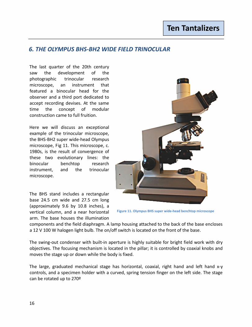
6. THE OLYMPUS BHS-BH2 WIDE FIELD TRINOCULAR
The last quarter of the 20th century saw the development of the photographic trinocular research microscope, an instrument that featured a binocular head for the observer and a third port dedicated to accept recording devises. At the same time the concept of modular construction came to full fruition. Here we will discuss an exceptional example of the trinocular microscope, the BHS-BH2 super wide-head Olympus microscope, Fig 11. This microscope, c. 1980s, is the result of convergence of these two evolutionary lines: the binocular benchtop research instrument, and the trinocular microscope. The BHS stand includes a rectangular base 24.5 cm wide and 27.5 cm long (approximately 9.6 by 10.8 inches), a vertical column, and a near horizontal arm. The base houses the illumination components and the field diaphragm. A lamp housing attached to the back of the base encloses a 12 V 100 W halogen light bulb. The on/off switch is located on the front of the base. The swing-out condenser with built-in aperture is highly suitable for bright field work with dry objectives. The focusing mechanism is located in the pillar; it is controlled by coaxial knobs and moves the stage up or down while the body is fixed. The large, graduated mechanical stage has horizontal, coaxial, right hand and left hand x-y controls, and a specimen holder with a curved, spring tension finger on the left side. The stage can be rotated up to 270º
Figure 11. Olympus BHS super wide-head benchtop microscope
17
Ten Tantalizers
The above-the-stage optics includes a sextuple nosepiece fit with five Olympus dry objectives, 2X to 40X, and a 60x oil-immersion. A magnification changer, placed between the nosepiece and the microscope head provides magnification factors from 1X to 1.5X. The head is designated by Olympus as a “SW Super Widefield Attachment.” The binocular portion has tubes with adjustment for interpupillary distance. On the right side there is a knob that can be pulled or pushed to direct all the light to the trinocular tube, to split it between the binocular and the trinocular tube, or to send it exclusively to the binocular. The 10X eyepieces have a diameter of 30.5 mm and carry individual diopter adjustment. The robust trinocular tube was designed to support even the heaviest photographic equipment. The tube holds a projection eyepiece, here 2.5X. This is a highly functional, all-purpose microscope. The BHS trinocular is not only a tantalizer, but a tantalizer that delivers!

18
Ten Tantalizers
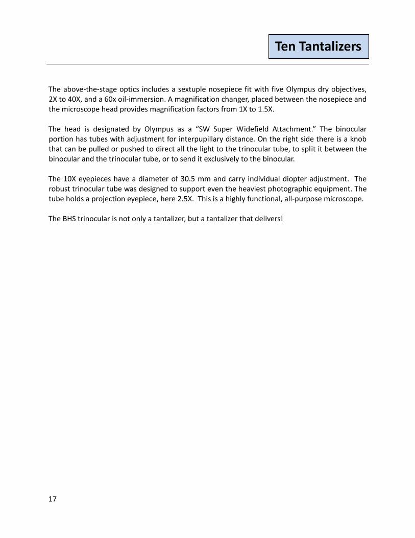
7. The Solar Microscope
Figure 12. A solar microscope c. 1775
This solar microscope, Fig. 12, is fairly large, perhaps larger than it might appear from the
photograph. It measures about 17" long and the brass plate is about 6" square. It is almost more
akin to a slide projector than a microscope, where the bulb is replaced by the sun, and the
typical beaded screen by a wall.
In its "hay days" during the second half of the 18th and the 19th century it was often mounted in
a shutter, with the mirror outside the shutter and the knobs and optical elements extending
inside. The two knobs, shown in Fig. 13 are used to control the mirror; the outermost knob
rotates the mirror relative to the flat mounting plate, and the innermost knob extends or
contracts the mirror against that plate. This flexibility allows the mirror to reflect sunlight from
any point in the sky, when the microscope is mounted in a wall or shutter. Typically, the room
with the internal optical elements is darkened, and the microscope focused to allow an image of
an object to be focused on an opposite wall.
The image is inverted and magnified, quite significantly enlarged, if the wall is some distance
from the "eyepiece". If use is extended for some time, the position of the mirror would need to
be changed to keep up with the movement of the sun.
19
Ten Tantalizers
Solar microscopes usually consisted in addition to the mirror of a large lens/condenser to focus
the light on the object and a smaller lens for projecting the image of the object onto the wall,
which acted as a screen.
U.S. President Thomas Jefferson is known to have owned, and used a solar microscope.
[Author's aside: When the U.S. Library of Congress (LOC) was burned by the British, Jefferson
offered to sell his library, then the largest private collection of books in the United States, to the
LOC for approximately $24,000. The LOC accepted his offer and completed the purchase in
1815].
This solar microscope is fascinating not only for its unusual shape, but for the enormous images it can project. In fact the title of an article by Dr. Peter Heering (Heering, 2004) begins with Fleas LIke Elephants, Lice Like Bears: 18th Century Solar Microscope .... The article notes that it was unclear whether the solar microscope was used to further scientific knowledge or simply for
entertainment, as he also indicates by the second half of his title, Projections Between
Enlightened Natural Philosophy and Amusement For Women and Children.
Figure 13. Solar microscope, dual knobs for mirror
orientation
20
Ten Tantalizers
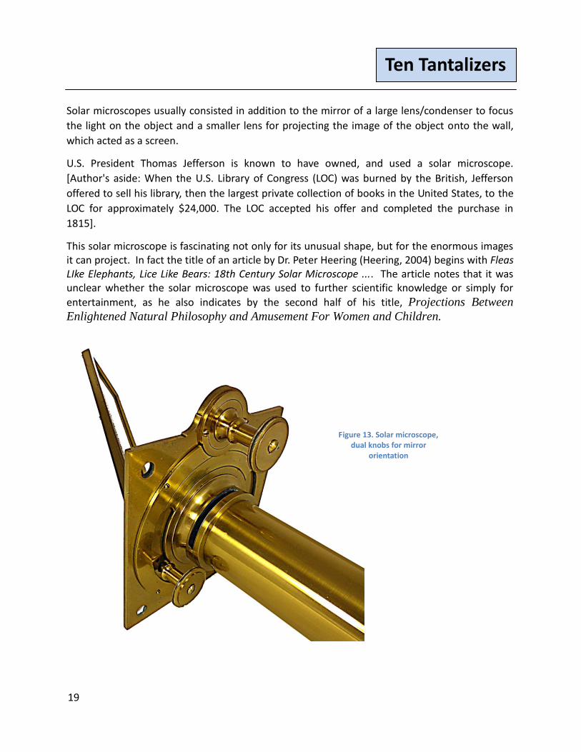
8. C. Vérick - Simple Dissecting Microscope
Figure 14. C. Vérick rue de la Parcheminerie, 2, Paris Simple dissecting microscope, c. 1880s

21
Ten Tantalizers
Many simple dissecting microscope are minimal in design and construction.
However, this cannot be said for this unusual, substantial, and to the authors, attractive dissecting microscope c. 1880s, manufactured by C. Vérick of Paris, Fig. 14. See excerpt in French, Fig. 15, from Eugène Trutat's 1883 book (Trutat ,1883). Trutat was a naturalist, geologist, and photographer. As a scientist, and writer about science, he was quite familiar with contemporary French dissecting microscopes. He was the Conservateur Du Musée D'Histoire Naturelle De Toulouse (Conservator of the Museum of Natural HIstory in Toulouse, France).
This Vérick dissecting microscope was "state of the art" at the time of its manufacture, just before the introduction of the first Greenough microscope was made by Zeiss in 1897. This is a relatively large simple microscope, with non-floating arm rests that rest on uprights and storage drawers.
After the introduction of the Greenough design, dissecting microscopes became even larger and more complex and many were no longer simple microscope designs.
Figure 15. Text and engraving from (Trutat ,1883)
22
Ten Tantalizers
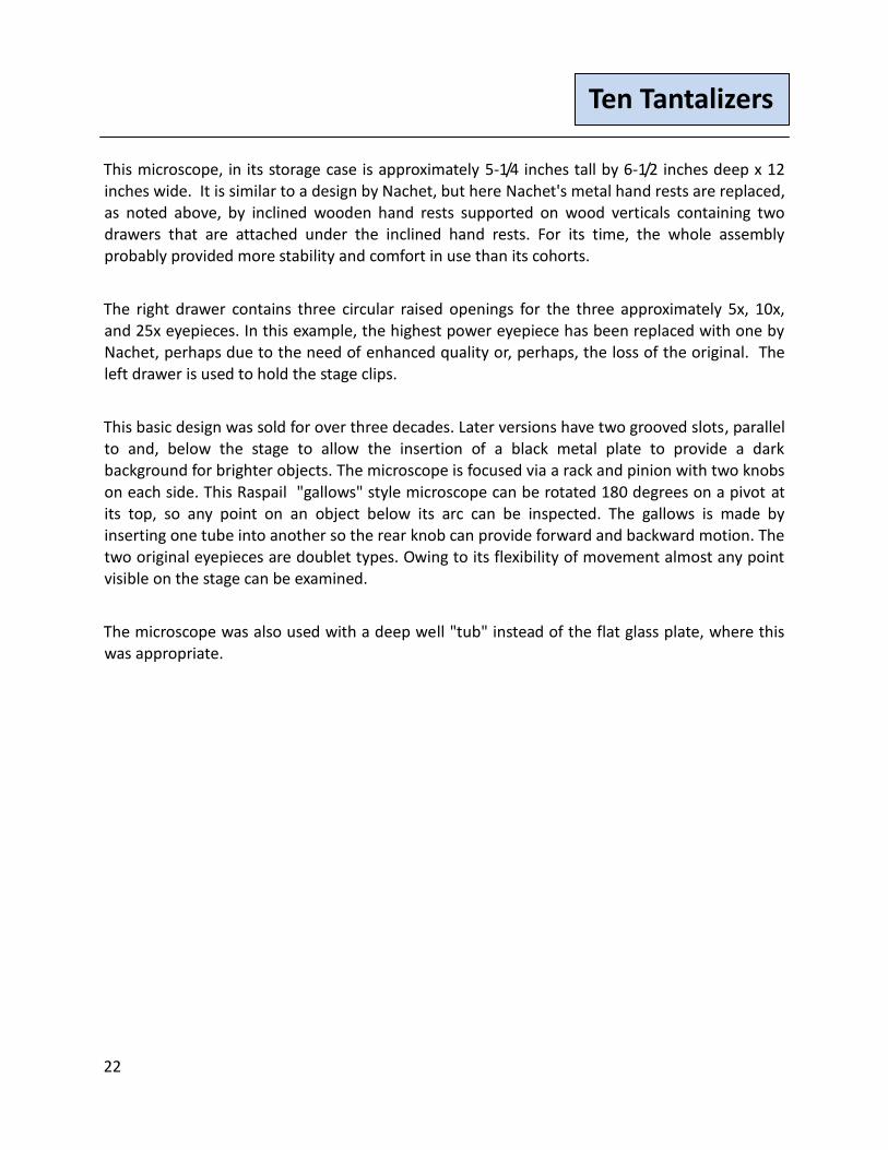
This microscope, in its storage case is approximately 5-1/4 inches tall by 6-1/2 inches deep x 12 inches wide. It is similar to a design by Nachet, but here Nachet's metal hand rests are replaced, as noted above, by inclined wooden hand rests supported on wood verticals containing two drawers that are attached under the inclined hand rests. For its time, the whole assembly probably provided more stability and comfort in use than its cohorts.
The right drawer contains three circular raised openings for the three approximately 5x, 10x, and 25x eyepieces. In this example, the highest power eyepiece has been replaced with one by Nachet, perhaps due to the need of enhanced quality or, perhaps, the loss of the original. The left drawer is used to hold the stage clips.
This basic design was sold for over three decades. Later versions have two grooved slots, parallel to and, below the stage to allow the insertion of a black metal plate to provide a dark background for brighter objects. The microscope is focused via a rack and pinion with two knobs on each side. This Raspail "gallows" style microscope can be rotated 180 degrees on a pivot at its top, so any point on an object below its arc can be inspected. The gallows is made by inserting one tube into another so the rear knob can provide forward and backward motion. The two original eyepieces are doublet types. Owing to its flexibility of movement almost any point visible on the stage can be examined.
The microscope was also used with a deep well "tub" instead of the flat glass plate, where this was appropriate.

23
Ten Tantalizers
9. Violin-Cased Small Monocular
Fig. 16, is a version of a microscope style popular during the 19th century. As they have no signature, or date information, dating them exactly is impossible. However, they do have
compartments for the smaller 1-1/2 x 1/2 inch microscope slides popular around the 1830s, so they can tentatively be dated c. 1830. The cases, shown here, are lined with royal purple plush.
“Violin-cased” microscopes were sold in both Georgian, and Victorian periods. They are all brass, monocular, and have button objectives.
The picture shows two violin-cased examples. What makes these instruments so interesting is both the unique “violin-case” shape of their storage cases and their size. Both cases here are almost the same size, measuring about 5 1/8 inches long by 1 3/4 inches tall (at their tallest), and about 2 1/4 inches wide, which is typical. Each has a single plan mirror and a pivot joint at the base allowing the microscope to tilt for use. These are smaller versions of bench instruments. Their optical quality is definitely sub-par, they will only accommodate smaller slides, and their focusing leaves much to be desired. However, there can be no question of their charm as display items, even though their performance is clearly less than desired.
Figure 16. Small brass monocular microscopes in 'violin-cases'
24
Ten Tantalizers
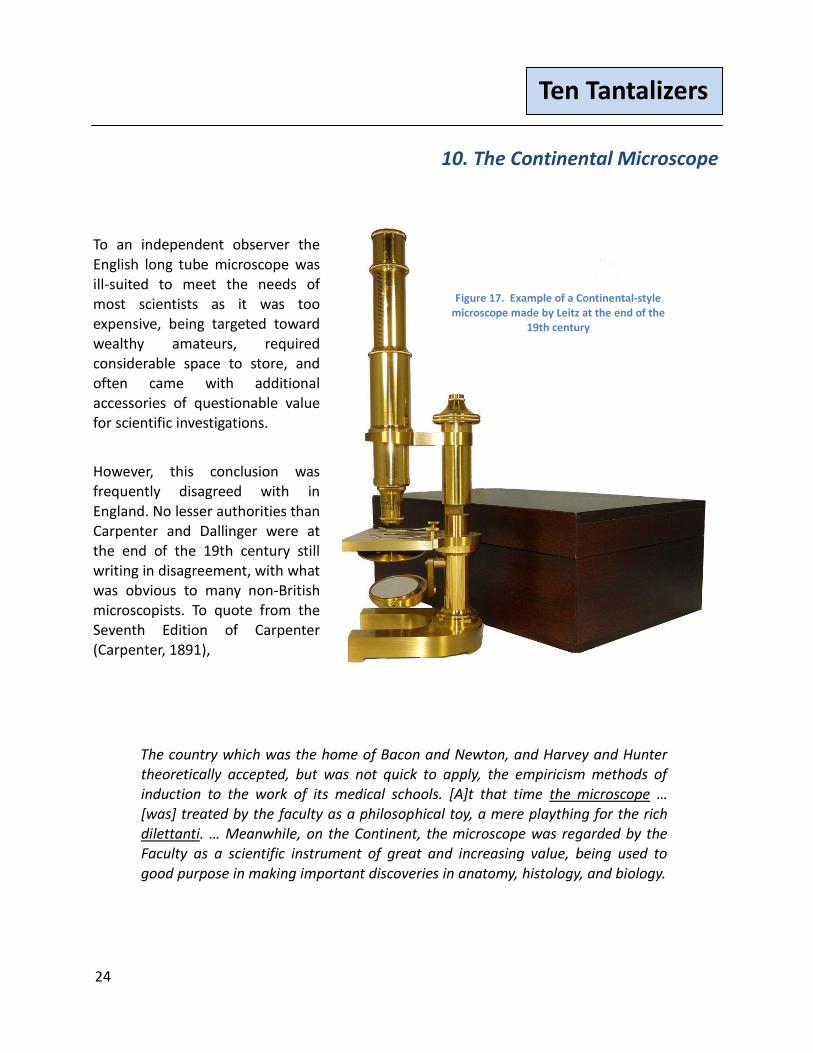
10. The Continental Microscope
To an independent observer the English long tube microscope was ill-suited to meet the needs of most scientists as it was too expensive, being targeted toward wealthy amateurs, required considerable space to store, and often came with additional accessories of questionable value for scientific investigations.
However, this conclusion was frequently disagreed with in England. No lesser authorities than Carpenter and Dallinger were at the end of the 19th century still writing in disagreement, with what was obvious to many non-British microscopists. To quote from the Seventh Edition of Carpenter (Carpenter, 1891),
The country which was the home of Bacon and Newton, and Harvey and Hunter theoretically accepted, but was not quick to apply, the empiricism methods of induction to the work of its medical schools. [A]t that time the microscope … [was] treated by the faculty as a philosophical toy, a mere plaything for the rich dilettanti. … Meanwhile, on the Continent, the microscope was regarded by the Faculty as a scientific instrument of great and increasing value, being used to good purpose in making important discoveries in anatomy, histology, and biology.
Figure 17. Example of a Continental-style microscope made by Leitz at the end of the
19th century

25
Ten Tantalizers
This was gradually realised in [England that] … because it was on the Continent that the investigations referred to had been made – it was nothing less than the Continental microscope that was sought after and obtained. Because early observations of a histological character (and therefore of a nature to lie beyond the sphere of the lay amateur) had been successfully made with a certain form of microscope on the Continent, it was practically argued that this must be the most suitable instrument for such a purpose; but this was an inference made without knowledge of or reference to the well-known English models.
-- (Carpenter, 1891) Pages 209-210
Nonetheless, at the end of the 19th century the European Continental stand held prominence in the minds of most scientists and physicians. One of the earlier style Continental stands is shown in Fig. 17. This is a 20th century reproduction by Leitz, in accordance with Leitz' internal records of its 1899 stand. This stand shown here is typical of Continental stands made in Europe at the close of the 19th Century. Leitz along with Zeiss lead the development of the Continental-style microscope.
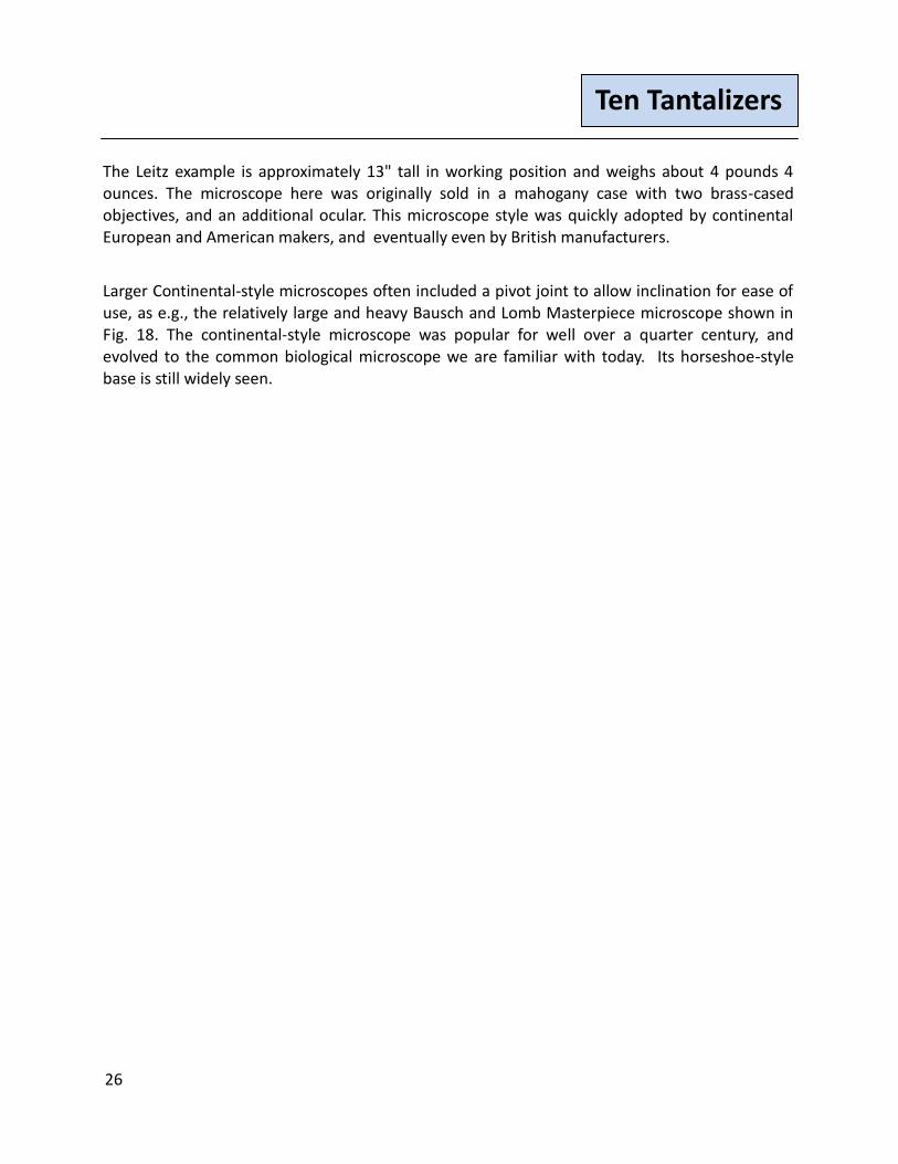
Figure 18. Bausch and Lomb Masterpiece microscope, a later Continental-style microscope. c 1896
26
Ten Tantalizers
The Leitz example is approximately 13" tall in working position and weighs about 4 pounds 4 ounces. The microscope here was originally sold in a mahogany case with two brass-cased objectives, and an additional ocular. This microscope style was quickly adopted by continental European and American makers, and eventually even by British manufacturers.
Larger Continental-style microscopes often included a pivot joint to allow inclination for ease of use, as e.g., the relatively large and heavy Bausch and Lomb Masterpiece microscope shown in Fig. 18. The continental-style microscope was popular for well over a quarter century, and evolved to the common biological microscope we are familiar with today. Its horseshoe-style base is still widely seen.

27
Ten Tantalizers
References and End Notes
Beale, Lionel Smith (1870) How to Work with the Microscope, Fourth Edition. Lindsay and Blakiston: Philadelphia Bracegirdle, Brian (1996) Notes on Modern Microscope Manufacturers. Oxford: Quekett Microscopical Club Carpenter, William B. (1891)The Microscope and its Revelations by the Late William B. Carpenter,
C.B., M.D., LL.D., F.R.S. Seventh Edition (and W.H. Dallinger)
del Cerro, Manuel (January 2007) The Griffith Club Microscope, a Queen of Grace and Utllity
Amongst Microscopes. Micscape Magazine
Doyle, Arthur Conan (1892) The Boscombe Valley Mystery. The Adventures of Sherlock Holmes, London: George Newnes English, Mary P. (1987) Modecai Cubitt Cooke. Victorian Naturalist, Mycologist, Teacher & Eccentric. Dorchester, Dorset. [Author's note: Dr. English was a distant relative of Dr. Cooke's and was herself a mycologist. She had unprecedented access to family members who remembered him, and as a mycologist she could appreciate and explain his contributions.] This is is the definitive biography of this great Victorian scientist Hartley, W. G. (1993) The Light Microscope: Its Use and Development. Oxford: Senecio Publishing Company Heering, Peter. (2004) Fleas Like Elephants, Lice Like Bears: 18th Century Solar Microscope Projections Between Enlightened Natural Philosophy and Amusement For Women and Children. Fifth International Conference for History of Science in Science Education Keszthely, Hungary Hogg, Jabez. The Microscope, its history, construction, and application, being a familiar introduction to the use of the instrument, and the study of microscopial science. 6th ed. George Routledge: London, 1867. page 109. Kreindler, R.Jordan and Yuval Goren (May 2011), Baker's Traveller's Microscope, Micscape Magazine
Kreindler, R. Jordan and Yuval Goren (November 2011), The TWX-1 Folded-Optics Microscope, Micscape Magazine Kreindler, R. Jordan (June 2012), Greenough Microscopes Part 2a, Micscape Magazine
28
Ten Tantalizers
Moginie (undated) The name "Moginie" may be originally of Swiss origin, see the book Moginie: An 18th Century Adventure: The life and adventures in the east of an 18th century Swiss soldier of fortune.
Spitta, Edmund .J. (1907) Microscopy: The Construction, Theory, and Use of the Microscope. New York: E. P. Dutton and Company, p 318 Trutat, Eugène (1883) Conservateur Du Musée D'Histoire Naturelle De Toulouse. Traité Élémentaire Du Microscope: Le Microscope et Son Emploi. Paris: Gauthiers-Villars, p. 22 Turner, G.L'E. (1980) Dr. M. Cooke's Microscope designed by W. Moginie. (reprinted in) Essays on
the History of the Microscope. Senecio Publishing: Oxford, pp 131-139. Both authors have corresponded on and off with Mr. Turner for several decades. He was always helpful
and generous with his time, and careful in stating his opinions. The information in this paper was not available at the time of Turner's original 1978 publication.
__________
©2011, 2012 Text and photographs (except as noted) by the authors.
The authors welcome any suggestions for corrections or improvement. They can be reached at:
R. Jordan Kreindler: [email protected]
Manuel del Cerro: [email protected]
__________
Published in the online magazine Micscape, November 2012,
Please report any Web problems or offer general comments to the Micscape Editor.
Micscape is the on-line monthly magazine of the Microscopy UK web site at
www.microscopy-uk.org.uk