Temporomandibular joint: conservative care of TMJ ... · Temporomandibular joint: conservative care...
8
J Can Chiropr Assoc 2009; 53(3) 165 0008-3194/2009/165–172/$2.00/©JCCA 2009 Temporomandibular joint: conservative care of TMJ dysfunction in a competitive swimmer Erik Yuill, BPHE, BSc, MSc* Scott D. Howitt, BA, CK, CSCS, DC, FCCSS(C), FCCRS(C)** Objective: To detail the progress of a patient with TMJ dysfunction and headaches due to swimming, who underwent a conservative treatment plan featuring soft tissue therapy, spinal manipulative therapy, and rehabilitation. Clinical Features: The most important features were initial bilateral temporal headaches and persistent left sided TMJ pain brought about by bilateral breathing while swimming. Conventional treatment aimed at decreasing hypertonic muscles, increasing hyoid mobility, improving TMJ mobility, resolving cervical restrictions, and improving digastric facilitation. Intervention and Outcome: The conservative treatment approach utilized in this case involved soft tissue therapy, hyoid mobility treatment, TMJ mobilization, spinal manipulative therapy, and digastric facilitation. Outcome measures included subjective pain ratings, range of motion, and motion palpation of the cervical spine. Conclusion: A patient with bilateral temporal headaches and TMJ pain due to bilateral breathing while swimming appeared to be relieved of his pain after three treatments of soft tissue therapy, hyoid mobility treatment, spinal manipulative therapy, and digastric facilitation. (JCCA 2009; 53(3):165–172) Objectif : Expliquer en détail les progrès d’un patient souffrant du dysfonctionnement d’une articulation temporomandibulaire (ATM) et de maux de tête reliés à la natation, à qui l’on a prescrit un traitement conservateur comprenant un travail des tissus mous, une manipulation rachidienne et une réadaptation. Caractéristiques cliniques : La plus importante manifestation du dysfonctionnement se présentait d’abord sous forme de céphalées temporales bilatérales et de douleur persistante ATM du côté gauche, causée par la respiration bilatérale pendant la natation. Le traitement conventionnel visait à adoucir les muscles hypertoniques, accroître la mobilité hyoïdienne, améliorer la mobilité ATM, éliminer les contraintes cervicales et améliorer le fonctionnement du muscle digastrique. Intervention et résultat : La méthode de traitement conservateur utilisé dans le présent cas a consisté à centrer le traitement sur les tissus mous, la mobilité hyoïdienne, la mobilité ATM, la manipulation rachidienne et le travail du muscle digastrique. L’indicateur des résultats a inclus des cotes subjectives de classification de la douleur, la portée du mouvement et la palpation du mouvement de la colonne cervicale. Conclusion : Un patient souffrant de céphalées temporales bilatérales et de douleurs ATM attribuables à la respiration bilatérale pendant la natation semble être soulagé de sa douleur après trois traitements d’une thérapie des tissus mous, d’un traitement de la mobilité hyoïdienne, d’une manipulation rachidienne et d’un * Clinic Intern, CMCC ** Assistant Professor, Clinical Education, Canadian Memorial Chiropractic College, 6100 Leslie St., Toronto, Ontario, M2H 3J1. Phone: (416) 226-6780 x7233. Fax: (416) 488-0470. Email: [email protected] © JCCA 2009.
-
Upload
nguyenlien -
Category
Documents
-
view
225 -
download
0
Transcript of Temporomandibular joint: conservative care of TMJ ... · Temporomandibular joint: conservative care...
J Can Chiropr Assoc 2009; 53(3) 165
0008-3194/2009/165–172/$2.00/©JCCA 2009
Temporomandibular joint:conservative care of TMJ dysfunction in a competitive swimmerErik Yuill, BPHE, BSc, MSc*Scott D. Howitt, BA, CK, CSCS, DC, FCCSS(C), FCCRS(C)**
Objective: To detail the progress of a patient with TMJ dysfunction and headaches due to swimming, who underwent a conservative treatment plan featuring soft tissue therapy, spinal manipulative therapy, and rehabilitation.
Clinical Features: The most important features were initial bilateral temporal headaches and persistent left sided TMJ pain brought about by bilateral breathing while swimming. Conventional treatment aimed at decreasing hypertonic muscles, increasing hyoid mobility, improving TMJ mobility, resolving cervical restrictions, and improving digastric facilitation.
Intervention and Outcome: The conservative treatment approach utilized in this case involved soft tissue therapy, hyoid mobility treatment, TMJ mobilization, spinal manipulative therapy, and digastric facilitation. Outcome measures included subjective pain ratings, range of motion, and motion palpation of the cervical spine.
Conclusion: A patient with bilateral temporal headaches and TMJ pain due to bilateral breathing while swimming appeared to be relieved of his pain after three treatments of soft tissue therapy, hyoid mobility treatment, spinal manipulative therapy, and digastric facilitation.(JCCA 2009; 53(3):165–172)
Objectif : Expliquer en détail les progrès d’un patient souffrant du dysfonctionnement d’une articulation temporomandibulaire (ATM) et de maux de tête reliés à la natation, à qui l’on a prescrit un traitement conservateur comprenant un travail des tissus mous, une manipulation rachidienne et une réadaptation.
Caractéristiques cliniques : La plus importante manifestation du dysfonctionnement se présentait d’abord sous forme de céphalées temporales bilatérales et de douleur persistante ATM du côté gauche, causée par la respiration bilatérale pendant la natation. Le traitement conventionnel visait à adoucir les muscles hypertoniques, accroître la mobilité hyoïdienne, améliorer la mobilité ATM, éliminer les contraintes cervicales et améliorer le fonctionnement du muscle digastrique.
Intervention et résultat : La méthode de traitement conservateur utilisé dans le présent cas a consisté à centrer le traitement sur les tissus mous, la mobilité hyoïdienne, la mobilité ATM, la manipulation rachidienne et le travail du muscle digastrique. L’indicateur des résultats a inclus des cotes subjectives de classification de la douleur, la portée du mouvement et la palpation du mouvement de la colonne cervicale.
Conclusion : Un patient souffrant de céphalées temporales bilatérales et de douleurs ATM attribuables à la respiration bilatérale pendant la natation semble être soulagé de sa douleur après trois traitements d’une thérapie des tissus mous, d’un traitement de la mobilité hyoïdienne, d’une manipulation rachidienne et d’un
* Clinic Intern, CMCC** Assistant Professor, Clinical Education, Canadian Memorial Chiropractic College, 6100 Leslie St., Toronto, Ontario, M2H 3J1.
Phone: (416) 226-6780 x7233. Fax: (416) 488-0470. Email: [email protected] © JCCA 2009.
Temporomandibular joint: conservative care of TMJ dysfunction in a competitive swimmer
166 J Can Chiropr Assoc 2009; 53(3)
key words : temporomandibular, joint, headache, manipulation
IntroductionThe temporomandibular joint (TMJ) is a complex junc-tion in the human skull incorporating disk, masticatorymuscles, and cervicocranial innervation. The prevalenceof TMJ pain in the general population is reported to be25%.1 Common signs associated with TMJ discomfortinclude popping, clicking, muscle tenderness, joint ten-derness, and decreased opening of the jaw.1 This joint hasalso been suggested to be a key propagating factor in oraland cervical disorders as well as headaches. TMJ paincommonly occurs with capsulitis, synovitis, meniscalderangement, tendonitis, degenerative joint disease, andinfection.2 The main movements which the TMJ is re-sponsible for are the opening and closing of the mouth.Proper opening mechanics involves both mandible de-pression and chin retrusion. Alternatively mouth closingincludes mandible elevation and chin protrusion.2
The National Board of Chiropractic Examiners(NBCE) 2005 survey of 2574 chiropractors in the USAconfirms that TMJ pain or Temporal Mandibular Disor-der (TMD) is a condition that is commonly seen. Theirdata rated TMD complaints as being a condition that‘sometimes’ (26–50%) presents to a chiropractors office.In fact, the 2003 paper by Raphael et al reported that intheir survey of women with TMD, that 22% of themchose complementary alternative therapies, which in-cluded chiropractic.3
Early signs of TMJ dysfunction vary from one patientto another, however commonly reported findings include:headache and facial pain, impaired jaw mobility, clickingor crepitus, pain in the TMJ and ears, masticatory musclepain, “stuffy” sensation in the ears, eustachian tube dys-function, and dizzy spells.4
Other predisposing factors can include joint specific is-sues such as joint laxity, anatomical variation, capsular ormuscular inflammation, repetitive motion, and static ar-ticular stress.
An aquatic sport which can lead to TMJ pain is swim-ming. To date there have not been any investigations toassess the prevalence of TMJ dysfunction in swimmers.The majority of reported swimming injuries includeshoulder, neck, and back injuries due to repetitive over-use and microtrauma brought on by poor technique andbiomechanics.5–9 In order to maximize force productionwhile in the water swimmers must position themselves inuncommon anatomical positions that subject the athletesto repetitive strain of numerous structures and tissues inthe upper limb and spine.5,9 In fact, the neck can be sub-jected to sustained and repetitive movements which canlead to overuse injury. Fifty-five (55%) of total cervicalmovement (most prominently rotation) is provided by theatlanto-axial joint (C1-C2), which houses the trigeminalspinal tract subnucleus and C1-C2 dorsal horns.5 It is notsurprising that the neck and its related structures cancause radiating pain to the shoulder and facial structures.In the older swimmer, disc dysfunction and spondylosismay also impinge on nerve roots at these levels as well asC4, C5 and C6 resulting in radiating pain to the shoulderjoint and beyond. Such an injury would make it difficultto swim due to the additional load placed on the cervi-cocranial structures.5
The number of strokes a freestyle swimmer takes in apractice is considered to be approximately 2500. Assum-ing they are taking a breath every three strokes, thistranslates into the swimmer turning their head over 800times per workout.5 This is further complicated if the ma-jority of the swimmers breaths are unilaterally (only ro-tating the head in one direction for breathing in a frontcrawl) as it could lead to muscle imbalances. Such dys-functions can be further irritated by postural alterationssuch as forward head carriage. Repetitively turning thehead from the axis of rotation at C1-C2, into a stressedposition while breathing, can cause the neck to adopt ahyperextended and rotated position. It has been suggested
traitement facilitant le fonctionnement du muscle digastrique.(JACC 2009; 53(3):165–172)
mots clés : temporomandibulaire, articulation, mal de tête, manipulation
E Yuill, SD Howitt
J Can Chiropr Assoc 2009; 53(3) 167
that the overuse of a hyperextended cervical spine canpredispose the swimmer to cervicogenic headaches.5
Thus bilateral breathing (breathing to the left when theright arm is extended overhead and to the right when theleft arm is extended overhead) during the front crawl ispromoted in swimmers from an early age to enhancemuscle balance.
The term for a headache occurring during physical ac-tivity is called “benign exertional headache.” Althoughswimming headaches are rare, they are often described assudden, severe, exploding, and pulsating.10,11 It has beensuggested that vascular factors are involved in the patho-genesis of swimming headaches.10 Increased levels ofCO2 in the blood, due to insufficient ventilation whileswimming, could possibly give rise to cerebral vasodila-tation, resulting in increased intracranial pressure leadingto an exertional headache.10 Another possible explanationfor swimming induced headaches could be neuronal irri-tation. Cervicogenic headaches while swimming mayalso be the result of nerve entrapments in hypertonic cer-vical muscles brought on by the repetitive rotation andhyperextension of the neck. Such a mechanism could alsobe used to explain a TMJ dysfunction brought on byswimming via relay through the previously mentionedtrigeminal spinal tract subnucleus and C1-C2 dorsal horntransitional zone.
The purpose of this case report was to describe a pa-tient who experienced TMJ pain and headaches duringswimming while training for a triathlon. The patient un-derwent a successful, simple, non-invasive chiropractictreatment plan using manual procedures and rehabilita-tive training for the TMJ musculature as well as undergo-ing technique and postural education for their swimmingstroke.
Case ReportThis case report involves a 31 year old male recreationaltriathlete who developed headaches and TMJ pain whileattempting to incorporate bilateral breathing into his free-style swimming training regime. Initially the patient wasbreathing every 2 or 4 strokes only rotating his head tothe left side. Breathing bilaterally every 3 strokes intro-duced an additional right sided rotation breathing. On hisfirst day of initially attempting to bilaterally breathe thepatient experienced a bilateral temporal headache afterhis swim which was relieved by AdvilTM. Two days later
while attempting to bilaterally breathe for the secondtime, the patient experienced another bilateral temporalheadache (again relieved by AdvilTM) and left sided jawpain while in the pool. The next day while attempting tobilaterally breathe for the third time the patient experi-enced a mild headache, extreme jaw pain, and an inabilityto open his mouth more than 50% which prompted thepatient to present for treatment.
The patient was a physically fit health care professionalwho reported to work-out daily (either swimming, biking,running, or weightlifting, each 2–3 times per week). Hedenied any previous incidences of TMJ pain or dental is-sues, but did report previous cervicogenic headaches as astudent, that resolved with postural exercises. No previousmotor vehicle accidents were reported however the patientacknowledge that he suffered several concussions in hisyouth playing minor sports. Occasional knee discomfortassociated with his run training was noted however the pri-mary complaint was his jaw pain and dysfunction whichwas aggravated by his swim training.
The subject presented with decreased hyoid mobility(less lateral motion from left to right) and a dysfunctionalactive mouth opening pattern where pterygoid hyper-tonicity/jaw jut was prominent. No headache or neck painwas noted at this time, but bilateral TMJ pain (with theleft side greater than the right) was reported at the initialassessment. The subject rated his TMJ pain 7 out of 10on Visual Analog Scale (VAS), while Neck Disability In-dex was zero out of 50. The centric relation protractiontest (TMJ compression) and biting/jaw clenching did notreproduce pain. Motion palpation revealed the left TMJto be subjectively hypermobile while the right TMJ wasfound to be hypomobile. A cervical spine screen foundright rotation painful and decreased by 25% while left ro-tation was full but painful at end range. Bilateral restric-tions were found with motion palpation at C0, C1, andC2. Jackson’s, Spurling’s, and Cervical Compressiontests (as described by Vizniak’s Clinical ChiropracticHandbook) were all found to be negative for facet jointand nerve root involvement, however left Kemps test wasfound to cause pain on the left at C1-C2 without radia-tion. Soft tissue palpation revealed tight and tender ster-nocleidomastoid (SCM), upper trapezius, levator scapula,superior and inferior oblique, rectus capitis minor, lateralpterygoids and masseter muscles bilaterally. Deep neckflexors were also found to be weak, as indicated by an
Temporomandibular joint: conservative care of TMJ dysfunction in a competitive swimmer
168 J Can Chiropr Assoc 2009; 53(3)
endurance test in which the patient could only maintain achin tucked position for 10 seconds and reported the sen-sation of a headache “coming on” at the conclusion of thetest.
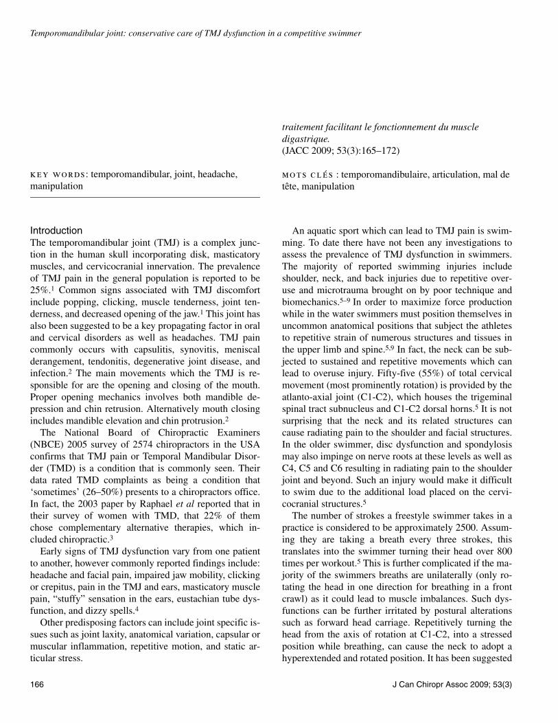
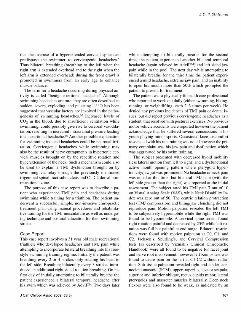
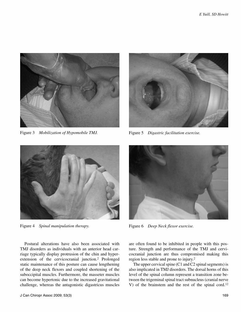
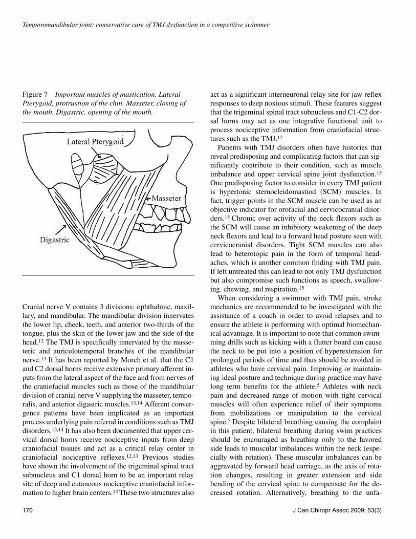
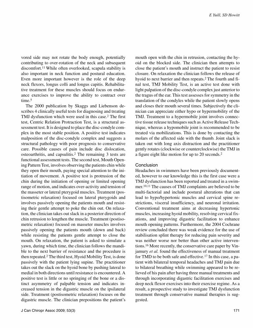
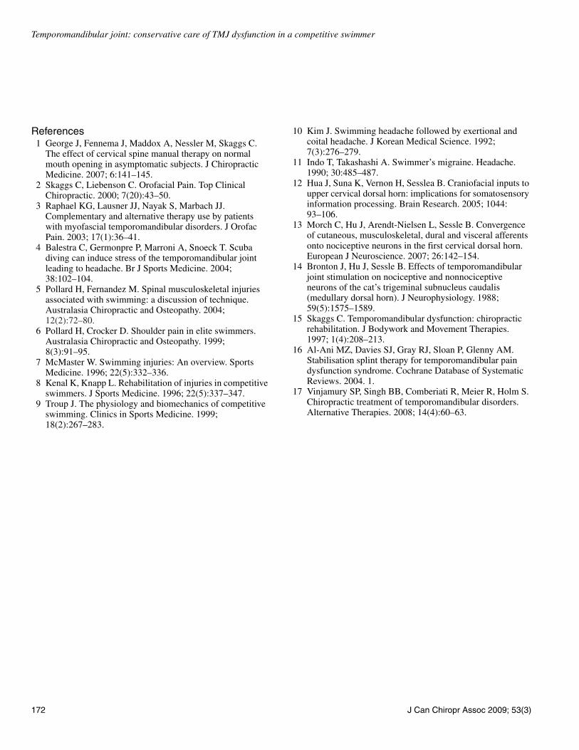
TreatmentThe patient was treated with 3 sessions of soft tissue ther-apy consisting of Active Release Technique® (ART®) tothe SCM, upper trapezius, levator scapula, superior andinferior oblique, and rectus capitis minor muscles (seeFigure 1); hyoid mobility treatment done passively withthe patient lying supine and the slack taken out on the hy-oid bone by pushing lateral to medial in both directionsuntil resistance was encountered (see Figure 2); TMJ mo-bilization done passively taking out the joint slack and ro-tating the joint in a figure eight like motion (see Figure3); spinal manipulative therapy was performed by rotaryadjustment to C1-C2 (see Figure 4); and digastric exer-cises were accomplished by an isometric facilitation, eve-ry other day over the course of 1 week. Additionally thepatient was instructed to continue digastric facilitationexercises by placing the tongue on the roof of his mouthwhile opening his jaw (see Figure 5) and deep neck flexorexercises (chin tucks) at home 2 to 3 times daily (see Fig-ure 6). The patient returned to the pool 1 week later witha swimming coach for stroke assessment and reported nopain. These interventions combined with 1 week of restresulted in relief from headaches and TMJ pain as the pa-tient resumed his biweekly swim training with a VAS ofzero out of 10. At follow up one month later the patientreported no reoccurrence of headache or TMJ pain, ex-hibited an improved mouth opening pattern and full rangeof TMJ and cervical spine motion. He also demonstratedan improved deep neck flexor endurance test to 45 sec-onds, and VAS remained zero out of 10.
DiscussionThe three main categories of TMJ disorders are myofas-cial pain, internal derangement, and degenerative destruc-tion.2 The myofascial category is the most common andoften involves pain not only in the muscles of masticationbut also muscles of the neck and shoulders.2 A number ofmuscles can be involved with movement of the TMJ, how-ever it has been suggested that the most important are thedigastricus, masseter, and lateral pterygoid.2 (see Figure7) The primary action of the digastricus is to aid in open-
ing of the mouth, whereas the masseter muscle is chieflyresponsible for closing the mouth. The masseter is assistedin this task by the medial pterygoid and temporalis mus-cles. The lateral pterygoids bilaterally stabilize the TMJand contribute to protrusion of the chin during mouthopening.2 Manual treatments for relaxation, facilitation,and mobilization of these muscles have been shown to besuccessful in dealing with TMJ pain.2
Figure 1 Active release technique for the SCM.
Figure 2 Hyoid mobility treatment.
E Yuill, SD Howitt
J Can Chiropr Assoc 2009; 53(3) 169
Postural alterations have also been associated withTMJ disorders as individuals with an anterior head car-riage typically display protrusion of the chin and hyper-extension of the cervicocranial junction.2 Prolongedstatic maintenance of this posture can cause lengtheningof the deep neck flexors and coupled shortening of thesuboccipital muscles. Furthermore, the masseter musclescan become hypertonic due to the increased gravitationalchallenge, whereas the antagonistic digastricus muscles
are often found to be inhibited in people with this pos-ture. Strength and performance of the TMJ and cervi-cocranial junction are thus compromised making thisregion less stable and prone to injury.2
The upper cervical spine (C1 and C2 spinal segments) isalso implicated in TMJ disorders. The dorsal horns of thislevel of the spinal column represent a transition zone be-tween the trigeminal spinal tract subnucleus (cranial nerveV) of the brainstem and the rest of the spinal cord.12
Figure 3 Mobilization of Hypomobile TMJ.
Figure 4 Spinal manipulation therapy.
Figure 5 Digastric facilitation exercise.
Figure 6 Deep Neck flexor exercise.
Temporomandibular joint: conservative care of TMJ dysfunction in a competitive swimmer
170 J Can Chiropr Assoc 2009; 53(3)
Cranial nerve V contains 3 divisions: ophthalmic, maxil-lary, and mandibular. The mandibular division innervatesthe lower lip, cheek, teeth, and anterior two-thirds of thetongue, plus the skin of the lower jaw and the side of thehead.12 The TMJ is specifically innervated by the masse-teric and auriculotemporal branches of the mandibularnerve.13 It has been reported by Morch et al. that the C1and C2 dorsal horns receive extensive primary afferent in-puts from the lateral aspect of the face and from nerves ofthe craniofacial muscles such as those of the mandibulardivision of cranial nerve V supplying the masseter, tempo-ralis, and anterior digastric muscles.13,14 Afferent conver-gence patterns have been implicated as an importantprocess underlying pain referral in conditions such as TMJdisorders.13,14 It has also been documented that upper cer-vical dorsal horns receive nociceptive inputs from deepcraniofacial tissues and act as a critical relay center incraniofacial nociceptive reflexes.12,13 Previous studieshave shown the involvement of the trigeminal spinal tractsubnucleus and C1 dorsal horn to be an important relaysite of deep and cutaneous nociceptive craniofacial infor-mation to higher brain centers.14 These two structures also
act as a significant interneuronal relay site for jaw reflexresponses to deep noxious stimuli. These features suggestthat the trigeminal spinal tract subnucleus and C1-C2 dor-sal horns may act as one integrative functional unit toprocess nociceptive information from craniofacial struc-tures such as the TMJ.12
Patients with TMJ disorders often have histories thatreveal predisposing and complicating factors that can sig-nificantly contribute to their condition, such as muscleimbalance and upper cervical spine joint dysfunction.15
One predisposing factor to consider in every TMJ patientis hypertonic sternocleidomastiod (SCM) muscles. Infact, trigger points in the SCM muscle can be used as anobjective indicator for orofacial and cervicocranial disor-ders.15 Chronic over activity of the neck flexors such asthe SCM will cause an inhibitory weakening of the deepneck flexors and lead to a forward head posture seen withcervicocranial disorders. Tight SCM muscles can alsolead to heterotopic pain in the form of temporal head-aches, which is another common finding with TMJ pain.If left untreated this can lead to not only TMJ dysfunctionbut also compromise such functions as speech, swallow-ing, chewing, and respiration.15
When considering a swimmer with TMJ pain, strokemechanics are recommended to be investigated with theassistance of a coach in order to avoid relapses and toensure the athlete is performing with optimal biomechan-ical advantage. It is important to note that common swim-ming drills such as kicking with a flutter board can causethe neck to be put into a position of hyperextension forprolonged periods of time and thus should be avoided inathletes who have cervical pain. Improving or maintain-ing ideal posture and technique during practice may havelong term benefits for the athlete.5 Athletes with neckpain and decreased range of motion with tight cervicalmuscles will often experience relief of their symptomsfrom mobilizations or manipulation to the cervicalspine.5 Despite bilateral breathing causing the complaintin this patient, bilateral breathing during swim practicesshould be encouraged as breathing only to the favoredside leads to muscular imbalances within the neck (espe-cially with rotation). These muscular imbalances can beaggravated by forward head carriage, as the axis of rota-tion changes, resulting in greater extension and sidebending of the cervical spine to compensate for the de-creased rotation. Alternatively, breathing to the unfa-
Figure 7 Important muscles of mastication. Lateral Pterygoid, protrustion of the chin. Masseter, closing of the mouth. Digastric, opening of the mouth.
E Yuill, SD Howitt
J Can Chiropr Assoc 2009; 53(3) 171
vored side may not rotate the body enough, potentiallycontributing to over-rotation of the neck and subsequentdiscomfort.11 While often overlooked, scapula stability isalso important in neck function and postural education.Even more important however is the role of the deepneck flexors, longus colli and longus capitis. Rehabilita-tive treatment for these muscles should focus on endur-ance exercises to improve the ability to contract overtime.5
The 2000 publication by Skaggs and Liebenson de-scribes 4 clinically useful tests for diagnosing and treatingTMJ dysfunction which were used in this case.2 The firsttest, Centric Relation Protraction Test, is a structural as-sessment test. It is designed to place the disc-condyle com-plex in the most stable position. A positive test indicatesmalposition of the disc-condyle complex and suggests astructural pathology with poor prognosis to conservativecare. Possible causes of pain include disc dislocation,osteoarthritis, and capsulitis.2 The remaining 3 tests arefunctional assessment tests. The second test, Mouth Open-ing Pattern Test, involves observing the patients chin whilethey open their mouth, paying special attention to the ini-tiation of movement. A positive test is protrusion of thechin during the initiation of opening or limited openingrange of motion, and indicates over-activity and tension ofthe masseter or lateral pterygoid muscles. Treatment (pos-tisometric relaxation) focused on lateral pterygoids andinvolves passively opening the patients mouth and resist-ing their gentle attempt to poke the chin out. On relaxa-tion, the clinician takes out slack in a posterior direction ofchin retrusion to lengthen the muscle. Treatment (postiso-metric relaxation) focused on masseter muscles involvespassively opening the patients mouth (down and back)while resisting the patients gentle attempt to close themouth. On relaxation, the patient is asked to simulate ayawn, during which time, the clinician follows the mandi-ble to the next barrier of resistance and the procedure isthen repeated.2 The third test, Hyoid Mobility Test, is donepassively with the patient lying supine. The practitionertakes out the slack on the hyoid bone by pushing lateral tomedial in both directions until resistance is encountered. Apositive test is little or no springing of the bone or a dis-tinct asymmetry of palpable tension and indicates in-creased tension in the digastric muscle on the ipsilateralside. Treatment (postisometric relaxation) focuses on thedigastric muscle. The clinician prepositions the patient’s
mouth open with the chin in retrusion, contacting the hy-oid on the blocked side. The clinician then attempts toclose the patient’s mouth and instruct the patient to resistclosure. On relaxation the clinician follows the release ofhyoid to next barrier and then repeats.2 The fourth and fi-nal test, TMJ Mobility Test, is an active test done withlight palpation of the disc-condyle complex just anterior tothe tragus of the ear. This test assesses for symmetry in thetranslation of the condyles while the patient slowly opensand closes their mouth several times. Subjectively the cli-nician can appreciate either hypo or hypermobility of theTMJ. Treatment to a hypermobile joint involves connec-tive tissue release techniques such as Active Release Tech-nique, whereas a hypomobile joint is recommended to betreated via mobilizations. This is done by contacting themolars of the affected side with the thumb. Joint slack istaken out with long axis distraction and the practitionergently rotates (clockwise or counterclockwise) the TMJ ina figure eight like motion for up to 20 seconds.2
ConclusionHeadaches in swimmers have been previously document-ed, however to our knowledge this is the first case were aTMJ dysfunction has been reported and treated in a swim-mer.10,11 The causes of TMJ complaints are believed to bemulti-factorial and include postural alterations that canlead to hypo/hypertonic muscles and cervical spine re-strictions, visceral insufficiency, and neuronal irritation.Conventional treatment aims at decreasing hypertonicmuscles, increasing hyoid mobility, resolving cervical fix-ations, and improving digastric facilitation to enhancemouth opening patterns. Furthermore, the 2004 Cochranereview concluded there was weak evidence for the use ofstabilisation splint therapy for reducing pain severity andwas neither worse nor better than other active interven-tions.16 More recently, the conservative care paper by Vin-jamury et al. found the effectiveness of manual treatmentfor TMD to be both safe and effective.17 In this case, a pa-tient with bilateral temporal headaches and TMJ pain dueto bilateral breathing while swimming appeared to be re-lieved of his pain after having three manual treatments andthrough incorporating digastric facilitation exercises anddeep neck flexor exercises into their exercise regime. As aresult, a prospective study to investigate TMJ dysfunctiontreatment through conservative manual therapies is sug-gested.
Temporomandibular joint: conservative care of TMJ dysfunction in a competitive swimmer
172 J Can Chiropr Assoc 2009; 53(3)
References1 George J, Fennema J, Maddox A, Nessler M, Skaggs C.
The effect of cervical spine manual therapy on normal mouth opening in asymptomatic subjects. J Chiropractic Medicine. 2007; 6:141–145.
2 Skaggs C, Liebenson C. Orofacial Pain. Top Clinical Chiropractic. 2000; 7(20):43–50.
3 Raphael KG, Lausner JJ, Nayak S, Marbach JJ. Complementary and alternative therapy use by patients with myofascial temporomandibular disorders. J Orofac Pain. 2003; 17(1):36–41.
4 Balestra C, Germonpre P, Marroni A, Snoeck T. Scuba diving can induce stress of the temporomandibular joint leading to headache. Br J Sports Medicine. 2004; 38:102–104.
5 Pollard H, Fernandez M. Spinal musculoskeletal injuries associated with swimming: a discussion of technique. Australasia Chiropractic and Osteopathy. 2004;12(2):72–80.
6 Pollard H, Crocker D. Shoulder pain in elite swimmers. Australasia Chiropractic and Osteopathy. 1999; 8(3):91–95.
7 McMaster W. Swimming injuries: An overview. Sports Medicine. 1996; 22(5):332–336.
8 Kenal K, Knapp L. Rehabilitation of injuries in competitive swimmers. J Sports Medicine. 1996; 22(5):337–347.
9 Troup J. The physiology and biomechanics of competitive swimming. Clinics in Sports Medicine. 1999; 18(2):267–283.
10 Kim J. Swimming headache followed by exertional and coital headache. J Korean Medical Science. 1992; 7(3):276–279.
11 Indo T, Takashashi A. Swimmer’s migraine. Headache. 1990; 30:485–487.
12 Hua J, Suna K, Vernon H, Sesslea B. Craniofacial inputs to upper cervical dorsal horn: implications for somatosensory information processing. Brain Research. 2005; 1044:93–106.
13 Morch C, Hu J, Arendt-Nielsen L, Sessle B. Convergence of cutaneous, musculoskeletal, dural and visceral afferents onto nociceptive neurons in the first cervical dorsal horn. European J Neuroscience. 2007; 26:142–154.
14 Bronton J, Hu J, Sessle B. Effects of temporomandibular joint stimulation on nociceptive and nonnociceptive neurons of the cat’s trigeminal subnucleus caudalis (medullary dorsal horn). J Neurophysiology. 1988; 59(5):1575–1589.
15 Skaggs C. Temporomandibular dysfunction: chiropractic rehabilitation. J Bodywork and Movement Therapies. 1997; 1(4):208–213.
16 Al-Ani MZ, Davies SJ, Gray RJ, Sloan P, Glenny AM. Stabilisation splint therapy for temporomandibular pain dysfunction syndrome. Cochrane Database of Systematic Reviews. 2004. 1.
17 Vinjamury SP, Singh BB, Comberiati R, Meier R, Holm S. Chiropractic treatment of temporomandibular disorders. Alternative Therapies. 2008; 14(4):60–63.