Temporomandibular joint ankylosis: Case-series of two ... · Temporomandibular joint ankylosis:...
6
103 Dental Hypotheses Jul-Sep 2014 / Vol 5 | Issue 3 Temporomandibular joint ankylosis: Case-series of two different surgical procedures Gholamreza Shirani, Farnoosh Mohammadi, Mahnaz Arshad 1 , Mohsen Shirazi 2 Departments of Oral and Maxillofacial Surgery, 1 Dental Implant Research Center and 2 Department of Orthodontics, School of Dentistry, Tehran University of Medical Sciences, Tehran, Iran Introduction Disabling interruptions to chewing and malformed facial development may be caused by bony and fibrous ankylosis of the temporomandibular joint (TMJ) as a result of trauma (31-98% of cases), local or systemic infection (10-49%), systemic disease (10%), or neoplasm. [1] Ankylosis of the TMJ is one of the most serious complications of condylar fracture. In the cases of trauma, it is hypothesized that intra- articular hematoma, with scarring and excessive bone formation, leads to hypomobility. Infection of the TMJ is most commonly the result of contiguous spread from otitis media or mastoiditis but may also result from hematogenous spread, including tuberculosis, gonorrhea, and scarlet fever. Systemic causes of TMJ ankylosis include ankylosing spondylitis, rheumatoid arthritis, and psoriasis. [1] Ankylosis can be classified on the basis of degree of limitation (partial or complete), location of the union (intracapsular versus extracapsular) and type of tissue involved (fibrosis, osseous, or fibro-osseous). The end result is severing limitation in the range of mandibular motion, which may interfere with speech, oral hygiene, and proper preparation of a food bolus for digestion as well as maintenance of nutrition. As the mandible develops, three growth centers are able to produce enlargement this complex bony structure, the condyles, the surface of the ramus, and the alveolus. [1,2] Initially, it was believed that the mandible was pushed down and forward by growth at the condylar Corresponding Author: Prof. Mahnaz Arshad, Dental Implant Research Center, School of Denstry, Tehran University of Medical Sciences, North Amirabad, Tehran, Iran. E-mail: [email protected] ORIGINAL RESEARCH ABSTRACT Introducation: The long-term outcome and clinical results of gaparthroplasty used for the treatment of condylar ankylosis of the mandible in children with application of postoperative activator appliances and costochondral rib graft are evaluated and compared. The purpose of this study was to compare the effect of gap arthroplasty and costochondral graft methods on reankylosis, a mount of mouth opening and growth. Materials and Methods: A non-randomized, retrospective clinical study of l0 cases (5-12 years old) of condylar ankylosis of the mandible, surgically treated during a 10 year period from 2002 to 2012 was performed. Four patients were treated by condylectomy and interpositional flap, whereas six were treated by condylectomy and immediate costachondral rib grafts. The first group underwent long-term postoperative therapy using removable activator appliances. Casts, radiographs, photographs, and computed tomography (CT) were used post surgically to evaluate rib graft, condylar growth and function, occlusion, facial, and condylar symmetry. Data were analyzed by SPSS 16 statistical software using Mann-Whitney, Paired T-test and Chi- square tests. Results: Children with long-standing condylar ankylosis of the mandible treated by condylectomy and interpositional flap showed more favorably when activators were used post-surgically. Conclusions: Gaparthroplasty with functional activator post-operatively can be considering for TMJ ankylosis. Key words: Costochondral graft, gap arthroplasty, temporomandibular joint ankylosis Access this article online Quick Response Code: Website: www.dentalhypotheses.com DOI: 10.4103/2155-8213.136754 [Downloaded free from http://www.dentalhypotheses.com on Sunday, August 24, 2014, IP: 37.98.49.227] || Click here to download free Android application for this journal
Transcript of Temporomandibular joint ankylosis: Case-series of two ... · Temporomandibular joint ankylosis:...
103Dental HypothesesJul-Sep 2014 / Vol 5 | Issue 3
Temporomandibular joint ankylosis: Case-series of two different surgical proceduresGholamreza Shirani, Farnoosh Mohammadi, Mahnaz Arshad1, Mohsen Shirazi2
Departments of Oral and Maxillofacial Surgery, 1Dental Implant Research Center and 2Department of Orthodontics, School of Dentistry, Tehran University of Medical Sciences, Tehran, Iran
Introduction
Disabling interruptions to chewing and malformed facial development may be caused by bony and fibrous ankylosis of the temporomandibular joint (TMJ) as a result of trauma (31-98% of cases), local or systemic infection (10-49%), systemic disease (10%), or neoplasm.[1] Ankylosis of the TMJ is one of the most serious complications of condylar fracture. In the cases of trauma, it is hypothesized that intra-articular hematoma, with scarring and excessive bone formation, leads to hypomobility. Infection of the TMJ
is most commonly the result of contiguous spread from otitis media or mastoiditis but may also result from hematogenous spread, including tuberculosis, gonorrhea, and scarlet fever. Systemic causes of TMJ ankylosis include ankylosing spondylitis, rheumatoid arthritis, and psoriasis.[1]
Ankylosis can be classified on the basis of degree of limitation (partial or complete), location of the union (intracapsular versus extracapsular) and type of tissue involved (fibrosis, osseous, or fibro-osseous). The end result is severing limitation in the range of mandibular motion, which may interfere with speech, oral hygiene, and proper preparation of a food bolus for digestion as well as maintenance of nutrition. As the mandible develops, three growth centers are able to produce enlargement this complex bony structure, the condyles, the surface of the ramus, and the alveolus.[1,2]
Initially, it was believed that the mandible was pushed down and forward by growth at the condylar
Corresponding Author: Prof. Mahnaz Arshad, Dental Implant Research Center, School of Dentistry, Tehran University of Medical Sciences, North Amirabad, Tehran, Iran. E-mail: [email protected]
original rESEarcH
A B S T R A C T
Introducation: The long-term outcome and clinical results of gaparthroplasty used for the treatment of condylar ankylosis of the mandible in children with application of postoperative activator appliances and costochondral rib graft are evaluated and compared. The purpose of this study was to compare the effect of gap arthroplasty and costochondral graft methods on reankylosis, a mount of mouth opening and growth. Materials and Methods: A non-randomized, retrospective clinical study of l0 cases (5-12 years old) of condylar ankylosis of the mandible, surgically treated during a 10 year period from 2002 to 2012 was performed. Four patients were treated by condylectomy and interpositional flap, whereas six were treated by condylectomy and immediate costachondral rib grafts. The first group underwent long-term postoperative therapy using removable activator appliances. Casts, radiographs, photographs, and computed tomography (CT) were used post surgically to evaluate rib graft, condylar growth and function, occlusion, facial, and condylar symmetry. Data were analyzed by SPSS 16 statistical software using Mann-Whitney, Paired T-test and Chi-square tests. Results: Children with long-standing condylar ankylosis of the mandible treated by condylectomy and interpositional flap showed more favorably when activators were used post-surgically. Conclusions: Gaparthroplasty with functional activator post-operatively can be considering for TMJ ankylosis.
Key words: Costochondral graft, gap arthroplasty, temporomandibular joint ankylosis
Access this article online
Quick Response Code:Website: www.dentalhypotheses.com
DOI: 10.4103/2155-8213.136754
[Downloaded free from http://www.dentalhypotheses.com on Sunday, August 24, 2014, IP: 37.98.49.227] || Click here to download free Android application for thisjournal
Shirani, et al.: Mandibular growth and mouth opening after two different surgical procedures in TMJ ankylosis
104 Dental Hypotheses Jul-Sep 2014 / Vol 5 | Issue 3
growth center. This concept has not been borne out by clinical research, in that the condylar cartilage in vitro possesses little independent growth potential and produces only a fraction of the growth pressure of epiphyseal cartilage. In addition, removal of the condylar cartilage in experimental animals has shown little effect on growth.[2] With the introduction of the functional matrix theory by Moss,[2] a new understanding of mandibular growth was born. This theory suggests that the mandible rather than being pushed down and forward is pulled there as a result of changes in the growing soft tissue envelope, or functional matrix surrounding it.[2] Therefore, growth at the condylar center is secondary and compensatory to these primary changes.
Ankylosis prevents the anterior and inferior distraction of the mandible by its soft tissue envelope (as stated in the functional matrix theory). The opposite condyle, in unilateral cases, deposits appositional bone along its posterior and superior aspects. This process result in a shortened ramus on the ipsilateral side and a normal or elongated ramus in the contralateral side, facial asymmetry and deviation of the chin midline toward the affected side are the result. There is a potential for significant growth disturbance in the growing patients.[3,4]
The purpose of this study was to compare the effect of gap arthroplasty and costochondral graft methods on reankylosis, a mount of mouth opening and growth.
Materials and Methods
This non-randomized, retrospective case series study, which covered the period between 2002 and 2012, involved a review of the records, charts, radiographs, computed tomography (CT) scans and photographs of 10 children ( four boys and six girls, ranging from
2-12-year-old with mean age of 6.7 years) 18 joints who had TMJ ankylosis. Eight children with bilateral TMJ ankylosis and two children with unilateral TMJ ankylosis. Each group consisted of four children with bilateral TMJ ankylosis and one child with unilateral TMJ ankylosis. In children up to 6-year-old, because of growth center they were treated with immediate costochondral rib grafts and children from 6-12-year-old treated with gap arthroplasty with interpositional flap at the Department of Oral and Maxillofacial Surgery, Tehran University of Medical Sciences, Tehran, Iran. This study approved by the Regional Bioethics Committee of Tehran Province and informed consent was written by all patients.
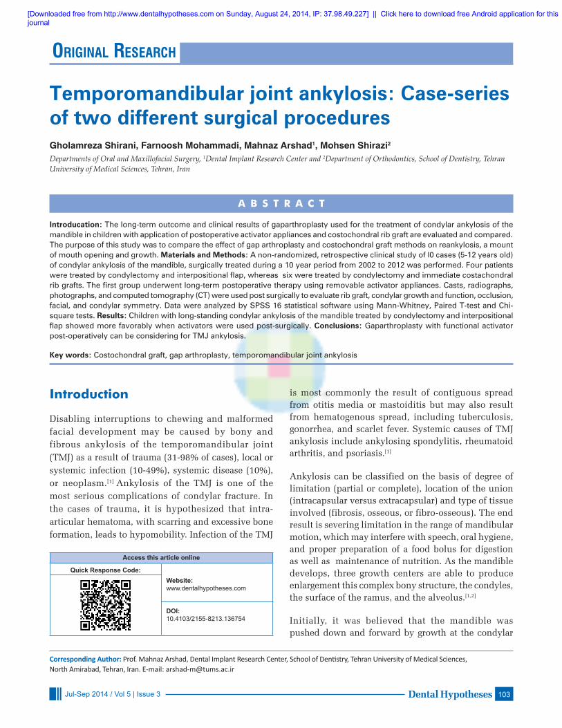
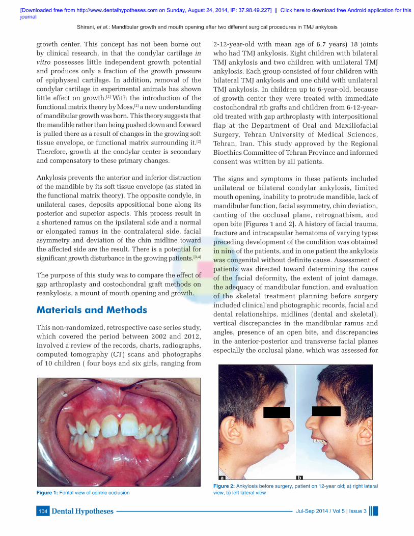
The signs and symptoms in these patients included unilateral or bilateral condylar ankylosis, limited mouth opening, inability to protrude mandible, lack of mandibular function, facial asymmetry, chin deviation, canting of the occlusal plane, retrognathism, and open bite [Figures 1 and 2]. A history of facial trauma, fracture and intracapsular hematoma of varying types preceding development of the condition was obtained in nine of the patients, and in one patient the ankylosis was congenital without definite cause. Assessment of patients was directed toward determining the cause of the facial deformity, the extent of joint damage, the adequacy of mandibular function, and evaluation of the skeletal treatment planning before surgery included clinical and photographic records, facial and dental relationships, midlines (dental and skeletal), vertical discrepancies in the mandibular ramus and angles, presence of an open bite, and discrepancies in the anterior-posterior and transverse facial planes especially the occlusal plane, which was assessed for
Figure 1: Fontal view of centric occlusionFigure 2: Ankylosis before surgery, patient on 12-year old; a) right lateral view, b) left lateral view
a b
[Downloaded free from http://www.dentalhypotheses.com on Sunday, August 24, 2014, IP: 37.98.49.227] || Click here to download free Android application for thisjournal
Shirani, et al.: Mandibular growth and mouth opening after two different surgical procedures in TMJ ankylosis
105Dental HypothesesJul-Sep 2014 / Vol 5 | Issue 3
transverse tilting and occlusal slanting. Radiographs and CT scans were evaluated for the extent of callous formation, condylar deformity, and length of the mandibular body and ramus on the right and left sides.
The radiographs were also used for the purpose of tracing and determining discrepancies contributing to the skeletal asymmetry such as ramus height, as well as assessment of extent of deviation of the dental and skeletal midlines relative to the midsagital plane.
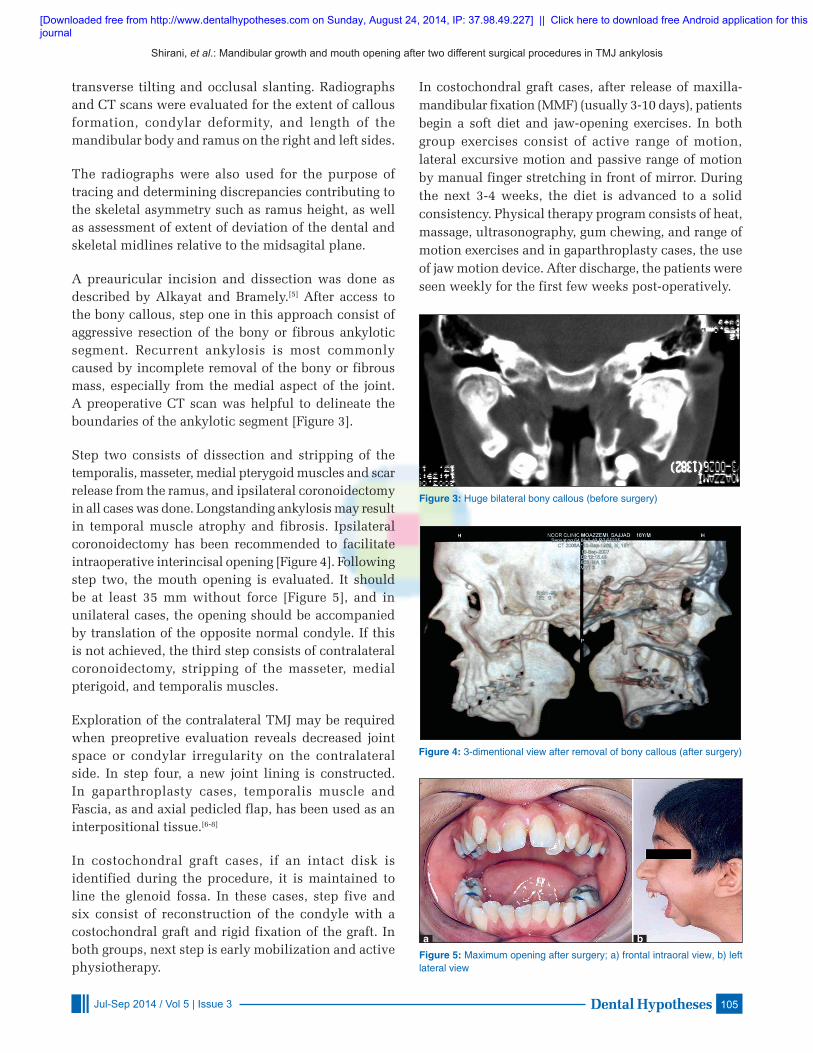
A preauricular incision and dissection was done as described by Alkayat and Bramely.[5] After access to the bony callous, step one in this approach consist of aggressive resection of the bony or fibrous ankylotic segment. Recurrent ankylosis is most commonly caused by incomplete removal of the bony or fibrous mass, especially from the medial aspect of the joint. A preoperative CT scan was helpful to delineate the boundaries of the ankylotic segment [Figure 3].
Step two consists of dissection and stripping of the temporalis, masseter, medial pterygoid muscles and scar release from the ramus, and ipsilateral coronoidectomy in all cases was done. Longstanding ankylosis may result in temporal muscle atrophy and fibrosis. Ipsilateral coronoidectomy has been recommended to facilitate intraoperative interincisal opening [Figure 4]. Following step two, the mouth opening is evaluated. It should be at least 35 mm without force [Figure 5], and in unilateral cases, the opening should be accompanied by translation of the opposite normal condyle. If this is not achieved, the third step consists of contralateral coronoidectomy, stripping of the masseter, medial pterigoid, and temporalis muscles.
Exploration of the contralateral TMJ may be required when preopretive evaluation reveals decreased joint space or condylar irregularity on the contralateral side. In step four, a new joint lining is constructed. In gaparthroplasty cases, temporalis muscle and Fascia, as and axial pedicled flap, has been used as an interpositional tissue.[6-8]
In costochondral graft cases, if an intact disk is identified during the procedure, it is maintained to line the glenoid fossa. In these cases, step five and six consist of reconstruction of the condyle with a costochondral graft and rigid fixation of the graft. In both groups, next step is early mobilization and active physiotherapy.
In costochondral graft cases, after release of maxilla-mandibular fixation (MMF) (usually 3-10 days), patients begin a soft diet and jaw-opening exercises. In both group exercises consist of active range of motion, lateral excursive motion and passive range of motion by manual finger stretching in front of mirror. During the next 3-4 weeks, the diet is advanced to a solid consistency. Physical therapy program consists of heat, massage, ultrasonography, gum chewing, and range of motion exercises and in gaparthroplasty cases, the use of jaw motion device. After discharge, the patients were seen weekly for the first few weeks post-operatively.
Figure 3: Huge bilateral bony callous (before surgery)
Figure 4: 3-dimentional view after removal of bony callous (after surgery)
Figure 5: Maximum opening after surgery; a) frontal intraoral view, b) left lateral view
a b
[Downloaded free from http://www.dentalhypotheses.com on Sunday, August 24, 2014, IP: 37.98.49.227] || Click here to download free Android application for thisjournal
Shirani, et al.: Mandibular growth and mouth opening after two different surgical procedures in TMJ ankylosis
106 Dental Hypotheses Jul-Sep 2014 / Vol 5 | Issue 3
In gaparthroplasty group, the functional device is constructed for muscle training according to functional matrix hypothesis. This device was activator appliance made of acrylic resin (Luxatemp; DGM, Hamburg, Germany) and stainless steel orthodontic wire for correction of open bite and mouth opening and closing pathway.
In unilateral gaparthroplasty group at each visit 1-2 mm of acrylic resin was removed from the superior inner part of the acrylic functional appliance on the ipsilateral side, using acrylic burs to allow for eruption of the teeth on that side and gradual maxillary compensation for the occlusal canting to occur. In bilateral group acrylic resin was removed from both sides. Chin deviation was also addressed by adjustment of the appliance in such a way that the patient would close the jaw in midline. The activator appliance was retained until correction of the deformity and restoration of symmetry and occlusion. During this period, new appliances need to be constructed as growth progresses. In bilateral cases, the same procedure was used except that acrylic was removed bilaterally from the superior inner portions of appliance to allow the eruption of the posterior teeth bilaterally. The patients are followed closely until completion of growth (post-puberty) and then periodically, because after completion of growth there is usually less chance for development of future occlusal or facial disharmony [Figure 6].
Data were analyzed by SPSS 16 statistical software. Mann-Whitney test was utilized to compare the extent of mouth opening after surgery using the two methods. Paired samples T-test was utilized to investigate and find a relationship between the extent of mouth opening before and after surgery in each method. Chi-square test was used to determine which surgery method would result in higher re-ankylosis. P-value less than 0.05 considered as significant.
Results
The mandibular growth was symmetric in bilateral ankylosis although in gaparthroplasty group was more than osteochondral group. In unilateral ankylosis, the facial and mandibular asymmetry improved after gaparthroplasty with appliance.
The average of maximum mouth opening (MIO) in gaparthroplasty technique increased from 7-32.7 mm and in osteochondral technique from 8-20.7 mm. MIO increased significantly more in gaparthroplasty group (P < 0.05).
In 8 cases with bilateral ankylosis, gonion and gnation (Go-Gn) difference with both side had no change after 1 year. In 2 cases with unilateral ankylosis, right and left Go-Gn difference was 8 mm pre-operatively and decreased to 4 mm, 1 year post-operatively. Go-Gn changes represents mandibular growth amount. An S-Gn change was more in gaparthroplasty with appliance group.
Mouth opening was significantly higher with gaparthroplasty than chostocondral graft (Mann-whitney, P = 0.055). The average mouth opening before surgery was 7.6 mm, which was increased to 25.5 mm after surgery. In gap arthroplasty method, the average of 7 mm before surgery was increased to 32.7 mm after the surgery. In costochondral graft method, the average of 8 mm before surgery was increased to 20.7 after surgery.
With regard to the symmetry of mandible growth in eight cases with two-sided ankylosis there was no difference in the length of left and right mandibles before and after the surgery. Therefore, the symmetry was maintained. However, in two cases with one-sided ankylosis such symmetry was not observed. In one of such cases left and right Go-Gn difference was 7 mm before the surgery, which was decreased to 3 mm one year after the surgery. In the other case, the difference was 9 mm which was decreased to 5 mm after one year.
The extent of mouth opening after surgery was significantly higher in gap arthroplasty method compared to costochondral graft method (Mann-Whitney, P = 0.055).
There was no significant relationship between re-ankylosis occurance and the surgery method (Chi-
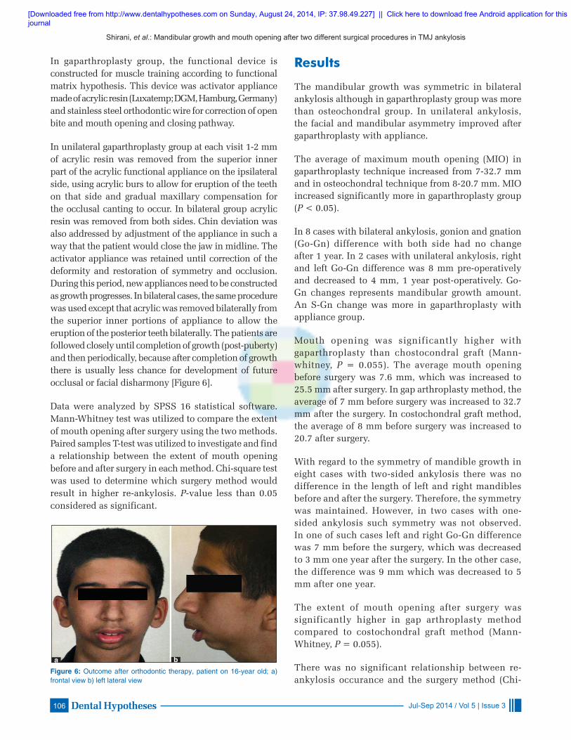
Figure 6: Outcome after orthodontic therapy, patient on 16-year old; a) frontal view b) left lateral view
a b
[Downloaded free from http://www.dentalhypotheses.com on Sunday, August 24, 2014, IP: 37.98.49.227] || Click here to download free Android application for thisjournal
Shirani, et al.: Mandibular growth and mouth opening after two different surgical procedures in TMJ ankylosis
107Dental HypothesesJul-Sep 2014 / Vol 5 | Issue 3
Square, P > 0.05), which might be due to the small sample size. However, further evaluations indicated that four cases out of six that went under costochondral graft method resulted in re-ankylosis with MIO of less than 25 mm after a year. However, none of the four cases that went under gap arthroplasty method resulted in re-ankylosis.
There was no significant relationship found between the extent of mouth opening before the surgery and re-ankylosis (P > 0.05). This means that there is no relationship between the low mouth opening extents before the surgery and the likelihood of re-ankylosis after the surgery.
Since there were only two cases with one-sided ankylosis, the sample size for such cases was not large enough for any statistically significant conclusion.
The increase in the distance from Go-Gn to Sella turcica was studied (gap arthroplasty and costochondral graft). This increase was significantly higher in gap arthroplasty compared to costochondral graft (P < 0.05). This indicates that gap arthroplasty method is more effective in causing mandible growth.
Discussion
The method chosen for treatment of TMJ ankylosis must not only provide a functional joint but also help restore facial symmetry. In children there is an additional requirement of allowing facial growth to proceed normally.[4,9-11]
Several study have shown that in children costochondral graft have the potential to grow, however, this factor alone does not mean that growth will always proceed normally, growth of these grafts is unpredictable, ranging from resorption to overgrowth necessitating secondary surgical procedures.[12-15] Unfortunately, uncontrolled and often excessive growth leads to asymmetry in facial profile and overgrowth in TMJ region which was also noted in these cases.
Fundamental research on the growth potential at the normal mandibular condyle suggest that the velocity of this growth is governed by a combination of intrinsic (cell-derived) and local extrinsic (environment modified) factors.[9-16]
The clinical findings in our cases supported the fact that external factors such as activators and jaw function
may indeed influence and guide mandibular growth after gaparthroplasty, and that these appliances may play a role in allowing favorable results to be obtained. Despite the fact that the extrinsic mechanisms affecting growth are unknown the role of activator appliances in effectively guiding and stimulating mandibular growth, coordinationg the arches, and correcting chin deviation is well-known. Using activator device also eliminate to need for additional surgery procedure, complication and risk of it.[8] Our Results showed that, gaparthroplasty with postoperative functional activator has better outcome than costochondral graft in cases of TMJ ankylosis.
This accords well with the soft tissue functional matrix theory that suggests the role of the soft tissue in mandibular growth and also suggests that functional matrix is more important than the condylar growth center when considering mandibular growth[2], present study would address the precise role of activator appliances and help the profession decide whether it is the activator alone that produces favorable results and prevents reankylosis, or if other factors are involved. Genetic factors may be influential in reankylosis, and growth pattern.
The clinical criteria used for postoperative evaluation of the patients included assessment of mouth opening, evaluation of the occlusion, occlusal canting, and facial symmetry. Paraclinical evaluations included CT scan and plain radiographs to determine graft position, joint form, function and symmetry. In all cases that were treated post-surgically with activator appliances, we were able to follow-up to date which made it possible to obtain a more than satisfactory occlusion and acceptable facial symmetry aa well as mandibular function.
Occlusal canting and dental midline relationships were also corrected in this group. The six patients who did not undergo appliance therapy postoperatively developed mechanisms affecting growth that are unknown.
In conclusion, gaparthroplasty with functional activator postopervatively has better outcome than costochondral grafts in cases of TMJ ankylosis.
References
1. Rowe NL. Ankylosis of the temporomandibular joint. Part 3. J R Coll Surg Edinb 1982;27:209-18.
2. Moss ML, Salentijn L. The primary role of functional matrices in facial growth. Am J Orthod 1969;55:566-77.
[Downloaded free from http://www.dentalhypotheses.com on Sunday, August 24, 2014, IP: 37.98.49.227] || Click here to download free Android application for thisjournal

Shirani, et al.: Mandibular growth and mouth opening after two different surgical procedures in TMJ ankylosis
108 Dental Hypotheses Jul-Sep 2014 / Vol 5 | Issue 3
3. Kent IN, Misiek DJ. Oral and maxillofacial Trauma. Vol 2. Philadelphia: Saunders, 1991.
4. Munro IR, Chen YR, Park BY. Simultaneous total correction of temporomandibular ankylosis and facial asymmetry. Plast Reconstr Surg 1986;77:517-29.
5. Fonseca. Oral and maxillofacial surgery. Vol 4. St Louise: Saunders; 2000.
6. Kent IN, Misiek. DJ, Akin RK, Hinds EC, Homsy CA. Tempromandibular joint condylar prosthesis: A ten year report. J Oral Maxillofac Surg 1983;41:245-54.
7. Guralnick WC, Kaban LB. Surgical treatment of mandibular hypomobility. J Oral Surg 1976;34:343-8.
8. Silvestri A, Natali G, Jnnetti G. Functional therapy in hemifacial microsomia: Therapeutic protocol for growing children. J Oral Maxillofac Surg 1996;54:271-8.
9. Kaban LB. Acquired abnormalities of the temporomandibulajoint, In: Kaban LB. Pediatric oral and Maxillofacial Surgery. Philadelphia: Saunders; 1990.
10. Bell WH, Profit WR, White RP. Surgical correction of dentofacial deformities. Vol. 2. Philadelphia: Saunders; 1980.
11. Lindqvist C, Pihakari A, Tasanen A, Hampf G. Autogenous costochondral grafts in temporo-mandibular joint arthroplasty.
A survey of 66 arthroplasties in 60 patients. J Maxillofac Surg 1986;14:143-9.
12. MacIntosh RB, Henny FA. A spectrum of application of autogenous costochondral grafts. J Maxillofac Surg 1977;5:257-67.
13. Heffez L, Doka HC. The Goldenhar syndrome: Diagnosis and early surgical management. Oral Surg Oral Med Oral Pathol 1984;58:2-9.
14. Munro IR, Phillips JH, Griffin G. Growth after construction of the temporomandibular joint in children with hemifacial microsomia. Cleft Palate J 1989;26:303-11.
15. Fukata K, Jackson IT, Topf JS. Facial lawn mower injury treated by a vascularized costochondral graft. J Oral Maxillofac Surg 1992;50:194-8.
16. Petrovic A, Stutzmann J, Oudet C. Condylectomy and mandibular growth in young rats. A quantitative study. Proc Finn Dent Soc 1981;77:139-50.
Cite this article as: Shirani G, Mohammadi F, Arshad M, Shirazi M. Temporomandibular joint ankylosis: Case-series of two different surgical procedures. Dent Hypotheses 2014;5:103-8.
Source of Support: Nil, Conflict of Interest: None declared.
[Downloaded free from http://www.dentalhypotheses.com on Sunday, August 24, 2014, IP: 37.98.49.227] || Click here to download free Android application for thisjournal