Transposition Evidence Mechanisms: DNA-mediated RNA-mediated.

Temporal Cell-mediated Immune Responses of CattleFollowing Experimental and NaturalExposure to Living Brucella abortus
J. M. B. Kaneene, R. D. Angus, D. W. Johnson,C. C. Muscoplat, R. K. Anderson and D. E. Pietz*
ABSTRACT
A study on cell-mediated immune responsesin cattle with different exposure experiencesto Brucella abortus was conducted by an invitro lymphocyte stimulation assay. The pur-pose of this study was to determine how soonthe cell-mediated immune responses would bedetected following experimental exposure toB. abortus and to study the cell-mediatedimmune trend following experimental andnatural exposure of cattle to B. abortus. Thefirst positive cell-mediated immune responsesoccurred one to two weeks after experimentalinoculation with living B. abortus strain 2308.The cell-mediated immune responses in theseanimals appeared at least one week beforethe appearance of B. abortus serum agglu-tinating antibodies. Animals which werenaturally infected with B. abortus biotypes 1and 2 demonstrated positive cell-mediatedimmune responses throughout the study.
ReSUMe
Cette experience visait 'a utiliser uneepreuve de stimulation des lymphocytes invitro, pour etudier l'immunite cellulaire chezdes bovins soumis a diverses infections expe-rimentales avec Brucella abortus. On espe-rait ainsi determiner la rapidite avec la-quelle on pouvait deceler les resultats de l'im-munite cellulaire, a la suite d'une infectionexperimentale avec B. abortus, ainsi que le
'Department of Large Animal Clinical Sciences, Collegeof Veterinary Medicine, University of Minnesota, St.Paul, Minnesota 55108 (Kaneene, Johnson and Musco-plat), National Animal Disease Center, VeterinaryServices Laboratories, Ames, Iowa 50010 (Angus andPietz) and Division of Epidemiology, School of PublicHealth, University of Minnesota, Minneapolis, Min-nesota 55455 (Anderson).Submitted July 17, 1978.
profil de cette immunite, a la suite d'une in-fection naturelle ou experimentale des bovinspar ce microbe. On obtint les premiers resul-tats positifs de l'immunite cellulaire, envi-ron une a deux semaines apres l'inoculationexperimentale de la souche vivante 2308 deB. abortus, i.e. au moins une semaine avantl'apparition des anticorps agglutinants seri-ques. Les animaux atteints d'une infectionnaturelle attribuable aux biotypes 1 et 2 deB. abortus demontrerent une reaction posi-tive d'immunite cellulaire, tout au long de1'experience.
INTRODUCTION
In our earlier studies, we reported thatlymphocytes from Brucella abortus infect-ed cattle (2-4) and Brucella suis infectedpigs (5) exhibited positive in vitro lym-phocyte stimulation responses when cul-tured with brucella antigens. Those ini-tial studies were primarily directed atdetermination of a positive response anddid not determine the duration of theresponse nor was it determined how soonafter infecting cattle or pigs could thecell-mediated immune (CMI) response bedetected by the lymphocyte stimulationtest (LST). Thus, there was interest tofollow infected animals and study thetrend of CMI responses over a period oftime. It was also desirable to determinehow soon after infection with B. abortus,the CMI response could be detected by thein vitro LST and to study the trend ofCMI thereafter. This experiment, there-fore, was designed to: i) follow the trendsof CMI responses in naturally infectedanimals, ii) experimentally inoculatecattle with living B. abortus to determine
Can. J. comp. Med.132

TABLE I. Experimental Groups of Animals in the Study Arranged According to Type of Exposureto B. dbortus
Age WhenExperimentally Brucella Brucella
Animal Animal Infected With Exposure IsolationGroup No. B. abortus Sex Status Results
Experimental1ba 15-16 months F Inoculation Neg.26 " M " "
Ib 43a ssF49 " M50 " F51 " M
Natural21 Not applicable F Infection -Pos.51 " F " "
II 57 " F "t74 " F " "
Not891 Not applicable F Nonexposed applicable892 " F " "893 " F " "894 " F " "
III 895 " F "900 M901 " F902 " F903 " M904 F
&Calfhood vaccinatedbEach animal in this group was inoculated with 2.86
how soon the CMI responses would be de-tected and study the trend of CMI re-sponses thereafter.
MATERIALS AND METHODS
ANIMAL HISTORY
Dairy cattle were used in this study andinformation on each animal is given(Table I). Animals in group I were keptat the National Animal Disease Labora-tories (NADL), Ames, Iowa, in separateunits. Animals no. 10 and 43 were vac-cinated with 5 ml of B. abortus strain 19vaccine subcutaneously (approximately5-15 x 109 cells/ml) at the age of threemonths. Animals in group I were pur-chased from the Tuberculosis School atAmes and had been exposed previously toMycobacterium five months prior to ino-culation of live B. abortus strain 2308.Animals 10, 26 and 43 were each inoculat-ed with 0.1 mg of live M. avium strain D4intratracheally. Animals 49, 50 and 51
X 106 live B. abortus strain 2308 conjunctively
were sensitized to M. bovis by inoculating(intradermally) 0.1 ml of Freund's in-complete adjuvant which had 1 mg/ml ofheat killed M. bovis. All the six animals ingroup I were inoculated with 0.5 ml (2.86x 10⁶ cells) of live B. abortus strain 2308conjunctively at the age of 16 months, fivemonths after exposure to Mycobacterium.To prove that the animals in group I haddeveloped delayed hypersensitivity, theywere skin tested using M. bovis and M.arium antigens. Secondly, lymphocyte sti-mulation test was conducted on peri-pheral blood lymphocytes from these ani-mals using M. bovis and M. avium purifiedprotein derivatives. These animals dev-eloped typical delayed hypersensitivityskin reactions and positive lymphocytestimulation responses.
PREPARATION OF LYMPHOCYTE SUSPENSION
Following collection, heparinized bloodwas subjected to the following procedures:The lymphocytes were purified accordingto the Ficoll-diatrizoate (FD) techniquepreviously described (2). Briefly, a FD
Volume43- April, 1979 133

solution' of specific gravity 1.077 wasused. Heparinized blood was mixed withan equal volume of 0.15 M NaCl solu-tion and this mixture was subsequentlylayered over the FD solution, using asterile needle and syringe. Approximately10 ml of diluted blood was layered over5 to 6 ml of FD in 18 ml tubes fitted withplastic covers. All samples were centri-fuged for 30 to 45 minutes with a forceof 400 g delivered at the FD-blood inter-face. The separation procedure was doneat ambient room temparture.Lymphocyte populations collected in
this manner had greater than 95% puremononuclear cells and a cell viability of99% or more, as determined by the trypanblue exclusion method. The lymphocyteswere washed twice in Hanks' balancedsalt solution (HBSS) and resuspended inRPMI-16402 complete tissue culture me-dium. Cell counts were obtained, using ahemacytometer and cell concentrationswere adjusted to 1.5 x 10 cells/ml.
CULTURE MEDIUM, MITOGEN AND ANTIGEN
Tissue culture medium was RPMI-16402supplemented with 25 ml of Hepes buffer,penicillin (100 U/ml), streptomycin (100,ug/ml) and bovine fetal serum3 (BFS;15% v/v). Concanavalin A (Con A)4 at aconcentration of 2 ug/ culture was usedas a positive control. A B. abortus-solubleantigen (BASA) prepared from cell ofB. abortus strain 1119-3 was used at aconcentration of 4.4 ,ig of protein/culture.The preparation of this antigen and someof the chemical analyses have been de-scribed (2).
CELL CULTURES
After cell dilutions were made, suspen-sions of cells from each animal (200 ,ul)were added (in triplicate) to wells of amicrotitration culture plate by using anautomatic dispenser. Con A were added toappropriate wells of microtitration cul-ture plates. As negative controls, tripli-cate wells for each animal were set upwith lymphocyte suspension but neither
lPharmacia Fine Chemicals, Piweataway, New Jersey.2Biolabs, Chicago, Illinois.3Gibco, Grand Island, New York.4Miles-Yeda, Rehovot, Israel.
Con A nor BASA was added. All cultureswere incubated for six days at 37°C in anincubator with a 5% C02-humidified airatmosphere. Approximately 16 to 18 hoursbefore termination, 1 ,uCi of methyl-3-Hthymidine5 (3HdR, 6.7 Ci/mmole) wasadded to each well. Cultures were termi-nated by cooling to 4°C and then the cellswere harvested for liquid scintillationcounting, as previously described (2).Animals in group II (Table I) were pur-chased from four different herds. B.abortus had been isolated from each ofthese herds prior to purchase of theseanimals. B. abortus was isolated repeat-edly from the milk, and from tissues afterslaughter, of each of the four animals ingroup II.Animals in group II were kept in sep-
arate isolation units of the College ofVeterinary Medicine, University of Min-nesota (U of M), while those in group IIIwere kept in a brucellosis-free herd ofthe Department of Animal Science, U ofM. All animals were bled once a week fora period of 18 to 24 weeks. Approximately30 ml of blood were collected by jugularvenipuncture from each animal. Twentyml of each blood sample were put intosterile tubes containing heparin' (50units/ml) and serum was recovered fromthe remaining 10 ml.
SEROLOGICAL TESTS
All sera collected were subjected to thefollowing tests: standard plate (SPT) andtube (STT) agglutination tests, bufferedbrucella antigen test (Card), Rivanol pre-cipitation-plate agglutination test (Riv),2-mercapto-ethanol tube test (2 ME) andacidified plate antigen test (APA) sup-plemental tests. These tests were con-ducted according to United States Depart-ment of Agriculture procedures (14).However, only the results of the STT arereported.
Brucella abortus ISOLATION ATTEMPTS
At the end of the experiment the ani-mals were slaughtered and the followingtissues were collected for isolation of
5Schwartz/Mann, Orangesburg, New York.
6Upjohn Company, Kalamazoo, Michigan.
Can. J. comp. Med.134
Brucella abortus: supramammary, mandi-bular, retropharyngeal, gastrohepatic, in-ternal and external iliacs and lumbarlymph nodes; spleen, udder tissue, part ofthe uterus, fetal membranes, testicles,liver, heart and bone marrow. The labo-ratory procedures used were those re-commended by United States Departmentof Agriculture (15).
RESULTS
EXPRESSION OF LYMPHOCYTESTIMULATION RESULTS
These are expressed in two ways: i)difference in counts per minute (ACPM)= mean cpm of triplicate cultures withouteither Con A or BASA subtracted frommean cpm of triplicate cultures with ConA or BASA, ii) stimulation index (SI) =mean cpm of triplicate culture with ConA or BASA divided by mean cpm of tri-plicate culture without either Con A orBASA.
LYMPHOCYTE STIMULATION
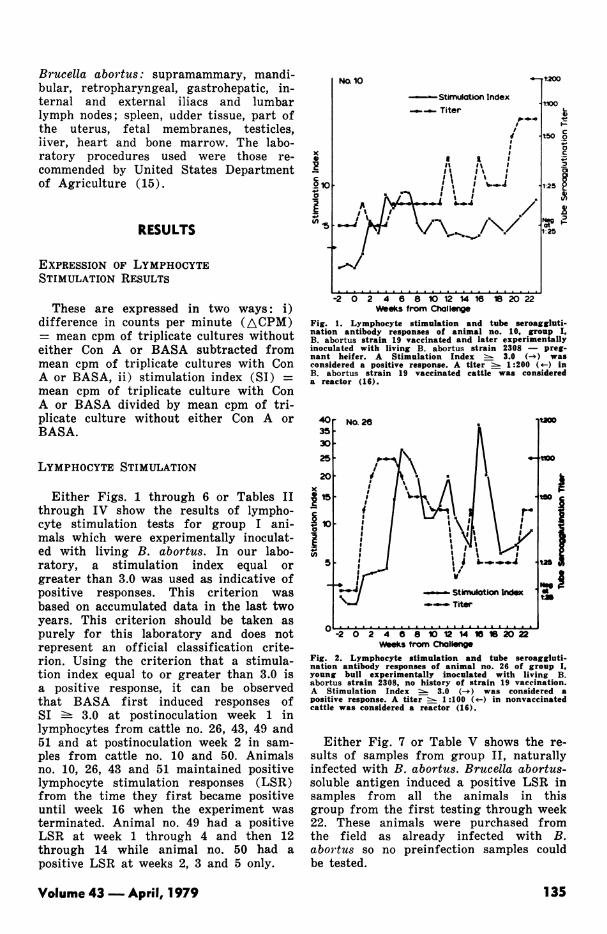
Either Figs. 1 through 6 or Tables IIthrough IV show the results of lympho-cyte stimulation tests for group I ani-mals which were experimentally inoculat-ed with living B. abortus. In our labo-ratory, a stimulation index equal orgreater than 3.0 was used as indicative ofpositive responses. This criterion wasbased on accumulated data in the last twoyears. This criterion should be taken aspurely for this laboratory and does notrepresent an official classification crite-rion. Using the criterion that a stimula-tion index equal to or greater than 3.0 isa positive response, it can be observedthat BASA first induced responses ofSI - 3.0 at postinoculation week 1 inlymphocytes from cattle no. 26, 43, 49 and51 and at postinoculation week 2 in sam-ples from cattle no. 10 and 50. Animalsno. 10, 26, 43 and 51 maintained positivelymphocyte stimulation responses (LSR)from the time they first became positiveuntil week 16 when the experiment wasterminated. Animal no. 49 had a positiveLSR at week 1 through 4 and then 12through 14 while animal no. 50 had apositive LSR at weeks 2, 3 and 5 only.
'C0
S~100
in.5
No. 10
Stimulation Index* _ Titer
I
t200
t,oo_t
at5 0o_._
tc
1:25 1
Ng3-1-25
-2 0 2 4 6 8 10 12 14 16 20 22Weeks from Challenge
Fig. 1. Lymphocyte stimulation and tube seroaggluti-nation antibody responses of animal no. 10, group I,B. abortus strain 19 vaccinated and later experimentallyinoculated with living B. abortus strain 2308 - preg-nant heifer. A Stimulation Index s 3.0 (-3) wasconsidered a positive response. A titer : 1:200 (4--) inB. abortus strain 19 vaccinated cattle was considereda reactor (16).
403530
25
20x
15
~10
U)
5,
No.26
PI I\"
.O ' a'
II i
II %.. i ~
I I'h---sI~~~~~~
II - StInuItlonI --- Titer
t2s aA
usmat
V-2 0 2 4 8 8 10 12 14 1 1 20 22Weeks trom Challeg
Fig. 2. Lymphocyte stimulation and tube seroaggluti-nation antibody responses of animal no. 26 of group I,young bull experimentally inoculated with living B.abortus strain 2308, no history of strain 19 vaccination.A Stimulation Index -- 3.0 (-e) was considered apositive response. A titer :- 1 :100 (4-) in nonvaccinatedcattle was considered a reactor (16).
Either Fig. 7 or Table V shows the re-sults of samples from group II, naturallyinfected with B. abortus. Brucella abortus-soluble antigen induced a positive LSR insamples from all the animals in thisgroup from the first testing through week22. These animals were purchased fromthe field as already infected with B.abortus so no preinfection samples couldbe tested.
Volume 43- April, 1979
. . . . . . . . . . . . . . . . . . . . . . . .
n -. . . . . . . . . . . . . . . ......
135

x0
° 10c
a
U,
OCWeeks from Challenge
Fig. 3. Lymphocyte stimulation and tube seroagglutina-tion antibody responses of animal no. 43, group I,B. abortus strain 19 vaccinated as a calf and laterexperimentally inoculated with living B. abortus strain2308 - nonpregnant heifer. A Stimulation Index!. 3.0 (-e) was considered a positive response. A titer=- 1:200 (-v) in B. abortus strain 19 vaccinated cattlewas considered a reactor (16).
No. 50
---Stimulation Index--- Titer
4.-
/1'l~~~~~
-2 0 2 4 6 8 10 12 14 16Weeks from Challenge
1:200
-1:100 0I--c.°
t50 a
0
._
0L
1:25 3
Neg 'Fat1:25
Fig. 5. Lymphocyte stimulation and tube seroagglutina-tion responses of animal no. 50, group I, experimentallyinoculated with living B. abortus strain 2308 - non-pregnant heifer with no history of strain 19 vaccina-tion. A Stimulation Index s 3.0 (-*) was considereda positive response. A titer : 1:200 (+-) in B. abortusstrain 19 vaccinated cattle was considered a reactor(16).
No. 49 Stimulation Index--- Titer
11:200
41100
r at
K.-.a~~~~~t°°°AIE°° .. .
_"E~~~~~~~--
1:50
1:25
Negat1:25
-2 0 2 4 6 8 10 12 14 16Weeks from Challenge
Fig. 4. Lymphocyte stimulation and tube seroagglutina-tion antibody responses of animal no. 49, group I,young bull experimentally inoculated with living B.abortus strain 2308 - no history of strain 19 vaccina-tion. A Stimulation Index 3.0 (-e) was considered
a positive response. A titer 1:200 (<-) in B. abortus
strain 19 vaccinated cattle was considered a reactor(16).
Results of samples from animals ingroup III, nonexposed controls are shown(Fig. 7 or Table VI). The data show thatBASA did not induce a positive LSR in
50.L 450 40 .
35 -
C
o 30 .
c 25 .CD
aL 15
0 4n in10.
ol ..' . . . . . . .
No. 51
4.i
---- Stimulation Index
Titer
t200
*t100kXI.-c0
150 g
t251
-
_- I.Negatt25
-2 0 2 4 6 8 10 12 14 16Weeks from Challenge
Fig. 6. Lymphocyte stimulation and tube seroagglutina-tion antibody responses of animal no. 51, group I,young bull experimentally inoculated with living Babortus strain 2308 - no history of strain 19 vaccina-tion.
samples from these animals.Concanavalin A induced significant
LSR in samples from all animals in thethree groups (Figs. 1 through 7 and
Can. J. comp. Med.
x
.24 10
E(.)
Dnoo
136

TABLE II. Lymphocyte Stimulation Induced by Con A and BASA in Lymphocytes from Cattleno. 10 and 43 of Group I
MitogenWeeks from and Lowest and Mean ACPM
Animal No. Challenge Antigen Highest ACPM ± SEM- 2 - Ob Con A 99,443 - 422,059 219,109 & 102,016
BASA 509-1,390 853 271
1 - 3 Con A 192,083 - 305,391 235,281 i 48,422BASA 431 - 1,512 977 + 187
10 ....... ........4-8 Con A 108,595 - 205,983 137,902 ± 29,955BASA 1,127 - 4,953 2,316 -+ 387
9 - 18 Con A 25,819 - 151,665 108,416 + 23,240BASA 1,001 - 2,588 817 + 162
19 - 22 Con A 113,678 - 213,167 167,423 + 24,172BASA 1,647 - 2,910 2,043 -+1 293
- 2 - 0 Con A 71,181 - 224,318 167,396 i 15,617BASA 2,086 - 4,530 961 + 759
1 - 6 Con A 121,645 - 240,783 172,897 -i 17,786BASA 553 - 1,083 2,961 + 476
43...................7 - 12 Con A 120,647 - 301,332 168,993 16,975
BASA 792 - 2,001 1,372 -+- 342
13 - 16 Con A 158,314 - 183,545 169,851 + 14,911BASA 600 - 2,637 1,088 + 301
&Experimentally inoculated with living B. abortus strain 2308bOne sample from every animal was tested every week, postinoculation. However, ACPM obtained eachweek are not presented. Instead, mean ACPM values of weeks, as specified in the left hand column, aregiven.
Tables II through VI) which indicatesthat the lymphocytes were immunolo-gically responding.
RESULTS OF B. abortus ISOLATIONATTEMPTS
Attempts to isolate B. abortus fromanimals in group I, experimentally ex-posed to B. abortus were unsuccessful.Among group II, naturally infected ani-mals, B. abortus biotype 1 was isolatedrepeatedly from the milk of cow no. 21and from tissues (after slaughter) fromcows no. 21, 51 and 74. Brucella abortusbiotype 2 was isolated repeatedly fromthe milk of cow no. 57. No isolation at-tempts were made in group III animals,since they were not exposed to B. abortus.
RESULTS OF SERUM TUBEAGGLUTINATION TEST
According to the United States Depart-ment of Agriculture procedures (16) an
officially vaccinated bovine animal witheither a plate or tube seroagglutinationtiter equal to or greater than 1:200 is anofficial "reactor", while a nonvaccinatedbovine animal with a titer equal to orgreater than 1:100 is an official "reac-tor". Animals no. 43, 49 and 50 of groupI did not develop any detectable sero-agglutination antibodies (Figs. 3, 4 and5). Animal no. 10 first developed a detect-able (1:25 dilution) seroagglutinationantibody titer on week 12 and did notdevelop to "reactor" level (Fig. 1). Ani-mal 26 (Fig. 2) first developed a detect-able titer by week 1 and reached a "reac-tor" level by week 3 postinoculation. Ani-mal no. 51 (Fig. 6) first developed adetectable titer by week 3 and reacheda "reactor" level by week 4 postinocula-tion. All animals in group II (where B.abortus field strain was isolated) hadtiter above the "reactor" level (1:100)throughout the project. There was nodetectable seroagglutination titers (1:25dilution) in sera from any of the animalsin group III (Brucella nonexposed con-trols Fig. 7).
Volume 43- April, 1979 137

TABLE III. Lymphocyte Stimulation Induced by Con A and BASA in Lymphocytes from Cattleno. 26 and 49 of Group 1a
WCCAnimal No.
26...................
Mitogeneeks from and:hallenge Antigen
2 - Ob Con ABASA
1 - 12 Con ABASA
13 - 18 Con ABASA
19 - 22 Con ABASA
Lowest andHighest ACPM
129,108 - 262,948553 - 1,375
118,062 - 176,647418 - 8,227
90,194 - 289,798968 - 22,267
52,690 - 143,913598 - 1,127
Mean ACPMi SEM
203,835 i1: 39,417903 4 245
148 051 ±i 19,8393,575 :+1 1,172
181,135 :+1 16,8716,355 i1: 1,791
83,318 + 17,403791 -i- 464
- 2 - 0 Con A 68,801 -443,268BASA 492 - 1,409
49...................
0 - 4 ConABASA
5 - 11 Con ABASA
163,227 - 172,3741,187 - 2,743
30,477 - 208,750104 - 759
12 - 16 Con A 115,012 - 199,148BASA 451 - 1,327
250,680 i 108,231950 ±fi 458
167,711 i 44,0331 846 ±fi 365
119,613 i 10,094408 :1i 301
145,427 11,0321,172 i1: 218
*Experimentally inoculated with living B. abortus strain 2308bOne sample from every animal was tested every week, postinoculation. However, ACPM obtained eachweek are not presented. Instead, mean ACPM values of weeks, as specified in the left hand column, aregiven
TABLE IV. Lymphocyte Stimulation Induced by Con A and BASA in Lymphocytes from Cattleno. 50 and 51 of Group Ia
We(ChAnimal No.
50...................
Mitogeneks from andallenge Antigen
2 - Ob Con ABASA
1 - 6 Con ABASA
7 - 16 Con ABASA
Lowest andHighest ACPM
107,505 - 254,278161 - 3,039
101,546 - 231,513907 - 4,222
51,895 - 183,467191 - 1,504
Mean ACPMi SEM
166,536 i 20,5022,105 :i 629
157,254 :i 20,1651,360 522
132,111 17,296641 -i± 356
- 2 - 0
51...................
Con A 117,148 - 191,525BASA 1,747 - 19,627
1 - 5 Con A 68,628- 117,589BASA 3,388 - 4,192
6 - 10 Con A 56,988 -133,489BASA 3,292 - 13,183
11 - 16 Con A 57,582 - 333,703BASA 1,700 - 6,181
152,483 21,5506,684 2,280
90,449 15,9033,539 1,147
108,670 :i: 11,53411,725 2,518
156,918 19,2083,314 2,025
-Experimentally inoculated with living B. abortus strain 2308bOne sample from every animal was tested every week, postinoculation. However, ACPM obtained eachweek are not presented. Instead, mean ACPM values of weeks, as specified in the left hand column, aregiven
Can. J. comp. Med.138

DISCUSSION
In group I animals, experimentally in-oculated with living B. abortus strain2308, BASA detected a positive LSR atweek 1 postinoculation with B. abortusstrain 2308 in four animals and at week2 in the remaining two animals. None ofthe animals developed a detectable sero-agglutination titer earlier than threeweeks. Thus, in group I, the CMI responsewas detected earlier than the humoralresponse.Four of these animals (nos. 10, 26, 43
and 51) had a similar trend of CMIresponse; once it reached a positive level,these animals remained positive through-out the study. Animals no. 49 and 50,however, developed a positive LSR andthen later returned to negative. One couldspeculate here that the animal's body wasfighting to eliminate the infection. Theperiod when LSR could not be detectedmight then represent the period after thebody eliminated the infection. During thatperiod, only very minimal populations oflymphocytes were sensitive, too few toproduce a detectable LSR by the lympho-cyte stimulation test (LST). This changefrom positive to negative LSR may alsobe a reflction of the variation of thelymphocyte stimulation assay.The failure to isolate B. abortus from
the animals in group I may be due to anumber of factors: i) these animals were
60
50
40-
x 30w0
z
Z 20
nLIO
0 2 4 6 8 10 12 14 16 18 20 22WEEKS
Fig. 7. Lymphocyte stimulation responses of B. abortusfield strain naturally infected cattle, group II, andbrucellae nonexposed control cattle, group III. A Stimula-tion Index 3.0 (-e) was considered a positive
response.
relatively young (15-16 months) andyoung cattle are relatively less susceptibleto B. abortus than adults (2), ii) three ofthe animals 26, 49 and 51 were males andmales are relatively less susceptible tobrucella infection than females (8,11) andiii) apart from animal 10, none of thefemale animals were pregnant and preg-nant cattle are more susceptible to bru-
TABLE V. Lymphocyte Stimulation Induced by Con A and BASA in Lymphocytes from Cattleno. 21, 51, 57 and 74 of Group Ila
Mitogenand Lowest and Mean ACPM
Weeks Antigen Highest ACPM ± SEM
1 - 3b ............................ Con A 59,611 -210,022 134,823 4 43,419BASA 7,673 -165,476 32,290 4,075
4 - 8........................... Con A 107,656 - 550,672 285,668 ± 71,861BASA 121,165 - 361,463 193,274 1,176
9 - 14 .......................... Con A 97,984 - 337,566 215,540 i69,197BASA 7,173 - 148,871 12,677 410
15 - 18 .......................... Con A 162,911 - 376,838 251,431 1 21,072BASA 9,107 - 159,007 48,081 i 2,744
19 - 22 .......................... Con A 203,254 - 473,922 301,713 4 27,009BASA 35,709 6,767
-Experimentally inoculated with living B. abortus strain 2308bOne sample from every animal was tested every week. However, ACPM obtained each week are notpresented. Instead, mean ACPM values of weeks, as specified in the left hand column, are given
Volume 43 - April, 1979
- Br abortus Field Infection (Group )- - -- Non-exposed Controls (Group M)
IF4-+-f-+++i+++ ttt4l,-IF*-t-+IF4I a -L-.&-.L-
31
139

TABLE VI. Lymphocyte Stimulation Induced by Con A and BASA in Lymphocytes from Cattlein Group III (Nonexposed Controls)
Mitogenand Lowest and Mean ACPM
Weeks Antigen Highest ACPM 4 SEM1 - 3a ............. . ..... Con A 95,512 - 211,676 161,671 i 2,134
BASA 264 - 2,358 813 120
4 - 8. . .............. Con A 36,566 - 882,653 216,904 i1 12,507BASA 807 - 2,127 1,027 ± 976
9 - 14 ............................. Con A 97,009 - 831,603 101,671 i 27,450BASA 703 - 1,812 899 203
15 - 18 . .............. Con A 60,709 - 515,400 332,271 i 29,372BASA 592 - 1,709 911 i 300
19 - 22.Con A 43,802 - 531,122 373,456 + 12,334BASA 413 - 1,126 667 216
aOne sample from every animal was tested every week. However, ACPM obtained each week are notpresented. Instead, mean ACPM valle.s OI weeks, as specified in the left hand column, are given
cella infection than nonpregnant ones(7,11). Pullinger (12) inoculated virulenttubercle bacilli and B. abortus into guineapigs, simultaneously, but the latter infec-tion generally failed to become establish-ed, whereas control animal inoculated withB. abortus (under the same conditions butwthout tubercle bacilli) became infected.Cattle in group I were sensitized or in-oculated with Mycobacterium. It is pos-sible that the Mycobacterium (though notgiven simultaneously with brucella asPullinger did) interfered with the estab-lishment of brucella infection.
It has been reported (1,6,8) that cellwalls prepared from Mycobacterium bovisstrain Bacillus Calmette-Guerin (BCG)may kill Listeria monocytogenes when in-jected in guinea pigs or when incubatedin vitro with macrophages and lympho-cytes from guinea pigs. The authors addedthat macrophages release an extracellularfactor which is then responsible for killingthe bacteria. From Pullinger's (12) reportand those of others (1,7,8) it can be spec-ulated that since animals in group I wereinoculated with Mycobacterium this mighthave interfered with the establishment ofbrucella infection. The mechanism of in-terference in this experiment is difficultto establish.
Mitchell et al (9) and Mokyr et al (10)reported that injection of BCG (a strainof M. bovis) alone into allogeneic miceelicited "pseudoimmune" T lymphocytescapable of killing leukemic cells in vitro,to which they had never been previouslyexposed. These authors added that splenic
T and some B lymphocytes were stimulatedinto DNA synthesis tenfold greater thannormal. From the reported studies (10,12)it could be speculated that when animalsin group I were inoculated with Myco-bacterium their cell-mediated immune sys-tems were activated. When viable B.abortus strain 2308 were inoculated the Tlymphocytes and macrophages were furtheractivated. It cannot be proved from thedata, however, that the lymphocyte stimu-lation responses detected by BASA in lym-phocytes from these animals was a sum-mation of a specific response due to bru-cella sensitization plus unspecific responsedue to Mycobacterium.Animals in group II exhibited positive
LSR throughout the study (Fig. 7 andTable V). While there was an obviousvariation from one date of testing to ano-ther, no animal had a response below thepositive mark (SI c 3.0). Thus it appearsfrom the data that once a field strain ofB. abortus is established in the body, theLST was positive as long as we were ableto isolate B. abortus. This observationseems to agree with Renoux (13) whoreported that both lymphocyte transfor-mation and migration inhibition testspersisted for several years when humansare exposed to brucellosis. One of the im-portant things to be determined is whetheranimals that develop detectable specificCMI responses retain these responsesthroughout the time they remain infectedand what happens to the CMI responseswhen the animal recovers from the infec-
Can. J. comp. Med.140

tion. Further studies are recommendedwhere brucella-infected cattle will be keptfor a long period, i.e. from infectionthrough recovery and the CMI responsesmonitored throughout.We believe that among the important
findings of this experiment are: i) cattlethat were experimentally inoculated withB. abortus strain 2308 developed detectableCMI responses by one to two weeks post-inoculation, ii) the CMI responses in theseanimals appeared at least one week earlierthan detectable seroagglutination titers,iii) apart from two animals which con-verted to negative, the rest of the animalsin group I had detectable positive CMIresponses from week 1 to the end of thestudy, iv) the naturally infected cattle hadpositive CMI responses throughout theentire experiment and v) BASA did notinduce lymphocyte stimulation responsesin lymphocytes from brucella nonexposedcontrols.
ACKNOWLEDGMENTS
This research was supported in part bygrants from the University of MinnesotaAgricultural Experiment Station and Vet-erinary Services, Animal Plant Health In-spection Service, U.S. Department of Agri-culture.The authors thank Ellen Sloane and
Diane Klausner for technical help.
REFERENCES
1. BAST, R.C., R.P. CLEAVELAND, B.H. LITTMAN,
B. ZBAR and HJ. RAPP. Acquired cellular immu-nity: extracellular killing of Listeria monocytogenesby a product of immunologically activated macro-phages. Cell. Immun. 10: 248-259. 1974.
2. KANEENE, J.M.B., D.W. JOHNSON, R.K ANDER-SON, R.D. ANGUS, D.E. PIETZ and C.C. MUSCO-PLAT. Kinetics of in vitro bovine lymphocyte im-muno-stimulation with a Brucella abortus antigen.Am. J. vet. Res. 39: 235-239. 1978.
3. KANEENE, J.M.B., D.W. JOHNSON, R.K. ANDER-SON, R.D. ANGUS, D.E. PIETZ and C.C. MUSCO-PLAT. Specific lymphocyte stimulation in cattlenaturally infected with strains of Brucella abortusand cattle vaccinated with BruceUa abortus Strain19. Am. J. vet. Res. 39: 385-389. 1978.
4. KANEENE, J.M.B., R.K. ANDERSON, D.W. JOHN-SON, C.C. MUSCOPLAT, P. NICOLETTI, R.D.ANGUS, D.E. PIETZ and DJ. KLAUSNER. Wholeblood lymphocyte stimulation assay for measurementof cell-mediated immune responses in bovine brucel-losis. J. clin. Microbiol. 7: 550-557. 1978.
5. KANEENE, J.M.B., R.K. ANDERSON, D.W. JOHN-SON, R.D. ANGUS, C.C. MUSCOPLAT, D.E. PIETZ,L.C. VANDERWAGEN and E. SLOANE. Cell-mediated responses in swine from a herd infectedwith Brucella suis. Am. J. vet. Res. 39: 1607-1611.1978.
6. KELLY, M.T. Activation of guinea pig macrophagesby cell walls of Mycobacterium bovis strain BCG.Cell. Immun. 26: 254-263. 1976.
7. MANTHEI, C.A. Brucella abortus in cattle. Interna-tional Encyclopedia of Veterinary Medicine 1: 487-499. 1966.
8. MIDDLEBROOK, C., B.J. SALMON and J.I.KREISBERG. Sterilization of Listeria monocytogenesby guinea pig peritoneal exudate cell cultures. Im-munology 14: 270-283. 1974.
9. MITCHELL, M.S., D. KIRKPATRICK, M.B. MOKYRand I. GERY. On the mode of action of BCG.Nature, New Biol. 243: 216-218. 1973.
10. MOKYR, M.C. and M.S. MITCHELL. Activation oflymphoid cells by BCG in vitro. Cell. Immun. 15:264-273. 1975.
11. NATIONAL ACADEMY OF SCIENCES. BrucellosisResearch: An Evaluation. A report by the sub-committee on Brucellosis. pp. 104-106. 1977.
12. PULLINGER, E.J. The influence of tuberculosisupon the development of Brucella abortus infection.J. Hyg., Camb. 36: 451-466. 1934.
13. RENOUX, G., A. PALAT, J.M. GUILLAUMIN etA.G. RENOUX. Hemagglutination passive, transfor-mation lymphoblastique et migration des leucocytesappliqu&es et diagnostic des brucellosis. Int. Symp.Brucellosis (II) Dev. Biol. 31: 145-156. 1976. Pre-sented June 2-4, 1975 at Rabat, Morocco. Publishedat Geneva, Switzerland.
14. U.S.D.A. Standard Agglutination Test Proceduresfor the Diagnosis of Brucellosis. National AnimalDisease Laboratory Diagnostic Reagents Manuals65D and 65E. 1970.
15. U.S.D.A. National Animal Disease Laboratory Diag-nostic Reagents Manuals 6SF. 1970.
16. U.S.D.A. Recommended Uniform Methods and Rules.Brucellosis Eradication. Animal and Plant HealthService. 91-1: 19-20. 1977.
Volume 43-April, 1979 141