Temperature-Inducible Suppressor: Construction Containing … › content › jb › 155 › 3 ›...
9
Vol. 155, No. 3 JOURNAL OF BACTERIOLOGY, Sept. 1983, p. 1417-1425 0021-9193/83/091417-09$02.00/0 Copyright © 1983, American Society for Microbiology Temperature-Inducible Amber Suppressor: Construction of Plasmids Containing the Escherichia coli serU- (supD-) Gene Under Control of the Bacteriophage Lambda PL Promoter DEBORAH A. STEEGE* AND JAMILA I. HORABIN Department of Biochemistry, Duke University Medical Center, Durham, North Carolina 27710 Received 18 February 1983/Accepted 6 June 1983 An Escherichia coli DNA fragment containing the structural gene serU132 for the nonsense suppressor tRNA2sr was identified and purified by being cloned into a plasmid vector. Information obtained from DNA sequence analysis was used to select a serU132 fragment for insertion downstream from the bacteriophage A PL promoter in two pBR322-A derivatives. In nonsense mutant strains bearing the resulting serU132 hybrid plasmids, the presence of the A c1857 repressor gene carried on the same plasmid or in a prophage genome permits thermal regulation of suppressor synthesis. The usefulness of mutations which confer thermal lability on a protein or RNA molecule and thereby set permissive and restrictive ex- perimental conditions for gene activity is well established. Although it is not always possible to obtain temperature-sensitive mutants, nonsense mutations in a gene can effectively be converted to temperature-sensitive mutations in two ways. Thermolabile suppressor tRNAs (23, 24) allow completion of a polypeptide chain at low tem- peratures, but do not suppress nonsense muta- tions at high temperatures. The converse situa- tion-no suppression at low temperature but rapid production of a suppressor at temperatures above 35°C-could be achieved if the gene spec- ifying a nonsense suppressor were located downstream from the major leftward promoter of bacteriophage A and its transcription were then controlled by the thermolabile c1857 repres- sor protein. The well-characterized amber suppressor tRNA encoded by the serUB32 (supD32) gene of Escherichia coli, which was mapped at 43 min on the Taylor and Trotter map (8, 10), is well suited for use in the construction of a plasmid vector that gives temperature-inducible suppres- sion. This tRNA (tRNAser) inserts the small neutral amino acid serine (22, 35, 38) and hence has a broad range of suppression activity. Our recent purification of the suppressor tRNA spe- cies made 32P-labeled tRNA available for use as a hybridization probe, and the complete tRNA sequence (32) now provides a source of restric- tion endonuclease cleavage site information. This report describes the identification of a DNA fragment bearing the serUl32 sequence, its characterization, and the steps used to con- struct serUJ32 derivatives of two pBR322-X plasmids that show temperature-inducible am- ber suppression in nonsense mutant strains. MATERIALS AND METHODS Nomenclature. The supD- allele used in these stud- ies was supD32, from the Garen strain S26rleA- (8). Since this allele was identified as the gene encoding an amber-suppressing seryl tRNA, the wild-type locus is now designated serU+ and the nonsense suppressor allele is designated serU132. The corresponding gene products are tRNAser and tRNAs". In accord with the format used for the E. coli map (2), Sup' and Sup- denote the suppressor-negative and suppressor-posi- tive phenotypes, respectively. Materials. Carrier-free 32p, was obtained from New England Nuclear Corp. and incorporated into [-y- 32P]ATP by the phosphate-ATP exchange reaction described by Glynn and Chapell (9) as given elsewhere (16). The sources of materials for nucleic acid extrac- tions and polyacrylamide gel electrophoresis were those previously reported (3, 33). Agarose was ob- tained from Bio-Rad Laboratories, and nitrocellulose sheets (BA85) were from Schleicher & Schuell Co. Chloramphenicol, ampicillin trihydrate, o-nitrophenyl- P-D-galactoside, and isopropyl-p-D-thiogalactoside (IPTG) were purchased from Sigma Chemical Co. Restriction endonucleases were obtained commercial- ly and used according to the suppliers' recommenda- tions. Other enzymes were obtained as follows: T4 DNA ligase (P.L. Biochemicals, Inc.), E. coli DNA polymerase I Klenow fragment (Bethesda Research Laboratories), and egg white lysozyme (LYSF grade; Millipore Corp.). Bacterial strains, bacteriophage, and plasmids. The E. coli strains used are listed in Table 1. The recAl allele was introduced into a spontaneous Thy- isolate of strain X7886, obtained via trimethoprim selection (18), by conjugation with the Hfr KL16 strain MA1079 (recAl Thy'), obtained from K. B. Low (Yale Univer- sity). The X dserU132 transducing phage (34) was maintained as a lysogen, with the helper prophage A 1417 on August 3, 2020 by guest http://jb.asm.org/ Downloaded from
Transcript of Temperature-Inducible Suppressor: Construction Containing … › content › jb › 155 › 3 ›...
Vol. 155, No. 3JOURNAL OF BACTERIOLOGY, Sept. 1983, p. 1417-14250021-9193/83/091417-09$02.00/0Copyright © 1983, American Society for Microbiology
Temperature-Inducible Amber Suppressor: Construction ofPlasmids Containing the Escherichia coli serU- (supD-) GeneUnder Control of the Bacteriophage Lambda PL Promoter
DEBORAH A. STEEGE* AND JAMILA I. HORABIN
Department ofBiochemistry, Duke University Medical Center, Durham, North Carolina 27710
Received 18 February 1983/Accepted 6 June 1983
An Escherichia coli DNA fragment containing the structural gene serU132 forthe nonsense suppressor tRNA2sr was identified and purified by being cloned intoa plasmid vector. Information obtained from DNA sequence analysis was used toselect a serU132 fragment for insertion downstream from the bacteriophage A PLpromoter in two pBR322-A derivatives. In nonsense mutant strains bearing theresulting serU132 hybrid plasmids, the presence of the A c1857 repressor genecarried on the same plasmid or in a prophage genome permits thermal regulationof suppressor synthesis.
The usefulness of mutations which conferthermal lability on a protein or RNA moleculeand thereby set permissive and restrictive ex-perimental conditions for gene activity is wellestablished. Although it is not always possible toobtain temperature-sensitive mutants, nonsensemutations in a gene can effectively be convertedto temperature-sensitive mutations in two ways.Thermolabile suppressor tRNAs (23, 24) allowcompletion of a polypeptide chain at low tem-peratures, but do not suppress nonsense muta-tions at high temperatures. The converse situa-tion-no suppression at low temperature butrapid production of a suppressor at temperaturesabove 35°C-could be achieved if the gene spec-ifying a nonsense suppressor were locateddownstream from the major leftward promoterof bacteriophage A and its transcription werethen controlled by the thermolabile c1857 repres-sor protein.The well-characterized amber suppressor
tRNA encoded by the serUB32 (supD32) gene ofEscherichia coli, which was mapped at 43 minon the Taylor and Trotter map (8, 10), is wellsuited for use in the construction of a plasmidvector that gives temperature-inducible suppres-sion. This tRNA (tRNAser) inserts the smallneutral amino acid serine (22, 35, 38) and hencehas a broad range of suppression activity. Ourrecent purification of the suppressor tRNA spe-cies made 32P-labeled tRNA available for use asa hybridization probe, and the complete tRNAsequence (32) now provides a source of restric-tion endonuclease cleavage site information.This report describes the identification of aDNA fragment bearing the serUl32 sequence,its characterization, and the steps used to con-struct serUJ32 derivatives of two pBR322-X
plasmids that show temperature-inducible am-ber suppression in nonsense mutant strains.
MATERIALS AND METHODSNomenclature. The supD- allele used in these stud-
ies was supD32, from the Garen strain S26rleA- (8).Since this allele was identified as the gene encoding anamber-suppressing seryl tRNA, the wild-type locus isnow designated serU+ and the nonsense suppressorallele is designated serU132. The corresponding geneproducts are tRNAser and tRNAs". In accord withthe format used for the E. coli map (2), Sup' and Sup-denote the suppressor-negative and suppressor-posi-tive phenotypes, respectively.
Materials. Carrier-free 32p, was obtained from NewEngland Nuclear Corp. and incorporated into [-y-32P]ATP by the phosphate-ATP exchange reactiondescribed by Glynn and Chapell (9) as given elsewhere(16). The sources of materials for nucleic acid extrac-tions and polyacrylamide gel electrophoresis werethose previously reported (3, 33). Agarose was ob-tained from Bio-Rad Laboratories, and nitrocellulosesheets (BA85) were from Schleicher & Schuell Co.Chloramphenicol, ampicillin trihydrate, o-nitrophenyl-P-D-galactoside, and isopropyl-p-D-thiogalactoside(IPTG) were purchased from Sigma Chemical Co.Restriction endonucleases were obtained commercial-ly and used according to the suppliers' recommenda-tions. Other enzymes were obtained as follows: T4DNA ligase (P.L. Biochemicals, Inc.), E. coli DNApolymerase I Klenow fragment (Bethesda ResearchLaboratories), and egg white lysozyme (LYSF grade;Millipore Corp.).
Bacterial strains, bacteriophage, and plasmids. TheE. coli strains used are listed in Table 1. The recAlallele was introduced into a spontaneous Thy- isolateof strain X7886, obtained via trimethoprim selection(18), by conjugation with the Hfr KL16 strain MA1079(recAl Thy'), obtained from K. B. Low (Yale Univer-sity). The X dserU132 transducing phage (34) wasmaintained as a lysogen, with the helper prophage A
1417
on August 3, 2020 by guest
http://jb.asm.org/
Dow
nloaded from
1418 STEEGE AND HORABIN
TABLE 1. Bacterial strainsStrain no. Description Source/reference
LS289 F- pro48 trpR55 trpA9605(Am) his-85(Am) ilv- L. Soll632 tsx-84 serU+
KL241(X c1857 S7) F- arg47 trp49(Am) lacZ53(Am) rpsL150 K. B. Low. Strain lysogenizedrel-I serU+ with X c1857 S7
DS122 F' lac pro/W3110 A(lac) ilv-632 argH trpR55 L. Soll strain LS540, F' lac protrpA9605(Am) his-85(Am) recA rpoB serU+ from J. Miller strain GM1
(27)DS68 F- A(attX-bio) arg47 trp49(Am) lacZ53(Am) 34
rpsL150 rel-I serUI32 (X c1857)S26rleX- Hfr (Cavalli) phoA4 rel-l tonA33 serU132 A. Garen (8) via E. P. HoffmanK38 Hfr (Cavalli) phoA4 rel-1 tonA22 serUJ (X) N. Zinder (14)K37 Hfr (Cavalli) phoA4 rel-I tonA22 serU132 (X) N. Zinder (14)K802 F- galK2 galT22 metBI lacYl supE44 hsdR2DS125 F- ara recAl A(lac-proB) galE rpsL Val' (+80 recAl derivative of strain
dlac+) X7886 (19), which carries atonB deletion extending intothe I gene (endpoint betweenamino acid positions 265 and273) on the +80 dlac phage.Strain is lacI-Z+
DS127 F- arg47 trp49(Am) lacZ53(Am) rpsL150 serUJ32 derivative of strainrel-1 serUJ32 KL241 via Pl transduction
c1857, in strain LS289. Wild-type phage fl and ambermutants suppressible by serU132 in gene IV (fl R12)and gene VIII (M13 8H1) have been described previ-ously (37). The amber mutant T4N58 (6) was used toscore suppressor phenotypes. The pBR322-A plasmidpKC30 (29), constructed by R. N. Rao, was obtainedfrom M. Rosenberg (Smith Kline and Beckman) andpropagated in strain N99(A+). This plasmid containsthe leftward promoter and the N gene of bacteriophageX (Fig. 1). A similar derivative of pBR322, pGW7,includes additional sequences encoding the cI857 re-pressor protein (G. Wilson and W. Konigsberg, un-published data). This plasmid was maintained in strainK802. Both the rich and minimal media used forbacterial growth and phage lysate preparations havebeen described previously (34). LG broth contained 10g of tryptone, 5 g of yeast extract, 5 g of NaCI, and 5 gof glycerol per liter.DNA preparation. Mixed lysates containing X c1857
and X dserU132 were prepared by heat induction of thelysogenic strain LS289. Broth cultures grown at 30°Cto a cell density of 2 x 10⁸ to 3 x 10⁸ per ml wereheated at 43°C for 12 min in the presence of 0.01 MMgSO4 and then shaken at 37°C until lysis was com-plete. After cell debris was removed by low-speedcentrifugation, the lysates were treated with 15 ,ug ofDNase I (D grade; Worthington Diagnostics) per mlfor 30 min at 37°C and concentrated in 10%o polyethyl-ene glycol-0.5 M NaCl (40). Phage pellets were sus-pended in phage buffer (0.006 M Tris-hydrochloridepH 7.5, 0.01 M MgSO4, 0.068 M NaCl, 0.5% gelatin[Sigma]) and banded initially in CsCl step gradientsprepared by layering CsCl solutions made in phagebuffer (po = 1.3, 1.5, and 1.7 g/ml). Final purificationwas accomplished by equilibrium CsCl density gradi-ent centrifugation (34). DNA was released from thepurified phage particles by incubation at 65°C in 1%sodium dodecyl sulfate, separated from protein by
potassium precipitation (26), and recovered by ethanolprecipitation. Plasmid DNAs were isolated on a smallscale by a sodium dodecyl sulfate lysis-phenol extrac-tion procedure (K. McKenney, personal communica-tion) and preparatively by CsCI-ethidium bromidecentrifugation after chloramphenicol amplification(20). DNA was recovered from agarose gels by trans-fer to DEAE paper (DE-81; Whatman, Inc.) (39) andfrom polyacrylamide gels by electrophoretic elution(12).Recombinant DNA methods. Standard methods were
used for restriction mapping and cloning (15). Forcalcium chloride transformations, X c1857 lysogenswere grown at 30°C and, to minimize prophage induc-tion, were heated for only 2 min at 42°C. 32P-labeledtRNAs" was prepared for use as a hybridizationprobe as reported elsewhere (32). Restriction endonu-clease fragments fractionated on 2% agarose slabs in0.04 M Tris-acetate (pH 7.9)-0.001 M EDTA werestained with ethidium bromide and photographed, andthen the gels were treated to successive 15-min washeswith two changes of 1.5 M NaCI-0.5 M NaOH and twochanges of 3.0 M NaCl-0.5 M Tris-hydrochloride (pH7.0). Transfers to nitrocellulose in 20x SSC (1 x SSC= 0.15 M NaCl, 0.015 M sodium citrate) were done bythe method of Southern (31), but the blots were rinsedbriefly with 2x SSC before being dried and baked.Hybridizations to 32P-labeled tRNA Ir were carriedout at 45°C for 24 h in 5x SSC-50% formamide. Afterremoval of the hybridization solution, nitrocellulosesheets were washed successively for 10 min with 50 mlof 5 x SSC-50% formamide, for 30 min with 200 ml of5x SSC-50% formamide, and then for several hourswith 250 ml of 2x SSC.
RESULTSPurification of the serU gene from A dserU132
DNA. The strategy adopted for placing serU
J. BACTERIOL.
on August 3, 2020 by guest
http://jb.asm.org/
Dow
nloaded from
TEMPERATURE-INDUCIBLE AMBER SUPPRESSION
XdsorUamHoere HoeM
I, I I/r
I Iv
tL
rexP LpDS3
ori
FIG. 1. Construction of temperature-inducible amber suppression vectors. The construction of plasmidspDS1, pDS2, and pDS3 is described in the text. When pDS2 and pDS3 DNAs isolated from independent cloneswere analyzed by digestion with HaeIII, fragments of two sizes were found to contain the insert. One was thesize expected for an insertion of a BstNI fragment of about 650 bp in the plasmid, and the other wasapproximately 250 bp smaller. Since DNA sequence analysis showed that the junction between the A N gene andthe 5' flanking region of serU was that expected, the smaller insert most likely contained a deletion in the regionmarked by asterisks (*), where there is a duplication of 158 bp in the vector DNA sequence and in the BstNIfragment. Plasmids containing the shorter insert were used for all further work. Solid bars, Region of serU132corresponding to the 90-nucleotide mature tRNA sequence. Boldface lines distinguish the sequences in pKC30and pGW7 of bacteriophage X origin from pBR322 sequences. The locations of the ampicillin resistance (Apr)determinant and the origin of plasmid DNA replication (ori) are shown, as is the direction of transcription ()from the X PL promoter and the position of the p-dependent termination site (tL).
gene expression under control of the bacteri-ophage A PL promoter was to clone a restrictionfragment of only a few hundred base pairs (bp)that contained the sequences required for tRNAprocessing and post-transcriptional modifica-tions but not a functional promoter. A search ofthe suppressor tRNAsae sequence identified anumber of restriction enzymes that did not havesites in the region of the serU gene correspond-ing to the mature tRNA. Of these, three that giveflush ends were selected for Southern blot analy-sis. A DNA mixture prepared from a lysatecontaining the defective transducing phage XdserUB32 (34) and the helper phage X c1857 wasdigested with AluI, HincII, and HaeIII. Afterbeing separated on a 2% agarose gel and trans-ferred to sheets of nitrocellulose, the restrictionfragments were hybridized with 32P-labeledtRNAsar. No hybridization was observed in
control experiments with X c1857 DNA. Byconstrast, two fragments that carried serU se-quences were generated in this digest from the XdserUl32-A cI857 DNA mixture by AluI and onefragment of more than 1.5 kilobases was gener-ated by HincII (Fig. 2). Based on other analyses,the larger AluI fragment was likely the result ofincomplete digestion. The sequence correspond-ing to tRNA2Ia was contained in a uniqueHaeIII fragment of approximately 570 bp. Thisfragment was not further reduced in size whendigested with HincII.
Construction of pDS1. Based on hybridizationdata, the A dserUJ32-A c1857 DNA preparationswere digested with HaeIII. DNA fragments inthe appropriate size range were eluted frompreparative 2% agarose gels and inserted intothe unique HpaI site of plasmid pKC30 (29),which is located in the X N gene (Fig. 1). The
VOL. 155, 1983 1419
on August 3, 2020 by guest
http://jb.asm.org/
Dow
nloaded from
1420 STEEGE AND HORABIN
dsgrCstWI
1i1
iz
acfcK
trim
w
cc
PtitqPIin
te
ecorTI
fri
A B C A B C (k b) contained a functional promoter for the serUPrimary structure of the serU gene. Nine nu-
cleotides downstream from the position in theserU132 gene corresponding to the tRNA 5' end,a recognition sequence for BglI occurs. Thepresence of this restriction site made it possiblefor us to characterize the insert in pDS1 bysequencing from this position in both directions,using the chemical methodology of Maxam andGilbert (16). The data obtained confirmed earlierresults suggesting that the HaeIII fragment inpDS1 had inserted in the correct orientation to
1 . 5 the A PL promoter. The junction between theinsert and the HpaI site of the X N gene (7) in thepKC30 vector was found 104 nucleotides up-stream from the position of the tRNA 5' end. A
00. 9 part of this region, and that corresponding to themature suppressor tRNA, is shown in Fig. 3. Inthe sequence, appropriately spaced regions were
-* - O.6 found that had substantial homology with boththe -10 region (5 of 7 nucleotides) and the -35
- 0. 4 5 region (6 of 8 nucleotides) consensus sequencesderived from analysis of E. coli promoters (25,30). The BstNI restriction site indicated betweenthose regions was well positioned for cleavage ofupstream DNA from that encoding the suppres-sor tRNA.
Construction of pDS2 and pDS3. The presenceofBstNI restriction sites in the 5' flanking regionof serU and in the plasmid vector 160 nucleo-
a b tides beyond the HpaI site in the A N gene (7)FIG. 2. Localization of serU DNA fragments in . made it possible to purify a fragment of aboutserU132 (a) Agarose gel separation. (b) Autoradio- 650 bp from pDS1 DNA. This was subsequentlyam of nitrocellulose sheet. Lanes A, AluI, B, HincII- repaired to give flush ends and then inserted intoHaeIII. Numbers indicate the size of HaeIII re- the HpaI sites of plasmids pKC30 and pGW7.
:riction fragments from plasmid pKG1800 (17) that pGW7 is also a pBR322 derivative, but containsere electrophoresed in the same gel. both the c1857 allele of the lambda gene encod-
ing the cI repressor protein and the rex gene inaddition to PL and the N gene. Using the steps
gation mixture was treated with HpaI to linear- outlined for construction of pDS1, we generated:e circular pKC30 DNA molecules that had not plasmids pDS2 and pDS3 (Fig. 1). As expected,:quired an insert, and was then used to trans- DNA sequence analysis revealed that now onlyrm a A c1857 S7 lysogenic derivative of strain 24 nucleotides separated upstream A N geneL241, which has amber mutations in lacZ and sequences in pDS2 and pDS3 DNAs from thepA. From the ampicillin-resistant transfor- position of the tRNA 5' end. More important,[ants with a Lac' Trp+ Sup- phenotype, one strains bearing these plasmids had the desiredas selected for further analysis. This clone phenotype: they were Sup' at 30°C and Sup- at)ntained a plasmid (pDS1) that had an electro- 42°C. The lacZ53 amber mutant strain KL241 (Ahoretic mobility in agarose gels consistent with c1857 S7)(pDS2) formed white (Lac-) coloniess having an insertion of one DNA fragment into on lactose MacConkey agar when it was platedKC30. The plasmid-bearing strain also had overnight at temperatures below 33°C, and pinkcreased levels of 32P-labeled tRNA2a after a (Lac') colonies at temperatures between 33 andmperature shift from 30 to 43°C. This suggest- 39°C; the induced lysogenic strain KL241 (A1 that pDS1 carried the serU gene in the correct c1857 S7) is not viable at higher temperatures.rientation to the bacteriophage APL promoter. Likewise, strain KL241(pDS3) formed whitehe fact that the plasmid conferred a Sup- colonies at or below 35°C and pink colonies athenotype under conditions in which expression higher temperatures. Both strain KL241 and theom the lambda promoter is repressed (30°C), pDS3-containing derivative showed reduced via-:)wever, raised the possibility that the insert bility above 40°C.
J. BACTERIOL.
on August 3, 2020 by guest
http://jb.asm.org/
Dow
nloaded from
TEMPERATURE-INDUCIBLE AMBER SUPPRESSION 1421
BSTN1 BGLIGGGACTGTTAAAATGCCAAATTTCCTGGCAICATGGCAACCATCTGAACGGAGAGATGCCGGAGCGGCTGAACGG
G
ACCGGTCTCTAAAACCGGAGTAGGGGCAACTCTACCGGGGGTTCAAATCCCCCTCTCTCCGCCACTTTATCAATG
FIG. 3. Partial sequence of the DNA insert in pDS1 which contains the serU132 structural gene. The polarityof 5'-end-labeled DNA strands analyzed by the method of Maxam and Gilbert (16) is shown by solid-head arrows
under the sequence. Bold underlining indicates the region corresponding to mature tRNA"', with the onlynucleotide that differs between the wild-type tRNA er and the suppressor tRNA species (32) shown (l) This
position corresponds to the middle nucleotide of the tRNA anticodon. In the region flanking the tRNA 5' end,nucleotides identical to the consensus sequences for the -10 (TATAATG) and -35 regions (TGTTGACA) of E.coli promoters (25, 30) are underlined, and an adenine residue appropriately positioned to serve as the RNA startpoint is indicated (e). The absence of a cleavage product in both pyrimidine ladders in the positioncorresponding to the G marked with an asterisk suggested that 5-methylcytosine was present in the complemen-tary DNA strand. BstNI and BglI restriction sites are shown.
Regulated synthesis of the serU132 suppressor.Suppression of the lacZ53 amber mutation togive active 3-galactosidase was used to monitorserU132 gene expression in strains bearing plas-mid pDS2 or pDS3. We first asked how effec-tively expression was limited under growth con-ditions that permitted repression of transcriptionfrom PL by the c1857 protein. P-Galactosidaseactivities were determined by the method ofMiller (18) for several bacterial strains culturedfor 12 h at 30°C with and without IPTG, aninducer of the lac operon (Table 2). In strainKL241 (X cI857 S7)(pDS2), which carries cI857on a prophage in the bacterial chromosome andserU132 on the multicopy plasmid, P-galacto-sidase levels were 6.8 and 3.1% that in theconstitutive (DS125) and induced (526rleX-)lacZ+ strains, respectively. The activity in
strain KL241(pDS3), which carries both c1857and serU132 on the multicopy plasmid, was one-third that in the pDS2-containing strain. Thissuggests that transcription from the PL promoterwas more effectively repressed in the strainbearing pDS3, presumably due to overproduc-tion of the cI857 product encoded on the multi-copy plasmid. In both plasmid-containingstrains, however, suppressor synthesis was lim-ited sufficiently to give a Lac- phenotype. Asshown by the lacI+ lacZ-(Am) serUJ32 strain(DS68), expression of the suppressor gene fromits natural context in the chromosome resultedin induced P-galactosidase levels 25-fold higherthan those observed in the pDS3-containingstrain.Thermal induction of serU132 gene expres-
sion was then monitored in plasmid-bearing
TABLE 2. Suppression of the lacZ53 amber mutation under conditions of c1857 repression'3-Galactosidase activity'
Strain Relevant genotype Without WithIPTG IPTG
DS125 lacI lacZ+ serU+ 1,260 1,220S26rleA- lacI lacZ+ serUJ32 11.8 2,670DS68 lacI+ lacZ53 serU132 7.8 703KL241 lacI lacZ53 serU+ 1.4 2.2KL241(X c1857 S7)(pDS2)C lacl lacZ53 serU+ (serUJ32) 4.6 83.9KL241(pDS3)C lacI+ lacZ53 serU+ (serUJ32) 3.1 27.4
a Strains were cultured for 12 h at 30°C in LB broth with or without 5 x 10⁻⁴ M IPTG, an inducer of the lac op-eron.
b Units are expressed as 1,000 times the o-nitrophenol absorbance at A420 per A6w unit of cells per min.c LB broth contained ampicillin (50 jig/ml).
VOL. 155, 1983
on August 3, 2020 by guest
http://jb.asm.org/
Dow
nloaded from
1422 STEEGE AND HORABIN
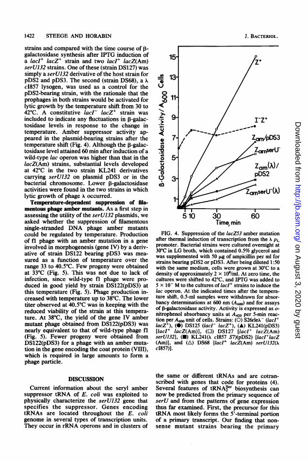
strains and compared with the time course of ,-galactosidase synthesis after IPTG induction ofa lacI+ lacZ+ strain and two lacI+ lacZ(Am)serU132 strains. One ofthese (strain DS127) wassimply a serU132 derivative of the host strain forpDS2 and pDS3. The second (strain DS68), a Xc1857 lysogen, was used as a control for thepDS2-bearing strain, with the rationale that theprophages in both strains would be activated forlytic growth by the temperature shift from 30 to42°C. A constitutive lacI- lacZ+ strain wasincluded to indicate any fluctuations in ,B-galac-tosidase levels in response to the change intemperature. Amber suppressor activity ap-peared in the plasmid-bearing strains after thetemperature shift (Fig. 4). Although the p-galac-tosidase level attained 60 min after induction ofawild-type lac operon was higher than that in thelacZ(Am) strains, substantial levels developedat 42°C in the two strain KL241 derivativescarrying serU132 on plasmid pDS3 or in thebacterial chromosome. Lower ,B-galactosidaseactivities were found in the two strains in whichlytic growth of phage X occurred.
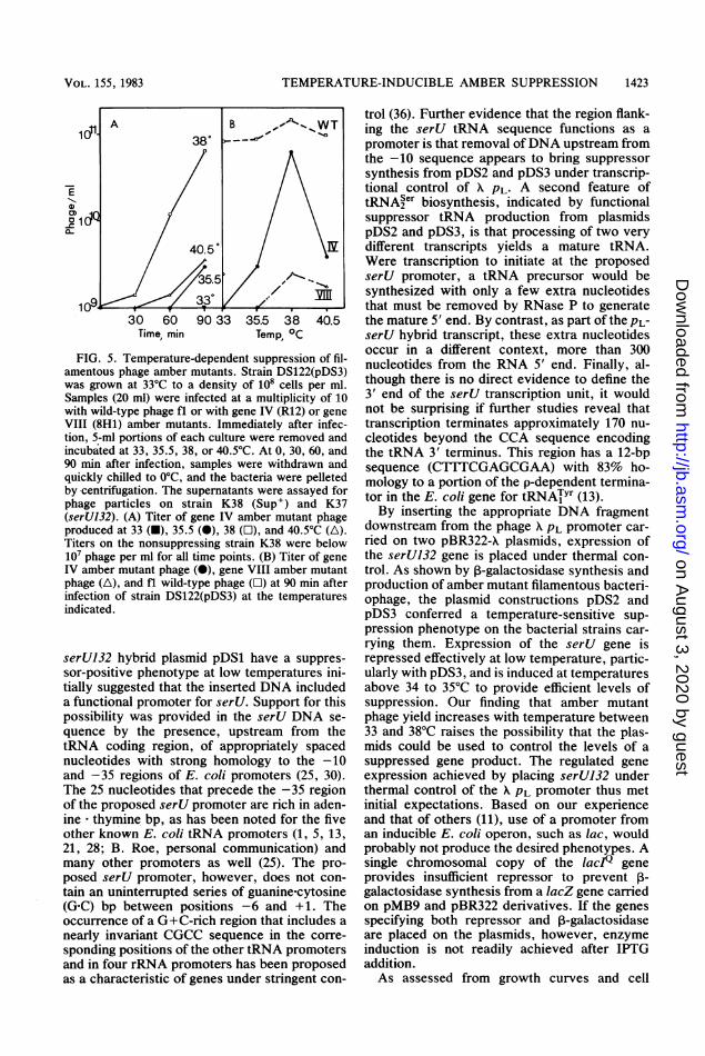
Temperature-dependent suppression of fila-mentous phage amber mutants. As a first step inassessing the utility of the serU132 plasmids, weasked whether the suppression of filamentoussingle-stranded DNA phage amber mutantscould be regulated by temperature. Productionof ft phage with an amber mutation in a geneinvolved in morphogenesis (gene IV) by a deriv-ative of strain DS122 bearing pDS3 was mea-sured as a function of temperature over therange 33 to 40.5°C. Few progeny were obtainedat 33°C (Fig. 5). This was not due to lack ofinfection, since wild-type fl phage were pro-duced in good yield by strain DS122(pDS3) atthis temperature (Fig. 5). Phage production in-creased with temperature up to 38°C. The lowertiter observed at 40.5°C was in keeping with thereduced viability of the strain at this tempera-ture. At 38°C, the yield of the gene IV ambermutant phage obtained from DS122(pDS3) wasnearly equivalent to that of wild-type phage fl(Fig. 5). Fewer progeny were obtained fromDS122(pDS3) for a phage with an amber muta-tion in the gene encoding the coat protein (VIII),which is required in large amounts to form aphage particle.
DISCUSSIONCurrent information about the seryl amber
suppressor tRNA of E. coli was exploited tophysically characterize the serU132 gene thatspecifies the suppressor. Genes encodingtRNAs are located throughout the E. coligenome in several types of transcription units.They occur in rRNA operons and in clusters of
- 13-
0011/
*> 9/I-Z+
7-;7 Zam/p)63
05'a Z /pDS3Yf 3/
amserUlY
510 30 60Time, min
FIG. 4. Suppression of the lacZ53 amber mutationafter thermal induction of transcription from the X PLpromoter. Bacterial strains were cultured overnight at30°C in LG broth, which contained 0.5% glycerol andwas supplemented with 50 ,ug of ampicillin per ml forstrains bearing pDS2 or pDS3. After being diluted 1:50with the same medium, cells were grown at 30°C to adensity of approximately 2 x 10⁸/ml. At zero time, thecultures were shifted to 42°C, and IPTG was added to5 x 10- M to the cultures of IacI+ strains to induce thelac operon. At the indicated times after the tempera-ture shift, 0.5-mi samples were withdrawn for absor-bancy determinations at 600 nm (A,o) and for assaysof j-galactosidase activity. Activity is expressed as o-nitrophenol absorbancy units at A420 per 5-min reac-tion per A6N unit of cells. Strains: (0) S26rleA- (IacI+lacZ+), (0) DS125 (lacI lacZ+), (A) KL241(pDS3)[lacI+ lacZ(Am)], (U) DS127 [lacI lacZ(Am)serU132], (U) KL241(X c1857 S7)(pDS2) [IacI+lacZ(Am)], and (A) DS68 [lacI+ lacZ(Am) serU132(XcI857)].
the same or different tRNAs and are cotran-scribed with genes that code for proteins (4).Several features of tRNAser biosynthesis cannow be predicted from the primary sequence ofserU and from the patterns of gene expressionthus far examined. First, the precursor for thistRNA most likely forms the 5'-terminal portionof a primary transcript. Our finding that non-sense mutant strains bearing the primary
J. BACTERIOL.
on August 3, 2020 by guest
http://jb.asm.org/
Dow
nloaded from
TEMPERATURE-INDUCIBLE AMBER SUPPRESSION 1423
30 60 90 33 35.5 38 40.5Time, min Temp, OC
FIG. 5. Temperature-dependent suppression of fil-amentous phage amber mutants. Strain DS122(pDS3)was grown at 33°C to a density of 108 cells per ml.Samples (20 ml) were infected at a multiplicity of 10with wild-type phage fl or with gene IV (R12) or geneVIII (8H1) amber mutants. Immediately after infec-tion, 5-ml portions of each culture were removed andincubated at 33, 35.5, 38, or 40.5°C. At 0, 30, 60, and90 min after infection, samples were withdrawn andquickly chilled to 0C, and the bacteria were pelletedby centrifugation. The supernatants were assayed forphage particles on strain K38 (Sup') and K37(serUB32). (A) Titer of gene IV amber mutant phageproduced at 33 (U), 35.5 (0), 38 (O), and 40.5°C (A).Titers on the nonsuppressing strain K38 were below107 phage per ml for all time points. (B) Titer of geneIV amber mutant phage (0), gene VIII amber mutantphage (A), and fl wild-type phage (O) at 90 min afterinfection of strain DS122(pDS3) at the temperaturesindicated.
serUJ32 hybrid plasmid pDS1 have a suppres-sor-positive phenotype at low temperatures ini-tially suggested that the inserted DNA includeda functional promoter for serU. Support for thispossibility was provided in the serU DNA se-
quence by the presence, upstream from thetRNA coding region, of appropriately spacednucleotides with strong homology to the -10and -35 regions of E. coli promoters (25, 30).The 25 nucleotides that precede the -35 regionof the proposed serU promoter are rich in aden-ine * thymine bp, as has been noted for the fiveother known E. coli tRNA promoters (1, 5, 13,21, 28; B. Roe, personal communication) andmany other promoters as well (25). The pro-posed serU promoter, however, does not con-tain an uninterrupted series of guanine-cytosine(G-C) bp between positions -6 and +1. Theoccurrence of a G+C-rich region that includes a
nearly invariant CGCC sequence in the corre-sponding positions of the other tRNA promotersand in four rRNA promoters has been proposedas a characteristic of genes under stringent con-
trol (36). Further evidence that the region flank-ing the serU tRNA sequence functions as apromoter is that removal ofDNA upstream fromthe -10 sequence appears to bring suppressorsynthesis from pDS2 and pDS3 under transcrip-tional control of X PL. A second feature oftRNAser biosynthesis, indicated by functionalsuppressor tRNA production from plasmidspDS2 and pDS3, is that processing of two verydifferent transcripts yields a mature tRNA.Were transcription to initiate at the proposedserU promoter, a tRNA precursor would besynthesized with only a few extra nucleotidesthat must be removed by RNase P to generatethe mature 5' end. By contrast, as part of the PL-serU hybrid transcript, these extra nucleotidesoccur in a different context, more than 300nucleotides from the RNA 5' end. Finally, al-though there is no direct evidence to define the3' end of the serU transcription unit, it wouldnot be surprising if further studies reveal thattranscription terminates approximately 170 nu-cleotides beyond the CCA sequence encodingthe tRNA 3' terminus. This region has a 12-bpsequence (CTTTCGAGCGAA) with 83% ho-mology to a portion of the p-dependent termina-tor in the E. coli gene for tRNATYr (13).By inserting the appropriate DNA fragment
downstream from the phage A PL promoter car-ried on two pBR322-A plasmids, expression ofthe serU132 gene is placed under thermal con-trol. As shown by ,B-galactosidase synthesis andproduction of amber mutant filamentous bacteri-ophage, the plasmid constructions pDS2 andpDS3 conferred a temperature-sensitive sup-pression phenotype on the bacterial strains car-rying them. Expression of the serU gene isrepressed effectively at low temperature, partic-ularly with pDS3, and is induced at temperaturesabove 34 to 35°C to provide efficient levels ofsuppression. Our finding that amber mutantphage yield increases with temperature between33 and 38°C raises the possibility that the plas-mids could be used to control the levels of asuppressed gene product. The regulated geneexpression achieved by placing serU132 underthermal control of the A PL promoter thus metinitial expectations. Based on our experienceand that of others (11), use of a promoter froman inducible E. coli operon, such as lac, wouldprobably not produce the desired phenotypes. Asingle chromosomal copy of the lacIf geneprovides insufficient repressor to prevent P-galactosidase synthesis from a lacZ gene carriedon pMB9 and pBR322 derivatives. If the genesspecifying both repressor and P-galactosidaseare placed on the plasmids, however, enzymeinduction is not readily achieved after IPTGaddition.As assessed from growth curves and cell
VOL. 155, 1983
on August 3, 2020 by guest
http://jb.asm.org/
Dow
nloaded from
1424 STEEGE AND HORABIN
viability determinations carried out with thenonsense mutant strains thus far examined andtheir corresponding derivatives bearing pDS2,pDS3, or the parent plasmids pKC30 or pGW7,the serU132 plasmids have no discernible effectper se on cell growth in the temperature rangefrom 30 to 39°C. At temperatures above 40°C,for reasons not yet clear, a general trend towardreduced viability appears to be more pro-nounced in some pDS3-containing strains. Inview of this, 39°C is routinely used as a condi-tion for the induction of suppressor synthesisthat is compatible with long-term cell growth.The viable range for pDS2-containing strains, inwhich the c1857 repressor must be provided by aprophage, could presumably be extended be-yond the range found in our studies by usingphage X strains that do not kill an induced host.Our motivation in undertaking this project wasto develop a temperature-inducible nonsensesuppression vector that could be useful in genet-ic selections, physiological studies of gene func-tion, and efforts to identify the polypeptideproducts of genes which are marked geneticallyby nonsense mutations. pDS3, which encodesboth the amber suppressor tRNA and the ther-molabile phage X c1857 repressor protein, ishopefully well suited for such applications.
ACKNOWLEDGMENTS
We thank R. E. Webster for assistance and interest in theproject and Leslie Petch for technical assistance. The con-struction of pDS3 was made possible by the generous contri-bution, from G. Wilson and W. Konigsberg, of plasmid pGW7before publication.
This work was supported by grant NP-367 from the Ameri-can Cancer Society. D.A.S. is supported in part by PublicHealth Service grant CA-14236 from the National CancerInstitute.
LITERATURE CITED1. An, G., and J. D. Friesen. 1980. The nucleotide sequence
of tufB and four nearby tRNA structural genes of Esche-richia coli. Gene 12:33-39.
2. Bachmann, B. J., and K. B. Low. 1980. Linkage map ofEscherichia coli K-12, edition 6. Microbiol. Rev. 44:1-56.
3. Cashman, J. S., R. E. Webster, and D. A. Steege. 1980.Transcription of bacteriophage fl: the major in vivoRNAs. J. Biol. Chem. 255:2554-2562.
4. Daniel, V. 1981. Biosynthesis of transfer RNA. Crit. Rev.Biochem. 9:253-292.
5. Duester, G., R. K. Campen, and W. M. Holmes. 1981.Nucleotide sequence of an Escherichia coli tRNA (Leu 1)operon and identification of the transcription promotersignal. Nucleic Acids Res. 9:2121-2139.
6. Edgar, R. S., G. H. Denhardt, and R. H. Epstein. 1964. Acomparative genetic study of conditional lethal mutationsof bacteriophage T4D. Genetics 49:635-648.
7. Franklin, N. C., and G. N. Bennett. 1979. The N proteinof bacteriophage lambda, defined by its DNA sequence, ishighly basic. Gene 8:107-119.
8. Garen, A., S. Garen, and R. C. Wilhelm. 1965. Suppressorgenes for nonsense mutations. I. The Su-l, Su-2, and Su-3genes of Escherichia coli. J. Mol. Biol. 14:167-178.
9. Glynn, I. M., and J. B. ChappeUl. 1964. A simple methodfor the preparation of 32P-labeled adenosine triphosphate
of high specific activity. Biochem. J. 90:147-149.10. Hoffman, E. P., and R. C. Wilhelm. 1970. Genetic map-
ping and dominance of the amber suppressor, Sul (supD)in Escherichia coli K-12. J. Bacteriol. 103:32-36.
11. Horowitz, H., and T. Platt. 1982. A termination site forlacI transcription is between the CAP site and the lacpromoter. J. Biol. Chem. 257:11740-11746.
12. Kramer, R. A., M. Rosenberg, and J. A. Steitz. 1974.Nucleotide sequences of the 5' and 3' termini of bacteri-ophage T7 early messenger RNAs synthesized in vivo:evidence for sequence specificity in RNA processing. J.Mol. Biol. 89:767-776.
13. Kipper, H., T. Sekiya, M. Rosenberg, J. Egan, and A.Landy. 1978. A p-dependent termination site in the genecoding for tyrosine tRNA SU3 of Escherichia coli. Nature(London) 272:423-428.
14. Lyons, L. B., and N. D. Zinder. 1972. The genetic map ofthe filamentous bacteriophage fl. Virology 49:45-60.
15. Maniatis, T., E. F. Fritsch, and J. Sambrook. 1982. Mo-lecular cloning. Cold Spring Harbor Laboratory, ColdSpring Harbor, N.Y.
16. Maxam, A. M., and W. Gilbert. 1980. Sequencing end-labeled DNA with base-specific chemical cleavages.Methods Enzymol. 65:499-560.
17. McKenney, K., H. Shimatake, D. Court, U. Schmeissner,C. Brady, and M. Rosenberg. 1981. A system to studypromoter and terminator signals recognized by Escherich-ia coli RNA polymerase, p. 383-415. In J. G. Chirikjianand T. S. Papas (ed.), Gene amplification and analysis,vol. 2: Structural analysis of nucleic acids. Elsevier/North-Holland, New York.
18. MIler, J. H. 1972. Experiments in molecular genetics.Cold Spring Harbor Laboratory, Cold Spring Harbor,N.Y.
19. Miller, J. H., D. Ganem, P. Lu, and A. Schmitz. 1977.Genetic studies of the lac repressor. I. Correlation ofmutational sites with specific amino acid residues: con-struction of a colinear gene-protein map. J. Mol. Biol.109:275-301.
20. Modrich, P., and D. Zabel. 1976. EcoRI endonuclease:physical and catalytic properties of the homogeneousenzyme. J. Biol. Chem. 251:5866-5874.
21. NakaJima, N., H. Ozeki, and Y. Shimura. 1981. Organiza-tion and structure of an E. coli tRNA operon containingseven tRNA genes. Cell 23:239-249.
22. Notani, G. W., D. L. Engelhardt, W. Konigsberg, andN. D. Zinder. 1965. Suppression of a coat protein mutantof the bacteriophage f2. J. Mol. Biol. 12:439-447.
23. Oeschger, M. P. 1980. Applications of temperature-sensi-tive suppressors to the study of cellular biochemistry andphysiology, p. 363-377. In D. S6ll, J. N. Abelson, andP. R. Schimmel (ed.), Transfer RNA: biological aspects.Cold Spring Harbor Laboratory, Cold Spring Harbor,N.Y.
24. Rasse-Messenguy, F., and G. R. flnk. 1973. Temperature-sensitive nonsense suppressors in yeast. Genetics 75:459-464.
25. Rosenberg, M., and D. Court. 1979. Regulatory sequencesinvolved in the promotion and termination of RNA tran-scription. Annu. Rev. Genet. 13:319-353.
26. Rosenberg, M., R. A. Kramer, and J. A. Steitz. 1974. T7early messenger RNAs are the direct products of ribonu-clease III cleavage. J. Mol. Biol. 89:777-782.
27. Schmeissner, U., D. Ganem, and J. H. Miller. 1977. Genet-ic studies of the lac repressor. II. Fine structure deletionmap of the lacI gene, and its correlation with the physicalmap. J. Mol. Biol. 109:303-326.
28. Seklya, T., R. Contreras, H. KUpper, A. Landy, and H. G.Khorana. 1976. Escherichia coli tyrosine transfer ribonu-cleic acid genes: nucleotide sequences of their promotersand of the regions adjoining C-C-A ends. J. Biol. Chem.251:5124-5140.
29. Shimatake, H., and M. Rosenberg. 1981. Purified X regula-tory protein cII positively activates promoters for lyso-
J. BACTERIOL.
on August 3, 2020 by guest
http://jb.asm.org/
Dow
nloaded from

TEMPERATURE-INDUCIBLE AMBER SUPPRESSION
genic development. Nature (London) 292:128-132.30. Skbenflst, U., R. B. Simpson, and W. Gilbert. 1980. E. coli
RNA polymerase interacts homologously with two differ-ent promoters. Cell 20:269-281.
31. Southern, E. M. 1975. Detection of specific sequencesamong DNA fragments separated by gel electrophoresis.J. Mol. Biol. 98:503-517.
32. Steege, D. A. 1983. A nucleotide change in the anticodonof an Escherichia coli serine transfer RNA results insupD- amber suppression. Nucleic Acids Res. 11:3823-3832.
33. Steege, D. A., M. C. Graves, and L. L. Spremnul. 1982.Euglena gracilis chloroplast small subunit rRNA: se-quence and base pairing potential of the 3' terminus,cleavage by colicin E3. J. Biol. Chem. 257:10430-10439.
34. Steege, D. A., and B. Low. 1975. Isolation and character-ization of lambda transducing bacteriophages for the sul +
(supD-) amber suppressor of Escherichia coli J. Bacteri-ol. 122:120-128.
35. Stretton, A. 0. W., and S. Brenner. 1%5. Molecular con-sequences of the amber mutation and its suppression. J.
Mol. Biol. 12:456-465.36. Travers, A. A. 1980. Promoter sequence for stringent
control of bacterial ribonucleic acid synthesis. J. Bacteri-ol. 141:973-976.
37. Webster, R. E., and J. S. Cashman. 1973. Abortive infec-tion of Escherichia coli with the bacteriophage fl: cyto-plasmic membrane proteins and the fl DNA-gene 5 pro-tein complex. Virology 55:20-38.
38. Weigert, M. G., and A. Garen. 1965. Amino acid substitu-tions resulting from suppression of nonsense mutations. I.
Serine insertion by the Su-I suppressor gene. J. Mol. Biol.12:448-455.
39. Winberg, G., and M.-L. Hammarsakold. 1980. Isolation ofDNA from agarose gels using DEAE-paper. Applicationto restriction site mapping of adenovirus type 16 DNA.Nucleic Acids Res. 8:253-264
40. Yamnoto, K. R., B. M. Alberts, R. Benzinger, L. Law-horne, and G. Treiber. 1970. Rapid bacteriophage sedi-mentation in the presence of polyethylene glycol and itsapplication to large-scale virus purification. Virology40:734-744.
VOL. 155, 1983 1425
on August 3, 2020 by guest
http://jb.asm.org/
Dow
nloaded from