Techniques of tear film evaluation by Raju Kaiti
18
TEAR FILM EVALUATION Raju Kaiti Optometrist Dhulikhel Hospital, Kathmandu University Hospital
-
Upload
raju-kaiti -
Category
Health & Medicine
-
view
412 -
download
0
Transcript of Techniques of tear film evaluation by Raju Kaiti

TEAR FILM EVALUATION
Raju Kaiti
Optometrist
Dhulikhel Hospital, Kathmandu University Hospital

STRUCTURE OF TEAR FILM
It is approximately:
7 micrometre thick
7microliter in volume
Wolff (1946): Precorneal layer Three layers
• Outer lipid layer• Intermediate aqueous layer• Inner mucous layer
2
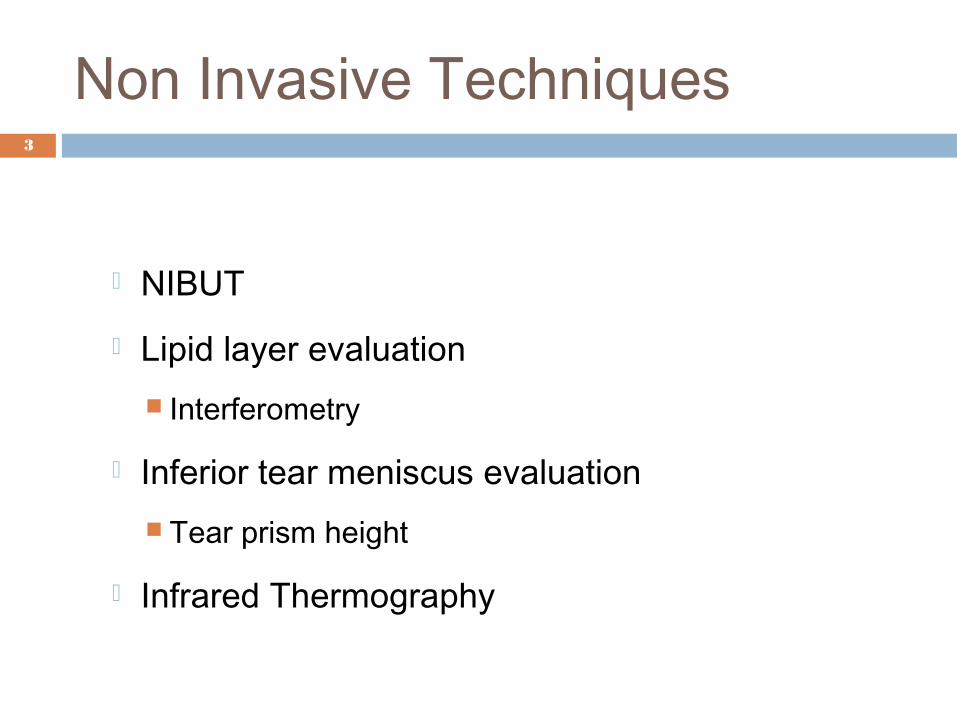
Non Invasive Techniques 3
NIBUT
Lipid layer evaluation Interferometry
Inferior tear meniscus evaluation Tear prism height
Infrared Thermography
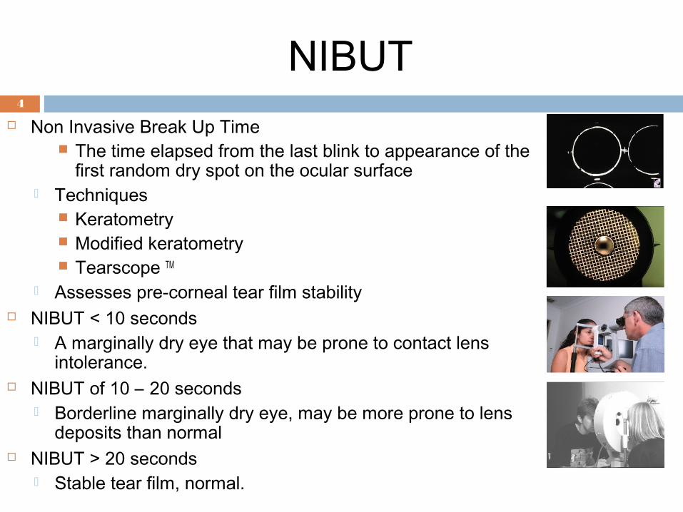
NIBUT4
Non Invasive Break Up Time The time elapsed from the last blink to appearance of the
first random dry spot on the ocular surface Techniques
Keratometry Modified keratometry Tearscope TM
Assesses pre-corneal tear film stability NIBUT < 10 seconds
A marginally dry eye that may be prone to contact lens intolerance.
NIBUT of 10 – 20 seconds Borderline marginally dry eye, may be more prone to lens
deposits than normal NIBUT > 20 seconds
Stable tear film, normal.
Assessment of inferior tear meniscus
5
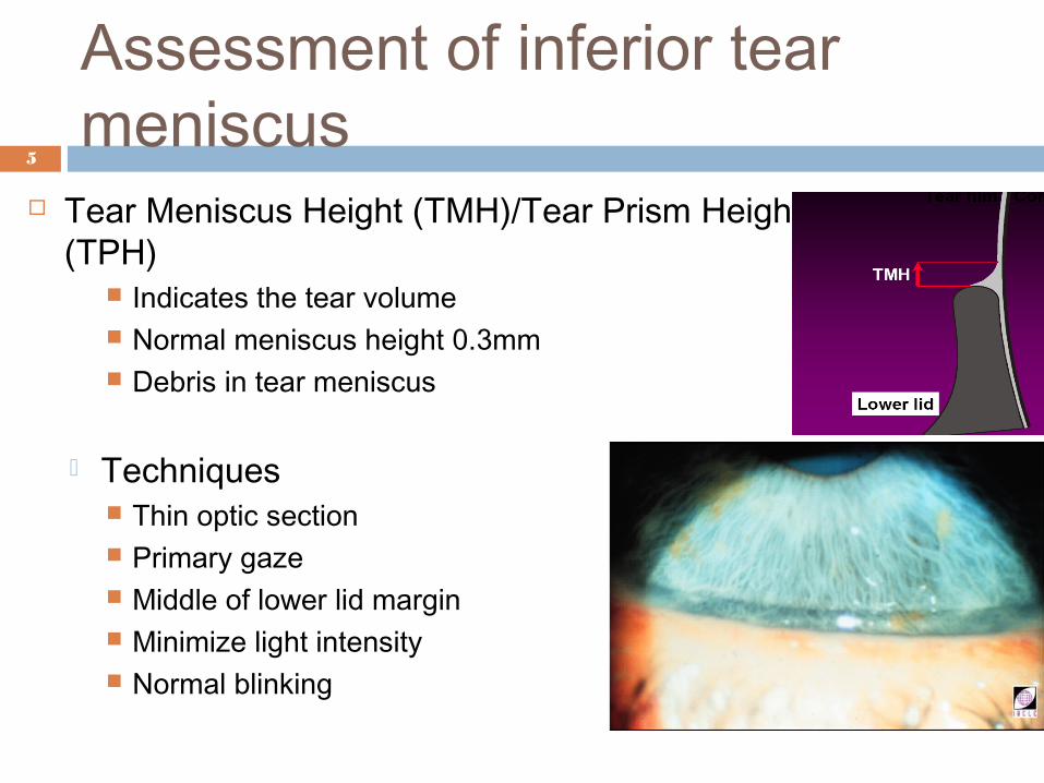
Tear Meniscus Height (TMH)/Tear Prism Height (TPH)
Indicates the tear volume Normal meniscus height 0.3mm Debris in tear meniscus
Techniques Thin optic section Primary gaze Middle of lower lid margin Minimize light intensity Normal blinking

Interferometry 6
Interference patterns Tear film interference patterns
(colored fringes)
Indicate the different
thicknesses of the component
layers
Lipid layer Colored fringes
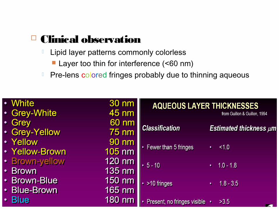
7 Clinical observation
Lipid layer patterns commonly colorless Layer too thin for interference (<60 nm)
Pre-lens colored fringes probably due to thinning aqueous

The typical marmoreal pattern seen in the lipid layer is absent in the dry eye
8

Infrared Thermography 9
The rate of change of corneal temperature, an analogue of the tear film evaporation rate is measured Evaporation produces cooling
Infrared thermograms confirm: Dry eyes have lower evaporation rates Corneal temperature fluctuates between blinks (an effect of
evaporation) Dry eyes have slower between-blink cooling (reduced
evaporation)
Blink evaluation 10
Blink Movement and Blink Rate Twitch blink Incomplete blink Forced blink Normal blink
Average blink rate : 12.5 / min During concentrated = 3 / min Free conversation = 29 /min

Invasive Techniques11
Schirmer’s Tear test Schirmer’s tear test 1 Basic secretion test Schirmer’s test 2
Tear Break Up Time
Phenol Red Thread Test
Fluorescein Staining
Rose Bengal Staining
Conjunctival Impression Cytology
Schirmer’s Tear test
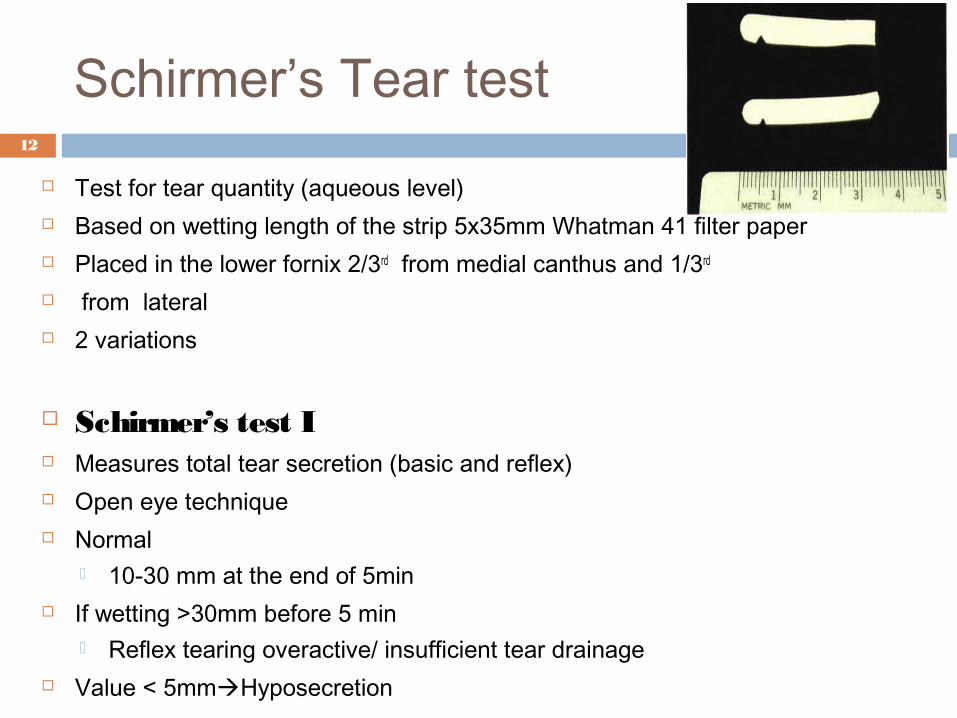
Test for tear quantity (aqueous level) Based on wetting length of the strip 5x35mm Whatman 41 filter paper Placed in the lower fornix 2/3rd from medial canthus and 1/3rd from lateral 2 variations
Schirmer’s test I Measures total tear secretion (basic and reflex) Open eye technique Normal
10-30 mm at the end of 5min If wetting >30mm before 5 min
Reflex tearing overactive/ insufficient tear drainage Value < 5mmHyposecretion
12

13
Basic Secretion Test Variant of Schirmer's test I measures basal tear secretion Topical anesthetic is applied Cul-de-sac is dried out before the strip is inserted Difference between Schirmer's I and BST
Schirmer’s 1 reading gives reflex secretion also Normal value > 10mm Basic Secretion of 3mm or less Abnormal
Schirmer’s Tear test II Measures reflex tear secretion Irritate nasal mucosa by rubbing it with cotton swab or smelling ammonia Measure wetting after 2min Wetting < 15mm Failure of reflex secretion Parasympathetic supply

14
Fluorescein Staining Fluorescein dye is used to detect epithelial defects on the anterior surface of the eye.Penetrates only the corneal epithelium at sites of interrupted continuity of the epithelial surface.Is enhanced with the use of cobalt blue filter that blocks extraneous light and highlights staining patterns.
Tear Break Up TimeTear film Break Up Time in cobalt blue filterInterval between the last blink & the first appearance of black spot in the fluorescein-stained tear film (No rn & Ha m il 1 9 7 3 )
A black island in the sea of green fluoresceinAssessment of tear film stability >10 sec Normal

Rose Bengal Staining15
Devitalized cells on the cornea and conjunctiva and mucus in the tear film detected using 1% rose bengal; highlighted by red punctate staining 0 to 3 graded scale•Three regions of the interpalpebral ocular surface are assessed
– the triangular wedge of the nasal interpalpebral conjunctiva,– the corneal surface– the wedge of the temporal conjunctiva
•The grade of each region is summed, and a score greater than 3.5 is considered indicative of dry eye
Conjunctival Impression Cytology
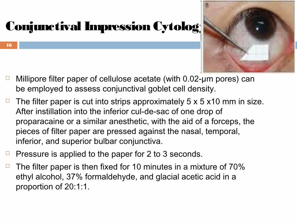
Millipore filter paper of cellulose acetate (with 0.02-µm pores) can be employed to assess conjunctival goblet cell density.
The filter paper is cut into strips approximately 5 x 5 x10 mm in size. After instillation into the inferior cul-de-sac of one drop of proparacaine or a similar anesthetic, with the aid of a forceps, the pieces of filter paper are pressed against the nasal, temporal, inferior, and superior bulbar conjunctiva.
Pressure is applied to the paper for 2 to 3 seconds. The filter paper is then fixed for 10 minutes in a mixture of 70%
ethyl alcohol, 37% formaldehyde, and glacial acetic acid in a proportion of 20:1:1.
16
Conjunctival Impression Cytology
Each paper is stained, using periodic acid–Schiff (PAS), hematoxylin and eosin, and Papanicolaou.
Under the light microscope, the epithelial cells are evaluated for morphology and density.
Nelson (1988) classified impression cytology of the conjunctiva in grades:
Stage 0: normal cellular structure Stage 1: early loss of goblet cells without keratinisation Stage 2: total loss of goblet cells with slight enlargement of
epithelial cells Stage 3: early and mild keratinisation Stage 4: moderate keratinisation Stage 5: advanced keratinisation Grades 1 and 2 are considered normal.
17

THANK YOU !!!!!!
18