Documents: 3.II. 214 -19 214 -19 ELECTRONIC TAX SALE.PDF 3 ...

EJMT 1(2) 2014 • Najnowsze technologie medyczne
1 Copyright © 2014 by ISASDMT
The validation of microscopic techniques for identification and differentiation of Microsporidia
Walidacja technik mikroskopowych w celu identyfikacji i różnicowania mikrosporydiów
StreszczenieOrganizmy należące do typu mikrosporydia (Regnum: Fungi) są przyczyną wie-lu poważnych chorób ludzi i zwierząt. Większość gatunków mikrosporydiów, wywołujących zakaźne choroby u ssaków, może być również przyczyną infekcji u ludzi. Dlatego do badań tych groźnych patogenów został wybrany jako or-ganizm modelowy owad – pszczoła miodna (Apis mellifera) oraz dwa gatunki mikrosporydiów: Nosema apis i Nosema ceranae, które są przyczyną nosemozy, groźnej choroby tych owadów. Próbki pobrane od zakażonych Nosema spp. pszczół obserwowano w jasnym polu widzenia, przy zastosowaniu kontrastu Nomarskiego (DIC) oraz z kontrastem fazowym. Ponadto obserwowano zarod-niki Nosema spp. przy użyciu skaningowej mikroskopii elektronowej (SEM). Badanie próbek jelit przy użyciu SEM, wykazało, że zarodniki mikrosporydiów tworzą warstwę równo pokrywającą światło jelita u zakażonych osobników. Podobna warstwa może powstawać podczas innych infekcji powodowanych przez inne mikrosporydia, tak jak w przypadku np. Encephalitozoon intestinalis, który jest przyczyną przewlekłej biegunki i zapalenia jelit u pacjentów z AIDS. Może to powodować niedożywienie i wyższą śmiertelność wśród pacjentów z AIDS dodatkowo zakażonych mikrosporydiozą układu pokarmowego.
dr Aneta A. Ptaszyńska
Department of Botany and Mycology, Institute of Biology and Biochemistry, Faculty of Biology and Biotechnology, Maria Curie-Skłodowska University, Lublin, Poland
European Journal of Medical Technologies 2014; 1(2): 1-5Copyright © 2014 by ISASDMT All rights reserved www. medical-technologies.euPublished online 24.01.2014
Cite this paper as: Ptaszyńska A.A. 2014. The validation of microsco-pic techniques for identification and differentiation of Microspori-dia. European Journal of Medical Technologies; 1(2): 1-5
Słowa kluczowe: mikrosporydiozy, Nosema apis; Nosema ceranae; nosemoza; mikroskopia optyczna, DIC; mikroskopia kontrastowo--fazowa; mikroskopia elektronowa; SEM; TEM
EJMT 1(2) 2014 • Najnowsze technologie medyczne
2 Copyright © 2014 by ISASDMT
IntroductionOrganisms belonging to the phylum Microsporidia in the regnum Fungi are causative agents of many seri-ous diseases of human and animals. Until now have been described more than 1200 microsporidian spe-cies belonging to approximately 150 genera. But only species belonging to ten genera have been reported from human hosts as agents of systemic, ocular, in-testinal and muscular infections, e.g.: Anncaliia (Bra-chiola, Nosema) algerae, A. (Brachiola) vesicularum, A. (Nosema) connori, Enterocytozoon bieneusi, En-cephalitozoon (Septata) intestinalis, E. hellem, E. cu-niculi, Microsporidium africanum, M. ceylonensis, Nosema ocularum, Pleistophora ronneafyi, Pleistopho-ra sp., Trachipleistophora hominis, T. anthropophthera, Tubulinosema acridophagus, Vittaforma corneae (Nosema corneum) [12]. The first detected human mi-crosporidiosis was in 1959 r. a case of 9-year-old boy who suffered from fever, headache, seizures and loss of consciousness. In the boy’s urine and cerebrospi-nal fluid were detected spores of Encephalotozoon sp. Since 1985, microsporidiosis has become an emergent problem for HIV/AIDS patients with chronic diar-rhea. Especially, infection caused by Enterocytozoon bieneusi were the most common ones.
Microsporidia are obligate intracellular spore-forming pathogens. The spore germinates in the tar-get tissue, e.g. in the intestines, extrudes polar tubule, inserts the tubule into the epithelial cell and injects the infective sporoplasms. Such a sporoplasm, inside a host cell, transforms into a meront and that initi-ated a merogony phase – a proliferactive stage of microsporidian life cycle, in which meronts are rep-licated. Then meronts develop into sporonts, which are characterized by a dense surface coat. Sporonts multiply and the sporogonic phase ends with spores formation. The spores can be excreted outside, but also they can immediately germinate and infect other cells of the same host organism. Spores can also pass across the placenta in animals and severely infect the foetus. The spores are extremely resistant with external stress factor and remain infective for long periods of time. Therefore, person to person contact, fecal-oral route, contamined air, water or food are the most frequent ways of microsopridiosis transmission [7,10,12].
Due to the microsporidian life cycle, the only eas-ily recognizable stages of their life are spores. Mi-crosporidia that infect humans have typically oval spores, which vary in size, from approximately 1 to 4 μm in diameter. Under transmission electron mi-
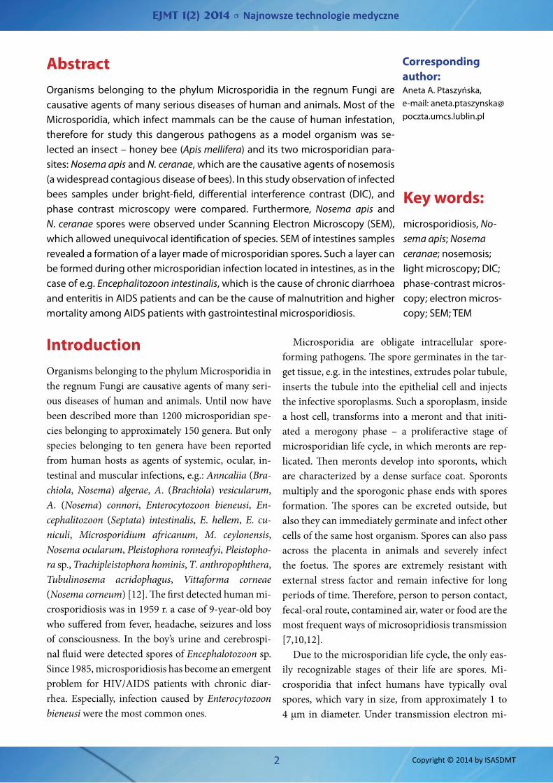
AbstractOrganisms belonging to the phylum Microsporidia in the regnum Fungi are causative agents of many serious diseases of human and animals. Most of the Microsporidia, which infect mammals can be the cause of human infestation, therefore for study this dangerous pathogens as a model organism was se-lected an insect – honey bee (Apis mellifera) and its two microsporidian para-sites: Nosema apis and N. ceranae, which are the causative agents of nosemosis (a widespread contagious disease of bees). In this study observation of infected bees samples under bright-field, differential interference contrast (DIC), and phase contrast microscopy were compared. Furthermore, Nosema apis and N. ceranae spores were observed under Scanning Electron Microscopy (SEM), which allowed unequivocal identification of species. SEM of intestines samples revealed a formation of a layer made of microsporidian spores. Such a layer can be formed during other microsporidian infection located in intestines, as in the case of e.g. Encephalitozoon intestinalis, which is the cause of chronic diarrhoea and enteritis in AIDS patients and can be the cause of malnutrition and higher mortality among AIDS patients with gastrointestinal microsporidiosis.
Key words:microsporidiosis, No-sema apis; Nosema ceranae; nosemosis; light microscopy; DIC; phase-contrast micros-copy; electron micros-copy; SEM; TEM
Corresponding author: Aneta A. Ptaszyńska, e-mail: [email protected]

EJMT 1(2) 2014 • Najnowsze technologie medyczne
3 Copyright © 2014 by ISASDMT
croscopy (TEM) mature spores can be differentiated to the species by the number of coils in cross-section and the location of parallel coils and tilt [5].
Most of the Microsporidia, which infect mammals can be the cause of human infestation, therefore for study this dangerous pathogens as a model organism was selected an insect – honeybee (Apis mellifera). Two microsporidian species, Nosema apis and N. cer-anae, are the causative agents of nosemosis, which is a widespread contagious disease of bees. This disease is associated with honeybee Colony Collapse Disor-der (CCD), manifested by rapid and massive loss of bees outside the hive and, consequently, extinction of bee colonies worldwide. Nosemosis causes many changes both at the level of individuals and colonies. Life expectancy of infected bees is reduced by one third. In families affected by Nosema spp., worker bees become lethargic and unwilling to work. Un-til now, it was thought that the spores of N. apis can be found only in the gut of bees, while N. ceranae in other tissues and glands [3,4,6]. Recent studies have shown that both N. apis and N. ceranae are not tissue-specific and their spores can be found in the midgut epithelium, Malpighian tubule system, hypopharyn-geal glands, salivary glands, and venom sacs [2,8]. Spores remaining in the glands are potential reser-voirs of infection [2].
In this study, the techniques based on the micros-copy to detect Microsporidia on the examples of hon-eybees infection caused by Nosema apis and N. cera-nae, are discussed.
Materials and methodsFor each method (light microscopy and SEM – scan-ning electron microscopy), the same suspension ob-tained by grinding 10 fresh whole bees in 10 ml of sterile water was examined. Additionally for SEM preparation, Nosema spp. infected bees’ intestines were also examined. The intestines were removed in-dividually, gently washed in distilled water to prevent contamination by a heamolymph and immediately immersed in 5% gluteraldehyde (v/v) in 0.1 M phos-phate buffer pH 7.3.
Light microscopyThe suspension was smeared on a microscope slide for examination as described by Cantwell [1]. The same samples were observed under bright-field, dif-ferential interference contrast (DIC), and phase con-trast microscopy using an Olympus BX 61 micro-scope.
Scanning Electron Microscopy (SEM) sample preparationThe specimens were fixed in 5% glutaraldehyde (v/v) in 0.1 M phosphate buffer pH 7.3 for 24 hours and then washed in a phosphate buffer prior to post fixa-tion in 1% osmium tetroxide in 0.1 M-phosphate buffer for 24 hours followed by washing in the same buffer. SEM samples were dehydrated by immersion for 15 min each in fresh solutions of 30%, 50%, 75%, 90%, and 100% acetone and critical point dried. The dried samples were mounted on specimen stubs us-ing a double side adhesive tape and coated with gold. Coated samples were viewed under a VEGA LMU scanning microscope at 30 KV, measured, and pho-tographed.
Results and discussion
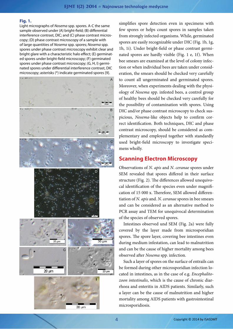
Light microscopyThere is a wide range of techniques for preparation the smear samples providing better recognition of Microsporidia [5,11]. The samples can be stained us-ing trichrome, Calcoflour white, Fungiflour, toluidine blue, methylene blue-azure, basic fuchsin and others [7]. Still, the simplest and fastest method is to use dif-ferent observation techniques of one sample under the light microscope. Currently, microscopes with the ability to view samples under DIC or phase con-trast are easily available. The easiest and most popu-lar way to observe Nosema spp. spores is to use light bright-field microscopy (Fig. 1a), but then the spores may be misidentified as other yeast cells or amoeba cysts. Under phase contrast microscopy, Nosema spp. spores exhibit clear and bright glare with a character-istic halo effect (Fig. 1c, 1d). Therefore, this technique
EJMT 1(2) 2014 • Najnowsze technologie medyczne
4 Copyright © 2014 by ISASDMT
simplifies spore detection even in specimens with few spores or helps count spores in samples taken from strongly infected organisms. While, germinated spores are easily recognizable under DIC (Fig. 1b, 1g, 1h, 1i). Under bright-field or phase contrast germi-nated spores are hardly visible (Fig. 1 e, 1f). When bee smears are examined at the level of colony infec-tion or when individual bees are taken under consid-eration, the smears should be checked very carefully to count all ungerminated and germinated spores. Moreover, when experiments dealing with the physi-ology of Nosema spp. infested bees, a control group of healthy bees should be checked very carefully for the possibility of contamination with spores. Using DIC and/or phase contrast microscopy to check sus-picious, Nosema-like objects help to confirm cor-rect identification. Both techniques, DIC and phase contrast microscopy, should be considered as com-plementary and employed together with standardly used bright-field microscopy to investigate speci-mens wholly.
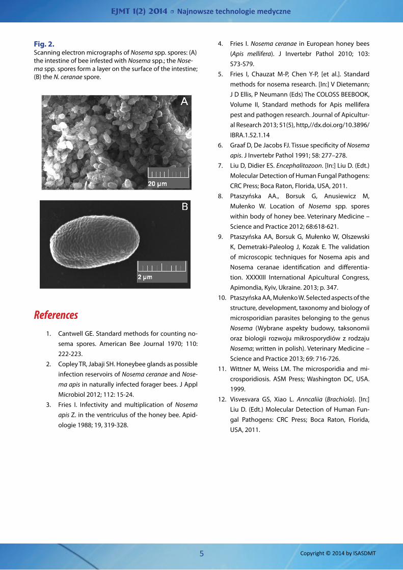
Scanning Electron MicroscopyObservations of N. apis and N. ceranae spores under SEM revealed that spores differed in their surface structure (Fig. 2). The differences allowed unequivo-cal identification of the species even under magnifi-cation of 15 000 x. Therefore, SEM allowed differen-tiation of N. apis and. N. ceranae spores in bee smears and can be considered as an alternative method to PCR assay and TEM for unequivocal determination of the species of observed spores.
Intestines observed und SEM (Fig. 2a) were fully covered by the layer made from microsporidian spores. The spore layer, covering bee intestines even during medium infestation, can lead to malnutrition and can be the cause of higher mortality among bees observed after Nosema spp. infection.
Such a layer of spores on the surface of entrails can be formed during other microsporidian infection lo-cated in intestines, as in the case of e.g. Encephalito-zoon intestinalis, which is the cause of chronic diar-rhoea and enteritis in AIDS patients. Similarly, such a layer can be the cause of malnutrition and higher mortality among AIDS patients with gastrointestinal microsporidiosis.
Fig. 1. Light micrographs of Nosema spp. spores. A-C the same sample observed under (A) bright-field; (B) differential interference contrast, DIC; and (C) phase contrast micros-copy; (D) phase contrast microscopy of a sample with of large quantities of Nosema spp. spores; Nosema spp. spores under phase contrast microscopy exhibit clear and bright glare with a characteristic halo effect; (E) germinat-ed spores under bright-field microscopy; (F) germinated spores under phase contrast microscopy; (G, H, I) germi-nated spores under differential interference contrast, DIC microscopy; asterisks (*) indicate germinated spores [9].
EJMT 1(2) 2014 • Najnowsze technologie medyczne
5 Copyright © 2014 by ISASDMT
References1. Cantwell GE. Standard methods for counting no-
sema spores. American Bee Journal 1970; 110: 222-223.
2. Copley TR, Jabaji SH. Honeybee glands as possible infection reservoirs of Nosema ceranae and Nose-ma apis in naturally infected forager bees. J Appl Microbiol 2012; 112: 15-24.
3. Fries I. Infectivity and multiplication of Nosema apis Z. in the ventriculus of the honey bee. Apid-ologie 1988; 19, 319-328.
4. Fries I. Nosema ceranae in European honey bees (Apis mellifera). J Invertebr Pathol 2010; 103: S73-S79.
5. Fries I, Chauzat M-P, Chen Y-P, [et al.]. Standard methods for nosema research. [In:] V Dietemann; J D Ellis, P Neumann (Eds) The COLOSS BEEBOOK, Volume II, Standard methods for Apis mellifera pest and pathogen research. Journal of Apicultur-al Research 2013; 51(5), http,//dx.doi.org/10.3896/IBRA.1.52.1.14
6. Graaf D, De Jacobs FJ. Tissue specificity of Nosema apis. J Invertebr Pathol 1991; 58: 277–278.
7. Liu D, Didier ES. Encephalitozoon. [In:] Liu D. (Edt.) Molecular Detection of Human Fungal Pathogens: CRC Press; Boca Raton, Florida, USA, 2011.
8. Ptaszyńska AA., Borsuk G, Anusiewicz M, Mułenko W. Location of Nosema spp. spores within body of honey bee. Veterinary Medicine – Science and Practice 2012; 68:618-621.
9. Ptaszyńska AA, Borsuk G, Mułenko W, Olszewski K, Demetraki-Paleolog J, Kozak E. The validation of microscopic techniques for Nosema apis and Nosema ceranae identification and differentia-tion. XXXXIII International Apicultural Congress, Apimondia, Kyiv, Ukraine. 2013; p. 347.
10. Ptaszyńska AA, Mułenko W. Selected aspects of the structure, development, taxonomy and biology of microsporidian parasites belonging to the genus Nosema (Wybrane aspekty budowy, taksonomii oraz biologii rozwoju mikrosporydiów z rodzaju Nosema; written in polish). Veterinary Medicine – Science and Practice 2013; 69: 716-726.
11. Wittner M, Weiss LM. The microsporidia and mi-crosporidiosis. ASM Press; Washington DC, USA. 1999.
12. Visvesvara GS, Xiao L. Anncaliia (Brachiola). [In:] Liu D. (Edt.) Molecular Detection of Human Fun-gal Pathogens: CRC Press; Boca Raton, Florida, USA, 2011.
Fig. 2. Scanning electron micrographs of Nosema spp. spores: (A) the intestine of bee infested with Nosema spp.; the Nose-ma spp. spores form a layer on the surface of the intestine; (B) the N. ceranae spore.