
Technical / Products Preparation - Plasma Solutions€¦ · Technical / Products Preparation. Who...
62
Plasma Solutions, LLC 800.353.1507 [email protected] Accelerate GF™ Natural Regenerative Medicine Treatments Technical / Products Preparation
Transcript of Technical / Products Preparation - Plasma Solutions€¦ · Technical / Products Preparation. Who...

Plasma Solutions, LLC 800.353.1507
Accelerate GF™Natural Regenerative Medicine Treatments
Technical / Products Preparation

Who is Plasma Solutions?
Plasma Solutions, LLC is a biologics company that develops, manufactures, and markets regenerative medicine products for the repair, restoration and revitalization of damaged and diseased cellular tissue for (i) musculoskeletal injuries and conditions, (ii) neuropa-thy, and (iii) chronic non-healing wounds.
• Plasma Solutions uses products such as ACCELERATE GF™ for treating joint and ten-don pain, ankle sprains, heel pain and plantar fasciitis.
• Plasma Solutions products are also used to treat a wide range of musculoskeletalconditions in addition to peripheral neuropathies.
• We have also found that our products can be used to treat chronic non-healingwounds and help to prevent amputations in high risk populations.

Today’s Objectives
• Give you the ability to process Accelerate GF
• Explain what makes Accelerate GF different.
• Show you the Science and make you comfortable with it.
• Give you resources
• Show you how to grow your patient base.

What’s a Platelet?
• Also called “thrombocyte”
• Small, disc shaped clear cell fragment
• Lifespan is 5-9 days
• Natural source of growth factors
• Circulate in the blood
• Lead the formation of blood clots
• Essential in healing process

Platelets Work
Platelets Work
Platelets are activated by damaged tissue. Primary hemostasis and initiation of the clotting cascade are just the beginning of the platelets role in heal-ing.
Upon activation they release Natural anabolic growth factors.
• Platelet derived GF
• Transforming GF
• Basic Fibroblast GF
• Vascular Endothelial GF
• Epidermal GF
• Nerve GF

Plasma• The liquid portion of your blood
•Mostly water (95%)
• Proteins, Electrolytes, glucose,
• Fibrinogen
• Hormones
• The body’s Protein Reserve
• Protects you from infection and blood disorders.
• Platelet Rich Plasma is simply plasma rich in platelets. This is achieved through centrif-ugation.
• Platelet concentration is increased from 2 to five times baseline.
• Generally considered golden.
• Platelet Poor Plasma is plasma which is poor or low in platelets. However, this plasma is rich in fibrinogen and proteins.
• A by-product of the centrifugation process.

Plasma Solutions• Through breakthrough science and technology, we have developed a proprietarymethod for activating and aggregating platelet rich and platelet poor plasma and increased levels of GF.
• This method has led to patient outcomes that are far more positive than those of com-peting products.
• Additionally, Plasma Solutions can provide the physician with the ability to customize treatments.

Procedure: Simplicity• Draw 8.5 - 17cc of the patient’s blood into the tube(s) Plasma Solutions provided vacutainertubes.
• Rotate the tubes 7 times to mix the anti-coagulant into the blood. Make certain that the patient’s name has been written on the tubes.
• Place the tubes in your Centrifuge and set it to the specifications ordered by the physi-cian. Start thecentrifugation.
• When centrifugation is complete, according to protocol, extract the Accelerate GF from the tubes with the preloaded activator/aggregator syringe.
• It’s ready for injection.
Injection• People hate shots.
• PRP injections are usually pretty painful.
• After the first PRP injection manyPeople don’t want more.
• Most patients say the Plasma Solutions injections are “painless”. Sometimes theydon’t know they had the injection.
• Patients don’t mind coming back forMore.

Closing Remarks• Technician vs Technologist
• Building patient loyalty
• Being the Expert Spokesperson
• Support: WHO TO CALL

Table of Contents• History of plasma-based therapy
• Pathophysiology
• Plasma product preparation
• Indications
• Contraindications
• Techniques

History of Plasma-Base Therapy• M. Ferrari in 1987
• Autologous transfusion component after an open heart operation
• Avoid homologous blood product transfusion

History cont.
National Center for Biotechnology Information
• 5200 Entries
– Orthopaedic– Sport medicine– Dentistry– Otolaryngology– Neurosurgery– Ophthalmology– Urology– Wound healing– Cosmetic– Cardiothoracic– Maxillofacialsurgery

• Platelets
Growth factors
– Cytokines (inflammation)
– Infection
– Osteogenesis
– Wound healing
– Maxillofacialsurgery
• Bio-Active Proteins
– Macrophages
– Mesenchymal stem cells
– Osteoblast
– Enhance tissue
regeneration/healing
Pathophysiology

• Platelets
Growth factors
– Cytokines (inflammation)
– Infection
– Osteogenesis
– Wound healing
– Maxillofacialsurgery
• Bio-Active Proteins
– Macrophages
– Mesenchymal stem cells
– Osteoblast
– Enhance tissue
regeneration/healing
Pathophysiology Cont.

Dense granules
SerotoninHistamineATPADPCalciumMagnesiumPyrophosphate
Alpha-granules
SerotoninAlbuminFibrinogenFibronectinVitronectinOsteonectinCalcitoninVon Willebrand FactorVonWillebrand antigenIIThrombospondinPlatelet factor 4lgG,lgA.lgMCl inhibitorPlasminogenPlasminogenactivatorinhibitor-1Platelet-derived
collagenase inh bitorHighmolecularweightkininogenAngiotensinogenProtein S 0<2-Antitrypsin 0<2-Macroglobulin 0<2-AntiplasminMultimerinPlatelet basic proteinThromboglobulinHistidine-richglycoproteinConnective tissueactivatingprotein IllNeutrophil-activatingprotein IIPlatelet-derivedgrowth factorCoagulation factor \/CoagulationfactorVIIISubstancePVasoactive intestinalpeptide>300 other proteins(Maguire et al.. 2002;Maguireand Fitzgerald,2003, Copoingeret al.,2004)
Lysosomal granules
Cathepsin DCathepsin ECarboxypeptidaseACarboxypeptidase BProline carboxypeptidaseN-acetyl-0-hexosaminidase!3-0-g lucuronidase!3-0-galactosidasea-o-mannosidase0<-L-arabinofuranosidasea-o-galactosidasea-L-fucosidase!3-0-fucosidase!3-0-g lucosidasea-o lucosidaseAcid phosphataseArylsulfatase
Pathophysiology Cont.

• Definition
Definition
– Higher concentration
than baseline
– Subjected to the
individual baseline
– 2.5 -3 times baseline (low))))
– 5 - 9 times baseline (high)
– Optimal level 2.5x
– Graziani et al. Clin Oral Implants Res.
2006 Apr;17(2):212-9.
• Preparation
– Low platelet count
• 2 components
– High platelet count
• 3 components
PRP Definition and Preparation

• Concentration of platelet (2.5X)
– TGF-b
– FGF
– PDGF
– EGF
– VEGF
– CTGF
• WBC
– Neutrophils
• 40 Hydrolytic enzymes
– Lymphocytes
• T/B cells, NK cells
– Monocytes/Macrophages
PRP (Buffy coat)

White Blood Cells

• Before Injection
– Anticoagulant (ACD)
• Binds to calcium
• PH
• Bio-Active Proteins
– Macrophages
• Injection
– Buffering the PH
• Thrombin
• Calcium chloride
• Mechanical trauma
Plasma-Base Product Preparation

• Tendinopathies
• Ligament injuries
• Muscle injuries
• Joints conditions
• Intervertebral discs
• Nerves(irritation)
• Fracture Non-Union
Plasma-Base Therapy Indications

• AbsoluteContraindication
– Platelet dysfunctionsyndrome
– Critical thrombocytopenia
– Hemodynamic instability
– Septicemia
– Local infection at the site of theproce-dure– Patient unwilling to accept the risks
• RelativeContraindications
– NSAIDs within 48 hours ofprocedure– Corticosteroid injection within 1 month ofprocedure
– Systemic use of corticosteroids with 2 weeks of procedure
– Tobacco use
– Fever or illness (recent)– Cancer (especially hematopoietic or of bone)– HGB < 10g/dl– Platelet count < 105/ul
Plasma Solutions Screening Recommendations

• Regenerative medicine on-demand• Customary treatment modalities• Amendable plasma-base products
Plasma Solutions Treatment Approach

• Anatomicalsite– Axial skeletal• Cervical, thoracic, lumbar, sacrum
– Appendicular skeletal• Shoulder, elbow, wrist, hand, hip, knee, ankle, foot
• Tissue involvement– Cartilage (joint), tendon, ligament, muscle, nerve
• Chronicity– Acute (1-4days), sub-acute (4days-3wks), chronic (3wk >)
Treatment Algorithms

• Anatomicalsite– Axial skeletal• Cervical, thoracic, lumbar, sacrum
– Appendicular skeletal• Shoulder, elbow, wrist, hand, hip, knee, ankle, foot
• Tissue involvement– Cartilage (joint), tendon, ligament, muscle, nerve
• Chronicity– Acute (1-4days), sub-acute (4days-3wks), chronic (3wk >)
Anatomical Landmarks and Injection Techniques

• Shoulder• Elbow• Wrist• Hip• Knee• Ankle• Spine (C/L)
Injections Techniques

Normal Anatomical Structures of the Shoulder

Injection Procedure to the Shoulder
1.Gleno-humeral joint
1. Subacromial joint
1. Acromioclavicular joint

Injection Procedures for the Shoulder Ligaments
1. Coracoacromial Ligament
2. Acromioclavicul ar Ligament
3. Coracohumeral Ligament
4. Articular capsule of shoulder

Injection Procedures to the Shoulder Tendons/Bursa
1. Supraspinatus tendon
2. Infraspinatus tendon
3. Teres minor tendon
4. Sub-acromial bursa
5. Sub-deltoid bursa

Injection Procedures to the Shoulder Tendons/Bursa
1. Subscapularis 2. Biceps Brachii long head

Normal Anatomical Structures of the Elbow

Injection Procedures to the Elbow Joints
1. Radial-capitulum joint2. Ulna-trochear joint

Injection Procedures to the Elbow Ligaments
1. Radial collateral ligament 2. Ulnar collateral ligament

Injection Procedures to the Elbow Ligaments
1. Ulnar collateral ligament

Injection Procedures for the Elbow Tendons
1. Common extensor tendons

Injection Procedures for the Elbow Tendons
1. Common flexor tendons

Normal Anatomical Structures of the Wrist

Injections Procedure to the Wrist Joints
1. Radial carpal joint
2. Ulnar carpal joint

Injection Procedures for the Wrist Ligaments
1. TFCC ligamentsa. Dorsal and palmar ulnarcarpalligaments

Injection Procedures to the Wrist Tendons
1. Common wrist extensor tendons

Injection Procedures to the Wrist Tendons
1. Common wrist flexor tendons

Injection Techniques for Dequervain’s Tendonitis
The needle is placed into the first extensor compartment, directed proximally toward the radial styloid process and sliding in parallel to the abductor and extensor tendons

Entrapment Syndrome

Normal Anatomical Structures of the Hip
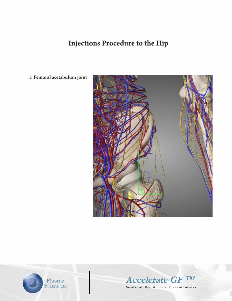
Injections Procedure to the Hip
1. Femoral acetabulum joint

Injections Procedures for the Hip Tendons/Bursa
1. Anterior hip flexors

Injections Procedures for the Hip Tendons/Bursa
1. Trochanteric Bursa

Normal Anatomical Structures of the knee
1. Anterior hip flexors

Injection procedure to the Knee
1. Medical/Lateral compartmentsof the knee
1. Patella-femoral joint

Normal Anatomical Structures of the Knee

Injection Procedure to the Knee Ligaments
1. Medial/lateral collateral ligament
2. ACL/PCL
3. Medial/lateral meniscus
4. Patella ligament

Injections Procedure to the Knee Tendons
1. Patella tendon
2. Quadriceps tendon

Normal Anatomical Structures of the Ankle

Injection Procedures to the Ankle Joints
1. Tibial-talar joint
1. Sub-talar joint

Injection Procedures to the Ankle Joints
1. Sub-talar joint

Normal Anatomical Structures of the Ankle

Injection Procedures for Ankle Ligaments
1. Anterior tibiofibular ligament
2. Anterior talofibular ligament
3. Calcaneofibular ligament

Normal Anatomical Structures of the Ankle

Injection Procedures for Ankle Ligaments
1. Anterior tibiofibular ligament
2. Anterior talofibular ligament
3. Calcaneofibular ligament

Injection Procedures to the Ankle Tendons
Peroneal longus/bervis
Tibialis posterior
Flexor digitorium longus
Flexor hallucis longus
Achilles tendon

Cervical Facet and Ligament Injection Procedure
Cervical facets
1. Cervical interspinous ligaments

Lumbar Facet and Ligament Injection Procedure
Lumbar facets
2. Lumbar interspinous ligaments


![PLASMA» •SOLUTIONS]...Global Plasma Solutions' patent pending technology produces positive and negative ions ( Cold Plasma ) in the air stream which treats the inside of the air](https://static.fdocuments.in/doc/165x107/5f2645b4ee76d960f10332f7/plasma-asolutions-global-plasma-solutions-patent-pending-technology-produces.jpg)


![[Solutions] Introduction to Plasma Physics and Controlled Fusion Plasma Physics](https://static.fdocuments.in/doc/165x107/55cf9d44550346d033ace210/solutions-introduction-to-plasma-physics-and-controlled-fusion-plasma-physics.jpg)

