teamwork 437 - KSUMSCksumsc.com/download_center/1st/1.Foundation Block/Team Work... · AND TISSUE...
21
▪ VERY IMPORTANT ▪ Extra explanation ▪ Examples ▪ Diseases names: Underlined ▪ Definitions * { إً ر س يُ م اً ل سهَ ان ك ن م إ لُ ح ل ا ة م ي ق ا م ف ك،ِ م لُ ح ل لِ ت ا ق... } ( 2 ) Cell Injury ( 1 ) . teamwork 437 Color Index :-
Transcript of teamwork 437 - KSUMSCksumsc.com/download_center/1st/1.Foundation Block/Team Work... · AND TISSUE...

▪ VERY IMPORTANT▪ Extra explanation ▪ Examples▪ Diseases names: Underlined▪ Definitions
سًرإ}* َ سهًلا ُمي ن كان لم إ الُحمة ي
ما ق ُحلِمك، ف
لل ات ِ {...ق
(2) Cell Injury ( 1 ).
teamwork 437
Color Index :-

OB
JEC
TIV
ES
:
• UNDERSTAND THE CONCEPT OF CELLS
AND T ISSUE ADAPTATION TO
ENVIRONMENTAL STRESS INCLUDING
THE MEANING OF : HYPERTROPHY ,
HYPERPLAS IA , APLAS IA , ATROPHY ,
HYPOPLAS IA , METAPLAS IA WITH THE IR
CL IN IC AL MANIFESTAT IONS .
• I S AWARE OF THE CONCEPT OF
HYPOXIC CELL IN JURY AND ITS MAJOR
C AUSES .
• UNDERSTANDS THE DEF INIT IONS AND
MECHANISMS OF FREE RADIC AL
IN JURY.

ADAPTATION TO ENVIRONMENTAL STRESS
• Cells are constantly adjusting their structure and function to accommodate changing
demands i.e. they adapt within physiological limits.
• As cells encounter physiologic stresses or pathologic stimuli, they can undergo
adaptation. The principal adaptive responses are :
adaptive responses
Hypertrophy
Hyperplasia
Metaplasia
Atrophy
• If the adaptive capability is exceeded or if the external stress is harmful, cell injury develops.
ReversibleIrreversible
*NOTE THAT : Within certain limits injury is reversible, and cells return to normal but severe or persistent stress results in irreversible injury and death of the affected cells.
ات ا للتغيرًلخارجية، طبيعية الخاليا أنها متكيفة جدا
. أثير الخارج بحيث تغير شكلها للتناسب وظيفيا مع الت
: يؤدى ( stress) هنا التأثير
كيف الخلية أما يكون بسبب طبيع فيسيولوج وتت-1Physiological)معه adaptation ) مثل عندما
.يكي حجم العضالت بسبب الرياضة
2- ( pathological adaptation)أو بسبب مرض
مثل تضخم عضله القلب عند ارتفاع الضغط
ممكن يحدث •Injuryلما يكون بسبب مرض
للخلية ، لو كان التأث injuryوهنا وال• ير تنقسم لنوعير
Mild ضعها فتستطيع الخلية بعد كذا الرجوع لوالطبيع
فيسبب موت too severe to adaptلكن لو كان •للخلية

• Hypertrophy: is an increase in the size of the tissue/organ due to the increase in the
size of the cells. In pure hypertrophy there are no new cells, just bigger cells containing
increased amount of structural proteins and organelles
– Increased demands lead to hypertrophy.
– Hypertrophy takes place in cells that are not capable of dividing e.g. striated
muscles.
– Hypertrophy can be physiologic or pathologic
• Examples of physiologic hypertrophy:
Physiologic Muscular hypertrophy. Exercise itself is normal but with talking anabolic steroid this gives pathologic hypertrophy to the muscle.
Physiologic hypertrophy of the uterus during pregnancy.
People who remove one kidney usually experience hypertrophy in the other one, because it compensates the other (removed) one .
breast during lactation

• Example of pathologic hypertrophy:
the cardiomyocytes (the cells of the heart) of the myocardium in heart failure (e.g.
hypertrophy in hypertension or aortic valve disease)
Myocardium facing persistent and increased work load, as in hypertension or with a
narrowed (stenotic) valve- ضيق الصمامات-. adapt by being hypertrophic to generate a
higher contractile force. If the increased demand is not relieved, such as if the
myocardium is subjected to reduced blood flow (ischemia) from an occluded coronary
artery, the cardiac cells may undergo injury. Myocardium may be reversibly injured if
the stress is mild or the arterial occlusion is incomplete or sufficiently brief, or it may
undergo irreversible injury and cell death (infarction).

• Hyperplasia: Increase in the size of an organ or tissue, caused by an increase in the
number of cells
- in response to hormonal stimuli or other growth factors or irritation
-Hyperplasia occurs in tissues that are able to divide or contain abundant tissue stem
cells, so some types of cells are unable to exhibit hyperplasia (nerve, cardiac, skeletal
muscle cells).
- hyperplasia can be induced by hormones (endometrial hyperplasia induced by
estrogen).
– Usually, hyperplasia occurs together with hypertrophy.
– Hyperplasia can be physiologic or pathologic
– Sometimes pathologic hyperplasia acts as the base for cancer to develop from.
Thus, patients with hyperplasia of the endometrium are at increased risk of developing
endometrial cancer.
Hyp
erp
lasi
a Physiologic
Pathologic
Hormonal hyperplasia e.g. the proliferation of the glands of the female breast at puberty and during pregnancy
Compensatory hyperplasia e.g. when a portion of liver is partially resected, the remaining cells multiply and restore the liver to its original weight.
are caused by abnormal excessive hormonal or growth factor stimulation e.g.1- excess estrogen leads to endometrial hyperplasia which causes abnormal menstrual bleeding
2- Benign prostatic hyperplasia.

• Atrophy : Opposite of Hypertrophy. Atrophy is the shrinkage in the size of the
cell.
– When a sufficient number of cells are involved, the entire organ decreases in size, becoming
atrophic. .
– Atrophic cells are not dead but have diminished function. In atrophy there is decreased
protein synthesis and increased protein degradation in cells.
• Causes of atrophy include:
-decreased workload or disuse(e.g. immobilization of a limb in fracture),
- loss of innervation (lack of neural stimulation to the peripheral muscles caused by
injury to the supplying nerve causes atrophy of that muscle)
- diminished blood supply
- inadequate nutrition
- loss of endocrine stimulation (e.g. the loss of hormone stimulation in menopause)
- aging: senile atrophy of brain can lead to dementia.
NOTE:
-HOW? Decreased nutritious supply or diseases, associated with decreased synthesis of cellular building blocks (proteins) and increased breakdown of cellular organelles
-In the human embryo, for example, a number of structures are transient and at birth have already undergone atrophy, e.g. The adrenal glands become smaller shortly after birth because an inner layer of the cortex has shrunk.
Dr.Rekabi ‘note :
Athletes have hypertrophy in their skeletal muscles (large muscles size), If these athletes use anabolic steroids (hormones to build their muscles), male athletes who use these steroids will eventually experience artrophy in their testicles; called testicular atrophy, and they may have liver cell injury because of the anabolic steroids.
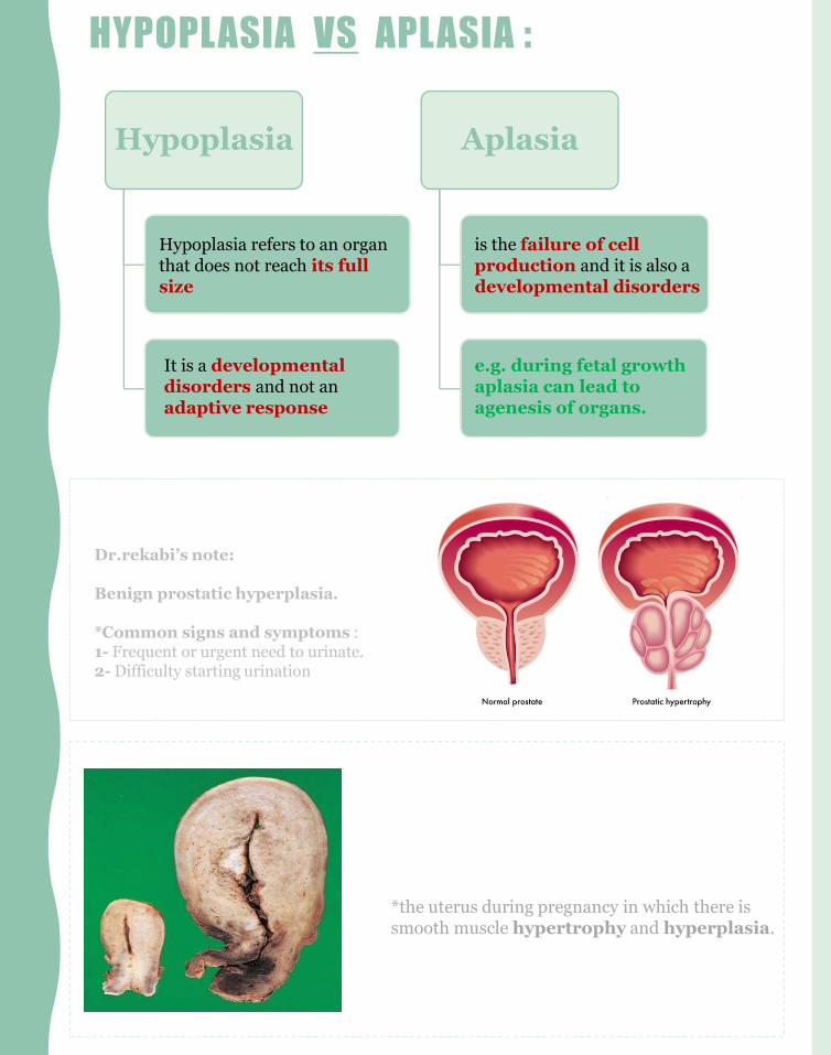
HYPOPLASIA VS APLASIA :
Hypoplasia Aplasia
Hypoplasia refers to an organ that does not reach its full size
It is a developmentaldisorders and not an adaptive response
is the failure of cell production and it is also a developmental disorders
e.g. during fetal growth aplasia can lead to agenesis of organs.
*the uterus during pregnancy in which there is smooth muscle hypertrophy and hyperplasia.
Dr.rekabi’s note:
Benign prostatic hyperplasia.
*Common signs and symptoms :1- Frequent or urgent need to urinate.2- Difficulty starting urination

• Metaplasia : In metaplasia the cells adapt by changing (differentiating) from one
type of cell into another type of cell. This is known as metaplasia.
– Here the cells sensitive to a particular causative agent are replaced by another cell
types better able to tolerate the difficult environment.
– Metaplasia Is usually a reversible provided the causative agent is removed.
Squamous metaplasia
Columnar metaplasia
Osseous metaplasia
Myeloid metaplasia
NOTES :، وطبيعتها - الخلية هنا تتعرض لمؤثر خارج
ب وتركيبها ال يستطيع مجابهه هذا المؤثر، طيوش تسوي ؟ تتحول لنوع أخر يكون باستطاعة
خلية لكن بالمقابل ما كان نوع ال.. تركيبته التحملاالصل موضوع بالمكان هذا عبث ولكن فأنه
باألساس يمتلك صفات يحتاجها النسيج .الموجود
فبالتال لما تتحول لشكل أخر راح تفقد هذه نحتاجها
.الصفات الت
☺راح تفهمون أكير مع األمثلة *
Squamous metaplasia:
Replacement of columnar
By squamous
-in cervix: replacement takes place at the squamocolumnar junction.
-In respiratory tract:-the pseudo stratified columnar epithelium of the bronchus is replaced by stratified squamous cell following chronic injury in chronic smokers.- The stratified squamous epithelium is able to survive better under
circumstances that the more fragile pseudo stratified columnar epithelium would not tolerate. Although the metaplastic stratified squamous epithelium will survive better, the important protective functions of pseudo stratified columnar epithelium are lost, such as mucus secretion and ciliary action.
- If the causative agent persists, it may provide the base (predispose to) for malignant transformation. In fact, it is thought that cigarette smoking initially causes squamous metaplasia, and later squamous cell cancers arise from it.
- Similarly squamous cell carcinoma of cervix also arises from the squamous metaplasia in the cervix. NOTE:
pseudostratified ciliatedطبيعة الخاليا المبطنة للنسيج التنفسي columnar
رى انها تقوم بمنع ذرات الغبار والميكروبات الدقيقة من الدخول لمج ciliaوظيفة ال
طيع المجابهة، التنفس، لكن عن تعرضها لماده النيكوتين في الدخان فأنها تتأذى وال تست
، لكن squamousلذا فأنها تحول لنوع أخر يستطيع التكيف مع هذا الوضع وهي ال
.بالمقابل فأن النسيج التنفسي سيخسر قدرته على منع المايكروبات

• Columnar cell metaplasia:
-It is seen in the esophagus in chronic gastro-esophageal reflux disease.The normal stratified squamous epithelium of the lower esophagus undergoes metaplastic transformation to columnar epithelium
- This change is called as Barrett’s esophagus and it can be precancerous and lead to development of adenocarcinoma of esophagus.
replacement of the
Squamous By columnar
Note:
وكما squamousعند نقصان حموضة المعدة، هذي الحموضة تصعد من المعدة للمريء وألن خاليا المريء عباره عن
فعشان تتكيف مع الوضع columnarنعرف انها خاليا رقيقة ما تتحمل الحموضة القوية، بعكس الحال في خاليا المعدة
.مثل المعدة columnarوتقدر تتحمل هذا الحمض تتحول ل
☺للمعدة؟ من أهم األسباب هو زياده التوتر، لهذا فأن زياده التوتر خطيرة phيقلل الايشطيب
• Osseous metaplasia: it is the formation of new bone at sites of tissue injury. Cartilaginous metaplasia may also occur.
• Myeloid metaplasia (extramedullary hematopoiesis): is the proliferation of hematopoietic tissue in sites other then the bone marrow such as liver or spleen.
يتصدى إي ؟ عشانليشفي بعض األحيان عندنا نتعرض ألصابه قوية يتحول مكان االصابة إلى عظم، يا ترى
.اصابه ثانية بكونه نسيج عظمي قوي

• When the cell is exposed to an injurious agent or stress, a sequence of events follows
that is loosely termed cell injury.
• Cell injury is reversible up to a certain point, but if the stimulus persists or is
severe enough from the beginning, the cell reaches a point of no return and suffers
irreversible cell injury and ultimately cell death.
• Cell death, is the ultimate result of cell injury.
Extra explanation:مثل ما عرفنا قبل، عندما تتعرض الخلية ألي مؤثر فأنها ممكن-
ها لكنها في حالة تستطيع أن ترجع لوضعCell injuryتتعرض ل
يكون الطبيعي، حتى توصل نقطة معينه ما عاد تقدر تتحمل فيها و
.Irreversible cell injuryالضرر كبير لدرجة يصير
بيكون واضح تحت Irreversibleفقط عندما تكون : نقطة مهمه-
Abnormal cell structure. المايكروسكوب
cell structureفغالباً يبين ال reversibleبينما في حالة ال-.طبيعي
For example: Myocardial cells become:
- after 1 to 2 minutes of ischemia: noncontractile. - after 20 to 30 minutes of ischemia: they
become necrotic.- after 2 to 3 hours of ischemia: myocytes may
not appear necrotic by electron microscopy.- after 6 to 12 hours of ischemia: myocytes
appear necrotic under light microscopy
• There are two principal patterns of cell death, necrosis and apoptosis.
Necrosis
-is the type of cell death that occurs after ischemia and chemical injury- it is always pathologic.
Apoptosis
occurs when a cell dies through activation of an internally controlled suicide program.

CAUSES OF REVERSIBLE AND IRREVERSIBLE INJURY
• Causes of both reversible and irreversible injury are the same. اللي يفرق هو قوه التأثير و المدة
الزمنية لكن االسباب نفسها
Causes of cell injury
1)Oxygen Deprivation
(hypoxic cell injury
2)Physical Agents e.g. mechanical
trauma, burns and deep cold,
sudden changes in atmospheric pressure, radiation,
and electric shock
3)Chemical Agents and Drugs e.g. oxygen in high
concentrations, poisons,
pollutants, insecticides,
industrial and occupational
hazards, alcohol and narcotic drugs
and therapeutic drugs.
4)Infectious Agents
5) Immunolog
ic agents e.g. thyroid
damage caused by
autoantibodies
6) Genetic Derangem
ents e.g. sickle cell anemia.
7) Nutritional Imbalance
s
* Also some cell types are more vulnerable to hypoxic injury then others e.g. Neurons are most susceptible followed by cardiac muscle, hepatocytes and then skeletal muscles.

OXYGEN DEPRIVATION:
• Oxygen Deprivation : means Oxygen deficiency. (hypoxic cell injury ). Its common
cause of cell injury and cell death.
– Hypoxia can be due to:
Causes of hypoxia
a) Ischemia (obstruction of arterial
blood flow), E.g. in myocardial infarction and
atherosclerosis.
b) Inadequate oxygenation of the
blood e.g. lung disease and carbon monoxide
poisoning
c) Decreased oxygen-carrying capacity of the
blood e.g. anemia
d) Inadequate tissue perfusion due to
cardiorespiratory failure, hypotension, shock..etc
• Depending on the severity of the hypoxic state, cells may adapt, undergo injury, or die.
Mechanism in hypoxic cell injury:
Hypoxia
Decreased oxidative phosphorylation
Decreased ATP
Membrane damage and proton pump failure
Loss of ion gradient
Anaerobic respiration leading to excess lactic acid
production
Membrane, organelle and nuclear damage
Release of Calcium from mitochondria
Calcium influx into cell from extracellular space which leads to
further injury
* We will talk about them in more more details next slides, don’t worry ☺

MECHANISM OF CELL INJURY :What happens when we have cell injury that may result in cell death?
1. Depletion of ATP
is caused by :
1- reduced supply of oxygen and nutrients.
2- mitochondrial damage
3-some toxins e.g.: cyanide
results in :
1- reduction of plasma membrane pumps activity → imbalance of substances
disturbing the solutes concentration causes the cell to swell and ER to dilate.
2- detachment of RER proteins > less protein synthesis.
3- increase in anaerobic glycolysis using stored glycogen → decrease in glycogen → lactic acid
accumulation → decrease intracellular pH →clumping of nuclear chromatin.
Extra explanation :تكمن في كونه ATPمثل ما نعرف أهميه ال-
مصدر للطاقة، طيب كيف ممكن يقل إنتاجه؟
لو ما فيه اوكسجين ماراح تتم عمليه -1
بداخل oxidative phosphorylationال
.ATPالمايتوكندريا، بالتالي ماراح يكون فيه
ه لو فيه ضرر بالمايتوكندريا، ما راح يكون في-2
ATP ألنه مكان إنتاجه☺
.نفسهATPتضر المايتوكندريا أو الtoxinsأي -3
Extra explanation :.Active transportعشان ال ATPتعتمد على ال Pumpsمثل ما نعرف إن بعض ال-
.down their constrictionجوا، فستبدأ االيونات تنتشر Kبرا وال Na، بتوقف عن ضخ الsodium-potassium pumpمثل -وتزيد ال osmolarityسيدخل لداخل الخلية ويتراكم ، وكما تعلمنا في الفيسيولوجي فأن زيادته داخل الخلية تقلل ال Naإذاً فأن ال-
volume =the cell will swell._______________________________
بكمية قليلة من عملية ATPراح تنتج oxidative phosphorylationمن ATPأيضاً كما نعلم انه عندما سيتوقف عمليه إنتاج ال-
.glycolysisاليتحلل بدوره للطاقة glucoseيتفكك منتجاً glycogenال-
DNAويخرب ال lactic acidنتيجة هذه العملية ستراكم ال-

• 2- mitochondrial damage:
- It is seen specially in hypoxic injury and cyanide poisoning.
- in case of ( low O2 supply/toxins/ radiation ) the mitochondria fails to produce ATP, and instead it produces oxygen derived free radicals. These are harmful to the cell and can lead to necrosis
• 3- influx of calcium activation of enzymes:
– ischemia causes an increase in intracellular calcium concentration, due to malfunction of the Na/Calcium pump
– when calcium enters the cell, because of the damage in the plasma membrane pumps, it participates in chemical reactions that will activate a number of enzymes that trigger apoptosis.
• 4)Ribosomal damage:
– - It is seen in alcohol damage of liver cells and with antibiotic use.
5) increased permeability of the cellular membrane: typically culminate in necrosis.
Several biochemical mechanisms contribute to membrane damage:
1. decreased phospholipid synthesis because the ATP is less available.
2. increased phospholipid breakdown : when the Ca levels are high, it activates the phospholipase enzyme that breaks down the membrane. “the products of the broken lipids also affect the membrane.”
3. oxygen free radicals.
4. cytoskeletal abnormalities: when the Ca levels are high it activates the protease enzyme that damages the cytoskeleton
The affected membranes are :
1.Mitochondrial membrane: → decrease of ATP formation →many deletions → necrosis.
2. Plasma membrane: → imbalance to the permeability → influx of fluids + ions / loss of cellular contents. the also can lose metabolic products thus further reducing the energy stores.
3. lysosomal membranes: → leakage of enzymes → digestion of cellular components → cell death by necrosis

MECHANISM OF CELL INJURY :
5. Free Radical Injury: Accumulation of oxygen-derived free radicals (oxidative stress):
-Free radicals are highly reactive and harmful atoms that have single unpaired electron
in an outer orbit.
-They are referred to as reactive oxygen species/free radicals. The free radicals are produced in our cells
through several ways, called as the free radical generating systems.
• The common free radicals are:
• superoxide anion radical (O2-)
• hydrogen peroxide (H2O2)
• hydroxyl ions (OH)
• Nitric oxide (NO) is an important chemical mediator generated by various cells and it can also
act as a free radical.
Extra explanation :
-Free radicals are: atoms that have lost 1 or more electrons from their outer orbit are usually harmful.
-Free radicals in the cell are usually from Oxygen.
-The free radicals are considered unstable, therefore its hyper reactive (searches for a way to become stable).
- therefore it must enter biochemical reactions in order to bind to another atom to form a new compound and become stable.
-This causes cell injuries. Why? Because the reaction it enters might be dangerous and toxic to the cell.
Free radicals are produced by:
1. Normal metabolism/ respiration:Small amounts of harmful reactive oxygen is produced as a bi-product of mitochondrial respiration during normal respiration (reduction-oxidation reactions that occur in normal metabolism)
2. Ionizing radiation injury:e.g. UV light, x-rays
3. Chemical toxicity:enzymatic metabolism of exogenous chemicals or drugs.
4. Oxygen therapy and reperfusion injury
5. Immune response or inflammation (neutrophil oxidative burst)
6. Transition metalse.g. iron and copper

MECHANISM OF CELL INJURY :
▪ Free radicals cause damage to lipids, proteins, and nucleic acids. The main
damaging effects of these reactive species are:
1. Lipid peroxidation of membranes
damage of membranes & organelles etc..
2. Oxidative modification of
proteins
protein fragmentation
3. DNA damage
lead to cell aging and malignant
transformation of cells.
*When they react with other molecules, they initiate a reaction to transform the other molecules into free radicals.
• How does our body fight the free radicals?
Certain substances in the cells remove/ inactivate the free radicals in order to minimize injury caused by them. They are called as the free radical scavenging system. They are:
- Antioxidants: vitamins E, A and C (ascorbic acid).- Enzymes: which break down hydrogen peroxide and superoxide anion e.g.Catalase, Superoxide dismutase, Glutathione peroxidase and mannitol.
NOTE: Any imbalance between free radical-generating and radical-scavenging systems results in oxidative stress causing cell injury.

MORPHOLOGICAL CHANGES OF REVERSIBLE AND IRREVERSIBLE CELL INJURY
• Earliest changes associated with cell injury are reversible. They are:
1) Swelling & vacuolization of cytoplasm called hydropic/ vacuolar degeneration.
Disturbance in cytoplasmic membrane permeability (gain of Na and loss of K)
2) Mild mitochondrial swelling. the rough endoplasmic reticulum and plasma
membrane damage.
3) Defect in protein synthesis. Due to the mitochondrial swelling and damage.
5) Mild eosinophilia of cytoplasm (due to decrease in cytoplasmic RNA)
6) cytoplasmic membrane blebs
-Fatty change: appearance of small or large lipid vacuoles in the cytoplasm (e.g. in
hepatocytes and myocardial cells)
تسمح ATP=>sodium potassium pumpيقل انتاج < =نقص االوكسجين
ب بسبللخليهيدخل الماء <=مما يزيد تركيزه الخليهداخل Naبدخول كميه اكبر من
osmosis= >cell swell= >cell organelles swell= >ل نتيجه
rough ER swell وبالتالي تتاثرالرايبوسوماتdefect protein synthesis الرايبوسوماتالموجود في RNAتزداد حامضيه السيتوبالزم بسبب نقص < =
*The order is
important

• Persistent or excessive injury, however, causes cells to pass the threshold into
irreversible injury. Irreversible injury is marked by
1.Severe mitochondrial damage with the appearance large, amorphous densities in mitochondria. (severe mitochondrial dilatation and damage. Large, shapeless (amorphous) densities are also present) → (lack of oxidative phosphorylation and ATP generation).
2.Severe plasma/cell membrane damage
3.Increased eosinophilia** (appears red under the microscope.)
4.Numerous myelin figures: a rolled-up or scroll-like arrangement of a lipid bilayer within a cell, superficially resembling the myelin sheath of nerves. Formed from the fragmentation of the lipid bilayer.
5.Rupture of lysosomes leakage and enzymatic digestion of cellular contents(lysosomal membranes results in the enzymatic dissolution of the injured cell)
-Nuclear damage:
1) karyolysis (dissolution), in this case the nuclear chromatin is lost, therefore there is a decrease in basophilia. Why a decrease in basophilia? the chromatin of the nucleus is lost; therefore hematoxylin stain has less DNA to bind with, hence there is less blue color or basophilia
2) pyknosis (shrinkage) in this case the nucleus shrinks and the DNA condenses into a solid mass. There is an increase in the nucleus’ basophilia. Why an increase in the basophilia? because the DNA comes together and the stain makes it look very dark blue
3) karyorrhexis (break down)(this happens after pyknosis) The pyknotic nucleus undergoes fragmentation.
NOTE:
**Why is there increased eosinophilia?
reason: the DNA is damaged → cannot form RNA → decrease RNA in cytoplasm.
Didn’t get it ? Remember that there are two dyes used when staining a cell; hematoxylin and
eosin. Hematoxylin a base and binds with acidic parts of the cell, especially the nucleus. Eosin is
an acid and binds with basic parts of the cell (parts of the cytoplasm). When hematoxylin binds
with parts of the cell this part appears in dark blue. On the other hand when eosin binds to
parts of the cell they appear pinkish. Usually, when the cell is active, lots of RNA are being
synthesized. (mRNA, tRNA, and rRNA). When the cell is strongly damaged, the DNA is no
longer capable of synthesizing RNA, which means that the cell is less capable of building
proteins. Less RNA in the cytoplasm means that there is less blue color of hematoxylin (RNA
is an acid and hematoxylin is a base). When the blue color is less, the pinkish color of eosin is
clearer; and that is what increased cytoplasmic eosinophilia means.

ME
MB
ER
S :
د ب ة آ ل ماج ن ي ث
ي - حرب روإن ال
ي - ي ي اء آلعن وق
ي -ف ي ي
آلش وهرة الج
ي -هرإب إن آلر رر
مري -رهف آلش
عل- روإن مش مشعد-
رة ال ي من
لم- تس آلشو لمن
ي - ي ي وف آلعن ن
ي -هرإب إن آلر رر
ي -اب ل عورت هدت
رف - الس اطمة ت ق
ري - سام المطي ي إث رم- اد آلف رت دإن - لن رإم ج غ
حي -س آلرإج ن لق ت
ي -اض ورة آلق ن
غ - لاء آلصون إ
ي -حطاب
م آلق ري ن جشن - ورة ب ن
ي -حطاب
وق آلق ش
د- ن دي آلعن ث
صل آلطحان ي ق
ي ماب ار آلي ب دالج عن
ق اجاد حمد ت م
د إ حمد آلرإش د ن العن دإلله ت عن
ي اب دإلله آلسرج عن
ي حرب إ حمد ال
ف س آلشي ن إ
ل سماعب دإود إ الد آلدوسري
ج
ر اي هد آلق ف
وف ن معي حمد ب م
ي هاب هد آلن
ف
ي ي دآلع معاد آلعن
إن ور شعد آلف
اري ف المس شي
ي ي م آلوهن مي ي
مي ي إ حمد آلرج
لاع د آلب ن رش م ي ج
آلن حمد
م
Gently contact us if you have any
questions/comments and
suggestions:
* EMAIL: [email protected]
* TWITTER : @pathology437
☺
teamwork 437
Resources:
1- females slides
2- Robbins book