TEAM 10 Assay Development and Alternative … 10/presentation.pdfTEAM 10 Assay Development and...
54
TEAM 10 Assay Development and Alternative Testing Table of Contents Brief description of the activity 2 Team members 3 Personnel training during 2007-2012 5 Overall Personnel competence in the framework of Team 10 7 Communication and intra- and extra-team collaboration 8 Equipment list and level of exploitation 9 On-going Projects 10 Patents and Awards 11 Other relevant informations for Team’s activity 2007-2012 12 Future Research Strategy 13 Projects/contracts finalized in the period 2007-2011 15 International Projects Proposals - not funded 19 Publication list 2007-2013 22 International and national Scientific Conferences 2007-2013 28 Representative Project Title Development of o Novel Immunoassay for the Very Early Detection of Biothreatening Bacterial Infections NATO SfP 982838/2007 34
Transcript of TEAM 10 Assay Development and Alternative … 10/presentation.pdfTEAM 10 Assay Development and...
TEAM 10
Assay Development and Alternative Testing
Table of Contents
Brief description of the activity 2 Team members 3 Personnel training during 2007-2012 5 Overall Personnel competence in the framework of Te am 10 7 Communication and intra- and extra-team collaborati on 8 Equipment list and level of exploitation 9 On-going Projects 10 Patents and Awards 11 Other relevant informations for Team’s activity 200 7-2012 12 Future Research Strategy 13 Projects/contracts finalized in the period 2007-201 1 15 International Projects Proposals - not funded 19 Publication list 2007-2013 22 International and national Scientific Conferences 2 007-2013 28 Representative Project Title Development of o Novel Immunoassay for the Very Early Detection of Biothreatening Bacterial Infections NATO SfP 982838/2007
34
2
TEAM 10
Assay Development and Alternative Testing Brief description of the activity
In the last 4 years our team was involved in the following research directions: A. Immune-based assay development and B. Cell-based assays.
A.Immune-based assay development
In the domain we are currently involved in innovative immune-detection in infectious diseases through the cooperation with University of Tubingen and University of Athens (NATO SfP 982838/2007).
Results. We have established a complete new testing approach for early bacterial infection testing. The innovative assay development consists in designing both new antibodies specific for C-terminal prothymosine peptide and immunoassay detection methods.
In the last 4 years we have published the relation between the in vitro and in vivo bacterial infection and the release of a C-terminal prothymosine peptide. In biological fluids the developed immune-assay can detect in experimental mouse models the mentioned peptide as early as 2 hours post-infection. The possibility to detect as early as 2 hours post-infection a marker by means of chemiluminometric testing is a major breakthrough in this domain. We have published the effect induced by this peptide on innate and adaptive immune cells both in normals and in cancer patients with focus on its adjuvant therapeutical possibilities.
Currently the sensitive test is in the validation step performed mutually in Romania and Germany on human infected samples. The preliminary results in infected children show the possibility to detect in plasma the peptide in concordance with their clinical evolution.
The on-going tests are establishing the specificity as the animal models were infected with various bacterial strains such as: Streptococae, Staphyloccocae, Salmonella, Corynebacterium, Klebsiella, Pasteurella. Time-course of the infection and the sensitivity limits of the test is evaluated.
Funds. The research funds obtained in the framework of this NATO SfP 982838/2007 project are for Romania 300,000 Euros.
Future - development of an original international patent and the technological transfer for developing an easy-to-perform, sensitive and quick test for bio-terrorist attack. Technology transfer is in advanced stages with Panatech Europe GmbH Company, Germany, a company that has been following the project evolution since the start.
The project is described further in this presentation.
B.Cell-based assays development
In this domain we are developing efficient technology/workflow for drug potency identification with emphasis on nano-drugs for controlled delivery.
Results. In the last 4 years through a long lasting collaboration with the National Institute for Chemistry Bucharest we have developed several classes of photosensitizing compounds with anti-tumoral effect. Target potency, cytotoxicity, and metabolic liabilities were evaluated

3
and the selected compounds entered the animal models testing. In animal models in vivo efficacy was tested when reliable models were available.
The hit-to-lead process induced a close collaboration between Assay Development Team and other teams, such as Proteomic and Biomarkers and Drug Development. When a promising lead series has been identified, besides publications, several composition-of-matter patents were accomplished.
The internationally recognized value of the patens was recognized by awards.
Recognition of the last 10 years long-standing work performed by us in this domain, resides in our affiliation to the international networks: COST D39 Metallo-Drug Design & Action (2006-2010); COST TD1002, European network on applications …in NanoMedicine and Life Sciences (2011-2015).
Through cooperation with the University of Lisbon and Technical Institute of Portalegre, we have developed several classes of nano-compounds intended to be intracellular trackers in tumor cells and indicators for minimum residual disease in blood circulation. The project has the following patents developed eith co-inventors from the University of Lisbon: OSIM Nr. ROBOPI2/ 2009 entitled “Tetrapirolic compound asymmetrically substituted – synthesis and biological evaluation” and RO125018-A0 “, OSIM 126761 A0 RO-BOPI 10/2011 entitled „Bio functionalized porphyrinic compound has tetrapyrrolic heterocycles class, synthesized and optimized for cellular load assays „
The involved laboratories are affiliated to the National Platform for Nanomedicine.
Prizes and awards
The project that was developed in our team entitled “Photochemotherapy innovative methods with nanostructured photosensitizers – from synthesis to clinical trial” funded from National Reserach Programm received the First Prize of the Romanian National Authority for Research on Health Domain in 2008.
The patent OSIM Mr. 00489/25.06.2008 entitled “Tetra-sulphonated porphyrin application for producing a dermatologic therapy –photosensitizer” received Gold medal at Brussels Innova 2008, Special Prize of Rudy Demotte, Minister President of the Walloon Government, Gold medal at The 37th International Exhibition of Inventions of Geneva 2009 and Special Prize of the Ministry of Education of Rusia, 2009; Gold medal at The International Fair for Innovation, Moscow, 2009.
In the cell-based assay development we have developed specific equipments for cell imaging that were subject for patent OSIM A/00351/2019 / 21.04.2010 entitled “Equipment and procedure for microwave irradiation in in vitro models with concomitant registration of biological behaviour in a fluorescence microscope”.
Funds. Developing several nationally granted projects the funds obtained by the research team in this domain heaves up to 850,000 Euros. Through MNT-ERA –NET 7050/2010 project the team was financed with 115,000 Euros.
Future of this research direction lays in increasing the nano-drug specificity with emphasis in targeting tumour receptors and tissue markers for not only a controlled delivery in time but as well in space. Until 2013 we will develop several others in the cell-based assay for efficient drug delivery.
4
TEAM MEMBERS First and Last Name Role in the
team Involvement
Background Age
Monica Neagu Team Leader 0,50
Seniour Researcher, CSII, PhD
50
Carolina Constantin Member 0,50
Seniour Researcher, CS III, PhD
40
Dan Ciotaru Member 0,3
Seniour Researcher, CS III, biologist
59
Mihaela Surcel Member 0,3 Seniour Researcher, CS III, chemist
40
Cristiana Tanase Member 0,3 Seniour Researcher, MD, PhD, CSII
58
Radu Albulescu Member 0,15
Seniour Researcher, PhD, CSI
52
Elena Codrici Member 0,2 Researcher, CS III, PhD
33
Daniela Popescu Member 0,25
Researcher, PhD student
35
Georgiana Dumitrascu Member 1 Researcher, Physician
25
Simona Mihai replacing Lucian Albulescu during his PhD at Utrecht University
Member 0,2 Researcher, Physician
25
Emilia Manole Member 0,3 Seniour Researcher, PhD, CS II
53
Gheorghita Izvoranu Member 0,3 Researcher, CS III, PhD
36
Alina Nita Member 0,2 Asistant Researcher 25 Angela Petrescu Member 0,5 Seniour Researcher,
PhD, CSIII 50
Technical personnel First and Last Name
Role in the team Involvement
Background Age
Mariana Caralicea Technician 0.5 Medical Assistant 60 Mariana Pisica Technician 0.5 Laboratory
technician 48
Irina Radu Technician 0,2 Student 23 Laurentiu Anghelache
Technician Veterinary Physician student
0.5 Animal Husbandry
25

5
Team 10 - Cumulated Hirsch index and cumulated cita tions for all the team members.
Team 10 – Personnel dynamics and number of paper /b ooks/chapter in books published 2007-2013
Personnel training during 2007-2012
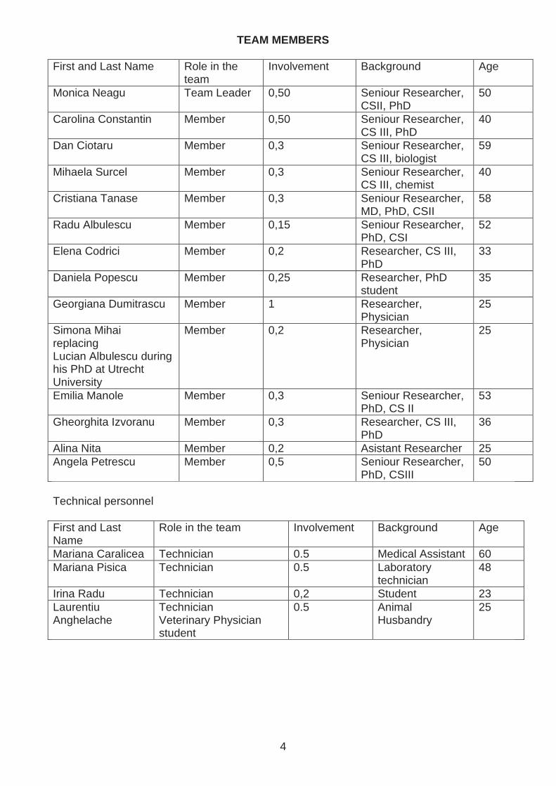
The personnel dynamics in our team is remarkable, namely we have a mean age of 41.5 years and equilibrated between young researchers and more experienced senior researchers and have hosted through the international collaborations several PhD students that fulfilled their thesis in subject developed by the research team. The knowledge up-grading of our team is continuous such as, each of the team members attends international courses on up-to-date technologies.
The following courses and training stages were attended:
2007
Profiling Kinases and Phospho-Sites with Antibody-Based Methods for Disease Biomarker; Drug Target Discovery and Protein Arrays for Biomarker Discovery and Protein Expression Profiling, Amsterdam, Holland; (Monica Neagu, Cristiana Tanase, Daniela Popescu)
Total citation and H-index
106
10
3026
34
104
63
48
19 2522 26 28
0
20
40
60
80
100
120
2006 2007 2008 2009 2010 2011 2012 2013
No. citation
Total H-index
0
2
4
6
8
10
12
14
16
18
20
2007200820092010201120122013
rese
arch
per
sone
l
pape
rs
book
s an
dch
apte
r(s)
research personel
papers
books and chapter(s)
6
Luminex Fundamental Assay Techniques – Protein, Oosterhout, Holand (Elena Codrici, Lucian Albulescu)
2D electrophoresis training, Praga, Czech Republic (Elena Codrici)
Measurement uncertainty in medical laboratories, CALILAB, Laboratory Quality Association, Bucharest (Elena Codrici)
Evaluator for International Project Management, Bucharest, Romania (Monica Neagu)
2008
Principles and applications of microfluidics in the life sciences – Training Course, Barcelona, Spain; (Monica Neagu, Carolina Constantin, Mihaela Surcel)
Microfabrication technologies for Microfluidic Devices Training Course, Barcelona, Spain; (Monica Neagu, Carolina Constantin, Mihaela Surcel)
ProteinChip SELDI-ToF MS Training Course, Malvern, USA; (Monica Neagu, Daniela Popescu, Cristiana Tanase)
Training stage at University of Athens, Laboratory of Human and Animal Physiology and Laboratory of Biochemistry and Molecular Biology, in the frame of NATO project SfP 982838/2007 (Carolina Constantin)
Course: CEI Spring Workshop for Young Researchers: DEVELOPING ENTREPRENEURIAL SKILLS FOR FUTURE CAREER, Poznan, Poland, (Carolina Constantin)
In vivo confocal microscopy training, Mavig, Bucharest, Romania; (Monica Neagu)
Quality management course, RENAR, Bucharest (Elena Codrici)
Course Real-Time PCR – TATAA Biocenter, Prague, Czech Republic (Gheorghita Isvoranu)
Master in Biostatistics, Statistic evaluation of peripheral immune circulatory iimune subpopulations in cutaneous melanoma patients diagnosed in stages 1-4, coordinator Prof.dr Denis Enachescu, June 2008, "Carol Davila" University of Medicine and Pharmacy, Bucharest, Mathematics Faculty, University of Bucharest (Angela Petrescu)
2009
Flow Cytometer BD FACSCANTO II Training Course – BD Biosciences, Heildelberg, Germany (Monica Neagu, Mihaela Surcel)
Training stage in mass spectrometry at University of Tuebingen, Germany in the frame of NATO project SfP 982838/2007 (Lucian Albulescu)
Cell Culture Seminar – LGC Standards, Bucharest, Romania (Carolina Constantin, Mihaela Surcel, Dan Ciotaru, Isvoranu Gheorghita)
Advanced BD FACSCANTO II and BD FACSDiva 6 Training, Bucharest, Romania. (Dan Ciotaru, Mihaela Surcel, Isvoranu Gheorghita)
Training auditors of Quality Management System in the clinical laboratories, according to SR EN 15189:2007 and ISO 19011:2003, FiaTest, Bucharest (Daniela Popescu, Elena Codrici)
2010
xCELLIgence Hand-on training Victor Babes Institute, Bucharest, Romania (Monica Neagu, Carolina Constantin, Daniela Popescu, Elena Codrici, Cristina Tanasei)
xCELLIgence Users Meeting, Munchen, Germany (Monica Neagu)
Intensive Educational Course in Clinical Immunology, Centre de Recherche des Cordelieres, Paris (Monica Neagu, Carolina Constantin)
7
10th Young Scientist Forum - Life of Molecules, Gothenburg (bursary Ionela Daniela Popescu)
Course: „SR EN ISO/CEI 17025 – General requirements for the competence of testing and calibration laboratories”, organized by the Romanian Society of Accreditation (RENAR), Bucharest, Romania (Isvoranu Gheorghita)
2011
xCELLIgence Users Meeting, Rome, Italy (Monica Neagu)
Continuing Education Course “Characterizing and applying physiologically-based pharmacokinetic models in risk assessment” – WHO, Paris, France. (Monica Neagu)
Continuing Education Course «CEC 2 – REACH : How to complete a Chemical Safety Report ?», WHO, Paris, France (Carolina Constantin)
Workshop Autumn Days of Cytometry, Romanian Cytometry Association, Bucharest, Romania. (Mihaela Surcel)
IBM SPSS Statistics, Bucharest (Codrici Elena, Ionela Daniela Popescu )
ProteinChip Pattern Analysis Software Training, Bucharest (Codrici Elena, Ionela Daniela Popescu )
Advanced Proteomics at The St. George’s Medical Biomics Centre, St.George’s University of London, UK (Codrici Elena, Ionela Daniela Popescu )
36 th FEBS CONGRESS, Torino, (bursaries for young researchers Codrici Elena, Ionela Daniela Popescu )
PhD thesis - PhD thesis Modulation of lymphoid cells with cytokine tandems for destruction of tumor cells in mammals; Date: 25.11.2011; Coordinator: Prof. dr. Calin Tesio, University of Bucharest (Gheorghita Isvoranu)
2012
Workshop Multicolor Cytometry Road Show, Bucharest, Romania (Isvoranu Gheorghita)
Cours for web site designers (basic), Top Quality Management, CNFPA licence (Angela Petrescu)
2013
Second Intensive Educational Course in Clinical Immunology, Centre de Recherche des Cordelieres, Paris (Monica Neagu, Carolina Constantin)
We have constant young Bachelor of Science degree personnel that are accomplishing their diplomas in the framework of the mentioned domains. The involved Laboratories are constantly hosting in PhD students or post-doctoral fellows in the framework of the mentioned international collaborations. We have hoasted PhD students Margarita Skopeliti, Pinelopi Samara, Kostas Voutsas from the University of Athens in 2007, 2008, 2010 respectively.
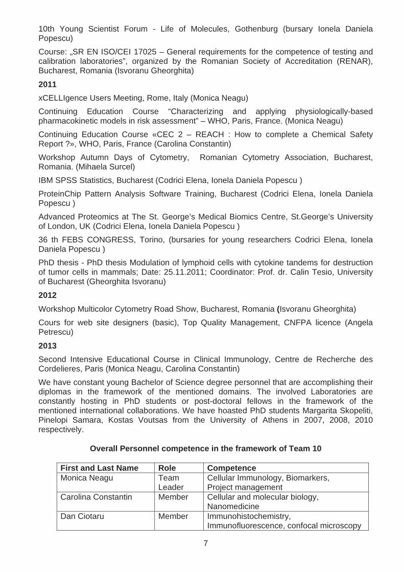
Overall Personnel competence in the framework of Te am 10
First and Last Name Role Competence Monica Neagu Team
Leader Cellular Immunology, Biomarkers, Project management
Carolina Constantin Member Cellular and molecular biology, Nanomedicine
Dan Ciotaru Member Immunohistochemistry, Immunofluorescence, confocal microscopy

8
Mihaela Surcel Member Flow-cytometry, confocal microscopy Cristiana Tanase Member Biomarkers, Translational medicine Radu Albulescu Member Pharmacology, Pharmacodynamics Elena Codrici Member 2D electrophoresis Daniela Popescu Member SELDI-ToF-MS Simona Mihai Member X-Map Array Georgiana Dumitrascu Member ELISA Emilia Manole Member Western-blot Gheorghita Isvoranu Member Animal models Alina Nita Member Economic manager Angela Petrescu Member Bioinformatics and biostatistics Mariana Caralicea Member Electrophoresis Mariana Pisica Member Laboratory technician Irina Radu Member Secretary and correspondence Laurentiu Anghelache Member Animal Husbandry
Communication and intra- and extra-team collaborati on
Extra team collaboration
Team 10 closely collaborates with Team 5 Proteomic biomarkers and Team 6 Immunomodulation-Immunodiagnosis having common research projects and therefore numerous joint publications and communications.
Team 10 collaborates with all the other institute’s teams in various projects were team member’s 10 competence is required.
Intra-team collaboration
Intra-team communication and collaboration is sustained by journal clubs and team workshops that are organized in the following occasions:
• Project proposals for debating the future aim, objectives, work plans
• Project approval and paperwork appending for research contract
• For project’s evolution monitorization at scheduled time-points; discussions for both scientific and administrative issues
• Scientific drafts papers of the team members
• Scientific published paper that are of interest for the team’s preoccupations
• Scientific communications of the results
• Virtual meetings with our international partners
Ad-hoc meetings are always welcomed for brain-storming of particular scientific issues and immediate results discussion.

9
Equipment list and level of exploitation
The laboratories involved in the Assay development team have high-throughput technology with specialized software. Although the equipment is recently purchased (2007-2012) de level of exploitation by the described team is over 75%. Domain Equipment
Year of Aquisition
Level of exploitation (%)
2D electrophoresis 2010 75 2D-DIGE (Typhoon 9000) 2011 25 SELDI-ToF-MS 2008 50 Western blotting 2005 100 Multiplex xMAP® technology 2008 100 Protein microarray 2009 75
Proteomics
Experion microelectrophoresys system 2007 100 Complete unit cell culture manipulation and biobank storage
2007-2012 100
Automatic Cell Counter Countess 2012 75 Complete ELISA lines 2009 100 xCELLigence impedance measurement 2010 50 Confocal microscopy 2007 100 Varioskan Multimode Reader for time-resolved fluorescence, chemiluminescence
2009 75
Flow Cytometers: BD FACSCalibur, BD FACSCanto II
2010 100
Cellular physiology testing
Cellular Separation System: VarioMACS (Miltenyi Biotec)
2010 100
10
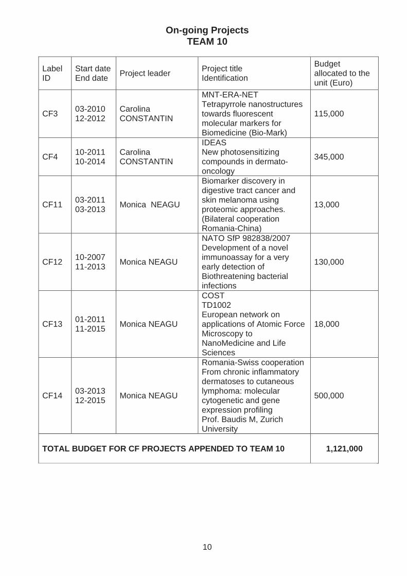
On-going Projects TEAM 10
Label ID
Start date End date
Project leader Project title Identification
Budget allocated to the unit (Euro)
CF3 03-2010 12-2012
Carolina CONSTANTIN
MNT-ERA-NET Tetrapyrrole nanostructures towards fluorescent molecular markers for Biomedicine (Bio-Mark)
115,000
CF4 10-2011 10-2014
Carolina CONSTANTIN
IDEAS New photosensitizing compounds in dermato-oncology
345,000
CF11 03-2011 03-2013
Monica NEAGU
Biomarker discovery in digestive tract cancer and skin melanoma using proteomic approaches. (Bilateral cooperation Romania-China)
13,000
CF12 10-2007 11-2013
Monica NEAGU
NATO SfP 982838/2007 Development of a novel immunoassay for a very early detection of Biothreatening bacterial infections
130,000
CF13 01-2011 11-2015
Monica NEAGU
COST TD1002 European network on applications of Atomic Force Microscopy to NanoMedicine and Life Sciences
18,000
CF14 03-2013 12-2015
Monica NEAGU
Romania-Swiss cooperation From chronic inflammatory dermatoses to cutaneous lymphoma: molecular cytogenetic and gene expression profiling Prof. Baudis M, Zurich University
500,000
TOTAL BUDGET FOR CF PROJECTS APPENDED TO TEAM 10 1,121,000
11
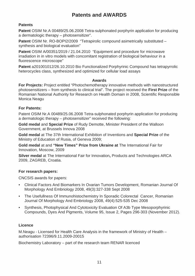
Patents and AWARDS Patents
Patent OSIM Nr.A 00489/25.06.2008 Tetra-sulphonated porphyrin application for producing a dermatologic therapy – photosensitizer”.
Patent OSIM Nr. RO-BOPI2/2009 “Tetrapirolic compound asimetrically substituted – synthesis and biological evaluation”
Patent OSIM A/00351/2019 / 21.04.2010 “Equipment and procedure for microwave irradiation in in vitro models with concomitant registration of biological behaviour in a fluorescence microscope”
Patent a201001012/26.10.2010 Bio Functionalized Porphyrinic Compound has tetrapyrrolic heterocycles class, synthesized and optimized for cellular load assays
Awards For Projects: Project entitled “Photochemotherapy innovative methods with nanostructured photosensitizers – from synthesis to clinical trial”. The project received the First Prize of the Romanian National Authority for Research on Health Domain in 2008, Scientific Responsible Monica Neagu For Patents: Patent OSIM Nr.A 00489/25.06.2008 Tetra-sulphonated porphyrin application for producing a dermatologic therapy – photosensitizer” received the following:
Gold medal and Special Prize of Rudy Demotte, Minister President of the Walloon Government, at Brussels Innova 2008
Gold medal at The 37th International Exhibition of Inventions and Special Prize of the Ministry of Education of Rusia, of Geneva 2009;
Gold medal at and “New Times” Prize from Ukraine at The International Fair for Innovation, Moscow, 2009
Silver medal at The International Fair for Innovation, Products and Technologies ARCA 2009, ZAGREB, Croatia.
For research papers:
CNCSIS awards for papers:
• Clinical Factors And Biomarkers In Ovarian Tumors Development, Romanian Journal Of Morphology And Embriology 2008, 49(3):327-338 Sept 2008
• The Usefullness Of Immunohistochemistry In Sporadic Colorectal Cancer, Romanian Journal Of Morphology And Embriology 2008, 49(4):525-535 Dec 2008
• Synthesis, Photophysical And Cytotoxicity Evaluation Of A3b Type Mesoporphyrinic Compounds, Dyes And Pigments, Volume 95, Issue 2, Pages 296-303 (November 2012).
Licence
M.Neagu - Licensed for Health Care Analysis in the framework of Ministry of Health – authorisation 72396/9.11.2009-20015
Biochemistry Laboratory – part of the research team RENAR licenced

12
Other relevant informations for Team’s activity 2007-2012
Membership
Team leader is an active member of the Commission for Advanced Therapies – European Medicine Agency since 2010, Founding member of the Romanian Dermato-oncology Society – 2010, Member of Romanian Division of International Academy of Pathology – 2009, International Society of Porphyrins and Phthalocyanines – 2007. International Society of Program Management – 2007
Team leader is Member of the Editorial Board for the journal Recent Patents in Biomarkers, Bentham Science Publishers, Guest Editor for 2012 Special Issue Transcriptomics biomarkers for diagnosis, prognosis and therapy monitoring in cancer Recent Patents in Biomarkers , Bentham Science Publishers.
Team leader is Member of the Editorial Board for the journal World Journal of Methodology Number ID:02445884.
Team leader and one team member is evaluator for EuroNanoMed Projects. One team member is National Contact Point for FP7 projects and Member of the Editorial Board for Journal of Immunoassay&Immunochemistry.
Members of the Assay Development team are actively peer-reviewing for the following scientific journals:
Pigments and Dyes, Photochemical and Photobiological Sciences, Patents in Biomarkers, Archives of Gerontology and Geriatrics, International Journal of Photoenergy, Romanian Biotechnological Letters, Romanian Archives of Microbiology and Immunology, Materials Science and Engineering B, Journal of Clinical Laboratory Analysis, International Journal of Nanomedicine, Current Biomarker Findings, Drug Design, Development and Therapy, Journal of Inflammation Research.
Teaching activity:
Team leader is Lecturer in Immunology for Romanian Sciety of Immunology training course since 2002.
Team leader and members of Team 10 are involved in teaching programmes as lecturers in 3 EU Funded Projects :
POSDRU nr. 31081, 1.2 University for future “Dermato-oncology development as multidisciplinary domain for medical training and international cooperation”
POSDRU nr. 58819. „Training platform for implementing high technology in the health care system”
POSDRU nr. 59497 “Training for implementing in the Immunology Laboratory of high technology”

13
Future Research Strategy for Team 10
Research
We have developed a research plan in the framework of overall institute’s strategic directions aiming to improve early detection and diagnosis, develop effective and efficient treatments with the designated role of improving the quality of health care and patient’s quality of life. We are foreseen the development of partnerships with institutes that are synthesizing nanomedicines, enlarging our participation in national and international networks on this field. The constant collaboration with clinical units orients our research in targeted nanomedicine.
We will develop new assays for innovative therapies using nanotechnology and other advanced technologies focusing on:
Drug design for targeted therapy
• Complex toxicology evaluation for new drugs and/or improved classical drugs
• In accordance with the research directions of the institution we will emphasizes on translational medicine through its Diagnostic Centre and will encourage the clinical trials participation enlarging thus the public-private collaboration.
• Patents are foreseen as well in the future taking into account our previous experience and success in the field.
We will orient in the future our team toward discovery of novel therapy targets using gene silencing, and will take on board new biomarkers from the integromics domain providing new insights in the mechanisms underlying tumorigenesis. Developing the Analytic approaches of innovative therapies we anticipate intelligent drug delivery new research direction that will drive various scientific programs within the institute in the near future.
As we already have from our team members’ evaluators for FP7 Health Research Programm, IMI, EuroNanoMed and Commission for Advanced Therapy (EMEA) we are aiming at enlarging the number of peer-reviewers and scientific Board members in international prestigious journal.
Searching for common projects in private-public cooperation as Excellence Poles is one of our future goals.
Education and training
As over 50% of the team memebers are engaged in the ongoing research projects funded by European Structural Funds in order to provide knowledge for researchers of the described institute’s teams we will increase in house expertise under supervision of the team leader from University of Brussels in proteomics. In the same direction, as 75% of the team members are involved in the ongoing training projects funded by European Structural Funds in order to provide knowledge for medical personnel employed by clinical laboratories appended to EU development regions of Romania we will continue to provide training and expertise in new technologies concerning molecular diagnosis (series of lectures and hands-on training in: molecular cytogenetics, gene microarray, proteomics, flow cytometry, in situ hybridization, PCR, archived tissue molecular histopathology, molecular imaging, spectrometry, EU project management, in vivo histopathology).
Due to the international projects we are currently involved in, continuous training of the research personnel is performed in the framework of the scientific strategic directions.
The continuous task of scientific knowledge up-dating through training/seniour mobility/invited speaker at international level is one of our future goals as well.

14
Human Resources
The team is commited to attract and retain qualified direct research, medical care and technical staff. As we are actively training young Master and PhD students we will train and attract young research personnel suited for the scientific objective strategy.
Subjected Projects in international competitions
Infect-ERA ERA-NET
Title Evaluation of molecular interactions in severe co-infections of respiratory tract, with influenza virus and respiratory syncytial virus co-infected with S.pneumoniae and Streptococci from oral cavity, Clamydophila pneumonia, Mycoplasma pneumoniae, in order to improve the diagnosis and treatment strategies in Romania
Dead-line – first step 19th of April 2013
Cooperation with:
Cantacuzino Institute, Dr. Mariana Pana, Dr.Cristina Tecu – Microbiology Reference Center
Technical University of Kaiserslautern, Prof. Dr. Hakenbeck – Microbiology Professor, Prof. Dr. Bruckner - Microbiology Professor The Institute of Microbiology, Immunology and Biotechnology, Lodz, Poland, TBA
15
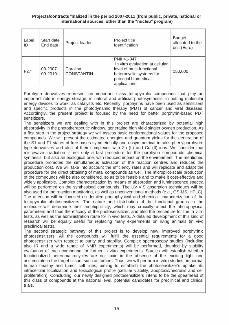
Projects/contracts finalized in the period 2007-201 1 (from public, private, national or international sources, other than the "nucleu" prog ram)
Label ID
Start date End date Project leader Project title
Identification
Budget allocated to the unit (Euro)
F27 09-2007 09-2010
Carolina CONSTANTIN
PNII 41-047 In vitro evaluation at cellular level of multi-functional heterocyclic systems for potential biomedical applications
150,000
Porphyrin derivatives represent an important class tetrapyrrolic compounds that play an important role in energy storage, in natural and artificial photosynthesis, in putting molecular energy devices to work, as catalysts etc. Recently, porphyrins have been used as sensitisers and specific products in the photodynamic therapy (PDT) of cancer and viral diseases. Accordingly, the present project is focused by the need for better porphyrin-based PDT sensitizers. The sensitizers we are dealing with in this project are characterized by potential high absorbtivity in the phototherapeutic window, generating high yield singlet oxygen production. As a first step in the project strategy we will assess basic conformational values for the proposed compounds. We will present the estimated energies and quantum yields for the generation of the S1 and T1 states of free-bases symmetrically and unsymmetrical tetrakis-phenylporphyrin-type derivatives and also of their complexes with Zn (II) and Cu (II) ions. We consider that microwave irradiation is not only a fast procedure for the porphyrin compounds chemical synthesis, but also an ecological one, with reduced impact on the environment. The mentioned procedure promotes the simultaneous activation of the reaction centres and reduces the production cost. We will take into account the efficiency rates and will replicate and adapt the procedure for the direct obtaining of metal compounds as well. The micropilot-scale production of the compounds will be also considered, so as to be feasible and to make it cost-effective and widely applicable. Complex characterization by means of absorption and luminescence spectra will be performed on the synthesised compounds. The UV-VIS absorption techniques will be also used for the reaction monitoring, as well as unconventional methods (e.g., GS-MS, HPLC). The attention will be focused on detailed photophysical and chemical characterization of the tetrapyrrolic photosensitizers. The nature and distribution of the functional groups in the molecule will determine their amphiphilicity, which may crucially affect the photophysical parameters and thus the efficacy of the photosensitizer, and also the procedure for the in vitro tests, as well as the administration route for in vivo tests. A detailed development of this kind of research will be equally useful for replacing many experiments on living animals (in vivo preclinical tests). The second strategic pathway of this project is to develop new, improved porphyrinic photosensitizers. All the compounds will fulfill the essential requirements for a good photosensitizer with respect to purity and stability. Complex spectroscopy studies (including also IR and a wide range of NMR experiments) will be performed, doubled by stability evaluation of each compound for further in vitro experiments. Studies will establish whether functionalized heteromacrocycles are not toxic in the absence of the exciting light and accumulate in the target tissue, such as tumors. Thus, we will perform in vitro studies on normal human healthy and tumor cell lines, aiming to establish the photosensitizer’s uptake, its intracellular localization and toxicological profile (cellular viability, apoptosis/necrosis and cell proliferation). Concluding, our newly designed photosensitizers intend to be the spearhead of this class of compounds at the national level, potential candidates for preclinical and clinical trials.
16
F28 09-2007 10-2010
Carolina CONSTANTIN
PNII 11-035 Cavitand and coronand structures - new nanotechnologies approach for antitumoral compunds
160,000
Nanospheres of carbon, known as fullerenes have attracted attention of scientific community due to their possible applications in biomedicine. Fullerenes, a novel carbon allotrope, comprise variable carbon atoms number (C60, C70…C200), arranged like a condensed ring aromatic compound with extended _ systems. The international studies regarding the biological properties of fullerenes and their derivatives are nowadays focused on their antitumoral and pharmacological effects and also on their involvement in oxidative stress. Unlike conventional antineoplazic agents, the antitumoral efficiency of these nanoparticles is not characterized by a direct attack on tumour cells. Beside this, photochemical activation of some types of fullerenes and singlet oxygen generation, contribute to the antitumoral effects and define the dual nature of this nanoparticles. Therefore, the major objective of the proposed project is represented by the selection of nanostructures compounds like fullerenes characterized by antineoplazic potential. The specific objectives of the study will be considered as following: the chemical synthesis and physical-chemical characterization of C60, C70 fullerenes including structure optimization for their application in biomedicine; the functionalization of fullerenes structure in order to improve their solubility in biological fluids; the toxicological profile determination for these nanostructures compounds, by in vitro assays with standard normal and tumoral cell lines. Thus, the toxicological studies will be very helpful for choosing the concentration domain in which the nanocompounds are cytotoxic in relation to different time points of action and cellular type. The degree of fullerenes incorporation in cell systems and subcellular localization will be analyzed by confocal and electronic microscopy. The fullerenes mechanisms of action at cellular level in normal and neoplazic cells will be evaluated by assessing the cell multiplication rate correlated with cell cycle. Based on their structure – biological toxicity report, the fullerenes compounds will be selected and further tested in a experimental model of photodynamic therapy with these nanostructures as photosenzitizers. The proposed project will be accomplished in a multidisciplinary partnership (physicians, biochemists, biologists, chemists, physicists), able to complete and aprofundate the studies concerning the in vitro effects of fullerenes in biological systems. The methodology used in this project comprises up-to-date techniques of physics, cellular and molecular biology. The proposed theme is enclosed in general objective of Programme 4 by supporting the collaboration between national research institutes with a long experience in the main interest domains (cellular biology, pathology, immunology and chemistry). The multidisciplinary partnership is involved in a very actual and newly field: identification of new antitumoral agents recruited from nanotechnology area. By its topic, the study fit in the 1.7.6 objective of PNCDI II as a result of investigating some nanocompounds with biomedical application. In addition, the subject of the propose project is in cope with FP7 priority domains: HEALTH-2007-1.3. Predicting suitability, safety and efficacy of therapies; 1.3-1: Novel alternative testing strategies for use in pharmaceutical discovery and development; HEALTH-2007-2.1.2. Systems biology - 2.1.2-4: Developing an integrated in vitro, in vivo and systems biology modeling approach to understanding apoptosis in the context of health and disease. By reason of such sort of determinations are not yet initiated at national level, the aim of the present study is represented by preclinical, in vitro testing of fullerenes like nanostructures, in order to define the cytotoxic/pharmacological profile of mentioned above compounds.
F29 10-2006 09-2008
Cornel URSACIUC Team members Dan Ciotaru, Mihaela Surcel
CEEX - BIOTECH Control of human cells and bacterial interaction with NANO structured surfaces: strategies for the creation of "INTeligent "biosurfaces
38,000

17
F31 09-2007 09-2009
Cornel URSACIUC Team members Dan Ciotaru, Mihaela Surcel
PNII CAPACITIES Upgrading of Biobank for tumor cells and nucleic acids by attaching a imunogenomics laboratory for molecular screening in cancer
421,000
The project proposes the development of the “Victor Babeş” Institute biobank for tumor cells and nucleic acids (BTCNA), by attaching an immunogenomics laboratory for cancer molecular screening. The project involves the upgrading of BTCNA and Immunopathology Lab, in order to form an integrated cellular and molecular medicine system, focused principally on complex cancer investigation.The molecular screening in cancer constitutes a targeted and gradually gene investigation, finally differentiating between tumors belonging to the same group. Immunogenomics associates the molecular screening with immunologic investigation, adding to the gene data the immune status and antitumor immune response characterisation. The Immunogenomics lab will be attached to BTCNA in order to enhance the investigation efficiency by complex sample processing and testing. BTCNA patrimony represents permanently a research material for clinical and experimental oncology, this making stringently needed the applying of immunogenomic specific molecular and cellular techniques.The achievement of the Immunogenomics unit became a necessary and possible objective for “Victor Babeş” Institute institutional development for several reasons: The institute includes BTCNA, which will contain the Immunogenomics unit and also includes an immunology department which performs tumor immunodiagnosis and molecular screening. There is the necessary preoccupation for enhancement the equipment rank with the aim of performant determinations.The institute has the organization and logistic facility to realise the proposed development.
F33 11-2006 09-2008
Cornel URSACIUC Team members Dan Ciotaru, Mihaela Surcel
CEEX - BIOTECH New strategies of cell therapy by using progenitors from umbilical cord blood
88,000
F34 10-2006 09-2008
Cornel URSACIUC Team members Mihaela Surcel
CEEX - BIOTECH The role of membrane lipids in tumor cell response to chemotherapy
50,000
F35 10-2008 09-2011
Cornel URSACIUC Team members Dan Ciotaru, Mihaela Surcel
PNII Bioinformatics system based on artificial intelligence for monitoring of the immune system in correlation with human body metabolic processes
37,000
F43 10-2005 07-2008 Cristiana TANASE
CEEX - MATNANTECH Network of integrated research for nanomedicine (Nanobiotechnology for health)
8,000
F44 10-2005 11-2008 Cristiana TANASE
CEEX - MATNANTECH Condensate systems with phtalocyanine and other metallic complexes carrying oxygen with applications in sensors of ecological and medical interest.
28,000
18
F45 10-2008 12-2011
Dan CIOTARU Team members Mihaela Surcel
PNII Medical-biological complex study of novel usage of potentially therapeutic environmental factors in salt mines and caves for balneoclimatic health and tourism
29,000
The research theme of the project is new, mostly applied with fundamental studies and support activities and dissemination. The project aim is to develop partnership and a comprehensive and mulltidisciplinar study in health care (cellular and molecular immunology, microbiology, cell biology, biochemistry and physiological processes on laboratory animals with experimentally induced pathologies and some groups of patients with chronic inflammatory or allergic) environmental natural resources, with potential therapeutic factors, poorly studied and valued, as salt mines and karst caves not yet used for medical purposes in the country, (specific morphological investigations, microclimatic, physico-chemical, microbiological). The project involves the development of new technical solutions and innovative artificial simulation (modeling) for the proper use of the curative potential of environmental parameters of salt mines and karst caves (speleoterapeutic and national and international balneoclimatic medical-tourism). The study includes - the experience gained in the field in different European countries, obtaining new scientific data through fundamental research and applied studies of the environmental healing factors and their therapeutic effects, biomedical studies on metabolism, clinical trials and in laboratory animals with induced pathologies, in patients with asthma, chronic obstructive bronchitis and skin wounds, to stimulate research and experiment on patients with different pathologies; - implementation of models and optimization of innovative therapies in treating diseases such as asthma, chronic bronchitis and various inflammatory and allergic skin wounds. Preliminary studies and analysis of the field, nationally and internationally, have highlighted several issues in speleotherapy. Complexity, multidisciplinary nature and many issues need to be addressed activities, methodologies and new environmental and medical technologies or innovative solution that will increase compatibility and competitiveness of European research area.Thus, in order to achieve the project objectives and solving health problems are expected experimental and clinical studies of potential therapeutic effects of underground environmental factors on physiological processes and mechanisms, evaluation of potential therapeutic and salt mines investigated, will be developed conceptual models and made models/solutions/technologies speleoterapeutic experimental room with artificial environment and parameters potentially curative salt mine and cave.
F46 10-2008 12-2011
Dan CIOTARU
PNII Study of innovative therapies in infantile hemangiomas selectively applied depending on the form of the disease and the stage of evolution
70,000
F109 10-2008 12-2011
Monica Neagu
PNII Development of protocols for coherent optical multiple fractionated irradiation (SIFROC) inphotodynamic therapy using methyl-aminolevulinate (MAL-PDT) in premalignant and malignant non-melanoma cutaneous diseases
82,000
F110 09-2005 11-2008
Monica NEAGU
CEEX - BIOTECH Anti-aging effects induced by photodynamic therapy with 5-aminolevulinic acid - molecular mechanisms
111,000
19
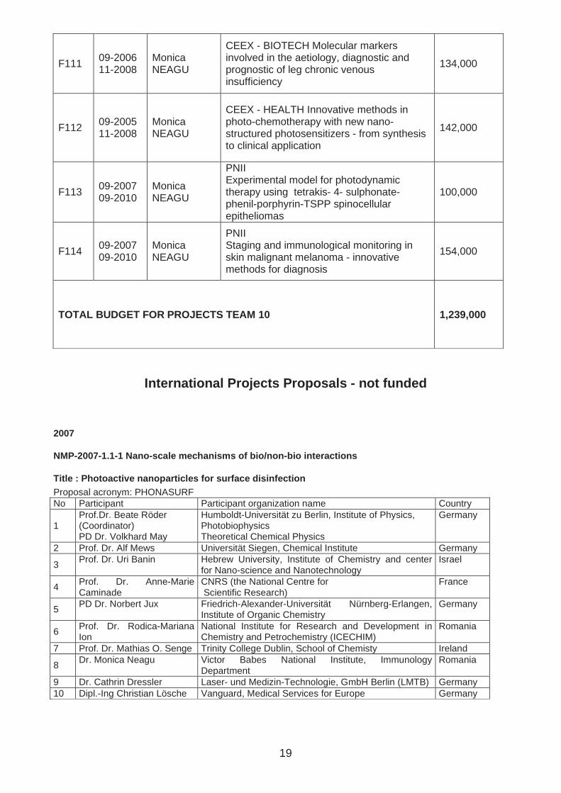
F111 09-2006 11-2008
Monica NEAGU
CEEX - BIOTECH Molecular markers involved in the aetiology, diagnostic and prognostic of leg chronic venous insufficiency
134,000
F112 09-2005 11-2008
Monica NEAGU
CEEX - HEALTH Innovative methods in photo-chemotherapy with new nano-structured photosensitizers - from synthesis to clinical application
142,000
F113 09-2007 09-2010
Monica NEAGU
PNII Experimental model for photodynamic therapy using tetrakis- 4- sulphonate-phenil-porphyrin-TSPP spinocellular epitheliomas
100,000
F114 09-2007 09-2010
Monica NEAGU
PNII Staging and immunological monitoring in skin malignant melanoma - innovative methods for diagnosis
154,000
TOTAL BUDGET FOR PROJECTS TEAM 10 1,239,000
International Projects Proposals - not funded
2007
NMP-2007-1.1-1 Nano-scale mechanisms of bio/non-bio interactions
Title : Photoactive nanoparticles for surface disin fection Proposal acronym: PHONASURF No Participant Participant organization name Country
1 Prof.Dr. Beate Röder (Coordinator) PD Dr. Volkhard May
Humboldt-Universität zu Berlin, Institute of Physics, Photobiophysics Theoretical Chemical Physics
Germany
2 Prof. Dr. Alf Mews Universität Siegen, Chemical Institute Germany
3 Prof. Dr. Uri Banin Hebrew University, Institute of Chemistry and center
for Nano-science and Nanotechnology Israel
4 Prof. Dr. Anne-Marie Caminade
CNRS (the National Centre for Scientific Research)
France
5 PD Dr. Norbert Jux Friedrich-Alexander-Universität Nürnberg-Erlangen,
Institute of Organic Chemistry Germany
6 Prof. Dr. Rodica-Mariana Ion
National Institute for Research and Development in Chemistry and Petrochemistry (ICECHIM)
Romania
7 Prof. Dr. Mathias O. Senge Trinity College Dublin, School of Chemisty Ireland
8 Dr. Monica Neagu Victor Babes National Institute, Immunology
Department Romania
9 Dr. Cathrin Dressler Laser- und Medizin-Technologie, GmbH Berlin (LMTB) Germany 10 Dipl.-Ing Christian Lösche Vanguard, Medical Services for Europe Germany
20
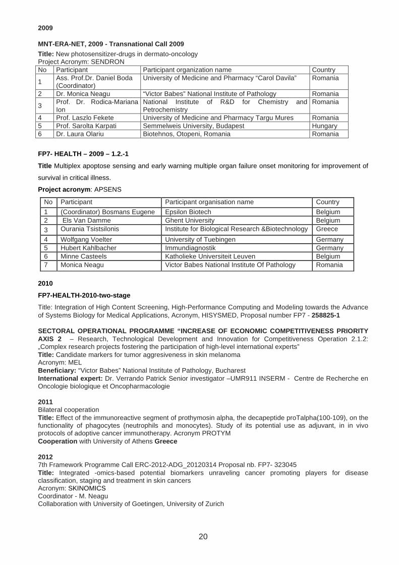
2009
MNT-ERA-NET, 2009 - Transnational Call 2009
Title: New photosensitizer-drugs in dermato-oncology Project Acronym: SENDRON No Participant Participant organization name Country
1 Ass. Prof.Dr. Daniel Boda (Coordinator)
University of Medicine and Pharmacy “Carol Davila” Romania
2 Dr. Monica Neagu “Victor Babes” National Institute of Pathology Romania
3 Prof. Dr. Rodica-Mariana Ion
National Institute of R&D for Chemistry and Petrochemistry
Romania
4 Prof. Laszlo Fekete University of Medicine and Pharmacy Targu Mures Romania 5 Prof. Sarolta Karpati Semmelweis University, Budapest Hungary 6 Dr. Laura Olariu Biotehnos, Otopeni, Romania Romania
FP7- HEALTH – 2009 – 1.2.-1
Title Multiplex apoptose sensing and early warning multiple organ failure onset monitoring for improvement of
survival in critical illness.
Project acronym : APSENS
No Participant Participant organisation name Country
1 (Coordinator) Bosmans Eugene Epsilon Biotech Belgium 2 Els Van Damme Ghent University Belgium 3 Ourania Tsistsilonis Institute for Biological Research &Biotechnology Greece
4 Wolfgang Voelter University of Tuebingen Germany 5 Hubert Kahlbacher Immundiagnostik Germany 6 Minne Casteels Katholieke Universiteit Leuven Belgium 7 Monica Neagu Victor Babes National Institute Of Pathology Romania
2010
FP7-HEALTH-2010-two-stage
Title: Integration of High Content Screening, High-Performance Computing and Modeling towards the Advance of Systems Biology for Medical Applications, Acronym, HISYSMED, Proposal number FP7 - 258825-1 SECTORAL OPERATIONAL PROGRAMME “INCREASE OF ECONOMI C COMPETITIVENESS PRIORITY AXIS 2 – Research, Technological Development and Innovation for Competitiveness Operation 2.1.2: „Complex research projects fostering the participation of high-level international experts” Title: Candidate markers for tumor aggresiveness in skin melanoma Acronym: MEL Beneficiary: “Victor Babes” National Institute of Pathology, Bucharest International expert: Dr. Verrando Patrick Senior investigator –UMR911 INSERM - Centre de Recherche en Oncologie biologique et Oncopharmacologie 2011 Bilateral cooperation Title: Effect of the immunoreactive segment of prothymosin alpha, the decapeptide proTalpha(100-109), on the functionality of phagocytes (neutrophils and monocytes). Study of its potential use as adjuvant, in in vivo protocols of adoptive cancer immunotherapy. Acronym PROTYM Cooperation with University of Athens Greece 2012 7th Framework Programme Call ERC-2012-ADG_20120314 Proposal nb. FP7- 323045 Title: Integrated -omics-based potential biomarkers unraveling cancer promoting players for disease classification, staging and treatment in skin cancers Acronym: SKINOMICS Coordinator - M. Neagu Collaboration with University of Goetingen, University of Zurich
21
National Projects Proposals that were not funded National Research Project - PN-II-PT-PCCA-2011-3 Title: Hesperedine and flavonoids Nanocrystals – in vivo cuantification – by confocal microscopy, Acronym: NANO-HESP Coordinator: Dr. Carolina Constantin Title: Immunomics of melanoma: interferon-stimulated genes (ISGs) products as candidate biomarkers for monitoring tumor evolution and personalized medicine, Acronym: MELIM Coordinator: Dr. Monica Neagu Title: Menstrual Blood Endometrial Regenerative (Stem) Cells - an opportunity for reparative medicine”/MenERC/LS3 Coordinator: : CS II Dr. Cornel Ursaciuc, Members: Dan Ciotaru, Mihaela Surcel Title: Protocol for the diagnosis of premature birth risk using imaging, immunological, morphological, proteomic, immunogenic predictive markers; antepartum and immediate postpartum dynamic assessment”/ DPPRB/LS7 Coordinator: : CS II Dr. Cornel Ursaciuc, Members: Dan Ciotaru, Mihaela Surcel Title: Tuberculin reagent with increased specificity, demonstrated by T cell response against specific antigens encoded in Mycobacterium tuberculosis genome”/PPD specific/LS9 Coordinator: : CS II Dr. Cornel Ursaciuc, Members: Dan Ciotaru, Mihaela Surcel Title: Flexible culture media for cell diferentation and selection”/ MEDIMED/PE4 Coordinator: CS II Dr. Cornel Ursaciuc, Members: Dan Ciotaru, Mihaela Surcel Title: Bioinformatic system based on artificial intelligence for the control of the proliferation of human cells through the management of the metabolism” CELLPROLIFCONT/LS2, Coordinator: CS II Dr. Cornel Ursaciuc, Members: Dan Ciotaru, Mihaela Surcel

22
Publication list 2007-2013 TEAM 10
(see Europass CVs of the team members )
2007
Synthetic porphyrins in experimental photodynamic therapy induce different antitumoral effect, Monica Neagu, et al, Journal of Porphyrins and Phtalocyanins, 2007 Jan;11 (1): 58-65 ISSN: 1088-4246
Laser effect in photodynamic therapy of tumors, Rodica-Mariana Ion, Dragos-Viorel Brezoi, Monica Neagu, et al Proc. SPIE Vol. 6606, 66061G (Apr. 25, 2007) 253-261, doi:10.1117/12.730203
Mechanisms in photodynamic therapy: photosensitizers and cellular localization on K562 cells Rodica-Mariana Ion, Monica Neagu, et al. SPIE -The International Society for Optical Engineering.13 July 2007 Vol: 6632, DOI: 10.1117/12.727968
Molecular changes in superficial bladder cancer. Vrabie CD, Petrescu A, Waller M. Rom J Morphol Embryol. 2007;48(2):131-8.
2008
Cell Investigations Simultaneous with Exposure to 2.45 GHz Microwaves, D. Martin, S. Cinca, I. Margaritescu, M. Neagu, et al, Journal of Microwave Power and Electromagnetic Energy 2008, vol 42 (4), pp. 751-754 ISSN: 0832-7832
Combined Microwave and Electron Beam Exposure Facilities for Medical Studies and Applications, D. Martin, S. Cinca, I. Margaritescu, M. Neagu, et al, Journal of Microwave Power and Electromagnetic Energy 2008, vol 42 (4), 771-774 ISSN: 0832-7832
TGF-beta2 involvements in open angle glaucoma, Stefan C, Dragomir L, Melinte-Dumitrica D, Ursaciuc C, Dobre M, Surcel M - Oftalmologia, 52, 110-112, 2008
Immune response in healthy ageing: immunosenescence versus immunodeficiency, M. Neagu, C. Constantin, G. Manda, R. Huica, M. Surcel, M. Dobre, D. State, D. Gradinaru, C. Ursaciuc, the FEBS Journal, 275 (1), 210, 2008, 33rd FEBS Congress Athens 2008, Greece
Prolonged treatment with interferon alpha and peginterferon induces rheumatoid arthritis syndrome and erythema nodosum. Ionescu C, Micu L, Constantinescu I, Hortopan M, Ursaciuc C, Voiculescu M. J Gastrointestin Liver Dis. 2008 Jun;17(2):211-2.
The usefulness of immunohistochemistry in sporadic colorectal cancer. Vrabie CD, Ceauşu M, Petrescu A, Waller M, Dina I. Rom J Morphol Embryol. 2008;49(4):525-35
Clinical factors and biomarkers in ovarian tumors development. Vrabie CD, Petrescu A, Waller M, Dina I. Rom J Morphol Embryol. 2008;49(3):327-38.
2009
Biomarkers of metastatic melanoma, Neagu Monica, et al, Biomarkers In Medicine, Volume 3, Number 1, February 2009 , pp. 71-89(19)
Research Highlights, Monica Neagu, et al, Biomarkers In Medicine, Vol. 3(4), 343 - 345, 2009.
Toxicological studies with cavitands organic structures in human immune cells experimental models, Constantin C., Neagu M. et al Proceedings of the 2nd European Congress of Immunology, Monduzzi Editore International Proceedings Division, pp 415 – 418, 2009.

23
SELDI-ToF-MS for prothymosin-alpha detection as bacterial infection apoptosis marker - NATO SfP Project, Neagu M., Constantin C., et al. Proceedings of the 2nd European Congress of Immunology, Monduzzi Editore International Proceedings Division, pp 419 – 422, 2009.
Biomarkers in the diagnosis and early detection of pancreatic cancer, Cristiana Pistol Tanase, Monica Neagu, et al Expert Opinion on Medical Diagnostics, Vol. 3, No. 5, Pages 533-546, 2009
Key signaling molecules in pituitary tumors, Cristiana Pistol Tanase, Monica Neagu, Radu Albulescu, Expert Review of Molecular Diagnostics, 9(8), 859-877, 2009
Serum markers in skin melanoma – preliminary study, Georgiana Dumitrascu, et al, Roum Arch Microbiol Immunol, vol 69, pages 125-135, November 2009
New Photosensitizers Versus Aminolevulinic Acid (ALA) in Experimental Photodynamic Therapy of Actinic Keratosis – A Case Report, Daniel Boda, Monica Neagu, Carolina Constantin, et al, Analele Stiintifice ale Universitatii Alexandru Ioan Cuza Din Iasi (Serie Noua)-Genetica si Biologie Moleculara, Tom X, Fascicola 3, pagina 61-69, 2009.
CD28 T-cell costimulatory molecule expression in pemphigus vulgaris, Alecu M, Ursaciuc C, Surcel M, Coman G, Ciotaru D, Dobre M, J Eur Acad Dermatol, 23, 288-91,2009.
Relation between NK cells subpopulations and tetraspanin membrane expression in malignant tumors, Mihaela Surcel, R. Huică, D. Ciotaru, Maria Dobre, Anamaria Belmega, Ioana Pîrvu, Gheorghiţa Isvoranu, C. Ursaciuc, European Congress of Immunology, Monduzzi Editore International Proceedings Division, 77-82, 2009.
In vitro hepatic differentiation of human bone marrow mesenchymal stem cells under differential exposure to liver-specific factors,. Chivu M, Dima SO, Stancu CI, Dobrea C, Uscatescu V, Necula LG, Bleotu C, Tanase C, Albulescu R, Ardeleanu C, Popescu I, Transl Res, 154, 122-13, 2009.
Functionalized polyvinyl alcohol derivatives thin films for controlled drug release and targeting systems: MAPLE deposition and morphological, chemical and in vitro characterization, Cristescu R, Popescu C, Popescu AC, Grigorescu S, Duta L, Mihailescu IN, Caraene G, Albulescu R, Albulescu L, Andronie A, Stamatin I, Ionescu A, Mihaiescu D, Buruiana T, Chrisey DB, Appl Surf Sci, 255, 5600-5604, 2009.
Curcumin derivatives with potential biological activity, Robu M, Tănase C, Boscornea C, Tomas S, Albulescu R, Revista de Chimie, 60, 76-80, 2009.
Caveolin-1 overexpression correlates with tumour progression markers in pancreatic ductal adenocarcinoma, Tanase CP, Dima S, Mihai M, Raducan E, Nicolescu MI, Albulescu L, Voiculescu B, Dumitrascu T, Cruceru LM, Leabu M, Popescu LM, Hinescu ME, J Mol Histol, 40, 23-9, 2009.
CD28 T-cell costimulatory molecule expression in pemphigus vulgaris Alecu M, Ursaciuc C, Surcel M, Coman G, Ciotaru D, Dobre M. J Eur Acad Dermatol Venereol. 2009 Mar; 23(3):288-91.
Inflammatory, degenerative and vascular lesions in long-term dialysed patients. Vrabie CD, Petrescu A, Waller M, Cojocaru M, Ciocâlteu A, Dina I. Rom J Intern Med. 2009; 47(2):149-59.
The histopathology analysis of the diffuse sclerosing variant of the papillary carcinoma of the thyroid: a distinctive and rare form. Vrabie CD, Terzea D, Petrescu A, Waller M. Rom J Morphol Embryol. 2009;50(4):743-8.
Boscencu R, Socoteanu R, Ilie M, Oliveira AS, Constantin C, Ferreira LFV, Synthesis, Spectral and Biological Evaluation of Some Mesoporphyrinic Zn(II) Complexes, Revista de Chimie (Bucharest), 60, 1006-1011, 2009.

24
2010
Fullerene-porphyrin nanostructures in photodynamic therapy, Carolina Constantin, Monica Neagu, et al, Nanomedicine, 5(2), February 2010 Pages 307-317
Antitumoral Effect of Calixarenes on Experimental Photodynamic Therapy with K562 Tumor Cell Line, Monica Neagu, et al, Romanian Journal of Biochemistry, issue 47(1), 2010, Pages 17-35.
Microwave synthesis, basic spectral and biological evaluation of some copper (II) mesoporphyrinic complexes.Boscencu R, Ilie M, Socoteanu R, Oliveira AS, Constantin C, Neagu M, et al. Molecules. 2010 May 25;15(5):3731-43.
Photodynamic Therapy on B16 Cells with Tetrasulphonated Porphyrin and Different Light Sources, Simona-Florentina Pop, Rodica-Mariana Ion, Monica Neagu and Carolina Constantin, Journal of Materials Science and Engineering, Volume 4, Number 3, March 2010 (Serial Number 28), 10-16
Porphyrin (TPP)–Polyvinylpyrrolidone (PVP)–Fullerene (C60) Triad as Novel Sensitizer in Photodynamic Therapy Rodica Mariana Ion, Radu Claudiu Fierascu, Monica Neagu, Carolina Constantin, Crina Stavaru, Science of Advanced Materials, Vol. 2, 223–229, 2010
Biomarkers Discovery In Cancer – up-dates In methodology, Cristina Tacu, Monica Neagu, et al, Roum Arch Microbiol Immunol, vol 69(1), 2010, 48-55
Immune-related biomarkers for diagnosis/prognosis and therapy monitoring of skin melanoma, Monica Neagu, et al, Expert Review of Molecular Diagnostics, 10(7), 2010, 897-921.
Statistical correlations between peripheral blood lymphocyte subpopulations and tumour inflammatory infiltrate in stage I of skin melanoma Mariana Costache, Monica Neagu, et al, Romanian Journal of Morphology and Embryology vol. 51 no. 4, 2010, 693-699.
Nano-engineered materials based on fullerenes: synthesis and biomedical applications, Fierascu, RC Dumitriu, I, Ion RM, Neagu, M , et al, Advanced Topics In Optoelectronics, Microelectronics, And Nanotechnologies V Book Series: Proceedings of SPIE-The International Society for Optical Engineering Volume: 7821, Article Number: 78211J DOI: 10.1117/12.882045 Published: 2010.
Experimental model of chemically induced carcinogenesis in mouse strains - B. Marinescu, Gheorghiţa Isvoranu, Carolina Constantin, C. Coman, Sabina Zurac, C. Căruntu, D. Boda, Monica Neagu, Mihaela Călin, Revista Romana de Medicina Veterinara, vol 20(4), 2010.
Effects of menadione, hydrogen peroxide, and quercetin on apoptosis and delayed luminescence of human leukemia Jurkat T-cells, Baran I, Ganea C, Scordino A, Musumeci F, Barresi V, Tudisco S, Privitera S, Grasso R, Condorelli DF, Ursu I, Baran V, Katona E, Mocanu MM, Gulino M, Ungureanu R, Surcel M, Ursaciuc C, Cell Biochem Biophys, 58, 169-79, 2010.
Regulatory T cells and TH1/ TH2 cytokines as immunodiagnosis keys in systemic autoimmune diseases, Ursaciuc C, Surcel M, Ciotaru D, Dobre M, Pirvu IR, Munteanu AN, Alecu M, Huica R - Rom Arch Microbiol Immunol, 69, 79-84, 2010.
Biomarkers predictive for the clinical response in rheumatoid arthritis, Agache M, Berghea F, Simion S, Taran L, Bojinca VC, Predeteanu D, Ionescu R, Ursaciuc C, Ciotaru D, Parvu M, Balanescu A, Med Int, 7, 33-40, 2010.
Autoantibodies in connective tissue diseases:correlation with the clinical manifestations, Heretiu L, Predeteanu D, Ursaciuc C, Ciotaru D, Surcel M - Med Int, 7, 67-74, 2010.

25
Correlation between delayed luminescence and oxidative stress-induced apoptosis in human leukaemia Jurkat T-cells. I.Baran, C. Ganea, A. Scordino, F. Musumeci, V. Barresid, S. Tudiscob, S. Privitera, R. Grasso, D. F. Condorelli, I. Ursu, V. Baran, E. Katona, M. M. Mocanu, M. Gulino, R. Ungureanu, M. Surcel, C. Ursaciuc, Activity Report Istituto Nazionale Di Fisica Nucleare Laboratori Nazionali Del Sud, pp. 242-245; Edit. Arti Grafiche Le Ciminiere Catania, Italia; ISSN: 1827-1561, 2010.
Applications of SELDI-TOF technology in cancer biomarkers discovery, Ionela Daniela Popescu, Radu Albulescu, Elena Raducan, Anca Dinischiotu, Cristiana Tanase, Rom Biotechnol Lett, 15 (5), 5654-5667, 2010.
Baran I, Ganea C, Scordino A, Musumeci F, Barresi V, Tudisco S, Privitera S, Grasso R, Condorelli DF, Ursu I, Baran V, Katona E, Mocanu MM, Gulino M, Ungureanu R, Surcel M, Ursaciuc C, Effects of menadione, hydrogen peroxide, and quercetin on apoptosis and delayed luminescence of human leukemia Jurkat T-cells, Cell Biochem Biophys, 58, 169-79, 2010.
Codorean E., Nichita C., Albulescu L., Raducan E., Popescu I.D., Ionită A.C., Albulescu R., Correlation of xMAP and ELISA cytokine profiles; development and validation for immunotoxicological studies in vitro, Roumanian Archives of Microbiology and Immunology, 69, 13-19, 2010.
B. Marinescu, Gheorghiţa Isvoranu, Carolina Constantin, C. Coman, Sabina Zurac, C. Căruntu, D. Boda, Monica Neagu, Mihaela Călin, Experimental model of chemically induced skin carcinogenesis in mice, Romanian Veterinary Medicine Magazine, 20, 97-104, 2010.
Ursaciuc C, Surcel M, Ciotaru D, Dobre M, Pirvu IR, Munteanu AN, Alecu M, Huică R. Regulatory T cells and TH1/TH2 cytokines as immunodiagnosis keys in systemic autoimmune diseases.Roum Arch Microbiol Immunol. 2010 Apr-Jun;69(2):79-84.
2011
Tissular and soluble microRNAs for diagnostic improvement and therapy in digestive tract cancers, Radu Albulescu, Monica Neagu, et al, Expert Review of Molecular Diagnostics, 11(1), 2011, 101-120
Patented Biomarker Panels in Early Detection of Cancer, Monica Neagu, et al, Recent Patents on Biomarkers, 1(1), 2011, 10-24.
Application of 3D hydrogel microarrays in molecular diagnostics: advantages and limitations.Tanase CP, Albulescu R, Neagu M. Expert Rev Mol Diagn. 2011 Jun;11(5):461-4. doi: 10.1586/ERM.11.30.
Sensitizer localization and immune response in photodynamic therapy of B16 cells, Pop SF , Ion RM , Neagu M, Constantin, C LASER PHYSICS Volume: 21 Issue: 3 Pages: 576-581 DOI: 10.1134/S1054660X11050239 Published: MAR 2011
Diaconeasa A, Boda D, Neagu M, Constantin C, Căruntu C, Vlădău L, Guţu D. The role of confocal microscopy in the dermato-oncology practice. J Med Life. 2011 Jan-Mar;4(1):63-74. Epub 2011 Feb 25.
Cellular mechanisms and photon propagation in low level laser therapy, Iulian Ionita, Adrian Iftime, Carmen Fulga, Maria-Magdalena Mocanu, Mihaela Surcel, Cornel Ursaciuc, Eva Katona, /, Proc. 2011 E-Health And Bioengineering Conference (EHB), Eds. Hariton –Gr. T. Popa University of Medicine and Pharmacy Publishing House, 303-306, 2011.
Pop SF, Ion RM , Neagu M, Constantin, C, Sensitizer localization and immune response in photodynamic therapy of B16 cells, LASER PHYSICS, 21(3), 576-581, 2011

26
Identification of telocytes in skeletal muscle interstitium: implication for muscle regeneration. Popescu LM, Manole E, Serboiu CS, Manole CG, Suciu LC, Gherghiceanu M, Popescu BO. J Cell Mol Med. 2011 Jun;15(6):1379-92.
Dynamics of endothelial progenitor cells following sevoflurane preconditioning. Popescu M, Munteanu A, Isvoranu G, Suciu L, Pavel B, Marinescu B, Zagrean L. Roum Arch Microbiol Immunol. 2011 Jul-Sep;70(3):109-13.
2012
Research Highlights, Neagu M, Radu Albulescu, Cristiana Tanase, Biomarkers In Medicine, Vol. 6(2), 821 - 824, 2012.
Apoptosis in seborrheic keratoses: an open door to a new dermoscopic score. Simionescu O, Popescu BO, Costache M, Manole E, Spulber S, Gherghiceanu M, Blum A. J Cell Mol Med. 2012 Jun;16(6):1223-31.
Transcriptomics in Cancer - Stages Toward Patents in Biomarkers? Monica Neagu and Daniel Boda, Recent Patents on Biomarkers, Volume 2, Number 2, May 2012, Pp.75-82
Patents in miRNAs Biomarkers for Gastrointestinal Cancer Diagnostics, Radu Albulescu and Cristiana Tanase, Recent Patents on Biomarkers, Volume 2, Number 2, May 2012, Pp.83-92
Revising Skin Cancers by Means of Epigenetic Markers, Carolina Constantin and Sabina Zurac, Recent Patents on Biomarkers, Volume 2, Number 2, May 2012 pp. 93-98.
Synthesis, photophysical and cytotoxicity evaluation of A3B type mesoporphyrinic compounds, Luís F. Vieira Ferreira, Diana P. Ferreira, Anabela S. Oliveira, Rica Boscencu, Radu Socoteanu, Mihaela Ilie, Carolina Constantin, Monica Neagu, Dyes and Pigments, Volume 95, Issue 2, Pages 296-303 (November 2012).
Photodynamic Properties Of Aluminium Sulphonated Phthalocyanines In Human Displazic Oral Keratinocytes Experimental Model C. Matei, M. Tampa, R.M. Ion, M. Neagu, C. Constantin, Digest Journal of Nanomaterials and Biostructures Vol. 7, No. 4, October -December 2012, p. 1535-1547.
Nestin and caveolin-1 in the diagnosis of GISTs. Enache S, Arsene D, Iosif C, Stoicea M, Grigore A, Petrescu A, Enache V, Ardeleanu C. Rom J Morphol Embryol. 2012;53(1):41-6.
Spectrum of morphologic alterations of regression in cutaneous melanoma--potential for improving disease prognosis.Zurac S, Negroiu G, Petrescu S, Andrei R, Tebeica T, Popp C, Musţată R, Neagu M, Constantin C, Solovan C, Chiţu V, Reboşapcă A, Andreescu B, Marinescu I, Stăniceanu F. Rom J Intern Med. 2012 Apr-Jun;50(2):145-53.
The C-terminal decapeptide of prothymosin alpha is responsible for its stimulatory effect on the functions of human neutrophils in vitro.Samara P, Ioannou K, Neagu M, Arnogiannaki N, Ardavanis A, Voelter W, Tsitsilonis O. Int Immunopharmacol. 2013 Jan;15(1):50-7. doi: 10.1016/j.intimp.2012.11.011. Epub 2012 Nov 29.
Matrix metalloproteinases underexpression in melanoma with regression S. Zurac, G. Negroiu, S. Petrescu, I. Tudose, R. Andrei, T. Tebeica, C. Popp, C. Solovan, M. Neagu, C. Constantin, F. Staniceanu, Virchows Arch (2012) 461 (Suppl 1):S1–S332
2013
Research Highlights: Highlights from the latest articles in biomarkers in medicine, Neagu M, Albulescu R, Tanase C. Biomark Med. 7(2):201-204, 2013.
The role of porphyrin precursor-based photodynamic therapy in dermatology Matei C, Tampa M, Georgescu S-R, Sarbu I, Ion R-M, Constantin C, Neagu M, MEDICINE IN EVOLUTION , XIX (1): 38-44,2013, ISSN 2065-376X
27
5-aminolevulinic acid photodynamic therapy in the treatment of cutaneous squamous cell carcinoma in situ (Bowen’s disease), Tampa M, Matei C, Georgescu S-R, Benea V, Sarbu I, Ion R-M, Constantin C, Neagu M, Popescu S, MEDICINE IN EVOLUTION, XIX (1): 45-50,2013, ISSN 2065-376X
Books and chapters
2009
E. Codorean, M. Tanase, L. Albulescu, I.D. Popescu, S.Mihai, A.Murariu, C. Tănase, Novel developmental immunotoxicology monitoring risk assessment for human populations from environmental pollution; alternative methods in vitro, Environmental Health Risk V, WIT press, Ed. CA Brebbia, 978-1-84564-201-3, 2009
2010
Chapter Advances in Pancreatic Cancer Detection, Cristiana Pistol Tanase, Monica Neagu, Radu Albulescu, Mihail Eugen Hinescu in Advances in Clinical Chemistry, 51, Pages 145-180, 2010 ISBN 13: 978-0-12-380981-0
2011
Chapter 9 Application of electron accelerators in conjunction with microwave sources for medical studies, Diana Martin, Gabriela Craciun, Irina Margaritescu, Monica Neagu, et al, in „Ultrasound and Microwave: Recent Advances in Organic Chemistry, 2011: ISBN: 978-81-7895-532-2, Editors: Jean Pierre Bazureau and Micheline Draye
Dermato-oncology 3-set volume, Volume I-III, Authors (in alphabetical order): Robert Ancuceanu, Corina Baican, Daniel Boda, Daciana Brănişteanu, Daniel Brănişteanu, Constantin Căruntu, Constantin Ciuce, Carolina Constantin, Rodica Cosgarea, Adriana Diaconeasa, Laszo Fekete, Julia Edit Fekete, Ana-Maria Forsea, Dan Forsea, Nicolae Fotin, Victor Georgescu, Daniela Guţu, Mihai Ioana, Gabriel Ianoşi, Simona Ianoşi, Rodica Ion, Mihaela Leventer, Francisc Mixich, Roxana Mustaţă, Monica Neagu, Dragoş Pieptu, Florian Popa, Cătălin Popescu, Raluca Popescu, Sanda Popescu, Caius Solovan, Simona Şenilă, Florica Stăniceanu, Laurenţiu Vlădău, Cristiana Tănase, Dragoş Teodorescu-Brânzeu, Sabina Zurac, Editura Universitară „Carol Davila” Bucureşti 2011, ISBN: 978-973-708-542-9
2012
Chapter 5 Immune-therapy in cutaneous melanoma – efficacy immune markers, Monica Neagu, Carolina Constantin in "Advancements in Tumor Immunotherapy and Cancer Vaccines ", vol 58, pages 83-106, InTech, February 2012, ISBN 978-953-307-998-1.
Chapter 5 The immune system - a hidden treasure for biomarker discovery in cutaneous melanoma, Monica Neagu, In Gregory S. Makowski, editor: Advances in Clinical Chemistry, Burlington: Academic Press vol 58, 2012 pp. 89-140 ISBN: 978-0-12-394383-5.
Immunology Compedium, Ursaciuc C, Neagu M, Manda G, Medical Life Ed, 2012

28
International and national Scientific Conferences 2007-2013
2007
The 10th International Congress on Photodynamic The rapy, Shanghai, China, 29-31 martie, 2007
Experimental photodynamic therapy with calix[8] and calix[6]arenes in K562 tumor cell line, Monica Neagu, Rodica Ion, et al
Parameters optimization for photodynamic therapy of oral dysplasic keratinocytes with 5-aminolevulinic acid, Carolina Constantin, Monica Neagu, et al
Bio-Rad Proteomics Roadshow, Bucuresti, 27 aprilie 2007
Photodynamic therapy – associated immune response in melanoma animal model, Monica Neagu, et al
The 13th International Congress of Immunology, Rio de Janeiro, Brazilia, 21 – 25 august 2007
Photodynamic therapy - associated immune response in melanoma animal model, M. Neagu, et al.
Evaluation Of IL-6 And TNFalpha in the sera of patients with psoriasis treated with photodynamic therapy (ALA-PDT), D. Boda, Adriana Diaconeasa, Monica Neagu, et al.
European Biomarkers Summit and Proteomics Europe, A msterdam, 4-5 September 2007
Inflammatory markers in leg ulcer fluid from chronic venous insufficieny (CVI), Monica Neagu et al.
NCRI Cancer Conference, Birmingham, 30 Septembrie - 3 Octombrie 2007
Skin melanoma treated with photodynamic therapy protocols generates specific immune response – experimental animal model, M. Neagu et al
44th Congress of the European Societies of Toxicolo gy, Amsterdam, 4 - 7 Octombrie, 2007
The effect of novel nucleoside analogues on normal and neoplastic immune cells, Ionela Neagoe, Monica Neagu, Carolina Constantin, Gina Manda
Al 4-lea Simpozion National de Patologie Bucuresti, 31 Octombrie – 2 Noiembrie 2007
Photodynamic therapy with 5 – aminolevulinic acid – optimization for experimental system with displastic oral keratinocytes, Carolina Constantin, Monica Neagu, et al
Simpozionul Aniversar al Institutului Na ţional ”Victor Babe ş”- The (Un)predictable Future of Cellular and Molecular Medicine şi al 4-lea Simpozion Na ţional de patologie, Bucure şti, Romania, 2007
The variation of cellular immunological parameters values in different age groups from romanian adult population, Mihaela Surcel, Maria Dobre, R. Huică, D. Ciotaru, C. Ursaciuc, Ioana Culea, Doina Barac, Adina Munteanu, Ioana Pîrvu, Silvia Sorca, Mariana Caralicea,
The variation of humoral immunological parameters values in relation with age in romanian adult population, Maria Dobre, Mihaela Surcel, C. Ursaciuc, D. Ciotaru , Doina Barac, Ioana Culea, R. Huică, Adina Munteanu, Ioana Pîrvu, Silvia Sorca, Mariana Caralicea,
A XXXVII-a Conferin ţă Naţional ă de Imunologie, Ia şi, Romania, 2007
Modificările valorilor serice ale C1-inhibitor şi C4 în angioedem, C.Ursaciuc, Mihaela Surcel, R.Huică, Maria Dobre, D.Ciotaru, Doina Barac,
Variaţia valorilor parametrilor imunologici celulari la diferite grupe de varstă în populaţia adulta din Romania, Mihaela Surcel, Maria Dobre, R.Huică, D.Ciotaru, C.Ursaciuc, Ioana Culea,
Variaţia valorilor parametrilor imunologici umorali in functie de varstă la populaţia adulta din Romania, Maria Dobre, Mihaela Surcel, C.Ursaciuc, D.Ciotaru, Doina Barac, Ioana Culea,
Simpozion Na ţional de Cercetare Ştiin ţific ă Medical ă de Excelen ţă, Sibiu, Romania, 2007
Variaţia valorilor parametrilor imunologici în funcţie de vârstă la populaţia adultă din România, C.Ursaciuc, Maria Dobre, Mihaela Surcel, D.Ciotaru, Doina Barac, Ioana Culea, Florentina Vlădăreanu,
2008
Al IV-lea Simpozion "Acad. Nicolae Cajal", Bucurest i, 26 - 28 martie 2008
Raspunsul imun in procesul de imbatranire normala – imunosenescenta versus imunodeficienta, Monica Neagu, Carolina Constantin, et al.

29
33rd FEBS Congress and 11th IUBMB Conference (FEBS Journal, 2008, Vol. 275, Supp. 1) Atena, 28 Iunie – 3 Iulie, 2008
Immune response in healthy ageing - immunosenescence versus immunodeficiency, Monica Neagu, Carolina Constantin, et al.
Soluble matrixmetalloproteinases quantification in leg ulcer fluid from chronic venous insufficiency, Monica Neagu, et al
Signaling proteins profile in pancreatic cancer, C. Tanase, et al.
Fifth International Conference on Porphyrins and Ph thalocyanines, Moscova, 6-11 Iulie 2008
Fullerenes-Porphyrin compounds as anti-tumoral agents in photodynamic therapy experimental model, Carolina Constantin, Monica Neagu, et al.
20th Meeting of the European Association for Cancer Research, European Journal of Cancer Supplements, 2008, Vol. 6, Issue 9, pg. 151, Lyon, 5-8 iulie 2008
Signaling profile pathways involved in pancreatic cancer progression, C. Tanase, et al.
A 38-a Conferinta Nationala de Imunologie, Busteni, 24-26 septembrie 2008
Studii funcţionale asupra liniei celulare K562 în prezenţa nanosferelor de carbon derivatizate, Carolina Constantin, Monica Neagu, et al.
Cuantificarea matrix metaloproteinazelor solubile din fluidul recoltat de la pacienţii diagnosticaţi cu insuficienţă cronică venoasă, Monica Neagu, et al.
Valorile normale ale parametrilor imunologici umorali in functie de varsta la populatia adulta din Bucuresti, M.Dobre, M.Surcel, D.Barac, D.Ciotaru, R.Huica, G.Isvoranu, I.Pirvu, C.Stavaru, I.Culea, F.Vladareanu, D.State, I.Pertache, D.L.Radu, C.Ursaciuc,
Expresia unor molecule din superfamilia tetraspaninelor la nivelul subpopulatiilor celulare NK, R.Huica, D.Ciotaru, M.Surcel, A.Belmega, S.Sorca, C.Ursaciuc,
Izolarea si caracterizarea subpopulatiilor celulare NK CD56 bright si CD56 dim, M.Surcel, D.Ciotaru, R.Huica, A.Belmega,S.Sorca, M.Dobre,D Barac, A.Munteanu, G.Isvoranu C.Ursaciuc,
Evaluarea imunogenomica in cancerul de colon metastazat, M.Dobre, M.Surcel, D.Barac, D.Ciotaru, R.Huica, G.Isvoranu, I.Pirvu, A.Munteanu, E.Bratucu, C.Cirimbei, C.Ursaciuc,
Simpozion Viasan, Sinaia, 26 septembrie 2008
Metode inovative de fotochemoterapie cu noi fotosensibilizatori nanostructurati – de la sinteza la studiu clinic - Proiect CEEX 18/2005 – SANATATE, Rodica-Mariana Ion, Monica Neagu, Daniela Baconi, Dragos Brezoi, Silvia Patachia, Daniel Boda, Mihai Alecu
Parametrii imunologici umorali: valori normale in functie de varsta la populatia adulta din Bucuresti, M.Dobre, M.Surcel, D.Barac, D.Ciotaru, R.Huica, G.Isvoranu, I.Pirvu, C.Stavaru, I.Culea, F.Vladareanu, D.State, I.Pertache, D.L.Radu, C.Ursaciuc,
15th International Congress on In Vitro Toxicology, Stockholm (ESTIV INVITOX 2008), September 25-28, 2008
Preliminary cytotoxicity investigation with newly synthetized tetrapyrrolic compounds, Carolina Constantin, et al.
Fullerenes functionalized with porphyrin compounds as anti-tumoral agents in photodynamic therapy experimental model, Monica Neagu, et al.
European Biomarkers Sumitt, Lisabona, 16-17 octombr ie 2008
Metastatic Skin Melanoma Biomarkers, Monica Neagu, Carolina Constantin, et al.
Al 5-lea Simpozion National de Patologie, Sesiunea anuala a Institutului "Victor Babes", Bucuresti, 4- 8 noiembrie 2008
Nucleofection – new protocol for antiprothymosin alpha-chitinase construct transfection, Carolina Constantin, Monica Neagu, et al.
Detectia fragmentului C-terminal al protimozinei-alpha prin metodologia SELDI – Aplicatie Proiect NATO, Monica Neagu, Carolina Constantin, et al.
Markeri serici in melanomul cutanat, Georgiana Dumitrascu, et al.
Utilizarea microsferelor fluorescente in analiza microparticulelor prin citometrie de flux, R.Huica, D.Ciotaru, M.Surcel, A.Belmega, C.Ursaciuc,

30
Analiza subpopulatiilor celulare NK din sange si suspensii celulare, M.Surcel, D.Ciotaru, R.Huica, A.Belmega,S.Sorca, M.Dobre,D Barac, A.Munteanu, G.Isvoranu C.Ursaciuc,
Al 4-lea Congres National de Citometrie, Predeal, R omania
Age related lymphocyte immunophenotyping in Bucharest adult population, C.Ursaciuc, M.Surcel, M.Dobre, I.Pirvu, I.Culea, F.Vladareanu, D.State, I.Pertache,
Comparative view on regulatory T cell distribution in malignant tumors and autoimmune diseases, I.Pirvu, M.Surcel, M.Dobre, C.Ursaciuc, E.Bratucu, C.Cirimbei, L.Belusica, M.Voiculescu,D.Ciotaru, R.Huica, G.Isvoranu
2009
Bio-Rad Proteomics road-show, Bucuresti, 29 aprilie 2009
Serum Protein Profiling for Biomarker Discovery in Skin Melanoma - SELDI-ToF MS Application Monica Neagu
Biomarker World Congress, Philadelphia, 27-29 mai, 2009
New soluble biomarkers discovery in relation with cancer progression - SELDI-ToF-MS, Monica Neagu, Carolina Constantin, et al.
2nd European Congress of Immunology, Berlin, Septem ber 13 – 16, 2009
Immune Cell Phenotyping After Salmonella Typhymurium Infection In Murine Experimental Models, A.D. Iancu, C. Dinu, M. Neagu, D.L. Radu
SELDI-ToF-MS For Prothymosin-Alpha Detection As Bacterial Infection Apoptosis Marker - NATO SFP Project, M. Neagu, C. Constantin, et al.
The In Vitro Photodynamic Effect Of Functionalized Fullerenes on Human Leukocytes, C. Stavaru, A.D. Iancu, C. Constantin, M. Neagu, D.L. Radu, R.M. Ion
Toxicological Studies With Cavitands Organic Structures In Human Immune Cells Experimental Models, C. Constantin, M. Neagu, et al.
A XXXIX-a Conferin ţă Naţional ă de Imunologie, C ălim ăneşti-Căciulata, 24-26 Septembrie 2009
Markeri Serici in Melanomul Cutanat, Dumitraşcu Georgiana, et al.
Studiul unor noi Biomarkeri in Melanomul Cutanat, Neagu Monica, Constantin Carolina, et al.
Nanostructuri de tip Cavitand – Studii Functionale in Model Experimental cu Celule Imune Umane, Constantin Carolina, Neagu Monica, et al.
Biomarkeri predictivi pentru evoluţia bolilor reumatice de tip inflamator in cursul tratamentului imunosupresor, Maria Dobre, Gheorghiţa Isvoranu, D. Ciotaru, R. Huică, Mihaela Surcel, Doina Barac, Ioana Pirvu, Anamaria Belmega, Andra Balanescu, C. Ursaciuc,
Optimizarea unui protocol qRT-PCR pentru evaluarea expresiei genice relative a testraspaninelor în cancerul pancreatic, Gheorghiţa Isvoranu, R. Huică, Maria Dobre, D. Ciotaru, Mihaela Surcel, Doina Barac, Anamaria Belmega, Ioana Pîrvu, C. Ursaciuc,
Subpopulaţiile NK în suspensiile celulare din tumori maligne; corelaţii cu distribuţia în sânge, C. Ursaciuc, Mihaela Surcel, D. Ciotaru, R. Huică, Maria Dobre, Ioana Ruxandra Pîrvu, E. Brătucu, C. Cirimbei, D. Mischianu, O. Bratu, Gheorghiţa Isvoranu, Anamaria Belmega,
Al 6-lea Simpozion National de Patologie "Sesiunea anuala a Institutului "Victor Babes", Bucuresti, 3- 5 noiembrie 2009
Studiul unor noi biomarkeri in melanomul cutanat utilizand tehnologia SELDI-ToF-MS, Monica Neagu, Carolina Constantin, et al.
Markeri Serici In Melanomul Cutanat, Georgiana Dumitrascu, et al.
Microscopia confocala de reflectanta – metoda moderna de diagnostic in dermato-oncologie Adriana Diaconeasa, Monica Neagu, Carolina Constantin, Daniel Boda
Studiul celulelor NK intratumorale prin imunofluorescenţă şi citometrie de flux, Mihaela Surcel, D. Ciotaru, Anamaria Belmega, Ioana Ruxandra Pârvu, Maria Dobre, R. Huică, Gheorghiţa Isvoranu, E. Brătucu, C. Cirimbei, D. Mischianu, O. Bratu, Silvia Sorca, C. Ursaciuc,
Evaluarea expresiei genice relative a tetraspaninelor în cancerul pancreatic – optimizarea unui protocol qRT-PCR, Gheorghiţa Isvoranu, R. Huică, Maria Dobre, D. Ciotaru, Mihaela Surcel, Doina Barac, Anamaria Belmega, Ioana Pîrvu, C. Ursaciuc,

31
Tehnologie VarioMACS pentru separarea celulelor NK din sânge periferic şi tumori, Anamaria Belmega, Ioana Ruxandra Pîrvu, Mihaela Surcel, Maria Dobre, R. Huică, Gheorghiţa Isvoranu, D. Ciotaru, E. Brătucu, C. Cirimbei, D. Mischianu, O. Bratu, D. Nanu, Claudia Mehedinţu, Silvia Sorca, C. Ursaciuc,
Tendinţa de evoluţie a biomarkerilor predictivi în cursul tratamentului imunosupresor pentru bolile reumatice de tip inflamator, D. Ciotaru, Maria Dobre, Gheorghiţa Isvoranu, R. Huică, Mihaela Surcel, Doina Barac, Ioana Pirvu, Anamaria Belmega, Andra Bălanescu, F. Berghea, C. Ursaciuc,
Model experimental de studiu al factorilor pro- şi anti-angiogenici pe celule endoteliale, Maria Dobre, Mihaela Surcel, R. Huică, Monica Neagu, Carolina Constantin, Sanda Hristescu, Gheorghiţa Isvoranu, Ioana Ruxandra Pîrvu, D. Ciotaru, C. Ursaciuc,
Aspecte teoretice si practice ale analizei microparticulelor celulare circulante prin citometrie de flux, R.Huică, D.Ciotaru, Mihaela Surcel, Maria Dobre, Anamaria Belmega, Simona Huica, C.Ursaciuc,
BIT’s 2nd Annual Congress and Expo of Molecular Dia gnostics (CEMD-2009), Beijing November 19-21, 2009
New soluble biomarkers discovery in relation with skin melanoma progression - SELDI-ToF-MS Monica Neagu, Carolina Constantin, et al.
7th Conference on Nuclear and Particle Physics, 11- 15 Nov, 2009, Egipt (NUPPAC’09)
Application of Electron Accelerators in Conjunction with Microwave Sources in Medical Studies, D.Martin,G. Craciun, I. Margaritescu, M. Neagu,E. Manaila,D.Ighigeanu,D. Chirita
Al 5-lea Congres Na ţional De Citometrie, 25-27 mai 2009, Sinaia ,Romani a
NK subpopulations in malignant tumors, C. Ursaciuc, R. Huică, Mihaela Surcel, D. Ciotaru, Maria Dobre, Anamaria Belmega, Ioana Pîrvu, Gheorghiţa Isvoranu,
Separation and phenotypic characterization of NK cell subpopulations, Mihaela Surcel, D. Ciotaru, R. Huică, Anamaria Belmega, Ioana Pîrvu, Maria Dobre, Gheorghiţa Isvoranu, C. Ursaciuc,
Cell “dust”: cui prodest? optimization of flow cytometric analysis of microparticles, R. Huică, Mihaela Surcel, D. Ciotaru, Anamaria Belmega, Maria Dobre, Ioana Pîrvu, Gheorghiţa Isvoranu, C. Ursaciuc,
2010
The 6th Balkan Congress of Immunology of BAIS, The 40th National Conference of RSI, 28 April – 1st May, Sibiu, Romania, 2010
Immune-related biomarkers in skin melanoma, Monica Neagu, Carolina Constantin, Michael Murphy
Complex combinations with macrocylic ligands – an in vitro approach on tumor cell lines, Constantin Carolina, et al.
Study of non-invasive markers for diagnosis and prognosis of non-alcoholic steato-hepatitis, M.Dobre, D.Ciotaru, M.Surcel, I.R.Pirvu, R.Huica, G.Isvoranu, L.Micu, I.Copaci, C.Ursaciuc,
Experimental model on HUVEC endothelial cell line to study various cytokines involvement in tumor growth and regression, D.Ciotaru, M.Surcel, M.Dobre, I.R. Pirvu, R.Huica, G.Isvoranu, A.Diaconeasa, D.Boda, C.Ursaciuc,
Helper and regulatory T-lymphocyte evaluation during biologic and immunosuppresive therapy for rheumatoid arthritis, C. Ursaciuc, Mihaela Surcel, R. Huică, D. Ciotaru, Ioana Ruxandra Pîrvu, Carmen Ardeleanu, Georgeta Butur, Denisa Predeţeanu, Maria Dobre, Silvia Sorca,
NK cell subpopulations in metastatic cancer, C. Ursaciuc, Mihaela Surcel, D. Ciotaru, R. Huică, Maria Dobre, Ioana Ruxandra Pîrvu, E. Brătucu, C. Cirimbei, D. Mischianu, O. Bratu, Gheorghţa Isvoranu,
Balkan Flow User’s Meeting, Rovinj, Croatia, May 31 -2nd June, 2010
Intracellular reactive oxygen species quantification in relation to healthy aging Monica Neagu, Carolina Constantin, Daniela Gradinaru
NK-cell subpopulations in blood, primary tumors and metastasis, C. Ursaciuc, Mihaela Surcel, D. Ciotaru, R. Huică, Maria Dobre, Ioana Ruxandra Pîrvu, E. Brătucu, C. Cirimbei, D. Mischianu, O. Bratu, Gheorghiţa Isvoranu
Gene-Environment Multidisciplinary Meeting, July 5, 2010, SZAB Székház, Szeged
Nanocompounds for the photodynamic therapy of cancer, Monica Neagu, Carolina Constantin
14th International Congress of Immunology, August 2 2-27, 2010, Kobe, Japan
Cytokine monitoring in experimental therapy using skin melanoma mouse model, Monica Neagu, Carolina Constantin, et al.

32
The effect of potential anti-tumoral drugs - substituted calixarenes - on human immune cells, Carolina Constantin, Monica Neagu, Sanda Hristescu and Rodica-Mariana Ion
First Conference of the Romanian Dermato-oncology S ociety, Bucharest, 25-26 sept, 2010
Proteomic approaches for biomarker discovery in cutaneous melanoma, Monica Neagu, Carolina Constantin, Adriana Diaconeasa, Daniel Boda, Michael Murphy
Cytokine monitoring in experimental therapy using skin melanoma mouse model, Monica Neagu, Carolina Constantin, Adriana Diaconeasa, Cristiana Tanase and Daniel Boda
ETP Nanomedicine General Assembly & Annual Forum 20 10, 14-15 octombrie, Milano
Nanostructures for photodynamic therapy applications, Carolina Constantin, Monica Neagu, Rodica Ion
Planet xMAP Europe 2010, Vienna, 2010
Immune-monitoring in cutaneous melanoma mouse model, Monica Neagu, Carolina Constantin, Lucian Albulescu, Diana Martin, Nicusor Iacob, Daniel Ighigeanu, Constantin Matei
Annual Session”Victor Babe ş” National Institute. Al 7-lea Simpozion Na ţional de Patologie, 9 – 12 noiembrie 2010, Bucure şti, România
Determinarea celulelor T reg în singele periferic la pacienţii cu artrită reumatoidă, Ursaciuc C., Surcel Mihaela, Huica R., Pîrvu Ioana Ruxandra,Dobre Maria, Munteanu Adriana Narcisa, Sorca Silvia, Ciotaru D.
Monitorizarea tratamentului imunosupresor al bolilor reumatice de tip inflamator prin biomarkeri predictivi, Maria Dobre, D. Ciotaru, Adriana Narcisa Munteanu‚ Gheorghiţa Isvoranu, Mihaela Surcel, R. Huică, Ioana Ruxandra Pirvu, Silvia Sorca, Mariana Caralicea, Andra Balanescu, F. Berghea, C. Ursaciuc,
Caracterizarea celulelor NK din placentă şi analiza activităţii antitumorale a extractelor placentare, Adriana Narcisa Munteanu, Mihaela Surcel, Radu Huică, Ioana Ruxandra Pîrvu, Maria Dobre, Silvia Sorca, Mariana Caralicea, Claudia Mehedinţu, Dan Ciotaru, Cornel Ursaciuc,
Investigatii imunologice pe loturi de animale de laborator supuse curei de terapie experimentala in mediul subteran din saline cu proprietati potential curative, Ciotaru D., Dobre Maria, Surcel Mihaela, Pirvu Ioana Ruxandra, Isvoranu Gheorghita, Munteanu Adriana, Huica R., Sorca Silvia, Caralicea Mariana, Simionca I., Ursaciuc C.,
Evaluarea expresiei genice relative a tetraspaninelor în cancer, Maria Dobre, R. Huică, Gheorghiţa Isvoranu, Adriana Narcisa Munteanu, D. Ciotaru, Mihaela Surcel, Ioana Ruxandra Pîrvu, Silvia Sorca, Mariana Caralicea, C. Ursaciuc,
Tehnici semi-automate de procesare a datelor de citometrie în flux în studiul subpopulaţiilor celulare rare şi/sau slab fluorescente, R. Huică, Mihaela Surcel, Ioana Pîrvu, Maria Dobre, D. Ciotaru., Silvia Sorca, C. Ursaciuc,
Seminar of Advanced Therapies: "An alternative in d ermato-oncology", Bucharest, Romania. 2010
Terapii celulare avansate în dermatologie – Comisia Europeana CAT [Advanced cellular therapies in dermatology – The Committee for Advanced Therapies]. Monica Neagu.
Cutaneous lymphomas: from gene to clinic. Timisoara. Romania. 2010
Angiogeneza în limfoamele cutanate [Angiogenesis in cutaneous lymphomas]. Cristiana Tănase, Monica Neagu, Carolina Constantin, Adriana Diaconeasa, Daniel Boda.
2011
47th International Congress of European Toxicology Societies Congress EUROTOX, 28-31 August 22-27, 2011, Paris, France
Potential intracellular tracker capacity of novel synthetic metalloporphyrins" - Monica Neagu, Carolina Constantin, Rica Boscencu, Mihail Eugen Hinescu, Anabela Sousa Oliveira and Luis Filipe Vieira Ferreira
NANOPROSPECT Project, Workshop „Research Direction in nanopharmaceuticals” 20 Mai 2011
xCELLigence – real-time monotoring of living cell physiology, Monica NEAGU
41st Annual Immunology Conference, Timi şoara, 2011 – Physiology, Official Journal of the Ro manian Society of Physiological Sciences, ISSN 1223-2076, 2011
Pro-and anti-inflammatory cytokines in cured wistar rats in salt mines with potentially curative properties, Ioana Ruxandra Pîrvu, D. Ciotaru, Mihaela Surcel, Adriana Narcisa Munteanu, Maria Dobre, R. Huică, Silvia Sorca, Mariana Caralicea, I. Simionca, C. Ursaciuc,
33
Study of several cytokines involved in non-alcoholic STEATOHEPATItis, D. Ciotaru, Mihaela Surcel, Adriana Narcisa Munteanu, Ioana Ruxandra Pîrvu, Maria Dobre , R.Huică, L. Micu, I. Copaci, Silvia Sorca, Mariana Caralicea, C.Ursaciuc,
Proliferative and antiproliferative study on the effects of some biochemical markers involved in growth and regression of infantile hemangiomas, Adriana Narcisa Munteanu, D. Ciotaru, Mihaela Surcel, Ioana Ruxandra Pîrvu, Maria Dobre, R. Huică, Adriana Diaconeasa, D. Boda, Silvia Sorca, Mariana Caralicea, C. Ursaciuc,
Conferin ţa Naţional ă de Speleoterapie, Turda, 2011
Biomarkers involved in skin wounds and burns healing, in wistar rats treated in cacica and dej salt mines, D. Ciotaru, Ioana Ruxandra Pirvu, Mihaela Surcel, Adriana Narcisa Munteanu, Maria Dobre, R. Huica, I. Simionca, C. Ursaciuc,
Genetics in dermato-oncology. Craiova. Romania. 201 1
Direcţii de cercetare în terapia genică a melanomului cutanat [Research directions in gene therapy for cutaneous melanoma]. Monica Neagu, Carolina Constantin, Sabina Zurac, Caius Solovan, Daniel Boda.
Analiza transcriptomică în tumorile cutanate [Transcriptomic analysis of skin tumors]. Monica Neagu, Carolina Constantin, Sabina Zurac, Caius Solovan, Daniel Boda
Terapii genice în melanomul cutanat [Gene therapy in cutaneous melanoma]. Robert Ancuceanu, Monica Neagu.
Carolina Constantin, M. Neagu, A. Diaconeasa, D. Boda. Epigenetica în melanomul cutanat - noi ţinte terapeutice [Epigenetics in cutaneous melanoma – new therapeutic targets] Dermatopathology of skin tumors. Timisoara. Romania 2011
Nevul "displazic" în patologia actuală [Dysplastic nevus in current pathology] Razvan Andrei, Sabina Zurac, Florica Stăniceanu, Monica Neagu, Nicolae Fotin
Celule modificate genetic – o provocare continuă în cadrul strategiilor terapeutice [Genetically modified cells – an ongoing challenge in therapeutic strategies]. M. Neagu, C. Constantin, A. Diaconeasa, D. Boda.
News in diagnosis and prognosis in dermato-oncology. Bucharest, Romania. 2011
Actualităţi în imunoterapie – Comisia Europeană de Terapii Avansate [News in immunotherapy – The Committee for Advanced Therapies] Monica Neagu, Adriana Diaconeasa, Daniel Boda.
Markeri imuni în imunoterapia melanomului cutanat [Immune markers in cutaneous melanoma immunotherapy] Monica Neagu, Laszlo Fekete, Daniel Boda.
The role of chemotherapy in the treatment of skin t umors. Iasi, Romania, 2011.
Terapii celulare avansate în dermatologie – Comisia Europeana CAT [Advanced cellular therapies in dermatology – The Committee for Advanced Therapies]. Monica Neagu, Daniel Boda.
Chimioterapia in carcinomul spinocelular si alte tumori maligne si pre-maligne [Chemotherapy in squamous cell carcinoma and other malignant and pre-malignant tumors]. C. Constatin, Monica Neagu, Adriana Diaconeasa, Victor Georgescu, Daniel Boda.
Photodynamic therapy – an alternative in dermato-on cology. Bucharest, Romania, 2011
Structuri de tip cavitand – potentiali fotosensibilizatori in terapia fotodinamica a cancerului [Cavitation technology in photodynamic therapy of cancer]. Monica Neagu, Carolina Constantin, D. Boda, Adriana Diaconeasa, Rodica-Mariana Ion.
Nanocompusi in terapia fotodinamica a cancerului [Nanocomposites in photodynamic therapy of cancer]. Monica Neagu, Carolina Constantin, D. Boda, Adriana Diaconeasa, Rodica-Mariana Ion.
Terapia fotodinamica in dermato-oncologie [Photodynamic therapy in dermato-oncology]. Rodica-Mariana Ion, Monica Neagu, Carolina Constantin, D. Boda, Adriana Diaconeasa.
2012
International Symposium Quantitative Biology & Cyto kine Signalling, 21-26 January 2012 Engelberg, Switzerland.
Cytokine profiling in cutaneous melanoma experimental model, Monica Neagu, Carolina Constantin, Lucian Albulescu, Cristiana Tanase

34
Representative Project
Title
DEVELOPMENT OF A NOVEL IMMUNOASSAY FOR THE VERY EARLY DETECTION OF
BIOTHREATENING BACTERIAL INFECTIONS
NATO SfP 982838/2007
Project Directors
VOELTER, Wolfgang, Prof. Dr. Dr. h. c. mult., Interfakultaeres Institut fuer Biochemie, University of Tuebingen, Hoppe-Seyler-Strasse 4, D-72076, Tuebingen, Germany, NPD
NEAGU, Monica, Ph.D., Head of Immunobiology Laboratory, “Victor Babes” National Institute of Pathology and Biomedical Sciences, 99-101 Splaiul Independentei, 050096 Bucharest, Romania Bucharest, Romania, PPD
Project Co-Directors
RADU, Dorel Lucian, M.D., Ph.D., Associate Professor, “Cantacuzino” National Institute of Research and Development for Microbiology and Immunology, 103 Splaiul Independentei, C.P. 1-525, 050096, Bucharest, Romania
VORGIAS, Constantinos, Ph.D., Associate Professor, Department of Biochemistry and Molecular Biology, Faculty of Biology, University of Athens, Panepistimiopolis, Ilissia, 15784, Athens, Greece
TSITSILONIS, Ourania, M.D., Ph.D., Lecturer, Department of Animal and Human Physiology, Faculty of Biology, University of Athens, Panepistimiopolis, Ilissia, 15784, Athens, Greece
2007-2013 Existing knowledge
ProTalpha is an immunoregulatory thymic polypeptide involved in the enhancement of cellular immune responses as well as in cell proliferative events. ProTalpha is an ubiquitous, essential and abundant molecule that is common for all living cells, as it regulates gene transcription by binding to nuclear histones.
Recent reports have shown that when cells undergo apoptosis, activated caspases cleave off the C-terminus of proTalpha, and thus deplete the polypeptide of its nuclear localization signal. As a consequence, truncated proTalpha is relocalised to the cytoplasm where it can no longer exert its chromatin-remodelling function. The C-terminal proTalpha peptide(s) are further released in the extracellular space by a presently unknown mechanism, where they may act as “SOS”-alarm signals to nearby cells, triggering the initiation of immune responses.
The immunologic activity of the C-terminus of proTalpha, in terms of induction of T-cell proliferation and NK cell cytotoxicity, has been reported by our research team. In our in vitro studies, we have been able to show that C-terminal peptides of proTalpha exert functionally

35
equal to the intact proTalpha immunoenhancing roles on immune cells isolated from peripheral blood of healthy individuals and cancer patients.
Immune functions of ProTalpha Intracellularly ProTa is responsible for chromatin remodeling, cell proliferation, differentiation, and apoptosis. Extracellularly ProTa has cytokine-like functions, including antiviral (via Toll-like receptor 4 type I IFN induction), anticancer, antifungal, anti-ischemic activities and the ability to function as an adjuvant for vaccines (Mosoian Future Med. Chem. (2011) 3(9), 1199–1208)
Function of ProT-alpha - acidic region of ProTalpha + H1 (histone) induces an unfolding of the 30 nm chromatin fibers (Segade & Gomez-Marquez, 1999) As it is a project developed in the NATO framework we aimed addressing the possibility to detect bacteria infection by using this type of peptide. The bacteria, proposed as models for the study of immune-cell apoptosis followed by proTalpha peptide(s)’ excretion into blood plasma, induce upon infection different clinical symptoms. In particular, the bacterial strains that we have used both in murine models and in humans are: Streptococcus spp, Pasteurella spp, Klebsiella spp, Salmonella spp, Corynebacterium spp The aim of the project is to quantify proTalpha C-terminal peptide(s) in biological samples in infected experimental animal models and subsequently in infected humans and establish an ELISA test for very early infection detection. The specific objectives that we have developed:
1. Elucidation of the relevance of the C-terminal peptide fragments of pr oTalpha’s excretion to early apoptosis of monocytes/macrophag es induced upon their in vitro infection with pathogenic bacteria (eg. Streptococcus spp, Pasteurella spp, Klebsiella spp, Salmonella spp, Corynebacterium spp).
2. Elucidation of the C-terminal peptide fragments of proT alpha ’s effect on lymphocyte and neutrophils functions.
3. Development of a very sensitive and specific diagno stic tool (immunoassay in the format of an ELISA) for the detection of the released into blood plasma proTalpha fragments.
4. Validation of this diagnostic tool in infected animal models
5. Validation of this diagnostic tool in infected and non-infected humans at the onset and during the course of infectious diseases
6. Implementation of the end-results of the project towards commercialization
36
Results in the framework of objective 1
Detection of released proTa peptide(s)
In the objective that aims up-dating decapeptide proTalpha(100-109)’s immune functions we have used several monocyte lines. Human monocyte/macrophage cell lines were triggered to undergo apoptosis using specific apoptotic simuli (eg. by actinomycin D and TNF-alpha). Apoptosis was monitored via flow cytometry (annexin V stain). Cell supernatants and cytoplasmic cell extracts were collected acidified and filtrated in order to obtain peptides with molecular weight less than 10 kDa. The eluants were lyophilized and all samples finally resuspended in 50% methanol, 1% TFA (in order to proceed to mass spectrometry evaluation).
Macrophages are known to be one of the initial responders to bacterial infection. While infection of macrophages with bacteria induces a pro-inflammatory response, apoptosis is also elicited. Different signals triggered during bacterial invasion and phagocytosis are also essential for regulation of apoptosis in phagocytic cells and simultaneous activation of survival signals antagonizes the pro-apoptotic activities of pathogens. Microbe induced apoptosis of phagocytes plays an important role in the bacterial pathogenesis, which may represent a pathogenic strategy to eliminate key immune cells and to initiate inflammation. Therefore, in addition to the virulence factors produced by pathogens, factors such as the activation status of host cells and the route of bacterial entry, which might affect the apoptotic pathways, may have a profound effect on cell fate and the subsequent inflammatory responses.
Although literature related to cellular models of infection is somewhat abundant, a relation between apoptotic events and post-infection time was not reported. Using various standard and live bacterial strains, we studied the determination of apoptotic events in two cell lines with monocytic characteristics. Supernatants from cellular models of infection were subjected to mass spectrometry using SELDI-ToF-MS to detect relevant peptides associated to apoptosis.
Monocytic characteristic cell lines were evaluated for their phagocytic capacity and standard parameters: U937 - Human histiocytic lymphoma with monocytic like characteristics and THP-1 - human monocytes.
Bacterial strains for experimental in vitro infecti on was obtained from Cantacuzino National Institute collection: Streptococcus pneumoniae (pneumococcus - id collection 433); Streptococcus pyogenes group A (id collection 44); Neisseria meningitidis group B (meningococcus - id collection 118serogroup B, encapsulated, L2 immunotype), while the following strains were isolated from pathological samples: Salmonella enteridis, Klebsiella pneumoniae, Escherichia coli K1. Bacterial internalization, cell viability, or apoptosis, were recorded as means and standard errors of the means (SEM), with significance defined as results having p values of < 0.05.
Experimental infection. After cultivation cells were suspended in serum-free, antibiotic-free RPMI 1640 medium, and infected with bacterial strains at MOI of 25:1 using a freshly made PBS suspension of McFarlane 2.1 using various cell concentrations. Infected cultures were incubated at 370C, 5%CO2 at various time points and supernatants were removed.
Cell culture supernatants were harvested at 30 min, 1, 2 and 3h post-infection, and proteases inhibitors added. Cells supernatants were obtained and sterilized through 22µm filtration. Infected cells were lyzed and the titers of intracellular bacteria were determined by serial dilution of cell lysates on agar plates. The number of live intracellular organisms was determined using the colony-forming unit method.
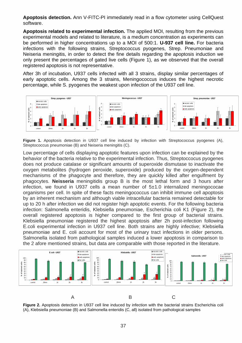
37
Strep. pyogenes - U937
0
1
2
3
4
5
6
control 20min 1h 2h 3h
% o
f sta
ined
cel
ls
necrotic cells
late apoptosis
early apoptosis
E coli - U937
0102030405060708090
100
control 20min 1h 2h 3h
% o
f sta
ined
cel
ls
necrotic cells
late apoptosis
early apoptosis
live cells
Klebsiella - U937
0102030405060708090
100
control 20min 1h 2h 3h
% o
f sta
ined
cel
ls
necrotic cells
late apoptosis
early apoptosis
live cells
Salmonella - U937
0102030405060708090
100
control 20min 1h 2h 3h
% o
f sta
ined
cel
ls
necrotic cellslate apoptosisearly apoptosislive cells
Strep. pneumoniae - U937
0
1
2
3
4
5
6
control 20min 1h 2h 3h
% o
f sta
ined
cel
ls
necrotic cells
late apoptosis
early apoptosis
Meningococcus - U937
0
1
2
3
4
5
6
control 20min 1h 2h 3h
% o
f sta
ined
cel
ls
necrotic cells
late apoptosis
early apoptosis
Apoptosis detection. Ann V-FITC-PI immediately read in a flow cytometer using CellQuest software.
Apoptosis related to experimental infection. The applied MOI, resulting from the previous experimental models and related to literature, is a medium concentration as experiments can be performed in higher concentrations up to a MOI of 500:1. U-937 cell line. For bacteria infections with the following strains, Streptococcus pyogenes, Strep. Pneumoniae and Neiseria meningitis, in order to detect the fine details regarding the apoptosis induction we only present the percentages of gated live cells (Figure 1), as we observed that the overall registered apoptosis is not representative.
After 3h of incubation, U937 cells infected with all 3 strains, display similar percentages of early apoptotic cells. Among the 3 strains, Meningococcus induces the highest necrotic percentage, while S. pyogenes the weakest upon infection of the U937 cell line.
Figure 1. Apoptosis detection in U937 cell line induced by infection with Streptococcus pyogenes (A), Streptococcus pneumoniae (B) and Neiseria meningitis (C).
Low percentage of cells displaying apoptotic features upon infection can be explained by the behavior of the bacteria relative to the experimental infection. Thus, Streptoccocus pyogenes does not produce catalase or significant amounts of superoxide dismutase to inactivate the oxygen metabolites (hydrogen peroxide, superoxide) produced by the oxygen-dependent mechanisms of the phagocyte and therefore, they are quickly killed after engulfment by phagocytes. Neisseria meningitidis group B is the most lethal form and 3 hours after infection, we found in U937 cells a mean number of 5±1.0 internalized meningococae organisms per cell. In spite of these facts meningococcus can inhibit immune cell apoptosis by an inherent mechanism and although viable intracellular bacteria remained detectable for up to 20 h after infection we did not register high apoptotic events. For the following bacteria infection: Salmonella enteridis, Klebsiella pneumoniae, Escherichia coli K1 (Figure 2), the overall registered apoptosis is higher compared to the first group of bacterial strains. Klebsiella pneumoniae registered the highest apoptosis after 2h post-infection following E.coli experimental infection in U937 cell line. Both strains are highly infective; Klebsiella pneumoniae and E. coli account for most of the urinary tract infections in older persons. Salmonella isolated from pathological samples induced a lower apoptosis in comparison to the 2 afore mentioned strains, but data are comparable with those reported in the literature.
A B C
Figure 2. Apoptosis detection in U937 cell line induced by infection with the bacterial strains Escherichia coli (A), Klebsiella pneumoniae (B) and Salmonella enteridis (C, all) isolated from pathological samples

38
Strep. pyogenes - THP-1
0
5
10
15
20
25
30
control 20min 1h 2h 3h
% o
f sta
ined
cel
ls
necrotic cells
late apoptosis
early apoptosis
Strep.pneumoniae - THP-1
0
5
10
15
control 20min 1h 2h 3h
% o
f sta
ined
cel
ls
necrotic cells
late apoptosis
early apoptosis
Meningoccocus- THP-1
0123456789
10
control 20min 1h 2h 3h
% o
f sta
ined
cel
ls
necrotic cells
late apoptosis
early apoptosis
E coli - THP-1
0
20
40
60
80
100
control 20min 1h 2h 3h
% o
f sta
ined
cel
ls
necrotic cells
late apoptosis
early apoptosis
live cells
Klebsiella - THP-1
0102030405060708090
100
control 20min 1h 2h 3h
% o
f sta
ined
cel
ls
necrotic cells
late apoptosis
early apoptosis
live cells
Salmonella - THP-1
0102030405060708090
100
control 20min 1h 2h 3h
% o
f sta
ined
cel
ls
necrotic cells
late apoptosis
early apoptosis
live cells
THP-1 cell line. For Streptococcus pyogenes, Streptococcus pneumoniae and Neiseria meningitis THP-1 cell line presented high percentages of necrotic cells (Figure 3). After 3h of infection with Streptococcus pyogenes only 50% of the cells were still alive. S. pyogenes after 2h post-infection induces the highest necrotic pattern, followed by S. pneumoniae and meningococcus. The apoptotic pattern differs in THP-1 cellular models of infection compared to U937 cell line.
Figure 3. Apoptosis detection in THP-1 cell line induced by infection with Streptococcus pyogenes (A), Streptococcus pneumoniae (B) and Neiseria meningitis (C).
In the case of THP-1 cellular model of infection, as expected, high necrotic percentages were registered for bacteria isolated from pathological samples (Figure 4). Salmonella induced the death of over 50% of the cells within the first 3h. Results match the apoptotic patterns reported in the literature incriminating caspase-3 for apoptosis, but not caspase-8 or 9. The higher resistance of U937 cell line to experimental infection compared to THP-1 to all the tested bacteria is in concordance with previously reported experimental models of infection. Taking into account that when entering the host most of the bacteria are subjected to antibody attack we would consider using opsonized bacteria in our experimental models to mimic more the physiological model of infection.
A B C
Figure 4. Apoptosis detection in THP-1 cell line induced by infection with bacterial strains Escherichia coli (A), Klebsiella pneumoniae (B) and Salmonella enteridis (C), all isolated from pathological samples.
Neagu et al, Infection and Immunity, 2013 (in preparation)
Peptide detection in infection cellular models
Non-fractionated samples of cellular supernatants sterielized through filtration for removal of free bacteria were profiled on chromatographic SELDI ProteinChip arrays using various chemistry and further analyzed via TOF-MS.
Automatic peak detection yielded a total of over 1400 peaks analyzing only the 500-2000 Da region. The post-infection spectra were subtracted from the following controls spectra: RPMI1640, inhibitor cocktail, non-infected culture cells supernatants.
Fragments at 1087Da, 1230.27Da, 1430Da along with various small peptides around 500Da can be detected and accumulate with the time of infection (Figure 1). Spectrum registered on H50 array for U937 cells supernatants post-infection with Klebsiella at 2h post-infection proved the appearance of fragments of 500Da (Figure 2).
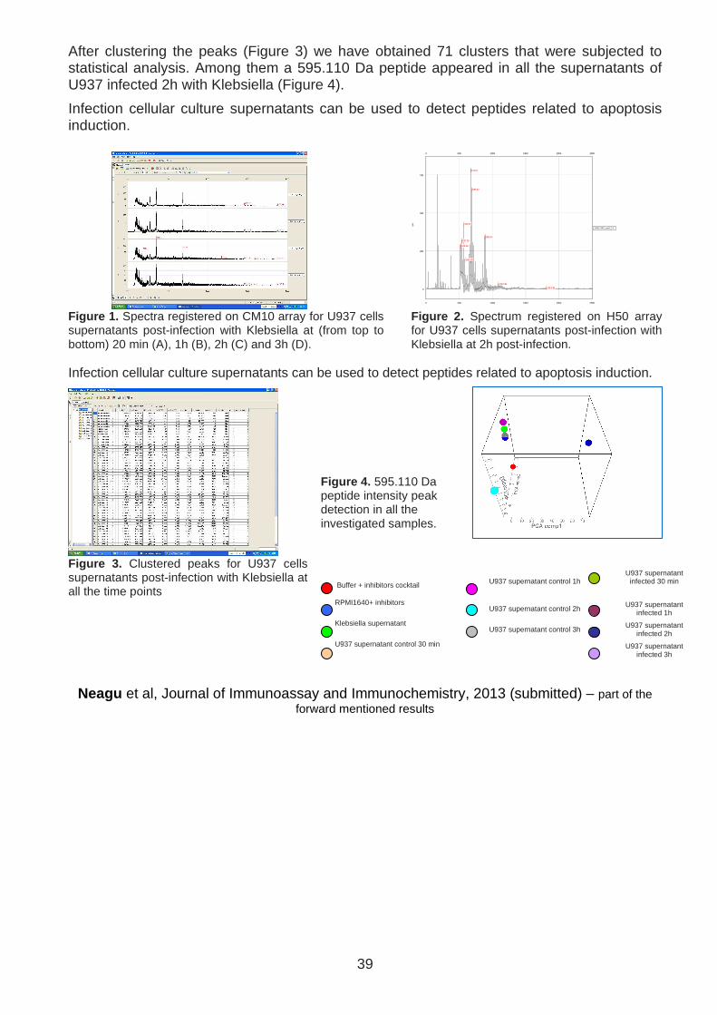
39
After clustering the peaks (Figure 3) we have obtained 71 clusters that were subjected to statistical analysis. Among them a 595.110 Da peptide appeared in all the supernatants of U937 infected 2h with Klebsiella (Figure 4).
Infection cellular culture supernatants can be used to detect peptides related to apoptosis induction.
0 500 1000 1500 2000 2500
519.98523.98
537.93
567.87
586.88
671.67
687.62
876.66892.64
1087.89
1813.350
250
500
750
uA
1050173492_spotF_2.4
0 500 1000 1500 2000 2500 Figure 1. Spectra registered on CM10 array for U937 cells supernatants post-infection with Klebsiella at (from top to bottom) 20 min (A), 1h (B), 2h (C) and 3h (D).
Figure 2. Spectrum registered on H50 array for U937 cells supernatants post-infection with Klebsiella at 2h post-infection.
Infection cellular culture supernatants can be used to detect peptides related to apoptosis induction.
Figure 4. 595.110 Da peptide intensity peak detection in all the investigated samples.
Figure 3. Clustered peaks for U937 cells supernatants post-infection with Klebsiella at all the time points
Buffer + inhibitors cocktail
U937 supernatant control 1h
U937 supernatant
infected 30 min
RPMI1640+ inhibitors U937 supernatant control 2h U937 supernatant infected 1h
Klebsiella supernatant U937 supernatant control 3h U937 supernatant infected 2h
U937 supernatant control 30 min U937 supernatant infected 3h
Neagu et al, Journal of Immunoassay and Immunochemistry, 2013 (submitted) – part of the forward mentioned results

40
Results in the framework of objective 2
Effect of ProT-alpha and its peptides on peripheral neutrophils
Conclusion of the published results
ProTα(100–109) increases the basic functional responses of neutrophils to levels similar to those induced by proTα. Neutrophils’ activation with proTalpha or proTalpha(100-109) duplicate the phagocytic capacity. The increase of phagocytic capacity in cancer patients was not so high as in healthy donors. Nevertheless, proTalpha or proTalpha(100-109) restored the reduced phagocytic capacity of cancer patients neutrophils to the levels of healthy donors’ control (Figure 1,2,3,5). This seems reasonable because an important proportion of circulating neutrophils in cancer patients are immature with low phagocytic capacity.
ProTα(100–109) and proTα increase the cytotoxicity of neutrophils against cancer cell targets. Neutrophils from cancer patients that were treated with fMLP, proTα(100–109), or proTα killed MCF-7 breast cancer targets more efficiently (Figure 4). In general, neutrophils from cancer patients showed a slightly increased ability to kill three of the four cell lines, in agreement with their registered ability to produce and release more ROS. Nevertheless, although these neutrophils were derived from the peripheral blood of patients with breast cancer, no preferential lysis of the two human breast cancer cell lines (MCF-7 and SKBR3) was observed. The overall low cytotoxicity recorded, in conjunction with lysis of all cancer cell targets, suggest that neutrophils are less efficient killer cells than either cytotoxic T cells (CTLs) or NK cells and mediate limited non-specific cytotoxic responses.
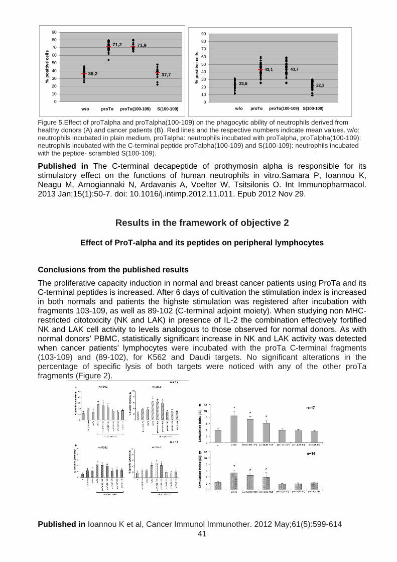
41
36,2
71,2 71,9
37,7
0
10
20
30
40
50
60
70
80
90
w/o pr οΤα prοΤα(100-109) S(100-109)
% p
ositi
ve c
ells
23,6
43,1 43,7
22,3
0
10
20
30
40
50
60
70
80
90
w/o proT α proTα(100-109) S(100-109)
% p
ositi
ve c
ells
Figure 5.Effect of proTalpha and proTalpha(100-109) on the phagocytic ability of neutrophils derived from healthy donors (A) and cancer patients (B). Red lines and the respective numbers indicate mean values. w/o: neutrophils incubated in plain medium, proTalpha: neutrophils incubated with proTalpha, proTalpha(100-109): neutrophils incubated with the C-terminal peptide proTalpha(100-109) and S(100-109): neutrophils incubated with the peptide- scrambled S(100-109).
Published in The C-terminal decapeptide of prothymosin alpha is responsible for its stimulatory effect on the functions of human neutrophils in vitro.Samara P, Ioannou K, Neagu M, Arnogiannaki N, Ardavanis A, Voelter W, Tsitsilonis O. Int Immunopharmacol. 2013 Jan;15(1):50-7. doi: 10.1016/j.intimp.2012.11.011. Epub 2012 Nov 29.
Results in the framework of objective 2
Effect of ProT-alpha and its peptides on peripheral lymphocytes
Conclusions from the published results
The proliferative capacity induction in normal and breast cancer patients using ProTa and its C-terminal peptides is increased. After 6 days of cultivation the stimulation index is increased in both normals and patients the highste stimulation was registered after incubation with fragments 103-109, as well as 89-102 (C-terminal adjoint moiety). When studying non MHC-restricted citotoxicity (NK and LAK) in presence of IL-2 the combination effectively fortified NK and LAK cell activity to levels analogous to those observed for normal donors. As with normal donors’ PBMC, statistically significant increase in NK and LAK activity was detected when cancer patients’ lymphocytes were incubated with the proTa C-terminal fragments (103-109) and (89-102), for K562 and Daudi targets. No significant alterations in the percentage of specific lysis of both targets were noticed with any of the other proTa fragments (Figure 2).
Published in Ioannou K et al, Cancer Immunol Immunother. 2012 May;61(5):599-614
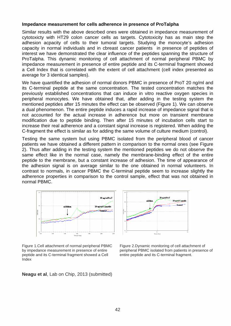
42
Impedance measurement for cells adherence in presen ce of ProTalpha
Similar results with the above descrbed ones were obtained in impedance measurement of cytotoxicty with HT29 colon cancer cells as targets. Cytotoxicity has as main step the adhesion acpacity of cells to their tumoral targets. Studying the monocyte’s adhesion capacity in normal individuals and in cbreast cancer patients in presence of peptides of interest we have demonstrated the clear influence of the peptides spanning the structure of ProTalpha. This dynamic monitoring of cell attachment of normal peripheral PBMC by impedance measurement in presence of entire peptide and its C-terminal fragment showed a Cell Index that is correlated with the extent of cell attachment (cell index presented as average for 3 identical samples).
We have quantified the adhesion of normal donors PBMC in presence of ProT 20 ng/ml and its C-terminal peptide at the same concentration. The tested concentration matches the previously established concentrations that can induce in vitro reactive oxygen species in peripheral monocytes. We have obtained that, after adding in the testing system the mentioned peptides after 15 minutes the effect can be observed (Figure 1). We can observe a dual phenomenon. The entire peptide induces a rapid increase of impedance signal that is not accounted for the actual increase in adherence but more on transient membrane modification due to peptide binding. Then after 15 minutes of incubation cells start to increase their real adherence and a constant signal increase is registered. When adding the C-fragment the effect is similar as for adding the same volume of culture medium (control).
Testing the same system but using PBMC isolated from the peripheral blood of cancer patients we have obtained a different pattern in comparison to the normal ones (see Figure 2). Thus after adding in the testing system the mentioned peptides we do not observe the same effect like in the normal case, namely the membrane-binding effect of the entire peptide to the membrane, but a constant increase of adhesion. The time of appearance of the adhesion signal is on average similar to the one obtained in normal volunteers. In contrast to normals, in cancer PBMC the C-terminal peptide seem to increase slightly the adherence properties in comparison to the control sample, effect that was not obtained in normal PBMC.
Figure 1.Cell attachment of normal peripheral PBMC by impedance measurement in presence of entire peptide and its C-terminal fragment showed a Cell Index
Figure 2.Dynamic monitoring of cell attachment of peripheral PBMC isolated from patients in presence of entire peptide and its C-terminal fragment.
Neagu et al , Lab on Chip, 2013 (submitted)
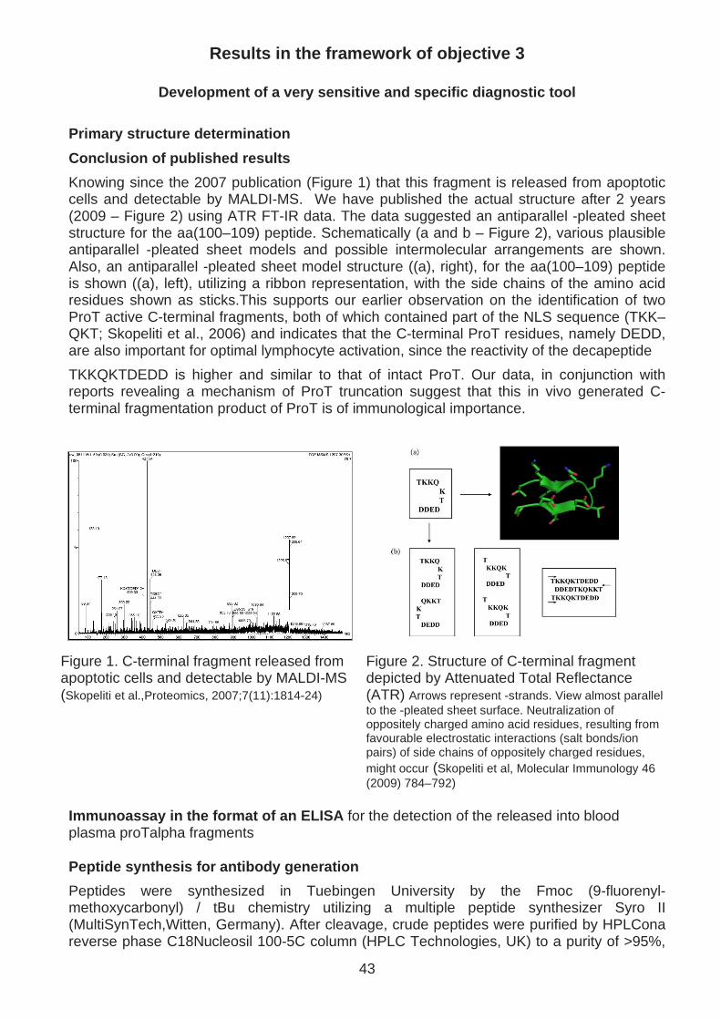
43
Results in the framework of objective 3
Development of a very sensitive and specific diagno stic tool
Primary structure determination
Conclusion of published results
Knowing since the 2007 publication (Figure 1) that this fragment is released from apoptotic cells and detectable by MALDI-MS. We have published the actual structure after 2 years (2009 – Figure 2) using ATR FT-IR data. The data suggested an antiparallel -pleated sheet structure for the aa(100–109) peptide. Schematically (a and b – Figure 2), various plausible antiparallel -pleated sheet models and possible intermolecular arrangements are shown. Also, an antiparallel -pleated sheet model structure ((a), right), for the aa(100–109) peptide is shown ((a), left), utilizing a ribbon representation, with the side chains of the amino acid residues shown as sticks.This supports our earlier observation on the identification of two ProT active C-terminal fragments, both of which contained part of the NLS sequence (TKK–QKT; Skopeliti et al., 2006) and indicates that the C-terminal ProT residues, namely DEDD, are also important for optimal lymphocyte activation, since the reactivity of the decapeptide
TKKQKTDEDD is higher and similar to that of intact ProT. Our data, in conjunction with reports revealing a mechanism of ProT truncation suggest that this in vivo generated C-terminal fragmentation product of ProT is of immunological importance.
Figure 1. C-terminal fragment released from apoptotic cells and detectable by MALDI-MS (Skopeliti et al.,Proteomics, 2007;7(11):1814-24)
Figure 2. Structure of C-terminal fragment depicted by Attenuated Total Reflectance (ATR) Arrows represent -strands. View almost parallel to the -pleated sheet surface. Neutralization of oppositely charged amino acid residues, resulting from favourable electrostatic interactions (salt bonds/ion pairs) of side chains of oppositely charged residues, might occur (Skopeliti et al, Molecular Immunology 46 (2009) 784–792)
Immunoassay in the format of an ELISA for the detection of the released into blood plasma proTalpha fragments Peptide synthesis for antibody generation
Peptides were synthesized in Tuebingen University by the Fmoc (9-fluorenyl-methoxycarbonyl) / tBu chemistry utilizing a multiple peptide synthesizer Syro II (MultiSynTech,Witten, Germany). After cleavage, crude peptides were purified by HPLCona reverse phase C18Nucleosil 100-5C column (HPLC Technologies, UK) to a purity of >95%,

44
using a linear gradient of 5–80% acetonitrile in 0.05% trifluoroacetic acid for 45 min. All peptides were characterized by MALDI-ToF-MS and results were in all cases in agreement with the calculated masses. We have syntehized the peptides persented in Panel 1.
Primary structure of human ProT:
acSDAAVDTSSEITTKDLKEKKEVVEEAENGRDAPANGNANEENGEQEADNEVDEEEE EGGEEEEEEEEGDGEEEDGDEDEEAESATGKRAAEDDEDDDVDTKKQKTDEDD
Figure 1. Amino acid sequence of synthetic peptides spanning the ProT C-terminus.
Generation of monoclonal antibodies in mice
BALB/c mice were immunized with emulsified (with CFA or IFA) synthetic peptides (single peptide) via intraperitoneal (i.p.) injections performed at 4-week intervals. After 2-3 booster injections, spleen cells will be aseptically isolated and fused with the murine B-cell myeloma cell lines SP2 or NSO. Cell fusion will be performed at 37oC in the presence of polyethylene glycol (PEG), which promotes membrane fusion. The pellet obtained will be plated into tissue culture plates in hypoxanthine-aminopterin-thymidine (HAT)-containing medium which allows selection only of fused cells. After feeding over 2-3 weeks, the supernatants of the hybridomas will be screened for specific IgG production, using direct ELISA. Positive clones were propagated and recloned by limiting dilution. When a stable hybridoma producing the IgG of interest was obtained, large scale production of supernatant or ascites in mice inoculated i.p. with the monoclonal antibody hybridoma cells was performed. These monoclonal antibody preparations were purified and used as positive control antibodies.
Monoclonal antibody generation used the above mentioned antigens: 10-100 µg in the below presented schedule
After selection we have obtained 43 clones; 23 clones with high specificity; out of which 2 clones highest mAb concentration ; Clones 3A2 and 6C5 genetically unstable - did not produce stable monoclonal antibodies.
Monoclonals secreting hybridomas were done in E-Biotech BVBA, Zonhoven, Belgium (CEO Dr Eugene Bosmans)
and AusMAb (Australian Monoclonal Antibodies) (CEO Dr. Alfio Comis)
Hybridomas were tested Genetically mapped in the Biochemistry and Molecular Biology by Prof. Kostas Vorgias – University of Athens

45
Generation of Polyclonal antibody
As tyhe monoclonal antibodyes did not have stable clones we have tested in the mean time the polyclonal antibodies generated according to a standard protocol. New Zealand white rabbits were intradermally or subcutaneously injected at many sites on their back with ProTa, ProTa[1-28], ProTa [87-109], or ProTa[101-109] conjugated to KLH
Conjugation was performed according to the glutaraldehyde method. The immunogens were injected as emulsions (1:1, v/v) in complete Freund’s adjuvant. The animals were boosted initially after 6 weeks and subsequently every 4 weeks. Blood was collected 2 weeks after each booster injection. Antisera (source of antibodies G) were obtained with low-speed centrifugation of whole blood and stored at -350C.
Klimentzou P et al, Journal of Histochemistry & Cytochemistry Volume 56(11): 1023–1031, 2008
Polyclonal antibodies were purified on affinity chromatography using modified HaloTag-based procedure (Ohana RF et al, Protein Expr Purif. 2011 Apr;76(2):154-64) against synthetic and natural decapeptide proTalpha(100-109) – PATENT PENDING
SELDI-ToF-MS affinity capture technology to evident iate Ab-Ag specific complex
Conclusions from the results
We have been applying several Protocols: namely 3 types of SELDI chips : NP20 (neat protein) Q10 (weak anion exchanger) and CM10 (weak cation exchanger) and two types of matrix CHCA and SPA. For developing the affinity capture mass spectrometry assay we have been testing 2 protocols with/without protein A for antibody fixation on the chip high and low intensity laser conditions.
1000 2000 3000
-0.050
0.050.1
uA 1051003472_spotA_2.15
-0.1-0.05
00.050.1
uA 1051003472_spotB_2.15
-0.050
0.050.1
uA 1051003472_spotC_2.15
-0.050
0.050.1
uA 1051003472_spotD_2.15
-0.050
0.050.1
uA 1051003472_spotE_2.15
-0.05
00.05
0.1
uA 1051003472_spotF_2.15
-0.050
0.05
0.1
uA 1051003472_spotG_2.15
-0.050
0.050.1
uA 1051003472_spotH_2.15
1000 2000 3000
30000 40000 50000
47268.06
00.10.20.30.40.5
uA Protein A
33476.89
47462.960
0.1
0.2
0.3
uA Protein A+ A b
33530.25
46924.170
0.2
0.4
0.6
0.8
uA Ab
33579.37
47108.030
0.2
0.4
0.6
uA Protein A+(Ab +Peptide)
33566.47
47195.920
0.2
0.4
0.6
0.8
uA
Protein A+(Ab +Scrambled Peptide)
47292.25
0
0.25
0.5
0.75
1
uA Protein A+Peptide
47221.19
0
0.2
0.4
0.6
0.8
uA Protein A+Scrambled Peptide
0
0.01
0.02
0.03
uA Peptide
30000 40000 50000 A B - E
60000 70000 80000
0
0.01
0.02
0.03
uA Protein A
67148.25
0
0.2
0.4
0.6
uA Protein A+ Ab
67257.34
0
0.25
0.5
0.75
uA Ab
67366.50
0
0.2
0.4
0.6
uA Protein A+(Ab +Peptide)
67419.42
0
0.25
0.5
0.75
1
uA
Protein A+(Ab +Scrambled Peptide)
0
0.01
0.02
0.03
uA Protein A+Pept ide
0
0.01
0.02
0.03
0.04
uA Protein A+Scrambled Peptide
0
0.01
0.02
0.03
uA Peptide
60000 70000 80000
130000 140000 150000
0
0.005
0.01
0.015
uA Protein A
135633.09
00.0250.05
0.0750.1
0.125
uA Protein A+ Ab
135516.56
0
0.05
0.1
uA Ab
136070.24
00.0250.05
0.0750.1
uA Protein A+(A b +Peptide)
136057.35
0
0.05
0.1
0.15
uA
Protein A+(A b +Scrambled Peptide)
00.0050.01
0.0150.02
0.025
uA Protein A+Peptide
0.01
0.02
0.03
uA Protein A+Scrambled Peptide
00.00250.005
0.00750.01
0.0125
uA Peptide
130000 140000 150000 1150 1200 1250 1300
0
0.5
1
uA Protein A
00.5
11.5
2
uA Protein A+ Ab
01234
uA Ab
1209.15
1221.490
25
50
75
uA Protein A+(Ab +Peptide)
1208.33
02.5
57.510
12.5
uA
Protein A+(Ab +Scrambled Peptide)
1193.66
1207.75
1219.761230.54 1264.080
100
200
300
uA Protein A+Peptide
1208.96
0
5
10
15
uA Protein A+Scrambled Peptide
1194.52
1208.67
1221.00
1231.05 1265.240250500750
1000
uA Peptide
1150 1200 1250 1300 Figure 1. SELDI-ToF mass spectrum of Ab fragments spoted on NP (A) Q10 or CM10 chips (B-E). At m/z ratio Ab fragments were depicted A: 33,666-33,477; Protein A at m/z (47,268-47,108); B:
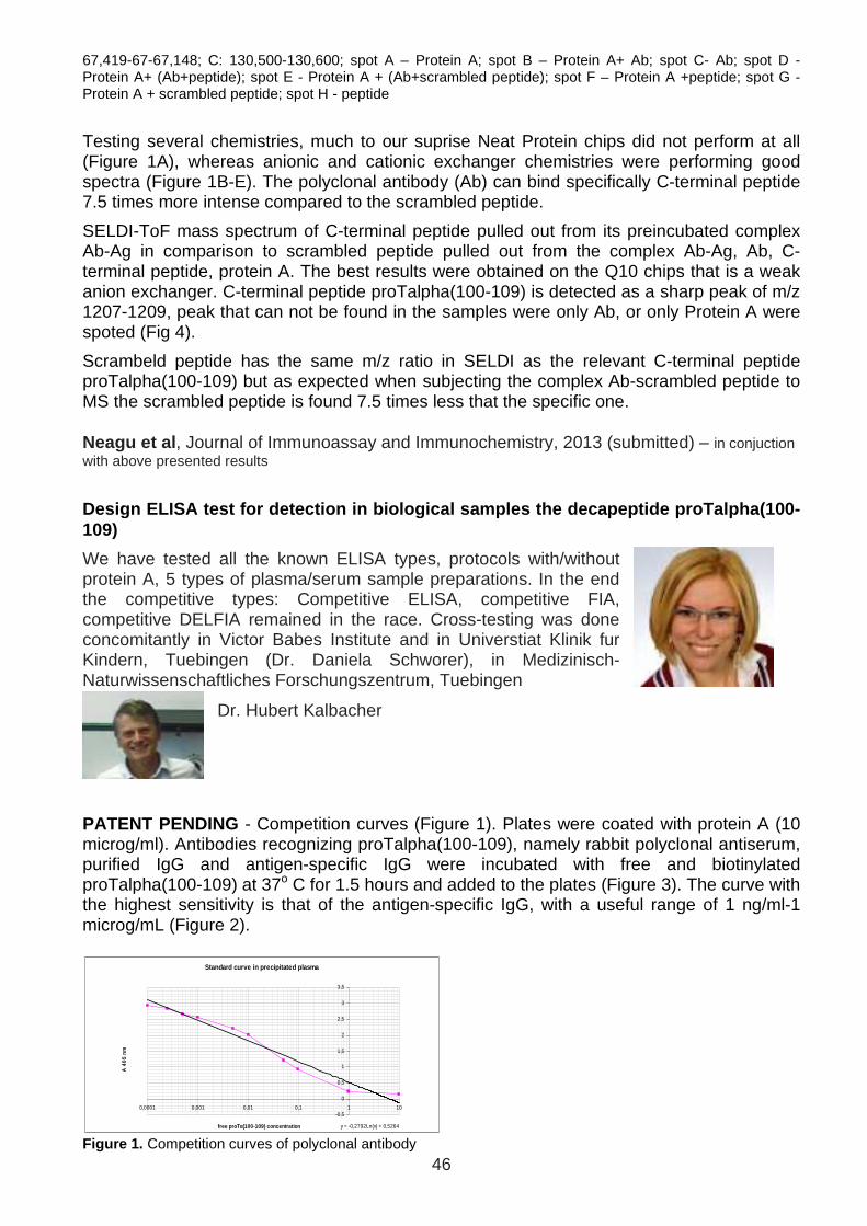
46
67,419-67-67,148; C: 130,500-130,600; spot A – Protein A; spot B – Protein A+ Ab; spot C- Ab; spot D - Protein A+ (Ab+peptide); spot E - Protein A + (Ab+scrambled peptide); spot F – Protein A +peptide; spot G - Protein A + scrambled peptide; spot H - peptide
Testing several chemistries, much to our suprise Neat Protein chips did not perform at all (Figure 1A), whereas anionic and cationic exchanger chemistries were performing good spectra (Figure 1B-E). The polyclonal antibody (Ab) can bind specifically C-terminal peptide 7.5 times more intense compared to the scrambled peptide.
SELDI-ToF mass spectrum of C-terminal peptide pulled out from its preincubated complex Ab-Ag in comparison to scrambled peptide pulled out from the complex Ab-Ag, Ab, C-terminal peptide, protein A. The best results were obtained on the Q10 chips that is a weak anion exchanger. C-terminal peptide proTalpha(100-109) is detected as a sharp peak of m/z 1207-1209, peak that can not be found in the samples were only Ab, or only Protein A were spoted (Fig 4).
Scrambeld peptide has the same m/z ratio in SELDI as the relevant C-terminal peptide proTalpha(100-109) but as expected when subjecting the complex Ab-scrambled peptide to MS the scrambled peptide is found 7.5 times less that the specific one. Neagu et al , Journal of Immunoassay and Immunochemistry, 2013 (submitted) – in conjuction with above presented results
Design ELISA test for detection in biological sampl es the decapeptide proTalpha(100-109)
We have tested all the known ELISA types, protocols with/without protein A, 5 types of plasma/serum sample preparations. In the end the competitive types: Competitive ELISA, competitive FIA, competitive DELFIA remained in the race. Cross-testing was done concomitantly in Victor Babes Institute and in Universtiat Klinik fur Kindern, Tuebingen (Dr. Daniela Schworer), in Medizinisch-Naturwissenschaftliches Forschungszentrum, Tuebingen
Dr. Hubert Kalbacher
PATENT PENDING - Competition curves (Figure 1). Plates were coated with protein A (10 microg/ml). Antibodies recognizing proTalpha(100-109), namely rabbit polyclonal antiserum, purified IgG and antigen-specific IgG were incubated with free and biotinylated proTalpha(100-109) at 37o C for 1.5 hours and added to the plates (Figure 3). The curve with the highest sensitivity is that of the antigen-specific IgG, with a useful range of 1 ng/ml-1 microg/mL (Figure 2).
Standard curve in precipitated plasma
y = -0,2792Ln(x) + 0,5264
-0,5
0
0,5
1
1,5
2
2,5
3
3,5
0,0001 0,001 0,01 0,1 1 10
free proT α(100-109) concentration
A 4
05 n
m
Figure 1. Competition curves of polyclonal antibody
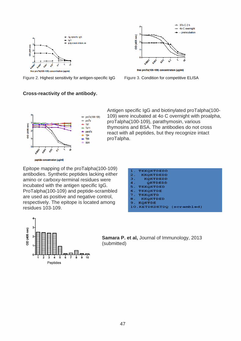
47
Figure 2. Highest sensitivity for antigen-specific IgG Figure 3. Condition for competitive ELISA Cross-reactivity of the antibody.
Antigen specific IgG and biotinylated proTalpha(100-109) were incubated at 4o C overnight with proalpha, proTalpha(100-109), parathymosin, various thymosins and BSA. The antibodies do not cross react with all peptides, but they recognize intact proTalpha.
Epitope mapping of the proTalpha(100-109) antibodies. Synthetic peptides lacking either amino or carboxy-terminal residues were incubated with the antigen specific IgG. ProTalpha(100-109) and peptide-scrambled are used as positive and negative control, respectively. The epitope is located among residues 103-109.
Samara P. et al, Journal of Immunology, 2013 (submitted)

48
Results in the framework of objective 4
Validation of this diagnostic tool in infected anim al models
Mouse models of infection – effect of experimental infection on innate and adaptive immune cells
Experimental models were performed in Cantacuzino Institute. Immune cell phenotyping and respiratory burst activity of peripheral neutrophils using flow cytometry analysis The analysis was performed in order to establish the percentage of B, T (CD4+ and CD8+) and NK cell sets and to establish the effect of experimental infection on peripheral neutrophils.
We used two murine models to determine lymphocyte sets in whole blood and spleen; murine strains are represented by Balb/c and C57 Black (C57bl) mice, wheater conventional or SFP strains. BALB/c is frequently used for a variety of immunological studies, as being highly susceptible to infection. C57bl is the most widely used strain has low susceptibility to tumors, high susceptibility to parasitic and viral infections. Mice from the following strains: Balb/c and C57BL/6 were infected intraperitoneally with one dose of Klebsiella oxytoca (50 germs/ 0.5 mL/ mouse). We obtained serum from not infected mice and infected mice. Peripheral blood was colected at selected time intervals after the infection (0 h, 2h, 24h and 72h).
In peripheral blood of SPF Balb/c variant has similar B220+ percentage compared to SPF C57/bl strain, but higher percentage of CD4+ and CD8+ cells compared to the same population in SPF Balb/c mice. In the spleen of both murine strains we have determined a higher percentage of B220, CD4+ and NK1.1 compared to the same populations in peripheral blood. In Balb/c (conventional and SPF) mice the percentage of CD8+ cells is higher in spleen then in peripheral blood. The lymphocyte sets in spleen from both SPF strains indicate a similar percentage of B220 and CD8+ lymphocytes. In SPF Balb/c mice the percentage of CD4+ cells is higher in comparison to SPF C57/bl mice. Higher values for NK1.1 cells in SPF C57/bl compared to SPF Balb/c mice.
The results from peripheral blood cells populations in SPF Balb/c and SPF C57bl mice are indicated in Figure 1.
Phenotyping the peripheral blood lymphocytes we observed that in the conventional Balb/c strain the CD4+ population is considerably higher then in conventional C57/bl mice.
The results from spleen cells populations in SPF Balb/c and SPF C57bl mice are indicated in Figure 3.
0
20
40
60
80
100
B220 CD4 CD8 NK1.1.
%Balb/c
0
20
40
60
80
100
B220 CD4 CD8 NK1.1.
%C57/bl
a b
Figure 1. Lymphocyte sets isolated from spleen in conventional Balb/c (a) and C57bl (b) mice after 72h post-
infection
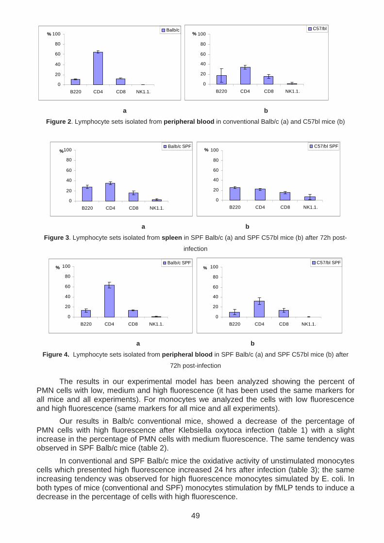
49
0
20
40
60
80
100
B220 CD4 CD8 NK1.1.
%Balb/c
0
20
40
60
80
100
B220 CD4 CD8 NK1.1.
%C57/bl
a b
Figure 2 . Lymphocyte sets isolated from peripheral blood in conventional Balb/c (a) and C57bl mice (b)
0
20
40
60
80
100
B220 CD4 CD8 NK1.1.
%Balb/c SPF
0
20
40
60
80
100
B220 CD4 CD8 NK1.1.
%C57/bl SPF
a b
Figure 3. Lymphocyte sets isolated from spleen in SPF Balb/c (a) and SPF C57bl mice (b) after 72h post-
infection
0
20
40
60
80
100
B220 CD4 CD8 NK1.1.
%Balb/c SPF
0
20
40
60
80
100
B220 CD4 CD8 NK1.1.
%C57/bl SPF
a b
Figure 4. Lymphocyte sets isolated from peripheral blood in SPF Balb/c (a) and SPF C57bl mice (b) after
72h post-infection
The results in our experimental model has been analyzed showing the percent of PMN cells with low, medium and high fluorescence (it has been used the same markers for all mice and all experiments). For monocytes we analyzed the cells with low fluorescence and high fluorescence (same markers for all mice and all experiments).
Our results in Balb/c conventional mice, showed a decrease of the percentage of PMN cells with high fluorescence after Klebsiella oxytoca infection (table 1) with a slight increase in the percentage of PMN cells with medium fluorescence. The same tendency was observed in SPF Balb/c mice (table 2).
In conventional and SPF Balb/c mice the oxidative activity of unstimulated monocytes cells which presented high fluorescence increased 24 hrs after infection (table 3); the same increasing tendency was observed for high fluorescence monocytes simulated by E. coli. In both types of mice (conventional and SPF) monocytes stimulation by fMLP tends to induce a decrease in the percentage of cells with high fluorescence.

50
The percentage of PMN cells with high fluorescence from conventional and SPF C57/bl mice decreases after infection (tables 5 and 6). The percentage of monocytes with high fluorescence in the same types of mice also decreases after the infection (tables 7 and 8); monocytes stimulation by E. coli, compared to unstimulated cells, induced an increase in the percentage of high fluorescent cells in conventional and SPF C57/bl mice, before and after Klebsiella infection.
Was also observed that in Balb/c mice (conventional and SPF) the percentage of phagocytic cells with high fluorescence is higher compared to C57/bl mice (conventional and SPF), both before and after infection (tables 1-8).
Table 1. PMN cells activity before and 24 hrs after infection using Balb/c conventional mice
PMN cells
Before infection Low fluorescence Medium fluorescence High fluorescence
% Mean % Mean % Mean
cell-fluorescence 99.70±0.16 35.60±6.19 0.28±0.13 746.60±140.04 0.00 0.00
NS+DHR123 1.76±1.34 147.80±44.76 81.02±5.33 2151.80±250.86 17.24±4.89 6402.60±767.99
E. coli 2.20±0.78 104.80±41.63 81.50±3.44 1826.40±49.98 16.26±3.56 5892.00±227.00
fMLP 1.36±0.34 102.00±29.92 85.14±4.15 2101.20±170.10 13.48±4.03 6106.60±605.54 24hrs after infection Low fluorescence Medium fluorescence High fluorescence
% Mean % Mean % Mean
cell-fluorescence 99.92±0.08 24.40±2.97 0.08±0.08 431.60±467.43 0.00 0.00
NS+DHR123 0.94±0.42 196.60±22.31 87.18±4.47 2072.00±131.52 11.88±4.11 4912.40±287.76
E. coli 0.94±0.51 185.80±26.40 89.76±3.27 1680.40±81.85 9.28±2.98 4243.80±188.11
fMLP 0.90±0.42 204.80±14.20 91.36±3.36 1913.60±57.04 7.70±3.01 4808.20±411.51 Table 2. PMN cells activity before and 24 hrs after infection using SPF Balb/c mice
PMN cells
Before Infection Low fluorescence Medium fluorescence High fluorescence
% Mean % Mean % Mean
cell-fluorescence 99.96±0.05 29.80±1.10 0.04±0.05 206.00±291.15 0.00 0.00
NS+DHR123 0.36±0.33 151.60±90.59 79.32±10.46 2307.80±234.00 20.32±10.51 4643.40±179.89
E. coli 0.62±0.57 55.80±51.83 86.06±6.74 1880.00±193.18 13.32±6.82 4390.40±224.03
fMLP 0.24±0.22 140.60±56.40 85.56±4.68 2246.60±191.20 14.20±4.54 4564.20±168.23 24hrs after infection Low fluorescence Medium fluorescence High fluorescence
% Mean % Mean % Mean
cell-fluorescence 99.88±0.10 22.75±2.75 0.13±0.10 816.00±206.27 0.00 0.00
NS+DHR123 4.28±3.13 195.80±68.21 86.98±4.18 1757.00±311.01 8.74±5.73 4764.80±290.85
E. coli 2.62±1.97 225.00±16.57 88.66±2.80 1601.80±120.53 8.78±1.89 4608.40±169.57
fMLP 3.10±2.04 222.20±3.56 89.94±2.86 1637.00±338.29 6.98±1.50 5236.60±440.08 Table 3. Monocyte cells activity before and 24 hrs after infection using Balb/c conventional mice
Monocyte population
Low fluorescence High fluorescence
Before infection % Mean % Mean
cell-fluorescence 99.96±0.09 24.40±2.19 0.04±0.09 164.60±368.06
NS+DHR123 66.18±11.55 508.00±30.50 33.82±11.55 1222.60±97.87
E. coli 61.38±4.01 487.20±34.29 38.62±4.01 1440.60±137.70
fMLP 64.70±12.41 494.80±15.02 35.30±12.41 1345.80±138.02 24hrs after infection Low fluorescence High fluorescence
% Mean % Mean
cell-fluorescence 99.94±0.09 21.40±0.55 0.06±0.09 679.80±1102.13
NS+DHR123 52.28±16.66 584.60±17.46 47.72±16.66 962.60±63.56
E. coli 34.36±8.07 554.80±28.27 65.64±8.07 1396.20±174.73

51
fMLP 70.88±7.90 551.20±20.80 29.12±7.90 999.00±30.89 Table 4. Monocyte cells activity before and 24 hrs after infection using SPF Balb/c mice
Monocyte population
Low fluorescence High fluorescence Before infection % Mean % Mean
cell-fluorescence 100.00±0.00 23.60±2.30 0.00 0.00
NS+DHR123 65.54±11.14 525.80±31.21 34.46±11.14 1390.20±203.62
E. coli 44.80±10.30 526.40±56.29 55.20±10.30 1631.00±94.64
fMLP 66.22±6.56 511.40±27.40 33.78±6.56 1480.80±197.27 24hrs after infection Low fluorescence High fluorescence
% Mean % Mean
cell-fluorescence 100.00±0.00 21.25±0.50 0.00 0.00
NS+DHR123 58.70±14.53 563.80±50.81 41.30±14.53 1016.00±89.48
E. coli 31.44±8.81 573.20±17.81 68.56±8.81 1433.60±200.31
fMLP 72.68±11.40 548.40±29.62 27.32±11.40 1221.20±245.13 Table 5. PMN cells activity before and 24 hrs after infection using C57/bl conventional mice
PMN cells
Before infection Low fluorescence Medium fluorescence High fluorescence
% Mean % Mean % Mean
cell-fluorescence 99.88±0.11 25.20±1.92 0.12±0.11 557.40±481.82 0.00 0.00
NS+DHR123 5.26±4.96 132.00±79.69 87.58±6.92 1452.40±282.22 7.18±8.63 6502.40±740.05
E. coli 4.06±4.05 127.20±67.20 89.66±5.27 1430.40±86.62 6.38±3.55 5012.40±973.17
fMLP 3.62±2.90 108.80±29.41 87.76±5.31 1464.20±198.60 8.62±5.42 6563.20±871.47 24hrs after infection Low fluorescence Medium fluorescence High fluorescence
% Mean % Mean % Mean
cell-fluorescence 99.36±0.59 30.20±1.92 0.64±0.59 511.60±299.06 0.00 0.00
NS+DHR123 4.58±3.06 156.20±64.63 94.12±2.98 1125.20±191.10 1.38±0.58 6161.40±921.82
E. coli 8.64±5.57 139.40±46.74 88.96±5.25 1100.40±89.11 2.50±1.92 5327.80±748.01
fMLP 3.94±2.65 161.40±35.74 93.86±2.32 1086.40±210.17 2.28±0.50 6609.00±735.81 Table 6. PMN cells activity before and 24 hrs after infection using SPF C57/bl mice
PMN cells
Before infection Low fluorescence Medium fluorescence High fluorescence
% Mean % Mean % Mean
cell-fluorescence 100.00±0.00 31.40±5.13 0.00 0.00 0.00 0.00
NS+DHR123 1.84±2.02 105.40±58.90 85.58±4.73 1735.40±146.39 12.58±6.08 5248.80±954.64
E. coli 3.82±3.33 120.40±58.48 88.60±5.26 1339.00±100.21 7.66±5.54 4271.00±157.00
fMLP 2.56±1.37 103.40±60.25 86.84±1.06 1658.00±122.15 10.62±1.26 4756.40±304.30
24hrs after infection Low fluorescence Medium fluorescence High fluorescence
% Mean % Mean % Mean
cell-fluorescence 99.50±0.71 32.80±6.46 0.65±0.79 273.25±324.25 0.00 0.00
NS+DHR123 3.10±2.50 214.80±20.12 92.84±4.46 1244.40±315.74 4.18±3.81 4997.20±574.26
E. coli 5.84±2.94 212.00±17.44 92.88±3.10 1019.40±118.21 1.50±0.57 5220.80±794.37
fMLP 2.94±1.68 191.60±37.29 93.46±3.91 1194.40±279.42 3.70±3.34 5717.80±651.36 Table 7. Monocyte cells activity before and 24 hrs after infection using C57/bl conventional mice
Monocyte population Before infection Low fluorescence High fluorescence
% Mean % Mean
cell-fluorescence 100.00±0.00 24.80±1.48 0.00 0.00
NS+DHR123 96.32±1.82 330.20±49.00 3.68±1.82 1600.80±601.96
E. coli 81.20±7.52 365.80±45.04 18.80±7.52 1390.60±420.08

52
fMLP 96.20±1.55 321.40±32.60 3.80±1.55 1818.60±559.97
24hrs after infection Low fluorescence High fluorescence
% Mean % Mean
cell-fluorescence 100.00 24.60±1.34 0.00 0.00
NS+DHR123 98.54±0.72 215.00±26.88 1.46±0.72 1846.00±386.79
E. coli 91.78±2.45 274.80±21.57 8.22±2.45 1661.40±341.62
fMLP 98.00±1.27 231.20±22.86 2.00±1.27 2132.20±856.12 Table 8. Monocyte cells activity before and 24 hrs after infection using SPF C57/bl mice
Monocyte population
Low fluorescence High fluorescence Before infection % Mean % Mean
cell-fluorescence 100.00 23.60±1.14 0.00 0.00
NS+DHR123 90.74±10.17 354.20±74.35 9.26±10.17 1209.00±224.19
E. coli 79.32±7.03 397.60±31.97 20.68±7.03 1293.20±208.27
fMLP 90.46±5.91 358.60±60.35 9.54±5.91 1380.60±234.07
24hrs after infection Low fluorescence High fluorescence
% Mean % Mean
cell-fluorescence 100.00 25.00±1.00 0.00 0.00
NS+DHR123 95.16±4.73 247.40±31.82 4.84±4.73 1529.00±408.59
E. coli 89.26±7.36 291.20±22.84 10.74±7.36 1403.60±301.99
fMLP 93.96±6.16 272.80±47.06 6.04±6.16 1494.60±108.48
Mouse models– ELISA testing in experimental infecti on
Using this immunoassay, we performed preliminary quantification of proTalpha(100-109) in sera of mice, infected with the strain presented below.
Table 1: Bacterial strains used to infect the mice
Bacterial strains Gram Usual route of infection Clinical signs
Pasteurella multocida - Skin, respiratory system Soft tissue inflammation
Corynebacterium urealyticum + Urinary system, skin Chronic cystitis
Streptococcus pyogenes + Throat, skin Haemolysis, toxic shock
syndrome
Klebsiella oxytoca - Gastrointestinal system Sepsis
Salmonella typhimurium - Gastrointestinal system Model of human typhoid
fever
Balb/C and C57BL/6 strains were used, concentional and SFP. Blood withdrawal was performed at 0, 2, 24 and 72 hours after infection. All the serum samples were precipitated overnight with acetone and 50 µl of each serum was analyzed with the standardized ELISA.
The animals were maintained under two different breeding conditions: mice maintained under normal conditions (conventional) and mice free of micro-organisms and parasites from their birth (germ-free). So, germ-free mice have received far fewer antigenic stimuli than conventional mice. The mice were infected by intraperitoneal inoculation with one dose of

53
bacterial suspension (50 CFU in 0.5 ml/ mouse). Blood was collected from the mice before infection (0 hours) and at 2, 24 and 72 hours post-infection. All the sera were precipitated with cold acetone before tested in the ELISA, in order to remove plasma proteins and reduce non-specific interactions.
Depending on the mouse strain and the type of bacteria that infected them, differences in proTalpha(100-109) peptide’s concentration as determined in murine sera, were observed. In general, the amount of the decapeptide proTalpha(100-109) before infection was marginal to undetectable, while proTalpha(100-109) gradually increased over time and for most pathogens peaked at 72 hours (Fig. 6-10). The following figures show the results obtained by ELISA analysis of mice sera, where each circle presents the values determined from each single mouse. These bacterial strains are known to cause massive apoptosis of the animals’ macrophages and, therefore, we assume that following proTalpha’s intracellular fragmentation, the decapeptide proTalpha(100-109) is excreted in the serum where it could act as a danger signal.
Fig. 6: Pasteurella multocida infection Fig. 7: Corynebacterium urealyticum infection
Fig. 8: Streptococcus pyogenes infection Fig. 9: Klebsiella oxytoca infection
Fig.10: Salmonella typhimurium infection
Samara et al, Journal of Immunological Methods, 201 3 (in preparation)

54
Overall Conclusions of the project
• ProTalpha or proTalpha(100-109) can restore the reduced phagocytic capacity of cancer patients neutrophils to the levels of healthy donors’ control.
• ProTalpha or proTalpha(100-109) reduced the intracellular and extracellular ROS production both in normal and cancer patients
• ProTalpha or proTalpha(100-109) increase lymphocyte proliferation of PBMC from normals and cancer patients during the autologous mixed lymphocyte reaction.
• ProTalpha or proTalpha(100-109) in combination with low-dose IL-2 significantly increase non MHC-restricted (NK and LAK) cell activity of PBMC from cancer subjects.
• Peripheral monocytes adhesion properties are enhanced by proTalpha and not by proTalpha(100-109) in normals and less in cancer patients
• Monoclonal Ab not stable
• Polyclonal highly purified Abs - Patent
• SELDI-ToF-MS can be used as an affinity capture technology to evidentiate Ab-Ag specific complex
• Optimized methodology for specific peptide identification from an Ab-Ag complex using known synthetic peptides; the optimization of the protocol showed that in our case the chemistry of weak anion exchanger gives the best spectra
• The synthetized C-terminal peptide proTalpha(100-109) depicted in spectra (m/z 1207-1209) is convergent to the MS prior tests (m/z 1207)
• The Mass spectrum pulled out the relevant C-terminal peptide from a Ab-Ag
• ELISA competitive test – Patent
• Sensitivity useful range 0.1 ng/ml-1 microg/ml.
• The antibodies do not cross react with all peptides, but they recognize intact proTalpha.
• The epitope is located among residues 103-109.
• ELISA detected proTalpha(100-109) in vitro induced apoptosis in cell cultures.
• ELISA detected proTalpha(100-109) in vivo release in plasma of mice experimentally infected was correlated with the time and type of infection as early as 2hrs post-experimental bacterial infection - Patent