TB Meningitis Article - wfpiweb.org Meningitis Article.pdf · WFPI TB Corner July 2016 Discussion...
9
WFPI TB Corner July 2016 Introduction TBM is the most severe and life-threatening form of tuberculosis in children [3,4] in which one out of every 300 untreated primary TB infections is complicated by TBM [5]. Its peak incidence is in young children under 4-5 years of age [6] although it may occur at any age. Children are most vulnerable and frequently affected by TBM due to their poor immune response to contain the Mycobacterium tuberculosis bacteria within the lungs [7]. The risk of progression of primary pulmonary TB to TBM is greater in children than in adults. Involvement of the central nervous system in tuberculosis occurs during hematogenous spread of the disease. Despite advances in its management and control around the world, the effort to eradicate this disease continues to be difficult and it still poses high risk of death which estimated to vary from 15 to 32% [5,6,8]. Neurologic sequelae are noted in more than half of the affected patients [6,7]. Radiologic imaging plays an important role in the diagnosis and management of TBM. CT scan is an established modality for evaluating TBM, its features, progression, and complications. It is also important in providing differential diagnoses for disease entities that may present similarly. Clinical Scenario The patient is an 18 year old male admitted at a pediatric hospital for persistent severe biparietal headache, dizziness, vomiting and changes in sensorium. He was being treated as a case of bacterial meningitis. It was disclosed that the patient was diagnosed with left ankle septic arthritis one month prior to admission. He had no known history of primary tuberculosis, but had contact with infected family members. Chest X-Ray was done and showed clear lungs without mediastinal lymphadenopathy. A Mantoux tuberculin skin test done demonstrated an induration of 20 mm. Physical examination and ultrasound of the neck also revealed cervical lymphadenopathies. The patient was referred to our institution for CT scan of the brain. Andres MM et al. TB Corner 2016; 2(5):1-9 1 Abstract Tuberculous meningitis (TBM) is the most severe form of mycobacterial infection [1] and the most frequent form of central nervous system tuberculosis [2]. The diagnosis of TBM relies on both clinical assessment and radiologic imaging features. Early and prompt diagnosis is imperative for better outcome. Clinical presentations may overlap with other disease entities and are very non-specific. CT plays vital role in the early diagnosis and monitoring of the course of the disease. The CT triad of basal enhancement, hydrocephalus and infarct strongly suggest TBM [3]. This review is an effort to examine and discuss the current literature in our understanding of TBM and to demonstrate the myriad of CT imaging features with focus on basal cistern involvement pattern, and to provide differential diagnoses. Key Facts TB Meningitis Clinical Presentation: Non- specific o Adults: typical meningeal signs o Children: prodrome lasting 2-8 weeks o Headache: Adults > children o Seizures: Children > Adults o Cranial nerve palsies: VI>III>IV>VII TUBERCULOUS MENINGITIS Basal Cistern Enhancement Pattern on CT imaging Mariaem M. Andres MD, Jacqueline Austine U. Uy MD, and Maricar P. Reyes-Paguia MD St. Luke’s Medical Center, Philippines
Transcript of TB Meningitis Article - wfpiweb.org Meningitis Article.pdf · WFPI TB Corner July 2016 Discussion...

WFPI TB Corner July 2016
Introduction TBM is the most severe and life-threatening form of tuberculosis in children [3,4] in which one out of every 300 untreated primary TB infections is complicated by TBM [5]. Its peak incidence is in young children under 4-5 years of age [6] although it may occur at any age. Children are most vulnerable and frequently affected by TBM due to their poor immune response to contain the Mycobacterium tuberculosis bacteria within the lungs [7]. The risk of progression of primary pulmonary TB to TBM is greater in children than in adults. Involvement of the central nervous system in tuberculosis occurs during hematogenous spread of the disease. Despite advances in its management and control around the world, the effort to eradicate this disease continues to be difficult and it still poses high risk of death which estimated to vary from 15 to 32% [5,6,8]. Neurologic sequelae are noted in more than half of the affected patients [6,7]. Radiologic imaging plays an important role in the diagnosis and management of TBM. CT scan is an established modality for evaluating TBM, its features, progression, and complications. It is also important in providing differential diagnoses for disease entities that may present similarly.
Clinical Scenario The patient is an 18 year old male admitted at a pediatric hospital for persistent severe biparietal headache, dizziness, vomiting and changes in sensorium. He was being treated as a case of bacterial meningitis. It was disclosed that the patient was diagnosed with left ankle septic arthritis one month prior to admission. He had no known history of primary tuberculosis, but had contact with infected family members. Chest X-Ray was done and showed clear lungs without mediastinal lymphadenopathy. A Mantoux tuberculin skin test done demonstrated an induration of 20 mm. Physical examination and ultrasound of the neck also revealed cervical lymphadenopathies. The patient was referred to our institution for CT scan of the brain.
Andres MM et al. TB Corner 2016; 2(5):1-9 �1
Abstract
Tuberculous meningitis (TBM) is t h e m o s t s e v e r e f o r m o f mycobacterial infection [1] and the most frequent form of central nervous system tuberculosis [2]. The diagnosis of TBM relies on both clinical assessment and radiologic imaging features. Early a n d p r o m p t d i a g n o s i s i s imperative for better outcome. Clinical presentations may overlap with other disease entities and are very non-specific. CT plays vital role in the early diagnosis and monitoring of the course of the disease. The CT triad of basal enhancement, hydrocephalus and infarct strongly suggest TBM [3]. This review is an effort to examine and discuss the current literature in our understanding of TBM and to demonstrate the myriad of CT imaging features with focus on basal cistern involvement pattern, and to prov ide d i fferent ia l diagnoses.
Key Facts
TB Meningitis
Clinical Presentation: Non-specific o Adults: typical meningeal
signs o Children: prodrome
lasting 2-8 weeks o Headache: Adults >
children o Seizures: Children > Adults o Cranial nerve palsies:
VI>III>IV>VII
TUBERCULOUS MENINGITIS Basal Cistern Enhancement Pattern on CT imaging
Mariaem M. Andres MD, Jacqueline Austine U. Uy MD, and Maricar P. Reyes-Paguia MD St. Luke’s Medical Center, Philippines
WFPI TB Corner July 2016
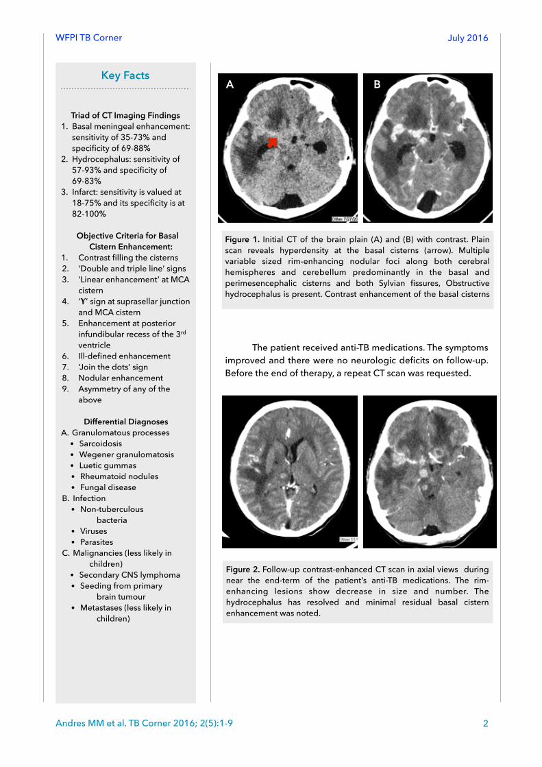
The patient received anti-TB medications. The symptoms improved and there were no neurologic deficits on follow-up. Before the end of therapy, a repeat CT scan was requested.
Andres MM et al. TB Corner 2016; 2(5):1-9 �2
Key Facts
Triad of CT Imaging Findings 1. Basal meningeal enhancement:
sensitivity of 35-73% and specificity of 69-88%
2. Hydrocephalus: sensitivity of 57-93% and specificity of 69-83%
3. Infarct: sensitivity is valued at 18-75% and its specificity is at 82-100%
Objective Criteria for Basal Cistern Enhancement:
1. Contrast filling the cisterns 2. ‘Double and triple line’ signs 3. ‘Linear enhancement’ at MCA
cistern 4. ‘ϒ’ sign at suprasellar junction
and MCA cistern 5. Enhancement at posterior
infundibular recess of the 3rd ventricle
6. Ill-defined enhancement 7. ‘Join the dots’ sign 8. Nodular enhancement 9. Asymmetry of any of the
above
Differential Diagnoses A. Granulomatous processes
• Sarcoidosis • Wegener granulomatosis • Luetic gummas • Rheumatoid nodules • Fungal disease
B. Infection • Non-tuberculous
bacteria • Viruses • Parasites
C. Malignancies (less likely in children)
• Secondary CNS lymphoma • Seeding from primary
brain tumour • Metastases (less likely in
children)
Figure 1. Initial CT of the brain plain (A) and (B) with contrast. Plain scan reveals hyperdensity at the basal cisterns (arrow). Multiple variable sized rim-enhancing nodular foci along both cerebral hemispheres and cerebellum predominantly in the basal and perimesencephalic cisterns and both Sylvian fissures, Obstructive hydrocephalus is present. Contrast enhancement of the basal cisterns
A B
Figure 2. Follow-up contrast-enhanced CT scan in axial views during near the end-term of the patient’s anti-TB medications. The rim-enhancing lesions show decrease in size and number. The hydrocephalus has resolved and minimal residual basal cistern enhancement was noted.

WFPI TB Corner July 2016
Discussion
Pathogenesis
The pathogenesis of TBM is a two-step process. 1) It begins with the Mycobacterium tuberculosis bacilli entering the host by droplet inhalation, which intensifies within the lungs, spreads to the regional lymph nodes, and then continues towards the hematogenous route [5,9]. 2) Hematogenous seeding of TB bacilli into the central nervous system with predilection into the interpeduncular and suprasellar cisterns creates a granulomatous hypersensitivity reaction that incites production of exudates [9]. These exudates surround and penetrate the cortical and meningeal blood vessels thereby producing inflammation and vasculitis, CSF pathway obstruction, and obliterative endarteritis leading to infarction [9,10].
Clinical Presentation
A family history of TB is elicited in 50 to 60%, along with a personal history of previous infection in 10% of adults and 50% of children [11]. Adults usually develop the classical meningeal signs of fever, headache and stiff neck in cases of TBM. Children on the other hand, can present with a prodrome of malaise and myalgia that lasts for 2-8 weeks before signs of meningeal irritation arise. Early diagnosis of TBM in the pediatric population is especially difficult [11] as onset may be insidious, and its variety of symptoms is non-specific. Clinical epidemiological data show that TBM develops most often within 3 months of primary infection in children [9,11]. Headache less commonly occurs in children compared to adults, although the former more often present with seizures [12].
Thwaites and colleagues stated differences in the clinical signs and symptoms of TBM between adults and children [13] as enumerated in the table below.
SYMPTOMS CLINICAL SIGNS
CHILDREN Early symptoms are non-specific and include cough, fever, vomiting (without diarrhea), malaise, and weight faltering
Initial apathy or irritability that progresses to meningism, decreased level of consciousness, signs of increased intracranial pressure (often bulging anterior fontanelle and palsy of abducens nerve) and focal neurological signs (most often hemiplegia)
ADULTS Non-specific prodrome of malaise, weight loss, low-grade fever, and gradual onset of headache over 1–2 weeks; followed by worsening headache, vomiting, and confusion, leading to coma and death if untreated
Variable degrees of neck stiffness; cranial nerve palsies (VI>III>IV>VII) develop as disease progresses and confusion and coma deepen; monoplegia, hemiplegia, or paraplegia in about 20% of cases
Andres MM et al. TB Corner 2016; 2(5):1-9 �3
Table 1. Common Clinical Features of Tuberculous Meningitis in Children and Adults (Source: Thwaites GE, Van Toorn R, and Schoeman J. Tuberculous Meningitis: more questions, still few answers. Lancet Neurol, 2013; 12:999-1010). DOI: 10.101016/S1474-4422(13)70168-6).

WFPI TB Corner July 2016
Radiologic Features
CT scanning is an established diagnostic modality in the detection of TBM. It is widely used and aids in distinguishing TBM from other similar TBM-like entities [14,15]. Although not regarded as a substitute for microbiological evaluation, CT scan has the advantage of image acquisition speed, easy accessibility, and non-invasiveness, which aids in rapid assessment and diagnosis. Due to these reasons, CT plays a major role in the early and prompt detection of TBM. This in turn ensures a better patient prognosis.
The triad of hydrocephalus, infarct, and basal meningeal enhancement make up the CT imaging features of TBM [2,3,4,5,17,18]. Combination of these features increases the specificity for the disease.
Hydrocephalus is the most frequent abnormality noted in TBM [16]. It is demonstrated on CT as either symmetric or asymmetric dilatation of the ventricles. Exudates in the cisterns are responsible for CSF pathway obstruction and subsequent development of this finding. This imaging predictor has sensitivity of 57-93% and specificity of 69-83% [4,15].
Inflammatory exudates are predominantly distributed in the basal subarachnoid cisterns around the circle of Willis. The most commonly affected vessels in children are the perforating vessels at the base of the brain and lenticulostriate branches of the middle cerebral artery. These explain why the ischemic infarctions are usually located in the basal ganglia, anterior limbs of the internal capsules, and thalami [16]. These exudates surround and infiltrate the vessels at the base of the brain causing inflammation and intimal damage leading to panarteritis, thrombosis, obstruction, and then eventually ischemic infarction. Infarction is demonstrated on CT as areas of hypoattenuation with loss of gray-white matter delineation. Its sensitivity is valued at 18-75% and its specificity is at 82-100% [4,15]. Infarction is related to poor outcome in patients with TBM and is associated with increased risk of neurologic complications.
On non-contrast CT images, meningeal involvement is isodense or hyperdense relative to the basal cisterns due to the accumulation of inflammatory exudates. Contrast-enhanced CT images provide better detection of the presence of meningeal involvement, infarction, and hydrocephalus. Basal meningeal enhancement as a predictor of TBM, and has a sensitivity of 35-73% and specificity of 69-88% [4,15].
Basal cistern enhancement is non-specific, and is not pathognomonic for TBM when it presents in isolation. Other predictors such as hydrocephalus, infarcts and TB elsewhere in the body should aid in pointing towards TB as the etiology. Several granulomatous processes aside from TB may also affect the basilar meninges, which include sarcoidosis, Wegener granulomatosis, luetic gummas, rheumatoid nodules and fungal disease that produce similar nodular basal cistern enhancement [19]. Other infectious agents such as non-tuberculous bacteria, viruses, and parasites may also give rise to abnormal enhancement [20]. Secondary CNS lymphoma, seeding from a primary brain tumor, or metastatic disease usually from breast and prostate cancers can cause similar appearances [19], however these are less considered in the pediatric population.
Andres MM et al. TB Corner 2016; 2(5):1-9 �4
WFPI TB Corner July 2016
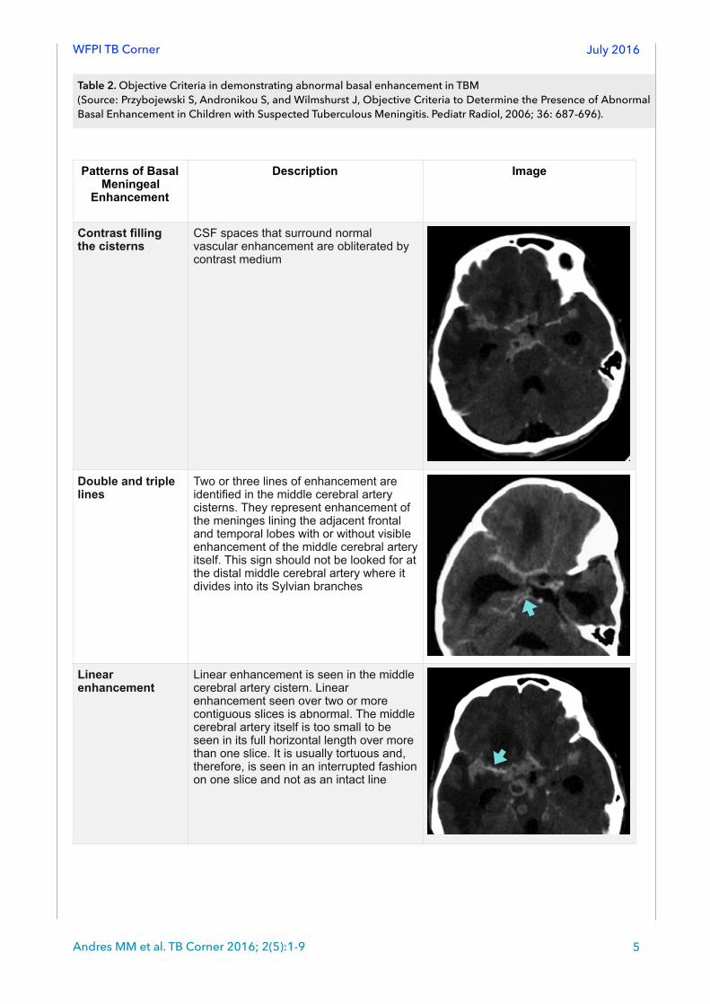
Patterns of Basal Meningeal
Enhancement
Description Image
Contrast filling the cisterns
CSF spaces that surround normal vascular enhancement are obliterated by contrast medium
Double and triple lines
Two or three lines of enhancement are identified in the middle cerebral artery cisterns. They represent enhancement of the meninges lining the adjacent frontal and temporal lobes with or without visible enhancement of the middle cerebral artery itself. This sign should not be looked for at the distal middle cerebral artery where it divides into its Sylvian branches
Linear enhancement
Linear enhancement is seen in the middle cerebral artery cistern. Linear enhancement seen over two or more contiguous slices is abnormal. The middle cerebral artery itself is too small to be seen in its full horizontal length over more than one slice. It is usually tortuous and, therefore, is seen in an interrupted fashion on one slice and not as an intact line
!
!
�
Andres MM et al. TB Corner 2016; 2(5):1-9 �5
Table 2. Objective Criteria in demonstrating abnormal basal enhancement in TBM (Source: Przybojewski S, Andronikou S, and Wilmshurst J, Objective Criteria to Determine the Presence of Abnormal Basal Enhancement in Children with Suspected Tuberculous Meningitis. Pediatr Radiol, 2006; 36: 687-696).

WFPI TB Corner July 2016
Y-sign The Y-sign is seen at the junction of the suprasellar and middle cerebral artery cistern. Pure vessel enhancement in this region lacks an arm of the ‘Y’ because the posterior communicating artery is not often seen on CT due to its small size
Infundibular recess of the third
The posterior aspect of the infundibular recess of the third ventricle in the suprasellar cistern is enhanced. There is no known vessel that lies here that can be confused with meningeal enhancement
Ill-defined edge There is an ill-defined edge to the enhancement, as opposed to the sharply marginated enhancement of normal vessels
�
!
!
Andres MM et al. TB Corner 2016; 2(5):1-9 �6

WFPI TB Corner July 2016
Conclusion Tuberculous meningitis is the most common presentation of CNS Tuberculosis. CT scanning is an established diagnostic modality in detection of TBM due to its accessibility, non-invasiveness, and rapid acquisition of images for evaluation.
There is a common triad of findings for TBM that includes abnormal basal cistern enhancement (sensitivity of 35-73% and specificity of 69-88%), hydrocephalus (sensitivity of 57-93% and specificity of 69-83%), and infarction (sensitivity is valued at 18-75% and its specificity is at 82-100%). Several patterns of basal meningeal enhancement are stated in the literature although these are non-specific,
Join the dots Normal enhancement of the Sylvian vessels is seen as separate dots because the branches are seen in cross section. Abnormal enhancement is present when the dots are joined by linear enhancement
Nodular enhancement
Nodular enhancement is always pathological as normal meninges and vessels are smooth and sharply marginated
Asymmetry Asymmetry of any of the above features aids recognition of abnormal basal enhancement as comparison can be made with the normal side
�
!
�
Andres MM et al. TB Corner 2016; 2(5):1-9 �7
WFPI TB Corner July 2016
and are not pathognomonic for TBM especially when presenting in isolation. Hence, findings of basal cistern enhancement should be correlated with other associated imaging and clinical presentations.
References 1. Duque-Silva A, Robsky K, Fllod J, and Barry P. Risk Factors for Central Nervous System
Tuberculosis. Pediatrics, 2015; 136(5). DOI: 10.1542/peds.2014-3958. 2. Theron S, Andronikou S, Grobbelaar, Steyn F, Mapukata A, Du Plessis J. Localized Basal
Meningeal Enhancement in Tuberculous Meningitis. Pediatr Radiol, 2006; 36:11182-1185. DOI: 10.1007/s00247-006-312-1
3. Andronikou, Wieselthaler N, Smith B, Douis H, Fieggen G, Van Toorn R, and Wilmshurst J. Value of Early Follow-up CT in Pediatric Tuberculous Meningitis. Pediatr Radiol, 2005; 35:1092-1099. DOI:10.1007/s00247-005-1549-9.
4. Andronikou, Savvas and Wieselthaler, Nicky. Modern Imaging of Tuberculosis in Children: Thoracic, Central Nervous System and Abdominal Tuberculosis. Pediatr Radiol, 2004; 34: 861-875. DOI: 10.1007/s00247-004-1236-2.
5. Reyes-Paguia MP, Laya BF, De Jesus JM, and Piedad RO. Accuracy of Established Cranial CT Scan Findings in the Diagnosis of Cranial Tuberculosis. St. Luke’s Journal of Medicine, 2011; 7(1): 33-42.
6. Torok ME. Tuberculous Meningitis: Advances in Diagnosis and Treatment. British Medical Bulletin, 2015;113:117-131. DOI: 10.1093/bmb/ldv003.
7. Principi N and Esposito S. Diagnosis and Therapy of Tuberculous Meningitis in Children. Tuberculosis, 2012;92:377-383. http://dx.doi.org/10.1016/j.tube.2012.05.011.
8. Chiang SS, Khan FA, Milstein MB et al. Treatment Outcomes of Childhood Tuberculous Meningitis: A Systematic Review and Meta-Analysis. Lancet Infect Dis, 2014; 14(10):947-957.
9. Van Rensburg PJ, Andronikou S, Van Toorn R, and Pienaar M. Magnetic Resonance Imaging of Tuberculosis of the Central Nervous System in Children with Tuberculous Meningitis. Pediatr Radiol, 2008; 38:1306-1313.
10.Donald Pr and Schoeman JF. Tuberculous Meningitis. N Engl J Med, 2004; 351:1719-1720. 11.Chatterjee S. Brain tuberculomas, tubercular meningitis, and post-tubercular hydrocephalus in
children. J Pediatr Neurosci. 2011;6 (3):96. 12.Changal KH, Raina AH. Central Nervous System Manifestations of Tuberculosis: A Review Article.
Mycobacterial Diseases. 2014; 4(2). 13.Thwaites GE, Van Toorn R, and Schoeman J. Tuberculous Meningitis: more questions, still few
answers. Lancet Neurol, 2013; 12:999-1010). DOI: 10.101016/S1474-4422(13)70168-6). 14.Nabukeera-Barungi N, Wilmshurst J, Rudzani M, Nuttall J. Presentation and outcome of
tuberculous meningitis among children: experiences from a tertiary children’s hospital. African Health Sciences.2014;14(1):143-9.
15.Botha H, Ackerman C, Candy S, Carr JA, Griffith-Richards S, and Bateman KJ. Reliability and Diagnostic Performance of CT Imaging Criteria in the Diagnosis of Tuberculous Meningitis. PLoS ONE, 2012; 7(6):1-8 e38982 DOI: 10.1371/journal.pone..0038982
16.Wallace RC, Burton EM, Barrett FF, Leggiadro RJ, Gerald BE, and Lasater OE. Intracranial Tuberculosis in Children: CT Appearance and Clinical Outcome. Pediatr Radiol, 1991; 21:241-246.
17.Andronikou, S, Wilmshurst J, Hatherill M, and Van Toorn R. Distribution of Brain Infarction in Children with Tuberculous Meningitis and Correlation with Outcome Score at 6 months. Pediatr Radiol, 2006;36:1289-1294. DOI: 10.1007/s00247-006-0319-7.
Andres MM et al. TB Corner 2016; 2(5):1-9 �8

WFPI TB Corner July 2016
18.Przybojewski S, Andronikou S, and Wilmshurst J. Objective Criteria to Determine the Presence of Abnormal Basal Enhancement in Children with Suspected Tuberculous Meningitis. Pediatr Radiol, 2006; 36:687-696.
19.Smirniotopoullos J, Murphy F, Rushing E, Rees J, Schroeder J. Patterns of Contrast Enhancement in the Brain and Meninges1. Radiographics, 2007;27(2):523-551.
20.Harisinghani M, McLoud T, Shepard J, Ko J, Shroff M, Mueller P. Tuberculosis from Head to Toe. Radiographics, 2000;20:449-470.
Corresponding author: Mariaem Andres St. Luke’s Medical Center Philippines [email protected]
Andres MM et al. TB Corner 2016; 2(5):1-9 �9