TAXONOMY, COMPARATIVE ANATOMY AND ......Instructions for use Title TAXONOMY, COMPARATIVE ANATOMY AND...
95
Instructions for use Title TAXONOMY, COMPARATIVE ANATOMY AND PHYLOGENY OF JAPANESE CATSHARKS, SCYLIORHINIDAE Author(s) NAKAYA, Kazuhiro Citation MEMOIRS OF THE FACULTY OF FISHERIES HOKKAIDO UNIVERSITY, 23(1), 1-94 Issue Date 1975-12 Doc URL http://hdl.handle.net/2115/21861 Type bulletin (article) File Information 23(1)_P1-94.pdf Hokkaido University Collection of Scholarly and Academic Papers : HUSCAP
Transcript of TAXONOMY, COMPARATIVE ANATOMY AND ......Instructions for use Title TAXONOMY, COMPARATIVE ANATOMY AND...

Instructions for use
Title TAXONOMY, COMPARATIVE ANATOMY AND PHYLOGENY OF JAPANESE CATSHARKS,SCYLIORHINIDAE
Author(s) NAKAYA, Kazuhiro
Citation MEMOIRS OF THE FACULTY OF FISHERIES HOKKAIDO UNIVERSITY, 23(1), 1-94
Issue Date 1975-12
Doc URL http://hdl.handle.net/2115/21861
Type bulletin (article)
File Information 23(1)_P1-94.pdf
Hokkaido University Collection of Scholarly and Academic Papers : HUSCAP

I ~ !
r
I
TAXONOMY, COMPARATIVE ANATOMY AND PHYLOGENY
OF JAPANESE CATSHARKS, SCYLIORHINIDAE*
Kazuhiro NAKAYA
Faculty of Fisheries, Hokkaido University, Hakodate, Japan
Abstract
Systematics of Japanese sharks in the family Scyliorhinidae has never been studied yet. These sharks were investigated externally and internally in order to review their classification and to make their interrelationships clear. Japanese scyliorhinid sharks were classified into seven genera and twelve species including three new species. A phyletic position was given to each' genus, based on several internal features, and a phyletic tree was presumed for the Japanese scyliorhinid genera.
Contents
Page I. Introduction ............ , . . . . . . . . . . . . . . . . . . . . . . . . . . . . . . . . . . . . . . . . . . . . . . . 2 II. Acknowledgments ................ , ............................. , . . . . . . . . . 3 III. Materials and methods . , .... , ............. , . . . . . . . . . . . . . . . . . . . . . . . . . . . . . . . 4 IV. Taxonomy ..................... ,....................................... 6
1. Family Scyliorhinidae .................................................. 6 2. Key to genera of Japan and its adjacent waters ............ , ........ " ., .. . 6 3. Genus Pentanchus Smith and Radcliffe, 1912 .............................. 7
Pentanchus sp. . ............. , ....... , ............................... ,. 8 4. Genus CephaloscyZlium Gill, 1861 ........................................ 8
Cephaloscyllium umbratile Jordan and Fowler, 1903 ........................ 9 5. Genus &yliorhinus Blainville, 1816 ...............................•...... 14
&yliorhinus torazame (Tanaka, 1908) .................................... 15 6. Genus Apristurus Garman, 1913 .... , " .... " ., ............. , ... , ... , .. ... 22
Apristurus japonicus sp. nov.. . . . . . . . . . . . . . . . . . . . . . . . . . . . . . . . . . . . . . . . . . .. 24 ApriStUTUS platyrhynchus (Tanaka, 1909) ..................... " ., . . . . . . . .. 28 Apristuruslongicephalu8 sp. nov. ........................................ 32 Apri8turus macrorhynchus (Tanaka, 1909) .....•.•........................ 36
7. Genus Galeus Rafinesque, 1810 .......................................... 40 Galeus sauteri (Jordan and Richardson, 1909).. . . . . . . . . . . . . . . . . . . . . . . . . . . .. 41 Galeus eastmani (Jordan and Snyder, 1904). . . . . . . . . . . . . . . . . . . . . . . . . . . . . . .. 46 Galeus nipponensis sp. nov. ............................................ 51
8. Genus Parmaturus Garman, 1906 ........................................ 57 Parmaturus pilo8U8 Garman, 1906 .. , .. " .. " ., ............. ,. . .. .. .... ... 58
9. Genus HalaelurU8 Gill, 1861 ............................................ 60 HalaeluTus burgeri (Muller and Henle, 1841) .............................. 61
* This work was submitted as a partial fulfillment of the requirements for Doctor's degree in Fisheries Science at Hokkaido University in 1974.
-1-

Mem. Fac. Fish. Hokkaido Univ. [XXllr, r
V. Comparative anatomy .................................................... 67 1. Chondrocranium ...................................................... 67 2. Pectoral fin .. . . . . . . . . . . . . . . . . . . . . . . . . . . . . . . . . . . . . . . . . . . . . . . . . . . . . . . .. 73 3. First dorsal fin ........................................................ 76 4. Vertebral calcification ....... " .............. " .. .. .. . . . . . . . . .. .. . . . . . .. 77 5. Extrabranchial cartilage . . . . . . . . . . . . . . . . . . . . . . . . . . . . . . . . . . . . . . . . . . . . . . .. 81 6. Reproductive system .................................................. 82
VI. General consideration .................................................... 84 VII. Summary .............................................................. 87
Literature cited . . . . . . . . . . . . . . . . . . . . . . . . . . . . . . . . . . . . . . . . . . . . . . . . . . . . . . . . .. 88
I. Introduction
The scyliorhinid sharks, or the catsharks, are comparatively small fishes and generally found in the waters of the temperate and tropical zones of the world oceans, inhabiting the bottom of shallow and deep waters.
Since Jordan and Fowler (1903) made the first revision of Japanese elasmobranch fishes, various ichthyologists such as Regan (1908), Pietschmann (1908), Garman (1913), Matsubara (1936, 1955), White (1937), Fowler (1941), Ishiyama (1958, 1967), and Lindberg and Legeza (1967) studied the classification of the sharks, skates and rays found in the waters around Japan. However, the sharks have not yet been investigated enough, at least so far as the taxonomy is concerned, though the skates are extensively surveyed by Ishiyama (1950, 1951a, 1951b, 1952, 1955a, 1955b, 1958, 1967).
Regarding the scyliorhinid sharks of Japan, a new species was reported for the first time by Muller and Henle (1841). Since then, eight species in total were described as new by Jordan and Fowler (1903); Jordan and Snyder (1904), Garman (1906), Tanaka (1908, 1909), Jordan and Richardson (1909), and Jordan and Hubbs (1925). Recently, Matsubara (1955), in his key to Japanese fishes, recognized eight genera and ten species in Japanese Scyliorhinidae including two unrelated species and excluding an undefinite species. The present author, however, believes that the family warrants seven genera and nine species in Japanese waters.
Though relatively many studies have been done on the biology of other shark groups, the biology of Japanese catshark has hardly been investigated. The only work is that of Kudo (1959) who studied the reproduction of a scyliorhinid in the waters of southern Japan.
The phyletic interrelationships among living elasmobranch fishes are still poorly known. Regan (1906), Holmgren (1941, 1942) and recently Compagno (1973) discussed the interrelationships among elasmobranch fishes above family level, but they scarcely mentioned the relationships among those of the lower levels. While Ishiyama (1958), Stehmann (1970) and Hulley (1972) extensively worked on the phylogeny within the Rajidae, no work on sharks of the lower taxa has been made except that of White (1937), who examined many species and discussed the interrelationships at the generic level. White included several Japanese scyliorhinid sharks, but their interrelationships were not clearly revealed.
-2-

1975] NAKAYA: Taxonomy, anatomy and phylogeny of Japanese catsharks
Thus the present author felt it necessary to compare their characters and to investigate the interrelationships among the species of Scyliorhinidae. Such a study should also be important when considering the phylogeny of carcharhinid and sphyrnid sharks, because the scyliorhinid sharks are considered to occupy the most primitive position among these groups .
. The purposes of the present work are to review the classification of Japanese scyliorhinid sharks, and to consider their phyletic interrelationships.
. On the basis of the phyletic study, the author proposes the following new classification of Japanese scyliorhinid sharks including three new species. Species which were not examined are noted by an asterisk.
Family Scyliorhinidae Genus Pentanchus Smith and Radcliffe, 1912
Pentanchus sp. * Genus Cephaloscyllium Gill, 1861
Cephaloscyllium umhratile Jordan and Fowler, 1903 Genus Scyliorhinus Blainville, 1816
Scyliorhinus torazame (Tanaka, 1908) Genus Apristurus Garman, 1913
Apristurus japonicus sp. nov. Apristurus platyrhynchus (Tanaka, 1908) Apristurus longicephalus sp. nov. Apristurus macrorhynchus (Tanaka, 1909)
Genus Galeus Rafinesque, 1810 Galeus sauteri (Jordan and Richardson, 1909) Galeus eastmani (Jordan and Snyder, 1904) Galeus nipponensis sp. nov.
Genus Parmaturus Garman, 1906 Parmaturus pilosus Garman, 1906*
Genus Halaelurus Gill, 1861 Halaelurus burgeri (Muller and Henle, 1841)
II. Acknowledgments
The present author expresses his sincere thanks to Dr. Shun Okada, former Professor of Hokkaido University, for his guidance in the course of this study and critical reading of the manuscript.
The author also wishes to thank the former Professor Hidejiro Niiyama and Professor Teruyoshi Kawamura of Hokkaido University, for critical reading of the manuscript.
The author wishes to express his heartfelt thanks to the following persons who assisted in this study: Associate Professor Tatsuyoshi Masuda of Tokyo University of Fisheries, for advice in collecting samples; Dr. Hideo Ootaki of Seikai Regional Fisheries Research Institute, Mr. Hung-chia Yang of Taiwan Fisheries Research Institute, Mr. Kazuyuki Teshima of Shimonoseki University of Fisheries, Mr. Juzo Sato of Choshi Trawler Fisheries Association and Mr. Yukio
-3-

Mem. Fac. Fish. Hokkaido Univ. paID, I
Tanaka of Shimonoseki Fish Market, for collection of sharks; Mr. Koji Sugishita at Hakodate, for long term collection of great many samples during this study; Dr. Reizo Ishiyama of Tokyo University of Fisheries, Dr. Tokiharu Abe of Tokai Regional Fisheries Research Institute, Mr. Torao Sato of University of Tokyo, Mr. Hirokazu Kishimoto of Tokai University, Mr. Yojiro Imaoka of Shimane Fisheries Experimental Station and Mr. Hiroshi Kabasawa of Keikyu Aburatsubo Marine Park Aquarium, for loan of shark specimens; Dr. Akira Ochiai of Kochi University, Drs. Allan O. DeLacy and Arthur D. Welander of University of Washington, Dr. G.E. Maul of Museu Municipal do Funchal in Madeira, and Mr. Shinji Kudo of Nansei Regional Fisheries Research Institute, for offer of invaluable specimens and samples; Dr. Lo-chai Ohen of Oalifornia State University in San Diego, for taking and shipping of the X-ray films; Dr. Hiroshi Kasahara of F.A.O. in Rome, for help in getting specimens; Mr. Ken-ichi Yano of Ise Jingu Historical and Agricultural Museum, for information on the specimens in the Museum; Dr. Stewart Springer of Mote Marine Laboratory in Florida and Mr. Susumu Kato of Tiburon Laboratory in Oalifornia, for advice and information on the classification of scyliorhinid sharks; Miss Jean B. Kellerman of MissionaryUnited Ohurch of Ohrist in Japan, for correction of English expression; Professor Takao Igarashi and Associate Professor Kunio Amaoka of Laboratory of Marine Zoology, Hokkaido University, for critical comments and advice in the course of this study.
III. Materials and methods
The specimens were collected at Hakodate in Hokkaido, Shimokita in Aomori Prefecture, Ohoshi in Ohiba Prefecture, Shimonoseki in Yamaguchi Prefecture and Millase in Kochi Prefecture. Specimens from Hakodate and Aomori were caught by the long lines. Other specimens were collected by means of trawlers. They are all deposited in the Laboratory of Marine Zoology, Faculty of Fisheries, Hokkaido University (HUMZ), and are preserved in 10 percent formalin.
Some specimens were loaned from University of Tokyo (ZIUT) and Tokai University (MSM). Some embryos and egg capsules were borrowed from Keikyu Aburatsubo Marine Park Aquarium and Shimane Fisheries Experimental Station. Many specimens and X-ray films were given from Taiwan Fisheries Research Institute, University of Washington and Scripps Institution of Oceanography (S1O).
Proportional measurements for all specimens were made according to the method described by Bigelow and Schroeder (1948) and expressed in percent of total length. Number of vertebrae was counted by taking softex films. Skeletal parts were examined after being stained by alizarin red.
The specimens used in the comparative anatomy and in counting vertebrae were picked out from those used in the taxonomical study. The fully matured specimens were dissected as far as possible in the anatomical study. They are listed in Tables 1 and 2.
-4-

1975] NAKAYA: Taxonomy, anatomy and phylogeny of Japanese catsharks
Table 1. Sex, Total length and locality of the specimens used far comparative anatomy.
Species I Sex I Number of Total
I Locality specimens length (mm)
Oephal08cyllium <5 1 383 Kii Suido Channel umbratile ~ 1 435
Scyliarhinua <5 6 413-500 Hakodate torazame ~ 7 418-472
ApriBturUB <5 2 640,666 Choshi japonicus ~ 0
Galeua eaatmani <5 1 330 Kochi, Kii Suido ~ 2 361, 380 Channel
Galeus sauteri <5 1 376 Tungkang ~ 1 383
Galeua nipponenaiB <5 1 576 Kochi, Kii Sudio ~ 1 586 Channel
HalaeluTUs bUrgeri <5 1 407 Kochi, East China ~ 2 400,456 Sea
Table 2. Sex, total length and locality of the specimena used for inapection of vertebral number.
Species
Oephaloscyllium umbratile
Scyliorhinus tara'zame
ApriBtuTU8 japonicua
Apri8tUrUB platyrhynchua
Apristurua longicephalua
Apri8tuTUs macrorhynchus
Galeua eaatmani
Galeua sauteri
Galeua nipponenaiB
HalaelUTUS burgeri
<5 ~
<5 ~
<5 ~
<5 ~
C; ~
C; ~
C; ~
C; ~
C; ~
<5 ~
Number of specimens
4 4
36 63
10 2
0 1
1 0
3 3
3 8
3 8
6 6
1 1
-5-
I Total I length (mm) Locality
313-458 Kochi 356-657
242-490 Hakodate, Aomori, 210-478 East China Sea
654-711 Choshi 600,626
- Tokyo 654
375 Kochi -
173-383 Sagami Bay, Tokyo, 130-674 Suruga Bay
263-332 Kochi 356-376
372-379 Keelnng, Tungkang 374-398
514-596 Kii Suido Channel, 518-624 Kochi
407 Kochi, East China 456 Sea .

Mem. Fac. Fish. Hokkaido Univ. [xxm, I
IV. Taxonomy
1. Family Scyliorhinidae
Body slender. Two dorsal fins (rarely only one dorsal fin) rather small, without spines on anterior margins. First dorsal origin posterior to origin of pelvic fin. Pectoral fin small to large; its corners rounded. Anal fin present; its base short to long. Caudal fin rather small; its axis only slightly elevated or straight; lower anterior corner not expanded as a definite lobe. Caudal peduncle slender, without pit or keel. Inner margins of pelvic fins more or less united posterior to cloaca. Head rounded or flattened. Snout short or elongated; its anterior tip moderately rounded. Eye small to moderate with sub ocular fold, but without nictitating membrane. Spiracle moderate behind eye. Gill openings small; five in number; 5th, or 4th and 5th gill openings above base of pectoral fin; 5th shortest. Nostrils not connected with mouth by a groove, or rarely connected by a groove but without a prominent nasal barbel. Labial grooves developed or absent. Teeth small, numerous, with several cusps; several series functional. Modified enlarged denticles present or absent on the upper margin of caudal fin.
Rostral cartilages three, united anteriorly. Supraorbital crest present or absent. Suborbital shelf present. Basals of pectoral fin three; metapterygium largest. Radials of pectoral fin mostly on metapterygium. Labial cartilages two or three. Extrabranchial cartilages four on upper and three or four on lower side. Vertebral calcification variable in type.
Oviparous or ovoviviparous. Remarks
Tanaka (1915) treated Oalliscyllium venustum as a species in the family Scyliorhinidae because of its oviparity. Succeeding workers such as Matsubara (1936, 1955), Nakamura (1936), Fowler (1941) and Teng (1962), also considered Proscyllium habereri and Oalliscyllium venustum as members of scyliorhinid sharks. However, the present author excludes them from Scyliorhinidae because of their anteriorly located 1st dorsal fin and some internal characters. They are considered as members of triakid sharks.
2. Key to genera of Japan and its adjacent waters
1a. Only one dorsal fin ................. . Pentanchus Smith and Radcliffe, 1912 1 b. Two dorsal fins
2a. Denticles along dorsal margin of anterior part of caudal fin normal and same as those of body
3a. Snout thick and short; mucous pores on head not prominent; anal base short and as long as 2nd dorsal base or longer, but less than two times; anal and caudal fins greatly separated
4a. Black spot absent, or small in number and indistinct, if present 5a. Snout in front of mouth short, less than 1/2 of mouth width; 2nd
-6-

1975] NAKAYA: Taxonomy, anatomy and phylogeny of Japanese catsharks
2nd dorsal fin and anal fin almost opposite; no labial groove on both jaws; body soft ................ Oephaloscyllium Gill, 1861
5b. Snout in front of mouth 1/2 of mouth width or more; anal fin much anterior to 2nd dorsal fin; a small labial groove on lower jaw; body not soft but stiff ........ Scyliorhinus Blainville, 1816
4b. Many distinct black spots smaller to greater than iris on body and fins ................. r ..................... . Halaelurus Gill, 1861
3b. Snout flattened dorsoventrally and long; mucous pores on head very prominent; anal base very long and at least two times as long as that of 2nd dorsal fin, or greater; anal and caudal fins separated only by a notch .. . .. .. .... . ... . ......... ....... ... . . . . . .. Apristurus Garman, 1913
2b. A distinct crest of enlarged modified denticles along dorsal margin of anterior part of caudal fin
6a. Snout very short, 2/3 of mouth width, or 4/3 of horizontal diameter of eye, which equal to or greater than length from tip of snout to posterior end of nasal aperture ............................. . Parmaturus Garman, 1906
6b. Snout long, equal to or slightly smaller than mouth width, or 5/3 of horizontal diameter of eye or more, which always less than length from tip of snout to posterior end of nasal aperture ..................... .
. . . . . . . . . . . . . . . . . . . . . . . . . . . . . . . . . . . . . . . . . . . . Galeus Refinesque, 1810
3. Genus Pentanchus Smith and Radcliffe, 1912
PentanchU8 profundicolu8 Smith and Radcliffe, 1912, p. 490, pI. 42 (Type species: Pentanchu8 profundicolus Smith and Radcliffe). - Garman, 1913, p. 95. - Jordan and Hubbs, 1925, p. 100. - Herre, 1925, p. 127. - (in part) Fowler, 1941, p. 52.
Sharks characterized by five branchial apertures, the last three of which are above the base of the pectoral fin; elongated body, nearly straight tail; long, flat snout; inferior mouth; pluriserial, pluricuspid, erect teeth, similar in both jaws; minute spiracles; imbricate denticles; a single small dorsal fin without spine; long caudal fin, with large lower lobe; long anal fin; ventral fins inserted in advance of the center of the body; and large broad, pectoral fins. (Smith, 1912). Remarks
The genus Pentanchus is based on a single type specimen from the Philippine Archipelago. Since, only Jordan and Hubbs (1925) reported a specimen of this genus under the name "Pentanchus undescribed species" based on a mounted specimen in the Yamada Museum (now formally, the Ise Jingu Historical and Agricultural Museum) in Mie Prefecture. This valuable specimen had, very unfortunately, been lost and could not be examined.
Although the genus Pentanchus has sometimes been considered to be based on an abnormal specimen, Bigelow and Schroeder (1948) and Springer (1966) treated it separately, distinct from Apristurus after examination of the type specimen, in which they found no sign of mutilation or abnormality. Because he could not
-7-

Mem. Fac. Fish. Hokkaido Univ. [xxm, I
examine any specimens of this genus, the present author can say nothing on the status of Pentanchus at present and treats it separately according to Bigelow and Schroeder (1948) and Springer (1966) ..
Pentanchus sp.
PentanchUB undescribed species Jordan and Hubbs, 1925, p. 100 (short description on a mounted specimen in the Yamada Museum, Mie Prefecture, Japan)
The single dorsal fin is placed above the anal, which is twice as large; both these fins are low; the ventrals much larger than either, and inserted behind the middle of the body; pectoral small; caudal short and rather low. Gill openings five, the 1st much higher than the others, which are progressively shortened. The species seems to differ from Pentanchus profundicolus Smith and Radcliffe from Philippines in the larger ventrals and smaller pectorals. (Jordan and Hubbs, 1925).
4. Genus Cephaloscyllium Gill, 1861
Oephalo8cyllium Gill, 1861, p. 407 (Type species: Scyllium laticep8 Gill). - Jordan and Fowler, 1903, p. 602. - Garman, 1913, p. 78. - Matsubara, 1936, p. 34. - Fowler, 1941, p. 30. - Beebe and Tee-Van, 1941, p. 99. - Bigelow and Schroeder, 1948, p. 201. - Matsubara, 1955, p. 107. - Lindberg and Legeza, 1967, p. 40.
Scyllium (in part) Giinther, 1870, p. 400. Oatulu8 (in part) Jordan and Evermann, 1896, p. 23. ScyliorhinUB (in part) Regan, 1908, p. 453. ScylliorhinUB (in part) Barnard, 1925, p. 39.
Two dorsal fins; origin of 1st dorsal fin above base of pelvic fin. Second dorsal fin smaller than 1st dorsal fin; its origin about above that of anal fin. Oaudal fin relatively large. Oaudal and anal fins widely spaced. Denticles along upper margin of caudal fin normal and not enlarged. Head broad and rather flattened. Snout short; its tip rounded. Eye rather small. Fourth and 5th gill openings above base of pectoral fin. Nostrils much near mouth, but never connected with mouth by a groove. Labial grooves absent on upper and lower jaws. Mouth large and wide. Teeth alike in both jaws; one large primary cusp and a few lateral cusps.
Rostral cartilage short and weak. Supraorbital crest present in C. umbratile, but unknown in other species.
Body very soft; abdomen capable of swelling. Oviparous.
Remarks Cephaloscyllium is a genus with about eight nominal species: C. isabellum
(Bonnaterre, 1788) from Australia and New Zealand, C. ventriosum (Garman, 1880) from Ohile, C. uter (Jordan and Gilbert, 1896) from Oalifornia, C. umbratile Jordan and Fowler, 1903, from Japan, C. laticeps (Dumeril, 1853) from Australia, C. suJllans (Regan, 1921) from South Africa, C. formosanum Teng, 1962, from Formosa, and C. fasciatum Ohan, 1966, from South Ohina Sea.
-8-

1975] NAKAYA: Taxonomy, anatomy and phylogeny of Japanese catsharks
The sharks of this genus can swell their abdomen and this is a very peculiar habit among sharks.
Cephaloscyllium umbratile Jordan and Fowler, 1903 Japanese name: Nanuka-zame
Figs. 1",,3
Oephalo8cyZlium umbratile Jordan and Fowler, 1903, p. 602, fig. 1 (original description; Nagasaki, Japan). - Jordan and Richardson, 1909, p. 160 (distribution). - Garman, 1913, p. 80 (key, description). - Jordan, Tanaka and Snyder, 1913, p. 9, fig. 4 (distribution). - Tanaka, 1914, p. 24 (list). - Tanaka, 1931a, p. 13 (list). - Tanaka, 1931b, p. 7, fig. 11 (short description, distribution). - Matsubara, 1936, p. 35, fig. 25 (key, description, distribution). - White, 1937, p. 117 (key, phylogeny). - Okada, 1938, p. 116 (list). - Fowler, 1941, p. 32 (key, description). - Chen, 1948, p. 28 (key, distribution). - Kamohara, 194\), p. 59 (short description, distribution). - Kamohara, 1950, p. 3 (short description, distribution). - Kuroda, 1951, p. 315 (list). - Honma, 1952, p. 139 (list). - Mori, 1952, p. 18 (list). - Matsubara, 1955, p. 107, pI. 3, fig. 8 (key). - Mori, 1956, p. 2 (list). - Teng, 1962, p. 45 (key, description). - Chu, Chang and Cherng, 1963, p. 18, fig. 13 (description, distribution). - Chen, 1963, p. 29 (key, description, distribution). - Chu and Wang, 1964, p. 676 (distribution). - Chung, Kim and Kim, 1967, p. 8 (distribution). - Lindberg and Legeza, 1967, p. 40, fig. 24 (short description, distribution).
Oatulus laticep8. Jordan and Snyder, 1901, p. 38 (list) Scyliorhinus umbratili8. Regan, 1908, p. 459 (key, short description).
Materials Male - HUMZ 39379 (383 mm TL), HUMZ 42373 (383 mm TL), Kii Suido Channel
(33°30'-33°40'N, 134°30'-135°00'E), Aug. 6,1972; HUMZ 39371 (313 mm TL), Mimase, Dec. 19,1972; HUMZ 42374 (458 mm TL), Mimase, Dec. 20,1972; HUMZ 39370 (436 mm TL), Mimase, Dec. 23, 1972. Female - HUMZ 39989 (435 mm TL), Kii Suido Channel (same area as above), Aug. 6, 1972; HUMZ 39979 (539 mm TL), Mimase, Dec. 18, 1972; HUMZ 39382 (657 mm TL), Mimase, Dec. 21, 1972. Embryo - One female (109 mm TL), Hamada in Shimane Prefecture.
Diagnosis Snout short and rounded; mouth width greater than two times as long as snout
in front of mouth; labial grooves absent on both jaws; anal origin a little before origin of 2nd dorsal fin, but both almost opposite in position; body soft and abdomen capable of swelling.
Proportional measurements Trunk: breadth 10.2-14.2; height 9.8-15.5 Head: breadth 13.9-16.1; height 8.1-11.5 Eye: horizontal diameter 2.8-3.3 Mouth: width 9.9-11.4; height 4.6--4.9
-9-

Mem. Fac. Fish. Hokkaido Univ.
Nostrils: distance between inner ends 2.5-2.7 Labial grooves: upper 0; lower 0 Gill openings: 1st 1.5--2.6; 5th 1.1-1.8 First dorsal fin: vertical height 4.6-5.6; base length 6.7-8.1 Second dorsal fin: vertical height 2.3-2.8; base length 4.8-5.5 Anal fin: vertical height 3.0-3.5; base length 6.8-8.0 Oaudal fin: upper margin 23.1-27.0 Pectoral fin: outer margin 11.2-14.2
[xxm, I
Distance from tip of snout to: anterior nasal aperture 1.7-2.3; mouth 3.6-4.4; eye 4.5--6.0; 1st gill opening 14.5--16.5; 5th gill opening 18.4-20.3; pectoral fin 16.6-18.7; pelvic fin 42.8-47.0; 1st dorsal fin 46.6-51.7; 2nd dorsal fin 61.0-65.1; anal fin 59.1-63.5; upper caudal fin 72.5--76.0
Interspace between: 1st and 2nd dorsal fins 7.6-8.7; 2nd and upper caudal fins 6.2-7.1; anal and lower caudal fins 5.3-7.0
Distance from origin to origin 'of: pectoral and pelvic fins 24.8-28.7; pelvic and anal fills 15.4-17.5
Description (External): Body rather slender; depth of trunk and caudal peduncle about equal to each width; caudal axis a little elevated.
Head large; its length equal to 1/5 of total length. Snout short, broadly rounded (Fig. 2 a, b). Nasal apertures moderate; anterior-most of nostrils behind middle of snout in front of mouth. Mouth very wide; its width greater than two times as great as snout in front of mouth. Labial grooves completely absent (Fig. 2 b). Eye small, ovate; its horizontal diameter equal to distance from 1st to 3rd gill opening. Spiracle small, behind orbit and slightly below level of horizontal axis of eye. Gill openings short; 1st longest and about 2/3 of horizontal diameter of eye; 4th and 5th gill openings above base of pectoral fin. Pectoral fin moderate in size; length of outer margin equal to distance from anterior margin of eye to 3rd gill opening; its outer and inner corners moderately rounded; distal margin straight or slightly concave. First dorsal origin above anterior 1/3 of pelvic base; anterior margin convex; distal margin straight; free rear tip not elongated, but rounded. Second dorsal origin only slightly behind anal origin; its vertical height 1/2 of that of 1st dorsal fin; apex broadly rounded; distal margin concave; free rear tip slightly elongated and pointed. Pelvic origin a little anterior to 1st dorsal origin by distance of 1st gill opening. Anal origin slightly anterior to 2nd dorsal origin by distance of 1/2 of 1st gill opening; its anterior margin convex; apex very broadly rounded; distal margin concave; free rear tip elongated and pointed. Oaudal fin broad, without any crests of modified denticles on upper and lower anterior margins.
Denticles on body large, sparsely distributed and never overlapping; its shape over dorsolateral surface of trunk one cusped (Fig. 2 c).
Teeth in juvenile female small, numerous; typical teeth three cusped in both jaws; several series functional (Fig. 2 d).
Ground color of dorsolateral surface of body, upper surfaces of paired fins and dorsal fins brownish. Seven saddles and dirty blotches of dark brown on body, dorsal fins and caudal fin. No distinct blotches or bands on upper surface of
-10 -
..... .....
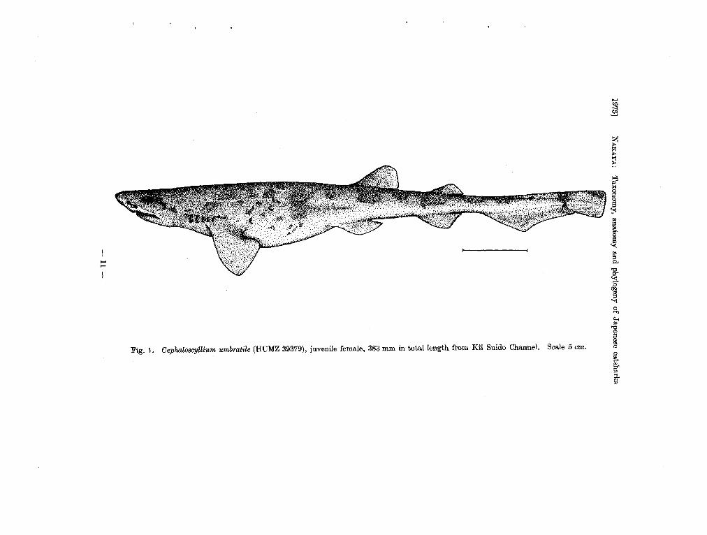
Fig. 1. Oephalo8cytlium umbratile (HUMZ 39379), juvenile female, 383 mm in total length from Kii Suido Channel. Scale 5 em.
..... <:P
.9
s ~
I j i
I So <!..j
1 "
f
Mem. Fae. Fish. Hokkaido Univ. [xxm, I
a ~
/ '., . .,.,,' ,;ii;
~ .~A" ... :i:'.
~
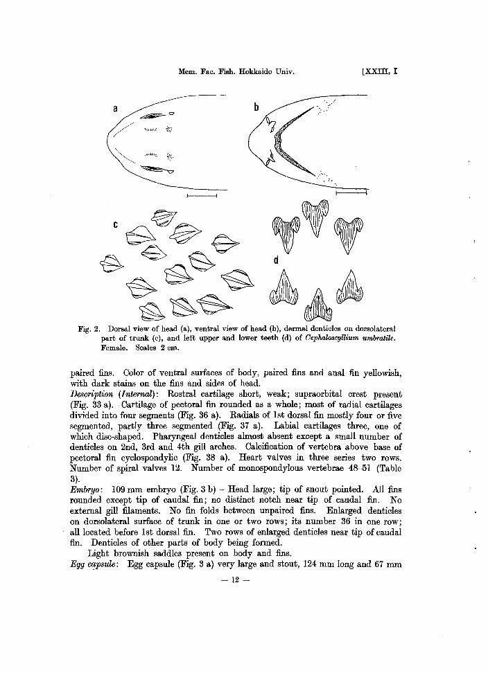
Fig. 2. Dorsal view of head (a), ventral view of head (b), dermal denticles on dorsolateral part of trunk (c), and left upper and lower teeth (d) of Oephalo8cyUium umhratile. Female. Scales 2 em.
paired fins. Oolor of ventral surfaces of body, paired fins and anal fin yellowish, with dark stains on the fins and sides of head. Description (Internal): Rostral cartilage short, weak; supraorbital crest present (Fig. 33 a). Oartilage of pectoral fin rounded as a whole; most of radial cartilages divided iuto four segments (Fig. 36 a). Radials of 1st dorsal fin mostly four or five segmented, partly three segmented (Fig. 37 a). Labial cartilages three, one of which disc-shaped. Pharyngeal denticles almost absent except a small number of denticles on 2nd, 3rd and 4th gill arches. Oalcification of vertebra above base of pectoral fin cyclospondylic (Fig. 38 a). Heart valves iu three series two rows. Number of spiral valves 12. Number of monospondylous vertebrae 48--51 (Table 3). Embryo: 109 mm embryo (Fig. 3 b) - Head large; tip of snout poiuted. All fins rounded except tip of caudal fin; no distinct notch near tip of caudal fin. No external gill filaments. No fin folds between unpaired fins. Enlarged denticles on dorsolateral surface of trunk iu one or two rows; its number 36 in one row; all located before 1st dorsal fin. Two rows of enlarged denticles near tip of caudal fin. Denticles of other parts of body beiug formed.
Light brownish saddles present on body and fins. Egg lX!psule: Egg capsule (Fig. 3 a) very large and stout, 124 mm long and 67 mID
-12 -

1975] NAKAYA: Taxonomy, anatomy and phylogeny of Japanese catsharks
Table 3. Frequency of occurrence of varioU8 numbers of 'TTW'TW8jJondylous vertebrae in Japanese scyliorhinid sharks.
Species
Oephaloscyllium umbratile
ScyliorhinU8 torazame
ApristuTU8 japonicU8 platyrhynchus longicephalus macrorhynchU8
GaleU8 eastmani sauteri nipponensis
HalaeluTU8 burgeri
a
b
Number of monospondylou8 vertebrae
2 29 43 15 9 1
1
452 2 3 4 2
1 1
1
141
372
3 2 4 3
1 124
Fig. 3. Egg capsule (a) and 109 mm embryo (b) from egg capsule of Oephaloscyllium umbratile. Scales 2 cm.
-13 -

Mem. Fac. Fish. Hokkaido Univ. [xxm, I
wide. Long tendrils present on four corners of anterior and posterior ends. Surface of the capsule smooth but with longitudinal streaks.
Color opaque and whitish yellow with yellow margin. Distribution: Hokkaido (Hakodate), Chiba Prefecture, Sagami Bay, Mie Prefecture (Kumanonada), Niigata Prefecture, Shimane Prefecture, Shikoku and Kyushu ill
Japan. China including Yellow Sea. Formosa (Kao-hsiung, Tungkang). Remarks
This species bears one big egg capsule in each oviduct, and the mode of reproduction is considered to be the single oviparity, which will be defined later in the anatomical section. This species is said to be abundant in the relatively deep waters of southern Japan (Matsubara, 1936, 1955).
5. Genus Scyliorhinus Blainville, 1816
Scyliorhinus Blainville, ISI6, p. 121 (Type species: Scyliorhinus caniculus, designated by Gill, IS61). - (in part) Regan, 1905, p. 453. - Fowler, 1941, p. 34. - Bigelow and Schroeder, 1945, p. 202. - Matsubara, 1955, p. lOS. - Tortonese, 1956, p. 125. - Springer, 1966, p. 597. - Lindberg and Legeza, 1967, p. 41. - Springer and S:tdowsky, 1970, p. S3.
Scyllium (in p:trt) Miiller and Henle, IS41, p. 3. Scylliorhinus (in part) Goode and Bean, IS95, p. 16. - (in part) Barnard, 1925, p. 3S. Oatulu8 (in part) Jordan and Evermann, IS96, p. 23. - (in part) Garman, 1913, p. 71.
- Matsubara, 1936, p. 36. Halaeluru8 Tanaka, 1912, p. 13. - (in part) Schmidt, 1931, p. 4.
Two dorsal fins; origin of 1st dorsal fin over or slightly anterior to rear end of base of pelvic fin. Pectoral fin small with rounded corners. Anal fin moderate; its origin anterior to origin of 2nd dorsal fin; base short. Caudal and anal fins widely spaced. Denticles along upper margin of caudal fin normal and not enlarged. Inner margins of male pelvic fin united posterior to cloaca for a long distance. Head rather flattened. Snout short; its tip rounded. Eye moderate. Fifth, or 4th and 5th gill openings above base of pectoral fin. Nostrils much near mouth, but not connected with mouth by a groove. A labial groove on lower jaw; none on upper. Teeth alike in both jaws; one large primary cusp with a few lateral cusps. Dermal denticles large and strongly ridged.
Rostral cartilage short and weak. Supraorbital crest present as far as known. Body stiff; abdomen incapable of swelling. Oviparous.
Remarks This genus contains about ten or less species from the world oceans. Species
of this genus are abundant in the Atlantic Ocean, especially in the Caribbean Sea, Gulf of Mexico and tropical to temperate zone of western Atlantic. The members of this genus resemble Oephaloscyllium externally, but greatly differ in having stiff body incapable of swelling.
- 14-

1975] NAKAYA: Taxonomy, anatomy and phylogeny of Japanese catsharks
Scyliorhinus torazame (Tanaka, 1908) Japanese name: Tora-zame
Figs. 4-9
Catulus torazame Tanaka, 1908, p. 6, pI. 2 (original description; Misaki, Japan). -Garman, 1913, p. 77 (key, description). -Tanaka, 1914, p. 24 (list). -Tanaka, 1931b, p. 5, fig. 8 (description, distribution). -Matsubara, 1936, p. 36, fig. 26 (key, description, distribution). -White, 1937, p. 117 (key, phylogeny). -Okada, 1938, p.1l7 (list). - Kamohara, 1950, p.4 (short description, distribution). -Honma, 1952, p.139 (list).
Halaelurus rudis Pietschmann, 1908, p. 2 (description). -Tanaka, 1912, p. 13 (description). 8cyliorhinus rudis. Regan, 1908, p. 457 (key, short description). Halaelurus torazame. Jordan, Tanaka and Snyder, 1913, p.1O (list). -Schmidt, 1930a" p.
48 (description of clasper). - Schmidt, 1931, p. 4 (short description). 8cyliorkinus torazame. Fowler, 1941, p. 36 (key, description). - Herre, 1949, p. 151
(distribution). -Mori, 1952, p. 18 (list). -Herre, 1953, p. 12 (list, distribution). -Okada, 1955, p. 10, fig. 9 (short description, distribution). -Matsubara, 1955, p. 108, pI. 3, fig. 9 (key). - Mori, 1956, p. 2 (list). - Chu, Chan and Cherng, 1963, p. 19., fig. 14 (key, description, distribution). - Chu and Wang, 1964, p. 675 (distribution). - Lindberg and Legeza, 1967, p. 41, fig. 25 (short description, distribution). - Ueno, 1971, p. 68 (distribution).
Materials Male-HUMZ 42375-42377 (440--500 mm TL), June 16,1970; HUMZ 42378 (457 mm
TL), HUMZ 42379 (413 mm TL), July 20,1970; HUMZ 42380 (458 mm TL), Apr. 28, 1971; HUMZ 42381 (455 mm TL), May 13, 1971; HUMZ 39392 (484 mm TL), June 11, 1971; HUMZ 42382 (490 mm TL), HUMZ 39981 (451 mm TL), HUMZ 42383 (481 mm TL), Apr. 22, 1972. Female - HUMZ 42384 (472 mm TL), May 27, 1969; HUMZ 42385 (42S mm TL), June 16, 1970; HUMZ 42386 (430 mm TL), July 20, 1970; HUMZ 42387 (440 mm TL), HUMZ 42388 (418 mm TL), Aug. 21, 1970; HUMZ 42389 (423 mm TL), HUMZ 42390 (454 mm TL), Aug. 24, 1970; HUMZ 42391 (245 mm TL), Aug. 25, 1970; HUMZ 42392 (432 mm TL), Apr. 22, 1971; HUMZ 42393 (434 mm TL), Apr. 28, 1971; HUMZ 39384 (478 mm TL), HUMZ 42394 (442 mm TL), June 3, 1971; HUMZ 42395 (456 mm TL), June 11,1971. Embryo-HUMZ 42396 (36 mm TL, male), March 17, 1971; HUMZ 42397 (58 mm TL, female), May 29, 1972; HUMZ 42398 (79 mm TL, female), Feb. 13, 1973.
All samples above collected at Hakodate.
Diagnosis
Snout short and rounded; mouth width nearly two times as long as snout in front of mouth, or less; labial groove only on lower jaw; anal and 2nd dorsal fins almost same in size, but anal fin located more anteriorly; dermal denticles large; skin relatively rough to touch.
Proporti<mal measurements
Trunk: breadth 9.4-13.1; height 9.4-13.2 Head: breadth 12.8-13.4; height 8.1-10.1
-15 -

t:::: CD
~ Io;j P> ? Io;j Iii' F'" .... "'<W
~ 0>
g: §1 ~.
Fig. 4. Scyliorhinus torazame (HUMZ 42383), adult male, 481 mm in total length from Hakodate. Scale 5 em.
~ 1-1

1975] NAKAYA: Taxonomy, anatomy and phylogeny of Japanese catsharks
Eye: horizontal diameter 2.9-3.5 Mouth: width 7.6-8.2; height 2.6-3.3 Nostrils: distance between inner ends 1.5-2.1 Labial grooves: upper 0; lower 1.0-1.3 Gill openings: 1st 1.4-2.1; 5th 0.6-1.2 First dorsal fin: vertical height 4.8-6.1; base length 6.4-7.4 Second dorsal fin: vertical height 3.1-3.8; base length 4.7-5.4 Anal fin: vertical height 2.9-3.7; base length 6.9-8.2 Oaudal fin: upper margin 20.7-23.6; lower margin 8.9-10.5 Pectoral fin: outer margin 11.4-12.7; inner margin 4.7-5.4 Distance from tip of snout to: anterior nasal aperture 2.2-2.6; mouth 3.7-
4.2; eye 4.6-5.2; 1st gill opening 13.3-14.4; 5th gill opening 17.0-19.7; pectoral fin 16.2-17.5; pelvic fin 38.6--42.8; 1st dorsal fin 47.7-52.6; 2nd dorsal fin 65.7-69.0; anal fin 58.5-62.6; upper caudal fin 76.4-79.3
Interspace between: 1st and 2nd dorsal fins 9.7-11.8; 2nd and upper caudal fins 4.1-7.0; anal and lower caudal fins 8.0-10.9
Distance from origin to origin of: pectoral and pelvic fins 22.5-25.9; pelvic and anal :fins 18.7-22.0
Description (External): Body slender; depths of trunk and caudal peduncle about equal to each width; caudal axis slightly elevated.
Head a little shorter than 1/6 of total length. Snout short, broadly rounded (Fig. 5 a, b). Nasal apertures large; anterior-most of nostrils behind middle of
. snout in front of mouth. Mouth wide; its width nearly two times as great as snout in front of mouth. A labial groove on lowerjaw, but upper one absent. Eye moderate, ovate; its horizontal diameter equal to distance from 1st to 4th gill opening. Spiracle moderate, behind orbit and slightly below level of horizontal axis of eye. Gill openings short; 1st longest and equal to 1/2 of horizontal diameter of eye; 5th, or 4th and 5th gill openings above base of pectoral fin. Pectoral fin moderate in size; length of outer margin equal to distance from anterior margin of eye to 3rd gill opening; its outer corner moderately rounded; inner corner rounded; distal margin slightly convex. First dorsal origin above end of pelvic base or a little anterior; its anterior margin straight or slightly convex; distal margin convex; free rear tip not elongated but rounded. Second dorsal origin over posterior half of anal base; fin itself similar in shape to 1st dorsal fin, but free rear tip more elongated and pointed. Pelvic fin moderate; its base almost wholly anterior to 1st dorsal origin. Olasper long, rod-like, tapering distally almost reaching to anal origin. Right and left pelvic fins united nearly to the tip. Anal origin a little anterior to middle of interdorsal space; its anterior and distal margin slightly convex; free rear tip elongated and pointed; end of base below anterior half of 2nd dorsal base. Oaudal fin moderate, without any crest of modified denticles on upper and lower anterior margins.
Denticles on body large and a little overlapping; its shape over dorsolateral surface of trunk three cusped (Fig. 5 c).
Teeth in adult male small, numerous; typical teeth five cusped in upper teeth; that of lower teeth five cusped but a pair of outermost cusps blunt; several series
- 17-

Mem. Fae. Fish. Hokkaido Univ. [XXIII, I
o ' ..
1----/
Fig. 5. Dorsal view of head (a), ventral view of head (b), dermal dentieles on dorsolateral part of trunk (c), and left upper and lower teeth (d) of ScyliorhinU8 torazame. Male. Scales 1 em.
functional (Fig. 5 d). Ground color of dorsolateral surface of body, dorsal sides of paired fins and
dorsal fins brownish. Ten odd saddles and blotches of dark brown on body, dorsal sides of paired fins and dorsal fins. Color of ventral surfaces of body, paired fins and anal fin yellowish with slight dirty stains on anal and margin of head. Description (Internal): Rostral cartilage very short, weak; modified supraorbital crest present (Fig. 33 b). Cartilage of pectoral fin rounded as a whole; radial cartilages divided mostly into three segments, but partly four segmented (Fig. 36 b). Radials of 1st dorsal fin mostly three segmented, partly four or five (Fig. 37 b). Labial cartilages two. Pharyngeal denticles almost absent except a small number of denticles on 2nd, 3rd and 4th gill arches. Calcification of vertebra above base of pectoral fin cyclospondylic (Fig. 38 b). Heart valves in three series two rows; some with a few small additional valves. Siphon of male under skin of abdomen short; its anterior tip not reaching pectoral fin. Number of spiral valves seven.
- 18-

1975] NAKAYA: Taxonomy, anatomy and phylogeny of Japanese catsharks
a
b
c
~~~ .... \ll(' .... : .......... .
d
Fig. 6. Egg capsule (a), 36 mm embryo (b), 58 mm embryo (c) and 79 mm embryo (d) from egg capsule of Scyliorhinu8 torazame. Scales 1 cm.
Number of monospondylous vertebrae 33-38 (Table 3). Embryo: 36 mm embryo (Fig. 6 b) - Head height greater than its width. Many external gill filaments from spiracle and gill openings. Paired:fins and unpaired fins clearly formed, but fin folds present between unpaired fins, and in front of 1st dorsal fin and anal fin. Enlarged denticles on dorsolateral surface of trunk being formed. No pigmentation on body and fins.
58 mm embryo (Fig. 6 c) - Head flattened dorsoventrally. External gill filaments few in number. Fin folds almost lost. Denticles on body very small.
79 mm embryo (Fig. 6 d) - All fins relatively large and rounded. Dermal denticles bristle-like, but soft. Enlarged denticles large and stiff; 32 in front of 1st dorsal fin and about seven between dorsal fins. Two rows of somewhat enlarged denticles near tip of caudal fin. General appearance almost same as adult, including color pattern. Blotches on body and fins brown and more distinct than those of adult.
-19 -

Mem. Fac. Fish. Hokkaido Univ. [XXIII, I
;:\" c: ~15r-----------r-------------~~----~---------4---
~10r---------~r---------~~~~ 0-
::: 5I::O:;;:::;;=;i)=Cii~fl".,.-aiP Fo4J60:t==---.sL----.J-u
(6)
30 Total
40 length
Fig. 7. Clasper length (%TL) in relation to total length.
50 \eM)
10·0t-------t---------t--:;01P'-"O--i----+_-
.... .t: Cl '0) 5.0 ~
"C co ~ 1.0~-------o~~~~~--------~--------------~---
(!)
0.5
30 Total length
Fig. 8. Testis weight in relation to total length.
Egg capsule: Egg capsule (Fig. 6 a) rather short, 55 mm long and 19 mm in greatest width. Posterior end truncated with long tendrils. Anterior end taper-ing with long tendrils at tip. .
Color of egg capsule translucent yellow and surface smooth.
-20-
1975] NAKAYA; Taxonomy, anatomy and phylogeny of Japanese catsharks
(6)
100~-------+----------~~~~~------T--
"C co c: o
(!) 1.01------1-----,,.c...~~--+--------t--
o
30 Total
40 length
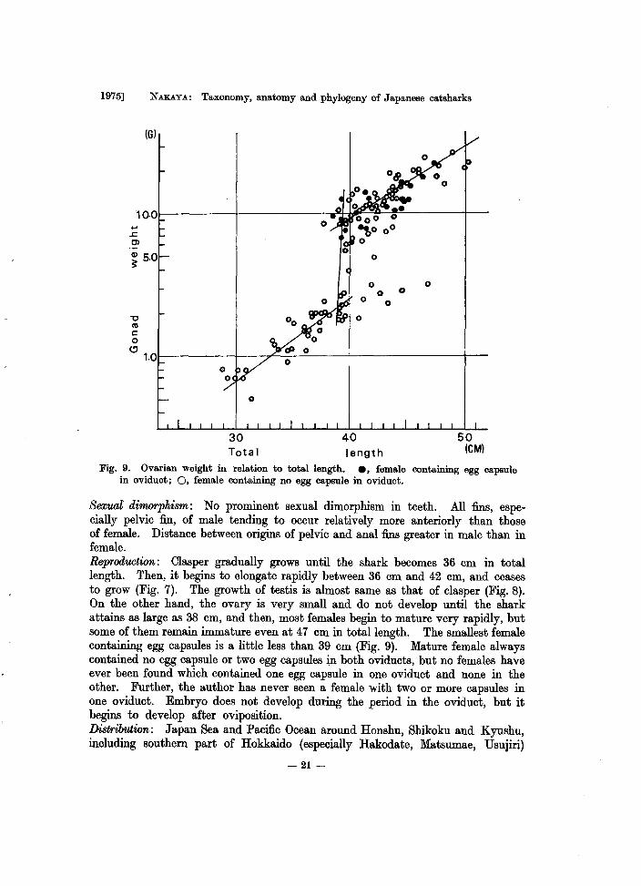
Fig. 9. Ovarian weight in relation to total length. e, female containing egg capsule in oviduct; 0, female containing no egg capsule in oviduct.
Sexual dimorphism : No prominent sexual dimorphism in teeth. All fins, especially pelvic fin, of male tending to occur relatively more anteriorly than those of female. Distance between origins of pelvic and anal fins greater in male than in female. Reprod'IJ.Ction: Clasper gradually grows until the shark becomes 36 cm in total length. Then, it begins to elongate rapidly between 36 cm and 42 cm, and ceases to grow (Fig. 7). The growth of testis is almost same as that of clasper (Fig. 8). On the other hand, the ovary is very small and do not develop until the shark attains as large as 38 cm, and then, most females begin to mature very rapidly, but some of them remain immature even at 47 cm in total length. The smallest female containing egg capsules is a little less than 39 cm (Fig. 9). Mature female always contained no egg capsule or two egg capsules in both oviducts, but no females have ever been found which contained one egg capsule in one oviduct and none in the other. Further, the author has never seen a female with two or more capsules in one oviduct. Embryo does not develop during the period in the oviduct, but it begins to develop after oviposition. Distribution: Japan Sea and Pacific Ocean around Honshu, Shikoku and Kyushu, including southern part of Hokkaido (especially Hakodate, Matsumae, Usujiri)
- 21-

Mem. Fac. Fish. Hokkaido Univ. [xxm, I
in Japan. China (Shangtung, Shanghai). Korea (Fusan, Mokpo, Quelpart Island). Philippines (Negros Oriental Province). Remarks
S. torazame is the only species of the genus in the Pacific Ocean and is one of the most northern species among Japanese scyliorhinid sharks, inhabiting even waters of Hokkaido. As this species can be seen abundantly all the year round in waters of Hakodate, most northern part of its distribution, it does not seem to migrate a long distance seasonally. In addition, the spawning ground is found in waters of Hakodate at the depth of about 100 meters.
Judging from the development of testes and claspers of the specimens from Hakodate, male begins to mature sexually at about 36 cm in total length and attains its full maturity at about 42 cm. On the other hand, female begins to mature at about 37 cm, and most of them seem to attain their full maturity at about 40 cm. However, some remain in immature condition even at 47 cm. The biological minimum appears to be about 38 cm in both sexes, and this species seems to grow a little larger than 50 cm.
Female contains always only one egg capsule in each oviduct during the period of pregnancy and seems to lay both eggs at a time after completion of the capsule before embryo in it develops. Then, next capsule comes down into oviduct. Two or more egg capsules have never been found in one oviduct. Therefore, multiple oviparity, as seen in Halaelurus burgeri, does not occur in the present species, and the mode of reproduction is single oviparity, definition of which will be mentioned later in the anatomical section.
6. Genus Apristurus Garman, 1913
ApristUru8 Garman, 1913, p. 96 (Type species: Scylliorhinus indicus Brauer). -Matsubara, 1936, p. 42. - Bigelow and Schroeder, 1948, p. 219. - Matsubara, 1955, p. 108. - Springer, 1966, p. 611.
Catulus (in part) Gilbert, 1891, p. 452. - (in part) Jordan and Evermann, 1896, p. 23. Scylliorhinus (in part) Goode and Bean, 1895, p. 16. -Jordan and Evermann, 1896, p.22.
- (in part) Barnard, 1925, p. 38. Scyliorhinus (in part) Regan, 1908, p. 453. - Tanaka, 1909, p. 1. - (in part) Weber, 1913,
p.595. PriStiuru8 (in part) Garman, 1913, p. 91. ParapristiuTUs Fowler, 1933, p. 237. Pentanchu8 (in part) Fowler, 1941, p. 52.
Two dorsal fins; origin of 1st dorsal fin above or posterior to base of pelvic fin. Second dorsal fin same as or larger than 1st dorsal fin; its origin above base of anal fin. Pectoral fin small to moderate in size. Anal fin large; its base long. Caudal fin relatively large; its origin much closer to end of base of anal fin. Denticles along upper margin of caudal fin normal or packed. Head large and greatly flattened. Snout very long and thin; its tip broadly rounded or moderately rounded. Eye moderate. Fifth, or 4th and 5th gill openings above base of pectoral fin. Nostrils large, at about middle of snout in front of mouth; each never connected with mouth by a groove. Large labial grooves on both jaws.
-22 -

1975] NAKAYA: Taxonomy, anatomy and phylogeny of Japanese catsharks
Teeth alike in both jaws; one large primary cusp and a few lateral cusps. Rostral cartilage long and strong. Supraorbital crest absent as far as known. Body soft, but abdomen incapable of swelling. Oviparous as far as known.
Remarks Apristurus is a genus containing 18 nominal species: A. brunneus (Gilbert,
1891), A. spongiceps (Gilbert, 1905), A. platyrhynchus (Tanaka, 1909), A. macrorhynchus (Tanaka, 1909), A. sibogae (Weber, 1913), A. herklotsi (Fowler, 1933), A. verweyi (Fowler, 1933), A. nasutus Buen, 1959, and A. kampae Taylor, 1972, from Pacific Ocean and its adjacent waters; A. profundorum (Goode and Bean, 1895), A. laurussoni (Saemundsson, 1922), A. atlanticus (Koefoed, 1927), A. riveri Bigelow and Schroeder, 1944, and A. maderensis Cadenat and Maul, 1966, from Atlantic Ocean; A. microps (Gilchrist, 1922) and A. saldanha (Barnard, 1925) from South African waters; A. indicus (Brauer, 1906) and A. investigatoris (Misra, 1959) from Indian Ocean.
As the sharks of this genus have soft body, it is difficult to take measurements and to give description precisely. These facts make the taxonomy of this group more difficult. A nomenclatural revision is needed in the species of this genus ApristufU,s.
All the species of this genus inhabit deep waters as far as known.
Key to species of Japan and its adjacent waters
1a. Distance between origins of pectoral and pelvic fins greater than that from tip of snout to pectoral origin; snout in front of mouth shorter than mouth width; slight prolongation in each interbranchial septum, but sometimes absent; number of monospondylous vertebrae 43-46 .... A. japonicus sp. nov.
lb. Distance between origins of pectoral and pelvic fins shorter than that from tip of snout to pectoral origin; snout in front of mouth far longer than mouth width; no prolongation in interbranchial septa
2a. Origin of 1st dorsal fin above interspace between pelvic and anal fins; mouth width greater than snout in front of posterior end of nostrils; outer margin of pectoral fin long and greater than distance from tip of snout to posterior end of eye; number of monospondylous vertebrae 38 ..... . . . . . . . . . . . . . . . . . . . . . . . . . . . . . . . . . . .. A. platyrhynchus (Tanaka, 1909)
2b. Origin of 1st dorsal fin above base of pelvic fin; mouth width smaller than snout in front of posterior end of nostril; outer margin of pectoral fin equal to or shorter than distance from tip of snout to center of eye 3a. Snout about two times as long as interorbitals; margins of snout
greatly concave before nostrils; snout tip somewhat pointed; number of monospondylous vertebrae 32 ......... . A. longicephalus sp. nov.
3b. Snout about 1.5 times as long as interorbitals; margins of snout before nostrils somewhat concave; snout tip rounded; number of monospondylous vertebrae 39-41 .. A. macrorhynchus (Tanaka, 1909)
-23 -

Mem. Fac. Fish. Hokkaido Univ. [XXIII, I
Apristurus japonicus sp. nov. Japanese name: Nihon-hera-zame
Figs. 10-11
Apri8turu8 maerorhynehUB. Matsubara, 1955, p. 108, pI. 4, fig. 10 (key, distribution). - Matsubara, 1965, p. 148, fig. 21 (short description, distribution).
Materials Holotype: HUMZ 40082, mature male, 697 mm TL, collected off Cape Daito, Chiba
Prefecture, Japan on May 25, 1973. Paratypes: HUMZ 39961, mature male, 690 mm TL; HUMZ 40075, mature female,
626 mm TL; HUMZ 40076--40081, mature males, 654-711 mm TL. Off Cape Daito, Chiba Prefecture, Japan on May 25, 1973.
Diagnosis Distance between origins of pectoral and pelvic :fins greater than distance from
tip of snout to origin of pectoral :fin, but shorter than that from snout tip to end of pectoral base; slight prolongation on each interbranchial septum, but sometimes absent; body stout.
Proportional measurements in howtype and paratypes (those in paratypes shown in parentheses) .
Trunk: breadth 8.2 (8.1-10.9); height 9.5 (9.7-12.1) Head: breadth 11.5 (10.2-12.4); height 8.8 (7.7-9.5) Eye: horizontal diameter 2.6 (2.4-3.0) Mouth: width 7.8 (8.0-8.3); height 2.7 (2.4-2.9) Nostrils: distance between inner ends 3.3 (3.0-3.5) Labial grooves: upper 3.0 (2.7-3.5); lower 2.1 (2.1-2.6) Gill openings: 1st 2.0 (2.0-2.3); 5th 1.8 (1.6-1.9) First dorsal:fin: vertical height 2.3 (2.1-3.0); base length 6.3 (6.4-7.2) Second dorsal fin: vertical height 3.1 (2.7-3.5); base length 6.8 (6.4-7.2) Anal :fin: vertical height 4.8 (4.0-5.5); base length 12.7 (12.7-14.9) Caudal :fin: upper margin 25.6 (25.0-26.7); lower margin 9.8 (8.4-10.4) Pectoral fin: outer margin 11.8 (11.4-13.7); inner margin 6.9 (4.7-7.7) Distance from tip of snout to: anterior nasal aperture 3.4 (3.3-4.3); mouth 6.6
(6.9-8.4); eye 7.6 (7.5-9.4); 1st gill opening 15.5 (14.8-17.5); 5th gill opening 20.5 (20.2-23.0); pectoral :fin 19.9 (19.1-21.5); pelvic :fin 45.3 (42.3-45.4); 1st dorsal:fin 49.8 (48.5-50.6); 2nd dorsal :fin 64.3 (63.5-65.4); anal fin 58.5 (55.8-58.6); upper caudal fin 73.2 (73.4-75.0)
Interspace between: 1st and 2nd dorsal :fins 8.7 (7.7-10.0); 2nd and upper caudal :fins 4.2 (2.6-4.2); anal and lower caudal :fins 0.8 (1.0-1.6)
Distance from origin to origin of: pectoral and pelvic fins 25.0 (19.5-24.9); pelvic and anal :fins 13.3 (12.5-15.6)
Description (Howtype): Body very slender, but stout; depth of trunk about equal
-24-

...... ~
.§3
~
I t
~ ~ ~ ~
I'd
~
f s. ~
Fig. 10. Holotype (HUMZ 40082) of Apri8turu8 japonicu8 sp. nov., adult male, 697 mm in total length from off Cape Daito ~ in Chiba Prefecture. Scale 5 cm. i
<>
f

Mem. Fac. Fish. Hokkaido Univ. [XXIII, I
to its width; depth of caudal peduncle much greater than its width; caudal axis nearly straight.
Head a little longer than 1/5 of total length. Snout flattened dorsoventrally; its tip rather narrowly rounded when looked from dorsal side; its length a little less than interdorsals (Fig. 11 a, b). Nasal apertures large, oblique; anterior-most of nostrils slightly before middle of snout in front of mouth. Mouth wide; its width a little greater than snout length in front of mouth. Labial grooves present on upper and lower jaws; lower one about 1/2 of distance between corner of mouth and symphysis; upper one a little less than l.5 times of the lower and slightly greater than internostrils (Fig. 11 b). Eye moderate, ovate; its horizontal diameter about 2/5 of interorbitals. Spiracle moderate, behind orbit .and slightly below level of horizontal axis of eye. Gill openings moderate in size; 4th longest; 5th above base of pectoral fin; prolongation of interbranchial septa present but slight. Pectoral fin moderate in size; length of outer margin a little greater than distance from snout tip to posterior edge of spiracle; its rear tip not reaching origin of pelvic fin by length of outer margin of pectoral fin. First dorsal fin smaller than 2nd dorsal fin; its origin above posterior half of base of pelvic fin; anterior margin a little convex; distal margin convex; its free rear tip not elongated. Second dorsal origin a little before middle of anal base; rear end of its base not reaching that of anal base only by distance of diameter of spiracle. Anal origin behind end of 1st dorsal base by distance of horizontal diameter of eye; end of its base separated from lower caudal fin by a notch formed by anal and caudal fins. Caudal fin moderate; its length about 1/4 of total length; its lower anterior corner only slightly expanded.
Distance between origins of pectoral and pelvic fins completely greater than that from snout tip to pectoral origin, but shorter than distance to rear end of pectoral base from snout tip.
Denticles on body small, rather overlapping and smooth to touch; its shape over dorsolateral surface of trunk three cusped; primary cusp longest (Fig. 11 c). Denticles on upper margin of caudal fin not packed but normal.
Teeth small, numerous; typical teeth three cusped and slender in upper jaw, lower one three cusped, some with a blunt additional cusp on inner side; several series functional (Fig. 11 d).
Color of dorsolateral and ventral surfaces of body and fins blackish brown. Inside of mouth blackish.
Number of monospondylous vertebrae 45 (Table 3). Description (Internal, not of holotype): Rostral cartilage very long; no supraorbital crest; antorbital and postorbital processes present, but both without any projections (Fig. 33 c). Cartilage of pectoral fin elongated anteroposteriorly; anterior radial cartilages divided into four segments; posterior radials into three segments (Fig. 36 c). Radials of 1st dorsal fin mostly three segmented (Fig. 37 c). Labial cartilages three. Pharyngeal denticles on hyoid and gill arches; ihose on upper and lower pharyngeal cavity very sparse. Calcification of vertebra above base of pectoral fin cyclospondylic (Fig. 38 c). Heart valves in three series two rows. Siphon of male short; its anterior tip not reaching pectoral fin. Number of spiral
-26 -

1975] NAKAYA: Taxonomy, anatomy and phylogeny of Japanese catsharks
Fig. 11. Dorsal view of head (a), ventral view of head (b), dermal denticles on dorsal part of trunk (c), and left upper and lower teeth (d) of holotype of Apristurus japcmicu8. Scales 2 cm.
valves 20. Number of monospondylous vertebrae 43--46 (Table 3). Sexual dimorphism: Teeth in male more slender and mostly three cusped, but those in female wide and mostly five cusped. Distribution: Chiba Prefecture (off Cape Daito) in Japan. Remarks
The present new species has such a remarkable character as the greatly separated paired fins, distance between which is completely greater than length from snout tip to pectoral fin.
A. japonicus is very closely related to an Atlantic A. maderensis in respects of the relation of above two dimensions, general appearance of body and feature of gill openings. But the present new species clearly differs from the latter in feature of dermal denticles. Denticles only slightly overlap (Fig. 11 c, and Matsubara, 1955, pI. 4, fig. lOF) and its number contained in a certain circle (2 mm in diameter) is about 37 in A. japonicus. In A. maderensis, however, they greatly overlap (Cadenat and Maul, 1966, fig. 4) and the number in that circle is about 56, based on a strip of skin from holotype of A. maderensis. As these differences are clear between the specimens of same size and sex of the two species, A. japonicus is considered to be distinct from A. maderensis.
This new species also somewhat resembles eastern Pacific A. brunneus and
-27 -

Mem. Fac. Fish. Hokkaido Univ. [XXIII, I
A. nasutus, whi,ch possess relatively long distance between origins of paired fins. But the new species is distinguishable from them by longer distance between origins of these fins than that between snout tip and pectoral fin. In addition, the number of monospondylous vertebrae falls in range of 43--46 in the new species, as against 39--41 in A. brunneus (HUMZ 40083--40084, 810-64-256, 810-66-36, 810-71-1, and Taylor, 1972), though it is unknown in A. nasutus.
On the basis of greatly separated paired fins and feature of gill openings, this new species is clearly separable from all the other species of Apristurus, except A. saldanha in which the dimension between paired fins is not given in the original description. However, the present new species is distinguishable from A. saldanha in another feature, that is, tip of pectoral fin separated from pelvic origin by a distance of snout length or pelvic base length in A. saldanha, but more greatly separated in A. japonicus.
This is the third species of this genus from Japan and its adjacent waters, bringing total number of species to nineteen.
Though Matsubara (1955, 1965) figured a shark under the name of A. macrorhynchus, his macrorhynchus is not correct, but should be identified as A. japonicus.
The egg capsule of this species has not been found yet, but the mode of reproduction appears to be the single oviparity, which will be defined later.
This species seems to be abundant in the deep waters off Chiba Prefecture. They are often caught by trawl in large amount and are landed at Choshi in Chiba Prefecture.
Apristurus platyrhynchus (Tanaka, 1909) Japanese name: Hera-zame
Figs. 12~14
Seyliorhinus platyrhynchus Tanaka, 1909, p. 4 (original description; Japan). - Jordan, Tanaka and Snyder, 1913, p. 10 (list).
ApristurUB platyrhynchus. Garman, 1913, p.98 (key, description). -Tanaka, 1914, p.24 (list). - Matsubara, 1936, p. 44, fig. 32 (key, description, distribution). - Okada, 1938, p. 117 (list). - Matsubara, 1955, p. 108 (key).
Pentanrihus platyrhynchus. (in part) Fowler, 1941, p. 57, not fig. 5 (key, description).
Materials ZIUT 3424 (654 mm TL, female) and its egg capsules from Tokyo.
Diagnosis Distance between origins of pectoral and pelvic fins equal to distance from tip
of snout to 1st or 2nd gill opening; origin of 1st dorsal fin above interspace between pelvic and anal fins; length of outer margin of pectoral fin greater than that to posterior end of eye from tip of snout.
-28 -

1975] NAKAYA: Taxonomy, anatomy and phylogeny of Japanese catsharks
ProJKYftional measurements Trunk: breadth 7.8; height 9.9 Head: breadth 12.2; height 7.8 Eye: horizontal diameter 3.0 Mouth: width 7.1; height 2.5 Nostrils: distance between inner ends 3.3 Labial grooves: upper 2.5; lower 1.7 Gill openings: 1st 1.5; 5th 1.3 First dorsal fin: vertical height 1.7; base length 3.6 Second dorsal fin: vertical height 3.0; base length 5.7 Anal fin: vertical height 3.6; base length 19.2 Caudal fin: upper margin 28.5; lower margin 8.6 Pectoral fin: outer margin 13.6; inner margin 6.3 Distance from tip of snout to: anterior nasal aperture 4.2; mouth 8.3; eye 9.1;
1st gill opening 17.3; 5th gill opening 20.8; pectoral fin 20.4; pelvic fin 37.3; 1st dorsal fin 50.0; 2nd dorsal fin 62.4; anal fin 52.0; upper caudal fin 72.9
Interspace between: 1st and 2nd dorsal fins 8.7; 2nd and upper caudal fins 4.3; anal and lower caudal fins 2.4
Distance from origin to origin of: pectoral and pelvic fins 18.1; pelvic and anal fins 14.7
Description (External): Body very slender; depth of trunk about equal to its width; depth of caudal peduncle greater than its width; caudal axis only slightly elevated.
Head about 1/5 of total length. Snout greatly flattened dorsoventrally; its tip rounded when looked from dorsal side (Fig. 13 a, b). Nasal apertures large, oblique; anterior-most of nostrils about middle of snout in front of mouth. Mouth wide; its width a little greater than length to posterior end of nostrils from tip of snout. Labial grooves present on both jaws; length of lower one 2/5 of distance between corner of mouth and symphysis; the upper a little less than internostrils (Fig. 13 b). Eye moderate, ovate; its horizontal diameter about 1/2 of interorbitals. Spiracle moderate, behind orbit and slightly below level of horizontal axis of eye. Gill openings short; 2nd or 3rd one longest and about 1/2 of horizontal diameter of eye; 4th and 5th gill openings above base of pectoral fin. Pectoral fin large in size; outer margin longer than that to posterior end of eye from snout tip; rear tip not reaching origin of pelvic by distance of 2/3 of mouth width; distal margin straight. First dorsal fin considerably smaller than 2nd dorsal fin; its origin above nearly middle of interspace between pelvic and anal bases, locating about midpoint of body; its vertical height and base length only a little greater than 1/2 of those of 2nd dorsal fin; anterior and distal margins convex; free rear tip not elongated but rounded. Second dorsal origin above middle of anal base; end of base not reaching end of anal base by distance of horizontal diameter of eye or slightly more; shape similar to 1st dorsal fin, but larger. Pelvic fin relatively large; fin itself ovoid when looked from below. Anal origin below base of 1st dorsal fin; rear end of its base a little separated from lower caudal fin. Caudal fin moderate; its lower
- 29-

~ ? b;I P> ? b;I til' ?" I:.¢ '<fjf ~
0
~ ~ §i ~.
Fig, 12. Apri8turU8 platyrhynchU8 (ZIUT 3424), adult female, 654 mm in total length from Tokyo. Scale 5 em.
~ ~

1975] NAKAYA: Taxonomy, anatomy and phylogeny of Japanese catsharks
a,::.:';':":. :~.,.~ ..
'.~'::::':"~~"~I:r;i~':~~ ':> ':';;::':~~':~"i'\ff.~ i:;.
':::.\.=:".:. ~ < . ..•... i~O·./~'
Fig, 13, Dorsal view of head (a), ventral view of head (b), dermal denticles on dorsolateral part of trunk (c), and left upper and lower teeth (d) of Apristurus platyrhynchus. Female. Scales 2 cm.
anterior corner a little expanded as a small lobe. Distance between origins of pectoral and pelvic fins equal to distance between
tip of snout and 1st or 2nd gill opening. Denticles on body small, greatly overlapping, and smooth to touch; its shape
over dorsolateral surface of trunk three pointed; primary cusp longest (Fig. 13 c). Denticles on upper margin of caudal fin not enlarged or not packed.
Teeth in adult female small, numerous; typical teeth three to five cusped in upper jaw; that of lower teeth five cusped; several series functional (Fig. 13 d).
Color uncertain because of long deposit, but at least plain colored without any markings in formalin. Inside of mouth blackish.
Number of monospondylous vertebrae 38 (Table 3). Egg capsule: Egg capsule (Fig. 14) rather long, 79 mm long and 19 mm in greatest width. Posterior end truncated without tendrils. Anterior end tapering, pointed at tip, but uncertain whether tendrils present or not. Capsule rather thick and finely striated longitudinally, surrounded by silky fibers.
Color of capsule opaque and light brown. Distribution: Tokyo and Sagami Bay.
- 31-
Mem. Fac. Fish. Hokkaido Univ. [xxm, I
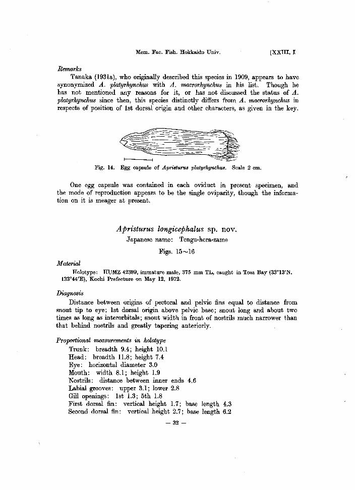
Remarks Tanaka (1931a), who originally described this species in 1909, appears to have
synonymized A. platyrhynchus with A. macrorhynchus in his list. Though he has not mentioned any reasons for it, or has not discussed the status of A. platyrhynchus since then, this species distinctly differs from A. macrorhynchus in respects of position of 1st dorsal origin and other characters, as given in the key.
r-----fl ~~
Fig. 14. Egg capsule of Apri8turu8 platyrhynchua. Scale 2 cm.
One egg capsule was contained in each oviduct in present specimen, and the mode of reproduction appears to be the single oviparity, though the information on it is meager at present.
Apristurus longicephalus sp. nov. Japanese name: Tengu-hera-zame
Figs. 15-16
Material Holotype: HUMZ 42399, immature male, 375 mm TL, caught in Tosa Bay (33°13'N,
133°44'E), Kochi Prefecture on May 12, 1972.
Diagnosis Distance between origins of pectoral and pelvic fins equal to distance from
snout tip to eye; 1st dorsal origin above pelvic base; snout long and about two times as long as interorbitals; snout width in front of nostrils much narrower than that behind nostrils and greatly tapering anteriorly.
Proportional measurements in holotype Trunk: breadth 9.4; height 10.1 Head: breadth 11.8; height 7.4 Eye: horizontal diameter 3.0 Mouth: width 8.1; height 1.9 Nostrils: distance between inner ends 4.6 Labial grooves: upper 3.1; lower 2.8 Gill openings: 1st 1.3; 5th 1.8 First dorsal fin: vertical height 1.7; base length 4.3 Second dorsal fin: vertical height 2.7; base length 6.2
- 32-

1975] NAKAYA: Taxonomy, anatomy and phylogeny of Japanese catsharks
Anal fin: vertical height 3.0; base length 15.0 Caudal fin: upper margin 33.0 Pectoral fin: outer margin 11.9; inner margin 6.0 Distance from tip of snout to: anterior nasal aperture 7.4; mouth 12.4; eye
12.7; 1st gill opening 22.2; 5th gill opening 25.4; pectoral fin 24.8; pelvic fin 38.7; 1st dorsal fin 45.6; 2nd dorsal fin 56.3; anal fin 48.5; upper caudal fin 67.2
Interspace between: 1st and 2nd dorsal fins 6.1; anal and lower caudal fins 1.3
·Distance from origin to origin of: pectoral and pelvic fins 15.1; pelvic and anal fins 9.1
Description (Holotype): Body very slender; depth of trunk about equal to its width; depth of caudal peduncle greater than its width; caudal axis nearly straight.
Head long, about 1/4 of total length. Snout greatly flattened dorsoventrally; snout in front of nostrils greatly narrower than that behind nostrils and greatly tapering anteriorly; its tip narrowly rounded when looked from dorsal side; length about two times as great as interorbitals. Nasal apertures large, oblique; anteriormost of nostrils behind middle of snout in front of mouth. Mouth wide; its width shorter than distance to posterior end of nostrils from tip of snout. Labial grooves present on both jaws; length of lower one about 1/2 of distance between corner of mouth and symphysis or a little longer; the upper one a little longer than the lower one (Fig. 16 a, b). Eye moderate, ovate; its horizontal diameter about 1/2 of interorbitals. Spiracle moderate, behind orbit and slightly below level of horizontal axis of eye. Gill openings short; 4th and 5th gill openings above base of pectoral fin. Pectoral fin large in size; length of outer margin slightly shorter than snout length; its rear tip not reaching origin of pelvics only by short distance. First dorsal fin smaller than 2nd dorsal fin; its origin before middle point of body and above posterior half of pelvic base; anterior margin convex; free rear tip not elongated but rounded. Second dorsal origin above middle of anal base; its rear end of base above end of anal base or slightly before; shape similar to 1st dorsal fin but larger. Pelvic fin moderate. Anal origin below 1st dorsal base; tip of its base separated from lower caudal fin by a deep notch formed by anal and caudal fins; its height a little lower than that of iower caudal fin. Caudal fin long, about 1/3 of total length; its lower anterior corner not expanded as a definite lobe.
Distance between origins of pectoral and pelvic fins equal to distance from tip of snout to posterior half of eye.
Denticles on body small, weak, widely spaced and velvety to touch; its shape over dorsolateral surfaces of trunk three cusped; primary cusp longest (Fig. 16 c). Denticles on upper margin of caudal fin not enlarged or not packed.
Teeth small, relatively sparse; typical teeth five cusped in both jaws; a few series functional (Fig. 16 d).
Color of upper and lower surfaces of body and fins blackish grey; anterior margin of fins black without any markings. Inside of mouth black.
Number of monospondylous vertebrae 32 (Table 3).
- 33-

~ >l>-
Fig. 15. Holotype (HUMZ 42399) of Apristuru8 longicephalus sp. nov., juvenile male 375 mm in total length from Tosa Bay in Kochi Prefecture. Scale 5 cm
~ ~
~ ?
~ F" ill ~ f. §'
§i ~.
~ 1-1

1975] NAKAYA: Taxonomy, anatomy and phylogeny of Japanese catsharks
"':.
b ~
.. ::~. . .... ,.,.,. • ." ~ w'
.'. ~ . '. ',:. ..-:: .....
: .. ~ ...... -... . 'pt,. '"
d ~'1
~~~ Fig. 16. Dorsal view of head (a), ventral view of head (b), dermal denticIes on dorsolateral
part of trunk (c), and left upper and lower teeth (d) of holotype of Apristurus longicephalus. Scales 1 em.
Distribution: Kochi Prefecture (Tosa Bay) in Japan Remarks
The present new species has distinctive characters such as very long head and shert distance between pectoral and pelvic fins.
This new species is closest to an Indian A. investigatoris in these respects, but the former is separable from the latter by the following characters. Anal origin locates below base of 1st dorsal fin in A. longicephalus (just behind posterior end of 1st dorsal base in A. investigatoris), and snout length in front of mouth is far greater than mouth width in the new species (nearly equal in A. investigatoris). In addition, two lateral cusps are present on both sides of primary cusp of teeth in A. longicephalus (one on one side and two on the other side in A. investigatoris).
On the other hand, when the present new species is compared with other species, A. longicephalus is distinguishable from three Japanese species by the features given in the key. Further, A. longicephalus can be discriminated from the other species by the characters below: tip of pectoral fin separated from origin of pelvic fin by only a very short interspace in A. longicephalus (by a far greater interspace in A. maderensis, A. brunneus, A. nasutus, A. saldanha, and A. spongiceps); origin of 1st dorsal fin above base of pelvic fin in the new species (above interspace
- 35-

Mem. Fac. Fish. Hokkaido Univ. [XXIII, I
between pelvic fin and anal fin in A. verweyi); anal base about equal to snout length in the new species (equal to length to 1st gill opening from snout tip in A. sibogae and A. herklotsi); origin of anal fin under base of 1st dorsal :fin in the new species (under or behind end of 1st dorsal base in A. indicus, A. laurussoni, A. riveri, A. atlanticus and A. profundorum); gill openings and eye normal in size in the new species (gill openings unusually high in A. kampae, and eye very small in A. microps).
Thus, this species is not identical with any other species of Apristurus from the world oceans. This is the twentieth species of this genus and the fourth species from Japan and its adjacent waters.
The holotype is an immature male and neither additional specimens nor egg capsules of this species has been collected yet.
Apristurus macrorhynchus (Tanaka, 1909) Japanese name: Naga-hera-zame
Figs. 17-19
Scyliorhinus macrorhynchus Tanaka, 1909, p. 1 (original description; Misaki, Japan). - Jordan, Tanaka and Snyder, 1913, p. 10 (list).
Apriaturua macrorhynchua. Garman, 1913, p.97 (key, description). -Tanaka, 1914, p.24 (list). - Schmidt, 1930, p. 630 (description). - Schmidt, 1931, p. 5 (distribution). - Tanaka, 1931a, p. 14 (distribution). - Tanaka, 1931b, p. 6, fig. 10 (short description, distribution). - Matsubara, 1936, p. 43, fig. 31 (key, description, distribution). - Okada, 1938, p. 117 (list). -- Chen, 1963, p. 33, fig. 11 (key, description, distribution).
Pentanchua macrorhynchua. Fowler, 1941, p. 56 (key, description, distribution). Pentanchua platyrhynchua. (in part) Fowler, 1941, p. 57, fig. 5 (key, description, distribution). not ApriaturuB macrorhynchus. Matsubara, 1955, p. 108, pI. 4, fig. 10 (key, distribution).
- Matsubara, 1965, p. 148, fig. 21 (short description, distribution).
Materials Male - ZIUT 21100 (383 mm TL), Sagami Bay, June 24,1906; MSM 71-1006 (173 mm
TL), Suruga Bay, Dec. 23, 1971; MSM 72-198 (183 mm TL), Suruga Bay, March 4, 1972. Female-ZIUT 3467 (674 mm TL), Tokyo; MSM 72-196 (c.a. 130mm TL), Suruga Bay, March 4, 1972; MSM 72-366 (393 mm TL), Suruga Bay, Apr. 27, 1972.
Diagnosis Distance between origins of pectoral and pelvic fins shorter than length to 1st
gill opening from snout tip; origin of 1st dorsal fin above base of pelvic fin; snout long but about 1.5 times as long as interorbitals.
Proportional measurements Trunk: breadth 6.8--8.4; height 6.8-8.1 Head: breadth 10.4-12.0; height 5.5-7.2 Eye: horizontal diameter 2.7
- 36-
1975] NAKAYA: Taxonomy, anatomy and phylogeny of Japanese catsharks
Mouth: width 6.8-7.2; height 2.5--2.8 Nostrils: distance between inner ends 3.0-3.3 Labial grooves: upper 3.0-3.4; lower 2.0-2.3 First dorsal fin: vertical height 2.2; base length 5.1-5.8 Second dorsal fin: vertical height 2.9-3.6; base length 5.7-6.5 Anal fin: vertical height 4.3-4.9; base length 18.2-19.1 Caudal fin: upper margin 27.1-32.1; lower margin 8.7-9.1 Pectoral fin: outer margin 11.3-12.5; inner margin 6.8-7.2 Distance from tip of snout to: anterior nasal aperture 5.0-5.4; mouth 9.6-
10.6; eye 10.0-11.7; 1st gill opening 18.0-19.9; 5th gill opening 22.2-23.7; pectoral fin 20.8-23.2; pelvic fin 36.6-37.7; 1st dorsal fin 42.2-45.0; 2nd dorasl fin 55.7-62.3; anal fin 49.3-50.7; upper caudal fin 67.9-71.2
Interspace between: 1st and 2nd dorsal fins 8.3-10.7; anal and lower caudal fins 0
Distance from origin to origin of: pectoral and pelvic fins 14.1-17.0; pelvic and anal fins 13.3-13.5
Description (External): Body very slender; depth of trunk about equal to its width; depth of caudal peduncle much greater than its width; caudal axis only slightly elevated.
Head a little longer than 1/5 of total length. Snout long and greatly flattened dorsoventrally; its tip rounded when looked from dorsal side (Fig. 18 a, b). Nasal apertures large, oblique; anterior-most of nostrils about middle of snout in front of mouth. Mouth wide; its width shorter than distance to posterior end of nostrils from snout tip. Labial grooves present on both jaws; the lower one a little less than 1/2 of distance between corner of mouth and symphysis; the upper about equal to internostrils. Eye moderate, ovate; its horizontal diameter about 2/5 of interorbitals. Spiracle moderate, behind orbit and slightly below level of horizontal axis of eye. Gill openings short; 4th longest and about 1/2 of horizontal diameter of eye; 4th and 5th gill openings above base of pectoral fin. Pectoral fin large in size; length of outer margin about equal to distance from snout tip to center of eye; its rear tip separated from origin of pelvic by distance of 1/2 of mouth width; distal margin straight. First dorsal fin a little smaller in size than 2nd dorsal fin; its origin in advance of middle point of body and above posterior half of pelvic base; anterior and distal margins convex; free rear tip not elongated but rounded. Second dorsal origin above middle of anal base or a little behind; rear end of its base anterior to end of anal base by distance of a little less than horizontal diameter of eye; shape similar to 1st dorsal fin but a little larger in size. Pelvic fin large; its base long. Anal origin below posterior half of 1st dorsal base; tip of its base separated from lower caudal fin only by a deep notch extending almost to fin base; height a little lower than that of lower caudal lobe. Caudal fin moderate; its lower anterior corner not expanded as a definite lobe.
Distance between origins of pectoral and pel vic fins shorter than distance to 1st gill opening from tip of snout.
Denticles on body small, a little overlapping and smooth to touch; its shape over dorsolateral surfaces of trunk three cusped; primary cusp longest (Fig. 18 c).
- 37-

~ F ~ lP ?
~ ~ ?" 00
~ ~ i'<'
~ §1 ~.
Fig. 17. Apristurus macrorhynchus (ZIUT 3467), adult female, 674 mm in total length from Tokyo. Scale 5 em.
~ t-I

1975] NAKAYA: Taxonomy, anatomy and phylogeny of Japanese catsharks
Denticles on upper margin of caudal fin normal and not packed. Teeth in adult female small, numerous; typical teeth three cusped in upper
teeth; that of lower teeth three to four cusped (Fig. 18 d). Lateral teeth four to five cusped in both jaws; several series functional.
Color of dorsolateral surfaces of body and fins uniformly light brownish grey; that of ventral surfaces of body and fins dirty white; anterior margins of fins, rims of mouth and nostrils black in the specimen preserved in alcohol. Inside of mouth black.
Number of monospondylous vertebrae 39-41 (Table 3). Egg capsule: Egg capsule (Fig. 19) short, stout and wide; its length 54 mm long and 23 mm wide. Posterior end truncated with a tuft of very fine silky fibers on each corner. Anterior end tapering with long tendrils. The egg capsule thick and finely striated longitudinally.
Color of egg capsule opaque and yellowish brown. Silky fibers on truncate end yellow. Distribution: Sagami Bay, Shizuoka Prefecture (Suruga Bay) and southern Japan.
"-:;~:::.
b .....••.• ~:~ •.......
. .. ~.~ ...... ~ ..
Fig. 18. Dorsal view of head (a), ventral view of head (b), dermal denticles on dorsolateral part of trunk (c), and left upper and lower teeth (d) of Apristurus macrorkynckus. Female. Scales 2 em.
-39 -
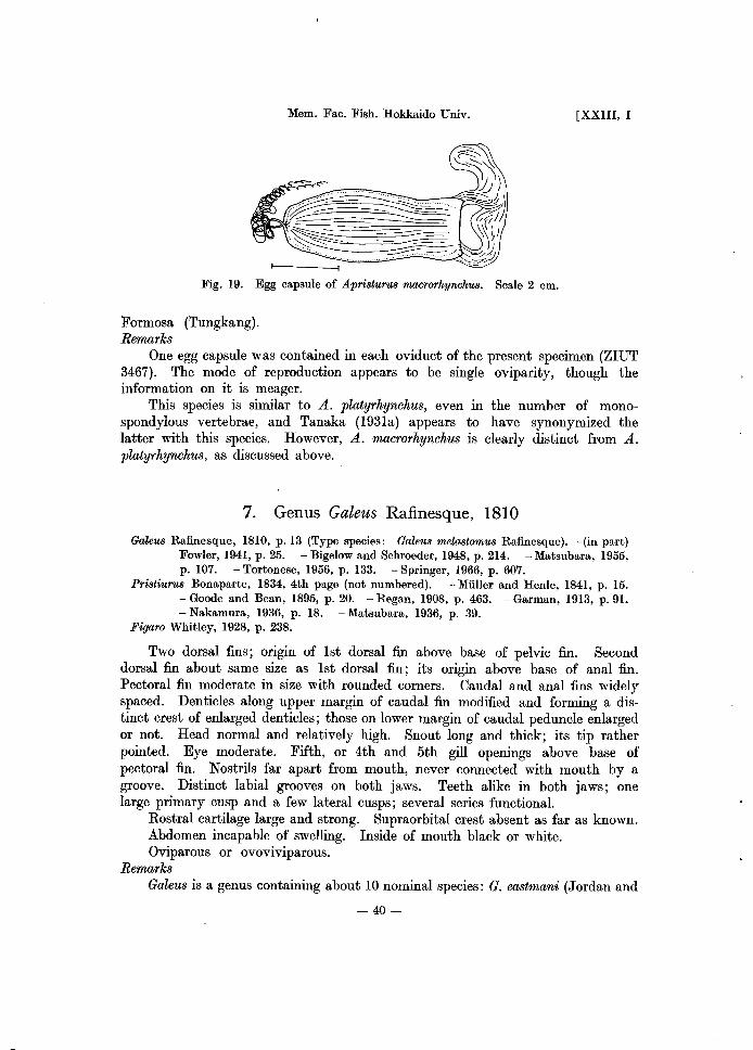
Mem. Fac. Fish. Hokkaido Univ.
I- I
Fig. 19. Egg capsule of Apristurus macrorhynchus. Scale 2 cm.
Formosa (Tungkang). Remarks
[XXIII, I
One egg capsule was contained in each oviduct of the present specimen (ZIUT 3467). The mode of reproduction appears to be single oviparity, though the information on it is meager.
This species is similar to A. platyrhynchus, even in the number of monospondylous vertebrae, and Tanaka (1931a) appears to have synonymized the latter with this species. However, A. macrorhynchus is clearly distinct from A. platyrhynchus, as discussed above.
7. Genus Galeus Rafinesque, 1810
Galeus Rafinesque, 1810, p.13 (Type species: Galeus melastomus Rafinesque). -(in p'trt) Fowler, 1941, p. 25. - Bigelow and Schroeder, 1948, p. 214. - Matsubara, 1955, p. 107. - Tortonese, 1956, p. 133. - Springer, 1966, p. 607.
Pristiurus Bonaparte, 1834, 4th page (not numbered). - Muller and Henle, 1841, p. 15. - Goode and Bean, 1895, p. 20. - Regan, 1908, p. 463. - Garman, 1913, p.91. - Nakamura, 1936, p. 18. - Matsubara, 1936, p. 39.
Figaro Whitley, 1928, p. 238.
Two dorsal fins; origin of 1st dorsal fin above base of pelvic fin. Second dorsal fin about same size as 1st dorsal fin; its origin above base of anal fin. Pectoral fin moderate in size with rounded corners. Caudal and anal fins widely spaced. Denticles along upper margin of caudal fin modified and forming a distinct crest of enlarged denticles; those on lower margin of caudal peduncle enlarged or not. Head normal and relatively high. Snout long and thick; its tip rather pointed. Eye moderate. Fifth, or 4th and 5th gill openings above base of pectoral fin. Nostrils far apart from mouth, never connected with mouth by a groove. Distinct labial grooves on both jaws. Teeth alike in both jaws; one large primary cusp and a few lateral cusps; several series functional.
Rostral cartilage large and strong. Supraorbital crest absent as far as known. Abdomen incapable of swelling. Inside of mouth black or white. Oviparous or ovoviviparous.
Remarks Galeus is a genus containing about 10 nominal species: G. eastmani (Jordan and
-40-

1975] NAKAYA: Taxonomy, anatomy and phylogeny of Japanese catsharks
Snyder, 1904), G. sauteri (Jordan and Richardson, 1909) and G. piperatus Springer and Wagner, 1966, from north Pacific and Gulf of California; G. boardmani (Whitley, 1908) from Australia; G. arae (Nichols, 1907) and G. cadenati Springer, 1966, from western Atlantic; G. melastomus Rafinesque, 1810, G. murinus (Collett, 1904), G. jenseni (Saemundsson, 1923) and G. polli Cadenat, 1959, from eastern Atlantic and Mediterranean Sea.
Members of this genus are distinctly characterized by the presence of a dermal crest of enlarged denticles on caudal fin. Though anothor scyliorhinid Parmaturus from Pacific Ocean has also such a crest of modified denticles on caudal fin, snout is very short and rounded in that genus. Somewhat different denticles from those of body are present on caudal fin of a species of Apristurus from Atlantic Ocean (Springer, 1966).
All the members of the family Scyliorhinidae had often been considered as oviparous (Matsubara, 1936; White, 1937), but Cadenat (1959) reported a new ovoviviparous scyliorhinid of Galeus from west Mrica. Afterwards, Springer (1966) considered the two species of western Atlantic Galeus as probably ovoviviparous, and then Bullis (1967) observed unencapsuled eggs in the oviduct of G. arae. On the other hand, G. melastomus and G. piperatus are known to be egg layers (Tortonese, 1956; Hubbs and Taylor, 1969; Collignon and Aloncle, 1972).
The further examination of mode of reproduction in the family Scyliorhinidae is needed for the better understanding of its taxonomy and evolution.
Key to species of Japan and its adjacent waters
1a. Body plain colored and without dark stain except tips of dorsal fins and caudal fin; interspace between anal and lower caudal fins shorter than horizontal diameter of eye; number of monospondylous vertebrae 31-34 ............................... . G. sauteri (Jordan and Richardson, 1909)
lb. Body with dark blotches or stains; interspace between anal and lower caudal fins much longer than horizontal diameter of eye
2a. Snout short; horizontal diameter of eye greater than distance from snout tip to anterior nasal aperture; number of monspondylous vertebrae 34-36 ................................ G. eastmani (Jordan and Snyder, 1904)
2b. Snout long; horizontal diameter of eye equal to or smaller than distance from snout tip to anterior nasal aperture; number of monospondylous vertebrae 38-40 ................................ G. nipponensis sp. nov.
Galeus sauteri (Jordan and Richardson, 1909) Japanese name: Taiwan-yamori-zame
Figs. 20-22
Pristiurus sauteri Jordan and Richardson, 1909, p. 160, pI. 63 (original description; Takao, Formosa). -Garman, 1913, p. 94 (key, description). -Tanaka, 1914, p.482 (list). - Tanaka, 1915, p. 645 (distribution). - Nakamura, 1936, p. 18, pI. 7, fig.
- 41-

Mem. Fac. Fish. Hokkaido Univ. [XXIII, I
1 (description). - Matsubara, 1936, p. 40, fig. 29 (key, description). - White, 1937, p. U8 (key, phylogeny). - Okada, 1938, p. 117 (list, distribution). - Okada and Matsubara, 1938, p. 8 (key). - Chen, 1948, p. 28 (key, list).
Galeu8 8auteri. Fowler, 1941, p. 28 (description). - Bigelow and Schroeder, 1948, p. 215 (key). - Matsubara, 1955, p. 107 (key). - Teng, 1962, p. 43, fig. 9 (description). - Chen, 1963, p. 27, fig. 8 (description). - Chu and Wang, 1964, p. 676 (list, distribution). - Fowler, 1967, p. 346 (list). - Yang, 1969, p. 124 (list).
Materials Male - HUMZ 39969 (379 mm TL), Keelung, Dec. 27, 1972; HUMZ 42405 (376 mm
TL), Tungkang, Feb. 1, 1973; HUMZ 39391 (372 mm TL), Tungkang, Feb. 20, 1973. Female - HUMZ 39406 (388 mm TL), Keelung, Dec. 27, 1972; HUMZ 39990 (376 mm TL), Tungkang, Feb. 1, 1973; HUMZ 39374 (382 mm TL), Tungkang, Feb. 10, 1973; HUMZ 39394 (374 mm TL), HUMZ 39978 (376 mm TL), HUMZ 39998 (398 mm TL), Tungkang, Feb. 20, 1973; HUMZ 40006 (394 mm TL), HUMZ 42406 (383 mm TL), Tungkang, Feb. 25, 1973.
Diagnosis Body plain colored; inside of mouth greyish; caudal peduncle very flattened
laterally; interspace between anal and caudal fins shorter than horizontal diameter of eye.
Proportional measurements
Trunk: breadth 8.0-9.8; height 9.0-13.6 Head: breadth 10.6-12.5; height 7.6-9.0 Eye: horizontal diameter 3.4-4.2 Mouth: width 7.2-9.2 Nostrils: distance between inner ends 1.9-2.3 Labial grooves: upper 1.5-1.8; lower 0.9-1.8 Gill openings: 1st 1.4-1.9; 5th 1.1-1.6 First dorsal fin: vertical height 2.7-3.5; base length 5.2-6.6 Second dorsal fin: vertical height 2.3-3.0; base length 4.6-6.0 Anal fin: vertical height 3.2-4.0; base length 12.6-15.2 Oaudal fin: upper margin 27.5-30.0; lower margin 8.9-10.8 Pectoral fin: outer margin 10.6-13.4; inner margin 5.9-7.1 Distance from tip of snout to: anterior nasal aperture 3.0-3.8; mouth 6.1-
7.2; eye 5.8-7.0; 1st gill opening 15.2-16.4; 5th gill opening 18.6-20.4; pectoral fin 17.5-19.8; pelvic fin 36.9-39.9; 1st dorsal fin 42.1--45.7; 2nd dorsal fin 60.4-62.6; anal fin 50.8-54.0; upper caudal fin 69.5-71.5
Interspace between: 1st and 2nd dorsal fins 11.7-13.8; 2nd and upper caudal fins 3.4-5.6; anal and lower caudal fins 2.2---4.5
Distance from origin to origin of: pectoral and pelvic fins 17.9-20.8; pelvic and anal fins 13.9-17.2
Description (External): Body slender; depth of trunk a little greater than its width. Oaudal peduncle high and its depth about 1.5 times as great as its width; caudal axis a little elevated.
-42 -

1975] NAKAYA: Taxonomy, anatomy and phylogeny of Japanese catsharks
-43 -

Mem. Fa.c. Fish. Hokka.ido Univ. [xxm, I
Head about 1/5 of total length. Snout moderate with many mucous pores above and below. Nasal apertures large; anterior-most of nostrils about middle of snout in front of mouth. Mouth wide; its width greater than snout in front of mouth. Labial grooves present on upper and lower jaws; both same length (Fig. 21 a, b). Eye large, ovate; its horizontal diameter equal to distance from 1st to 5th gill opening or slightly shorter. Spiracle moderate, behind orbit and slightly below level of horizontal axis of eye. Gill openings short; the longest shorter than 1/2 of horizontal diameter of eye; 5th, or 4th and 5th above base of pectoral fin. Pectoral fin moderate in size; length of outer margin equal to distance from anterior margin of eye to 2nd gill opening; its outer and inner corners moderately rounded; distal margin straight. First dorsal origin slightly behind middle of pelvic base; its anterior margin, distal margin and free rear tip rounded; the fin brush-shaped as a whole. Second dorsal fin a little smaller than 1st dorsal but similar in shape to 1st dorsal fin; its origin a little behind middle of anal base; free rear tip separated enough from crest of modified denticles on upper caudal fin by distance of 1/2 of horizontal diameter of eye. Pelvic fin moderate; rear end of base at middle of 1st dorsal base; right and left pelvic fins united a little proximally. Clasper moderate, but reaching origin of anal base in adult. Anal origin a little behind tip of 1st dorsal fin and in much advance of middle of interspace between 1st and 2nd dorsal fins; rear end of base below that of 2nd dorsal base; base a little longer than interspace between 1st and 2nd dorsal fins. Interspace between anal and lower caudal fins shorter than horizontal diameter of eye. Upper caudal with crest of modified denticles; its length equal to distance from 1st dorsal origin to origin of crest of modified denticles. No enlarged denticles on anterior margin of lower caudal fin and lower side of caudal peduncle.
Denticles over dorsolateral surface of trunk small, three cusped; a little overlapping; primary cusp longest (Fig. 21 c).
Teeth in adult small, numerous; typical teeth five cusped, similar in shape in both jaws; several series functional (Fig. 21 d).
Color of dorsolateral surface, dorsal fins and upper sides of paired fins yellowish brown and plain colored with dark brown shade on upper parts of dorsal fins and tips of lower and upper caudal fins. Color of ventral surfaces of body and paired fins white. Inside of mouth greyish. Description (Internal): Rostral cartilage long, relatively strong; no supraorbital crest; antorbital and postorbital processes present, and a projection present on each (Fig. 33 e). Cartilage of pectoral fin a little elongated anteroposter.iorly; radial cartilages divided into three segments (Fig. 36 e). Radials of 1st dorsal fin three segmented (Fig. 37 e). Labial cartilages thr.ee. Pharyngeal denticles on hyoid and gill arches, and anterior parts of upper and lower walls of pharyngeal cavity. Calcification of vertebra above base of pectoral fin cyclospondylic (Fig. 38 e). Heart valves in three series two rows. Siphon of male moderate, its anterior tip almost reaching pectoral fin. Number of spiral valves nine. Number of monospondylous vertebrae 31-34 (Table 3). Egg capsule: Egg capsule (Fig. 22) small, 35.5 mm long and 15 mm wide. Posterior end truncated with a bundle of long fine silky fibers on each corner. Anterior
-44-

1975] NAKAYA: Taxonomy, anatomy and phylogeny of Japanese catsharks
Fig.21. Dorsal view of head (a), ventral view of head (b), derm9.1 denticles on dorsol9.teral part of trunk (c), and left upper and lower teeth (d) of Galeus sauteri. Male. Scale 1 cm.
Fig. 22. Egg capsule of Galeus sauteri. Scale 1 cm.
margin a little tapering with long tendrils on each corner. Surface of egg capsule smooth and without any longitudinal streaks.
Color transparent and yellow. Sexual dimorphism: Distance between origins of pelvic and anal fins greater III
male than in female. No prominent sexual dimorphism in teeth. Distribution: Formosa (Keelung, Tungkang, Kao-hsiung)
- 45-

Mem. Fac. Fish. Hokkaido Univ. [xxm, I
Remarks
The specimens used are all matured and the sexual maturity is attained at a length less than 37 cm in both sexes. The mode of reproduction may be the single oviparity, which will be defined later.
This species may inhabit the waters of the southern most islands of Japan, though it commonly occurs in the water of Taiwan.
Galeus eastmani (Jordan and Snyder, 1904) Japanese name: Yamori-zame
Figs. 23 ....... 25
Pristiurus eastmani Jordan and Snyder, 1904, p. 230, pI. 60 (original description; Izu, Japan). - Regan, 1908, p. 464 (key, short description). - Garman, 1913, p. 93 (description). - Jordan, Tanaka and Snyder, 1913, p. II (list, distribution). - Tanaka, 1914, p. 24 (list). - Kamohara, 1931, p. 533 (list). - Matsubara, 1936, p. 39, fig. 28 (key, description, distribution). - White, 1937, p. ll8 (key, phylogeny). - (in part) Kamohara, 1955, p. I, pI. 1 (short description, distribution).
Pristiurus eastomani. Okada, 1938, p. ll7 (list, distribution). - Okada and Matsubara, 1938, p. 8 (key). - Kuroda, 1951, p. 315 (list).
Galeus eastmani. Fowler, 1941, p. 26 (key, description). - Bigelow and Schroeder, 1948, p. 215 (key). - Matsubara, 1955, p. 107, pI. 2 (key). - (in part) Kamohara, 1958, p. 4 (list). - Chu, Chang and Cherng, 1963, p. 17, fig. 12 (key, description, distribution). - Chu and Wang, 1964, p. 676 (distribution). - (in part) Kamohara, 1964, p. 6 (list). - Fowler, 1967, p. 345 (list).
Pristiurus eastmani. (in part) Kamohara, 1950, p. 4, fig. 3 (short description, distribution).
Materials Male - HUMZ 39396 (356 mm TL), HUMZ 39997 (319 mm TL), Kii Suido Channel
(33°30'-33°40'N, 134°30'-135°00'E), Aug. 6, 1972; HUMZ 39402 (318 mm TL), HUMZ 39407 (332 mm TL), HUMZ 39984 (305 mm TL), HUMZ 40013 (263 mm TL), Mimase, Oct. 13,1972; HUMZ 39377 (342 mm TL), HUMZ 40005 (3ll mm TL), HUMZ 42409 (330 mill TL), Mimase, Dec. 20, 1972. Female - HUMZ 40009 (393 mm TL), HUMZ 42408 (380 mm TL), Kii Suido Channel (same area as above), Aug. 6, 1972; HUMZ 39390 (360 mm TL), Mimase; HUMZ 39373 (376 mm TL), HUMZ 39376 (371 mm TL), HUMZ 39387 (358 mm TL), HUMZ 39393 (385 mm TL), HUMZ 39398 (358 mm TL), HUMZ 39408 (360 mm TL), HUMZ 39409 (384 mm TL), HUMZ 39970 (387 mm TL), HUMZ 39974 (356 mm TL), HUMZ 39987 (376 mm TL), HUMZ 39988 (373 mm TL), HUMZ 39995 (374 mm TL), HUMZ 40007 (370 mm TL), HUMZ 42407 (361 mm TL), Mimase, Oct. 13, 1972; HUMZ 39380 (363 mm TL), HUMZ 39993 (395 mm TL), Mimase, Dec. 20,1972; HUMZ 39967 (320 mm TL), Mimase, Dec. 21, 1972; HUMZ 39985 (372 mm TL), Mimase, Dec. 22, 1972.
Diagnosis
Body with dark blotches; origin of anal :fin under anterior half of interdorsals; inside of mouth white; snout in front of anterior nasal aperture shorter than horizontal diameter of eye.
-46 -

1975] NAKAYA: Taxonomy, anatomy and phylogeny of Japanese catsharks
-47 -

Mem. Fac. Fish. Hokkaido Univ.
Prop(Yrtional measurements
Trunk: breadth 7.0-8.7; height 7.0-8.9 Head: breadth 8.5-10.0; height 5.7-7.7 Eye: horizontal diameter 3.5-4.0 Mouth: width 5.8-6.4; height 2.5-3.2 Nostrils: distance between inner ends 1.8-2.2 Labial grooves: upper 1.5-1.9; lower 1.4-2.2 Gill openings: 1st 1.4-1.8; 5th 0.9-1.4 First dorsal fin: vertical height 3.3-4.2; base length 4.5-5.7 Second dorsal fin: vertical height 3.2-3.9; base length 5.1-6.1 Anal fin: vertical height 2.8-3.5; base length 10.7-12.7 Caudal fin: upper margin 26.2-30.4; lower margin 9.0-10.8 Pectoral fin: outer margin 9.4-13.2; inner margin 5.3-6.9
[XXIII, I
Distance from tip of snout to: anterior nasal aperture 2.7-3.3; mouth 5.8-6.6; eye 5.2-6.4; 1st gill opening 14.0-15.0; 5th gill opening 17.8-19.1; pectoral fin 17.1-19.1; pelvic fin 35.4-37.9; 1st dorsal fin 40.7-44.1; 2nd dorsal fin 60.1-63.8; anal fin 51.0-55.6; upper caudal fin 69.2-73.4
Interspace between: 1st and 2nd dorsal fins 14.2-15.9; 2nd and upper caudal fins 2.8-4.1; anal and lower caudal fins 5.9-7.7
Distance from origin to origin of: pectoral and pelvic fins 18.2-20.2; pelvic and anal fins 16.6-18.8
Description (External): Body slender; depth of trunk about equal to its width; caudal axis slightly elevated.
Head smaller than 1/5 of total length. Snout rather short, obtuse; its tip a little rounded; many mucous pores above and below. Nasal apertures large; anterior-most of nostrils about middle of snout in front of mouth. Mouth wide; its width slightly greater than snout in front of mouth. Labial grooves present on upper and lower jaws; both about same length (Fig. 24 a, b). Eye large, ovate; its horizontal diameter equal to distance from 1st to 5th gill opening. Spiracle moderate, behind orbit and slightly below level of horizontal axis of eye. Gill openings short; the longest less than 1/2 of horizontal diameter of eye; 5th, or 4th and 5th gill openings above base of pectoral fin. Pectoral fin moderate in size; length of outer margin equal to distance from anterior margin of eye to 2nd gill opening; its outer and inner corners moderately rounded; distal margin straight. First dorsal origin above posterior half of pelvic base; its anterior and distal margins slightly convex; free rear margin not elongated. Second dorsal fin a little smaller than 1st dorsal fin but similar in shape to 1st dorsal fin; its origin above posterior 1/3 of anal base; tip of fin separated from crest of modified denticles only by distance of 1/3 of horizontal diameter of eye. Clasper short, not reaching origin of anal fin even in adult. Pelvic fin moderate; rear end of base below anterior half of 1st dorsal base; right and left pelvic fins united proximally. Anal fin origin below anterior half of interspace between dorsal fins; rear end of its base below posterior half of 2nd dorsal base; posterior tip not reaching rear end of 2nd dorsal base; base long and two times as long as 2nd dorsal base. Interspace
-48 -

1975] NAKAYA: Taxonomy, anatomy and phylogeny of Japanese catsharks
Fig. 24. Dorsal view of head (a), ventral view of head (b), dermal denticles on dorsolateral part of trunk (c), and left upper and lower teeth (d) of Galeua Ila8tmani. Male. Scales 1 cm.
between anal and lower caudal fins great and two times as long as horizontal diameter of eye. Upper caudal fin with crest of mod:ified denticles; length of crest equal to distance from 1st dorsal origin to middle of free rear margin of 2nd dorsal fin; no enlarged denticles on anterior margin of lower caudal fin and lower side of caudal peduncle.
Denticles over dorsolateral surface of trunk small, three cusped; primary cusp longest (Fig. 24 c).
Teeth in adult male small, numerous; typical teeth :five cusped in upper and lower teeth, some with an additional small cusp on inner side; primary cusp longest; several series functional (Fig. 24 d).
Color of dorsolateral surface of trunk, upper surface of pectoral fin and dorsal fins grey or brownish grey. Dark saddles and blotches without definite edging present. Anal:fin white with black stain on anterior and distal margins. Color of ventral surfaces of body and paired fins white. Inside of mouth white. Description (Internal): Rostral cartilage long, relatively strong; no supraorbital crest; antorbital and postorbital processes present, and both having projections (Fig. 33 d). Cartilage of pectoral fin a little elongated anteroposteriorly; radial cartilages divided into three segments (Fig. 36 d). Radials of 1st dorsal fin three segmented (Fig. 37 d). Labial cartilages three. Pharyngeal denticles on hyoid and gill arches, and upper and lower anterior walls of pharyngeal cavity; those on
- 49-

Mem. Fac. Fish. Hokkaido Univ . [xxm, I
....... ......................
Fig. 25. Egg capsule of Galeus eaatmani. Scale 1 em.
lower fewer than those of the upper. Calcification of vertebra above base of pectoral fin cyclospondylic (Fig. 38 d). Heart valves in three series two rows. Siphon of male small; its anterior tip not reaching pectoral fin. Number of spiral valves seven. Number of monospondylous vertebrae 34-36 (Table 3). Egg capsule: Egg capsule (Fig. 25) small, slender and 60 rom long and 16 mm wide. Posterior end truncated without tendrils. Anterior end pointed with short tendrils of about 10 rom.
Color of egg capsule transparent yellow and its wall very smooth. Reproduction: A female of 321 mm in total length, smallest specimen on hand, has a small undeveloped ovary. A 360 rom female has developing eggs in ovary. In 368 rom female, however, ovary and shell gland develop completely, and all the larger females are fully matured. A 263 mm male, on the other hand, has very short undeveloped clasper, but a 305 mm male has large growing testis and long clasper, which extend beyond posterior tip of pelvic fin. A 318 mm and the larger males used have large testes and long stout claspers. Sex ratio: Present author has once investigated the sex ratios of this species at Mimase Fish Market in Kochi. One hundred and eleven specimens landed on October 11, 1972, by one ship, contained only three males, and 108 were females, which account for 97% of all. And further, only three of 46 specimens on the next day were males and 43 were females, which account for 93% of all. Sexual dimorphism : No prominent sexual dimorphism in teeth. Distance between origins of pelvic and anal fins a little greater in male than in female. Distribution: Shizuoka Prefecture (Izu), Mie Prefecture (Kumanonada), Shikoku (Kii Suido Channel, Tosa Bay) and Kyushu (Hyuganada, Tsushima Channel) in Japan. East China Sea. Remarks
Members of this genus are mostly small sharks. For example, G. piperatus, G. arae, G. cadenati and G. polli do not grow so large as 45 em (Springer, 1966; Krefft, 1968; Hubbs and Taylor, 1969). Only G. melastomus is known to attain 90 em (Springer, 1966). Japanese G. eastmani is also a small species and attains 40 em, but seems not to exceed 50 em, when judged from maximum size of the specimens examined and in literature.
Biological minimum of this species may be about 36-37 em in female, because ovary is developing in 360 mm female, but ovary and shell gland are fully developed in 368 mm and the larger females. On the other hand, male may mature
- 50-
1975] NAKAYA: Taxonomy, anatomy and phylogeny of Japanese catsharks
at about 31-32 em, because clasper of 305 mm male is small, but 318 mm and the larger males have fully developed testes and claspers.
Many adult females forced out egg capsules after capture at Mimase in October, and this fact may show that they are ready to lay their egg capsules in October. They have always only one capsule in each oviduct, and the mode of reproduction is defined as single oviparity, which will be mentioned later. Egg capsule of this species closely resembles those figured by Tortonese (1956) and photographed by Hubbs and Taylor (1969).
Sexual segregations have been reported in many kinds of sharks by various authors, i.e. Ford (1921), Harris (1952), Bachus, Springer and Arnold (1956), Springer (1960) and so on. In his general comments on social organization of sharks, Springer (1967) writes that the general tendency exists, but with more exception, for the adults to segregate by sex. Among catsharks, the sexual segregation is especially well known in the Caribbean marbled catshark, G. arae (Bullis, 1967). The adult female marbled catshark tends to occur in deeper waters and the male tends to distribute more evenly from shallow to deep waters. Although the data are meager, strong inclination of sex ratios in G. eastmani may suggest that at least sharp sexual segregation occurs also in this species.
Galeus nipponensis sp. nov. Japanese name: Nihon-yamori-zame
Figs. 26--28
Pristius ea8tmani. (in part) Kamohara, 1950, p. 4, fig. 3 (short description). Pristiurus ea8tmani. (in part) Kamohara, 1955, p. 1, pI. 1 (short description).
Materials Holotype: HUMZ 40012, mature male, 596 mm TL, collected at Mimase, Kochi
Prefecture, Japan on Dec. 20, 1972. Paratypes: Male-HUMZ 40002 (572 mm TL), Mimase, Oct. 13, 1972; HUMZ
39968 (517 mm TL), HUMZ 39972 (582 mm TL), HUMZ 40011 (564 mm TL), HUMZ 40016 (559 mm TL), Mimase, Dec. 21, 1972. Female - HUMZ 40000 (518 mm TL), HUMZ 40004 (572 mm TL), HUMZ 40008 (585 mm TL), Kii Suido Channel (33°30'-33°40'N, 134°30'-135°00'E), Aug. 6, 1972; HUMZ 39963 (518 mm TL), Mimase, Oct. 13, 1972; HUMZ 39971 (518 mm TL), HUMZ 39975 (624 mm TL), Mimase, Dec. 21, 1972.
Other materials: Male-HUMZ 39976 (563 mm TL), HUMZ 39986 (601 mm TL), HUMZ 42410 (576 mm TL), Mimase, Dec. 21, 1972; HUMZ 39367 (542 mm TL), HUMZ 39388 (541 mm TL), HUMZ 39960 (514 mm TL), HUMZ 39966 (573 mID TL), Mimase, Dec. 23, 1972. Female - HUMZ 39977 (520 mm TL), HUMZ 42411 (586 mm TL), Kii Suido Channel (same area as above), Aug. 6, 1972; HUMZ 39368 (586 mm TL), HUMZ 39372 (503 mm TL), Mimase, Dec. 21,1972; HUMZ 39965 (656 mm TL), Mimase, Dec. 23, 1972. Embryo - a newly hatched male (133 mm TL) and its capsule from Sagami Bay.
Diagnosis 130dy with dark blotches; origin of anal fin below middle or posterior half of
- 51-

Mem. Fac. Fish. Hokkaido Univ. [XXIII, I
interdorsals; inside of mouth white; snout in front of anterior nasal aperture longer than horizontal diameter of eye.
Proportional measurements in holotype and pa.ratypes (those in paratypes shown in parentheses)
Trunk: breadth 8.4 (6.9-9.6); height 9.1 (7.7-10.2) Head: breadth 9.6 (9.4-11.5); height 7.8 (6.6-8.5) Eye: horizontal diameter 3.3 (3.2-3.8) Mouth: width 6.7 (6.3-7.6); height 2.6 (2.4-3.2) Nostrils: distance between inner ends 1.6 (1.6-1.9) Labial grooves: upper 1.8 (1.6-2.1); lower 2.0 (1.9-2.4) Gill openings: 1st 1.3 (1.6-2.1); 5th 0.9 (0.9-1.3) First dorsal fin: vertical height 4.2 (3.6-5.2); base length 6.1 (5.8-6.7) Second dorsal fin: vertical height 3.7 (3.6-5.2); base length 5.9 (5.5-5.9) Anal fin: vertical height 3.5 (3.1-3.9); base length 10.3 (8.3-12.1) Caudal fin: upper margin 26.9 (26.1-28.2); lower margin 9.3 (8.4-10.9) Pectoral fin: outer margin 9.1 (9.1-11.1); inner margin 5.6 (5.3-6.3) Distance from tip of snout to: anterior nasal aperture 3.8 (3.4-4.2); mouth
6.8 (6.6-7.3); eye 6.4 (6.1-7.1); 1st gill opening 15.1 (14.7-16.2); 5th gill opening 19.2 (18.3-20.1); pectoral fin 17.8 (17.4-19.2); pelvic fin 37.2 (37.0-39.3); 1st dorsal fin 42.6 (43.3-45.5); 2nd dorsal fin 64.8 (63.4-65.5); anal fin 58.1 (56.6-60.1); upper caudal fin 72.3 (71.6-73.7)
Interspace between: 1st and 2nd dorsal fins 15.6 (13.5-15.6); 2nd and upper caudal fins 2.5 (2.0-3.2); anal and lower caudal fins 5.8 (4.3-6.5)
Distance from origin to origin of: pectoral and pelvic fins 19.3 (18.5-21.5); pelvic and anal fins 21.4 (18.7-23.4)
Description (Holotype): Body slender; depth of trunk about equal to its width; caudal axis straight or elevated a little.
Head a little shorter than 1/5 of total length. Snout rather pointed, and flattened dorsoventrally; many mucous pores above and below. Nasal apertures large; anterior-most of nostrils nearer to front of mouth than to snout tip. Mouth wide; its width slightly shorter than snout length in front of mouth. Labial grooves present on upper and lower jaws; the lower slightly longer (Fig. 27 a, b). Eye large, ovate; its horizontal diameter equal to distance from 1st to 4th gill opening. Spiracle moderate, behind orbit and slightly below level of horizontal axis of eye. Gill openings short; the longest shorter than 1/2 of horizontal diameter of eye; 5th above base of pectoral fin. Pectoral fin moderate in size; length of outer margin equal to distance from anterior margin of eye to 1st gill opening; its outer and inner corners moderately rounded; distal margin straight. First dorsal origin a little behind middle of pelvic base; its anterior margin and distal margin slightly convex; free rear margin not elongated. Second dorsal fin a little smaller than 1st dorsal fin, but similar in shape; its origin over posterior half of anal base; free rear tip separated from upper caudal crest of modified denticles only by distance from posterior margin of eye to spiracle. Pelvic fin moderate; rear end of its base
-52 -

Ol W
Fig. 26. Holotype (HUMZ 40012) of Gale'U8 nipponensi8 sp. nov., adult male, 596 mm in total length from Mimase in Kochi. Scale 5 cm.
......
~
[
I 1 ~
I sa,
~ i t

Mem. Fac. Fish. Hokkaido Univ. [XXIII, I
below anterior half of 1st dorsal base; 1/3 of right and left pelvic fins united proximally; tip rounded. Clasper long, slender; its tip beyond origin of anal fin. Anal origin a little behind middle of interspace between 1st and 2nd dorsal fins; rear end of its base a little behind middle of 2nd dorsal base; posterior tip not reaching rear end of 2nd dorsal base; base long but less than two times 2nd dorsal base; height about equal to that of 2nd dorsal fin. Interspace between anal and lower caudal fins great and about 1.5 times horizontal diameter of eye. Upper caudal with crest of modified denticles; length of crest slightly shorter than distance from origin of 1st dorsal fin to rear end of 2nd dorsal base; no enlarged denticles on anterior margin of lower caudal fin and lower side of caudal peduncle.
Denticles over dorsolateral surface of trunk small, three cusped; primary cusp longest (Fig. 27 c).
Teeth small, numerous; typical teeth three cusped, some four or five cusped; primary cusp longest; teeth similar in shape in both jaws; several series functional (Fig. 27 d).
Color of dorsolateral surface blackish grey; dark saddles without definite edging present. Upper surfaces of pectoral fin, dorsal fins blackish grey with white margins. Upper surface of pelvic fin greyish white. Upper side of clasper with darker shade. Anal fin greyish. Color of ventral surfaces of body and paired fins white. Inside of mouth white.
Number of monospondylous vertebrae 40. Description (Internal, not of type specimens): Rostral cartilage long, relatively strong; no supraorbital crest; antorbital and postorbital processes present; both having projections (Fig. 33 f). Cartilage of pectoral fin a little elongated anteroposteriorly; radial cartilages divided into three segments (Fig. 36 f). Radials of 1st dorsal fin three segmented (Fig. 37 f). Labial cartilages three. Pharyngeal denticles on hyoid and gill arches, and upper anterior wall of pharyngeal cavity. Those on lower anterior wall 'scattered sparsely in groups. Calcification of vertebra above base of pectoral fin cyclospondylic (Fig. 38 f). Heart valves in three series two rows; some with a few small additional valves. Siphon of male moderate; its anterior tip not reaching pectoral fin. Number of spiral valves seven. Number of monospondylous vertebrae 38-40 (Table 3). Embryo: Newly hatched embryo (133 mm TL, -Fig. 28 b) - Body slender; head about 1/5 of total length; tip of snout rounded. Caudal crest of modified denticles formed on upper margin of caudal fin. Enlarged denticles on dorsolateral surface of trunk 26; all before 1st dorsal fin. Many bristle-like dermal denticles on other parts of body and fins.
Ground color of dorsal side of body greyish white; many greyish saddles on body and fins. Anterior margins of dorsal fins and anal fin, anterior and middle parts of lower caudal lobe, and tip of upper caudal lobe blackish. Egg capsule: Egg capsule (Fig. 28 a) long, slender, and 89 mm long and 20 mm in greatest width. Posterior end truncated without tendrils. Anterior end somewhat tapering and narrowly rounded, and with a strip of membrane. Capsule rather thick, strong and finely striated longitudinally.
Color of egg capsule opaque and brown.
- 54-

1975] NAKAYA: Taxonomy, anatomy and phylogeny of Ja anese catsharks
"::' ... ~.:
!LA 1\ N~
Fig. 27. Dorsal view of head (a), ventral view of head (b), dermal denticles on dorsolateral part of trunk (c), and left upper and lower teeth (d) ofholotype of Galeus nipponenais. Scales 2 cm.
a ..... -.:..:.:.:...:.: ...... .
-"-----~~ .. -.~ ....... .
. _-- ...................... .
b
~--------=~ :; ~ ... ~~ ... ,,-
Fig. 28. Egg capsule (a) and 133 mm newly hatched embryo (b) of GaZeus nipponensis. Scales 2 cm.
-55-

Mem. Fac. Fish. Hokkaido Univ. [XXIII, I
Reproduction: Females of 518 mm and the smaller sizes have small undeveloped ovaries. However, growing ovary and shell gland are present in 522 mm female. Ovary fully develops in 556 mm female, but its shell gland is still small and undeveloped. The larger females have fully developed ovaries and shell glands. Though no egg capsule was found from oviduct of specimens used, an egg capsule with an embryo was obtained from the sea (Fig. 28 a, b). On the other hand, a male of 517 mm has very short clasper and small testis, but another male of the same size has growing testis and clasper. Males of 545 mm and the larger sizes have stout claspers and fully developed testes. Sexual dimorphism: Anal base shorter in male than in female. Distance between origins of pelvic and anal fins much longer in male than in female. Teeth of male mostly three cusped and partly five cusped, those of female mostly five cusped. Distribution: Sagami Bay and Shikoku (Kii Suido Channel and Tosa Bay) in Japan. Remarks
This new species G. nipponensis is very similar to another Japanese species G. eastmani in general shape of body and in color pattern, and in fact, these two have been treated as one species. For example, Kamohara (1950, 1955) apparently described and figured this new species under the name of G. eastmani. Because the samples of these two species on hand do not overlap in total length, the possibility still remains that the new species are the older individuals of G. eastmani. However, while G. eastmani attains its full maturity at length less than 40 cm in total length, biological minimum of the new species is 53-55 cm as discussed below. Therefore, morphological differences between the two are not due to proportional change with growth, but are apparently specific ones.
Springer (1966) divided the species of this genus into two groups on the hasis of color pattern. This new species apparently belongs to the blotched group. Most species of the group are small in size, which do not appear to grow so large as 45 cm (Springer, 1966; Krefft, 1968; Hubbs and Taylor, 1969), but G. melastomus is said to attain 80-90 cm or more in total length (Springer, 1966; Collignon and Aloncle, 1972). In respect of body size, the new species, which is considered to attain 70 cm or more, is closest to G. melastomus. However, the new species differes from the latter in having white mouth lining (black in G. melastomus). The present new species differs from other species of the blotched group in the following characters: white mouth lining in G. nipponensis (black in G. piperatus and bluish grey in G. arae); short anal base (only 8.3-12.1% of total length) in the new species (13.2-15.0% in G. cadenati); 38-40 monospondylous vertebrae in the new species (33-35 in G. poZZi). The new species is further distinguishable from an Australian G. boardmani in having caudal crest of enlarged denticles only on upper margin of caudal fin (on upper margin and lower side of caudal peduncle in the latter species).
Judging from above discussion, it is concluded that G. nipponensis is distinctly a new species, which is the third species from Japan and its adjacent waters.
The sexual maturity may be attained at a length of about 53-55 cm in both
- 56-

1975] N ARAYA: Taxonomy, anatomy and phylogeny of Japanese catsharks
sexes, because 52 cm male and female have growing gonads and claspers, and the specimens larger than 556 mm in female and 545 mm in male have fully matured gonads, shell glands and claspers.
The mode of reproduction is the oviparity, because an egg capsule with an embryo, was collected from the Sagami Bay, and reared in the Keikyu Aburatsubo Marine Park Aquarium until its hatching, and the newly born specimen was identified as G. nipponensis. Though no egg capsule has been found from the oviduct yet, the mode of reproduction may be the single oviparity, which will be defined later.
Sexual dimorphism is prominent in anal fin and its relative position, although it is not conspicuous in G. eastmani and G. sauteri. In G. nipponensis, end of anal base is in almost same position in both sexes, but anal origin in female is located more anteriorly than that in male by about 2% of total length. Consequently, its base length in male is 2% shorter than that of female. There are some instances of sexual dimorphism in the fin. For example, Heptranchias dakini, which belongs to Hexanchidae, had been separated from H. perlo mainly by the relative position of anal fin and dorsal fin. Garrick and Paul (1971) found this character merely reflecting sexual dimorphism within a single species. In Heptranchias, anal fin of female was slightly in advance of that of male, but lengths of anal bases of both sexes showed no difference. However, in G. nipponensis, the shape of anal fin is also much different in each sex. Garrick and Paul (1971) suggested that pelvic-anal length might be affected by clasper. In Galeus, G. eastmani and G. sauteri have short claspers, which never attain origin of anal fin even in its full maturity, but the fully developed clasper of G. nipponensis is very long and elongates beyond anal origin. As suggested by Garrick and Paul (1971), the short clasper may not need a longer pelvic-anal fin interspace, and the long clasper, which attains or exceeds origin of anal fin, might result in the elongation of the interspace and the shortening the base of anal fin in male of the present species.
8. Genus Parmaturus Garman, 1906
Parmaturus Garman, 1906, p. 203 (Type species: ParmaturU8 piZ08U8). - Garman, 1913, p.88. -Matsubara, 1936, p.37. -Fowler, 1941, p.29. -Bigelow and Schroeder, 1948, p. 199. - Matsubara, 1955, p. 107.
Catulus (in part) Jordan and Evermann, 1896, p. 23. Bcyliorhinus (in part) Regan, 1908, p. 453. Pri8tiurU8 Engelhardt, 1912, p. 644. Galeu8 (in part) Fowler, 1941, p. 25.
Body rounded, nearly as long as the tail. Snout short, ,thick very vascular. Nostrils large, without a nasoral groove, with two valves; anterior valves short, widely separated across the internarial space, without a cirrus; posterior valves short. Dorsals small; 1st dorsal above the ventrals, 2nd above the anal. Upper edge of caudal armed with modified scales somewhat as in species of the Pristiurus (Galeus) , but otherwise the general appearance approaches Oatulus
-57 -

Mem. Fac. Fish. Hokkaido Univ.
(Scyliorhinus) rather more. (Mter Garman, 1913). Remarks
[XXIII, I
This genus contains two nominal species, i.e. P. pilosus (Garman, 1906) from Japan and P. xaniurus (Gilbert, 1891) from California.
This genus is closely allied to genus Galeus, but Parmaturus should be considered to be distinct from Galeus (personal communication with Dr. S. Springer).
Parmaturus pilosus Garman, 1906 Japanese name: Imori-zame
Fig. 29
Parmaturus pilo8us Garman, 1906, p. 204 (original description; 34°59'N, 139°31'E off Japan). - Garman, 1913, p.89, pI. 8, figs. 1-6, pI. 50, fig. 2 (key, description). - Tanaka, 1914, p.25 (list).- Matsubara, 1936, p.38 (key, description, distribution). - Okada, 1938, p. 117 (list). - Fowler, 1941, p. 29 (key, description). - Matsubara, 1955, p. 107 (key).
Pristiurus hertwigi Engelhardt, 1912, p. 644 (description, distribution). -Tanaka, 1914, p. 24 (list).
Pristiurus pilosus. Tanaka, 1931a, p. 14 (distribution).
Diagnosis Enlarged modified denticles on upper margin of anterior part of caudal fin;
snout short, thick, about 4/3 of horizontal diameter of eye; anal fin relatively large and high; mucous pores prominent on snout.
Description: Body elongate, slender. Head broad, slightly arched above; snout broad, deep, blunt at the end as seen from the side, broadly rounded as seen from above. Tail long, compressed; caudal fin not deep, without a prominent subcaudal lobe, surmounted in 3/5 of its length from its origin by a couple of series of enlarged scales separated by four series of small ones forming a comparatively broad armature immediately below each edge of which there is a narrow space free from scales that is probably sensory, or light producing. Nostrils large, equal in width to their distance apart, or to their distance from the end of the snout which latter is about three times their distance from the mouth. Anterior nasal valves short, without cirri, with a well developed rounded lobe crossing the middle of the nasal cavity, widely separated from one another; posterior valves short, not continuous with the anterior around the inner edge of the nostril. Mouth large, in width equal to 2. 1/ 3 times the length, or 1. 1/ 2 times the length of the snout; labial folds short, passing around the angles, of equal length on the jaws. Teeth compressed, asymmetrical; cusps sharp, varying in number from five to seven or nine, upper more often with six of which the 4th from the inner edge is largest, lower commonly with five, the 3rd largest. Eye large, 3/4 as long as the snout. Spiracle small, near the eye, in width equal to the distance from the orbit. Gill
-58 -

g
Fig. 29. Parmaturu8 pilo8U8, taken at 34°59'N, 139°31'E off Japan. (After Garman, 1931).
.... <C>
~
r ~
j i [
I s., ~
~ g i€
l

Mem. Fac. Fish. Hokkaido Univ. [xxm, I
openings small, width of the foremost equal to half the length of the orbit, hindmost two above the pectoral, posterior one less than half the width of the foremost, much narrower than either of the first three.
Dorsal fins equal, middle of the base of the first dorsal in the middle of the total length, origin backward of that of the ventrals, base equal to 2/3 of its distance from the second dorsal. Base of second dorsal equal the length of the snout from the eyes, origin above the middle of the base of the anal, end of base opposite that of the anal base. Pectorals short, width about 3/4 of the length, which latter is equal to half the distance from the base to that of the ventral. Origin of ventrals little forward of that of the dorsal. Anal base about twice as long as that of a ventral. Scales minute, nearly erect, slightly bent backward, with a strong median cusp at each side of the base of which there is a much smaller one. The scales have a velvety appearance; they offer little more resistance when stroked toward the head.
Upper surfaces a warm brown, lower lighter, fins darker with blackish ends, inside of mouth and throat dark. (After Garman, 1913). Distribution: Sagami Bay and Mie Prefecture (Kumanonada) in Japan. Remarks
This species seems to be rare and has hardly been reported from Japan. Engelhardt (1912) described a shark under the name Pristiurus hertwigi from Yokohama and Aburatsubo, but this is certainly conspecific with P. pilosus. According to personal communication with Dr. Stewart Springer, the holotype of P. hertwigi were lost during bombing of Munch in World War II, but he had examined the type and he also concluded that P. hertwigi is a junior synonym of P. pilosus.
9. Genus H alaelurus Gill, 1861
HalaelurU8 Gill, 1861, p. 407 (Type species: Scyllium burgeri, designated by Gill, 1861). - Jordan and Fowler, 1903, p. 60. - Garman, 1913, p. 81. - (in part) Schmidt, 1931, p. 4. - Matsubara, 1936, p. 41. - Fowler, 1941, p. 41. - Hildebrand, 1946, p. 24. - Bigelow and Schroeder, 1948, p. 202. - Matsubara, 1955, p. 108. -Springer, 1966, p. 615.- Lindberg and Legeza, 1967, p. 42.
Scyllium (in part) Muller and Henle, 1841, p. 3. ScyliorhinU8 (in part) Regan, 1908, p. 453. ScylliorhinU8 (in part) Barnard, 1925, p. 38.
Two dorsal fins; origin of 1st dorsal fin above base of pelvic fin. Second dorsal fin smaller than 1st dorsal fin; its origin above or posterior to base of anal fin. Pectoral fin moderate with rounded corners. Caudal and anal fins widely spaced. Denticles along upper margin of caudal fin normal and not enlarged. Head broad and somewhat flattened. Snout short and rounded. Eye moderate. Fifth, or 4th and 5th gill openings above base of pectoral fin. Nostrils near mouth, but never connected with mouth by a groove. Labial grooves on both jaws, small to large. Teeth alike in both jaws; one large primary cusp and a few
-60-

1975] NAKAYA: Taxonomy, anatomy and phylogeny of Japanese catsharks
lateral cusps. Rostral cartilage short, and strong. Supraorbital crest present or absent. Body stiff; abdomen incapable of swelling. Oviparous as far as known.
Remarks Springer (1966) commented that the group of scyliorhinid sharks usually as
sembled in the genus Halaelurus seems to include leftovers and poorly known species and that it is in special need of nomenclatural revision. However, Springer and D'Aubrey (1972) made a key to all described species of the genus Halaelurus as defined by Bigelow and Schroeder (1948). They are: H. burgeri (Muller and Henle, 1841) from western Pacific, H. bivius (Muller and Henle, 1841) from southern South America, H. chilensis (Guichenot, 1848) and H. canescens (Gunther, 1878) from Peru and Chile, H. analis (Ogilby, 1885) from southern Australia and Tasmania, H. hispidus (Alcock, 1891) from Andaman Sea, H. quagga (Alcock, 1899) from India and Somalia, H. natalensis (Regan, 1904) from southeastern Mrica, H. vincenti (Zietz, 1908) from southern Australia, H. lutarius Springer and D'Aubrey, 1972, from east Africa, and H. boesemani Springer and D'Aubrey, 1972, from Somalia, Gulf of Aden, Amboina and western Australia.
Halaelurus burgeri (Miiller and Henle, 1841) Japanese name: Nagasaki-tora-zame
Figs. 30-32
Scyllium burgeri Miiller and Henle, 1841, p. 8, pI. 2 (original description; Japan). - Leigh Sharpe, 1926, p. 328, fig. 3 (description of clasper).
OutUlU8 burgeri. Jordan and Snyder, 1901, p. 38 (list, distribution). Hrilaeluru8 burgeri. Jordan and Fowler, 1903, p. 601 (key, description, distribution).
- Garman, 1913, p. 83 (key, description, distribution). - Jordan, Tanaka and Snyder, 1913, p. 10 (distribution). - Tanaka, 1914, p. 24 (list). -Tanaka, 1931a, p. 13 (distribution). - Schmidt, 1931, p. 4 (short description). -Matsubara, 1936, p. 41, fig. 30 (key, description, distribution). - White, 1937, p. 120 (key, phylogeny). - Okada, 1938, p. 117 (list). - Fowler, 1941, p. 44 (key, description, distribution). - Chen, 1948, p. 29 (key, short description, distribution). - Mori, 1952, p. 19 (list). - Herre, 1953, p. 11 (list, distribution). - Matsubara, 1955, p. 108 (key). - Kudo, 1959, p. 41 (reproduction). - Chu, Chang and Cherng, 1963, p. 20 (key, description, distribution). - Chen, 1963, p. 31 (key, description). -Chu and Wang, 1964, p. 676 (distribution). - Lindberg and Legeza, 1967, p. 43 (key, description, distribution). - Ueno, 1971, p. 68 (distribution). - Springer and D'Aubrey, 1972, p. 3, fig. 10 (key to world species).
Materials Male - HUMZ 39983 (407 mm TL), Mimase. Female - HUMZ 39369 (426 mm TL),
HUMZ 42400 (400 mm TL), East China Sea, Feb. 17, 1972; HUMZ 42401 (456 mm TL), Shimonoseki, Dec., 1972. Embryo - HUMZ 42402 (32 mm TL, female), HUMZ 42403 (50 mm TL, female), HUMZ 42404 (68 mm TL; female), Shikoku, Oct. 8, 1954.
- 61-

J ~ ~
~ ~
t. §'
~ Fig. 30. Halaeluru8 biirgeri (HUMZ 39983), adult male, 407 mm in total length from Mimase in Koehi. Scale 5 em.
~ )ooooj

1975] NAKAYA: Taxonomy, anatomy and phylogeny of Japanese catsharks
Diagnosis
Snout short and rounded; small or indistinct labial grooves on both jaws; anal and 2nd dorsal fins almost same in size, but anal fin more anteriorly located; many blackish spots smaller or larger than iris on body and fins.
Proportional measurements
Trunk: breadth 8.4-12.5; height 8.6-12.1 Head: breadth 10.1-13.8; height 7.0-7.6 Eye: horizontal diameter 3.2-3.3 Mouth: width 6.7-7.7; height 2.3-2.7 Nostrils: distance between inner ends 1.8-2.0 Labial grooves: upper 0.8; lower 0.5-0.9 Gill openings: 1st 1.4-2.0; 5th 0.7-1.0 First dorsal fin: vertical height 4.7-5.5; base length 6.9-7.4 Second dorsal fin: vertical height 1.5-2.1; base length 6.5-7.4 Anal fin: vertical height 2.9-3.2; base length 6.8-8.7 Caudal fin: upper margin 20.4-21.3 Pectoral fin: outer margin 10.6-11.6; inner margin 5.1-6.3 Distance from tip of snout to: anterior nasal aperture 2.1-2.5; mouth 4.2-
4.7; eye 4.2-5.0; 1st gill opening 12.1-13.5; 5th gill opening 15.8-17.9; pectoral fin 14.7-17.0; pelvic fin 33.9-39.2; 1st dorsal fin 39.8-43.2; 2nd dorsal fin 62.9-66.2; anal fin 56.0-58.5; upper caudal fin 76.9-79.4
Interspace between: 1st and 2nd dorsal fins 14.8-17.2; 2nd and upper caudal fins 7.2-8.2; anal and lower caudal fins 11.5--12.8
Distance from origin to origin of: pectoral and pelvic fins 17.8-23.7; pelvic and anal fins 20.5-21.5
Description (External): Body slender; depths of trunk, caudal region and caudal peduncle about equal to its width; caudal axis a little elevated.
Head shorter than 1/6 of total length. Snout short, broad, obtusely pointed; no prominent mucous pores on snout. Nasal apertures moderate;· anterior-most of nostrils about middle of snout in front of mouth. Mouth wide; its width equal to distance from snout tip to posterior margin of eye. Labial grooves small or indistinct (Fig. 31 a, b). Eye ovate; its horizontal diameter equal to distance from 1st to 4th gill opening. Spiracle moderate, behind orbit and slightly below level of horizontal axis of eye. Gill openings short; 1st longest and sub equal to 1/2 of horizontal diameter of eye; 5th, or 4th and 5th gill openings above base of pectoral fin. Pectoral fin moderate in size; length of outer margin equal to distance from anterior margin of eye to 4th gill opening; its outer and inner corners moderately rounded; distal margin straight. First dorsal origin above base of pelvic fin; its anterior margin convex; distal margin straight or slightly convex; free rear tip not elongated. Second dorsal fin a little smaller than 1st dorsal fin; its origin over posterior 1/3 of anal base; anterior margin convex; distal margin slightly concave; free rear tip a little elongated; base length subequal to 1st dorsal base. Pelvic fin long; its origin anterior to 1st dorsal fin by distance of snout in front of mouth. Clasper moderate, tapering distally and not reaching anal origin; right and left
- 63-
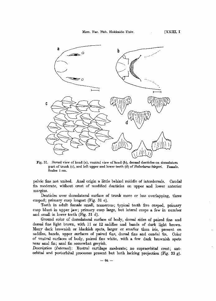
Mem. Fac. Fish. Hokkaido Univ.
a ~.O .... .. ' ,,'
[XXIII, I
Fig. 31. Dorsal view of head (a), ventral view of head (b), dermal denticles on dorsolatera part of trunk (c), and left upper and lower teeth (d) of Halaelu'1'U8 biirgeri. Female. Scales 1 cm.
pelvic fins not united. Anal origin a little behind middle of interdorsals. Caudal fin moderate, without crest of modified denticles on upper and lower anterior margins.
Denticles over dorsolateral surface of trunk more or less overlapping, three cusped; primary cusp longest (Fig. 31 c).
Teeth in adult female small, numerous; typical teeth five cusped, primary cusp blunt in upper jaw; primary cusp large, but lateral cusps a few in number and small in lower teeth (Fig. 31 d).
Ground color of dorsolateral surface of body, dorsal sides of paired fins and dorsal fins light brown, with 11 or 12 saddles and bands of dark light brown. Many dark brownish or blackish spots, larger or smaller than iris, present on saddles, bands, upper surfaces of paired fins, dorsal fins and caudal fin. Color of ventral surfaces of body, paired fins white, with a few dark brownish spots near anal fin; anal fin somewhat greyish. Description (Internal): Rostral cartilage moderate; no supraorbital crest; antorbital and postorbital processes present but both lacking projection (Fig. 33 g).
-64 -

1975] NAKAYA: Taxonomy, anatomy and phylogeny of Japanese catsharks
Cartilage of pectoral fin rounded as a whole; radial cartilages divided into three segments (Fig. 36 g). Radials of 1st dorsal fin three segmented (Fig. 37 g). Labial cartilages three. Pharyngeal denticles on hyoid and gill arches, and on anterior parts of upper and lower walls of pharyngeal cavity; those on lower wall fewer than the upper. Calcification of vertebra above base of pectoral fin cyclospondylic with some sign of diagonal calcification (Fig. 38 g). Heart valves in three series two rows; some with a few small additional valves. Siphon of male large; its anterior tip reaching pectoral fin. Number of spiral valves eight. Number of monospondylous vertebrae 33-34 (Table 3). Embryo: 32 mm embryo (Fig. 32 b) - Head not flattened. Many external gill filaments from spiracle and gill openings. All fins clearly formed, but fin folds present between unpaired fins and in front of 1st dorsal and anal fins. Enlarged denticles not formed on dorsolateral surface of trunk. No pigmentation on body and fins.
b
c
d
Fig. 32. Egg capsule (a), 32 rom embryo (b), 50 mm embryo (c) and 68 mm embryo (d) from egg capsule of HalaelUf"U8 bUrgeri. Scales 1 cm.
- 65-

Mem. Fac. Fish. Hokkaido Univ. [xxm, I
50 mm embryo (Fig. 32 c) -Head flattened dorsoventrally. No external gill filaments. Fin folds low and indistinct. All fins relatively small. Enlarged denticles being formed. Light brownish tint on body and fins.
68 mm embryo (Fig. 32 d) - Head flattened dorsoventrally. General appearance almost same as that of adult. Enlarged denticles being formed, large and about ten in number; all enlarged denticles located before 1st dorsal fin. Denticles of other parts of body being formed. No enlarged denticles near tip of caudal fin. Light brownish saddles present on body and fins. Egg capsule: Egg capsule (Fig. 32 a) wide and short; 51 mm long and 21 mm in greatest width. Posterior end truncated and anterior end tapering with long weak tendrils on each end. Color of egg capsule translucent yellow and its surface smooth. Distribution: Hokkaido (Otaru), Tokyo, Kanagawa Prefecture (Misaki), Kochi Prefecture, Nagasaki Prefecture and Miyazaki Prefecture (Totoro) in Japan. Korea (Mokpo). Formosa (Keelung, Kao-hsiung). China (southern part of East China Sea to South China Sea). Philippines. Amboyna. East Indies. India. Remarks
As pointed out by Springer and D'Aubrey (1972), the prominent labial grooves are shown in the original figure by Muller and Henle (1841), though they described that Halaelurus burgeri has almost no labial grooves. In the specimens used, labial grooves are indistinct or very small. However, these specimens have always three labial cartilages just like those in other species with distinct labial grooves. On the other hand, Scyliorhinus torazame has a very small labial groove only on lower jaw and Oephaloscyllium umbratile has no labial groove, but these two species have rather degenerated labial cartilages. Therefore, Halaelurus burgeri may have a tendency to lose labial grooves and may show a wide variation in this feature.
Kudo (1959) examined the reproduction and growth of embryo of this species. He suggested that the biological minimum of female is about 40 cm in total length, and the number of the egg capsules contained in the oviduct increases with growth, and that the eggs are laid one after another when the embryo grows as large as 70 mm in total length during December and April in the waters near Shikoku Island. It is very interesting that this species contains many egg capsules at one time and keeps the capsule in the oviduct until the embryo in it attains a certain length. Considering the evolution of the mode of reproduction in sharks, the scyliorhinid sharks are interesting, because two types of modes of reproduction, i.e. oviparity and ovoviviparity, are present. Scyliorhinid sharks had formerly been considered oviparous until Cadenat (1959) reported an ovoviviparous scyliorhinid, Galeus pollio
The present author considers that the mode of reproduction in this species is the intermediate between ovoviviparity and oviparity, latter of which is typically seen in Scyliorhinus torazame that contains only one egg capsule in one oviduct at a time and lays eggs before the embryo in it develops. This is further discussed in another chapter of the present paper.
- 66-

1975] NAKAYA: Taxonomy, anatomy and phylogeny of Japanese catsharks
V. Comparative anatomy
Many organs were dissected in order to consider the phyletic interrelationships among scyliorhinid sharks of Japan. They are skeletal, muscular, circulatory, digestive, reproductive and nervous systems. Although many of them did not show phyletically important differences within the family Scyliorhinidae, the present author gives consideration to such characters as chondrocranium, cartilages of pectoral and 1st dorsal fins, vertebral calcification pattern, extrabranchial cartilages and reproductive system in order to clarify the phyletic interrelationships among Japanese scyliorhinid sharks. In addition, such species were dissected for comparison as Halaelurus bivius, Proscyllium habereri, Mustelus schmidti, M. manazo, Triakis scyllia, Rhizoprionodon sp., Scoliodon macrorhinus, Negogaleus sp., Carcharhinus dussumieri, C. sorrah, Pterolamiops longimanus, Sphyrna lewini, Cirrhoscyllium japonicum, Chiloscyllium griseum, Orectolobus japonicus, Squalus acanthias and Etmopterus lucifer from Japanese waters, China Sea, Indian Ocean and Argentine waters. As for the vertebral numbers, the author divided them into monospondylous and diplospondylous vertebrae, though Springer and Garrick (1964) divided them into precaudal and caudal vertebrae. It will be more accurate and useful to divide them into mono- and diplospondylous vertebrae. Because the data on the vertebral numbers are not enough, and because the variation in monospondylous number seems to be very wide, the present author only gives the number of monospondylous vertebrae of Japanese scyliorhinid sharks (Table 3) and avoids the discussion.
1. Chondrocranium
Description The chondrocranium is mainly composed of rostral cartilage, nasal capsule,
orbit and auditory capsule. The rostral cartilage has been known to show the uniform shape on the same phyletic line, and the scyliorhinid, triakid and carcharhinid sharks have universally the triradiate rostral cartilage.
Chondrocranium of Cephaloscyllium (Fig. 33 a) is flatter and wider as a whole than that of Scyliorhinus. Rostral cartilage is also very short and weak. Nasal capsule is rounded and large. Supraorbital crest is present. Superficial ophthalmic branches of the trigeminal and facial nerves runl to dorsal side of chondrocranium through a cranial foramen on upper anterior part of orbit. Palatoquadrate levator muscle is small and is distributed between palatoquadrate cartilage and posterior part of supraorbital crest and never invades orbit.
Chondrocranium of Scyliorhinus (Fig. 33 b) is relatively flat and wide as a whole. Rostral cartilage is very short and weak. Nasal capsule is rounded and large. Supraorbital crest is present fundamentally, but variable in shape. A cranial foramen for the superficial ophthalmic branches of the trigeminal and facial nerves is present in most specimens used, and thus the typical supraorbital crest is formed. However, some, though very few, lack supraorbital crest in the strict sense, because a cranial foramen for the superficial ophthalmic branch is
- 67-

Mem. Fac. Fish. Hokkaido Univ. [XXllI, I
9
Fig. 33. Dorsal views of chondrocrania in scyliorhinid sharks. Scales 10 mm. a, Oephaloscyllium umbratile; b, Scyliorhinna torazame; c, ApristuTUs japonicna; d, Galena eastmani; e, Galena sauteri; f, Galeus nipponensis; g, Halaelurus burgeri. ao, antorbital process; pa, projection of antorbital process; po, postorbital process; pp, projection of postorbital process; sc, supraorbital crest.
not completely formed. A sheet of tendonous membrane is present over orbit to cover dorsal side of eye ball. Palatoquadrate levator muscle is small and is distributed between palatoquadrate cartilage and posterior part of supraorbital crest, and never invades orbit.
Anterior part of chondrocranium of .A.pristurus (Fig. 33 c) is flattened. Rostral cartilage is very long and slender. Nasal capsule projects anterolaterally. Supraorbital crest is absent, but antorbital and postorbital processes are present. They lack their anterior and posterior projections. A sheet of tendonous membrane, which covers the dorsal side of eye ball, is present between antorbital and postorbital processes. Superficial ophthalmic branches of the trigeminal and facial nerves run to dorsal side of chondrocranium at just posterior part of antorbital process, and never through any cranial foramina. Palatoquadrate levator muscle is small and is distributed between palatoquadrate cartilage and postorbital process, and never invades orbit.
Rostral cartilage of Galeus (Fig. 33 d-f) is very long and stout, and its anterior tip is square shaped. Nasal capsule is large. Supraorbital crest is not present, but antorbital and postorbital processes are present. Antorbital process has a projection posteriorly and postorbital process has also a projection anteriorly,
- 68-

1975] NAKAYA: Taxonomy, anatomy and phylogeny of Japanese catsharks
between which a sheet of tendonous membrane is present and it covers dorsal part of eye ball. Superficial ophthalmic branches of the trigeminal and facial nerves run to dorsal side of chondrocranium at just posterior part of antorbital process between the projection of antorbital process and the body of chondrocranium, and never through any cranial foramina. Palatoquadrate levator muscle is small and is distributed between palatoquadrate cartilage and postorbital process, never invading orbit.
Rostral cartilage of Halaelurus (Fig. 33 g) is moderate in length, and stout. Nasal capsule is large and expanded laterally. Supraorbital crest is absent, but antorbital process and postorbital process are present. Both processes lack their anterior and posterior projections. A sheet of tendonous membrane is present between antorbital and postorbital processes. Superficial ophthalmic branches of the trigeminal and facial nerves run to dorsal side of chondrocranium at just posterior part of antorbital process, and never through any cranial foramina. Palatoquadrate levator muscle is small and is distributed between palatoquadrate cartilage and postorbital process, and never invades orbit. Discussion
Among many characters of chondrocranium examined, the rostral cartilage and some characters near orbit showed remarkable differences. The length of rostral cartilage is, however, closely related with the length of snout, which is influenced by the environmental factors (White, 1936, 1937). Further, the snout length varies ontogenetically and is variable even between closely related species of same genus. Therefore, it is dangerous to use the character for consideration of phyletic interrelationships.
On the other hand, the certain characters near orbit, i.e. antorbital process, postorbital process and supraorbital crest, seem to be very useful to consider the interrelationships and phylogeny of sharks of scyliorhinid-carcharhinid line.
The author examined the characters of other species of scyliorhinid-carcha-rhlnid phyletic line and they are summarized in Table 4. .
There are two kinds of jaw suspensions among sharks. The amphistylic jaw suspension is seen in the primitive living and ancient sharks, i.e. Oklamydoselackus, Paleozoic cladodonts and Mesozoic hybodonts. As the shark became advanced, they got the hyostylic jaw suspension (Schaeffer, 1967), as present in all modern sharks. The jaw movements must have been limited in sharks with amphistylic suspension, but in sharks with hyostylic suspension, the jaws became more movable. As the effective feeding can be done by extending both jaws (Springer, 1966; Moss, 1962, 1972; Schaeffer, 1967), the feeding ability has greatly been improved through acquisition of hyostylic jaw suspension. Hence, the muscles associated with upper jaw projection must have also been greatly influenced. In sharks with hyostylic jaw suspension, the protrusion of upper jaw is done by dorsal head of quadratomandibularis muscle, palatoquadrate levator muscle and dorsal head of preorbitalis (Moss, 1962, 1972). The palatoquadrate levator muscle variously develops in each species of scyliorhinid-carcharhinid line, and hence the features near orbit associated with this muscle also vary greatly. Therefore, the present author investigated these characters in order to examine the
- 69-

Mem. Fac. Fish. Hokkaido Univ. [XXIII, I
Table 4. Presence of orbital proces8es and supraorbital crest, and pattern of di8tribution of palatoquadrate levator mU8cle in variou8 species of 8cyliorhinid-carcharhinid line.
Antorbital Postorbital Supraorbital Species process process crest
Halaelurus I + + -Apristurus + + -Galeus
I -It -It -
Cephal08cyllium - -
I +
Scyliorhinus - - + I
Halaeluru8 bivius + Proscyllium habereri + M ustelU8 manazo + Triakis 8cyllia + Rhizoprionodon sp. -It + Scoliodon -It +
macrorhinu8 N egogaleu8 sp. -It + Carcharhinus -It ±
dU8sumieri C. 80rrah -It ± Pterolamiop8 -It ±
longimanu8
+, present; -, ab8ent; *, proces8 with a projection; ±, process chiefly 8upporting infraorbital 8ensory canal.
Palatoquadrate levator muscle from palatoquadrate
cartilage to:
I postorbital process ditto ditto posterior p!1rt of crest ditto
---- -
ditto ditto ditto posterior p!1rt of crest and inside of orbit
postorbital process and dorsal side of skull
ditto
ditto ditto
ditto ditto
phyletic interrelationships of scyliorhinid sharks in Japan. The palatoquadrate levator muscle in scyIiorhinid sharks is distributed only
between palatoquadrate cartilage and posterior part of supraorbital crest, or postorbital process. Among Japanese scyIiorhinid genera, supraorbital crest is present in Cephaloscyllium and Scyliorhinus, but Galeus, Apristurus and Halaelurus have antorbital and postorbital processes (Fig. 34 a-d). The supraorbital crest is also present in Proscyllium and Mustelus (Fig. 34 e), and palatoquadrate levator muscle is small in them, similar to those of scyliorhinids. In Triakis, supraorbital crest is present, but the muscle becomes large and invades inside of orbit to occupy posterior part of orbit (Fig. 34 f). The palatoquadrate levator muscle in carcharhinid sharks develops greatly and this is distributed largely between palatoquadrate cartilage and dorsal part of chondrocranium through posterior part of orbit, and supraorbital crest is no longer present, but large antorbital process with posterior projection and a small rodlike postorbital process are present instead. A small amount of palatoquadrate levator muscle is also distributed to the small postorbital process (Fig. 34 g-i).
In short, they can be divided into the three fundamental types as below. A. Supraorbital Crest Type: Cephaloscyllium, Scyliorhinus, Proscyllium,
-70 -

1975] NAKAYA: Taxonomy, anatomy and phylogeny of Japanese catsharks
ap
f
I I I
'ok
I I I I I I I
0 10 I I
Fig. 34. Dorsal views of left half of chondrocrania in various species of scyliorhinidcarcharhinid line, showing areas of distribution of palatoquadrate levator muscle. a, Cephal08cyllium umbratile; b, Scyliorhinus torazame; c, Galeus nipponensis; d, Halaelurus burgeri; e, Mustelus manazo; f, Triakis scyllia; g, Negogaleus Bp.; h, Carcharhinus dussumieri; i, Pterolamiops longimanus. ap, position of attachment of palatoquadrate levator muscle; pq, palatoquadrate levator muscle; so, superficial ophthalmic branch of V and VII cranial nerves.
Mustelus, Triakis, Halaelurus (bivius) B. Orbital Process Type I (with small palatoquadrate levator muscle only
to postorbital process): Galeus, Apristurus, Halaelurus (burgeri) C. Orbital Process Type II (with voluminous palatoquadrate levator muscle
to dorsal side of chondrocranium and postorbital process): Negogaleus, Scoliodon, Rhizoprionodon, Carcharhinus, Pterolamiops
Supraorbital Crest Type also includes Scyliorhinus caniculus (Parker, 1878), Scylliogaleus and Hemitriakis (Compagno, 1970), and Halaelurus chilensis (Springer and D'Aubrey, 1972). All species of Halaelurus, except H. bivius and H. chilensis (Springer and D'Aubrey, 1972), belong to Orbital Process Type (probably to Type I). Advanced and intermediate carcharhinids of Compagno (1970) are expected to belong to Orbital Process Type II.
Though the available data on fossil sharks are of limited use, the present author
-71 -

Mem. Fac. Fish. Hokkaido Univ. [XXIII, I
considers that the ancestral sharks of scyliorhinid-carcharhinid line must have originally had supraorbital crest for protection of eye, as present in some known fossil sharks, and that the palatoquadrate levator muscle must have been small (Supraorbital Crest Type). These ancestral features seem to have been retained still in the modem Oephaloscyllium and Scyliorhinus.
Orbital Process Type I appears to be quite different from Type II, because one has a very little palatoquadrate levator muscle, and the other has a large voluminous palatoquadrate levator muscle, though both lack supraorbital crest. Therefore, these two types appear to have been derived independently from ancestral Supraorbital Orest Type. The former appears to have directed for the simplification of chondrocranium, and this seems to be the main evolutional trend in scyliorhinid sharks. In other words, the author considers that the supraorbital crest tends to disappear and to form separate antorbital and postorbital processes without proliferation of palatoquadrate levator muscle. The latter, however,
C~o
~
Fig. 35. Schematic views of hypothetical developments of left orbital processes, supraorbital crest and palatoquadrate levator muscle. Anterior up. A, Cephal08cyUium umbratile, Scyliorhinus torazame, HalaeluTUs bivius, Mustelus manazo; B, Galeus eastmani, Galeus sauteri, Galeus nipponensis; C, Halaelurus burgeri, ApristUTU8 japonicus; D. Trialci8 8cyllia; E, Hypothetical species; F, Scoliodon macrorhinus, Rhizoprionodon sp.; G, Negogaleus sp.; H, Carcharhinus dussumieri; I, Pterolamiop8 longimanus. ap, position of attachment of palatoquadrate levator muscle; pa, projection of antorbital process; pp, projection of postorbital process; pq, palatoquadrate levator muscle; sc, supraorbital crest; so, superficial ophthalmic branch of V and VII cranial nerves.
-72 -
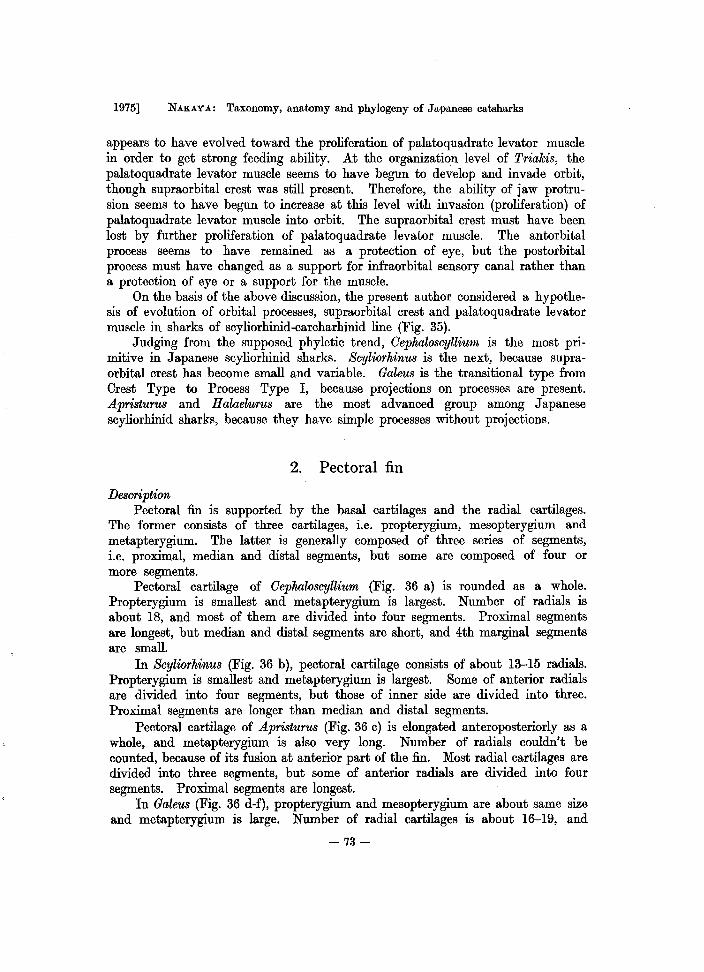
1975] NAKAY A: Taxonomy, anatomy and phylogeny of Japanese catsharks
appears to have evolved toward the proliferation of palatoquadrate levator muscle in order to get strong feeding ability. At the organization level of Triakis, the palatoquadrate levator muscle seems to have begun to develop and invade orbit, though supraorbital crest was still present. Therefore, the ability of jaw protrusion seems to have begun to increase at this level with invasion (proliferation) of palatoquadrate levator muscle into orbit. The supraorbital crest must have been lost by further proliferation of palatoquadrate levator muscle. The antorbital process seems to have remained as a protection of eye, but the postorbital process must have changed as a support for infraorbital sensory canal rather than a protection of eye or a support for the muscle.
On the basis of the above discussion, the present author considered a hypothesis of evolution of orbital processes, supraorbital crest and palatoquadrate levator muscle in sharks of scyliorhinid-carcharhinid line (Fig. 35).
Judging from the supposed phyletic trend, Oephaloscyllium is the most primitive in Japanese scyliorhinid sharks. Scyliorhinus is the next, because supraorbital crest has become small and variable. Galeus is the transitional type from Crest Type to Process Type I, because projections on processes are present. Apristurus and Halaelurus are the most advanced group among Japanese scyliorhinid sharks, because they have simple processes without projections.
2. Pectoral fin
Description Pectoral fin is supported by the basal cartilages and the radial cartilages.
The former consists of three cartilages, i.e. propterygium, mesopterygium and metapterygium. The latter is generally composed of three series of segments, i.e. proximal, median and distal segments, but some are composed of four or more segments.
Pectoral cartilage of Oephaloscyllium (Fig. 36 a) is rounded as a whole. Propterygium is smallest and metapterygium is largest. Number of radials is about 18, and most of them are divided into four segments. Proximal segments are longest, but median and distal segments are short, and 4th marginal segments are small.
In Scyliorhinus (Fig. 36 b), pectoral cartilage consists of about 13-15 radials. Propterygium is smallest and metapterygium is largest. Some of anterior radials are divided into four segments, but those of inner side are divided into three. Proximal segments are longer than median and distal segments.
Pectoral cartilage of Apristurus (Fig. 36 c) is elongated anteroposteriorly as a whole, and metapterygium is also very long. Number of radials couldn't be counted, because of its fusion at anterior part of the fin. Most radial cartilages are divided into three segments, but some of anterior radials are divided into four segments. Proximal segments are longest.
In Galeus (Fig. 36 d-f), propterygium and mesopterygium are about same size and metapterygium is large. Number of radial cartilages is about 16-19, and
-73-

Mem. Fac. Fish. Hokkaido Univ. [XXIII, I
Fig. 36. Dorsal views of cartilage of left pectoral fins in scyliorhinid sharks. Scales 10 mm. a, Oephaloscyllium umhratile; b, Scyliorhinus torazame; c, Apristurus japonicus; d, Galeus eastmani; e, Galeu8 sauteri; f, Galeus nipponensis; g, Halaelurus burgeri. ms, mesopterygium; mt, metapterygium; pr, propterygium; rd, radial cartilages.
they are divided into three segments, but never into four or more. Proximal segments are longest and distals are shortest.
Halaelurus (Fig. 36 g) has relatively large mesopterygium, which is larger than propterygium. Number of radial cartilages is about 14-15, and they are divided into three segments, but never into four or more. Proximal segments are longest and distals are shortest. Discussion
For the consideration of phyletic trend, pectoral fins of other species were examined and shown in Table 5. An Argentine scyliorhinid Halaelurus bivius has about 15 radials and Proscyllium habereri, which has been considered as an intermediate between Scyliorhinidae and Triakidae, has about 16 radials. Both have three series of radial segments and their distals do not elongate. Pectoral cartilages of Mustelus manazo, a typical triakid Triakis scyllia and a carcharhinid
-74 -

1975] NAKAYA: Taxonomy, anatomy and phylogeny of Japanese catsharks
Table 5. Numher of radial cartilages and segments of 1st dorsal and pectoral fins in various species of scyliorhinid.carcharhinid line.
Number of segments Number of radials Species
1st Dorsal I Pectoral 1st Dorsal I Pectoral
Oephaloscyllium umhratile 4- 4(3) I
13 18 Scyliorhinus torazame 3(4) 3(4) 12-14 13-15 Apristurus japonicus 3(4) 3(4) 16 1 Galeus eastmani 3 3
} 15-17 } 16-19 G. sauteri 3 3 G. nipponensis 3 3 Halaelurus bilrgeri 3 3 14 14-15
H. bivius 3 3 14 15 Pr08cyllium habereri 3 3 19 16 M ustelus manazo 3 3+ 23 20 Triakis scyllia 3+ 3+ 24 19 N egogaleus sp. 3+ 3+ 25 20 Oarcharhinus dussumieri 3+ 3+ 31 25 Sphyrna lewini 3+ 3+ 31 28
4-, four or more segmented; 4(3), mostly four segmented, but some three segmented; 3(4), m08tly three segmented, but some four segmented; 3+, distal segments elongated.
Negogaleus sp. consist of about 20 radial cartilages and their distal segments elongate and are longer than median segments. Further, Carcharhinus dussumieri and Sphyrna le'Uiini have 25 and 28 radial cartilages respectively. Their radials are divided into three segments, and the distal segments elongate greatly.
Judging from these data, there seem to be two phyletic tendencies in the characters of pectoral fin. One is the stabilization of number of radial segments and elongation of distal radials. This tendency was discussed by Regan (1906) and White (1936b, 1937) as far as the author knows. White (1937) also stated that the radials undergo further segmentation, and there is more segmentation in the primitive groups than in the more specialized ones, where the number becomes stabilized at three segments to a radial, and that if the fins are elongated, the radials are lengthened but there is no further segmentation. Compagno (1970) described that distal radials are much longer than proximals in his advanced and intermediate carcharhinids, i.e. Aprionodon, Carcharhinus, Galeocerdo, Hypoprion, Isogomphodon and so on. The author considers that the tendency above is valid in phyletic considerations. The other is the increase in number of radial cartilages and the break down of the anterior proximal segments instead of fusion. This tendency might be valid, but as the data are not enough, it is very dangerous to use the tendency for phyletic considerations. Therefore, this is not further discussed here.
Consequently, based on the former tendency, Cephaloscyllium, most of whose radials in pectoral fin are divided into four, is considered to be the most primitive group in Japanese Scyliorhinidae. Apristurus and Scyliorhinus, which have partly
-75 -

Mem. Fac. Fish. Hokkaido Univ. [XXIII, I
four segmented radials, are the intermediates. Galeus and Halaelurus, having three radial segments, are the advanced genera in the Japanese family.
3. First dorsal fin
Description First dorsal fin is supported by many radial cartilages, which are further
divided usually into three series of segments, i.e. proximal, median and distal segments.
First dorsal fin cartilage of Cephaloscyllium (Fig. 37 a) consists of about 13 radials. Proximal and median segments are long, but distal segments are subdivided into two to four small pieces. Some proximal segments are fused.
Scyliorhinus (Fig. 37 b) has 12-14 radials and most of them are divided into three segments, but some radials are divided into four by subdivision of distal segments into two pieces. Proximal and median segments are long. Some proximal segments are fused.
Radials of Apristurus (Fig. 37 c) are more obliquely arranged than those of Cephaloscyllium, Scyliorhinus, Galeus and Halaelurus. Number of radials is about 16 and most radials are divided into three segments, but some are divided into four segments. Median segments are longest. Some proximal segments are fused.
Galeus (Fig. 37 d-f) has 15-17 radial cartilages and they are relatively stiff.
~
Fig. 37. Lateral views of cartilage of 1st dorsal fins in scyliorhinid sharks. Scales 5 mm. a, Cephal08cyllium urribratile; b, Scyliorhinus torazame; c, ApristuffiB japonicus; d, Galeus eastmani; e, Galeus sauteri; f, Galeus nipponensis; g, HalaeluffiB burgeri.
-76 -

1975] NAKAYA: Taxonomy, anatomy and phylogeny of Japanese catsharks
Radial cartilages are divided into three segments and never into four or more. Median segments are longest. Last two proximal segments are fused.
Number of radial cartilages of Halaelu'fus (Fig. 37 g) is about 14 and the radials are divided into three segments and never into four or more. Median segments are longest, but distal and proximal segments are relatively long. Discussion
The radial number of some other sharks of scyliorhinid-carcharhinid line was examined (Table 5). Judging from the results, there seem to be two tendencies in the characters of 1st dorsal fin, just like those in pectoral fin. One is the increase in number of radial cartilages in the fin. This tendency might roughly be valid, but the data are too meager to discuss the phyletic interrelationships. Therefore, this is not further discussed here, too. The other tendency is the stabilization of number of radial segments and elongation of distal radials. The number of segments is universally three in P'foscyllium habe'fe'fi, Mustelus manazo, T'fiakis scyllia, Negogaleus sp., Ca'fcha'fhinus dussumieri and SphY'fna lewini. Distal segments of T'fiakis scyllia slightly elongate, though those of Mustelus manazo are still short. Negogaleus sp. and Ca'fcha'fhinus dussumie'fi have elongated distal segments almost as long as median segments. Further, those of SphY'fna lewini elongate very much, almost reaching margin of the fin, but they are divided into three, not four or more. Although White (1937) did not refer to the latter tendency in 1st dorsal fin, just the same trends as seen in pectoral fin are also apparently present in 1st dorsal fin. Concretely, the primitive groups have the subdivided radials into four or more segments and short distal segments. The more advanced groups, however, possess universally three segmented radial cartilages, and when a group becomes much more advanced, distal segments get elongated without subdivision.
Consequently, as no prominent elongation occurs in distal segments in Japanese genera of family Scyliorhinidae, it is impossible to find any trends in this character. However, judging from the subdivision, Cephaloscyllium is the most primitive group, because the radials are strongly subdivided. Scylio'fhinus and Ap'fistu'fuS are the intermediates among the members of Japanese Scyliorhinidae because they have partly four segmented radials. Galeus and Halaelu'fus, which have always three segmented radials, are the advanced groups.
4. Vertebral calcification
Desc'fiption Vertebral centra of elasmobranch fishes are calcified in various types, which
are generally classified cyclospondylic, asterospondylic and tectospondylic vertebrae. The pattern of calcification of vertebral centra was observed and described in the vertical plane cut transversely at the middle of a centrum, which were taken from the vertebral column above pectoral fin.
The condition of calcification in the centra of Cephaloscyllium (Fig. 38 a) is cyclospondylic and the peripheral calcification of centra is very thin and do not become thick.
-77 -

Mem. Fac. Fish. Hokkaido Univ. [XXIII, I
a
d f
Fig. 38. Transverse views of scyliorhinid vertebral calcification patterns, with calcified areas indicated in black. a, Oephaloscyllium umbratile; b, Scyliorhinus torazame; c, Apristurus japonicus; d, Galeus eastmani; e, Galeus sauteri; f, Galeus nipponensis; g, Halaelurus burgeri.
Scyliorhinus (Fig. 38 b) has about same condition as that of Cephaloscyllium and the calcification is cyclospondylic. Peripheral calcification of centra is very thin and do not become thick.
Calcified pattern of centra of Apristurus (Fig. 38 c) is cyclospondylic. Peripheral calcification of centra becomes thick, forming four blunt dorsal, ventral and lateral wedges.
Galeus (Fig. 38 d-f) has same condition as that of Apristurus and shows cyclospondylic condition. Peripheral calcification of centra becomes thick, forming four blunt dorsal, ventral and lateral wedges.
Central calcification of Halaelurus (Fig. 38 g) has diagonal development of calcification. Peripheral calcification of centra becomes thick, forming four blunt dorsal, ventral and lateral wedges. Discussion
The taxonomic value of the pattern of calcification in centra of elasmobranch fishes has been discussed by Hasse (1879), Ridewood (1921), White (1937), Goodrich (1958) and Applegate (1967). The pattern has been found to be very useful in taxonomy, as concluded by Ridewood (1921) that the species and genera, which are considered to be nearly related on other features, conform also with one another fairly well in the matter of the calcification of vertebral centra. The
-78 -

1975] NAKAYA: Taxonomy, anatomy and phylogeny of Japanese catsharks
Fig. 39. Transverse views of triakid and carcharhinid vertebral calcification patterns, with calcified areas indicated in black. a, Proscyllium habereri; b, Mustelus manazo; c, Triakis scyllia; d, Oarcharhinus dussumieri; e, Pterolamiops longimanus.
pattern of calcification has generally been divided into cyclospondylic, asterospondylic and tectospondylic types. These classifications, however, are not phyletic, because the same pattern can be found in sharks on different phyletic lines. For example, the cyclospondylic condition is seen in Scyliorhinidae and Squalidae. Ridewood (1921) recommended that the terms cyclo-, astero- and tectospondylic should be abandoned. Recently, Applegate (1967) tried to subdivide the vertebrae logically and separated them into eight morphotypes, but his subdivision appears to need more examination. The author, therefore, uses the classical terms in the following discussion.
In the living sharks of scyliorhinid-carcharhinid line, only the cyclospondylic and asterospondylic conditions are present, and some examples are shown in Fig. 39. Though the relationships with the modern scyliorhinids are not certain, one of the oldest sharks, Paleozoic cladodonts, had no centra (Dean, 1909; Schaeffer, 1967; Zangerl, 1973), and Triassic Hybodus had uncalcified centra (Wbite, 1937). Jurassic Protospinax, which has been anticipated as an ancestor of squaloids, shows strongly cyclospondylic condition (Dean, 1909). Palaeospinax from Lower Jurassic of England had cyclospondylic vertebrae. Though the systematic position is uncertain yet, Schaeffer (1967) considers Palaeospinax as an ancestor of either galeoids or squaloids. Other Jurassic Palaeoscyllium and Pristiurus, both of which are thought to be ancestors of modern Scyliorhinidae, had also cyclospondylic vertebrae just like those of living typical scyliorhinid sharks (White, 1937; Schaeffer, 1967). Above paleontological facts appear to show that the uncalcified vertebrae preceded the calcified vertebrae and that the cyclospondylic condition is the earliest type of calcification. The asterospondylic condition does not appear until Cretaceous and is the latest development (White, 1937).
-79 -

Mem. Fac. Fish. Hokkaido Univ. [XXIII, I
Fig. 40. Schematic transverse patterns of calcification of vertebral centra and their development, with calcified areas indicated in black. A, Cephaloscyllium umbratile, Scyliorhinus torazame; B, ApristurUB japonicus, Galeus eastmani, Galeus sauteri, Galeus nipponensis; C, HalaelurUB bUrgeri, HalaelurUB bivius, Proscyllium habereri; D, Mustelus manazo, MU8telus 8chmidti, Triakis 8cyllia; E, Scoliodon macrorhinu8, Rhizoprionodon sp., Carcharhinus dus8'Umieri; F, Pterolamiop8 longimanus.
Furthermore, when considering the strengthening of the vertebral column as an axial skeleton of body, the trend of modification must be from cyclospondylic condition to asterospondylic condition. The cyclospondylic condition IS
considered to be also strengthened by increasing peripheral calcifications of centra (Fig. 39). Though such evidences as mentioned above do not completely deny Regan's opinion (1906) that cyclospondylic vertebra in Pristiurus (=living genus Galeus) is a complete reversion from asterospondylic type, there is no positive proof that favors his opinion. Therefore, the present author considers that the cyclospondylic condition found in living scyliorhinids must have been retained since its occurrence in Mesozoic ancestors.
Consequently, the cyclospondylic condition is considered to be the most primitive and to evolve toward the asterospondylic condition in the sharks of scyliorhinid-carcharhinid line (Fig. 40).
Judging from above discussion, Cephaloscyllium and Scyliorhinus are most primitive, whose vertebrae show cyclospondylic condition and have only thin peripheral calcification without wedges. The intermediates are Galeus and Apristurus, both of which have cyclospondylic condition and peripheral calcification with four blunt wedges. Halaelurus is considered to be the most advanced genus among Japanese scyliorhinids, because its vertebrae have diagonal developments from central calcification, approaching asterospondylic condition.
- 80-

1975] NAKAYA: Taxonomy, anatomy and phylogeny of Japanese catsharks
5. Extrabranchial cartilage
Description Extrabranchial cartilages are the small pieces of cartilages supporting outer
margins of gill septa dorsally and ventrally, and usually consist of four upper and four lower cartilages.
Cephaloscyllium and Scyliorhinus have only three lower extrabranchial cartilages and four upper extrabranchial cartilages. Apristurus, Galeus and Halaelurus have four upper and four lower extrabranchial cartilages. These are shown in Table 6. Discussion
Data and discussions on extrabranchial cartilages are very few. Only Daniel (1928) dealt with the matter of extrabranchial cartilages of elasmobranch fishes as far as the author knows, and he stated that in a reduction of the number of
. extrabranchials the posterior cartilages are the 1st to be absent, and the 4th extrabranchial may be lacking ventrally, leaving three below and four above.
All the dissected species which are considered to be the higher group of the same phyletic line, i.e. Halaelurus bivius, Proscyllium habereri, Mustelus manazo, M. schmidti, Triakis scyllia, Negogaleus sp., Carcharhinus dussumieri and Sphyrna lewini, have universally four upper and four lower extrabranchial cartilages (Table 6).
White (1936) stated that the evolutional direction appears from sluggish shoredwelling shark similar to Jurassic catuloids (scyliorhinids) toward a freer pelagic life as seen at its best in the swift and voracious carcharhinids in Carcharhinida (=Scyliorhinidae, Triakidae, Carcharhinidae and Sphyrnidae of Bigelow and
Table 6. Number of extrabranchial cartilages in various species on 8cyliorhinid-carcharhinid line.
Species
Oephal08cyllium umbratile Scyliorhinus torazame A pristurus japonicus Galeus eastmani G. sauteri G. nipponensis Halaelurus burgeri
H. bivius Pro8cyllium habereri M ustelus manazo Triakis scyllia N egogaleu8 sp. Oarcharhinus dussumieri Sphyrna lewini
- 81-
Extrabranchials
upper
4 4 4 4 4 4 4
4 4 4 4 4 4 4
lower
3 3 4 4 4 4 4
4 4 4 4 4 4 4

Mem. Fac. Fish. Hokkaido Univ. [xxm, I
Schroeder, 1948), and it is generally considered that the bottom-dwelling habit may be primitive (White, 1937; Melouk, 1947). The gill region might have been affected by such a change of mode of life from benthic to pelagic as mentioned above. At any rate, it seems impossible to judge the phyletic trend of these cartilages by giving some reliable bases at present. Therefore, judging from only the above data (Table 6), those that lack 4th lower extrabranchial cartilage may be primitive and the presence of 4th lower extrabranchial cartilage may be an advanced character. In other words, the author considers that an extrabranchial cartilage was added according as a shark became advanced, contrary to Daniel (1928).
Consequently, Scylicwhinus and Oephaloscyllium are concluded to be more primitive than Apristurus, Galeus and Halaelurus in the extrabranchial cartilages.
6. Reproductive system
Description The fertilization is always internal in the living elasmobranch fishes, and
the development is either oviparous, ovoviviparous or viviparous. In sharks, all these three types of reproduction occur.
All Japanese scyliorhinid sharks are oviparous, but at least two kinds of oviparity appear to be present. One is seen in Oephaloscyllium, Scylicwhinus, Apristurus and Galeus, and they bear always only one egg capsule in each oviduct during the period of pregnancy and lay two eggs at a time. Embryo does not develop when the egg is laid. The other type is seen in Halaelurus, which bears many egg capsules in one oviduct, though the number of egg capsule varies with the size of the mother, and lays egg capsule one by one when the embryo grows to a certain length. Discussion
The present author defines three types of reproduction in sharks as below. Oviparity: Eggs are laid before hatching. Ovoviviparity: Eggs hatch and embryos develop within the mother with
out placental connection. Viviparity: Eggs hatch and embryos develop within the mother with a
connection through the yolk-sac placenta. Many cladodonts, hybodonts, Xenacanthus and all the Jurassic sharks, except
Oladoselache, had the clasper, apparatus for internal fertilization (Breder and Rosen, 1966; Schaeffer, 1967). Therefore, the fertilization was at least internal even in the ancient sharks, though their mode of reproduction is uncertain. In the evolution of mode of reproduction, the ovoviviparity would have been a later development than the oviparity, because there are some tendencies toward ovoviviparity in all living oviparous groups, and because the ovoviviparity is well developed in all the groups established in Cretaceous or later (White, 1937).
The present author considers the evolution of the mode of reproduction in sharks of scyliorhinid-carcharhinid line as follows (Table 7). Among the three
- 82-

1975] NAKAYA: Tax:onomy, anatomy and phylogeny of Japanese catsharks
Table 7. Cla8sification and evolution of mode of reproduction in 8hark8 of 8cyliorhinid-carcharhinid line.
Mode of Reproduction
Single Oviparity
Multiple Oviparity
Ovoviviparity
Viviparity
I Connection I Period of I I with mother Protection Japanese genera
No Short CephaZoscyZlium Scyliorhinus ApristurUB Galeus
No Long HaZaeZuru8
No Whole GaZeus (Atlantic) process MusteZus (manazo)
Yolk-sac Whole M usteZus (griseus) placenta process Carcharhinus
Sphyrna
Probable trend of evolution
Primitive
1 Advanced
types, the oviparity was the first developed, where only one egg capsule is contained in an oviduct. The capsule is protected for only a short time within the mother, and is laid before the embryo develops. This type of oviparity, which the author names the single oviparity, is seen in Oephaloscyllium, Scyliorhinus, Apristurus and Japanese Galeus. According as the single oviparity advanced, the period in oviduct, or the period of protection, became longer. Therefore, the embryo in capsule begins to develop even in oviduct and the next capsule comes down into oviduct. .As a result, many egg capsules are contained in an oviduct and the embryos develop in various degrees. The embryos are protected for relatively long time in the oviduct until they attain a certain length, and then an egg capsule is laid when the embryo in it grows to a certain length. This type of oviparity is seen in Halaelurus (Kudo, 1959), and the present author calls this the multiple oviparity. The author considers that the ovoviviparity is the most advanced oviparity, where the embryo is protected for whole process of development without direct connection with the mother. Some species of Atlantic Galeus have been known or suggested to be ovoviviparous (Cadenat, 1959; Springer, 1966; Bullis, 1967). The yolk-sac placenta may be formed according as ovoviviparity further advances, and the embryo is grown by nutrients from the mother through the yolk-sac placenta. This is the most advanced mode of reproduction, viviparity (viviparous placental type of Teshima and Mizue, 1972). The genus Mustelus may be transitional in the respect of reproduction, where both ovoviviparity and viviparity are present (Teshima and Koga, 1973). Advanced carcharhinid and sphyrnid sharks are all viviparous as far as known (Gilbert and Schlernitzauer, 1966; Schlernitzauer and Gilbert, 1966; Teshima and Mizue, 1972).
Judging from above trend of evolution of reproduction, Oephaloscyllium, Scyliorhinus and Apristurus, which belong to the single oviparity, are primitive, while Halaelurus is the advanced, because it shows the multiple oviparous condition. Genus Galeus contains some ovoviviparous species, though three Japanese species are considered to be the single oviparous species. When considering at
- 83-

Mem. Fac. Fish. Hokkaido Univ. [XXIII, I
generic level, the genus might be considered as a more advanced group than other oviparous genera.
VI. General consideration
The scyliorhinid sharks are generally thought to occupy the most primitive position of the scyliorhinid-carcharhinid phyletic line (Fig. 41; Regan, 1906; Garman, 1913; White, 1937; Bigelow and Schroeder, 1948; Tortonese, 1956; Compagno, 1970, etc.). Because the characters such as calcification of vertebral centrum, chondrocranium, fin skeleton, subocular fold, etc. are essentially same, the present author also considers that the scyliorhinid sharks are on the same phyletic line as the carcharhinids are.
White (1937), Schaeffer (1967) and Fowler (1967) consider the fossil genera Palaeoscyllium and Pristiurus as the most probable ancestors of the living scyliorhinid or more advanced forms. Fossils of this group are meager because of its inadequate preservation, although more paleontological evidences are needed in the consideration of the origin and phylogeny of sharks. It is therefore necessary to investigate them, bearing the thought of the functional improvement of each structure in mind.
Present author considers that the calcification of vertebral column and modification of orbital region are very important. When the vertebral column is considered as an axis of body, the trend of the modification, or strengthening, is very important. The modifications near the orbit are also important because
Sphyrnidae Batoidei
Isuridae, etc.
/ scyliorhinidae +Protospinax
+Palaeospinax
Fig. 41. Simplified interrelationships of elasmobranch fishes, showing the scyliorhinidcarcharhinid phyletic line (After White, 1937, modified by the present author). Scientific names were used according to Bigelow and Schroeder (1948, 1953). +, fossil genus.
-84-

1975] NAKAYA: Taxonomy, anatomy and phylogeny of Japanese catsharks
they are very closely related with the improvement of the feeding mechanism. The present author made a schematic graph based on the hypothetical evolution of the characters discussed in the preceding chapter (Fig. 42).
Considering the chondrocranium and the vertebral column most important, the schematic graph provides a clear picture of diphyletic differentiation in sharks of this phyletic line. One is the scyliorhinid stem, which consists of all the Japanese scyliorhinid genera, and the other is the carcharhinid stem, which consists of the higher groups. The carcharhinid stem must have differentiated directly from the ancestors of the lower organization level like modern Cephaloscyllium or Scyliorhinus, because some of the primitive carcharhinids have the chondrocrania of Supraorbital Crest Type in common with Cephaloscyllium and Scyliorhinus.
As for the scyliorhinid stem, the characters in Cephaloscyllium are all primitive, and the genus Cephaloscyllium is considered to occupy the most primitive position among Japanese scyliorhinid sharks.
First dorsal fin posterior First dorsal fin anterior
PTE 4 5 2 4
CAR ? SCO 4 5 2 4
RHI TRI
? 5 ?
4 2 MUS 4 3",4
PRO lor2 3 3 2 2
3 3
2 2 2
2 2
PDER
Fig. 42. Schematic graph showing level of organization of genera of scyliorhinid·carcharhinid line, based on the characters used in the present study. APR, Apri8turus; CAR, Carcharhinus; CEP, Cepluilo8cyllium; GAL, Galeus; HAL, HalaelurU8; MUS, Mustelus; PRO, Pro8cyllium; PTE, Pterolamiops; RHI, Rhizoprionodon; SCO, Scoliodon; SCY, ScyliorhinU8; TRI, Triakis. D, 1st dorsal fin; E, extrabranchial cartilage; P, pectoral fin; R, reproductive system. 1,....5, showing the more advanced condition in numerical order.
- 85-

Mem. Fac. Fish. Hokkaido Univ. [XXIII, I
Scyliorhinus has also primitive vertebra and chondrocranium like those of Cephaloscyllium. In addition, all the other characters are considered to be primitive as a whole, and it appears to occupy about the same phyletic position as that of Cephaloscyllium. However, Scyliorhinus may be a little more advanced group than Cephaloscyllium, because the former has the advanced supraorbital crest, pectoral fin and 1st dorsal fin, as discussed in the preceding chapter.
Galeus has already lost the supraorbital crest, but has projections still, which are regarded as the vestige of supraorbital crest. This is the intermediate condition between Supraorbital Crest Type and typical Orbital Process Type I. Vertebral column of Galeus is also intermediate between those of Scyliorhinus and Halaelurus. The segments of 1st dorsal and pectoral radials become stabilized. In addition, though the species of Japanese Galeus is the single oviparous, some of Atlantic Galeus attain the ovoviviparous condition. Considering above, Galeus may occupy the intermediate position between Scyliorhinus and Halaelurus.
Apristurus has typical orbital processes, which is the most advanced type in the scyliorhinid stem. However, vertebral column shows the intermediate condition. Pectoral and 1st dorsal cartilages are rather primitive and the reproduction is the primitive single oviparity. Apristurus also clearly occupies the position between Scyliorhinus and Halaelurus.
As for the relationship between Galeus and Apristurus, they have both advanced and primitive characters, and it is difficult to judge the level of organization of each, based on only above features. It is better to conclude at present that Galeus and Apristurus are the intermediate scyliorhinids between Scyliorhinus and Halaelurus.
Parmaturus, though no specimen was available, is considered to be closely related to Galeus, because of similarities in general external morphology.
Halaelurus has vertebra with diagonal development of calcification and orbital processes. Segments of pectoral and 1st dorsal radials are three, and the mode of reproduction is the multiple oviparity. Above all features were judged to be the most advanced characters in Japanese scyliorhinid sharks. Therefore, the genus Halaelurus appears to have generally attained the highest level of organization in Japanese Scyliorhinidae.
Putting all the evidences together, the presumable process of evolution III
Japanese Scyliorhinidae will be as follows and is shown in Fig. 43. Cephaloscyllium and Scyliorhinus must be the first to have branched off from
the base of the scyliorhinid stem. They have both retained their primitive characters until the present time, but Scyliorhinus attained a somewhat more advanced level than. that of Cephaloscyllium. They have become adapted to the bottom habitat of the shallow to deeper waters. Galeus and Apristurus must have branched off after Cephaloscyllium and Scyliorhinus had separated from the scyliorhinid stem. Parmaturus might also have branched off as early as Galeus. They appear to have bocome adapted to the deeper habitat. Halaelurus must have branched from the stem most recently among Japanese scyliorhinid genera, and appears to have become adapted to the bottom of the shallow waters.
-86 -

1975] NAKAYA: Taxonomy, anatomy and phylogeny of Japanese catsharks
Apristurus _-:I"::
Scyliorhinus Cephaloscyllium--__
Hypothetical Ancestor
Fig. 43. A presumable phyletic tree of sharks of scyliorhinid-carcharhinid phyletic line, showing the relations between Scyliorhinidae and the higher groups, and the interrelationships among Japanese genera in the family Scyliorhinidae.
VII. Summary
The sharks belonging to the family Scyliorhinidae in Japan and its adjacent waters were reviewed. Eight species, Cephaloscyllium umbratile, Scyliorhinus torazame, Apristurus platyrhynchus, Apristurus maCITorhynchus, Galeus sauteri, Galeus eastmani, Parmaturus pilosus and Halaelurus burgeri, were found to be distinct species. In addition to these, three new species were described under the names of Apristurus iaponicus, Apristurus longicephalus and Galeus nipponensis. Proscyllium habereri and Calliscyllium venustum were excluded from the family Scyliorhinidae because of their far anterior 1st dorsal fins, etc. As for Pentanchus, the author could not draw any conclusions, for the single specimen in Japan unfortunately has been lost. Thus, the Japanese scyliorhinid sharks numbered seven genera and twelve species including Pentanchus sp.
The phyletic interrelationships among Japanese scyliorhinid sharks were also investigated, based on external and internal characters. The modifications of characters near the orbit were found to be as useful as the calcification patterns of vertebra in studying the phylogeny of sharks of the scyliorhinid-carcharhinid line. Cephaloscyllium and Scyliorhinus were considered to occupy the most primitive position. Apristurus and Galeus were the intermediates. Parmaturus,
-87 -

Mem. Fac. Fish. Hokkaido Univ. [XXITI, I
though no specimen was available, was considered to be closely related to Galeus, because of similarities in external morphology. Halaelurus was regarded as the most advanced group among Japanese scyliorhinid sharks. A phyletic tree was presumed for Japanese genera of the family Scyliorhinidae from the results of the phyletic considerations.
Literature cited
(The author couldn't have access to those with an asterisk)
Alcock, A. (1891). On the deep-sea fishes collected by the Investigator in 1890-91. Ann. Mag. Nat. Hist., ser. 6, 8, 19-34.
*Alcock, A. (1899). A Descriptive Oatalogue of the Deep-sea Fishes in the Indian Museum. iii+211 p. 43 pIs. Calcutta.
Alexander, R. McN. (1967). Functional Design in Fishes. 16Op., 16 figs. Hutchinson University Library, London.
Applegate, S.P. (1967). A survey of shark hard parts. p.37-67, figs. 2.1-2.7, pIs. 2.1-2.4. In Gilbert, P.W. et al. (eds.), Sharks, Skates and Rays. xv+624 p. Johns Hopkins Press, Baltimore.
Bachus, R.H., Springer, S. and Arnold, E.L., Jr. (1956). A contribution to the natural history of the white-tip shark, Pterolamiops longimanus (Poey). Deep.Sea Research 3, 178-188.
Barnard, K.H. (1925). A monograph of the marine fishes of South Africa. Ann. South African Mus. 21, 1-418, pIs. 1-17.
Beebe, W. and Tee-Van, J. (1941). Fishes from the tropical eastern Pacific. Part 2. Sharks. Zoologica 26, 93-122, figs. 1-34, pIs. 1-2.
Bigelow, H.B. and Schroeder, W.C. (1944). New sharks from the western North Atlantic. Proc. New England Zool. Club 23, 21-36.
Bigelow, H.B. and Schroeder, W.C. (1948). Sharks. p. 56--576, figs. 6--106. In Tee-Van, J. et al. (eds.), Fishes of the Western North Atlantic. Part 1. xvii+576 p., 106 figs. Mem. Sears Found. Mar. Res., Yale Univ., New Haven.
Bigelow, H.B. and Schroeder, W.C. (1953). Sawfishes, guitarfishes, skates and rays. p. 1-514, figs. 1-117. Ibid. Part 2. xv+588 p., 127 figs.
*Bonaparte, C.L. (1834). Iconographia della Fauna Italica. Part 1-3. *Bonnaterre, J.P. (1788). Tableau Encyclopedique et Mlthodique des Trois Regnes de la
Nature .. Ichthyologie. vi+215 p., 102 pIs. Brauer, A. (1906). Die Tiefsee-Fische. 1. Systematischer teil. Deut. Tiefsee Expedition
15, 1-432, pIs. 1-18. Breder, C.M. and Rosen, D.E. (1966). Modes of Reproduction in Fishes. xv+941 p.
Natural History Press, New York. Buen, F. de. (1959). Notas preliminares sobre la fauna marina preabismal de Chile, con
descripcion de una familia de rayas, dOB generos y siete especies nuevos. Bol. Mus. Nac. Hist. Nat. Santiago 27, 171-201.
Bullis, H.R., Jr. (1967). Depth segregations and distribution of sex maturity groups in the marbled catshark, Galeus arae. p. 141-148, figs. 8.1-8.6. In Gilbert, P.W. et al. (eds.), Sharks, Skates and Rays. xv+624 p. Johns Hopkins Press, Baltimore.
Cadenat, J. (1959). Notes d'Ichtyologie ouest-africaine. xx. Galeus polli espece nouvelle ovovivipare de Scylliorhinidae. Bull. Inat. Franc. d'Afri. Noire, ser. A, 1, 395-409.
Cadenat, J. and Maul, G.E. (1966). Ditto. XLIII. Description d'une espece nouvelle du
- 88-

1975] NAKAYA: Taxonomy, anatomy and phylogeny of Japanese catsharks
genre Apruturus, Apristurus maderenBis. Ibid. 2, 769--782, figs. 1-4. Chan, W.L. (1966). New sharks from the South China Sea. J. Zool., London 148, 218--237. Chen, J.T.F. (1948). Notes on Formosan sharks. Quart. J. Taiwan Mus. 1, 21-45. (In
Chinese). Chen, J.T.F. (1963). A revision of the sharks of Taiwan. Biol. Bull., Dep. Biol. Coll.
Sci. Tunghai Univ. 19, 1-102, figs. 1-28. Chu, Y.T., Chang, C.L. and Cherng, C.T. (1963). Fuhes of East China Sea. 642 p.
Scientific Press. (In Chinese). Chu, Y.T. and Wang, Y.H. (1964). On the geographical distribution and faunal character
istics of the chondrichthian fishes of China. Acta Zool., Sinica 16, 674--Q89. (In Chinese with English abstract).
Chung, B.K., Kim, Y.M. and Kim, Y.S. (1967). Zoogeographical studies on the bottom fishes of the Korean coast in the Yellow Sea. Rep. Fish. Res. Dev. Agency (7), 5-27. (In Korean).
Collett, R. (1904). Four hitherto undescribed fishes from the depth south of the Faroe Islands. Christiania VidenBkabs-Selskabs Forhandlinger (9), 1-7.
Collignon, J. and Aloncle, H. (1972). Catalogue raisonne des poissons des mers Marocaines. lere partie. Cyclostomes, selaciens, holocephales. Bull. l'InBt. Peches Maritimes (19), 1-163, figs. 1-53.
Compagno, L.J.V. (1970). Systematics of the genus Hemitriakis. Proc. Calif. Acad. Sci., ser. 4, 38, 63-98, figs. 1-8.
Compagno, L.J.V. (1973). Interrelationships of living elasmobranchs. p. 15-61, figs. 1-5, pIs. 1-2. In Greenwood, P.H. et ai. (eds.), InterrelatiOnBhips of Fishes. xvi+536 p. Academic Press, London.
Daniel, J.F. (1928). The Elasmobranch Fishes. xi+332 p., 270 figs. Univ.Calif. Press, Berkeley.
Dean, B. (1909). Studies on fossil fishes (Sharks, chimaeroids and arthrodires). Mem. Amer. MU8. Nat. Hist. 9, 211-287, figs. 1-65.
*Dumeril, A.H.A. (1853). Monographie de la tribu des Scylliens ou Roussettes (poissons plagiostomes) comprenent deux especes nouvelles. Rev. Mag. Zool. 2, ser. 5, 8-25, 73-87, 119--130, pI. 1.
Engelhardt, R. (1912). Ueber einige neue Selachir-Foramen. Zool. Anz. 39, 643--648. Ford, E. 1921. A contribution to our knowledge of the life histories of the dogfishes
landed at Plymouth. J. Mar. Biol. Ass. U.K. 12, 468-505, figs. 1-19. Fowler, H.W. (1933). Descriptions of new fishes obtained 1907 to 1910, chiefly in the
Philippine Islands and adjacent Seas. Proc. Acad. Nat. Sci. Philadelphia 85, 233-271, figs. 1-34.
Fowler, H.W. (1941). The fishes of the groups Elasmobranchii, Holocephali, Isospondyli, and Ostarophysi obtained by the U.S. Bureau of Fisheries steamer "Albatross" in 1907 to 1910, chiefly in the Philippine Islands and adjacent seas. U.S. Nat. Mus., Bull. 100, 13, 1-879, ftgs. 1-30.
Fowler, H.W. (1967). A catalog of world fishes (VII). Quat. J. Taiwan Mus. 20, 341-366. Garman, S. (1880). New species of selachians in the Museum collection. Bull. Mus.
Compo Zool. 6, 167-172. Garman, S. (1906). New plagiostomia. Ibid. 46, 203-208. Garman, S. (1913). The plagiostomia. Mem. Mus. Compo Zool. Harvard Coll. 36, 1-515,
pIs. 1-75.
Garrick, J.A.F. and Paul, L.J. (1971). Heptranchias dakini Whitley, 1931, a synonym of H. perlo, the sharpsnouted sevengill or perlon shark, with notes on sexual dimorphism in this species. Zool. Publ. Victoria Univ. (54), 1-14, pIs. 1-2.
Gilbert, C.H. (1891). Descriptions of thirty four new species of fishes collected in 1888
-89-
•

• Mem. Fac. Fish. Hokkaido Univ. [xxm, I
and 1889, principally among the Santa Barbara Islands and in the Gulf of California. Proc. U.S. Nat. Mus. 14, 539-566.
Gilbert, C.H. (1905). The deep-sea fishes of the Hawaiian Islands. Bull. U.S. Fish Comm. 23, 575-713, figs. 230-276, pIs. 66-101.
Gilbert, C.H. and Thompson, J.U. (1905). Notes on the fishes of Puget Sound. Proc. U.S. Nat. Mus. 28, 973-988, figs. 1-3.
Gilbert, P.W. and Schlernitzauer, D.A. (1966). The placenta and gravid uterus of Oarcharhinus faleiformis. Oopeia, 1966, 451-457, figs. 1-7.
Gilchrist, J.D.R. (1922). Deep-sea fishes procured by the S.S. Pickle. Fish. Mar. Bioi. Surv. South Africa, Spec. Rep. (2), 41-79.
*Gill, T. (1861). Analytical synopsis of the order Squali and revision of the nomenclature of the genera, followed by "Squalorum generum novorum descriptiones dignosticae". Ann. Lyceum Natur. Hist. 7, 330~22.
Goode, G.B. and Bean, T.H. (1895). Oceanic ichthyology. U.S. Nat. Mus., Spec. Bull., 1-553, figs. 1-417.
Goodrich, E.S. (1958). Studies on the Structure and Development of Vertebrates. lxix+837 p., 754 figs. Dover Publication, New York.
*Guichenot, A. (1848). Peces de Chile. Zoologia, Fauna Ohilena 2, 5-327. Giinther, A. (1870). Oatalogue of the Fishes in the British Museum. Vol. 8. xxv+549 p.
London. Giinther, A. (1878). Preliminary notices of deep-sea fishes collected during the voyage of
H.M.S. Challenger. Ann. Mag. Nat. Hist., ser. 5, 2, 17-28. Harris, J.E. (1952). A note on the breeding season, sex ratio and embryonic development
of the dogfish, Scyliorhinus canicula L. J. Mar. Biol. Ass. U.K. 31, 269-275, figs. 1-3.
Hasse, J.C.R. (1879-1885). Das nf],turlische System der Elasmobranchier auf Grundlage des Baues und der Entwicklung ihrer Wirbelsiiule. Fisher, Jena.
Herre, A.W.C.T. (1925). Notes on Philippine sharks II. Philippine J. Sci. 26, U3-127, pI. 1.
Herre, A.W.C.T. (1949). A new labrid and other interesting Philippine fish records. Ibid. 78, 149-153.
Herre, A.W.C.T. (1953). Check list of Philippine fishes. Fish. Wildl. Serv., Ru. Rep. 20, 1-977.
Hildebrand, S.F. (1946). A descriptive catalog of the shore fishes of Peru. U.S. Nat. Mus., Bull. 189, 1-530.
Holmgren, N. (1941). Studies on the head in fishes. Part 2. Comparative anatomy of the adult selachian skull, with remarks on the dorsal fins in sharks. Acta Zool. 22, 1-100, figs. 1-74.
Holmgren, N. (1942). Ditto. Part 3. The phylogeny of elasmobranch fishes. Ibid. 23, 129-261, figs. I-54.
Honma, Y. (1952). A list of the fishes collected in the Province of Echigo, including Sado Island. Jap. J. Ichthyol. 2, 138-146.
Hubbs, C.L. and Taylor, L.R., Jr. (1969). Data on life history and characters of Galeus piperatus, a dwarf shark of Golfo de California. Fiskdir. Skr. Ser. HavUnders. 15, 310-330, figs. 1-7.
Hulley, P.A. (1972). The origin, interrelationships and distribution of southern Mrican Rajidae (Chondrichthyes, Batoidei). Ann. South African Mus. 60, 1-103, figs. 1-59.
Ishiyama, R. (1950). Studies on the rays and skates belonging to the family Rajidae, found in Japan and adjacent regions. 1. Egg-capsules of ten species. Japan. J. Ichthyol. 1, 30-36, figs. 1-2. (In Japanese).
-go-

1975] NAKAYA: Taxonomy, anatomy and phylogeny of Japanese catsharks
Ishiyama, R. (195111.). Ditto. 2. On the age-determination of Japanese blackskate, Raja fusca Garman. Bull. Soc. Sci. Fisk. 16, 112-1l8, figs. 1, pI. 1.
Ishiyama, R. (1951b). Ditto. 3. Age determination of Raja hollandi Jordan et Richardson, chiefly inhabiting in the waters of the East China Sea. Ibid. 16, 119-124, figs. 1-4. (In Japanese).
Ishiyama, R. (1952). Ditto. 4. A revision of three genera of Japanese rajids, with descriptions of one new genus and four new species mostly occurring in northern Japan. J. Skimon08eki Coll. Fisk. 2, 1-34, figs. 1-7.
Ishiyama, R. (195511.). Ditto. 5. Electric organ supposed as an armature. Bull. Biogeogr. Soc. Japan, 16-19, 271-277, figs. 1-4.
lahiyama, R. (1955b). Ditto. 6. Raja macrocauda, a new skate. J. Skimonoseki Coll. Fi8k. 4, 43-51, figs. 1-2.
Ishiyama, R. (1958). Studies on the rajid fishes (Rajidae) found in the waters around Japan. Ibid. 7, 193-394, figs. 1-86, pIs. 1-3.
Ishiyama, R. (1967). Fauna Japonica, Rajidae (Pi8ces). vi+84 p., 32 pIs. Tokyo Electrical Engineering College Press, Tokyo.
Jordan, D.S. and Evermann, B.W. (1896). The fishes of North and Middle America. Part 1. U.S. Nat. Mus., Bull. 47, 1-1240.
Jordan, D.S. and Fowler, H.W. (1903). A review of the elasmobranchiate fishes of Japan. Proc. U.S. Nat. Mus. 26, 593-674, figs. 1-10.
Jordan, D.S. and Hubbs, C.L. (1925). Record of fishes obtained by David Starr Jordan in Japan, 1922. Mem. Car. Mus. 10, 93-346, pIs. 5-12.
Jordan, D.S. and Richardson, R.E. (1909). A catalog of the fishes of Formosa. Ibid. 4, 159-204, pIs. 63-74.
Jordan, D.S. and Snyder, J.O. (1901). A preliminary check list of the fishes of Japan. Ann. Zool. Jap. 3, 31-159.
Jordan, D.S. and Snyder, J.O. (1904). On a collection of fishes made by Mr. Alan Owston in the deep waters of Japan. Smiths. Misc. Collect. 45, 230-240, pIs. 58-63.
Jordan, D.S., Tanaka, S. and Snyder, J.O. (1913). A catalogue of the fishes of Japan. J. Coll. Sci., Tokyo Imp. Univ. 33, 1-497, figs. 1-396.
Kamohara, T. (1931). Supplementary notes on the fishes collected in the vicinity of Kochi-shi. (I). Zool. Mag. 43, 533-544. (In Japanese).
Kamohara, T. (1949). Fiskes of tke Deep Sea. 189 p., 104 figs. Nihon Shuppan Sha, Osaka. (In Japanese).
Kamohara, T. (1950). Description of tke Fiskes from tke Province of T08a and Kisku, Japan. 288 p., 219 figs. Kochi Ken Bunkyo Kyokai, Kochi. (In Japanese).
Kamohara, T. (1955). Coloured Illustrations of tke Fiskes of Japan. xi+135 p., 64 pIs. Hoikusha, Tokyo. (In Japanese).
Kamohara, T. (1958). A catalogue of fishes of Kochi Prefecture (Province Tosa), Japan. Rep. Usa Mar. Biol. St. 5, 1-76.
Kamohara, T. (1964). Revised catalogue of fishes of Kochi Prefecture, Japan. Ibid. 11, 1-99, figs. 1-63.
Kato, S., Springer, S. and Wagner, M.H. (1967). Field guide to Eastern Pacific and Hawaiian sharks. U.S. Fisk Wildl. Serv., Circ. 271, 1-47, figs. 1-75.
Kim, Y.S., Kim, Y.M. and Choo. W.I. (1970). A study on distribution of demersal fishes near Korean waters in summer. Rep. Fisk. Res. (8), 139-197, figs. 1-15. (In Korean).
Koefoed, E. (1932). Fishes from the sea-bottom from the "Michael Sars" north Atlantic deep-sea expedition 1910. Rep. Sci. Result8 'Nortk Atlantic Deep Sea Exp. 1910 4, 1-148, pIs. 1-6.
Krefft, G. (1968). Knorpelfische (Chondrichthyes) aus dem tropischen Ostatlantik.
- 91-

Mem. Fac. Fish. Hokkaido Univ. [xxm, I
Atlantide Rep. 10, 33-76, figs. 1-2, pIs. 3-6. Kudo, S. (1959). Studies on the sexual maturation of female and on the embryo of
Japanese dog fish Halaelurus burgeri (Muller and Henle). Rep. Nankai Reg. Fish. Res. Lab. (11), 41--45, pIs. 1-2. (In Japanese).
Kuroda, N. (1951). A nominal list with distribution of the fishes of Suruga Bay, inclusive of the freshwater species found near the coast. Japan. J. Ichthyol. 1, 314-338.
Leight Sharpe, W.H. (1926). The comparative morphology of the secondary sexual characters of elasmobranch fishes. The clasper, clasper siphons, and clasper glands. Mem. 9. J. Morph. Physiol. 42, 321-334, figs. 1-14.
Lindberg, G.U. and Legeza, M.1. (1967). Fishes of the Sea of Japan and the Adjacent Areas of the Sea of Okhotsk and the Yellow Sea. Part 1. 198 p., 108 figs. Israel Program for Sci. Transl., Jerusalem.
Matsubara, K. (1936). Order Plagiostomi 1. Fauna Nipponica 15, 1-160, figs. 1-93. (In Japanese).
Matsubara, K. (1955). Fish Morphology and Hierarchy. I-III. xi+1605 p., 135 pIs. Ishizaki Shoten, Tokyo. (In Jap:mese).
Matsubara, K. (1965). New Illustrated Encyclopedia of the Fauna of Japan. Edited by K. Okada et aI., x+763 p., 2611 figs. Hokuryukan, Tokyo. (In Japanese).
Melouk, M.A. (1947). Why we consider the benthic mode of life as primitive among the elasmobranch fishes? Bull. Inat. Egypt. 30, 11-19.
Misra, K.S. (1947). A check-list of the fishes of India, Burma and Ceylon. Part 1. Elasmobranchii and Holocephali. Rec. Indian Mus. 45, 1--46.
Misra, K.S. (1959). A new scyliorhinid fish from the collections of the R.I.M.S. Investigator. Proc. First All-India Congr. Zool. 1959, 636-638, pI. 1.
Mori, T. (1952). Check list of the fishes of Korea. Mem. Hyogo Univ. Agr. 1, 1-228 Mori, T. (1956). Fishes of San-in District including Oki Islands and its adj:went waters
(Southern Japan Sea). Ibid. 2, 1-62, figs. 1-6. (In Japanese). Moss, S.A. (1962). The mechanism of upper jaw protrusion in sharks. Amer. Zool. 2,
133. Moss, S.A. (1972). The feeding mechanism of sharks of the family Carcharhinidae. J. Zool.
London 167, 423-436, figs. 1--4, pI. 1. Moy-Thomas, J.A. (1939). The early evolution and relationships of the elasmobranchs.
BioZ. Rev. Cambridge Phil. Soo. 14, 1-26, figs. 1-12. Moy-Thomas, J.A. (1971). Palaeozoio Fishes. 259 p., 158 figs. Chapman and Hall, London. Muller, J. and Henle, J. (1841). Systematische Beschreibung der Plagiostomen. 200 p., 60 pIs.
Viet und Comp., Berlin. Nakamura, H. (1936). Sharks of Taiwan. 54 p., 18 pIs. Taiwan Suisankai. (In Japanese). Nichols, J.T. (1927). A new shark from the continental slope off Florida. Amer. Mus. Nov.
(256), 1-2, fig. 1. Ogilby, J.D. (1885). Descriptions of three new fishes from Port Jackson. Proo. Linn. Soc.
N.S.W. 10, 445--447. Okada, Y. (1938). A Catalogue of Vertebrates of Japan. 412 p. Maruzen, Tokyo. Okada, Y. (1955). Fishes of Japan. 434 p., 391 figs. Maruzen, Tokyo. (In Japanese). Okada, Y. and Matsubara, K. (1938). Keys to the Fiskes and Fi8h-like Animals of Japan.
xl+584 p. Sanseido, Tokyo. (In Japanese). Parker, W.K. (1878). On the structure and development of the skull in sharks and
skates. Trans. Zool. Soo. London 10, 189-234, pIs. 34-42. Pietschmann, V. (1908). Japanische Plagiostomen. Sitzungsber. Kai8., Akad. Wiss.
Wien 117, 637-710, pIs. 1-2, figs. 1-14. Rafinesque, C.S. (1810). Caratteri di Alcuni Nuovi Generi e Nuove Specie di Animali e
Piante della Sioilia oonvarie Osservasioni sopra i Medesimi. Part 1-3. 105 p., 20
- 92-

1975] NAKAY.A: Taxonomy, anatomy and phylogeny of Japanese catsharks
pIs. Palermo. Regan, C.T. (1904). Descriptions of three new marine fishes from South Africa. Ann.
Mag. Nat. HiBt., 8er 7, 14, 128-130. Regan, C.T. (1906). A classification of the selachian fishes. Proc. Zool. Soc. London 2,
722-759. Regan, C.T. (1908). A synopsis of the sharks of the family Scyliorhinidae. Ann. Mag. Nat.
Hist., ser. 8, 1, 453-465. Regan, C.T. (1921). New fishes from deep water off the coast of Natal. Ibid., ser. 7, 9,
412-420. Ridewood, W.G. (1921). On the calcification of the vertebral centra in sharks and rays.
Phil. Trans. Roy. Soc. London, ser. B 210, 311-407, figs. 1-38. Saemundsson, B. (1922). Zoologiske Meddelelser fra Island. Vidensk Medd. Dansk
Naturhistorisk Foren i Kobenhavn 74, 159-201, pIs. 1-5. Schaeffer, B. (1967). Comments on elasmobranch evolution. p. 3-35, figs. 1.1-1.10.
In Gilbert, P.W. et al. (eds.), Sharks, Skates and Rays. xv+624 p. Johns Hopkins Press, Baltimore.
Schlernitzauer, D.A. and Gilbert, P.W. (1966). Placentation and associated aspects of gestation in the bonnethead shark, Sphyrna tiburo. J. Morph. 120, 219-231, figs. 1-3, pI. 1.
Schmidt, P.J. (1930a). A selachian clasper with a hundred hooks. Copeia 1930, 48-50, figs. 1-3.
Schmidt, P.J. (1930b). On two rare Japanese sharks Proscyllium habereri Hilgendorf and Apristurus macrorhynchus Tanaka. Camp. Rend. l'Ac'Ul. Sci. l'U.R.S.S. 1930, 627-631, figs. 1-2.
Schmidt, P.J. (1931). Fishes of Japan, collected in 1901. Trans. Pac. Comm. Acad. Sci. U.S.S.R. 2, 1-176, figs. 1-30.
Smith, H.M. (1912). Description of a new notidanoid shark from the Philippine Islands, representing a new family. Proc. U.S. Nat. Mus. 41, 489-491, pI. 42.
Springer, S. (1960). Natural history of the sandbar shark, Eulamia milberti. Fish Wildt. Serv., Fi8h. Bull. 61, 1-38, figs. 1-5.
Springer, S. (1961). Dynamics of the feeding mechanism of large galeoid sharks. Amer. Zool. 1, 183-185.
Springer, S. (1966). A review of western Atlantic cat sharks, Scyliorhinidae, with descriptions of a new genus and five new species. Fi8h Wildl. Serv., Fish. Bull. 65, 581-624, figs. 1-27.
Springer, S. (1967). Social organization of shark populations. p. 149-174. In Gilbert, P.W. et al. (eds.), Sharks, Skates and Rays. xv+624 p. Johns Hopkins Press, Baltimore.
Springer, S. and D'Aubrey, J.D. (1972). Two new scyliorhinid sharks from the east coast of Africa with notes on related species. Oceanogr. Res. Inst. Invest. Rep. (29), 1-19, figs. 1-4.
Springer, S. and Sadowsky, V. (1970). Subspecies of the western Atlantic cat shark, Scyliorhinus reliler. Proc. Biol. Soc. Wash. 83, 83-98, figs. 1-2.
Springer, S. and Wagner, M.H. (1966). GaZeus piperatu8, a new shark of the family Scyliorhinidae from the Gulf of California. Contrib. Sci. Los Angeles County Mus. (110), 1-9, figs. 1-2.
Springer, V.G. and Garrick, J.A.F. (1964). A survey of vertebral numbers in sharks. Proc. U.S. Nat. Mus. 116, 73-96, fig. 1.
Stehmann, M. (1970). Verleichend morphologische und anatomische Untersuchungen zur Neuordnung der Systematik der nordostatlantischen Rajidae (Chondrich. thyes, Batoidei). Arch. Fi8ch Wiss. 21, 73-164, figs. 1-27.
- 93-

Mem. Fac. Fish. Hokkaido Univ. [xxm, I
Tanaka, S. (1908). Notes on some Japanese fishes, with descriptions of fourteen new species. J. Coll. Sci., Tokyo Imp. Univ.23, 1-54, pIs. 1-4.
Tanaka, S. (1909). Descriptions of one new genus and ten new species of Japanese fishes. Ibid. 23, 1-27, pI. 1.
Tanaka, S. (1911). Figures and descriptions of the fishes of Japan. Daichi Shoin, Tokyo 1, 1-18, pIs. 1-5.
Tanaka, S. (1914). Amended list of sharks of Japan. Zool. Mag. 26, 482-485. (In Japanese).
Tanaka, S. (1915). Calliscyllium venustum is an oviparous species. Ibid. 27, 506--507. (In Japanese).
Tanaka, S. (1931a). On the distribution of fishes in Japanese waters. J. Fac. Sci., Tokyo Imp. Univ., sec. 4, 3, 1-90, pIs. 1-3.
Tanaka, S. (1931b). Japanese Fishes in Life Colours. lx+203 p., 355 figs. Ochi Shoin, Tokyo. (In Japanese).
Taylor, L.R., Jr. (1972). Apristurus kampae, a new species of scyliorhinid shark from the eastern Pacific Ocean. Copeia 1972, 71-78, figs. 1-5.
Teng, H.T. (1962). Studies on the classification and distribution of the elasmobranchiate fishes from Formosa. 304 p., 77 figs. Ph. D. Thesis, Kyoto Univ. (Mimeograph in Jap\tnese).
Teshima, K. and Mizue, K. (1972). Studies on sharks. 1. Reproduction in female sumitsuki shark, Carcharhinus dussumieri. Mar. Biol. 14, 222-231, figs. 1-11.
Teshima, K. and Koga, S. (1973). Ditto. V. Taxonomic characteristics of reproductive organs in Japanese Mustelus. Ibid. 23, 337-341, figs. 1-7.
Tomiyama, 1., Abe, T. and Tokioka, T. (1958). Encyclopaedia Zoologica Illustrated in Colours. II. 392 p., 1155 figs. Hokuryukan, Tokyo.
Tortonese, E. (1956). Fauna d'Italia. Vol. 2. Leptocardia, Cicrostomata, Selachii. 327 p., 163 figs. Edizioni Calderini, Bologna.
Ueno, T. (1971). List of marine fishes from the waters of Hokkaido and its adjacent regions. Sci. Rep. Hokkaido Fish. Exp. St. (13), 61-102, figs. 1-2. (In Japanese).
Weber, M. (1913). Die Fische der Siboga-expedition. Siboga- Expeditie. xii+710 p., 123 figs. E.J. Brill, Leiden.
White, E.G. (1936a). A classification and phylogeny of the elasmobranch fishes. Amer. Mus. Nov. (837), 1-16.
White, E.G. (1936b). Some transitional elasmobranchs connecting the Catuloidea with the Carcharinoidea. Ibid. (879), 1-22, figs. 1-10.
White, E.G. (1937). Interrelationships of the elasmobranchs with a key to the order Galea. Bull. Amer. Mus. Nat. Hist. 76, 25--138, figs. 1-66, pIs. 1-51.
Whitley, G.P. (1928). Studies in ichthyology. No.2. Rec. Austr. Mus. 16, 211-239, figs. 1-2, pIs. 16--18.
Yang, H.C. (1969). A check list of the deep-sea trawling ground fishes of Tungkang, Taiwan. Bull. Taiwan Fish. Res. Inst. 15, 123-137, pIs. 1-3. (In Chinese).
Zangerl, R. (1973). Interrelationships of early chondrichthyans. p. 1-14, figs. 1-9, pI. 1. In Greenwood, P.H. et al (eds.), Interrelationships of Fishes. xvi+536 p. Academic Press, London.
*Zietz, A.H. (1908). Description of a hitherto undescribed species of shark from Investigator Strait. Trans. Roy. Soc. S. Austr. 32, 287.
- 94-