
tautomerism and fragmentation of biologically active hetero - Doria
110
TURUN YLIOPISTON JULKAISUJA ANNALES UNIVERSITATIS TURKUENSIS SARJA - SER. A I OSA - TOM. 392 ASTRONOMICA - CHEMICA - PHYSICA - MATHEMATICA TURUN YLIOPISTO Turku 2009 TAUTOMERISM AND FRAGMENTATION OF BIOLOGICALLY ACTIVE HETERO ATOM (O, N)-CONTAINING ACYCLIC AND CYCLIC COMPOUNDS UNDER ELECTRON IONIZATION by Olli Martiskainen
Transcript of tautomerism and fragmentation of biologically active hetero - Doria

TURUN YLIOPISTON JULKAISUJAANNALES UNIVERSITATIS TURKUENSIS
SARJA - SER. A I OSA - TOM. 392
ASTRONOMICA - CHEMICA - PHYSICA - MATHEMATICA
TURUN YLIOPISTOTurku 2009
TAUTOMERISM AND FRAGMENTATION OF BIOLOGICALLY ACTIVE HETERO ATOM (O, N)-CONTAINING ACYCLIC
AND CYCLIC COMPOUNDS UNDER ELECTRON IONIZATION
by
Olli Martiskainen
From: Department of Chemistry University of Turku Turku, Finland Supervisor: Professor (Emeritus) Kalevi Pihlaja Department of Chemistry University of Turku Turku, Finland Custos: Professor (Emeritus) Kalevi Pihlaja Department of Chemistry University of Turku Turku, Finland Reviewers: Professor Pirjo Vainiotalo
Department of Chemistry University of Joensuu Joensuu, Finland Dr Pentti Oksman
Department of Chemistry University of Oulu Oulu, Finland Opponent: Professor Dietmar Kuck
Faculty of Chemistry Bielefeld University Bielefeld, Germany
ISBN 978-951-29-3863-6 (PRINT) ISBN 978-951-29-3864-3 (PDF) ISSN 0082-7002 Painosalama Oy − Turku, Finland 2009
Contents
3
CONTENTS Preface 5 Abstract 7 List of original publications 8 Abbreviations 9 1. INTRODUCTION 10 2. AIMS OF THE STUDY 12 3. TAUTOMERISM AND FRAGMENTATION MECHANISMS
UNDER EI 13 3.1 Prototropic and non-prototropic tautomerism 13
3.1.1 Prototropic tautomerism 13 3,1.2 Annular tautomerism 14 3.1.3 Non-prototropic tautomerism 14
3.2 Other types of tautomerism 16 3.2.1 Ring-chain tautomerism 16 3.2.2 Valence tautomerism 17
3.3 Keto-enol tautomerism 17 3.3.1 Some notes on keto-enol tautomerism and NMR 17 3.3.2 Some notes on substituent effects on keto-enol tautomerism 18 3.3.3 Keto-enol tautomerism in 2-phenacylpyridines [I] and
2-phenacylquinolines [VI] 20 3.3.4 Hammett substituent constants 22 3.3.5 Hammett substituent constants and quantitative structure–activity
relationships 24 3.3.6 Biologically active heterocycles and pharmaceutical effects 24
3.4 Fragmentation mechanisms in EIMS 27 3.4.1 General aspects 27 3.4.2 Rearrangements in EIMS 29
3.4.2.1 Metastable ions and fragmentation pathways 29 3.4.2.2 Distonic ions 30 3.4.2.3 Rearrangements 31
3.4.3 CO-loss under EI 32 3.4.4 Retro-Diels-Alder fragmentation 36
3.5 Mass spectrometry and keto-enol-tautomerism 38 3.5.1 Some notes on MS and tautomerization 38 3.5.2 The tautomerization of molecular ions 40
3.6 Materials and methods 42 3.6.1 MS measurements 42 3.6.2 NMR measurements 43 3.6.3 Linear fits and structures of molecules 43 3.6.4 The compounds studied 44
4. RESULTS AND DISCUSSION 45 4.1 2-Phenacylpyridines 1a−n [I] and 2-phenacylquinolines 2a-h [VI] 45

Contents
4
4.1.1 General fragmentations 45 4.1.2 Ions related to tautomers 50 4.1.3 Correlations with Hammett substituent constants for
2-phenacylpyridines 1a−n 52 4.1.4 Correlations with Hammett substituent constants for 2-
phenacylquinolidines 2a−h 54 4.1.5 Comparison of the results for 1a−n and 2a−h 57 4.1.6 The CO loss from 2-phenacylpyridines 1a−n and
2-phenacylquinolines 2a−h 58 4.2 8-Aryl-3,4-dioxo-2H,8H-6,7-dihydroimidazo[2,1-c][1,2,4]tri-
azines 3a−j [II] 60 4.3 Pyrrolo- and isoindoloquinazolinones 4−17 [III] 64
4.3.1. Structures and base peaks 64 4.3.2 Non-RDA-related stereospecific fragmentations 67 4.3.3 RDA related fragmentations
4.4 Aryl- and benzyl-substituted 2,3-dihydroimidazo[1,2-a]pyrimi- dine-5,7-(1H,6H)-diones 18−21 [IV] 73
4.5 Naphthoxazine, naphthpyrrolo-oxazinone and naphthoxazino-benzoxazine derivatives 22−29 [V] 79 4.5.1 General fragmentations 79 4.5.2 Comparison of regioisomers 85 4.5.3 Effects of substituents 87 4.5.4 Fragmentations of 1-(α-aminobenzyl)- and 1-aminomethyl-
2-naphthol derivatives 88 5. CONCLUSIONS 90 6. REFERENCES 92

Preface
5
Preface This thesis is based on work carried out at the Laboratory of Organic Chemistry and
Chemical Biology, Department of Chemistry, University of Turku, since early 2005. The
experimental work was carried out using mass spectrometer available at the Instrument
Centre of the Department of Chemistry.
First I would like to thank my supervisor Professor Emeritus Kalevi Pihlaja for providing
me the opportunity to work in the interesting field of mass spectrometry and structural
chemistry and for guidance and support.
I also want to thank our collaborators Prof. Ryszard Gawinecki, Prof. Dariusz Matosiuk,
Prof. (Emeritus) Géza Stájer and Prof. Ferenc Fülöp and their research groups in Poland
and Hungary for synthesizing and providing the compounds studied.
I am grateful for the reviewing and constructive criticism provided by Prof. Pirjo
Vainiotalo and Dr. Pentti Oksman.
I would like to thank the staff of the Instrument Center, Docent Petri Ingman, Kirsti
Wiinamäki and Jaakko Hellman for nice work atmosphere and coffee company, and also
other researchers and students from the University of Turku, Åbo Akademi and from
other universities. Kirsti also helped in recording mass spectra and always provided the
consumables for mass spectrometry, thank you for these years. I want to thank Doc.
Vladimir Ovcharenko for first teaching me the basics of mass spectrometry. Also Markku
Reunanen from Åbo Akademi has also given many useful tips regarding mass
spectrometers. Also the technical staff of the Chemistry Department is greatly
appreciated; special thanks go to Kari Loikas and Mauri Nauma for keeping the
instruments and computers alive.
Doc. Jari Sinkkonen, Doc. Petri Tähtinen and Dr. Henri Kivelä have always been helpful
in issues related to NMR and computational chemistry. We have also shared many

Preface
6
scientific and/or humorous discussions. Henri also recorded and analyzed the NMR
spectra of some compounds.
I am thankful to Prof. Juha-Pekka Salminen and my “room-mate” Dr. Maarit Karonen,
who have given many valuable practical tips. Also the company and friendship of other
PhD students, Jaana Liimatainen, Anu Tuominen, Johanna Moilanen, Matti Vihakas and
the former members of “Team Pihlaja”, is greatly appreciated. Thanks go also to other
former and present personnel and students in the Laboratory of Organic Chemistry and
Chemical Biology.
I am deeply grateful for funding from Magnus Ehrnrooth foundation, and for the financial
support from Prof. Emeritus Kalevi Pihlaja, Prof. Harri Lönnberg and Doc. Petri Ingman
for finishing the thesis. I am also grateful for travel funding from Palomaa-Erikoski
foundation, and Graduate School of Organic Chemistry and Chemical Biology.
I would like to thank “Team Utö” and “Minigolf & Bowling Buddies” and other friends. I
want to thank also my mother Marja-Leena, for supporting and believing in me all these
years.
Turku, March 2009

Abstract
7
Abstract
In this thesis a total of 86 compounds containing the hetero atoms oxygen and nitrogen
were studied under electron ionization mass spectrometry (EIMS). These compounds are
biologically active and were synthesized by various research groups. The main attention
of this study was paid on the fragmentations related to different tautomeric forms of 2-
phenacylpyridines, 2-phenacylquinolines, 8-aryl-3,4-dioxo-2H,8H-6,7-dihydroimidazo-
[2,1-c][1,2,4]triazines and aryl- and benzyl-substituted 2,3-dihydroimidazo[1,2-
a]pyrimidine-5,7-(1H,6H)-diones. Also regio/stereospecific effects on fragmentations of
pyrrolo- and isoindoloquinazolinones and naphthoxazine, naphthpyrrolo-oxazinone and
naphthoxazino-benzoxazine derivatives were screened. Results were compared with
NMR data, when available.
The first part of thesis consists of theory and literature review of different types of
tautomerism and fragmentation mechanisms in EIMS. The effects of tautomerism in
biological systems are also briefly reviewed.
In the second part of the thesis the own results of the author, based on six publications,
are discussed. For 2-phenacylpyridines and 2-phenacylquinolines the correlation of
different Hammett substituent constants to the relative abundances (RA) or total ion
currents (% TIC) of selected ions were investigated. Although it was not possible to
assign most of the ions formed unambiguously to the different tautomers, the linear fits of
their RAs and % TICs can be related to changing contributions of different tautomeric
forms. For dioxoimidazotriazines and imidazopyrimidinediones the effects of substituents
were rather weak.
The fragmentations were also found useful for obtaining structural information. Some
stereoisomeric pairs of pyrrolo- and isoindoloquinazolines and regiomeric pairs of
naphtoxazine derivatives showed clear differences in thir mass spectra. Some
mechanisms are suggested for their fragmentations.

List of original publications
8
List of original publications
This thesis is besed on the following publications that are referred to in the text by their Roman numerals. Some unpublished results are also presented in the text. [I]. Olli Martiskainen, Ryszard Gawinecki, Borys Ośmiałowski and Kalevi Pihlaja. “Electron ionization mass spectra and tautomerism of 2-phenacylpyridines”, Eur. J. Mass Spectrom., 2006; 12: 25–29. [II]. Olli Martiskainen, Krzysztof Sztanke, Dariusz Matosiuk and Kalevi Pihlaja. ”Electron ionization mass spectra of 8-aryl-3,4-dioxo-2H,8H-6,7-dihydroimidazo[2,1-c][1,2,4]triazines. Do they exhibit tautomerism in the gas phase?”, Rapid Commun. Mass Spectrom., 2006; 20: 2548–2552. ERRATUM Rapid Commun. Mass Spectrom., 2006; 20: 3163. [III]. Kalevi Pihlaja, Olli Martiskainen and Géza Stájer. “Does the electron ionization induced fragmentation of partly saturated stereoisomeric pyrrolo- and isoindoloquinazolinones show stereospecificity?” Rapid Commun. Mass Spectrom., 2007; 21: 653–660. [IV]. Olli Martiskainen, Henri Kivelä, Dariusz Matosiuk, Elzbieta Szacon, Marzena Rzadkowska and Kalevi Pihlaja. “Electron ionization mass spectra of aryl- and benzyl-substituted 2,3-dihydroimidazo[1,2-a]pyrimidine-5,7(1H,6H)-diones”, Rapid Commun. Mass Spectrom., 2007; 21: 3891–3897. [V]. Olli Martiskainen, Ferenc Fülöp, István Szatmári and Kalevi Pihlaja. “Electron Ionization Mass Spectra of Naphthoxazine, Naphthpyrrolo-oxazinone and Naphthoxazinobenzoxazine Derivatives”, ARKIVOC, 2009; (iii): 115–129. [VI]. Olli Martiskainen, Ryszard Gawinecki, Borys Ośmiałowski, Kirsti Wiinamäki and Kalevi Pihlaja. “Electron ionization mass spectra and tautomerism of substituted 2-phenacylquinolines”, Rapid Commun. Mass Spectrom., 2009; 23: 1075–1084. Article [I], Copyright © 2006 IM Publications, reprinted with permission. Articles [II], [III], [IV], [VI], Copyrights © 2006, 2007, 2009, respectively, John Wiley & Sons Ltd, reproduced with permission. Article [V], Copyright © 2009 Arkat USA Inc., reprinted with permission.

Abbreviations
9
Abbreviations Ar aryl group CID collision induced dissociation DMSO dimethyl sulfoxide DNA deoxyribonucleic acid EI electron ionization EIMS electron ionization mass spectrometry FFR field-free region GC gas chromatography IR infrared KER kinetic energy release MIKE mass-analyzed ion kinetic energy MS mass spectrometry NOESY nuclear Overhauser effect NMR nuclear magnetic resonance Ph phenyl group Py pyridine QET quasi-equilibrium theory QSAR quantitative structure–activity relationship Qui quinoline RA relative abundance RDA retro-Diels-Alder RNA ribonucleic acid TIC total ion current UV ultraviolet

Introduction
10
1. INTRODUCTION
The structural properties of various heterocyclic compounds have been subjected to under
extensive study at the University of Turku for a considerable time. This structural
information is needed during the investigation of biochemical reactions or in searches for
new compounds with pharmaceutical properties.
Theoretical calculations, nuclear magnetic resonance (NMR), gas chromatography (GC),
high-performance liquid chromatography (HPLC), ultraviolet (UV) and infrared (IR)
spectroscopies and mass spectrometry (MS) all give information about the structures of
organic molecules. Stereoisomeric and regioisomeric fragmentations are important in the
MS analysis of organic compounds, e.g. when synthetized molecules are to be identified,
or the purity of isomeric samples is to be detemined.
MS methods can be applied to the study of tautomerism in the gas phase. Tautomers are
interconvertible structural isomers. Tautomerism should not be confused with resonance;
resonance structures differ in the positions of electrons, whereas tautomerism involves the
movement of H or another atom and may result in changes in molecular geometry.
Tautomerism can affect chemical reactions; as an example, the oxidation of a ketone by a
strong oxidizing agent can proceed via tautomerization to the enol [1]. In solution,
enolization is enhanced by acid or base catalysis. Tautomeric equilibria can be shifted to
favor one of the tautomers through the use of different substituents with electron-donating
or electron-accepting properties. Tautomerism can be important in biochemical reactions,
even though the relative amount of the reactive tautomer may be small, an example being
the base pairing in deoxyribonucleic acid (DNA) or ribonucleic acid (RNA) [2,3].
Different tautomers may also have different pharmaceutical effects.
The amounts of the distinct tautomers can vary appreciably in the different states.
Tautomeric equilibria can be studied with the aid of X-ray diffraction, UV and IR
spectroscopy in the solid state, and NMR and UV spectroscopic methods in solution or in
the liquid state. Theoretical calculations can be applied to calculate the heats of formation

Introduction
11
and hence compare the stabilities of different tautomers in the gas phase. One useful
method with which to gain information about tautomerism in the gas phase is electron
ionization MS (EIMS). If the tautomeric system is transferred into the gas phase, external
factors such as solvents and intermolecular interactions can be excluded and the process
becomes unimolecular [4].

Aims of the study
12
2. AIMS OF THE STUDY
The aim of this study was to apply EIMS to obtain information on the tautomeric
equilibria and structures of heterocyclic or acyclic compounds containing the hetero
atoms O and N in the gas phase. Attention was paid in particular to the effects of different
substituents and various competitive fragmentation routes. The compounds studied
possess potential pharmacological activity.

Tautomerism and fragmentation mechanisms under EI
13
3. TAUTOMERISM AND FRAGMENTATION MECHANISMS UNDER EI
3.1 Prototropic and non-prototropic tautomerism
3.1.1 Prototropic tautomerism
Prototropic tautomerism involves the relocation of an H atom and a double bond. One
example of prototropic tautomerism is that between keto and enol forms (Fig. 1). The
keto tautomer possesses a CO group, while the enol form has a vinylic alcohol structure.
Increasing acidity of the α-H affects this tautomerism, favoring-the enol form.
Conjugated double bonds and intramolecular H-bonds can also stabilize the enol form.
CO
CH
CO
CH
keto form enol form
C
OO
H HC
OO
H
H
keto form enol form Figure 1. Keto-enol tautomerism and stabilization of the enol form through the
intramolecular H-bonding.
Other types of prototropic tautomerism are amine-imine tautomerism (e.g. in adenines
[5], amide-imidic acid tautomerism (related to asparagine-linked glycosylation [6]) and,
as a special case, lactam-lactim tautomerism (present in uracil and thymine [7]) (Fig. 2).

Tautomerism and fragmentation mechanisms under EI
14
N
O
N
OH
amide form imidic acid form
N
OH
N
OHH
lactam form lactim form
N
NH2
N
NH
amine form imine form
H
H
H
Figure 2. Other types of prototropic tautomerism.
Prototropic tautomerism can be studied by MS if the fragmentation patterns of the
tautomers are different [4,8]. The tautomeric studies in this work are limited to
prototropic tautomerism.
3.1.2 Annular tautomerism
This is a special case of prototropic tautomerism, where an H can occupy two or more
possible locations in a heterocyclic system, e.g. indazole, which can have 1H and 2H
tautomers.(Fig. 3) [9,10].
NH
NN
NH
Figure 3. 1H and 2H tautomers of indazole.
3.1.3 Non-prototropic tautomerism
Non-prototropic tautomerism involves the relocation of a substituent other than H, e.g.
the tautomerism of 1- and 2-(N,N-disubstituted aminomethyl)benzotriazoles (Fig. 4) [11].

Tautomerism and fragmentation mechanisms under EI
15
NN
N
NN
N
N
Ar
Me NMe Ar
Figure 4. Non-prototropic tautomerism between 1- and 2-(N,N-disubstituted
aminomethyl)benzotriazoles.
Other forms of non-prototropic tautomerism include acylotropism (transfer of acyl
group), methylotropism (transfer of a Me group) and aroylotropism (transfer of an Ar
group), transfer of N groups and elementotropism (transfer of halogens and metals).
Elementotropism includes chlorotropism (transfer of a Cl), and metallotropism (transfer
of a metal atom or a metal-containing group) [12,13].
Elementotropic migrations are very fast, which is often indicated by narrow averaged
signals in the 1H NMR spectra. Differentiation of these tautomers by MS is therefore
usually impossible. However, the slow migration of substituents on C atoms can make it
possible to differentiate non-prototropic tautomers. One example where MS has been
successfully applied is the isomerization of mercaptotetrazole to aminothiatriazole (Fig.
5) [14].
N NNN
SR1
R2
N NNN
SR1
R2
N NSN
NR2R1
N NSN
NR2
R1
aminothiatriazolemercaptotetrazole Figure 5. Isomerization of mercaptotetrazole to aminothiatriazole.

Tautomerism and fragmentation mechanisms under EI
16
3.2 Other types of tautomerism
3.2.1 Ring-chain tautomerism
In ring-chain tautomerism, a structural change occurs between an open-chain form and a
ring form through an H-transfer. This is an important process for monosaccharides such
as sugars. Glucose is a well-known example (Fig. 6), which can exist in five different
tautomeric forms in solution. Ring-chain tautomerism was first discovered by Emil
Fischer in the 1890s.
O H
H OHH OH
OH
HO HH OH
OHO
HOOH
OH
α-D-glucopyranose
OHO
HOOH
OH
OH
OH
β-D-glucopyranose
OHO
HO
OH
OH
OHO
HO
OH
OH
OH
OH
α-D-glucofuranose β-D-glucofuranose
D-glucose
Figure 6. The open-chain and ring tautomers of glucose.
Mass spectrometry has proved to be a relatively successful method for identification of
the ring and open-chain tautomers of organic compounds, because the fragmentations of
the molecular ions of the different tautomers often differ considerably [4]. The ring-chain
tautomerism of 1,3-O,N-heterocycles has been studied quite extensively with EIMS [15a-
g].
Tautomerism and fragmentation mechanisms under EI
17
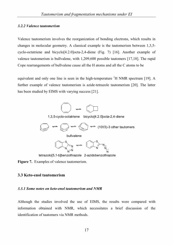
3.2.2 Valence tautomerism
Valence tautomerism involves the reorganization of bonding electrons, which results in
changes in molecular geometry. A classical example is the tautomerism between 1,3,5-
cyclo-octatriene and bicyclo[4.2.0]octa-2,4-diene (Fig. 7) [16]. Another example of
valence tautomerism is bullvalene, with 1,209,600 possible tautomers [17,18]. The rapid
Cope rearrangements of bullvalene cause all the H atoms and all the C atoms to be
equivalent and only one line is seen in the high-temperature 1H NMR spectrum [19]. A
further example of valence tautomerism is azide-tetrazole tautomerism [20]. The latter
has been studied by EIMS with varying success [21].
bullvalene
(10!/3)-3 other tautomers
N
SN
NN
2-azidobenzothiazoletetrazolo[5,1-b]benzothiazole
N
S
N NN
1,3,5-cyclo-octatriene bicyclo[4.2.0]octa-2,4-diene
Figure 7. Examples of valence tautomerism.
3.3 Keto-enol tautomerism
3.3.1 Some notes on keto-enol tautomerism and NMR
Although the studies involved the use of EIMS, the results were compared with
information obtained with NMR, which necessitates a brief discussion of the
identification of tautomers via NMR methods.

Tautomerism and fragmentation mechanisms under EI
18
Calculation of the relative amounts of keto and enol tautomers are based on the integral
intensities of the signals of H atoms α to CO group (-CH2-CO). For 2-phenacylpyridines
and 2-phenacylquinolines, the hydrogen exchange in solution is slow because in one of
the tautomers the H is bound to a neutral C atom. However, the hydrogen exchange is not
too slow to enable the differentiation of tautomers through the 15N and 13C chemical shifts
[22a,23,24].
In the case of fast H transfer between the individual tautomers, each nucleus gives only
one averaged signal. If the tautomers contain strongly electronegative basic centers such
as O and N, integration of the 1H NMR signals with a view to estimating the amounts of
tautomers is useless [25].
In 8-aryl-3,4-dioxo-2H,8H-6,7-dihydroimidazo[2,1-c][1,2,4]triazines, the occurrence of
amide-imidol tautomerism can be expected, but this requires H transfer between two
basic centers. The 1H NMR signal for an enolic/imidic H is slightly broadened, and the
signal at 11.42-11.89 ppm for this rather acidic H indicates that the equilibrium in
solution favors the 3-oxo form rather than the 3-hydroxy form [26].
3.3.2 Some notes on substituent effects on keto-enol tautomerism
The acidicity of an H α to the CO group is an important factor in keto-enol tautomerism.
The keto form is usually more favored and the enol form is rapidly tautomerized back to
the keto form. The more acidic the H, the more the equilibrium favors the enol form. The
solvent polarity also strongly affects the keto-enol equilibrium [27], polar solvents
generally favoring the keto form and apolar solvents the enol form. If the H is α to two
CO groups, the enol form becomes more favored because of the inductive electron
withdrawal of the two CO groups (Fig. 8).

Tautomerism and fragmentation mechanisms under EI
19
C
O Oδ+
CH
O OH
H H Figure 8. The inductive electron-withdrawal of two CO groups.
The tautomerism also depends on changes in π-electron conjugation. Conjugated double-
bonds help to stabilize the enol form. Another stabilizing factor is the formation of
internal H-bonds [28]. Steric crowding between the CO group and the substituents may
affect the relative amounts of the tautomers, as does the electrostatic repulsion between
two polar functionalities. This was observed in the tautomeric equilibria of cyclic α-nitro
ketones [29].
Electron-accepting groups destabilize the keto form and stabilize the enol form by
electron delocalization. This is seen, for example, in the enolization of 3-nitrobutan-2-one
in comparison with 2-butanone [30]. The substituents attached to aromatic groups affect
the conjugated system with resonance and inductive effects by repelling (electron-
donating groups or electron donors) or attracting electrons (electron-accepting groups or
electron acceptors) (Fig. 9).
X X X X
Electron donating group
X X
Electron accepting group
X X
Figure 9. Effect of electron donating and electron accepting groups on the aromatic ring.

Tautomerism and fragmentation mechanisms under EI
20
3.3.3 Keto-enol tautomerism in 2-phenacylpyridines [I] and 2-phenacylquinolines [VI]
The two series of compounds studied which most probably exhibit keto-enol tautomerism
were 2-phenacylpyridines and 2-phenacylquinolines.
2-Phenacylpyridines (K, ketimine form) are in equilibrium with (Z)-2-(2-hydroxy-2-
phenylvinyl)pyridines (O, enolimine form). The third possible tautomer (E, enaminone
form) is not detected in CDCl3 solution [23] or in aqueous solution [31,32]. The presence
of an intramolecular H-bond stabilizes form O [33]. With different substituents the
amount of form K in CDCl3 solution varies from the 99% for electron-donating
substituents to 7.8% for electron-accepting substituents [23]. This wide range makes 2-
phenacylpyridines ideal for a study in the gas phase, because at least the compounds
containing the strongest electron-donating or accepting groups may be expected to furnish
different mass spectrometric fragmentations.
In the presence of a strong electron-donating substituent in the aromatic ring form K of 2-
phenacylpyridines or -quinolines can attain the mesomeric form K’ besides form K form
(K↔K’) Similarly, forms E and O have mesomeric forms E’ and O’, respectively (Fig.
10) [23]. However, the increased stabilization of forms E’ and O’ by H-bonding is
contrasted by the lost aromaticity of both the benzene ring and the pyridine (Py) ring (in
2-phenacylpyridines) or the quinoline (Qui) ring (in 2-phenacylquinolines). In form K’
the Py and Qui rings remain aromatic [34]. Form E which has no possibility for an
internal H-bonding (e.g. in 2-ketomethylquinolines [35]) is usually less stable than form
K. Electron-accepting substituents make the methylene H atoms adjacent to the CO group
more acidic and their transfer to aza atoms becomes more favorable [34].

Tautomerism and fragmentation mechanisms under EI
21
N N
R
R
N
R
O
OH
OH
K O
E
1a-n R1=R2=H2a-h R1=R2=benzo
N
OH
RE'
N
O
RK'
N
OH
R
O'
Aromatic
Not aromatic
Not aromatic
Not aromatic
R2
R1
R2
R1
R2
R1
Not aromatic
Not aromatic
R2
R1
R2
R1
R2
R1
Figure 10. The stabilizing effect of the electron-donating substituent on ketimine form K’
and its destabilizing effect on enaminone forms E’ and O’.
On the other hand, benzo-annelated 2-phenacylquinolines do not prefer tautomer K since
its amount in solution is clearly less than for 2-phenacylpyridines, the maximum value
being 39.3% for the p-N(CH2)4-substituted compound (in CDCl3 solution, 303 K) [24].
X-ray crystallographic studies reveal only the presence of form E in the solid state [22a].
The effect of an intramolecular H-bond on form E stronger than that of an electron-
donating substituent on form K. Although the intramolecular H-bonding in form E is
weaker than that in form O, the π-electron delocalization is more effective in the former
[34].
The theoretical heats of formation of forms E and K, based on AM1 calculations [22b]
for some of the 2-phenacylquinolines studied [22a], are valid for isolated molecules in the
gas phase. Thus, the p-F-, p-Cl-, m-F- and p-CF3-substituted 2-phenacylquinolines prefer
form E in the gas phase. AM1 calculations reveal that form K has a lower heat of
formation than that of form E in the cases of the p-NMe2, p-OMe, p-Me and m-Me

Tautomerism and fragmentation mechanisms under EI
22
derivatives [22a]. Form E is therefore expected to be present in compounds with electron-
accepting substituents under mass spectrometric conditions.
3.3.4 Hammett substituent constants
The Hammett constants σ were first obtained from the ionization of organic acids in
solution. They are defined as:
0loglog KK −=σ
where K0 is the ionization constant of benzoic acid and K is the corresponding constant
for m- (σm) or p-substituted benzoic acid (σp). These constants have been successfully
used to compare the electronic effects of substituents on the rates and equilibria of
organic reactions [36]. Taft extended these principles to polar, steric and inductive and
resonance effects [37-40].
The substituents may push or pull electrons inductively or by resonance. The substituent
constants σp and σm can be split into field/inductive (σI) and resonance (σR) components
[38]:
σp = σI + σR
The field effect is the phenemenon that a charge separation will influence the energy
associated with the development of charge elsewhere in the molecule as a result of
through-space electrostatic interactions. The inductive effect means a transmission of
bond dipoles through the intervening bonds by successive polarization of each bond. The
field and inductive effects together are regarded as polar effects, expressed by substituent
constant σF [41]. However, the field effect outweighs the inductive effect [42,43].
Accordingly, σI is mainly due to the field effect component [44].
There are various ways to establish σI, such as the use of ionization constants for bicyclo-
octane carboxylic acids [45] or quinuclidines [46]. The tabulated σI (≡ F) values are
calculated from the results of the above two methods [47].

Tautomerism and fragmentation mechanisms under EI
23
For the resonance effect parameter R (≡σR), Swain and Lupton [48] made the assumption
that
σp = ασI + σR
The coefficient α does not differ much from 1 [47], and thus the resonance effect
parameter σR can be expressed as
σR = σp - σI
This definition of σR applies only to para substituents. The difference between σm and σp
for a given substituent is due to the possible difference between inductive (σI) and
resonance (σR) effects. The sensitivity to resonance effects is much larger for para than
for meta substituents [48]. Resonance contributions are present mainly with ortho and
para substituents, but ortho substituents are excluded from the Hammett treatment
because of steric effects [41]. Despite the fact that in general the resonance effects cannot
be taken as equal to zero, the constants for meta substituents are close to the
field/inductive parameters (σm ≈ σI).
Other contributions to the Hammett substituent constants are made by polarizibility (σα)
and electronegativity (σχ) effects [49]. However, in this work only the resonance and
field/inductive effects on tautomerism will be discussed.
When substituents are conjugated with a reaction center, the correlations with σp are poor.
σ+ constants were developed therefore for better representation of electrophilic reactions
where strong resonance occurs between electron-donating substituents and positively
charged reaction centers [50]. The positive charge may be located in the aromatic ring
and the conjugated substituent helps to delocalize the charge. Correspondingly σ−
constants are used for reactions where the substituent delocalizes the negative charge. In
literature the σp− constants differ from σp only for substituents that can accept electrons by
resonance (such as NO2 and CN) and conversely σp+ constants differ only for electron
donating substituents (such as NH2 or OMe) [48].

Tautomerism and fragmentation mechanisms under EI
24
The σp+ and σp
− values can be used to define resonance constants R+ and R−. However the
R− values for strong π-donating substituents are questionable as a result of strong
conjugation with electron-rich reaction centers [47]. Also for strong π-electron-accepting
groups with π-electron deficient centers R+ values are uncertain [49].
3.3.5 Hammett substituent constants and quantitative structure–activity relationships
(QSARs)
Hammett functions are among the variables used to study QSARs. In some cases, the
constants σ have been correlated with biological activity. The biological activity of
benzenesulfonamides against Escherichia coli and Mycobacterium smegmatis proved to
be correlated with σ [51], as was that of 2-hydroxy-6-methyl-7-arylamino-1,7-
dihydropurin-8-one against Agrobacterium tumefaciens and Arthobacter globiformis [52].
For diethyl phenyl phosphates, a correlation was found between the inhibition of insect
cholinesterase and σ [53]. However, in general problems arise with the application of
Hammett-type relationships to biological systems. This is because biological systems are
also affected by other factors, one of the most important being lipophilicity [54a].
Lipophilicity is predicted by log P, where P is the partition coefficient, i.e. the ratio of the
concentration of a compound as a neutral molecule in a hydrophobic organic solvent
(octanol) to its concentration in the aqueous phase [54b].
3.3.6 Biologically active heterocycles and pharmaceutical effects
In many biological and enzymatic processes, the rate-determining step is H-transfer [55].
Thus, the minor tautomeric forms of natural bases may play an important role in
substitution mutagenesis during DNA replication, i.e. the mutation caused by the pairing
of wrong base pairs [56,57]. This “rare tautomer hypothesis” is strengthened by the
experimental evidence of a direct correlation between the tautomeric constant (KT =
[amino]/[imino]) and the preferred nucleotide incorporation by the Klenow polymerase
[58]. Theoretical calculations on a base-pair analog N-methyl-P (6-methyl-3,4-dihydro-

Tautomerism and fragmentation mechanisms under EI
25
8H-pyrimido[4,5-c][1,2]oxazin-7-one) additionally point to the role of rare tautomers in
mutagenesis during DNA replication [59].
Enaminones (i.e. β-enaminones, compounds containing the conjugated system N-C=C-
C=O), which are possible pro-drugs [60-62] and therefore important intermediates in
organic synthesis, have also been reported to possess biological activity [63,64]. They are
interesting model compounds, with two basic centers and three possible tautomers. Some
2-phenacylpyridines have been noted to have anti-bacterial properties [65]. Enaminones
are used as intermediates in the synthesis of biologically active compounds, such as
oxytocin antagonists or compounds with anti-epileptic, molluscicidal or larvicidal
activities [66,67]. In this work, two series of compounds, phenacylpyridines and
phenacylquinolines, with possible enaminone and enolimine tautomers were subjected to
an EI study.
Fused imidazoline ring systems containing dioxo groups have been found to exert
analgesic opioid-like action without narcotic analgesic side-effects. The presence of two
polar CO groups and one hydrophobic moiety has been suggested to be responsible for
serotonergic activity, reducing the “head twitch” episodes in mice after 5-
hydroxytryptophan administration [68]. In the 8-aryl-3,4-dioxo-2H,8H-6,7-
dihydroimidazo[2,1-c][1,2,4]triazines studied in this work, amido-imido tautomerism is
possible. The tautomeric equilibrium has been observed to affect pharmacological
activity. In aqueous solution the p-Cl-substituted compound favors the enol form, and the
m-Cl-substituted one the keto form [26]. The p-Cl compound displays a serotonergic
effect and also acts on the opioid receptors. On the other hand, the m-Cl compound has an
antinociceptive effect, i.e. it reduces sensitivity to painful stimuli (Fig. 11). The p-Cl
compound exists mainly in the 3-hydroxy form, and accordingly transformation of the 3-
oxo group to a 3-hydroxy group is the main factor affecting the activity [26]. This
conflicts with the earlier model of the two oxo groups influencing the serotonergic
receptors, and means that the models for opioid and serotonergic activity require
adjustment.

Tautomerism and fragmentation mechanisms under EI
26
Figure 11. Possible H-bond-acceptor (HA) and donor (HD) interaction sites for opioid
activity and serotonergic activity of 8-(4-chlorophenyl)-3,4-dioxo-2H,8H-6,7-
dihydroimidazo[2,1-c][1,2,4]triazine [69]. Copyright © Elsevier 2004, reproduced with
permission.
The 8-aryl-3,4-dioxo-2H,8H-6,7-dihydroimidazo[2,1-c][1,2,4]triazines also have other
possible pharmaceutical applications. The p-Cl derivative displays high potency for the
inhibition of LS180 human Caucasian colon adenocarcinoma cells, HeLa Negroid cervix
epitheloid carcinoma and A549 human Caucasian lung cancer cells. The m-Cl derivative
inhibits HeLa cancer cells relatively strongly, but is completely inactive against LS180
and A549 cells. These differences are suggested to arise from the more lipophilic (log P =
1.28) nature of the electron-withdrawing substituent p-Cl [69].
Ar- and benzyl-substituted 2,3-dihydroimidazo[1,2-a]pyrimidine-5,7(1H,6H)-diones [70]
are structural modifications resembling 8-aryl-3,4-dioxo-2H,8H-6,7-dihydro-imidazo[2,1-
c][1,2,4]triazines. The o-MeO substituent has been observed by X-ray diffraction to exist
only as the 5-oxo/7-OH tautomer in the solid state [71]. These compounds may also have
pharmacological effects.
Cyclohexane/ene-fused pyrimido[2,1-a]isoindol-6-ones are of pharmacological
importance because their starting synthons and analogs exhibit biological effects and are

Tautomerism and fragmentation mechanisms under EI
27
applicable in therapy. Quinazolinone derivatives may have hypnotic and sedative
properties, and may be useful as analgesics, sedatives and hypertensives [72,73].
3.4 Fragmentation mechanisms in EIMS
3.4.1 General aspects
The formation of molecular ions follows the Franck-Condon principle [74], i.e. the
ionization is a fast vertical process. When an electron transition caused by an electron
beam or a photon beam occurs, the time for the transition is extremely short compared to
the vibration between the atoms, and therefore the structure of the molecule does not
change during the ionization.
Mass spectral fragmentations are well explained by the quasi-equilibrium theory (QET),
at least when the impact energy is sufficiently higher than the appearance energy or the
molecules are not very small [75]. The fragmentation takes a longer time than the
redistribution of energy to the different degrees of freedom. It requires the conversion of
internal electronic energy acquired during ionization into vibrational and rotational
energies [76a]. When the oscillating molecular ion has a sufficient amount of energy it
undergoes the fragmentation reaction. The fragments may have sufficient energy to
dissociate through a similar sequence of events, and the rearrangements of bonds may
also occur [76b]. Another theory similar to the QET, but for neutral molecules, is the
Rice-Ramsperger-Kassel-Marcus theory of unimolecular gas reactions in which the rate
at which the energized reactant molecule breaks down is treated as a function of the
energy that it contains, and the normal-mode vibrations and rotations too are taken into
account [77].
The EI fragmentation of the molecular ion produces a positively charged ion and a neutral
fragment (radical or molecule). Typical EI fragmentations result from a single-bond
cleavage where a radical is lost from the molecular ion via a σ-bond, homolytic or
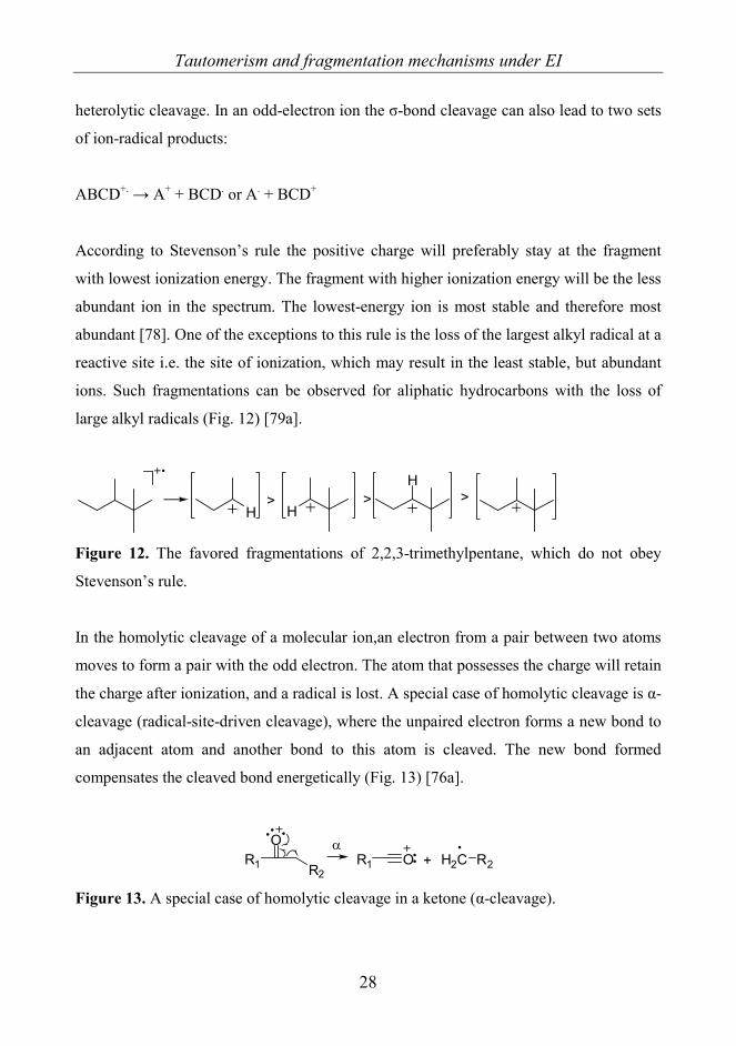
Tautomerism and fragmentation mechanisms under EI
28
heterolytic cleavage. In an odd-electron ion the σ-bond cleavage can also lead to two sets
of ion-radical products:
ABCD+. → A+ + BCD. or A. + BCD+
According to Stevenson’s rule the positive charge will preferably stay at the fragment
with lowest ionization energy. The fragment with higher ionization energy will be the less
abundant ion in the spectrum. The lowest-energy ion is most stable and therefore most
abundant [78]. One of the exceptions to this rule is the loss of the largest alkyl radical at a
reactive site i.e. the site of ionization, which may result in the least stable, but abundant
ions. Such fragmentations can be observed for aliphatic hydrocarbons with the loss of
large alkyl radicals (Fig. 12) [79a].
H>
H>
H>
Figure 12. The favored fragmentations of 2,2,3-trimethylpentane, which do not obey
Stevenson’s rule.
In the homolytic cleavage of a molecular ion,an electron from a pair between two atoms
moves to form a pair with the odd electron. The atom that possesses the charge will retain
the charge after ionization, and a radical is lost. A special case of homolytic cleavage is α-
cleavage (radical-site-driven cleavage), where the unpaired electron forms a new bond to
an adjacent atom and another bond to this atom is cleaved. The new bond formed
compensates the cleaved bond energetically (Fig. 13) [76a].
R1
O
R2R1 O H2C R2+
α
Figure 13. A special case of homolytic cleavage in a ketone (α-cleavage).
Tautomerism and fragmentation mechanisms under EI
29
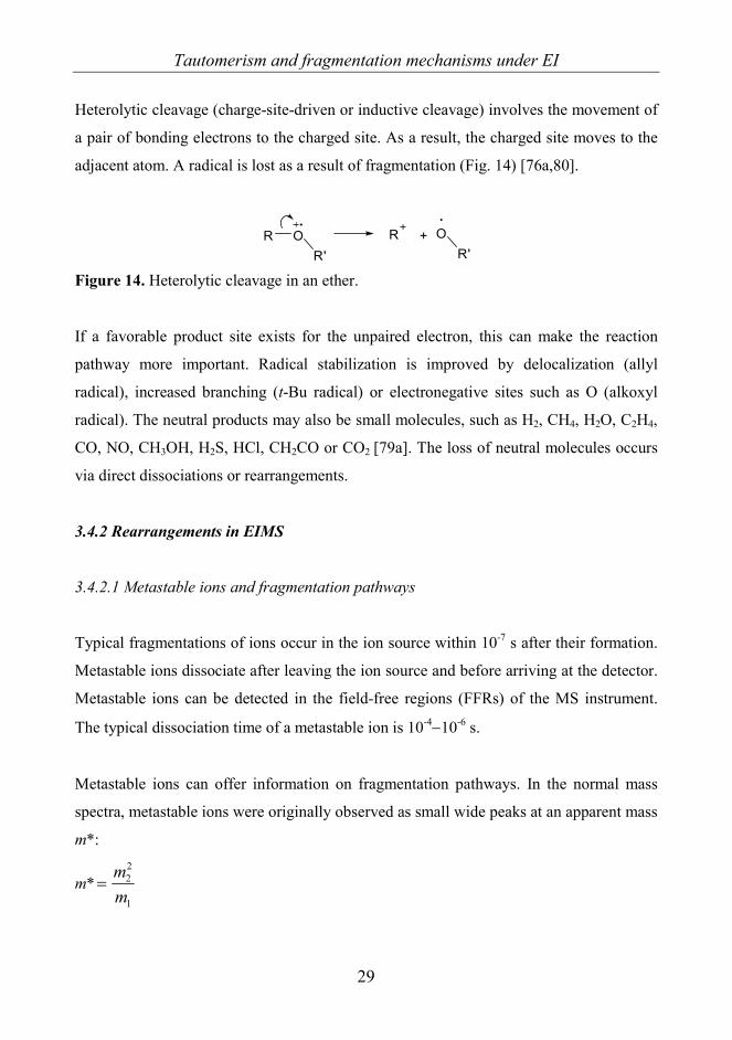
Heterolytic cleavage (charge-site-driven or inductive cleavage) involves the movement of
a pair of bonding electrons to the charged site. As a result, the charged site moves to the
adjacent atom. A radical is lost as a result of fragmentation (Fig. 14) [76a,80].
R OR'
R + OR'
Figure 14. Heterolytic cleavage in an ether.
If a favorable product site exists for the unpaired electron, this can make the reaction
pathway more important. Radical stabilization is improved by delocalization (allyl
radical), increased branching (t-Bu radical) or electronegative sites such as O (alkoxyl
radical). The neutral products may also be small molecules, such as H2, CH4, H2O, C2H4,
CO, NO, CH3OH, H2S, HCl, CH2CO or CO2 [79a]. The loss of neutral molecules occurs
via direct dissociations or rearrangements.
3.4.2 Rearrangements in EIMS
3.4.2.1 Metastable ions and fragmentation pathways
Typical fragmentations of ions occur in the ion source within 10-7 s after their formation.
Metastable ions dissociate after leaving the ion source and before arriving at the detector.
Metastable ions can be detected in the field-free regions (FFRs) of the MS instrument.
The typical dissociation time of a metastable ion is 10-4−10-6 s.
Metastable ions can offer information on fragmentation pathways. In the normal mass
spectra, metastable ions were originally observed as small wide peaks at an apparent mass
m*:
m*1
22
mm
=

Tautomerism and fragmentation mechanisms under EI
30
where m1 = mass of the precursor ion and m2 = mass of the product ion. The ions are
presumed to have a single charge.(z1 = z2 = 1). In theory, m* can be used to calculate the
masses for product ions. However, the abundance of metastable ions is often too low to
be seen in the normal mass spectra, so pathways are nowadays solved by using better
methods.
For a double-focusing mass spectrometer, the metastable transitions can be utilized by
linked scans. The product ions formed from a selected precursor ion can be identified by
keeping the ratio of the magnetic field B and the electrostatic field strength E constant;
this experiment is known as a linked scan at constant B/E [81a]. Similarly, the precursor
ions of selected product ions can be identified with a linked scan at constant B2/E [81a].
The fragmentation of precursor ions can be made more efficient and variable by
increasing the internal energy with a collision cell in the FFRs by introducing a collision
gas, such as He. This method is called collision-induced dissociation (CID) [81b].
3.4.2.2 Distonic ions
Distonic radical ions are odd electron ions in whích the radical and charge sites are
separated. They are important intermediates and products in dissociation reactions of
organic molecules. Distonic ions result from rearrangements, such as H-migration. They
can also be formed via ring opening. X- and γ-ray radiation too have been observed to
produce distonic radical cations [82a]. Some simple routes to distonic ions are presented
in Fig. 15 [82a].
O O
ethylene oxide
OO
HOH
CH2O+
1,2-dimethoxyethane
HONH2
-CH2ONH3
2-aminoethanol
trimethyl phosphate
OP
OOO
isomerization OHP
OOO
Figure 15. Examples of formation of distonic ions.

Tautomerism and fragmentation mechanisms under EI
31
3.4.2.3 Rearrangements
Rearrangement reactions make the mass spectra more complex to interpret, but they may
also yield information on stereochemical and structural problems. Gas-phase radical
cations that have low internal energy often dissociate via rearrangement processes.
Rearrangements tend to be reactions with low activation energies, while simple cleavages
require higher energies [83a-b]. The ions from rearrangements can be very abundant in EI
spectra, because of their low activation energies [83c]. Rearrangements are usually
associated with multiple-bond cleavages and the formation of new bonds, which requires
a favorable conformation. Due to the large negative activation entropies, the
rearrangement reactions are slower than simple cleavage reactions [83b-c], and they may
therefore occur in the metastable ion time frame.
There are numerous types of rearrangements, and only some of them can be discussed
here as examples. The most common and best-understood rearrangements are H-
rearrangements [79b]. Typically, an H atom moves away to another location within the
ion. One bond is broken and another bond is formed. An example of an H-rearrangement
is the McLafferty rearrangement (Fig. 16) [84a,b]. The H atom is transferred to a radical
cation site via a six-membered cyclic intermediate. A distonic radical cation is formed,
where the charge site and radical site are separated. The rearrangement is then followed
by charge- or radical-site-driven cleavage.
OR H ORH
ORH
ORH
ORH
radical-site-drivenrearrangement
charge-site-drivenrearrangement
Figure 16. McLafferty rearrangement.

Tautomerism and fragmentation mechanisms under EI
32
Displacement reactions are energetically favored since one bond is formed in
compensation for the one cleaved [79b]. The displacement arrangements may involve the
loss of halogen or alkyl radicals, resulting in cyclic cations (Fig. 17). Et
O Et O
O Me
+
O Me Figure 17. A displacement reaction of methyl (2Z)-2-heptenoate.
Elimination reactions involve the migration of H or some functional group with the
elimination of small stable neutrals. One example of an elimination with H-transfer is the
1,4-elimination of water from an alcohol and another is alkenyl radical elimination from
the dimethyl ester of cyclopentanediol (Fig. 18) [85a].
ROH
H OR
H
H2O+
O
O
Me
Me
O Me
O MeO
O Me
Me+C4H7H
R
Figure 18. 1,4-Elimination of water from an alcohol and elimination of an alkenyl radical
from the dimethyl ester of cyclopentanediol.
3.4.3 CO loss under EI
The loss of a CO molecule under EI is often observed in the mass spectra of diketones,
phenols, acetamides, esters, aromatic epoxides and chalcones. The ion [M–CO]+• has
variable relative abundances (RAs), ranging from very low to high. The loss of CO from
acyclic molecular ions containing a CO group (acyclic ketones) requires rearrangements
and/or cyclic intermediates.
Tautomerism and fragmentation mechanisms under EI
33
Acetylacetone (CH3COCH2COCH3) has been observed to exhibit the loss of CO, with a
RA of 10% [86]. This fragmentation requires the migration of a Me group. In
comparison, benzoylacetone (C6H5COCH2COCH3) does not exhibit CO loss, but the
presence of the tropylium ion indicates some phenyl migration [86]. For 2,2-dimethyl-3,5-
hexanedione, the migration of a t-Bu group involving an intermediate ion/neutral
complex has been suggested as the mechanism of CO loss [87].
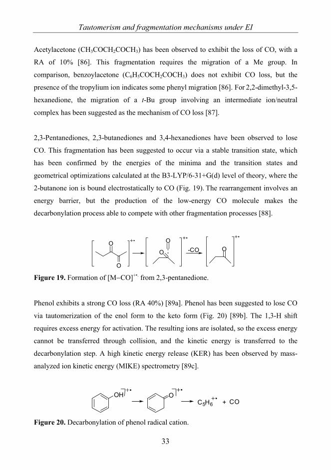
2,3-Pentanediones, 2,3-butanediones and 3,4-hexanediones have been observed to lose
CO. This fragmentation has been suggested to occur via a stable transition state, which
has been confirmed by the energies of the minima and the transition states and
geometrical optimizations calculated at the B3-LYP/6-31+G(d) level of theory, where the
2-butanone ion is bound electrostatically to CO (Fig. 19). The rearrangement involves an
energy barrier, but the production of the low-energy CO molecule makes the
decarbonylation process able to compete with other fragmentation processes [88].
O
OO
O
O-CO
Figure 19. Formation of [M−CO]+•. from 2,3-pentanedione.
Phenol exhibits a strong CO loss (RA 40%) [89a]. Phenol has been suggested to lose CO
via tautomerization of the enol form to the keto form (Fig. 20) [89b]. The 1,3-H shift
requires excess energy for activation. The resulting ions are isolated, so the excess energy
cannot be transferred through collision, and the kinetic energy is transferred to the
decarbonylation step. A high kinetic energy release (KER) has been observed by mass-
analyzed ion kinetic energy (MIKE) spectrometry [89c].
OH OC5H6 + CO
Figure 20. Decarbonylation of phenol radical cation.
Tautomerism and fragmentation mechanisms under EI
34
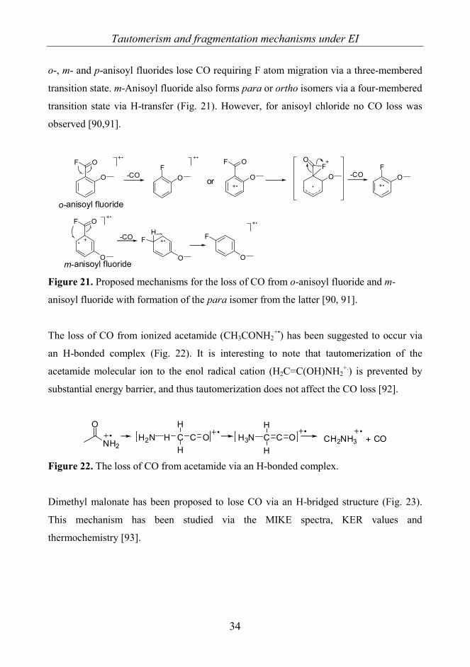
o-, m- and p-anisoyl fluorides lose CO requiring F atom migration via a three-membered
transition state. m-Anisoyl fluoride also forms para or ortho isomers via a four-membered
transition state via H-transfer (Fig. 21). However, for anisoyl chloride no CO loss was
observed [90,91].
F O
O -COF
O
o-anisoyl fluoride
m-anisoyl fluoride
F O
O
-CO
O
FH
O
F
or
F O
O -COF
OOF
O
Figure 21. Proposed mechanisms for the loss of CO from o-anisoyl fluoride and m-
anisoyl fluoride with formation of the para isomer from the latter [90, 91].
The loss of CO from ionized acetamide (CH3CONH2+•) has been suggested to occur via
an H-bonded complex (Fig. 22). It is interesting to note that tautomerization of the
acetamide molecular ion to the enol radical cation (H2C=C(OH)NH2+.) is prevented by
substantial energy barrier, and thus tautomerization does not affect the CO loss [92].
O
NH2H2N H C C H3N C CO O CH2NH3 + CO
H
HH
H
Figure 22. The loss of CO from acetamide via an H-bonded complex.
Dimethyl malonate has been proposed to lose CO via an H-bridged structure (Fig. 23).
This mechanism has been studied via the MIKE spectra, KER values and
thermochemistry [93].
Tautomerism and fragmentation mechanisms under EI
35
O
O
O
OH H
H
O
O
O
OH H
H
O
O
O
O
H
HH
-COO
O
O
H
HH
-CH2OO
OH
Figure 23. Proposed mechanism of CO loss from dimethyl malonate [93].
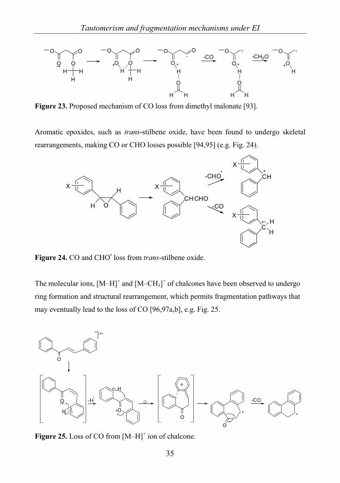
Aromatic epoxides, such as trans-stilbene oxide, have been found to undergo skeletal
rearrangements, making CO or CHO losses possible [94,95] (e.g. Fig. 24).
H
H
O
X
CHCHO
XCH
X
C
X
H
H
-CHO
-CO
Figure 24. CO and CHO• loss from trans-stilbene oxide.
The molecular ions, [M–H]+ and [M–CH3]+ of chalcones have been observed to undergo
ring formation and structural rearrangement, which permits fragmentation pathways that
may eventually lead to the loss of CO [96,97a,b], e.g. Fig. 25.
O
-H
O
H
-CO
OO
O
H
Figure 25. Loss of CO from [M–H]+ ion of chalcone.
Tautomerism and fragmentation mechanisms under EI
36
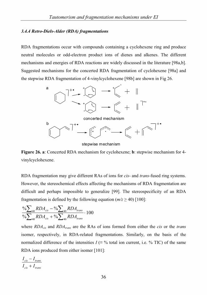
3.4.4 Retro-Diels-Alder (RDA) fragmentations
RDA fragmentations occur with compounds containing a cyclohexene ring and produce
neutral molecules or odd-electron product ions of dienes and alkenes. The different
mechanisms and energies of RDA reactions are widely discussed in the literature [98a,b].
Suggested mechanisms for the concerted RDA fragmentation of cyclohexene [98a] and
the stepwise RDA fragmentation of 4-vinylcyclohexene [98b] are shown in Fig 26.
concerted mechanism
stepwise mechanism
a
b
Figure 26. a: Concerted RDA mechanism for cyclohexene; b: stepwise mechanism for 4-
vinylcyclohexene.
RDA fragmentation may give different RAs of ions for cis- and trans-fused ring systems.
However, the stereochemical effects affecting the mechanisms of RDA fragmentation are
difficult and perhaps impossible to generalize [99]. The stereospecificity of an RDA
fragmentation is defined by the following equation (m/z ≥ 40) [100]:
100%%%%
40 40
40 40 ⋅+
−
∑ ∑∑ ∑
transcis
transcis
RDARDARDARDA
where RDAcis and RDAtrans are the RAs of ions formed from either the cis or the trans
isomer, respectively, in RDA-related fragmentations. Similarly, on the basis of the
normalized difference of the intensities I (≡ % total ion current, i.e. % TIC) of the same
RDA ions produced from either isomer [101]:
transcis
transcis
IIII
+−

Tautomerism and fragmentation mechanisms under EI
37
There have been attempts to explain EI-induced RDA reactions by comparing the degrees
of substitution at the bonds cleaved in the fragmentation. Compounds can be classified as
involving low, medium or high degrees of substitution (Fig. 27) [100]. The degree of
substitution is related to the critical energy of RDA fragmentation. The definition of
substitution is somewhat blurred, but the critical energy differences of cis and trans
isomers can still be used to explain RDA stereospecificity [100,101]. The critical energy
differences between cis and trans isomers cause the medium-substituted compounds to
give stereospecific RDA fragmentations, while the low and high-substituted compounds
exhibit low stereospecificity. When the degree of substitution is high, the critical energies
for RDA are low for both the cis and trans isomers, and when the degree of substitution is
low, the critical energies are high. For medium-substituted compounds, the critical energy
for trans isomers increases relative to that for cis isomers, leading to stereospecific RDA
fragmentations [100].
O
O
H
O
O
H
O
O
H
O
O
H
O
O
H
O
O
H
High
Medium
Medium
Low-medium
Low-medium
Low
Figure 27. Examples of fused cyclohexene systems with high, medium or low degrees of
substitution [100]. The sites mostly defining the classification are highlighted.
There are also results which indicate that the stereospecificity of an RDA process may
depend more on the molecular geometry (cis vs trans annelation) rather than the
substitution in the cyclohexene ring being cleaved [101]. Moreover, the stereospecificity
of the RDA reaction indicates a concerted single-step fragmentation mechanism [102].
RDA fragmentions may involve H-transfers. The even-electron dienophile cations
resulting from RDA fragmentations accompanied by H transfer are referred to either as
Tautomerism and fragmentation mechanisms under EI
38
(RDA+H) or as (RDA–H), corresponding to the addition of H to or the removal of H
from the dienophile, respectively [101]. The RDA+/–H processes and also RDA+2H or
RDA–2H are multi-step processes and often stereospecific [103,104].
In addition to the purely MS processes, RDA fragmentations may also occur via thermal
decomposition. Thermal decomposition may be problematic for methods requiring
vaporization of the sample by heating prior to ionization. Fast-atom bombardment or
liquid secondary ion MS methods may be more useful than EI for the study of regio- and
stereospecific RDA fragmentations because the samples are ionized at ambient
temperatures [105].
3.5 MS and keto-enol tautomerism
3.5.1 Some notes on MS and tautomerization
Although MS methods have long been used for structural investigations of organic
compounds, their application for the study of tautomerism in the gas phase has only
recently been recognized. The keto-enol tautomerism of β-diketones was the first case
studied by this means [87,106-109].
Different ionization energies and inlet temperatures were used to investigate their effects
on fragmentations related to keto-enol tautomerism of variously substituted β-diketones
[110]. The intensities I of the peaks (i.e. RA or % TIC) originating from the pure keto and
enol forms were presumed to obey the modified van’t Hoff equation CRT
HK +Δ
−=ln :
[ ][ ] )(lnlnln aC
RTHaKa
ketoenol
II
keto
enol ++Δ
−=+=+=
where K is the equilibrium constant for keto-enol equilibria, ΔH is the enthalpy difference
between the enol and keto forms, T is the absolute temperature, R is the gas constant, and
C and a are constants [110].

Tautomerism and fragmentation mechanisms under EI
39
The source temperature affects the tautomer ratio. At higher source temperatures for 2-
pentanone, it was observed that the amount of the enol tautomer increased [111]. Another
noteworthy fact was that the peak intensities depended not only on the tautomerism but
also on the differences in bond strengths [110].
In studies of tautomerism with MS, two important facts should be born in mind [11]:
1. The assignments of mass spectral fragmentations should be tautomer-specific, since the
corresponding abundance ratios should correlate to the keto/enol contents.
2. Ionization in the ion source is postulated to have no effect on the position of the
equilibrium, so that the results reflect the tautomer contents in the gas phase prior to
ionization.
The identification of peaks formed exclusively from either the keto or the enol form is
necessary to permit conclusions relating to the tautomerism [11]. The EI fragmentations
of β-ketoesters have been studied by GC-MS in an attempt to separate the tautomers, but
the problem was the non-negligible interconversion of the tautomers inside the column
[112]. However, the enol and keto forms of methyl and ethyl acetoacetate could be
separated by making use of GC retention times and mass spectra. It was seen that the
intermolecular stabilization of the enol form was higher for α-chloromethyl and α-
chloroethyl acetoacetate, which resulted in more enol form being present in the gas phase;
this indicates the effect of the electron accepting Cl substituent on the tautomeric
equilibria [112].
It is assumed that the equilibrium established at a certain temperature in the inlet system
will not be changed in the ion source, as the vapor pressure inside the mass spectrometer
is too low for the molecules to take part in collisions [110]. The energy barrier of
unimolecular isomerization from ketone to enol is high. Once formed, therefore, the
tautomer should retain its original structure in the gas phase, irrespective of the relative
stabilities of the isomers [113]. However, the radical cations may have sufficient energy
for tautomerization.

Tautomerism and fragmentation mechanisms under EI
40
The degree of enolization of ketones of the type R1(C=O)CHR2R3 is generally favored by
the increase of the steric effect exerted by the substituent at the position α to the CO
group. In general, the loss of OH from the molecular ion is assigned to the enol form and
the loss of R to the keto form, where R is the radical moiety that participates in the
enolization process next to the CO group (CHR2R3). Ion RA ratios [(M–R)+]/[(M–OH)+]
of selected ketones have been correlated with semi-empirical AM1 MO calculations of
the approximate equilibrium constants of enolization [114]. However, the stabilization of
the enol form by conjugation may lead to the absence of [M–R]+.
The loss of OH can also be used for the identification of other prototropic tautomers. For
example, supportive fragmentations and rearrangements have been found for imidol
forms of amides and their sulfur analogs, thioamides, such as the loss of H, OH/SH,
H2O/SH2, and the double H (McLafferty+1)-type rearrangement [115.] For lactones or
their sulfur analogs the OH/SH loss indicates the presence of the enol form, and the loss
of CX or CX2 (X = O or S) that of the keto form [116].
3.5.2 The tautomerization of molecular ions
Some mechanisms for the interconversion of molecular ions of the tautomers have been
observed.
Radical cations of phenol can tautomerize if they are sufficiently activated to undergo CO
loss [89]. The molecular ion of phenol, M+•, can acquire sufficient excess energy, with
ionization energies of 50-70 eV, whereas [M-CO]+• vanishes at energies < 15 eV [117a].
As mentioned earlier the excess energy from ionization is changed to kinetic energy when
CO is lost, and this compensates the energy required for the tautomerization reaction.
KER measurements on sterically crowded triaryl-substituted enols show that the enol
radical cations isomerize to excited ketones in a rate-determining step prior to
fragmentation (Fig. 28). This is achieved by a greater KER from the enol than from the
keto form [118].

Tautomerism and fragmentation mechanisms under EI
41
Ar2
Ar3
Ar1
OH
rate-determiningstep Ar2
Ar3
Ar1
OH
Ar2
Ar3
Ar1
O
Ar2
Ar3+
Ar1
O
Figure 28. The tautomerization of a triaryl-substituted enol radical cation.
The single and double McLafferty (or McLafferty+1) rearrangement have been studied
using deuterium-labeled ketones [117b]. It has been stated that the McLafferty
rearrangement of aliphatic ketones can produce enolic radical cations rather than keto
ions [117a,b] (Fig. 29). The enolization is simultaneous with the loss of alkene.
OH
H
HO
HO
HOH
OH H
singleMcLafferty ion
doubleMcLafferty ion
loss of alkene
O
keto ion (less favored)
no McLaffertyrearrangement
loss of alkene
McLaffertyrearrangement
Figure 29. The mechanisms for fragmentation of a ketone with a single McLafferty
rearrangement with a consecutive double McLafferty rearrangement [117a].
In general, the tautomerization of radical cations is quite rare, and the tautomerization of
neutral molecules is more important than that of radical cations. For example, the ion
abundances of lactones and related compounds have been correlated with the differences
in heats of formation between the keto and enol forms of the neutral molecules [116].

Tautomerism and fragmentation mechanisms under EI
42
This means that the impact of radical cations on tautomerization is at its minimum for
lactones and their sulfur analogs.
It would seem that the interpretation of MS results is not as straightforward as was once
believed. Although the effects of solvents and intermolecular interactions can be avoided,
new variables such as the source temperature and ionization energy appear together with
reactions of radical cations. Despite this MS can yield important information in studies of
tautomerism in the gas phase, especially when the results are compared with those of
theoretical semiempirical calculations.
3.6 Materials and methods
3.6.1 MS measurements
All measurements were made in the Instrument Centre in the Department of Chemistry at
the University of Turku between 2003 and 2008. The EI mass spectra were recorded on a
VG ZABSpec oaTOF mass spectrometer (VG Analytical, Division of Fisons,
Manchester, UK) equipped with the Opus V3.3X program package (Fisons Instruments,
Manchester, UK). The ionization energy was 70 eV and the source temperature was 160
°C. The acceleration voltage was 8 kV and the usual trap current was 200 μA.
Perfluorokerosine was used for calibration of the mass scale. A small amount of solid
sample dissolved in MeOH was placed into a quartz capillary tube and the MeOH was
evaporated off with hot air. Thereafter, the sample was transferred into the ionization
chamber via the solid inlet. The probe was sometimes heated in order to evaporate the
samples.
The fragmentation pathways were solved on the basis of B/E-linked scans (first field-free
region, i.e. FFR1) and in some cases also B2/E. The low-resolution spectra and B/E scans
were measured with a resolving power of 3000 (10% valley definition). The accurate
masses were determined by voltage scanning, at a resolving power of 6,000−10,000 for
small m/z values and > 10,000 for the larger values. Also collision induced dissociation

Tautomerism and fragmentation mechanisms under EI
43
(CID) was used to inspect the fragmentation pathways; He was applied as collision gas in
the FFR1. The gas flow was adjusted so that the beam transmission was 50%. Orthogonal
acceleration time-of-flight (oaTOF) measurements were made in some cases.
3.6.2 NMR measurements
Most of the compounds have been characterized earlier with NMR methods. However,
the NMR spectra for 2,3-dihydroimidazo[1,2-a]pyrimidine-5,7(1H,6H)-diones were
recorded and analyzed in our department by Dr. Henri Kivelä [IV]. The latter spectra
were acquired with a Bruker Avance 500 NMR spectrometer (Bruker BioSpin
Scandinavia AB, Taby, Sweden) operating at 500.13 MHz for 1H and at 125.77 MHz for 13C, equipped with a vendor-provided 5-mm direct or inverse detection Z-gradient probe
(BBO-5mm-Zgrad or BBI-5mm-Zgrad-ATM, respectively), the probe temperature set at
298 K. Because of some solubility problems in CDCl3, deuteriated dimethyl sulfoxide
(DMSO-d6) was used as solvent. The 1H spectra were referenced to internal SiMe4 (0.00
ppm) and the 13C spectra to the middle resonance line of the DMSO solvent signal (39.40
ppm). A standard one-dimensional (1D) 1H NMR spectrum and a 13C spectrum with
broad-band proton decoupling were run on each sample, supplemented by 2D gradient-
selected correlation spectroscopy (COSY), nuclear overhauser enhancement spectroscopy
(NOESY), multiplicity-edited heteronuclear single quantum correlation (HSQC) and
heteronuclear multiple bond correlation (HMBC) experiments for selected samples to
help with the assignment of signals. Vendor-provided pulse sequences were used
throughout the work. For NOESY, a mixing time of 0.3 s was employed, and the
heteronuclear experiments were optimized for a one-bond C,H coupling of 145 Hz and a
long-range coupling of 8–10 Hz.
3.6.3 Linear fits and structures of molecules
The linear functions for 2-phenacylpyridines were calculated by using linear regression
on the Origin 6.0 package (Microcal Software, Inc., Northampton, MA, USA).

Tautomerism and fragmentation mechanisms under EI
44
The linear functions for 2-phenacylquinolines were calculated by using linear regression
on the Origin 8 SR1 package (OriginLab Corporation, Northampton, MA, USA). As
default Origin 8 gives the coefficient of determination as adjusted R2. The value of
adjusted R2 increases only if the new term improves the model more than would be
expected by chance.
The structures of molecules in sections 4.3.2 and 4.5.2 have been drawn with the
ChemOffice Ultra 10.0 package (Cambridgesoft Corporation, Cambridge, Massachusetts,
USA) using Chem3D Pro 10.0 for MM2 energy minimizations.
3.6.4 The compounds studied
The compounds studied were obtained from different research groups. 2-
Phenacylpyridines 1a−n [I] (Scheme 1, p. 45) and 2-phenacylquinolines 2a−h [VI]
(Scheme 2, p. 46) were received from Prof. Ryszard Gawinecki (Department of
Chemistry, University of Technology and Life Sciences, Bydgoszcz, Poland), 8-aryl-3,4-
dioxo-2H,8H-6,7-dihydroimidazo[2,1-c][1,2,4]triazines 3a−j [II] (Scheme 6, p. 60) and
Ar- and benzyl-substituted 2,3-dihydroimidazo[1,2-a]pyrimidine-5,7(1H,6H)-diones
18−21 [IV] (Scheme 10, p. 73) from Prof. Dariusz Matosiuk (Department of Synthesis
and Chemical Technology of Pharmaceutical Substances, Professor Feliks Skubiszewski
Medical University, Lublin, Poland), pyrrolo- and isoindolo-quinazolinones 4−17 [III]
(Scheme 8, p. 65, and Table 8, p. 66) from Prof. (Emeritus) Géza Stájer (Institute of
Pharmaceutical Chemistry, University of Szeged, Hungary), and naphthoxazine,
naphthpyrrolo-oxazinone and naphthoxazino-benzoxazine derivatives 22−29 [V]
(Scheme 11, p. 79, and Table 14, p. 80) from Prof. Ferenc Fülöp (Institute of
Pharmaceutical Chemistry, University of Szeged, Hungary).
The syntheses have been published for 1a−n [119,120], 2a−h [125], 3a−j [26,121], 4−17
[122], 18−21 [70] and 22−29 [123,124].

Results and discussion
45
4. RESULTS AND DISCUSSION 4.1 2-Phenacylpyridines 1a−n [I] and 2-phenacylquinolines 2a−h [VI] 4.1.1 General fragmentations 2-Phenacylpyridines 1a−n (Scheme 1) and 2-phenacylquinolines 2a−h were selected for
study because strong effects of the substituents on the tautomeric equilibria were
expected to be seen in the gas phase as different fragmentations or different abundances
of ions for forms K, O and E. The results were also thought to give information about the
presence of internal hydrogen bonding in the gas phase. For 2-phenacylpyridines form E
is theoretically possible, but only forms K and O, i.e. (Z)-2-(2-hydroxy-2-
phenylvinyl)pyridine, have been observed. 2-Phenacylquinolines 2a−h (Scheme 2)
resemble the 2-phenacylpyridines, but instead of form O the other tautomer besides form
K is E, i.e. (Z)-2-benzoyl-methylene-1,2-dihydroquinoline. For 2-phenacylpyridines form
E and for 2-phenacylquinolines form O are not detected in solvents or in the solid state.
N N
R
R
N
R
O
OH
OH
K
O
E Scheme 1. Structures and possible tautomers of 2-phenacylpyridines 1a−n: R = a: H, b:
m-Me, c: p-Me, d: p-NH2, e: m-F, f: p-F, g: p-OMe, h: p-Cl, i: p-N(Me)2, j: p-NO2, k: p-
CF3, l: p-N(CH2)4, m: p-Br, n: m-Br. K = 2-phenacylpyridine (ketimine), O = (Z)-2-(2-
hydroxy-2-phenylvinyl)pyridine (enolimine), E = (Z)-1,2-dihydro-2-benzoylmeth-
ylenepyridine (enaminone).

Results and discussion
46
N
O
RK
N
O RH
E
N
OR
H
O Scheme 2. 2-Phenacylquinolines 2a−h and their possible tautomers. R = a: p-N(CH2)4, b:
p-NMe2, c: p-OMe, d: p-Me, e: m-Me, f: p-Cl, g: p-Br, h: p-CF3. K = 2-
phenacylquinoline (ketimine), E = (Z)-1,2-dihydro-2-benzoylmethylenequinoline
(enaminone), O = (Z)-2-(2-hydroxy-2-phenylvinyl)quinoline (enolimine).
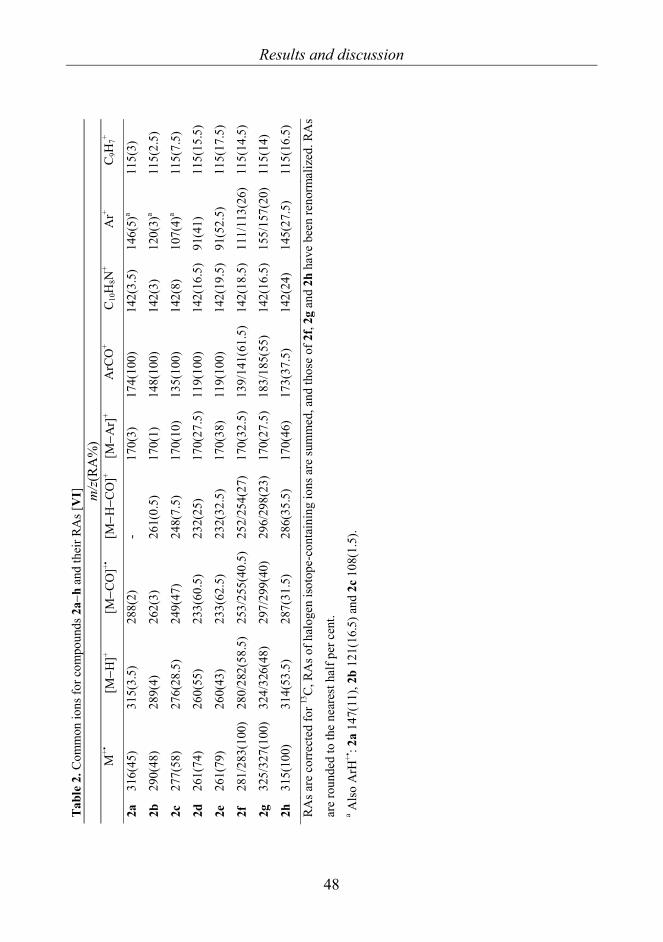
Common fragment ions with their RAs are presented for 1a−n in Table 1 [I] and for 2a−h
in Table 2 [VI], and their typical fragmentation pathways are illustrated in Schemes 3 and
4, respectively.

Results and discussion
47
Tab
le 1
. Com
mon
ions
for c
ompo
unds
1a−
n an
d th
eir R
As [
I].
m/z
(RA
%)
M
+•
[M−H
]+ [M
−OH
] +
[M−C
O] +
· [M
−HC
O] +
Py
CH
2CO
+ A
rCO
+ A
r+ Py
CH
2+ C 5
H5+
1a
197(
26)
196(
34.5
) 18
0(2)
16
9(47
) 16
8(33
) 12
0(9)
10
5(10
0)
77(6
1)
92(1
1.5)
65
(10)
1b
211(
22)
210(
28.5
) 19
4(3.
0)
183(
51)
182(
34)
120(
10.5
) 11
9(10
0)
91(6
0)
92(1
0.5)
65
(26)
1c
211(
13)
210(
20)
194(
1)
183(
39.5
) 18
2(20
) 12
0(4.
5)
119(
100)
91
(49)
92
(10.
5)
65(2
0)
1d
212(
14.5
) 21
1(3.
5)
195(
0.5)
18
4(14
.5)
183(
3)
120(
100)
a 12
0(10
0)a
92(2
2)
92(2
2)
65(1
7)
1e
215(
45.5
) 21
4(42
) 19
8(3)
18
7(55
) 18
6(43
.5)
120(
18)
123(
100)
95
(60)
92
(20)
65
(14)
1f
215(
9)
214(
11)
198(
1)
187(
24.5
) 18
6(14
) 12
0(3.
5)
123(
100)
95
(42)
92
(5.5
) 65
(5)
1g
227(
7)
226(
5.5)
21
0(0.
5)
199(
24)
198(
4.5)
12
0(0.
5)
135(
100)
10
7(5)
92
(14.
5)
65(4
)
1h
231/
233(
19)
230/
232(
23.5
) 21
4/21
6(2.
5)
203/
205(
39)
202/
204(
26)
120(
6.5)
13
9/14
1(10
0)
111/
113(
39)
92(9
) 65
(9)
1i
240(
24)
239(
2)
223(
1)
212(
2)
211(
1)
120(
0.5)
14
8(10
0)
120(
4)
92(3
) 65
(3.5
)
1j
242(
100)
24
1(57
) 22
5(3.
5)
214(
50.5
) 21
3(58
) 12
0(32
) 15
0(46
) 12
2(+)
92
(48.
5)
65(2
2)
1k
265(
72)
264(
75)
248(
4.5)
23
7(54
.5)
236(
52)
120(
27.5
) 17
3(10
0)
145(
69)
92(2
8)
65(1
8)
1l
266(
23)
265(
2)
249(
+)
238(
1.0)
23
7(0.
5)
120(
+)
174(
100)
14
6(4)
92
(1)
65(3
)
1m
275/
277(
46)
264/
276(
40.5
) 25
8/26
0(2.
8)
247/
249(
58.5
) 24
6/24
8(35
) 12
0(10
.5)
183/
185(
100)
15
5/15
7(37
.5)
92(1
4)
65(1
3)
1n
275/
277(
62.5
) 27
4/27
6(43
.5)
258/
260(
4.5)
24
7/24
9(62
.5)
246/
248(
55)
120(
24.5
) 18
3/18
5(96
.5)
155/
157(
51)
92(2
3)
65(2
0)
RA
s are
cor
rect
ed fo
r 13C
, RA
s of h
alog
en is
otop
e-co
ntai
ning
ions
are
sum
med
, and
thos
e of
1h,
1m
and
1n
have
bee
n re
norm
aliz
ed. R
As
are
roun
ded
to th
e ne
ares
t hal
f per
cen
t. a E
lem
enta
l com
posi
tion
C7H
6NO
+ .
Results and discussion
48
Tabl
e 2.
Com
mon
ions
for c
ompo
unds
2a−
h an
d th
eir R
As [
VI]
m
/z(R
A%
)
M+•
[M
−H]+
[M−C
O]+•
[M
−H−C
O]+
[M−A
r]+
ArC
O+
C10
H8N
+ A
r+ C
9H7+
2a
316(
45)
315(
3.5)
28
8(2)
-
170(
3)
174(
100)
14
2(3.
5)
146(
5)a
115(
3)
2b
290(
48)
289(
4)
262(
3)
261(
0.5)
17
0(1)
14
8(10
0)
142(
3)
120(
3)a
115(
2.5)
2c
277(
58)
276(
28.5
) 24
9(47
) 24
8(7.
5)
170(
10)
135(
100)
14
2(8)
10
7(4)
a 11
5(7.
5)
2d
261(
74)
260(
55)
233(
60.5
) 23
2(25
) 17
0(27
.5)
119(
100)
14
2(16
.5)
91(4
1)
115(
15.5
)
2e
261(
79)
260(
43)
233(
62.5
) 23
2(32
.5)
170(
38)
119(
100)
14
2(19
.5)
91(5
2.5)
11
5(17
.5)
2f
281/
283(
100)
28
0/28
2(58
.5)
253/
255(
40.5
) 25
2/25
4(27
) 17
0(32
.5)
139/
141(
61.5
) 14
2(18
.5)
111/
113(
26)
115(
14.5
)
2g
325/
327(
100)
32
4/32
6(48
) 29
7/29
9(40
) 29
6/29
8(23
) 17
0(27
.5)
183/
185(
55)
142(
16.5
) 15
5/15
7(20
) 11
5(14
)
2h
315(
100)
31
4(53
.5)
287(
31.5
) 28
6(35
.5)
170(
46)
173(
37.5
) 14
2(24
) 14
5(27
.5)
115(
16.5
)
RA
s are
cor
rect
ed fo
r 13C
, RA
s of h
alog
en is
otop
e-co
ntai
ning
ions
are
sum
med
, and
thos
e of
2f,
2g a
nd 2
h ha
ve b
een
reno
rmal
ized
. RA
s
are
roun
ded
to th
e ne
ares
t hal
f per
cen
t. a A
lso
ArH
+•: 2
a 14
7(11
), 2b
121
(16.
5) a
nd 2
c 10
8(1.
5).

Results and discussion
49
N
O
R
-H
[M-H]
-CO-HCO
[M-CO]
[M-H-CO]or
[M-HCO]
-OH.
R
O
ArCO
R
N
O
N CH2
PyCH2CO
PyCH2
-C6H6N-Ar
-PyCH2
-ArCO
-CO
-C6H5N
M
[M-OH]
-C6H5N
Ar
-CO
-CO
Scheme 3. The general fragmentation pathways of 1a−n.
N
O
R-H.
-OH.[M-H]+
[M-OH]+
[M-HCO]+[M-CH3]+
2c-h-CO
-HCO.
[M-CO]+.
C16H11N+.
-HR
2c-h-(H+CO+R) C11H8NO+
-Ar.
C9H7+
C10H8N+
-CO-(CO+HCN)
-HCN
2d-fArCO+
ArH+.
-C10H8N.
-C11H7NO
2d-g-R.
C17H12NO+
-CO
Ar+
2a,b,c-CH3
.
2b-h
2c-e-CH3
.
2c,d,e
*traces for 2b,f-h
2d,e
-H.
[M-CH3-CO]+
-C11H8NO .
m/z 217m/z 115
m/z 170
m/z 142
m/ z 246
C10H6N+
m/z1402a,2c-h
-R.
[M-CO-CH3]+-HCO
[M-CH3-HCO]+.2c-e
-H2
*
-CO-CO
2f-h-HR C6H3
+
m/z 75trace for 2b
2d,2e-C2H2
C5H5+ (2d,2e)
m/ z 65
2b,2c-CH3
. C7H7N+. (2b)
C6H4O+. (2c)
2a-C3H5
.
-C3H6
C7H7N+. m/z 105C7H6N+ m/ z 104
m/z 105
m/z 92
2h-HF
C7H3F2+
m/z 125
2c-CH2O
C6H5+
m/z 77
m/z 190C14H8N+
-C2H3.
2g,h
C9H6N+
m/z 128
-CO+(Ar.-H.)2f-h
M+.
Scheme 4. The general fragmentation pathways of 2a−h.

Results and discussion
50
All compounds studied gave clear M+. peaks; for the p-NO2-substituted compound 1j and
the halogen-substituted 2f−h this was the base peak. It is worth mentioning that the
substituent effects of the NO2 group are different for benzophenone-type molecular ions,
due to the possible alteration in the structure of the NO2 substituent in which the oxygen
atom becomes attached to the same carbon atom to which the nitrogen had earlier been
attached (Fig. 30) [126].
N
O
O
OR
Figure 30. The alteration in the structure of the NO2 substituent.
4.1.2 Ions related to tautomers
There were fragmentations indicating tautomerism for 2-phenacylpyridines 1a−n. The
RAs of [M–OH]+ were slightly higher for compounds with stronger electron acceptors,
i.e. for the compounds most likely to exhibit form O, but in general they were quite low
(RA <7%). For 2-phenacylquinolines 2a−h [M−OH]+ was observed (RA <5%) for all
compounds except 2a, and [M−OH]+ was again slightly more favored with electron-
withdrawing substituents. Only traces of [M–OH]+ were observed for compounds with
strong electron-donating groups. Since the OH loss can in general be assigned to the enol
form [114,116], and since the fragmentation cannot occur from form K, this
fragmentation may indicate the presence of form O or E.
For 2-phenacylpyridines 1a−n and 2-phenacylquinolines 2a−h, the ion Ar+ was found to
be more abundant for electron-withdrawing substituents as were [M−ArCO]+, [M−Ar]+,
[M−H]+ and M+•. The ions Ar+ and [M−Ar]+ may be formed from both tautomers, but
[M−Ar]+ should have different structures (Fig. 31). Although the RA of ArCO+ was 100%
for most compounds, its % TIC decreased for stronger electron-withdrawing substituents,
possibly as an indication of an increased contribution of form O or E. This can be

Results and discussion
51
explained by the dissociation of the bond α to the CO group, which is easier for the σ-
bond of form K than for the double bond of form O (Fig. 31).
Forms O and E seem to favor the formation of [M−Ar]+, and form K that of ArCO+. The
intramolecular H-bond may make the formation of ArCO+ from form O (and also form E)
less favorable. The ion ArH+• from 1i,l and 2a−c may be specific for form K.
N
O
R
K
O
R
ArCO+
N
OR
H
N
OH
[M-Ar]+
O
R1
R2
R1
R2
R1
R2
1: R1=R2=H2: R1=R2=benzo
N
O[M-Ar]+
R1
R2
Figure 31. The formation of ArCO+ from form K and the formation of [M−Ar]+ from
forms K and O.
The RAs of ions [M–CO]+• and [M–HCO]+ were greater for compounds with electron
acceptors. These two losses were weak for the compounds most likely to exist in form K,
i.e. for the compounds 1d,i,l and 2a,b with the most negative substituent constants.
Therefore, the structure of form O or E is favorable for (H)CO loss. The formation of
ions [M–CO]+• and [M–HCO]+ requires rearrangements. CO and HCO losses were
observed for 2-phenacylpyridines 1a−n and 2-phenacyl-quinolines 2a−h; the suggested
mechanism is discussed in section 4.1.6. It should be noted that, for 2b−e, [M−CO]2+ was
present with RAs of 4 to 8%.
Comparisons of compounds presumed to exhibit the keto form have been made with EI
data recorded in the US National Institute of Standards and Technology database [89].

Results and discussion
52
The carbon analogs of 2-phenacylpyridines, e.g. 1,2-diphenylethan-1-one, 1-(4-
bromophenyl)-2-phenylethanone and diphenylethanedione, give very weak molecular
peaks and no [M−H]+ or [M−CO]+• is noted. None of these compounds contains an
internal H-bond that would stabilize the enol form, and thus only the keto tautomer is
present in the gas phase.
The RAs of the molecular ions for 1j and 2f−2h, with the strongest electron acceptors,
were 100%.Increase of the molecular ion may be another indication of the increasing
form E or O. The molecular ion of a stable enol form is often more abundant than that of
the keto form [112,127,128]. The reason for this behavior may be that the positive enol
ions are thermodynamically more stable than the corresponding keto ions [129].
4.1.3 Correlations with Hammett σ for 2-phenacylpyridines 1a−n
Hammett σp or σm functions were used to correlate the RAs and % TICs of ions. Even
better correlations were obtained by omitting the inductive parameter from the
calculations and using the resonance effect parameter σR. The best correlations are
presented in Table 3, and the linear fit of ArCO+ (% TIC vs σp or σm) is depicted in Fig.
32. The presence of the ion ArCO+ seems to be related to form K, and that of the ions
[M−H]+, [M−CO]+•, [M−HCO]+ and [M−Ar]+ ions to form O.
Dual-substituent parameter analysis of the equilibrium constant KT, based on the NMR
results, has shown that the resonance substituent effect predominates over the inductive
effect [23]. The better correlations of the ion RAs with σR than with σp or σm indicate that
the substituents of 2-phenacylpyridines may affect the tautomerism mainly by resonance
in the gas phase.

Results and discussion
53
Table 3. RAs or % TICs of the ions from 1a−n which correlate with the Hammett
constants σ (σm or σp) or σR and the parameters for the linear fits.
Parameters for y = a + bx
Ion y x a ± error b ± error R Notes
ArCO+ % TIC σm or σ p 26.2±1.2 −(23.1±2.8) -0.926
ArCO+ % TIC σR 18.2±2.0 −(30.3±4.4) -0.924
[M–H]+ % TIC σm or σ p 5.44±0.45 5.7±1.0 0.859
[M–H] + % TIC. σR 7.9±0.5 8.5±1.1 0.943
[M–CO]+. RA σm or σ p 38.7±3.0 34.0±6.8 0.834
[M–CO]+. RA σR 49.0±3.1 45.7±6.8 0.922
[M–HCO]+ RA σm or σ p 27.4±2.4 39.6±5.5 0.907
[M–HCO]+ RA σR 49.0±3.2 45.7±6.8 0.922
[M–HCO]+ % TIC σm or σ p 5.5±0.4 5.3±0.9 0.876
[M–HCO]+ % TIC σR 7.2±0.4 7.5±0.8 0.961
PyCH2CO+ % TIC σm or σ p 1.6±0.2 4.0±0.6 0.912 1d and 1i excluded
PyCH2CO+ % TIC σR 3.0±0.3 5.5±1.0 0.920 1d and 1i excluded
Ar+ RA σR 57.4±2.8 53.4±9.1 0.966 1g and 1j excluded
[M−H]+ log10(RA) σR 1.62±0.05 1.44±0.11 0.978
[M−HCO]+ log10(RA) σR 1.61±0.07 1.60.±0.13 0.974
1l values are excluded because of missing σ values for the pyrrolidino substituent.

Results and discussion
54
Figure 32. ArCO+ (% TIC) vs σm or σp for 2-phenacylpyridines 1a−n excluding 1l.
4.1.4 Correlations with Hammett σ for 2-phenacylquinolines 2a−h
For the 2-phenacylquinolines the RAs and % TICs correlated with the Hammett
substituent constants σm, σp, σR and σ+ and resonance constant R+ (Tables 4 and 5).
Pyrrolidino-substituted 2a was excluded from the calculations, since its σ values in the
literature were obtained by using a different method [130]. In fact, the σp or σR values of
pyrrolidino are slightly smaller than those of NMe2. The RAs of common ions from 2a
and 2b were generally similar. The linear fit of ArCO+ (% TIC) vs σm or σp is presented in
Fig. 33.

Results and discussion
55
Table 4. RAs or % TICs of the ions from 2b−h which correlate with the Hammett
constants σ (σm or σp) or σR and the parameters for the linear fits.
Parameters for y = a + bx Fragment ion y x a±error b±error Adjusted R2 [M−H]+ RA σ 43±5 37±10 0.690 [M−H]+ RA σR 58±5 50±10 0.819 [M−H]+ % TIC σ 7.7±0.6 5.9±1.5 0.734 [M−H]+ % TIC σR 10±1 7.4±1.8 0.777 [M−HCO]+ RA σ 23±3 24±7 0.653 [M−HCO]+ RA σR 30±2 32±4 0.951 [M−HCO]+ % TIC σ 3.9±0.4 4.2±0.9 0.806 [M−HCO]+ % TIC σR 5.3±0.3 5.4±0.5 0.962 ArCO+ % TIC σ 16±1 -22±3 0.935 ArCO+ % TIC σR 8.2±1.9 -28±4 0.911 C10H8N+ RA σ 16±2 15±3 0.796 C10H8N+ RA σR 21±1 19±2 0.971 C10H8N+ % TIC σ 2.8±0.2 2.2±0.3 0.922 C10H8N+ % TIC σR 3.6±0.2 2.7±0.4 0.931 C9H7
+ RA σ 13±2 10±4 0.594 C9H7
+ RA σR 16±1 14±2 0.921 C9H7
+ % TIC σ 2.3±0.2 1.4±0.3 0.800 C9H7
+ % TIC σR 2.8±0.1 1.8±0.2 0.941 [M−Ar]+ RA σ 28±3 31±8 0.733 [M−Ar]+ RA σR 37±3 40±5 0.947 [M−Ar]+ % TIC σ 4.7±0.4 5.3±0.9 0.854 [M−Ar]+ % TIC σR 6.5±0.4 6.6±0.8 0.937 2a is excluded because of missing σ values for the pyrrolidino substituent. σR values available only for para substituents.
ArCO+ seems to be related to form K, while [M−H]+, [M−HCO]+ and [M−Ar]+ are related
to form E or O. In contrast with the situation in 2-phenacylpyridines (presumed to attain
form O), the RA of [M−CO]+• did not correlate with the substituent constants for 2-
phenacylquinolines (form E), even when the RAs of [M−CO]2+ were included. This may
be due to the different conjugation of form E relative to that of form O.
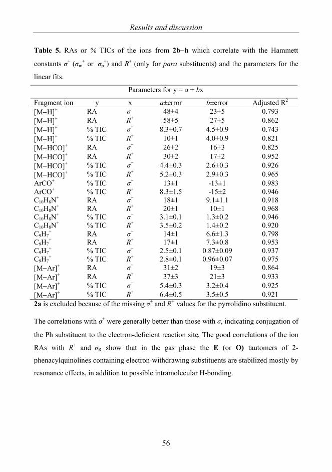
Results and discussion
56
Table 5. RAs or % TICs of the ions from 2b−h which correlate with the Hammett
constants σ+ (σm+ or σp
+) and R+ (only for para substituents) and the parameters for the
linear fits.
Parameters for y = a + bx
Fragment ion y x a±error b±error Adjusted R2 [M−H]+ RA σ+ 48±4 23±5 0.793 [M−H]+ RA R+ 58±5 27±5 0.862 [M−H]+ % TIC σ+ 8.3±0.7 4.5±0.9 0.743 [M−H]+ % TIC R+ 10±1 4.0±0.9 0.821 [M−HCO]+ RA σ+ 26±2 16±3 0.825 [M−HCO]+ RA R+ 30±2 17±2 0.952 [M−HCO]+ % TIC σ+ 4.4±0.3 2.6±0.3 0.926 [M−HCO]+ % TIC R+ 5.2±0.3 2.9±0.3 0.965 ArCO+ % TIC σ+ 13±1 -13±1 0.983 ArCO+ % TIC R+ 8.3±1.5 -15±2 0.946 C10H8N+ RA σ+ 18±1 9.1±1.1 0.918 C10H8N+ RA R+ 20±1 10±1 0.968 C10H8N+ % TIC σ+ 3.1±0.1 1.3±0.2 0.946 C10H8N+ % TIC R+ 3.5±0.2 1.4±0.2 0.920 C9H7
+ RA σ+ 14±1 6.6±1.3 0.798 C9H7
+ RA R+ 17±1 7.3±0.8 0.953 C9H7
+ % TIC σ+ 2.5±0.1 0.87±0.09 0.937 C9H7
+ % TIC R+ 2.8±0.1 0.96±0.07 0.975 [M−Ar]+ RA σ+ 31±2 19±3 0.864 [M−Ar]+ RA R+ 37±3 21±3 0.933 [M−Ar]+ % TIC σ+ 5.4±0.3 3.2±0.4 0.925 [M−Ar]+ % TIC R+ 6.4±0.5 3.5±0.5 0.921 2a is excluded because of the missing σ+ and R+ values for the pyrrolidino substituent. The correlations with σ+ were generally better than those with σ, indicating conjugation of
the Ph substituent to the electron-deficient reaction site. The good correlations of the ion
RAs with R+ and σR show that in the gas phase the E (or O) tautomers of 2-
phenacylquinolines containing electron-withdrawing substituents are stabilized mostly by
resonance effects, in addition to possible intramolecular H-bonding.
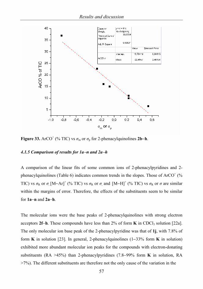
Results and discussion
57
Figure 33. ArCO+ (% TIC) vs σm or σp for 2-phenacylquinolines 2b−h.
4.1.5 Comparison of results for 1a−n and 2a−h
A comparison of the linear fits of some common ions of 2-phenacylpyridines and 2-
phenacylquinolines (Table 6) indicates common trends in the slopes. Those of ArCO+ (%
TIC) vs σR or σ, [M−Ar]+ (% TIC) vs σR or σ, and [M−H]+ (% TIC) vs σR or σ are similar
within the margins of error. Therefore, the effects of the substituents seem to be similar
for 1a−n and 2a−h.
The molecular ions were the base peaks of 2-phenacylquinolines with strong electron
acceptors 2f−h. These compounds have less than 2% of form K in CDCl3 solution [22a].
The only molecular ion base peak of the 2-phenacylpyridine was that of 1j, with 7.8% of
form K in solution [23]. In general, 2-phenacylquinolines (1−33% form K in solution)
exhibited more abundant molecular ion peaks for the compounds with electron-donating
substituents (RA >45%) than 2-phenacylpyridines (7.8−99% form K in solution, RA
>7%). The different substituents are therefore not the only cause of the variation in the
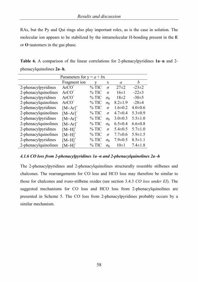
Results and discussion
58
RAs, but the Py and Qui rings also play important roles, as is the case in solution. The
molecular ion appears to be stabilized by the intramolecular H-bonding present in the E
or O tautomers in the gas phase.
Table 6. A comparison of the linear correlations for 2-phenacylpyridines 1a−n and 2-
phenacylquinolines 2a−h.
Parameters for y = a + bx Fragment ion y x a b 2-phenacylpyridines ArCO+ % TIC σ 27±2 -23±2 2-phenacylquinolines ArCO+ % TIC σ 16±1 -22±3 2-phenacylpyridines ArCO+ % TIC σR 18±2 -30±5 2-phenacylquinolines ArCO+ % TIC σR 8.2±1.9 -28±4 2-phenacylpyridines [M−Ar]+ % TIC σ 1.6±0.2 4.0±0.6 2-phenacylquinolines [M−Ar]+ % TIC σ 4.7±0.4 5.3±0.9 2-phenacylpyridines [M−Ar]+ % TIC σR 3.0±0.3 5.5±1.0 2-phenacylquinolines [M−Ar]+ % TIC σR 6.5±0.4 6.6±0.8 2-phenacylpyridines [M−H]+ % TIC σ 5.4±0.5 5.7±1.0 2-phenacylquinolines [M−H]+ % TIC σ 7.7±0.6 5.9±1.5 2-phenacylpyridines [M−H]+ % TIC σR 7.9±0.5 8.5±1.1 2-phenacylquinolines [M−H]+ % TIC σR 10±1 7.4±1.8 4.1.6 CO loss from 2-phenacylpyridines 1a−n and 2-phenacylquinolines 2a−h The 2-phenacylpyridines and 2-phenacylquinolines structurally resemble stilbenes and
chalcones. The rearrangements for CO loss and HCO loss may therefore be similar to
those for chalcones and trans-stilbene oxides (see section 3.4.3 CO loss under EI). The
suggested mechanisms for CO loss and HCO loss from 2-phenacylquinolines are
presented in Scheme 5. The CO loss from 2-phenacylpyridines probably occurs by a
similar mechanism.
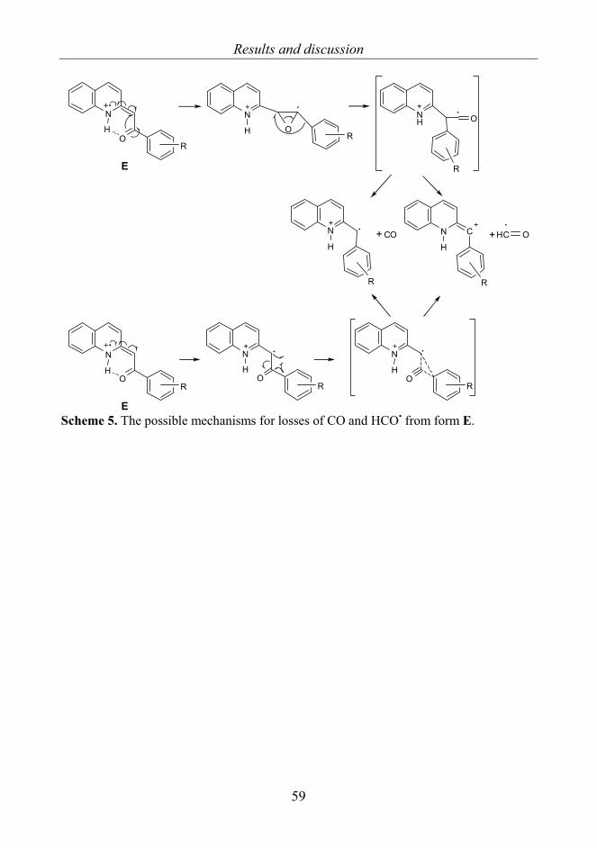
Results and discussion
59
N C
H
N
OH
N
O
NH O
HC O
H
N
HCO+
RR
R
RR
E
N
OH
R
E
N
OR
N
OR
H H
+
Scheme 5. The possible mechanisms for losses of CO and HCO• from form E.

Results and discussion
60
4.2 8-Aryl-3,4-dioxo-2H,8H-6,7-dihydroimidazo[2,1-c][1,2,4]triazines 3a−j [II]
Two tautomers are possible: 8-aryl-2,6,7,8-tetrahydroimidazo[2,1-c][1,2,4]triazine-3,4-
dione (the amide form with a 3-oxo group) and 3-hydroxy-8-aryl-7,8-
dihydroimidazo[2,1-c][1,2,4]triazin-4(6H)-one (the imidol form with 3-OH group)
(Scheme 6).
N
N NHN
R
O O
N
N NN
R
O OH
A B
8
34
76
5
219
Scheme 6. Structures of compounds 3a−j (R = a: H, b: o-Me, c: p-Me, d: o,m-diMe, e: o-
OMe, f: p-OMe, g: o-Cl, h: m-Cl, i: p-Cl, j: m,p-diCl) and possible tautomeric forms A/B.
A = 8-aryl-3,4-dioxo-2H,8H-6,7-dihydroimidazo[2,1-c][1,2,4]triazine (amide form), B =
3-hydroxy-8-aryl-7,8-dihydroimidazo[2,1-c][1,2,4]triazin-4(6H)-one (imidol form). The
numbering used for the assignment of NMR signals is depicted for A.
As noted in section 3.3.6, the NMR methods demonstrate that the 3-oxo form is favored
in solution (DMSO-d6) [26]. With the use of molecular modeling for m- and p-Cl-
substituted compounds it has been calculated that in general the 3-oxo form is more stable
than the 3-OH form in the gas phase. However, in the aqueous solution the heats of
formation for the 3-oxo and 3-OH tautomers indicate that the p-Cl-substituted compound
favors the 3-OH form [26], and thus 3i is the strongest candidate to exhibit
fragmentations related to the enol form.
The 3-oxo and 3-OH tautomers were expected to display different fragmentations, such as
the loss of two CO groups (A) or an OH radical (B). The problem arises from the
situation that the compounds generally favor the A form. Moreover, there is no stabilizing

Results and discussion
61
hydrogen bond even in the B form; this also explains why it is less favorable than the A
form, in contrast to the E or O tautomers of 2-phenacylpyridines and –quinolines which
possess the stabilizing hydrogen bond.
The main ions are listed in Table 7 [II]. The specific fragmentations of Cl-substituted
compounds are presented in Scheme 7.
The molecular ions exhibit the base peaks. Common ions for all compounds 3a–j were
[M−H]+, [M−CO]+•, C9H9N4+ (corresponding to 3d ions C10H11N4
+ and 3j ions
C9H9N4Cl+), Ar+, C3H4NO+, HOCN+• and C2H4N+. Generally the RAs of common ions
generally varied very little for differently substituted compounds. The RA of [M−H]+ was
low for all compounds. No OH loss was detected; this could be an indication of the
absence of the 3-OH form. CO losses were observed (RA <15%) for all compounds
except for 3b with o-Me substituent. For other ortho-substituted compounds the RA of
[M−CO]+• was low. For Cl-substituted compounds an Ar group migration to triazine ring
after CO loss was observed; this was confirmed by losses of C2H5N2•, C3H4N2O and
C3H4N3O• and further fragmentations of the corresponding product ions (Scheme 7). The
fragmentation involving C2H5N2• loss cannot occur if the Ar group remains in its original
position; an H-transfer is also required. Loss of C3H4N2O does not necessarily require H-
transfer.

Results and discussion
62
Tabl
e 7.
Mai
n io
ns, m
/z(%
), fo
rmed
und
er E
I at 7
0 eV
from
com
poun
ds 3
a−j
m/z
(%)
Com
poun
d
M
+•
[M−H
]+ [M
−CO
]+•C
9H9N
4+ A
r+ C
3H4N
O+
HO
CN
+•
C2H
4N+
3a (H
) 23
0(10
0)
229(
3)
202(
15)
173(
5)
77(2
2)
70(1
.5)
43(3
) 42
(4)
3b (o
-Me)
24
4(10
0)
243(
3)
- 17
3(9)
a
187(
2.5)
b 91
(33)
70
(3)
43(1
) 42
(9)
3c (p
-Me)
24
4(10
0)
243(
3.5)
21
6(10
.5)
173(
8)
187(
2.5)
b 91
(12)
70
(1)
43(4
) 42
(5)
3d (o
,m-d
iMe)
25
8(10
0)
257(
3)
230(
0.3)
18
7(9)
c 10
5(11
) 70
(3)
43(1
) 42
(7)
3e (o
−OM
e)
260(
100)
25
9(3)
23
2(0.
3)
173(
8)
203(
2)d
107(
1)
70(2
) 43
(6)
42(5
)
3f (p
-OM
e)
260(
100)
25
9(1.
4)
232(
2.4)
17
3(1.
4)
107(
1)
70(1
) 43
(8)
42(4
) 3g
(o-C
l) 26
6(33
)e
264(
100)
f 26
5(tra
ce)
263(
0.2)
23
8(1.
2)
236(
3.7)
17
3(12
) 11
3(4)
11
1(14
) 70
(4)
43(7
) 42
(8)
3h (m
-Cl)
266(
33)e
264(
100)
f 26
5(2)
26
3(2.
2)
238(
4.4)
23
6(13
.5)
173(
15)
113(
5)
111(
15)
70(3
) 43
(2.5
) 42
(6)
3i (p
-Cl)
266(
32)e
264(
100)
f 26
5(1)
26
3(2)
23
8(3.
5)
236(
10.6
) 17
3(11
) 11
3(4)
11
1(13
.5)
70(3
) 43
(2)
42(5
)
3j (m
,p-d
iCl)
302(
11)g
300(
66)h
298(
100)
i
301(
1)
299(
2.3)
29
7(1.
6)
274(
1.6)
27
2(8.
2)
270(
12.8
)
209(
3)j
207(
8)k
14
9(2)
14
7(7)
14
5(12
)
70(5
) 43
(4)
42(8
)
a Con
tain
s 4%
of C
10H
11N
3+ . b C
10H
11N
4+ . c C
10H
11N
4+ + C
11H
13N
3+ 7:3
. d C
10H
11N
4O+ .
e Ions
in th
is ro
w c
onta
in o
ne 37
Cl a
tom
. f Io
ns in
this
row
con
tain
one
35C
l ato
m.
g Ions
in th
is ro
w c
onta
in tw
o 37
Cl a
tom
s. h Io
ns in
this
row
con
tain
s one
37C
l and
one
35C
l ato
m.
i Ions
in th
is ro
w c
onta
in tw
o 35
Cl a
tom
s. j C
9H8N
437C
l+ . k C9H
8N435
Cl+ .

Results and discussion
63
M[M-Cl]
m/z 2293g (68%)
C11H9N4O2Cl (3g-i)(3j C11H8N4O2Cl2 )
[M-CO] [Ar N(1)] C8H4N2OClm/z 179
(3j: m/z 213)
m/z 151(3j: m/z 185)
-CO
C7H5N2Clm/z 152
(3j: m/z 186)
-HCN
C6H4NClm/z 125
(3j: m/z 159)C5H4Clm/z 99
(3j: m/z 133)
C7H5NClm/z 138
(3j: m/z 172)
C6H4Clm/z 111
(3j: m/z 145)
m/z 173(3j: m/z 207)
3g-i
-C2O2
-C2O2H -C2O2NH
C9H8N4m/z 172
C9H8N3m/z 158
-C2O2N
C9H9N3m/z 159
-HCN2
-C2HN2
C7H4N2Cl
-HCN
-C2H5N2
N
NNH
N
O O
R
-C3H4N2O-C3H4N3O
3g-j
m/z 264(3j: m/z 298)
C9H9N4
Scheme 7. Specific fragmentations for the Cl-substituted derivatives 3g–j (R = g: o-Cl, h:
m-Cl, i: p-Cl, j: m,p-diCl) (m/z values for the m,p-diCl derivative 3j are shown in
parentheses). Ar→N(1) means migration of the Ar moiety to N(1).
For all compounds the ion C9H9N4+ (C10H11N4
+ for o,m-diMe and C9H8N4Cl+ for m,p-
diCl-substituted ones) was observed, corresponding to the loss of substituent R• and two
CO groups. This may be an indication of the prevalence of the keto form in the gas phase,
because the enol form could not lose two CO groups but would instead undergo at least
OH loss. Since 3a (unsubstituted) gives the ion C9H9N4+, it is obvious that the H is lost
from the Ph group, as is the case with the other substituents R.
The presence of HOCN+• gave slight indications of tautomerism. Although its RA was
low (<10%), it increased for p- and m-substituted compounds as an exponential function
of σm and σp, and thus it would seem to be produced from the enol form. However, the
B2/E scans indicate that this HOCN+• is formed from the ion C3H4NO+, which does not
require the presence of the 3-OH form.

Results and discussion
64
The fragmentations involving the losses of CO and COCO generally prove the prevalence
of the amido form in the gas phase, and the absence of [M–OH]+ is a sign that no relevant
amount of the imidol form is present. The minor amounts of [M−HCO]+ (RA 0–2%) may
be indicative of the unfavorable imidol form. Due to the lack of H atoms near the OH
group and also because of the ring structure, the usual rearrangements and
fragmentations, such as the loss of H2O, that would support the presence of the imidol
form cannot occur.
4.3 Pyrrolo- and isoindoloquinazolinones 4−17 [III]
4.3.1. Structures and base peaks
The structures of pyrrolo- and isoindoloquinazolinones 4−17 are shown in Scheme 8 and
the names of the compounds are given in Table 8. The set of pyrrolo- and
isoindoloquinazolinones was studied in order to obtain information on the
stereochemistry of annulated heterocyclic compounds 5,6,10,11,12 and 13. The effects of
unsaturated rings and of aromatic groups on the fragmentations were additionally
investigated. The compounds studied might possibly participate in amide-imidol
tautomerism. However, no indication of such tautomerism was detected.
For 6 and 11 the molecular ion, and for 10 [M−H]+ was the base peak. The other common
base peaks were due either to RDA-related fragmentations (8,9,12,13,16 and 17) or to
loss of the Ar group (7 and 15). For 4 [M-C3H5O]+, for 5 [M−C4H6−C6H4Cl]+ and for 14
[M−C6H4CH3−C4H6]+ were the base peaks. The RAs of the molecular ions were weak for
compounds containing fused norbornane or norbornene skeletons (7,8,9,15,16 and 17).

Results and discussion
65
N
HNArH
HO
O
N
HNArH
HO
O
N
HNAr H
HO
O
N
NH
ArH
H
O
ON
NH
Ar HH
O
O N
NH
Ar HH
O
O
N
HNHH
HO
O
N
HNH
H
HO
O
N
HNHH
HO
O
N
HNH
H
HO
O
N
HNAr
H
HO
O
N
NH
ArH
H
O
O
N
NH
Ar
HH
O
O NHN
Ar HH
O O
Ar = p-chlorophenyl (4-9) or p-tolyl (14-17)
4 5 6
7 8 9
10 11 12
13 14 15
16 17
Scheme 8. Structures of compounds 4−17, pyrroloquinazolinones (4−9) and
isoindoloquinazolinones (10−17), or partly saturated pyrroloquinazolinones (4−6),
benzologs (10−14), methylene-bridged derivatives (7−9, 15 and 16) and a bisacyl
compound (17).
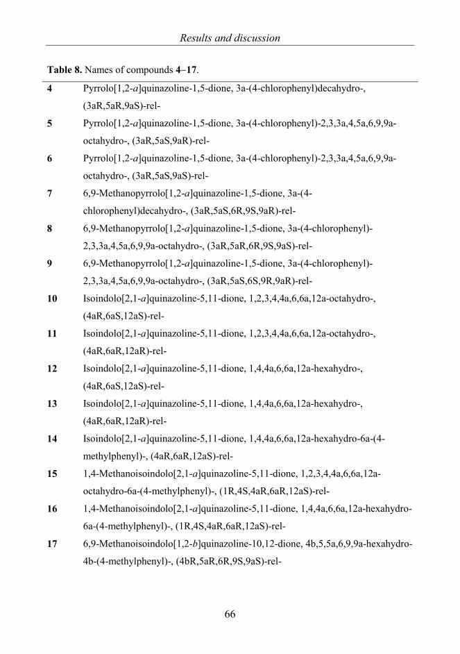
Results and discussion
66
Table 8. Names of compounds 4−17.
4 Pyrrolo[1,2-a]quinazoline-1,5-dione, 3a-(4-chlorophenyl)decahydro-,
(3aR,5aR,9aS)-rel-
5 Pyrrolo[1,2-a]quinazoline-1,5-dione, 3a-(4-chlorophenyl)-2,3,3a,4,5a,6,9,9a-
octahydro-, (3aR,5aS,9aR)-rel-
6 Pyrrolo[1,2-a]quinazoline-1,5-dione, 3a-(4-chlorophenyl)-2,3,3a,4,5a,6,9,9a-
octahydro-, (3aR,5aS,9aS)-rel-
7 6,9-Methanopyrrolo[1,2-a]quinazoline-1,5-dione, 3a-(4-
chlorophenyl)decahydro-, (3aR,5aS,6R,9S,9aR)-rel-
8 6,9-Methanopyrrolo[1,2-a]quinazoline-1,5-dione, 3a-(4-chlorophenyl)-
2,3,3a,4,5a,6,9,9a-octahydro-, (3aR,5aR,6R,9S,9aS)-rel-
9 6,9-Methanopyrrolo[1,2-a]quinazoline-1,5-dione, 3a-(4-chlorophenyl)-
2,3,3a,4,5a,6,9,9a-octahydro-, (3aR,5aS,6S,9R,9aR)-rel-
10 Isoindolo[2,1-a]quinazoline-5,11-dione, 1,2,3,4,4a,6,6a,12a-octahydro-,
(4aR,6aS,12aS)-rel-
11 Isoindolo[2,1-a]quinazoline-5,11-dione, 1,2,3,4,4a,6,6a,12a-octahydro-,
(4aR,6aR,12aR)-rel-
12 Isoindolo[2,1-a]quinazoline-5,11-dione, 1,4,4a,6,6a,12a-hexahydro-,
(4aR,6aS,12aS)-rel-
13 Isoindolo[2,1-a]quinazoline-5,11-dione, 1,4,4a,6,6a,12a-hexahydro-,
(4aR,6aR,12aR)-rel-
14 Isoindolo[2,1-a]quinazoline-5,11-dione, 1,4,4a,6,6a,12a-hexahydro-6a-(4-
methylphenyl)-, (4aR,6aR,12aS)-rel-
15 1,4-Methanoisoindolo[2,1-a]quinazoline-5,11-dione, 1,2,3,4,4a,6,6a,12a-
octahydro-6a-(4-methylphenyl)-, (1R,4S,4aR,6aR,12aS)-rel-
16 1,4-Methanoisoindolo[2,1-a]quinazoline-5,11-dione, 1,4,4a,6,6a,12a-hexahydro-
6a-(4-methylphenyl)-, (1R,4S,4aR,6aR,12aS)-rel-
17 6,9-Methanoisoindolo[1,2-b]quinazoline-10,12-dione, 4b,5,5a,6,9,9a-hexahydro-
4b-(4-methylphenyl)-, (4bR,5aR,6R,9S,9aS)-rel-

Results and discussion
67
4.3.2 Non-RDA-related stereospecific fragmentations
The loss of an Ar group was observed for cyclohexane-, cyclohexene- and norbornane-
fused compounds but not for norbornenes. Of these the cyclohexene derivatives 5 and 14,
in which the Ar group and the annelation H atoms were cis to each other, had the least
abundant ions [M–Ar]+, with RA = 19% and 30%, respectively.
Stereoisomers 5 and 6 have clearly different EI spectra (Fig. 34, Table 9). The molecular
ion and the ion [M–Ar]+ were more abundant for 6. Other ions differentiating 5 and 6
were [M–C7H8NO]+ (C10H9NO35Cl+), [M–C3H5O]+ (C14H12N2O235Cl+), [M–NH3CO]+
(C16H14NO35Cl+) and C4H7N2O+. The ion [M–NH3]+• was slightly more abundant for 5.
The elemental composition of [M–NH3]+• was confirmed by accurate mass
measurements to be C17H14NO235Cl+•, but clarification of the mechanism of its formation
would require extensive isotope labeling.
Table 9. Selected EI-induced ions of stereoisomers 5 (cis) and 6 (trans) and their isotope-
corrected RAs.
M+• [M–C6H4Cl]+ [M–C7H8NO]+ [M–C3H5O]+ [M–NH3CO]+ C4H7N2O+ [M–NH3]+•
5 318(21)
316(70)
205(19) 196(15)
194(50)
261(4)
259(17)
273(8)
271(27)
99(8) 301(2)
299(6)
6 318(31)
316(100)
205(58) 196(30)
194(96)
259(3)
273(2)
271(5)
99(27) 301(<1)
299(2)

Results and discussion
68
Figure 34. The EI mass spectra of 5 and 6.
For pyrrolo[2,1-b][1,3]oxazin-6-one derivatives [131] with cis annelation hydrogens, the
loss of C3H5O• was more favored than for the trans forms. This is suggested to involve a
regiospecific H-transfer to the carbonyl oxygen [131]. For 5 and 6, a corresponding
fragmentation mechanism was observed. From the structure in Fig. 35, it can be seen that
the cis form should favor H-transfer to the carbonyl oxygen more than the trans. In fact, a
more abundant [M–C3H5O]+ is observed for 5 than for 6. In comparison 4 contains a
flexible cycloalkane ring, and [M–C3H5O]+ is the base peak. Norbornane-containing
compound 7 likewise fragmented by the loss of C3H5O•. The loss of C7H5O• for benzo-
fused compounds, corresponding to the loss of C3H5O• without benzo-fusion, was missing
completely.
N
N
O
O
ArH
H
H
H
HH
H
H
HH
H
H
H
N
N
O
O
ArH
H
H
H H
H
H
H
HH
H
HH
5 (cis) 6 (trans) Figure 35. A structural explanation for the stereoselective loss of C3H5O• from 5 and 6.
Structures are optimized using MM2 minimum energy calculations.

Results and discussion
69
The structures of 5 and 6 reveal that the cyclohexene ring in 6 causes more steric strain in
the C6H4Cl group, and as a consequence the ions [M-C6H4Cl]+ and [M–C7H8NO]+ are
more abundant for the trans isomer. These two fragmentations are useful for
differentiating stereoisomers.
For benzo-fused compounds, the ion C8H7NO+• at m/z 133, i.e. a possible 2,3-dihydro-1H-
isoindol-1-one radical cation (Fig. 36) was observed for 10–13; for trans 11,this was
slightly more favorable than for cis 10. C8H7NO+• has also been observed for pyrrolo[2,1-
b][1,3]oxazin-6-ones [131].
NH
O
C8H7NOm/z 133
Figure 36. The 2,3-dihydro-1H-isoindol-1-one radical cation (C8H7NO+•).
Stereoisomers 10 and 11 were also distinguishable (Table 10, Fig. 37). The loss of NH3
from the molecular ion was more abundant for 11 (with trans annelation H atoms). The
composition of [M–NH3]+• was confirmed (C15H13NO2+•) by accurate scans, but the
mechanism for its formation is unknown. [M–C3H7]+ and [M–C2H5]+ were more abundant
for 11. From the structures of 10 and 11, it can be seen that the cyclohexane ring in the cis
form 10 favors the twist-boat conformation, whereas the trans form 11 favors the chair
conformation (Fig. 38). This may be the reason for the difference in the RAs of [M–
C3H7]+ and [M–C2H5]+.

Results and discussion
70
Table 10. Selected EI-induced ions of stereoisomers 10 (cis) and 11 (trans) and their
isotope-corrected RAs.
M+• [M–H]+ [M–NH3]+• [M–C2H5]+ [M–C3H7]+
10 256(97.5) 255(100) 239(4.5) 227(6) 213(20)
11 256(100) 255(82) 239(46) 227(23) 213(74)
Figure 37. The EI mass spectra of 10 and 11.
NO
N
H
O
HH
H
H
H
H
H
H
H H
H
H
H
H
H
N
O
N
H
O
H
H
H
H
H
HH
H
HH
H
H
H
H
H
10 11 Figure 38. MM2 optimized minimum energy structures of 10 and 11.

Results and discussion
71
For stereoisomers 12 and 13, the difference was not so clear. The ions [M–NH3]+• (RA
6% and 1% respectively), C14H11NO+• (RA 4% and 1% respectively), and those at m/z 182
(C13H12N+ for 12 3.5% and C12H8NO+ for 12 3%, traces for 13) were slightly more
abundant for 12, but in general the RAs were very close to each another.
The relative configuration of the Ar group had no effect on the fragmentation of
norbornene-fused compounds 8 and 9.
4.3.3 RDA-related fragmentations
Of the studied compounds, saturated 4,7,10,11 and 15 cannot exhibit RDA-related
fragmentations [III]. The RDA fragmentation may be highly stereospecific, and it was
therefore considered possible to distinguish the stereoisomers via different RAs of RDA-
related fragmentations.
Compounds containing fused cyclohexene rings gave RDA fragments [M–C4H6]+•. The
RDA fragmentations for the cyclohexene-fused stereoisomers 5,6 and 12,13 were non-
stereospecific. Additionally for 12 and 13 the base peak corresponded to the non-
stereospecific (RDA–H)- type fragmentation, leading to the ion [M–C4H7]+.
Norbornene-fused compounds exhibit fragment ions [M–C5H5]+, resulting from RDA
fragmentation with H-transfer (RDA+H). For the stereoisomeric pair 8 and 9, this
fragmentation proved to be non-stereospecific, i.e. the configuration of the Ar group did
not cause any stereospecificity. For norbornene-fused 2,3-dihydrothiazolo[3,2-
a]pyrimidin-5-ones and 3,4-dihydropyrimido[2,1-b]thiazin-6-ones RDA+H
fragmentations were observed, together with normal RDA fragmentations [132].
It is interesting to note that the RDA fragmentations were highly stereospecific for the
pyrrolo[2,1-b][1,3]oxazin-6-one derivatives [131], but not for the structurally similar
pyrrolo- and isoindoloquinazolinones. This is probably caused by stepwise fragmentation
with ring opening involving the CO group (Scheme 9). This ring opening may be similar

Results and discussion
72
to that of trans-3,4,4a,5,8,8a-hexahydro-8a-1(2H)-naphthalenone (or trans-Δ6-octalone-1)
[98a].
N
O
HNO
N
O
HNO
N
O
HNO
+
5,6,12,13 Scheme 9. Possible mechanism for stepwise RDA of pyrrolo- and
isoindoloquinazolinones.
The lack of stereospecificity of RDA or related fragmentations suggests a stepwise RDA
mechanism, at least for compounds 5, 6, 12 and 13. For the compounds containing a
norbornene group, 8 and 9, RDA+H was more favored than the formation of [M–R]+ (R =
H, C6H4CH3 or C6H4Cl). The molecular ions of bicyclic compounds are not very stable,
which is the case for the norbornene and norbornane compounds. The favored RDA-type
fragmentations may be caused by the greater release of the ring strain energy of
norbornene-fused compounds under fragmentation. As compared with the ring strain
energy of cyclohexene, which is estimated to be –1.3 to 10.5 kJ mol-1, the ring strain
energy of norbornene is very large: 80.4–90.4 kJmol-1. In comparison, the ring strain of
cyclohexane is 0–5.7 kJ mol-1 and that of the norbornane ring is 60.3–69.5 kJ mol-1 [133].
The RDA+H process, where the charge remains on the protonated dienophile, seems to
be a general feature of bicyclo[2.2.1]heptene compounds with carbonyl, amide, ester or
imide substituents at positions 5 and 6 [134,135]. It has been suggested that the driving
force for the H-migration for the RDA+H process is the relative stability of the neutral
fragment, i.e. the cyclopentadienyl radical [136]. It has further been observed that, for
norbornenes RDA-related fragmentations seem to be energetically more favored than the
Ar loss. [M–Ar]+ was the base peak for norbornane derivatives 7 and 15, whereas this ion
was missing from norbornene derivatives.

Results and discussion
73
4.4 Aryl- and benzyl-substituted 2,3-dihydroimidazo[1,2-a]pyrimidine-5,7-(1H,6H)-
diones 18−21 [IV]
The Ar-substituted pyrimidinediones 18−21 (Scheme 10) were studied by using EIMS
and NMR methods [IV]. Compound 20d was previously has been studied by
crystallographic methods; it exists as the 7-hydroxy-5-oxo tautomer in the solid state.
This OH-tautomer affects the formation of strong intermolecular resonance-assisted H-
bonding, in the solid crystal [71]. However in the gas phase stabilization of the OH-form
by intermolecular H-bonding cannot occur. In the gas phase the intermolecular bonds that
hold the molecules together in the solid or the liquid state are usually broken during
vaporization. No peaks due to dimers or polymers were seen in the EI mass spectra of
18−21.
R
N
N NO
O
N
N NO
O
N
N NO
O
N
N NO
OCl
R R
N
N NO
O R'
N
N NOH
O R'
N
N NO
HO R'A B C
18a-h 19 20a-h 21a-e,g,h
R' = H 18, 19= Ar 20, 21
12
3 45
6
788a
Scheme 10. Structures of compounds 18−21 and their possible tautomers A, B and C. 18:
1-phenyl-2,3-dihydroimidazo[1,2-a]-pyrimidine-5,7(1H,6H)-diones, 19: 1-benzyl-2,3-
dihydroimidazo[1,2-a]-pyrimidine-5,7(1H,6H)-dione, 20: 6-benzyl-1-phenyl-2,3-
dihydroimidazo[1,2-a]-pyrimidine-5,7(1H,6H)-diones, 21: 6-(o-chlorobenzyl)-1-phenyl-
2,3-dihydro-imidazo[1,2-a]pyrimidine-5,7(1H,6H)-diones. 18a−h, 19, 20a−h and
21a−e,g,h (R = a: H, b: 2-Me, c: 4-Me, d: 2-OMe, e: 4-OMe, f: 2-Cl, g: 3-Cl, h: 4-Cl).
The numbering used for the assignment of the NMR signals is depicted for A.

Results and discussion
74
The common fragmentations are shown in Tables 11-13. Abundant molecular ion peaks
were seen for compounds 18−20, except for 19 and o-Cl-substituted 18f. Compounds 21
gave weak molecular ions, caused by the ready loss of o-Cl. Loss of substituent R• was
also very strong for o-OMe substituted 18d. Various fragmentations related to CO groups
were observed. CO loss from the molecular ion was found for 18 and 19. For the 6-
benzyl-substituted pyrimidinediones 20, the ion [M–CO]+• co-existed with [M–C2H4]+•
with the same nominal mass, with the p-Cl derivative 20h having the most [M–C2H4]+•
and the o-OMe derivative 20d the most [M–CO]+•; generally, the compounds with
electron-accepting substituents (20f–h) slightly favored C2H4 loss, whereas compounds
with electron-donating substituents favored CO loss. For 20, ions [M–HCO]+ were
detected, co-existing with [M–C2H5]+. However, [M–HCO]+ was always preferred to [M–
C2H5]+. CO or HCO• loss was not detected for 21.
The ion [M–COCHCO]+• (or [M–COCCOH]+•) and/or its complementary ion at m/z 69
was present in the spectra of compounds 18 and 19. Also losses of CH1-3CO(•) too were
detected. Losses of substituent R• and C3O2 from the molecular ion were seen for o-
substituted compounds. The ion C9H10N3+ formed at m/z 160 was observed for 18b (RA
4%), 18d (30.5%) and 18f (33%) and also for 18a (4%). For 19, carbon suboxide C3O2
was lost after the loss of C7H7•, i.e. tropylium radical, leading to the ion C3H6N3
+ (RA
4%). The tropylium ion C7H7+ also formed the base peak for 19. For compounds 20, a
route [M–C6H5–C3O2]+ requiring skeletal rearrangements was confirmed. For 20a–e, an
interesting fragmentation was the direct loss of C10H8O2, i.e. (CO)2CHCH2C6H5 or
C10H7O2•. For 20, fragmentations involving the loss of the moiety (CO)CHCH2C6H5 and
(±H) (i.e. C9H7-9O(•)) from the molecular ion were seen. For compounds 21 similar losses
of neutral fragments C9H6-8OCl(•) were detected, the precursor ions being [M–Cl–C6H4]+
or [M–C6H4Cl]+.

Results and discussion
75
Tabl
e 11
. Com
mon
ions
, m/z
(RA
s m
ostly
≥4%
), fo
r 18a
−h a
nd 1
9. R
As
are
isoto
pe-c
orre
cted
for 13
C, 1
8b,d
are
reno
rmal
ized
. RA
s ar
e
roun
ded
to th
e ne
ares
t hal
f per
cen
t.
m
/z (%
RA
)
M+•
[M
−H]+
[M−C
O]+•
[M
−CH
CO
]+ [M
−CH
2CO
]+•[M
−CH
3CO
]+[M
−CO
CH
CO
]+
[M−R
]+ [M
−R−C
OC
CO
]+
18a
229(
100)
22
8(6)
20
1(15
.5)
188(
60)
18
7(19
)
186(
20)
160(
4)
228(
6)
160(
4)
18b
243(
100)
24
2(52
) 20
1(3)
20
2(24
)
201(
6)
20
0(16
) 17
4(14
) 22
8(9)
16
0(4)
18c
243(
100)
24
2(7)
21
5(14
) 20
2(54
)
201(
25)
20
0(18
) 17
4(4)
−
160(
1)
18d
259(
100)
25
8(13
) 23
1(3)
21
8(33
.5)
21
7(13
)
216(
23)
190(
4)
228(
89)
160(
30.5
)
18e
259(
100)
25
8(4)
23
1(8)
21
8(34
)
217(
30)
21
6 (9
.5)
− 22
8 (tr
ace)
16
0(2)
18f
265(
20)
263(
65)
264(
2)
262(
2)
237(
3)
235(
9)
224(
15)
222(
48)
223(
4)
221(
13)
222(
9)
220(
26)
196(
2)
194(
5)
228(
100)
16
0(33
)
18g
265(
30)
263(
100)
26
4(2)
26
2(5)
23
7(6)
23
5(19
) 22
4(23
) 22
2(74
) 22
3 (7
) 22
1(20
) 22
2(5)
22
0(16
) 19
6(1)
19
4(4)
−
−
18h
265(
30)
263(
100)
26
4(3)
26
2(5)
23
7(5)
23
5(16
) 22
4(17
) 22
2(54
) 22
3(8)
22
1(24
) 22
2(5)
22
0(14
) 19
6(1,
8)
194(
4)
− −
19
243(
56)
242(
14)
215(
1)
202(
3)
20
1(3)
200(
3)
174(
5)
[M−C
7H7]+
152(
9)
84(4
)
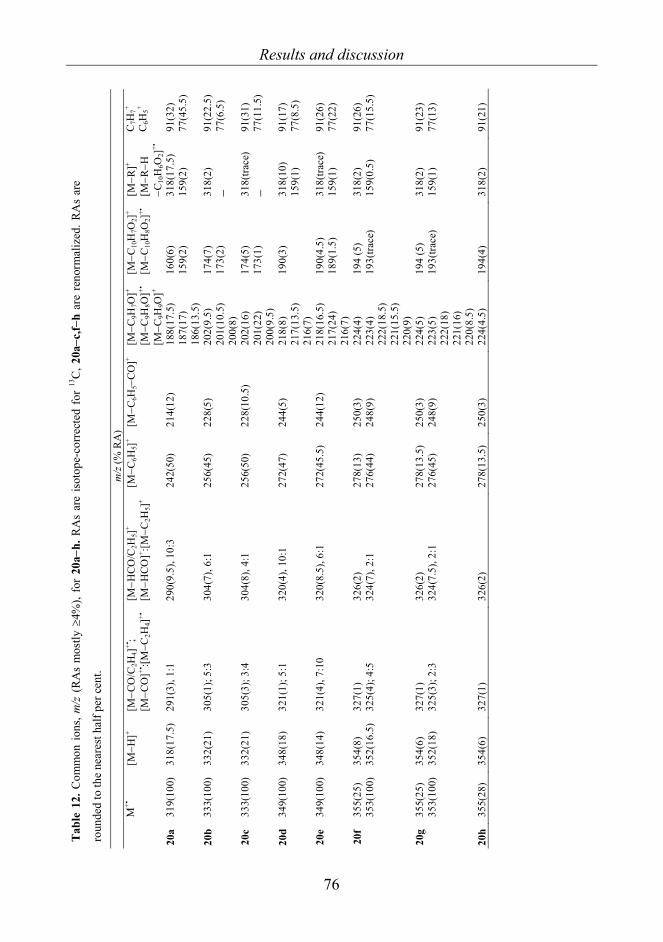
Results and discussion
76
Tab
le 1
2. C
omm
on io
ns, m
/z (
RA
s m
ostly
≥4%
), fo
r 20
a−h.
RA
s ar
e is
otop
e-co
rrec
ted
for
13C
, 20a
−c,f−
h ar
e re
norm
aliz
ed. R
As
are
roun
ded
to th
e ne
ares
t hal
f per
cen
t.
m
/z (%
RA
)
M+•
[M
−H]+
[M−C
O/C
2H4]
+•;
[M−C
O]+•
:[M−C
2H4]
+•
[M−H
CO/C
2H5]
+ [M
−HCO
]+ :[M−C
2H5]
+ [M
−C6H
5]+ [M
−C6H
5−C
O]+
[M−C
9H7O
]+ [M
−C9H
8O]+•
[M
−C9H
9O]+
[M−C
10H
7O2]
+
[M−C
10H
8O2]
+•
[M−R
]+
[M−R
−H
−C10
H6O
2]+•
C7H
7+
C6H
5+
20a
319(
100)
31
8(17
.5)
291(
3), 1
:1
290(
9.5)
, 10:
3 24
2(50
) 21
4(12
) 18
8(17
.5)
187(
17)
186(
13.5
)
160(
6)
159(
2)
318(
17.5
) 15
9(2)
91
(32)
77
(45.
5)
20b
333(
100)
33
2(21
) 30
5(1)
; 5:3
30
4(7)
, 6:1
25
6(45
) 22
8(5)
20
2(9.
5)
201(
10.5
) 20
0(8)
174(
7)
173(
2)
318(
2)
− 91
(22.
5)
77(6
.5)
20c
333(
100)
33
2(21
) 30
5(3)
; 3:4
30
4(8)
, 4:1
25
6(50
) 22
8(10
.5)
202(
16)
201(
22)
200(
9.5)
174(
5)
173(
1)
318(
trace
) −
91(3
1)
77(1
1.5)
20d
349(
100)
34
8(18
) 32
1(1)
; 5:1
32
0(4)
, 10:
1 27
2(47
) 24
4(5)
21
8(8)
21
7(13
.5)
216(
7)
190(
3)
318(
10)
159(
1)
91(1
7)
77(8
.5)
20e
349(
100)
34
8(14
) 32
1(4)
, 7:1
0 32
0(8.
5), 6
:1
272(
45.5
) 24
4(12
) 21
8(16
.5)
217(
24)
216(
7)
190(
4.5)
18
9(1.
5)
318(
trace
) 15
9(1)
91
(26)
77
(22)
20f
355(
25)
353(
100)
35
4(8)
35
2(16
.5)
327(
1)
325(
4); 4
:5
326(
2)
324(
7), 2
:1
278(
13)
276(
44)
250(
3)
248(
9)
224(
4)
223(
4)
222(
18.5
) 22
1(15
.5)
220(
9)
194
(5)
193(
trace
) 31
8(2)
15
9(0.
5)
91(2
6)
77(1
5.5)
20g
355(
25)
353(
100)
35
4(6)
35
2(18
) 32
7(1)
32
5(3)
; 2:3
32
6(2)
32
4(7.
5), 2
:1
278(
13.5
) 27
6(45
) 25
0(3)
24
8(9)
22
4(5)
22
3(5)
22
2(18
) 22
1(16
) 22
0(8.
5)
194
(5)
193(
trace
) 31
8(2)
15
9(1)
91
(23)
77
(13)
20h
355(
28)
354(
6)
327(
1)
326(
2)
278(
13.5
) 25
0(3)
22
4(4.
5)
194(
4)
318(
2)
91(2
1)

Results and discussion
77
Tabl
e 13
. Com
mon
ions
, m/z
(RA
s mos
tly ≥
4 %
), fo
r 21a
−e,g
,h. R
As a
re is
otop
e-co
rrec
ted
for 13
C. R
As a
re ro
unde
d to
the
near
est h
alf p
er-c
ent.
m
/z (%
RA
)
M+•
[M
−Cl]+
[M−H
Cl]+•
[M
−H−H
Cl]+
[M−C
l−C
6H4]
+•
[M−C
9H6O
Cl]+
[M−C
9H7O
Cl]+•
[M−C
9H8O
Cl]+
C7H
6Cl+
Oth
er io
ns
21a
353(
3)
318(
100)
317(
2.5)
31
6(4)
24
2(4)
18
8(3)
18
7(3)
18
6(2.
5)
125(
3)
[M
−Cl]2+
: 158
.5(3
), 77
(10)
21
b 36
7(2)
33
2(10
0)
33
1(4)
33
0(3.
5)
256(
5)
202(
3)
20
1(2)
200(
3)
125(
4)
11
8(4)
, 117
(4),
91(1
0),6
5(5)
21
c 36
7(3)
33
2(10
0)
331(
4)
330(
6)
256(
5)
202(
5)
201(
5)
20
0(2)
12
5(4)
C9H
6OC
l+ : 165
(4),
120(
4), 9
1: C
7H7+ (4
) +C
6H5N
+•(6
), 6
5(4)
21
d 38
3(3)
34
8(10
0)
34
7(3)
34
6(4)
27
2(4.
5)
218(
2.5)
21
7(3)
21
6(2.
5)
125(
6.5)
C9H
8N3O
+ : 174
(3.5
),
120:
C7H
6NO
+ (5),
77(5
) 21
e 38
3(5)
34
8(10
0)
347(
3.5)
34
6(5.
5)
272(
5)
218(
5)
217(
6)
216(
2)
125(
4.5)
13
6(4.
5), 7
7(5)
21
g 38
9(1)
38
7(1)
35
4(28
) 35
2(10
0)
353(
2)a
351(
2.5)
35
2(-)
b 35
0(4)
27
8(1)
27
6(3)
22
4(1)
22
2(3.
5)
223(
1)
221(
2.5)
22
2(0.
5)
220(
2)
125(
6)
11
1(4.
5)
21h
389(
1)
387(
1.5)
35
4(30
) 35
2(10
0)
353(
5)a
351(
3)
352(
-)b
350(
6)
278(
1)
276(
4)
224(
1)
222(
4)
223(
1)
221(
3.5)
22
2(0.
5)
220(
2)
125(
6)
31
8(3.
5), 1
11(5
.5),
75(5
) a C
alcu
late
d va
lue
afte
r rem
oval
of 13
C is
otop
ic p
eak.
b Im
poss
ible
to re
solv
e [M
−H−H
Cl]+ w
ith 37
Cl f
rom
[M−C
l]+ with
35C
l.

Results and discussion
78
The formation of [M–OH]+ was detected only for compounds 20a–h, but their RAs were
low. The OH and HCO• losses may indicate the presence of the enol tautomer in 20, in
contrast with 8-aryl-3,4-dioxo-2H,8H-6,7-dihydroimidazo[2,1-c][1,2,4]triazines (18a–j)
where the keto form was indicated by the lack of [M–OH]+ and [M–HCO]+. Similar
fragmentations, involving the losses of CH1-3CO(•) (18 and 19), C9H7-9O(•) (20), and C9H6-
8OCl(•) (21), which require the migration of H-atoms, may indicate different tautomeric
forms.
The 1H and 13C NMR spectra of 18a,d,g, 19, 20d,g and 21d,g, measured in DMSO-d6
solution at 298 K, show the presence of the 5-OH or 7-OH form, but not the dioxo form.
The weak NOESY correlation between the OH peak and the 1-Ar substituent indicates
that either 7-OH-5-oxo form or the spectral average of 7-OH and 5-OH forms prevails if
the interconversion is fast on the NMR time scale). Only for compound 19 was a small
amount of dioxo-form present, i.e. 5% of that of the OH form, based on the integrated
intensities.
For 3a−j, the predominance of the keto form was supported by fragmentations involving
the losses of CO and COCO but not those of OH or HCO. Similarly, for 18–21 no
fragmentations related to the Ar group migration were found. In conclusion, in view of
the fragmentations requiring H-migrations involving the CO groups, and also the
presence of the ions [M–OH]+ and/or [M–HCO]+, it appears that some amount of enol is
present for 18–21. This is most clearly seen for 18d, 19 and 20. In comparison with
crystallographic and NMR methods, it can be concluded that the intermolecular H-
bonding in the enol form is the main stabilizing effect, which is lost in the gas phase.

Results and discussion
79
4.5 Naphthoxazine, naphthpyrrolo-oxazinone and naphthoxazino-
benzoxazine derivatives 22−29 [V]
4.5.1 General fragmentations
The structures of 22−29 are presented in Scheme 11, and the names in Table 14.
O
N
O
O
N
O
O
N
O
O
N
O
23b
Me Et O
H H
H H
H
H
H
H H H
23c 23d 24b
O
N
O
23a
O
N
O
24a
O
H
H
H
H
O
N
O
22a
O
N
O
22b
O
HN
25a
O
O
HN
27b
N
O
N
26
O
HN
25b
O
O
HN
27a
N
O
N
29a
O
O
N
28
O
Me HO
N
29b
O
H
Scheme 11. Naphthoxazinobenzoxazines (22a, 22b, 23a−d, and 24a,b), naphth-oxazines
(25a,b, 26 and 27a,b) and naphthpyrrolo-oxazinones (28 and 29a,b).

Results and discussion
80
Table 14. The names of compounds 22−29 22a Naphth[1’,2’:5,6][1,3]oxazino[3,4-c][1,3]benzoxazine 22b Naphth[1’,2’:5,6][1,3]oxazino[3,2-c][1,3]benzoxazine 23a (8R*,15bS*)-8-Phenylnaphth[1’,2’:5,6][1,3]oxazino[3,4-c][1,3]benzoxazine 23b (7aR*,15S*)-15-Phenyl-7aH,13H,15H-naphth[1’,2’:5,6][1,3]oxazino[3,2-
c][1,3]benzoxazine 23c (7aR*,13R*,15S*)-13-Methyl-15-phenyl-7aH,13H,15H-
naphth[1’,2’:5,6][1,3]oxazino[3,2-c][1,3]benzoxazine 23d (7aR*,13R*,15S*)-13-Ethyl-15-phenylnaphth[1’,2’:5,6][1,3]oxazino[3,2-
c][1,3]benzoxazine 24a (8R*,15bS*)-8-Phenylnaphth[1’,2’:5,6][1,3]oxazino[3,4-c][1,3]benzoxazin-10-
one 24b (7aR*,15S*)-15-Phenylnaphth[1’,2’:5,6][1,3]oxazino[3,2-c][1,3]benzoxazin-13-
one 25a 2,3-Dihydro-1H-naphth[1,2-e][1,3]oxazin-3-one 25b 1-Phenyl-2,3-dihydro-1H-naphth[1,2-e][1,3]oxazin-3-one 26 3-Phenyl-1H-naphth[1,2-e][1,3]oxazine 27a 3-Phenylimino-2,3-dihydro-1H-naphth[1,2-e][1,3]oxazine 27b 1-Phenyl-3-Phenylimino-2,3-dihydro-1H-naphth[1,2-e][1,3]oxazine 28 7a-Methyl-8,9-dihydro-7aH,10H,12H-naphth[1,2-e]pyrrolo[2,1-b][1,3]oxazin-
10-one 29a 7aH,12H,14H-Naphth[1’,2’:5,6][1,3]-oxazino[2,3-a]isoindol-12-one 29b (7aR*,14S*)-14-Phenyl-7aH,12H,14H-naphth[1’,2’:5,6][1,3]oxazino[2,3-
a]isoindol-12-one
The 1,3-oxazine ring has been widely studied for ring-chain tautomeric equilibria. The
compounds studied, 22–29, cannot exhibit ring-chain tautomerism, but mass
spectrometric fragmentations involving ring openings are possible, such as the one
observed for 3,1-benzoxazino[1,2-c]-[1,3]benzoxazines (Scheme 12) [137].
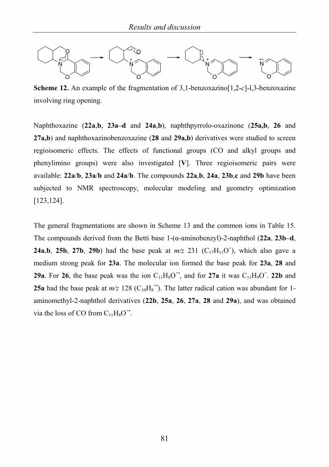
Results and discussion
81
N
O
O
N
O
O
N
O
N
O Scheme 12. An example of the fragmentation of 3,1-benzoxazino[1,2-c]-l,3-benzoxazine
involving ring opening.
Naphthoxazine (22a,b, 23a–d and 24a,b), naphthpyrrolo-oxazinone (25a,b, 26 and
27a,b) and naphthoxazinobenzoxazine (28 and 29a,b) derivatives were studied to screen
regioisomeric effects. The effects of functional groups (CO and alkyl groups and
phenylimino groups) were also investigated [V]. Three regioisomeric pairs were
available: 22a/b, 23a/b and 24a/b. The compounds 22a,b, 24a, 23b,c and 29b have been
subjected to NMR spectroscopy, molecular modeling and geometry optimization
[123,124].
The general fragmentations are shown in Scheme 13 and the common ions in Table 15.
The compounds derived from the Betti base 1-(α-aminobenzyl)-2-naphthol (22a, 23b–d,
24a,b, 25b, 27b, 29b) had the base peak at m/z 231 (C17H11O+), which also gave a
medium strong peak for 23a. The molecular ion formed the base peak for 23a, 28 and
29a. For 26, the base peak was the ion C11H8O+•, and for 27a it was C11H9O+. 22b and
25a had the base peak at m/z 128 (C10H8+•). The latter radical cation was abundant for 1-
aminomethyl-2-naphthol derivatives (22b, 25a, 26, 27a, 28 and 29a), and was obtained
via the loss of CO from C11H8O+•.

Results and discussion
82
O
N
R2
R3R1
-H.
[M-H]+
-OH. -CH3N
22a, 23a, 26,27a,b, 28,29a,b
22a,23a,24a,28,29a,b 22a
-C8H7NO22b,23a,b
22a,23a-d,24a,b
C17H11O+
-COH.22a,29b
-HCO.
C16H11+ C16H10
+.
-C8H5NO29a,b
-C8H5NO2
24a,b
C11H9O+
C11H8O+.
27a,28
22b*,25a*,28*,29a*
[M-OH]+
C10H8+.
25b,27b,29b: -C6H5.
25b, 27b,29a,b
C10H6+.
C8H6+.
C6H6+.
C6H5+
C10H7+
-CO
[M-C6H5]+
*22b,25a,26,27a,28,29a:C11H8O+.
, m/z 15623a-d,24a,b,25b, 27b,29b:C17H12O+., m/z 23224a: C24H16O2
+., m/z 336
C18H12O2+.
23b-d
[M-C17H12O]+.
23c: -Me.
23d: -Et.C8H6NO+
-C17H12O
m/z 203 m/z 202
m/z 260
24a,25a,b: -CONH23d: -C10H11NO
23c: -C9H9NO22b-C11H9O.
24a-C17H11O2
. 28: -C5H7NO26: -C7H5N27a,b: -C7H6N2
m/z 132
m/z 128
m/z 156
m/z 157
m/z 78m/z 102
m/z 126
m/z 127
m/z 77
22b,26,27a,28: -C11H8O
23b: C8H7NO+.
m/z 13328: C5H7NO+.
m/z 97
26: C7H5N+.
m/z 10327a: C7H6N2
+.
m/z 118
m/z 231
22-29M
Scheme 13. The general fragmentation routes of compounds 22–29.

Results and discussion
83
Tabl
e 15
. Com
mon
ions
for 2
2−29
and
thei
r RA
s m/z
(% R
A).
RA
s are
cor
rect
ed fo
r 13C
isot
opes
and
thos
e of
22b
, 25a
, 27a
and
29a
ha
ve b
een
reno
rmal
ized
. RA
s are
roun
ded
to th
e ne
ares
t hal
f per
cen
t. m
/z (%
RA
) C
ompo
und
M+•
[M
− H
]+ [M
− O
H]+
[M −
CO
NH
]+
[M −
CO
NH
2]+•
C17
H12
O+•
C
17H
11O
+C
16H
11+
C16
H10
+•
C11
H9O
+ C
11H
8O+•
C
10H
8+•
22a
289(
82.5
) 28
8(32
.5)
272(
33)
- 23
2(1)
23
1(10
0)
203(
2)
202(
8)
- 12
8(1.
5)
22b
289(
6)
- -
- -
-
15
7(2.
5)
156(
51)
128(
100)
23a
365(
100)
36
4(4)
34
8(67
.5)
- 23
2(3)
23
1(27
)
202(
8)
- -
23b
365(
5.5)
-
- -
232(
23)
231(
100)
20
3(3)
20
2(21
) -
- 23
c 37
9(3)
-
- -
232(
22)
231(
100)
20
3(3)
20
2(22
) -
- 23
d 39
3(2.
5)
- -
- 23
2(24
) 23
1(10
0)
203(
3)
202(
22)
- -
24a
379(
36.5
) -
362(
4)
336(
9)
- 23
2(12
) 23
1(10
0)
20
2(9)
-
-
24b
379(
10.5
) -
- -
232(
24)
231(
100)
20
3(3)
20
2(15
) -
- 25
a 19
9(46
) -
- 15
6(96
) 15
5(4)
-
-
15
7(4)
15
6(96
) 12
8(10
0)
25b
275(
31)
- -
232(
19)
231(
100)
23
2(19
)
231(
100)
20
3(3)
20
2(14
) -
-
26
259(
54)
258(
2)
- -
- -
203(
3)
202(
14)
- 156(
100)
12
8(70
)
27a
274(
68)
273(
32)
- -
- -
157(
100)
15
6(40
) 12
8(76
)
27b
350(
29)
349(
4.5)
-
- 23
2(16
) 23
1(10
0)
203(
3)
202(
15)
- 12
8(2)

Results and discussion
84
For 23b–d, 24b,25b,27b and 29b the mechanism for the formation of the ion C17H11O+ at
m/z 231 and a nitrogen radical is presented in Scheme 14. The fragmentation corresponds
to an RDA-H and it is exceptional since the charge does not remain on the nitrogen. For
22a, 23a and 24a, however, the fragmentation mechanism is more complicated, because
it requires a cleavage of a benzo-bound oxygen bond. For 22a, 23a and 24, an abundant
ion [M–C17H11O2]+ is also formed (Table 16, p. 86), i.e. the ion containing the nitrogen
has a positive charge. For 23b–d a nitrogen radical cation [M–C17H12O]+• was also
formed, although the RAs were low.
For 29b, C17H11O+ was also formed by a loss of C8H5N from [M–OH]+; for 22a, a minor
route to C17H11O+ consisted in C2H3N loss from [M–OH]+. For 22a, the loss of COH•
from [M–CH3N]+• gave the ion C17H11O+, as confirmed by B2/E scans.
O
N
R2
HR3
O
23b-d, 24b, 25b, 27b, 29b
N
R2
R3
H+
RDA-H
Scheme 14. The formation of C17H11O+ from 23b–d, 24b, 27b and 29b.
The ion C17H11O+ (22a, 23a–d, 24a,b, 25b, 27b and 29b) at m/z 231 can lose CO and
HCO•, the product ions being C16H11+ and C16H10
+•, respectively. C11H8O+ (22b, 25a, 26,
27a, 28 and 29a) gave analogously the ions C10H8+• and C10H7
+.
Very weak CO loss was detected only for compounds 28 and 29a. For 22a, 23a, 24a, 28,
29a, and 29b, the ion [M–OH]+ had RAs between 4 and 67.5%, being most abundant for
22a, 23a, 28 and 29a. This fragmentation requires H-migration and ring opening, and
solving the exact mechanism would require deuterium labeling. Suggested mechanisms
for OH• loss from 23a are presented in Scheme 15. For 23a, the B/E scans of [M–OH]+

Results and discussion
85
gave no significant signals; the ion therefore appears to be unusually stable, the positive
charge probably being stabilized by aromatic groups.
O
N
O
H
O
N
O
O
N
O
H
H H
O
N
OH H
OH
N
O
- OH - OH
O
N
H
N
O
O
N
O
H
Scheme 15. Possible ring openings of 23a and the consequent OH loss.
4.5.2 Comparison of regioisomers
22a–24a are derivatives of 1-(α-aminobenzyl)-2-naphthol, while 22b–24b are derivatives
of 1-aminomethyl-2-naphthol, the mass spectra of these regioisomeric pairs were

Results and discussion
86
expected to be very different. The ions useful for differentiating the regioisomers 22a–
24a vs 22b–24b are listed in Table 16.
Table 16. Ions useful for differentiating regioisomeric pairs (22–24)a/b and their RAs.
Ion 22a
O
N
O
22b
O
N
O
23a
O
N
OH H
23b
O
N
OH H
24a
O
N
O OH H
24b
O
N
OOH H
M+• 82.5 6 100 5.5 36.5 10.5
[M–OH]+ 33 - 67.5 - 4 -
C17H11O2+ 22 - 6 - 13 -
C16H10O+• 11 - 6 - 7 -
C15H9+ 19 - 6 - 10 -
C8H7NO+ 52 - 14 - - -
C17H11O+ 100 - 27 100 100 100
C10H8+ 1.5 100 - - - -
C8H6NO+
C7H5O+
C6H5+
-
-
-
25
13
20
-
-
-
7
5
6
52
5
18
-
-
-
Also only for 23a: C8H8N+ (77) and C7H7+ (44)
The formation mechanism of [M–OH]+ may be different for 22a, 23a and 24a. For 22a
and 23a the OH loss may involve ring opening (Scheme 15). The formation of the ion
[M–OH]+ from 24a may be due to the geometry since the tertiary α-hydrogen vicinal to
Ph group can migrate to the CO oxygen (Figure 39). The formation of the ions C7H5O+
and C17H11O2+ may be affected by the boat form of the benzo-bound oxazine ring. On the
other hand the tertiary α-H migration is unfavorable for 24b (Figure 40). It should be
noted that the configuration of the H-atoms in the calculated structure corresponding to
the global energy minimum for 24a in Ref. [124] was incorrect; hence the structure in
Figure 39 was established by using MM2 minimum energy optimization.

Results and discussion
87
O
N
O O
H H
H
H
H
H
H
H
HH
H
H
H H
H
HH
a: C7H5O+
b: C17H11O2+
b
b
a
a
a
Figure 39. Suggested formation of ions C7H5O+ (a) and C17H11O2
+ (b) from 24a.
Structure was optimized by using MM2 minimum energy calculations.
O
NO
H
H
OH
H
H
H
H H
H
H
H
H
H
HH
H
H
Figure 40. MM2 optimized minimum energy structure for 24b. The migration of H-atom
to CO oxygen is not favorable.
4.5.3 Effect of substituents
Me- (23c) and Et-substituted (23d) compounds yielded very similar spectra. The
fragments C9H9NO+• and C10H11NO+• were formed exclusively from 23c or 23d,
respectively. These ions also lost Me• or Et• and the product ion C8H6NO+ was more
Results and discussion
88
abundant for 23d. The spectra of 25a and its Ph analog 25b differed as expected.
Accordingly, for 25a the base peak was given by the ion C10H8+• and for 25b by the ion
[M–CONH2]+, i.e. C17H11O+. For 25a the RA of [M–CONH2]+ was only 4%, but that of
[M–CONH]+• was 96%.
Compound 27a furnished an abundant ion [M–H]+ and its Ph analog 27b an abundant ion
[M–Ph]+, both giving weak ions [M–HNPh]+. However, 27a did not give the ion [M–
Ph]+, which would require the cleavage of the Ph–N bond, therefore for 27b an α
cleavage of the Ph–C bond explains the relatively abundant Ph• loss.
4.5.4 Fragmentations of 1-(α-aminobenzyl)-2-naphthol and 1-aminomethyl-2-naphthol
derivatives
The ions useful for identifying 1-(α-aminobenzyl)-2-naphthol (22a, 23b–d, 24a,b, 25b,
27b and 29b) and 1-aminomethyl-2-naphthol (22b, 25a, 26, 27a, 28 and 29a) derivatives
are listed in Table 17. The ions C17H12O+•, C17H11O+, C11H8O+• and C17H11O2+ and the
complementary ions [M–C17H12O]+•, [M–C11H8O]+ and [M–C17H11O2]+ were formed
logically from the structures studied. For 24b, 25a,b, 27b and 29a,b (i.e. the structures
with CO or imino substituents, except 28), the complemetary ions were not seen.

Results and discussion
89
Tabl
e 17
. RA
s of i
ons C
17H
12O
+•, C
17H
11O
+ , C11
H8O
+• a
nd C
17H
11O
2+ and
thei
r com
plem
enta
ry io
ns fo
r 22–
29.
Ion
22a
O
N
O
22b
O
N
O
23a
O
N
O HH
23b
O
N
OH
H
23c
O
N
OM
e HH
H
23d
O
N
OE
t H
H
H
24a
O
N
OO
HH
24b
O
N
OO H
H
C 1
7H12
O+•
C 17H
11O
+ 1 10
0 - -
3 27
23
100
22
100
24
100
12
100
24
100
C 11H
8O+
- 51
-
- -
- -
- C 1
7H11
O2+
22
- 6
- -
- 13
-
[M–C
17H
12O
]+•
[M–C
17H
11O
2]+
- 78
- - - 77
14
-
11
- 6 -
- 52
- - [M
–C11
H8O
]+ -
52
- -
- -
- -
Ion
25a
O
H NO
25b
O
H NO
26
O
N
27a
O
H NN
27b
O
H NN
28
O
N
O
Me
29a
O
N
O
H
29b
O
N
O
H
C 1
7H12
O+•
C 17H
11O
+ -
19
100
- -
16
100
- 2
14
100
C 11H
8O+
96
- 10
0 40
-
72
26.5
-
C 17H
11O
2+ -
- -
- -
- -
- [M
–C17
H12
O]+•
[M–C
17H
11O
2]+
- - - -
- - - -
2 - - -
- - - -
[M–C
11H
8O]+
- -
5 15
-
28
- -

Conclusions
90
5. CONCLUSIONS The EIMS revealed linear correlations of the Hammett σ constants and the RAs and/or %
TICs of the ions for variously substituted 2-phenacylpyridines 1a−n and 2-
phenacylquinolines 2a−h. The substituents therefore clearly affected the fragmentations.
The strongest electron donors presumably favor form K, while the electron acceptors
favor form O (1a−n) or E (2a−h) as in solution or in the solid state. The OH loss is
expected to occur from form E or O. The increasing RAs of the molecular ion and
[M−H]+ may indicate the increase of forms E and O, because the molecular ion is often
more abundant for enol than for keto tautomers. Besides intramolecular H-bonds, the E or
O tautomers are possibly stabilized by the resonance effect of the Ph ring substituents.
For 1a−n and 2a−h the % TIC of ArCO+ appeared to be an indicator for form K, as were
those of [M−Ar]+ for forms E and O. The losses of CO and HCO• also seemed to be
related to forms E and O. [M−CO]+• correlated with the Hammett σ constants for 1a−h,
but not for 2a−h; this may be caused by the different conjugation of forms O and E.
For dioxoimidazotriazines 3a−j and pyrimidinediones 18−21, the tautomerism was not
clear. For 3a−j and 18−21, the substituents had only minor effects on the ion RAs. For
3a−j, the lack of OH loss and the fragmentations involving the losses of CO and COCO
show that the amido form predominates in the gas phase, though the fragmentations
involving the loss of HOCN may indicate a small amount of the enol form. On the other
hand, 18−21 exhibit [M–OH]+ peaks and many of the observed fragmentations require H-
migrations; thus at least a small amount of the enol form appears to be present.
For pyrrolo- and isoindoloquinazolinones 4−17, the geometries of the molecules affected
the fragmentations. Stereospecific fragmentations were observed most clearly for
stereoisomeric pairs 5,6 and 10,11. However, the RDA-related fragmentations were non-
stereospecific, indicating a stepwise mechanism, the first step possibly involving the CO
group-induced ring opening.

Conclusions
91
Naphthoxazine derivatives 22−29 displayed some regiospecific fragmentations, which
were useful for differentiating regioisomers with the same nominal mass. The
regiospecific pairs (22–24)a/b could be clearly distinguished by using the RAs of the ions
M+•, [M–OH]+, C17H11O2+, C16H10O+• and C15H9
+. The compounds derived from 1-(α-
aminobenzyl)-2-naphthol generally gave a strong peak at m/z 231 (C17H11O+), while the
1-aminomethyl-2-naphthol derivatives exhibited an ion at m/z 156 (C11H8O+•) instead.
This is useful for identifying regioisomeric compounds with the same nominal masses.

References
92
6. REFERENCES
[1] Fessenden RJ, Fessenden JS, Logue MW. “Organic Chemistry 6th edition”,
Brooks/Cole Publishing Company, USA, 1998, p. 578.
[2] Watson JD, Crick FHC. “Molecular Structure of Nucleic Acids: A Structure for
Deoxyribose Nucleic Acid”, Nature, 1953; 171: 737–738.
[3] Watson JD. “The Double Helix”, Penguin Books, New York, USA, 1968.
[4] Terent’ev PB, Kalandarishvili AG. “Application of Mass Spectrometry for the
Analysis of Organic Tautomeric Compounds”, Mass Spectrom. Rev., 1996; 15: 339–363.
[5] Pullman B, Berthod H, Dreyfus M. “Amine-Imine Tautomerism in Adenines”,
Theoret. Chim. Acta (Berl.), 1969; 15: 265–268.
[6] Imperiali B, O'Connor SE, Hendrickson T, Kellenberger C. “Chemistry and biology
of asparagine-linked glycosylation”, Pure Appl. Chem., 1999; 71: 777–787.
[7] Daniels M. “Tautomerism of Uracil and Thymine in Aqueous Solution: Spectroscopic
Evidence”, Proc. Nat. Acad. Sci. USA, 1972; 69: 2488–2491.
[8] Allegretti PE, de las Mercedes Schiavoni M, Castro EA, Furlong JJP. “Tautomeric
Equilibria Studies by Mass Spectrometry”, Nature Precedings, 2008:
<http://hdl.handle.net/10101/npre.2008.1842.1>
[9] Minkin VI, Garnovskii DG, Elguero J, Katritzky AR, Denisko OV. “The tautomerism
of heterocycles: Five-membered rings with two or more heteroatoms”, Adv. Heterocycl.
Chem., 2000; 76: 157–323.

References
93
[10] Alkorta I and Elguero J. “Theoretical estimation of the annular tautomerism of
indazoles”, J. Phys. Org. Chem., 2005; 18: 719–724.
[11] Lindsay Smith JR, Sadd JS. “Isomerism of 1- and 2-(N,N-disubstituted
aminomethyl)benzotriazoles. Investigation by nuclear magnetic resonance spectroscopy”,
J. Chem. Soc., Perkin Trans.1,, 1975; 12: 1181–1184.
[12] Lutsenko IF. “O- and C-isomeric organoelement derivatives of keto-enol systems:
rearrangements and elementotropism”, Pure Appl. Chem., 1972; 30: 409–426.
[13] Alkorta I, Goya P, Elguero J, Singh SP. “A simple approach to the tautomerism of
aromatic heterocycles. A Review”, Natl. Acad. Sci. Lett., 2007; 30: 139–159.
[14] Shurukhin YV, Klyuev NA, and Grandberg II. “Similarities Between Thermolysis
And Mass Spectrometric Fragmentation Of Tetrazoles (Review)”, Chem. Heterocycl.
Comp., 1985; 21: 605–620.
[15] a) Juhász M, Lázár L, Fülöp F. “Substituent effects in the ring-chain tautomerism of
4-alkyl-2-aryl substituted oxazolidines and tetrahydro-1,3-oxazines”, J. Heterocycl.
Chem., 2007; 44: 1465–1473.
b). Juhász M, Fülöp F, Pihlaja K. “Substituent effects on the gas-phase ring-chain
tautomerism of 3,4,5,6-tetrahydro-2H-1,3-oxazines”, Rapid Commun. Mass Spectrom.,
2007; 21: 3701–3710.
c) Pihlaja K, Juhász M, Kivelä H, Fülöp F. “Substituent effects on the ring-chain
tautomerism of some 1,3-oxazolidine derivatives”, Rapid Commun. Mass Spectrom.,
2008; 22: 1510–1518.
d) Vainiotalo P, Ronkanen S, Fülöp F, Pihlaja K. “Gas-Phase Ring-Chain Tautomerism in
1,3-oxazolidines”, Tetrahedron, 1990; 46: 3683–3692.
e) Vainiotalo P, Fulöp F, Pihlaja K. “Gas-phase Ring-chain Tautomerism in 1,3-
Oxazines. Does it exist?“, Org. Mass Spectrom., 1991; 26: 438–442.

References
94
f) Vainiotalo P, Fulöp F, Bernath G, Pihlaja K “Mass Spectrometric Intramolecular
Cyclization Reactions of Some 2-N-Phenyliminoperhydro-1,3-oxazines”, J. Heterocyclic
Chem., 1989; 26: 1453–1459.
g) Vainiotalo P, Savolainen P-L, Ahlgrén M, Mälkönen PJ, Vepsäläinen J. ”Identification
of the Condensation Products of 1,2- and 1,3-Amino Alcohols with Keto Esters by NMR
Spectroscopy, Mass Spectrometry and X-Ray Crystallography Studies. Open or Ring
Forms?”, J. Chem. Soc., Perkin Trans. 2, 1991; 5: 735–741.
[16] Cope AC, Haven Jr AC, Ramp FL, Trumbull ER. “Cyclic Polyolefins. XXIII.
Valence Tautomerism of 1,3,5-Cycloöctatriene and Bicyclo [4.2.0]octa-2,4-diene”, J. Am.
Chem. Soc., 1952; 74: 4867–4871.
[17] Doering W von E, Roth W. “A rapidly reversible degenerate Cope rearrangement
Bicyclo[5.1.0]octa-2,5-diene”, Tetrahedron, 1963; 19: 715–737.
[18] Ault A. “The Bullvalene Story. The Conception of Bullvalene, a Molecule That Has
No Permanent Structure”, J. Chem. Educ., 2001; 78: 924–927.
[19] Schröder G. “Preparation and Properties of Tricyclo[3,3,2,04,6]deca-2,7,9-triene
(Bullvalene)”, Angew. Chem., Int. Ed. Eng., 1963; 2: 481–482.
[20] Postovskii I Ya, Bednyagina NP, Senyavina LB, Sheinker Yu N. “Study of the
azide-tetrazole tautomerism by means of infrared spectroscopy” Izvestiya Akademii Nauk
SSSR, Seriya Fizicheskaya, 1962; 26: 1298–1300.
[21] Klyuev NA, Adanin VM, Postovskii I Ya, Azev Yu A. “Mass-spectrometric study of
azido-tetrazole tautomerism in the pyrimido-as-triazine series”, Chem. Heterocyc. Comp.,
1983; 19: 451–453.
[22] a) Kolehmainen E, Ośmiałowski B, Krygowski TM, Kauppinen R, Nissinen M,
Gawinecki R. “Substituent and temperature controlled tautomerism: multinuclear

References
95
magnetic resonance, X-ray, and theoretical studies on 2-phenacylquinolines”, J. Chem.
Soc., Perkin Trans. 2, 2000; 6: 1259–1266.
b) Dewar MJS, Zoebisch EG, Healy EF, Stewart JJP. “Development and use of quantum
mechanical molecular models. 76. AM1: a new general purpose quantum mechanical
molecular model”, J. Am. Chem. Soc., 1985; 107: 3902–3909.
[23] Kolehmainen E, Ośmiałowski B, Nissinen M, Kauppinen R, Gawinecki R.
“Substituent and temperature controlled tautomerism of 2-phenacylpyridine: the
hydrogen bond as a configurational lock of (Z)-2-(2-hydroxy-2-phenylvinyl)pyridine”, J.
Chem. Soc., Perkin Trans. 2, 2000; 11: 2185–2191.
[24] Gawinecki R, Ośmiałowski B, Kolehmainen E and Kauppinen R. ”Long-range
substituent and temperature effect on prototropic tautomerism in 2-
(acylmethyl)quinolines”, J. Phys. Org. Chem., 2001; 14: 201–204.
[25] Gawinecki R, Kuczek A, Kolehmainen E, Osmiałowski B, Krygowski TM,
Kauppinen R. ”Influence of Bond Fixation in Benzo-Annulated N-Salicylideneanilines
and Their ortho-C(=O)X Derivatives (X = CH3, NH2, OCH3) on Tautomeric Equilibria in
Solution”, J. Org. Chem., 2007; 72: 5598–5607.
[26] Sztanke K, Fidecka S, Kędzierska E, Karczmarzyk Z, Pihlaja K, Matosiuk D.
“Antinociceptive activity of new imidazolidine carbonyl derivatives. Part 4. Synthesis
and pharmacological activity of 8-aryl-3,4-dioxo- 2H, 8H-6,7 -dihydroimidazo[2,1-
c][1,2,4]triazines”, Eur. J. Med. Chem., 2005; 40: 127–134.
[27] Emsley J, Freeman NJ. “β-diketone interactions : Part 5. Solvent effects on the keto
enol equilibrium”, J. Mol. Struct., 1987; 161: 193–204.
[28] Yoshida Z, Ogoshi H, Tokumitsu T. “Intramolecular hydrogen bond in enol form of
3-substituted-2,4-pentanedione”, Tetrahedron, 1970; 26: 5691–5697.

References
96
[29] Simmons T, Love RF, Kreuz KL, “The Structure of α-Nitro Ketones”, J. Org.
Chem., 1966; 31: 2400–2401.
[30] Fontana A, De Maria P, Siani G, Pierini M, Cerritelli S, Ballini R. “Equilibrium
Constants for Ionisation and Enolisation of 3-Nitrobutan-2-one”, Eur. J. Org. Chem.
2000; 8: 1641–1646.
[31] Carey ARE, Eustace S, More O'Ferrall RA, Murray BA. “Keto-enol and imine-
enamine tautomerism of 2-, 3-, and 4-phenacylpyridines”, J. Chem. Soc., Perkin Trans.
2,, 1993; 11: 2285–2296.
[32] Katritzky AR, Kucharska HZ, Rowe JD. “Potentially tautomeric pyridines. VI. 2-, 3-
, and 4-Phenacylpyridines”, J. Chem. Soc., 1965; 3093–3096.
[33] More O’Ferrall RA, Murray A, “1H and 13C NMR spectra of α-heterocyclic ketones
and assignment of keto, enol and enaminone tautomeric structures”, J. Chem. Soc., Perkin
Trans. 2, 1994; 12: 2461–2470.
[34] Gawinecki R, Kolehmainen E, Loghmani-Khouzani H, Ośmiałowski B, Lovász T
and Rosa P. “Effect of π-electron delocalization on tautomeric equilibria -
benzoannulated 2-phenacylpyridines”, Eur. J. Org. Chem. 2006, 12, 2817–2824.
[35] Greenhill JV, Loghmani-Khouzani H, Maitland DJ. ”Tautomerism in ketomethyl
quinolines. Part 3. 4-Ketomethylquinolines“, Can. J. Chem., 1991; 69: 696–701.
[36] Hammett LP. “The Effect of Structure upon the Reactions of Organic Compounds.
Benzene Derivatives”, J. Am. Chem. Soc., 1937; 59: 96–103.
[37] Taft Jr RW. “Polar and Steric Substituent Constants for Aliphatic and o-Benzoate
Groups from Rates of Esterification and Hydrolysis of Esters”, J. Am. Chem. Soc,. 1952;
74: 3120–3128.

References
97
[38] Taft Jr RW, Lewis IC. “The General Applicability of a Fixed Scale of Inductive
Effects. II. Inductive Effects of Dipolar Substituents in the Reactivities of m- and p-
Substituted Derivatives of Benzene”, J. Am. Chem. Soc., 1958; 80: 2436–2443.
[39] Taft Jr RW, Lewis IC. “Evaluation of Resonance Effects on Reactivity by
Application of the Linear Inductive Energy Relationship-IV. Hyperconjugation Effects of
Para-alkyl Groups”, Tetrahedron, 1959; 5: 210–232.
[40] Taft Jr RW, Lewis IC. “Evaluation of Resonance Effects on Reactivity by
Application of the Linear Inductive Energy Relationship. V. Concerning a σR Scale of
Resonance Effects”, J. Am. Chem. Soc., 1959; 81: 5343–5352.
[41] Carey FA, Sundberg RJ. “Structural Effects on Stability and Reactivity“ in
“Advanced organic chemistry Part A: Structure and Mechanisms, 5th edition”, Springer
Science, USA, 2007, p. 253–388.
[42] Wilcox CF, Leung C. “Transmission of Substituent Effects. Dominance of Field
Effects”, J. Am. Chem. Soc., 1968; 90: 336–341.
[43] Dewar MJS, Marchand AP. “Substituent Effects. V. Further Evidence Concerning
the Nature of the Inductive Effect”, J. Am. Chem. Soc., 1966; 88: 354–358.
[44] Liotta CL, Fisher WF, Greene Jr GH, and Joyner BL. “Mechanism of Transmission
of Nonconjugative Substituent Effects. IV. Analysis of the Dissociation Constants of 6-
Substituted Spiro [3.3] heptane-2-carboxylic Acids”, J. Am. Chem. Soc., 1972; 94: 4891–
4897.
[45] Roberts JD, Moreland Jr WT. “Electrical Effects of Substituent Groups in Saturated
Systems. Reactivities of 4-Substituted Bicyclo [2.2.2]octane-1-carboxylic Acids”, J. Am.
Chem. Soc., 1953; 75: 2167–2173.
References
98
[46] Grob CA, Schlageter MG. “The Derivation of Inductive Substituent Constants from
pKa Values of 4-Substituted Quinuclidines. Polar effects. Part I” Helv. Chim. Acta., 1976;
59: 264–276.
[47] Hansch C, Leo A, Taft RW. “A Survey of Hammett Substituent Constants and
Resonance and Field Parameters”, Chem. Rev., 199l; 97: 165–195.
[48] Swain CG, Lupton Jr EC. “Field and resonance components of substituent effects”,
J. Am. Chem. Soc., 1968; 90: 4328–4337.
[49] Taft RW, Topsom RD. “The Nature and Analysis of Substitutent Electronic Effects”,
Prog. Phys. Org. Chem., 1987; 16: 1–83.
[50] Brown HC, Okamoto Y. “Electrophilic Substituent Constants”, J. Am. Chem. Soc.,
1958; 80: 4979–4987.
[51] Seydel JK. “Prediction of in Vitro Activity of Sulfonamides, Using Hammett
Constants or Spectrophotometric Data of the Basic Amines for Calculation”, Mol.
Pharmacol., 1966; 2: 259–265.
[52] Sharma P, Sharma S, Rane N. “Synthesis and in vitro antimicrobial activities of 2-
hydroxy-6-methyl-7-(arylamino)-1,7-dihydropurin-8-ones”, Bioorg. Med. Chem., 2004;
12: 3135–3139.
[53] Fukuto TR, Metcalf RL. “Pesticidal Activity And Structure: Structure and
Insecticidal Activity of Some Diethyl Substituted Phenyl Phosphates”, J. Agr. Food
Chem., 1956; 4: 930–935.
[54] a) Hansch C. “A Quantitative Approach to Biochemical Structure-Activity
Relationships”, Acc. Chem. Res., 1969; 2: 232–239.

References
99
b) Leo A, Hansch C, Elkins D. "Partition coefficients and their uses", Chem. Rev. 1971;
71: 525–616.
[55] Kohen A, Klinman JP. “Enzyme Catalysis: Beyond Classical Paradigms”, Acc.
Chem. Res., 1998; 31: 397–404.
[56] Watson JD, Crick FHC. “Genetical implications of the structure of deoxyribonucleic
acid”, Nature, 1953; 171: 964–967.
[57] Topal MD, Fresco JR. “Complementary base pairing and the origin of substitution
mutations”, Nature, 1976; 263: 285–289.
[58] Harris VH, Smith CL, Cummins WJ, Hamilton AL, Adams H, Dickman M, Hornby
DP, Williams DM. “The Effect of Tautomeric Constant on the Specificity of Nucleotide
Incorporation during DNA Replication: Support for the Rare Tautomer Hypothesis of
Substitution Mutagenesis”, J. Mol. Biol., 2003; 326: 1389–1401.
[59] Podolyan Y, Gorb L, Leszczynski J. “Rare Tautomer Hypothesis Supported by
Theoretical Studies: Ab Initio Investigations of Prototropic Tautomerism in the N-
Methyl-P Base”, J. Phys. Chem. A, 2005; 109: 10445–10450.
[60] Fedor LR. “Acid-catalyzed hydrolysis of (R)-C[(l-methyl-3-oxo-l-butenyl)amino]-3-
isoxazolidinone, prodrug of cycloserine”, Int. J. Pharm. 1984; 22: 197–205.
[61] Murakami T, Yata N, Tamauchi H, Nakai J, Yamazaki M, Kamada A. “Studies on
absorption promoters for rectal delivery preparations. I. Promoting efficacy of enamine
derivatives of amino acids for the rectal absorption of β-lactam antibiotics in rabbits.”
Chem. Pharm. Bull., 1981; 29: 1998–2004.

References
100
[62] Garro JC, Manzanares GD, Zamarbide GN, Ponce CA, Estrada MR, Jáuregui EA.
“Geometrical isomerism, tautomerism and conformational charges of 2-propenal-3-amine
in its neutral and protonated forms” J. Mol. Struct. (Theochem) 2001; 545: 17–27.
[63] Scott KR, Edafiogho IO, Richardson EL, Farrar VA, Moore JA, Tietz EI, Hinko CN,
Chang H, El-Assadi A, Nicholson JM. “Synthesis and anticonvulsant activity of
enaminones. 2. Further structure-activity correlations” J. Med. Chem., 1993; 36: 1947–
1955.
[64] Edafiogho IO, Hinko CN, Chang H, Moore JA, Mulzac D, Nicholson JM, Scott KR.
“Synthesis and anticonvulsant activity of enaminones”, J. Med. Chem. 1992; 35: 2798–
2805.
[65] Beckett AH, Kerridge KA, Clark PM, Smith WG. “Preparation and antibacterial
activity of 2-phenacylpyridine and related ketones.” J. Pharm. Pharmacol., 1955; 7: 717–
730.
[66] Bell IM, Erb JM, Freidinger RM, Gallicchio SN, Guare JP, Guidotti MT, Halpin RA,
Hobbs DW, Homnick CF, Kuo MS, Lis EV, Mathre DJ, Michelson SR, Pawluczyk JM,
Pettibone DJ, Reiss DR, Vickers S, Williams PD, Woyden CJ. “Development of Orally
Active Oxytocin Antagonists: Studies on 1-(1-{4-[1-(2-Methyl-1-oxidopyridin-3-
ylmethyl)piperidin-4-yloxy]-2- methoxybenzoyl}piperidin-4-yl)-1,4-
dihydrobenz[d][1,3]oxazin-2-one (L-372,662) and Related Pyridines”. J. Med. Chem.,
1998; 41: 2146–2163.
[67] Epifano F, Genovese S, Curini M. “Ytterbium triflate catalyzed synthesis of β-
enaminones”, Tetrahedron Lett., 2007; 48: 2717–2720 and references cited therein.
[68] Matosiuk D, Fidecka S, Antkiewicz-Michaluk L, Dybala I, Koziol AE. “Synthesis
and pharmacological activity of new carbonyl derivatives of 1-aryl-2-iminoimidazolidine
References
101
Part 3. Synthesis and pharmacological activity of 1-aryl-5,6(1H)dioxo-2,3-
dihydroimidazo[1,2-a]imidazoles”, Eur. J. Med. Chem., 2002; 37: 845–853.
[69] Sztanke K, Pasternak K, Rzymowska J, Sztanke M, Kandefer-Szerszeń M, Dybała I,
Kozioł AE. “Identification of antitumour activity of novel derivatives of 8-aryl-2,6,7,8-
tetrahydroimidazo[2,1-c][1,2,4]triazine-3,4-dione and 8-aryl-4-imino-2,3,7,8-
tetrahydroimidazo[2,1-c][1,2,4]triazin-3(6H)-one” Bioorg. Med. Chem., 2007; 15: 2837–
2849.
[70] Rzadkowska M, Szacon E, Fidecka S, Kedzierska E, Matosiuk D. “Nowe pochodne
1–arylo–5,7 (1H) diokso–2,3–dihydroimidazo[1,2–a]pirymidyny i spos˘b ich
wytwarzania” PL Patent Appl. 2004; 366270.
[71] Wysocki W, Matosiuk D, Karczmarzyk Z, Rzadkowska M, Urbanczyk-Lipkowska
Z. “6-Benzyl-7-hydroxy-1-(2-methoxyphenyl)-2,3-dihydro-1H,7H-imidazo[1,2-a]pyri-
midin-5-one”, Acta Crystallogr. E, 2006; E62: o2548–o2550.
[72] Houlihan WJ. “1,2,3,3a,4,5-Hexahydropyrrolo[1,2-a]quinazolin-5-one and
1,2,3,4,5,6-hexahydro-4aH-pyrido[1,2-a]quinazoline-1,6-dione antihypertensives”, Patent
Application, 1969; US 64-409949 19641109.
[73] Houlihan WJ. “Central nervous system active isoindoloquinazolines”, US Patent
Application, 1970; US 66-591694 19661103.
[74] Tate JT, Lozier WW. “The dissociation of nitrogen and carbon monoxide by electron
impact”, Phys. Rev., 1932; 39: 254–269.
[75] Lester GR. “Theory of mass spectra” Brit. J. Appl. Phys., 1963; 14: 414–421.
[76] a) de Hoffmann E, Stroobant V. “Mass Spectrometry Principles and Applications.
Second Edition”, John Wiley & Sons Ltd, Chichester, Great Britain 2001, pp. 207–237.

References
102
b) Rosenstock HM, Wallenstein MB, Wahrhaftig AL, Henry Eyring H. “Absolute Rate
Theory for Isolated Systems and the Mass Spectra of Polyatomic Molecules”, Proc. Nat.
Acad. Sci., 1952; 38: 667–678.
[77] Marcus RA. “Unimolecular dissociations and free radical recombination reactions”,
J. Chem. Phys., 1952; 20: 359–364.
[78] Stevenson DP. “Ionization and dissociation by electronic impact. The ionization
potentials and energies of formation of sec.-propyl and tert.-butyl radicals. Some
limitations on the method”, Discuss. Faraday Soc., 1951; 10: 35–45.
[79] McLafferty FW. “Interpretation of Mass Spectra, Third Edition”, University Science
Books, USA, 1980. a) pp. 45-74; b) pp. 155–175.
[80] Sparkman OD. “Mass Spec Desk Reference. Second Edition” Global View
Publishing, Pittsburgh, Pennsylvania 2006. p. 37.
[81] a) Bruins AP, Jennings KR, Evans S. “The observation of metastable transitions in a
double-focussing mass spectrometer using a linked scan of the electric sector and
magnetic sector fields.” Int. J. Mass Spectrom. Ion Phys., 1978; 26: 395–404.
b) Veith HJ. “Mass analysed collisional activation mass spectra of organic salt cations
produced by field desorption linked magnetic and electric field scanning” Org. Mass
Spectrom., 1978; 13: 280–283.
[82] a) Shchapin I Yu, Feldman VI, Belevskii VN, Khoroshutin AV, Bobylyova AA.
“Ion-molecule reactions and thermal isomerization of tricyclo[4.3.0.03,7]nona-4,8-diene
radical cations to tricyclo[4.2.1.04,9]nona-2,7-diene radical cations in a γ -irradiated frozen
Freon matrix” Radiat. Phys. Chem., 1999; 55: 559–563.
b) Stirk KM, Kiminkinen LKM, and Kenttämaa HI. “Ion-Molecule Reactions of Distonic
Radical Cations”, Chem. Rev., 1992; 92: 1649–1665.

References
103
[83] a) Falick AM. In Applications of Mass Spectrometry to Organic Stereochemistry,
Splitter JS, Tureček F (eds), VCH Publishers, Inc.: New York, 1994; p. 28.
b) Beckey HD. “A new rule concerning comparative interpretation of electron impact and
field ionization mass spectra”, Int. J. Mass Spectrom. Ion Phys., 1968; 1: 93–97.
c) Beckey HD, Hey H, Levsen K, Tenschert G. “Study of the kinetics of fast
unimolecular decomposition processes and of organic rearrangement reactions by field
ionization mass spectrometry”, Int. J. Mass Spectrom. Ion Phys., 1969; 2: 101–123.
[84] a) McLafferty FW, “Mass Spectrometric Analysis Broad Applicability to Chemical
Research” Anal. Chem., 1956; 28: 306–316.
b) Kingston DGI, Bursey JT, Bursey MM, “Intramolecular hydrogen transfer in mass
spectra. II. McLafferty rearrangement and related reactions” Chem. Rev., 1974;
74: 215–242.
[85] Winkler J, Grützmacher H-F. “Zum Mechanismus massenspektrometrischer
Fragmentierungsreaktionen-II: Methoxygruppenwanderungen und Ringfragmentierungen
in den Massenspektren der Methylather cyclischer Polyalkohole”, Org. Mass. Spectrom.,
1970; 3: 1117-1138.
[86] Bowie JH, Williams DH, Lawesson SO, Schroll G. “Studies in Mass Spectrometry.
IX.1 Mass Spectra of β-Diketones” J. Org. Chem., 1966; 31: 1384–1390.
[87] Grützmacher H-F, Masur M. “Loss Of CO From The Molecular Ions of 2,2-
Dimethyl-3,5-Hexandione”, Org. Mass Spectrom., 1988; 23: 223–227.
[88] Wang X, Holmes JL. “The unexpected decarbonylation of ionized 2,3-pentanedione
and related diketones”, Int. J. of Mass Spectrom., 2006; 249–250: 222–229.
[89] a) Linstrom PJ, Mallard WG, Eds. NIST Chemistry Webbook, NIST Standard
Reference Database Number 69, January 2009, National Institute of Standards and
Technology, Gaithersburg MD, 20899 (http://webbook.nist.gov).
References
104
b) Beynon JH, Lester GR, Williams AE. “Some Specific Molecular Rearrangements in
the Mass Spectra of Organic Compounds”, J. Phys. Chem., 1959; 63: 1861–1868.
c) Russell DH, Gross ML, Nibbering NMM. “Keto-Enol Tautomerism of Gas-Phase
Ions. Structure of Reactive C6H6O Radical Cations”, J. Am. Chem. Soc., 1978; 100:
6133–6137.
[90] Tajima S, Tobita S, Mitani M, Akuzawa K, Sawada H, Nakayama M. “Loss of CO
from the Molecular Ions of o-, m- and p-Anisoyl Fluorides, CH3OC6H4COF, with
Fluorine Atom Migration”, Org. Mass Spectrom., 1991; 26: 1023–1026.
[91] Tajima S, Siang LF, Fujishige M, Nakajima S, Sekiguchi O. “Collision-induced
dissociation spectra versus collision energy (collision-induced dissociation curve) using a
quadrupole ion trap mass spectrometer. II. Loss of CO from ionized o-, m and p-anisoyl
fluoride, CH3OC6H4COF+•” J. Mass Spectrom., 2000; 35: 1144–1146.
[92] Drewello T, Nikolaus Heinrich N, Wilfried P. M. Maas WPM, Nibbering NMM,
Weiske T, Schwarz H. “Generation of the distonic ion CH2NH3•+: nucleophilic
substitution of the ketene cation radical by ammonia and unimolecular decarbonylation of
ionized acetamide”, J. Am. Chem. Soc., 1987; 109: 4810–4818.
[93] Tobita S, Ogino K, Ino S, Tajima S. “On the mechanism of CO loss from the
metastable molecular ion of dimethyl malonate”, Int. J. Mass Spectrom. Ion Process.,
1988; 85: 31–42.
[94] Audier HE, Dupin JF, Fétizon M, Hoppilliard T. “Mass Spectra of Aromatic
Epoxides: Cases of Alkyl and Aryl Migration”, Tetrahedron Lett., 1966; 7: 2077–2081.
[95] Fetizon M, Henry Y, Aranda G, Audier H-E, De Luzes H. “Spectrometrie De Masse
Des Epoxydes Phenyles-II”, Org. Mass Spectrom., 1974; 8: 201–216.

References
105
[96] Ardanaz C, Kavka J, Guidugli FH, Favretto D, Traldi P. “An Unexpected Methyl
Loss Observed in Electron Ionization of Chalcones”, Rapid Commun. Mass Spectrom.,
1998; 12: 139–143.
[97] a) Ronayne J, Williams DW, Bowie JH. “Studies in Mass Spectrometry. XIX.1
Evidence for the Occurrence of Aromatic Substitution Reactions upon Electron Impact”,
J. Am. Chem. Soc., 1966; 88: 4980–4984.
b) Ardanaz CE, Traldi P, Vettori U, Kavka J, Guidugli F. “The Ion-trap Mass
Spectrometer in Ion Structure Studies. The Case of [M-H]+ Ions from Chalcone”, Rapid
Commun. Mass Spectrom., 1991; 5: 5–10.
[98] a) Budzikiewicz H, Brauman JI, Djerassi C. “Massenspektrometrie und Ihre
anwendung auf strukturelle und stereochemische Probleme—LXVII: Zum retro-Diels-
Alder-zerfall organischer Moleküle unter Elektronenbeschuss”, Tetrahedron, 1965; 21:
1855–1879.
b) Tureček F, Hanuš V. “Retro-Diels-Alder reaction in mass spectrometry”, Mass
Spectrom. Rev., 1984; 3: 85–152.
[99] Bohlmann F, Fischer C-H, Förster J, Mather W, Schwarz H. “Untersuchungen zum
Einfluss der Stereochemie auf die Elektronenstossinduzierte “Retro-Diels-Alders”-
Reaktion”, Org. Mass Spectrom., 1975; 10: 1141–1146.
[100] Mandelbaum A. “Stereochemical Effects in the Retro-Diels-Alder Fragmentation”
in Applications of Mass Spectrometry to Organic Stereochemistry, Edited by Splitter F,
Tureček F. VCH Publishers Inc New York USA, 1994, pp. 299–324.
[101] Pihlaja K, Ovcharenko VV, Stájer G. “Stereospecific Fragmentation Processes in
Cycloalkane/Cycloalkene-Fused Isomers of Saturated Pyrrolo[2,1-b][1,3]oxazin-6-one
Derivatives”, J. Am. Soc. Mass Spectrom., 1999; 10: 393–401.

References
106
[102] Denekamp C, Weisz A, Mandelbaum A. “Stereospecificity and Concertedness of
Retro-Diels-Alder Fragmentation in Some Diester Systems Upon Chemical Ionization”,
J. Mass Spectrom., 1996; 31: 1028–1032.
[103] Eguchi S. “Hydrogen Transfer in the Retro Diels-Alder Fragmentation of Oxygen-
Containing Heterocyclic Compounds. II – Chromones”, Org. Mass Spectrom., 1979; 14:
345–349.
[104] Morlender-Vais N, Mandelbaum A. “Stereospecific Retro-Diels–Alder
Fragmentation of Stereoisomeric 3-Methoxy- and 3,6-Dialkoxytricyclo [6.2.2.02,7]
dodeca-9-enes upon Electron Ionization”, J. Mass Spectrom., 1998; 33: 229–241.
[105] Ovcharenko VV, Pihlaja K, Stájer G. “Stereospecific Fragmentations in the Mass
Spectra of Stereoisomeric Isoindoloquinazolines”, J. Am. Soc. Mass Spectrom., 2003; 14:
1049–1056.
[106] Schamp N, Vandewalle M. “Organic mass spectrometry. I. Acyclic 1,3-diketones”,
Bull. Soc. Chim. Belges, 1966; 75: 539–550.
[107] Vandewalle M, Schamp N, De Wilde H. “Studies in organic mass spectrometry. III.
1,3-Cyclohexanediones”, Bull. Soc. Chim. Belges, 1967; 76: 111–122.
[108] Vandewalle M, Schamp N, De Wilde H. “Studies in organic mass spectrometry.
IV. Fragmentation of 2-alkyl-1,3-cyclohexanediones”, Bull. Soc. Chim. Belges, 1967; 76:
123–132.
[109] Bowie JH, Grigg R, Lawesson SO, Schroll G, Williams DH. “Rearrangement
reactions of some simple ketones and esters upon electron impact”, Chem. Commun.,
1965; 17: 403.

References
107
[110] Zamir L, Jensen BS, Larsen E. ”An Investigation Of Tautomeric Equilibria By
Means Of Mass Spectrometry”, Org. Mass Spectrom., 1969; 2: 49–61.
[111] Wood KV, McLuckey SA, Cooks RG. “The effect of ion source temperature on the
fragmentation of 2-pentanone”, Org. Mass Spectrom., 1986; 21: 11–14.
[112] Allegretti PE, Schiavoni MM, Di Loreto HE, Furlong JJP, Della Védova CO.
“Separation of keto-enol tautomers in β-ketoesters: a gas chromatography-mass
spectrometric study”, J. Mol. Struct., 2001; 560: 327–335.
[113] Levsen K, Schwarz H. “Gas-phase chemistry of collisionally activated ions”, Mass
Spectrom. Rev., 1983; 2: 77–148.
[114] Allegretti PE, Gavernet L, Castro EA, Furlong JJP. “Spectrometric and theoretical
evidences for the occurrence of tautomeric structures of selected ketones”, J. Mol. Struct.
(Theochem.), 2000; 532: 139–142.
[115] Allegretti PE, Castro EA, Furlong JJP. “Tautomeric equilibrium of amides and
related compounds: theoretical and spectral evidences”, J. Mol. Struct. (Theochem.),
2000; 499: 121–126.
[116] Allegretti PE, Cortizo MS, Guzmán C, Castro EA, Furlong JJP. “Tautomerism of
lactones and related compounds. Mass spectrometric data and theoretical calculations”,
ARKIVOC, 2003; (x): 24–31.
[117] a) MacLeod JK, Thomson JB, Djerassi C. “Mass spectrometry in structural and
stereochemical problems—CXXII Possible operation of tautomerism before and after
electron-impact induced fragmentation of molecular ions” Tetrahedron, 1967; 23: 2095–
2103.

References
108
b) Budzikiewicz H, Fenselau C, Djerassi C. “Mass spectrometry in structural and
stereochemical problems—XCII: Further studies on the McLafferty rearrangement of
aliphatic ketones”, Tetrahedron, 1966; 22: 1391–1398.
[118] Uggerud E, Drewello T, Schwarz H, Nadler EB, Biali SE, Rappoport Z. “The Gas-
Phase Ion Chemistry Of Crowded, Triaryl-Substituted Keto/Enol Pairs”, Int. J. Mass
Spectrom. and Ion Process., 1986; 71: 287–302.
[119] Goldberg NN, Barkley LB, Lewine R. “The Synthesis of Heterocyclic Nitrogen-
containing Ketones. I. Ketones Derived from 2-Picoline”, J. Am. Chem. Soc.,1951; 73:
4301–4303.
[120] Beckett AH, Kerridge KA. “Some alkyl and acyl derivatives of 2-
phenacylpyridine”, J. Chem. Soc., 1954: 2948–2951.
[121] Sztanke K, Matosiuk D, Fidecka S. ”Preparation of new 3-hydroxy-4-oxo-8-aryl-
7,8-dihydro-6H-imidazo[2,1-c][1,2,4]triazines as antinociceptive agents.” PL196752
(B1), 20080131 Patent written in Polish (2008). Application: PL 2003-361373
20030721.
[122] Sohár P, Csámpai A, Szabó AE, Stájer G. “Preparation and structure of pyrrolo-
and isoindoloquinazolinones”, J. Mol. Struct., 2004; 694: 139–147.
[123] Szatmári I, Hetényi A, Lázár L, Fülöp F. “Transformation reactions of the Betti
base analog aminonaphthols”, J. Heterocyclic Chem., 2004; 41: 367–373.
[124] Heydenreich M, Koch A, Klod S, Szatmári I, Fülöp F, Kleinpeter E. “Synthesis
and conformational analysis of naphth[1’,2’:5,6][1,3]oxazino[3,2-c][1,3]benzoxazine and
naphth[1’,2’:5,6][1,3]oxazino[3,4-c][1,3]benzoxazine derivatives”, Tetrahedron, 2006;
62: 11081–11089.
References
109
[125] Fukata G, O’Brien C, More O’Ferrall RA. “Enamine–imine tautomerism of benzyl-
and phenacyl-quinolines”, J. Chem. Soc., Perkin Trans. 2 1979; 6: 792–795.
[126] Bursey MM, McLafferty FW. “Substituent Effects in Unimolecular Ion
Decompositions. II. A Linear Free Energy Relationship between Acyl Ion Intensities in
the Mass Spectra of Substituted Acylbenzenes1,2” J. Am. Chem. Soc., 1966; 88: 529–536.
[127]. Salman RS, Saleh NAI. “Mass Spectral Study of Tautomerism in Some Schiff
Bases”, Spectros. Lett., 1998; 31: 1179–1189.
[128]. Salman RS, Kamounah FS. “Mass Spectral Study of Tautomerism in Some 1-
Hydroxy-2-naphthaldehyde Schiff Bases”, Spectros. Lett., 2002; 35: 327–335.
[129] Holmes JL, Lossing FP. “Gas-phase heats of formation of keto and enol ions of
carbonyl compounds“, J. Am. Chem. Soc. 1980; 102: 1591–1595.
[130] Zakrzewska A, Gawinecki R, Kolehmainen E, Ośmiałowski B. “13C-NMR Based
Evaluation of the Electronic and Steric Interactions in Aromatic Amines”, Int. J. Mol. Sci.
2005; 6: 52–62.
[131] Pihlaja K, Ovcharenko VV, Stájer G. “Stereospecific Fragmentation Processes in
Cycloalkane/Cycloalkene-Fused Isomers of Saturated Pyrrolo[2,1-b][1,3]oxazin-6-one
Derivatives”, J. Am. Soc. Mass Spectrom., 1999; 10: 393–401.
[132] Pihlaja K, Himottu M, Fülöp F, Huber I, Bernáth G, Ovcharenko V,
“Stereochemical Effects in the Electron Ionization Mass Spectra of Cycloalkane-
(a1kene)-fused 2,3-Dihydro-5H-thiazolo[3,2-a]pyrimidine-5-ones and 3,4-Dihydro-
2H,6H-pyrimido[2,l-b]thiazin-6-ones”, Rapid Comm. Mass Spectrom., 1996; 10: 721–
726.

References
110
[133] Khoury PR, Goddard JD, Tam W. “Ring strain energies: substituted rings,
norbornanes, norbornenes and norbornadienes”, Tetrahedron, 2004; 60: 8103–8112.
[134] Weisz A, Mandelbaum A. “One and two hydrogen atom migrations in the retro-
Diels-Alder fragmentation of norbornene and bicyclo [2.2.2] octene derivatives under
electron impact”, Org. Mass Spectrom., 1989; 24: 37–40.
[135] Partanen T, Vainiotalo P, Stájer G, Bernáth G, Pihlaja K. “Electron Impact and
Chemical Ionization Mass Spectra of Norbornane/ene di-exo and di-endo-fused 1,3-
oxazin-2(1H)-ones and 1,3-oxazine-2(lH)-thiones”, Org. Mass Spectrom., 1990; 25: 615–
619.
[136] Mandelbaum A, Weisz A, Karpati A. “Non-stereospecific hydrogen migrations
accompanying Retro-Diels-Alder fragmentations in some adducts of cyclopentadiene and
1,3-cyclohexadiene under electron impact”, Int. J. Mass Spectrom. Ion Phys. 1983; 47:
415–418.
[137] Joutsiniemi K, Vainiotalo P, Pihlaja K, Lázár L, Fülöp F, Bernáth G. “Electron
Ionization Mass Spectrometry of Some Substituted, Stereoisomeric, Partly Saturated 1,3-
and 3,l-Benzoxazino-1,3-benzoxazines“, Rapid Comm. Mass Spectrom., 1995; 9: 1035–
1037.