
Taste & the Gustatory System June 16, 2011. Brainstorming Why is taste important? Why did we develop...
38
Taste & the Gustatory System June 16, 2011
-
Upload
reynard-quentin-harris -
Category
Documents
-
view
216 -
download
2
Transcript of Taste & the Gustatory System June 16, 2011. Brainstorming Why is taste important? Why did we develop...

Taste & the Gustatory System
June 16, 2011

Brainstorming
• Why is taste important? Why did we develop a sense of taste?
• What do you think is the neurological basis of taste?

Mmm, Mmm Good.
• Chemicals in your food act on taste cells located in the taste buds of your tongue and mouth.
• 5 Basic Tastes– Bitter– Sweet– Sour– Salty– Umami
Chandrashekar, J., M. A. Hoon, et al. (2006). "The receptors and cells for mammalian taste." Nature 444(7117): 288-294.

First Tastes
• Information processing begins in the taste cells.– Receptors are
located on microvilli of the taste cells.
• Different tastes are represented by different receptor types.
http://faculty.washington.edu/chudler/taste.html
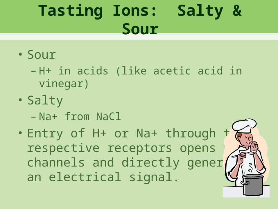
Tasting Ions: Salty & Sour
• Sour– H+ in acids (like acetic acid in vinegar)
• Salty– Na+ from NaCl
• Entry of H+ or Na+ through the respective receptors opens ion channels and directly generatesan electrical signal.
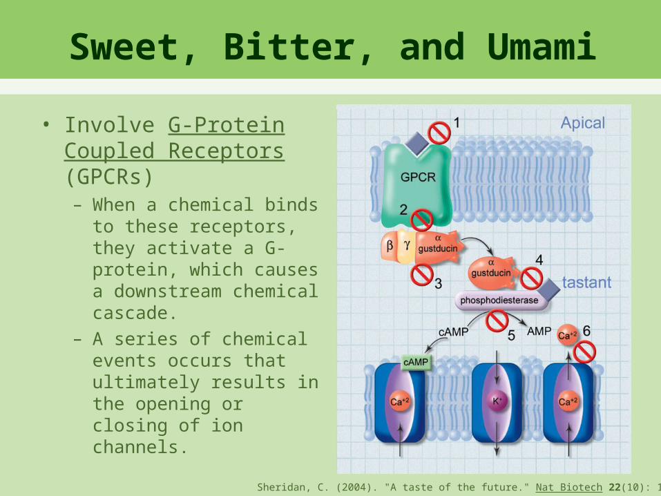
Sweet, Bitter, and Umami
• Involve G-Protein Coupled Receptors (GPCRs)– When a chemical binds
to these receptors, they activate a G-protein, which causes a downstream chemical cascade.
– A series of chemical events occurs that ultimately results in the opening or closing of ion channels.
Sheridan, C. (2004). "A taste of the future." Nat Biotech 22(10): 1203-1205.

• Sweet:– T1R2 and T1R3 receptors– Activates Ca2+ channels
• Umami:– T1R1 and T1R3 receptors– Activates Ca2+ channels
• Bitter:– T2R receptors interacting with gustducin
Sweet, Bitter, and Umami

Taste Cell Summary
• Ion Channels– Sour: H+– Salty: Na+
• GPCRs– Sweet:
T1R2 + T1R3– Umami:
T1R1 + T1R3– Bitter:
T2R
Eisenstein, M. (2010). "Taste: More than meets the mouth." Nature 468(7327): S18-S19.
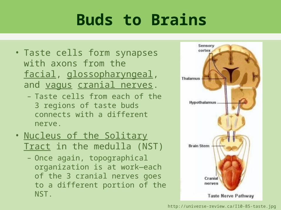
Buds to Brains
• Taste cells form synapses with axons from the facial, glossopharyngeal, and vagus cranial nerves.– Taste cells from each of the 3
regions of taste buds connects with a different nerve.
• Nucleus of the Solitary Tract in the medulla (NST)– Once again, topographical
organization is at work—each of the 3 cranial nerves goes to a different portion of the NST.
http://universe-review.ca/I10-85-taste.jpg
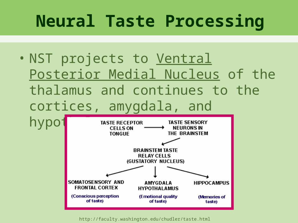
Neural Taste Processing
• NST projects to Ventral Posterior Medial Nucleus of the thalamus and continues to the cortices, amygdala, and hypothalamus
http://faculty.washington.edu/chudler/taste.html

Tasty Visions Experiment
Class Discussion
• What Influences Taste?• Sound Medicine: Neurobiology of
Taste

Neurobiology of Eating
• What brain region controls eating?
• 1940, Hetherington & Ranson – Lesions of the
ventromedial hypothalamus cause obesity & excess food intake in rats
– Lesions of the lateral hypothalamus caused decreased eating & starvation
https://wiki.brown.edu/confluence/display/BN0193S04/Historical+Background

What Does This Mean?
• Could the lateral hypothalamus be the brain’s control center for hunger, while the ventromedial hypothalamus is the center for satiety (feeling full)
• Oversimplified…there’s more to this story than that!– The brain & body have a host of
chemicals to help induce hunger and satiety.
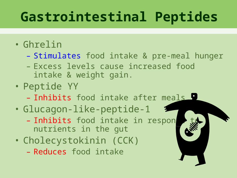
Gastrointestinal Peptides
• Ghrelin– Stimulates food intake & pre-meal hunger– Excess levels cause increased food intake &
weight gain.
• Peptide YY– Inhibits food intake after meals
• Glucagon-like-peptide-1– Inhibits food intake in response to
nutrients in the gut
• Cholecystokinin (CCK)– Reduces food intake

Peripheral Hormones
• Leptin & Insulin– Both hormones reduce food intake.
• Both circulate in the body in response to fat content and enter the central nervous system in proportionate levels.– Receptors expressed in brain areas
important for energy
• Leptin is a protein encoded for by the ob gene.

Leptin, Body Fat, & Food
• Douglas Coleman discovered in the 1960s that the ob genes indicates to the brain that fat reserves are at a normal level.
• So what would we expect from mice lacking the ob gene?
• In the 1990s, Jeffrey Friedman began studying leptin.– What happens if we give an ob/ob mouse
leptin?– Reverses the obesity until normal levels of
adipose tissue are reached.

Ob/Ob Mice
http://en.wikipedia.org/wiki/Ob/ob_mouse

Energy homeostasis
Schwartz et al Nature 2000 404:661
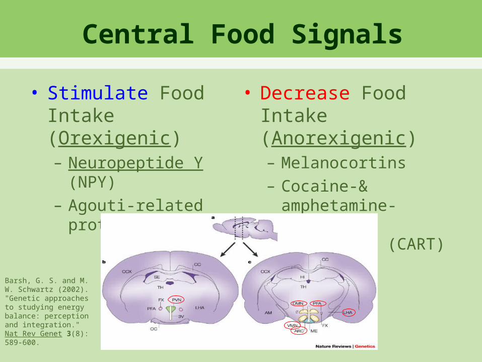
Central Food Signals
• Stimulate Food Intake (Orexigenic)– Neuropeptide Y
(NPY)– Agouti-related
protein (AgRP)
• Decrease Food Intake (Anorexigenic)– Melanocortins – Cocaine-&
amphetamine-regulated transcript (CART)
Barsh, G. S. and M. W. Schwartz (2002). "Genetic approaches to studying energy balance: perception and integration." Nat Rev Genet 3(8): 589-600.
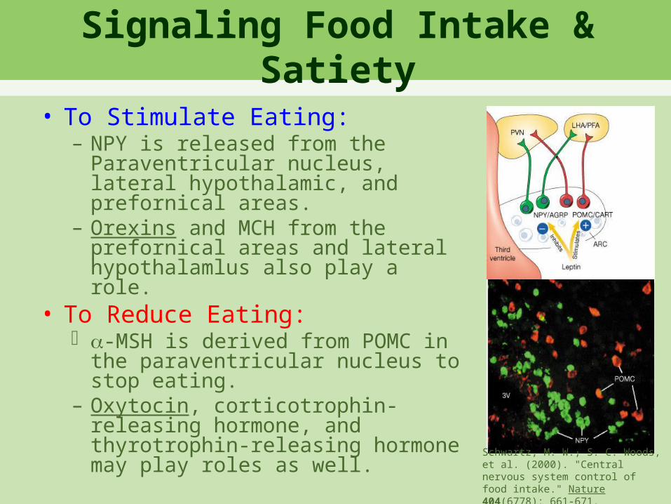
Signaling Food Intake & Satiety
• To Stimulate Eating:– NPY is released from the
Paraventricular nucleus, lateral hypothalamic, and prefornical areas.
– Orexins and MCH from the prefornical areas and lateral hypothalamlus also play a role.
• To Reduce Eating: -MSH is derived from POMC in
the paraventricular nucleus to stop eating.
– Oxytocin, corticotrophin-releasing hormone, and thyrotrophin-releasing hormone may play roles as well.
Schwartz, M. W., S. C. Woods, et al. (2000). "Central nervous system control of food intake." Nature 404(6778): 661-671.

Other Factors
• Brain Stem• Reward Pathways
– Dopamine– Serotonin– Endocannabinoids– Opioids– Cholinergic systems

Anorectic Peptides (Inhibit Feeding Behavior) Orexigenic Peptides (Stimulate Feeding BehaviorAbbreviation Full Name Location Abbreviation Full Name Location-MSH Alpha-melanocyte-
stimulatinghormone
Arcuatenucleus
NPY Neuropeptide Y Arcuatenucleus
CART Cocaine- andamphetamine-regulated transcript
Arcuatenucleus
AgRP Agouti-relatedpeptide
Arcuatenucleus
OT Oxytocin PVN MCH Melanin-concentratinghormone
Lateralhypothalamicarea
TRH Thyrotropin-releasing hormone
PVN Orexin Lateralhypothalamicarea
Summary
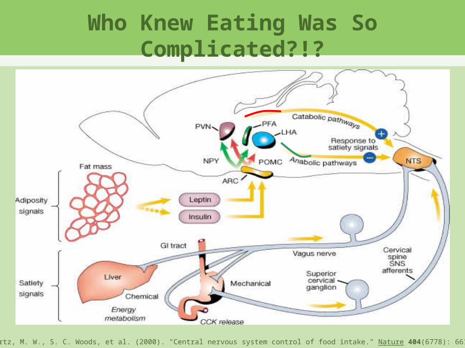
Who Knew Eating Was So Complicated?!?
Schwartz, M. W., S. C. Woods, et al. (2000). "Central nervous system control of food intake." Nature 404(6778): 661-671.

Hypothalamus

Eating Disorder Treatment Plans
http://fitnesslines.com/health-tips/what-is-an-eating-disordercausessymptoms-and-treatment-for-eating-disorders-in-children/

Smell Discrimination Experiment
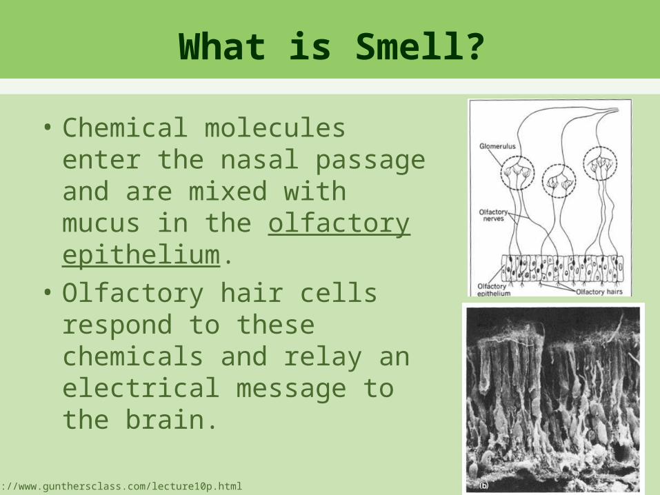
What is Smell?
• Chemical molecules enter the nasal passage and are mixed with mucus in the olfactory epithelium.
• Olfactory hair cells respond to these chemicals and relay an electrical message to the brain.
http://www.gunthersclass.com/lecture10p.html
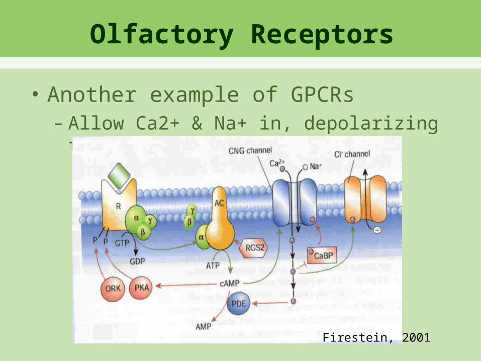
Olfactory Receptors
• Another example of GPCRs– Allow Ca2+ & Na+ in, depolarizing the
cell
Firestein, 2001
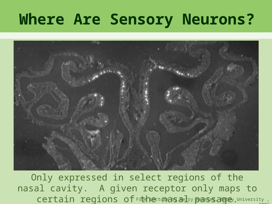
Only expressed in select regions of the nasal cavity. A given receptor only maps to certain regions of the nasal passage.
Where Are Sensory Neurons?
From Lecture by Kerry Ressler, Emory University , 2006

From Lecture by Kerry Ressler, Emory University , 2006

Olfactory Bulb
• Olfactory information is processed in the olfactory bulb.– Consists of glomeruli:
round regions without cell bodies that receive input from olfactory nerves.
– Each neuron sends only 1 axon to 1 glomerulus in the olfactory bulb.
Firestein 2001

Does This Organization Seem Familiar?
From Lecture by Kerry Ressler, Emory University , 2006

Patterning Creates Scents
• The patterns of glomeruli that are stimulated is important for determining the odor sensed.
From Lecture by Kerry Ressler, Emory University , 2006
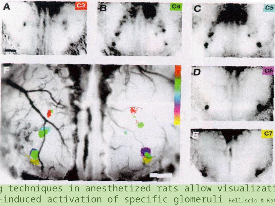
Imaging techniques in anesthetized rats allow visualization of odor-induced activation of specific glomeruli Belluscio & Katz, 2001

Special Scent Processing
• Further processing occurs in the olfactory cortex, hippocampus, amygdala, and hypothalamus
From Lecture by Kerry Ressler, Emory University , 2006

Changes in Smell
• NPR “Five Senses, Minus One: Living Without Smell”
• “The Dog Beneath the Skin”
Current Research in Taste and Smell


![Research articleLipopolysaccharide-induced inflammation ...proteins, and cytokines in taste buds [15-17]. MCP-1 expression in taste papillae can also be upregulated by gustatory nerve](https://static.fdocuments.in/doc/165x107/60b0d0698d3dc116c9376b32/research-articlelipopolysaccharide-induced-inflammation-proteins-and-cytokines.jpg)


