Targeting Notch Signaling with a Notch2/Notch3 Antagonist ...Targeting Notch Signaling with a...
13
Cancer Therapy: Preclinical Targeting Notch Signaling with a Notch2/Notch3 Antagonist (Tarextumab) Inhibits Tumor Growth and Decreases Tumor-Initiating Cell Frequency Wan-Ching Yen, Marcus M. Fischer, Fumiko Axelrod,Christopher Bond, Jennifer Cain, Belinda Cancilla,William R. Henner, Rene Meisner, Aaron Sato, Jalpa Shah,Tracy Tang, Breanna Wallace, Min Wang, Chun Zhang, Ann M. Kapoun, John Lewicki, Austin Gurney, and Timothy Hoey Abstract Purpose: The Notch pathway plays an important role in both stem cell biology and cancer. Dysregulation of Notch signaling has been reported in several human tumor types. In this report, we describe the development of an antibody, OMP-59R5 (tarextu- mab), which blocks both Notch2 and Notch3 signaling. Experimental Design: We utilized patient-derived xenograft tumors to evaluate antitumor effect of OMP-59R5. Immunohis- tochemistry, RNA microarray, real-time PCR, and in vivo serial transplantation assays were employed to investigate the mechan- isms of action and pharmacodynamic readouts. Results: We found that anti-Notch2/3, either as a single agent or in combination with chemotherapeutic agents was efficacious in a broad spectrum of epithelial tumors, including breast, lung, ovarian, and pancreatic cancers. Notably, the sensitivity of anti- Notch2/3 in combination with gemcitabine in pancreatic tumors was associated with higher levels of Notch3 gene expression. The antitumor effect of anti-Notch2/3 in combination with gemcita- bine plus nab-paclitaxel was greater than the combination effect with gemcitabine alone. OMP-59R5 inhibits both human and mouse Notch2 and Notch3 function and its antitumor activity was characterized by a dual mechanism of action in both tumor and stromal/vascular cells in xenograft experiments. In tumor cells, anti- Notch2/3 inhibited expression of Notch target genes and reduced tumor-initiating cell frequency. In the tumor stroma, OMP-59R5 consistently inhibited the expression of Notch3, HeyL, and Rgs5, characteristic of affecting pericyte function in tumor vasculature. Conclusions: These findings indicate that blockade of Notch2/3 signaling with this cross-reactive antagonist antibody may be an effective strategy for treatment of a variety of tumor types. Clin Cancer Res; 21(9); 2084–95. Ó2015 AACR. Introduction The Notch pathway is one of several key pathways linked to both stem cell biology and cancer (1). In particular, dysregulated Notch2 and/or Notch3 activity has been associated with several human tumor types, including lung (2), ovarian (3), breast (4), pancreatic (5, 6), and colon cancers (7). In addition to its role in develop- mental biology and cancer, Notch signaling also plays a key role in tumor vasculature and pericytes (8). Thus, therapeutic strategies directed at inhibition of the Notch2/3 pathway on tumors and vasculature may offer promise for the treatment of solid tumors. Accumulating evidence has indicated that tumors are frequent- ly composed of heterogeneous cell types that vary in their ability to initiate new tumor growth and that cancer stem cells (CSC, also referred to as tumor-initiating cells) drive tumor growth and progression and are relatively resistant to many existing therapies, including conventional chemotherapy and radiation treatments (9, 10). Because aberrant Notch signaling has been implicated in cancer, cancer stem cells, and tumor vasculature, therapeutic strategies that effectively target Notch signaling could have a major impact on cancer patient survival. Although inhibition of Notch receptor cleavage enzymes by gamma-secretase inhibitors (GSI) have been developed and progressed in the clinic, the therapeutic utility of these compounds is limited due to intestinal toxicity resulting from pan-Notch inhibition (11). In the current study, we report the development of a novel cross-reactive anti- body OMP-59R5 that selectively inhibits the function of both Notch2 and Notch3. We evaluated antitumor effect of anti- Notch2/3 antibody and its mechanisms of actions using patient-derived xenograft (PDX) models. Materials and Methods Antibody generation and characterization OMP-59R5 was generated by panning the HuCAL GOLD phage-display library (MorphoSys AG; ref. 12) with recombinant Notch2 extracellular domain (EGF1–12) containing the ligand- binding site. DNA fragments encoding the fAb generated from the phage display library were subcloned into a full-length human IgG2 expression vector and subsequently expressed into CHO cells and purified. To determine the ability of OMP-59R5 to block OncoMed Pharmaceuticals, Inc., Redwood City, California. Note: Supplementary data for this article are available at Clinical Cancer Research Online (http://clincancerres.aacrjournals.org/). Current address for C. Bond: CDRD—The Centre for Drug Research and Development, Vancouver, British Columbia; current address for A. Sato: Sutro Biopharma, Inc., South San Francisco, California. Corresponding Author: Wan-Ching Yen, OncoMed Pharmaceuticals Inc., 800 Chesapeake Drive, Redwood City, CA 94063. Phone: 650-995-8273; Fax: 650- 298-8600; E-mail: [email protected] doi: 10.1158/1078-0432.CCR-14-2808 Ó2015 American Association for Cancer Research. Clinical Cancer Research Clin Cancer Res; 21(9) May 1, 2015 2084 on June 16, 2020. © 2015 American Association for Cancer Research. clincancerres.aacrjournals.org Downloaded from
Transcript of Targeting Notch Signaling with a Notch2/Notch3 Antagonist ...Targeting Notch Signaling with a...

Cancer Therapy: Preclinical
Targeting Notch Signaling with a Notch2/Notch3Antagonist (Tarextumab) Inhibits Tumor Growthand Decreases Tumor-Initiating Cell FrequencyWan-Ching Yen, Marcus M. Fischer, Fumiko Axelrod, Christopher Bond, Jennifer Cain,Belinda Cancilla,William R. Henner, Rene Meisner, Aaron Sato, Jalpa Shah, Tracy Tang,Breanna Wallace, Min Wang, Chun Zhang, Ann M. Kapoun, John Lewicki, Austin Gurney,and Timothy Hoey
Abstract
Purpose: The Notch pathway plays an important role in bothstem cell biology and cancer. Dysregulation of Notch signalinghas been reported in several human tumor types. In this report, wedescribe the development of an antibody, OMP-59R5 (tarextu-mab), which blocks both Notch2 and Notch3 signaling.
Experimental Design: We utilized patient-derived xenografttumors to evaluate antitumor effect of OMP-59R5. Immunohis-tochemistry, RNA microarray, real-time PCR, and in vivo serialtransplantation assays were employed to investigate the mechan-isms of action and pharmacodynamic readouts.
Results:We found that anti-Notch2/3, either as a single agent orin combination with chemotherapeutic agents was efficacious ina broad spectrum of epithelial tumors, including breast, lung,ovarian, and pancreatic cancers. Notably, the sensitivity of anti-Notch2/3 in combination with gemcitabine in pancreatic tumors
was associated with higher levels of Notch3 gene expression. Theantitumor effect of anti-Notch2/3 in combination with gemcita-bine plus nab-paclitaxel was greater than the combination effectwith gemcitabine alone. OMP-59R5 inhibits both human andmouseNotch2 andNotch3 function and its antitumor activity wascharacterized by a dual mechanism of action in both tumor andstromal/vascular cells inxenograft experiments. In tumorcells, anti-Notch2/3 inhibited expression of Notch target genes and reducedtumor-initiating cell frequency. In the tumor stroma, OMP-59R5consistently inhibited the expression of Notch3, HeyL, and Rgs5,characteristic of affecting pericyte function in tumor vasculature.
Conclusions: These findings indicate that blockade ofNotch2/3 signaling with this cross-reactive antagonist antibodymay be an effective strategy for treatment of a variety of tumortypes. Clin Cancer Res; 21(9); 2084–95. �2015 AACR.
IntroductionTheNotchpathway is one of several key pathways linked to both
stem cell biology and cancer (1). In particular, dysregulatedNotch2and/or Notch3 activity has been associated with several humantumor types, including lung (2), ovarian (3), breast (4), pancreatic(5, 6), and colon cancers (7). In addition to its role in develop-mental biology and cancer, Notch signaling also plays a key role intumor vasculature and pericytes (8). Thus, therapeutic strategiesdirected at inhibition of the Notch2/3 pathway on tumors andvasculature may offer promise for the treatment of solid tumors.
Accumulating evidence has indicated that tumors are frequent-ly composed of heterogeneous cell types that vary in their abilityto initiate new tumor growth and that cancer stem cells (CSC, also
referred to as tumor-initiating cells) drive tumor growth andprogression and are relatively resistant tomany existing therapies,including conventional chemotherapy and radiation treatments(9, 10). Because aberrant Notch signaling has been implicated incancer, cancer stem cells, and tumor vasculature, therapeuticstrategies that effectively target Notch signaling could have amajor impact on cancer patient survival. Although inhibition ofNotch receptor cleavage enzymes by gamma-secretase inhibitors(GSI) have been developed and progressed in the clinic, thetherapeutic utility of these compounds is limited due to intestinaltoxicity resulting from pan-Notch inhibition (11). In the currentstudy, we report the development of a novel cross-reactive anti-body OMP-59R5 that selectively inhibits the function of bothNotch2 and Notch3. We evaluated antitumor effect of anti-Notch2/3 antibody and its mechanisms of actions usingpatient-derived xenograft (PDX) models.
Materials and MethodsAntibody generation and characterization
OMP-59R5 was generated by panning the HuCAL GOLDphage-display library (MorphoSys AG; ref. 12) with recombinantNotch2 extracellular domain (EGF1–12) containing the ligand-binding site. DNA fragments encoding the fAb generated from thephage display library were subcloned into a full-length humanIgG2 expression vector and subsequently expressed into CHOcells and purified. To determine the ability of OMP-59R5 to block
OncoMed Pharmaceuticals, Inc., Redwood City, California.
Note: Supplementary data for this article are available at Clinical CancerResearch Online (http://clincancerres.aacrjournals.org/).
Current address for C. Bond: CDRD—The Centre for Drug Research andDevelopment, Vancouver, British Columbia; current address for A. Sato:Sutro Biopharma, Inc., South San Francisco, California.
Corresponding Author: Wan-Ching Yen, OncoMed Pharmaceuticals Inc., 800Chesapeake Drive, Redwood City, CA 94063. Phone: 650-995-8273; Fax: 650-298-8600; E-mail: [email protected]
doi: 10.1158/1078-0432.CCR-14-2808
�2015 American Association for Cancer Research.
ClinicalCancerResearch
Clin Cancer Res; 21(9) May 1, 20152084
on June 16, 2020. © 2015 American Association for Cancer Research. clincancerres.aacrjournals.org Downloaded from

ligand binding to humanNotch2 andNotch3 receptors, HEK 293cells were transfected with cDNA expression vector encodinghuman Notch2 as well as a second vector encoding GFP,pcDNA-GFP, to mark the transiently transfected cells. SpecificOMP-59R5 binding was assessed by determining the presence ofcells positive for GFP signal and PE signal. For epitope mapping,HEK 293 cells were transiently transfected with expression vectorsencoding humanNotch2, humanNotch1, or humanNotch1withresidues 383–387 mutated to the corresponding human Notch2residues. Cells were also cotransfected with a plasmid encodingGFP to mark those cells that received transfected plasmid. Cellswere incubated with OMP-59R5 and fluorescent secondaryantibody and then examined by FACS. The ability of antibodiesto block activation of Notch signaling was determined by invitro luciferase reporter assays as described previously (13). Thebinding affinities of OMP-59R5 were determined using a Bia-core 2000. The data were fit using the simultaneous global fitequation to yield affinity constants (Kd) for each protein.Detailed assay protocols are described in Supplementary Mate-rials and Methods.
In vivo animal studiesThe establishment and characterization of minimally passed
human tumor xenograft models were performed as previouslydescribed (13, 14). Sources of surgically removed patient tumorsare CHTN (OMP-PN4, OMP-OV38), Asterand (OMP-LU40), andMRI (OMP-LU68). These tumors were established at OncoMedPharmaceuticals, Inc. OMP-PN8, PN16, and PN17 and wereobtained from Dr. Diane Simeone and UM-PE13 from Dr.Michael Clarke at the University of Michigan (Ann Arbor, MI).Cells were injected subcutaneously to NOD/SCID mice for effi-cacy studies. Treatment started when the mean tumor volumesreached about 100mm3.OMP-59R5was dosed at 40mg/kg everyother week. Gemcitabine was given from 5 to 40 mg/kg inpancreatic tumor xenografts in Supplementary Table S3, exceptas indicated, that is, Fig. 4B and Fig. 4C. All chemotherapeuticagents were given once weekly. Both antibody and chemothera-peutic agents were administered intraperitoneally. The proce-dures for tumorigenicity study were described previously (14).CSC frequency was determined using L-Calc Version 1.1 software(StemCell Technologies). Differences in frequency betweengroups were analyzed by the likelihood-ratio test. Difference ofP < 0.05 was considered significantly different. Detailed in vivo
studies and in vivo limiting dilution assay (LDA) protocols aredescribed in Supplementary Materials and Methods.
Real-Time RT-PCR, mRNA sequencing, and microarray geneexpression
Tumor RNAswere isolatedusing theRNeasy Fibrous TissueMiniKit (Qiagen) with DNAseI treatment. Five-hundred nanogramstotal RNA was reverse transcribed into cDNA. Quantitative real-time PCR was performed in an ABI 7900 HT Fast Real Time PCRSystem and analyzed using the SDSv2.3 (Applied Biosystems). Theresults were normalized with the housekeeping gene GADPH. Allprimer and probe sets were obtained fromApplied Biosystems. Formicroarray, sampleswere amplifiedusing theOvationRNAAmpli-fication System V2 (NuGEN), adjusted for array background,normalized signal intensity with GCRMA algorithm in the open-source Bioconductor software (www.bioconductor.org), and pro-filed for mRNA expression using Affymetrix both HG-U133plus2human andMG-430 2.0mouse chips to assess treatment effects onhuman tumor cells and onmouse stroma cells independently. Theprobe sets that were not species-specific were omitted from theanalysis. Data were subjected to the Gene Set Enrichment Analysis(GSEA). Genes differentially expressed between two groups wereidentified with the Bayesian t test (Cyber-T; ref. 15). Bayesian one-way ANOVA and Tukey HSD post hoc test were performed forexperiments with more than two groups. The significantly regu-lated genes between groups or time points were chosen based onthe P value <0.05 and absolute fold change �1.5. The sequencinglibraries were prepared following the protocols supplied by Illu-mina for sequencing mRNA samples (Illumina CA). The FASTQsequence reads were aligned to human genome hg19 and mousegenome mm9 with tophat (v2.0.9) and bowtie (v1.0.0). After thealignment of sequence reads, the uniquely mapped reads werecounted for each gene byHTSeq python script (v0.5.3p9). The rawcounts per gene in each xenograft tumor were then normalized asRPKM (reads per kilobase per million mapped reads) to representthe expression level of the gene in the tumor.
HistologyWhole tumors were excised and processed into formalin-fixed,
paraffin-embedded (FFPE) slides or as nonfixed optimal cuttingtemperature (OCT, Tissue-Tek, Sakura) embedded slides. Immu-nohistochemistry slides were scanned using Imagescope (Scan-Scope AT, Aperio). Total nuclei, Ki67-labeled cells, staining area,and intensity were quantified by Aperio software after excludingnecrotic regions. The primary antibody for immunohistochem-istry (IHC) was anti-Ki-67 (Vector, VP-RM04) and for immuno-fluorescence were anti-CD31 (BD Biosciences, 550274) and anti-Desmin clone D93F5 (Cell Signaling Technology). Antipimoni-dazole (Hypoxyprobe) was used for intratumoral hypoxia detec-tion. Detailed protocols for vascular perfusion, IHC, and immu-nofluorescence detections were described in SupplementaryMaterials and Methods.
Statistical analysisData for tumor measurements are expressed as mean � SEM.
Differences in mean values between groups were analyzed by thenonparametric t test. Multiple comparisons used a two-wayANOVA test followed by the Bonferroni post-test comparison.Differences of P < 0.05 are considered significantly different.Software for statistical analysis was by GraphPad Prism5 (Graph-Pad Software Inc.).
Translational Relevance
Aberrant Notch signaling has been implicated in cancer,cancer stem cells, and tumor vasculature. We report the devel-opment of OMP-59R5, an antibody that selectively inhibitsthe function of both Notch2 and Notch3. Our data demon-strate that OMP-59R5 was efficacious in inhibiting the growthof various epithelial tumors with minimal intestinal toxicity.Interference with Notch2/3 signaling by OMP-59R5 delayedtumor recurrence, decreased cancer stem cell frequency, andmodulated the function of tumor vasculature. These findingsprovide evidence for the utility of targeting Notch2 andNotch3 for cancer treatment. Anti-Notch2/3 (tarextumab) iscurrently advancing in phase II clinical testing for treatment ofpancreatic and small-cell lung cancers.
Antitumor Effect of Notch2/3 Antagonist (Tarextumab)
www.aacrjournals.org Clin Cancer Res; 21(9) May 1, 2015 2085
on June 16, 2020. © 2015 American Association for Cancer Research. clincancerres.aacrjournals.org Downloaded from

ResultsOMP-59R5 is a cross-reactive Notch2 and Notch3 antagonist
OMP-59R5 was identified using phase display (12) and wasinitially selected against human Notch2. Surface plasmon reso-nance-binding studies determined that this antibody demon-strates high-affinity binding toNotch2 andNotch3, withminimalbinding to Notch1 and no detectable binding to Notch4 (Sup-
plementary Fig. S1A). OMP-59R5 blocks Notch ligand DLL4 andJAG1 binding to human Notch2 (Fig. 1A). Analysis of deletionmutations identified EGF10 of Notch2 extracellular domains asrequired for binding (Note: due to the variance in the number ofEGF repeats between the individual Notch family members, thecorresponding EGF repeat of EGF10 in Notch2 is EGF9 inNotch3). Mutagenesis analysis revealed that OMP-59R5 bindsto full-length human Notch2 and Notch1 with residues 383–387
A
B
C hNotch1
0.001 0.01 0.1 1 10 1000
100,000
200,000
300,000
400,000
500,000
OMP-59R5No antibodyNo ligand
Control mAb
mg/mL
OMP-59R5 (PcDNA3)No antibody (PcDNA3)No ligand (PcDNA3)
Control mAb(PcDNA3)RL
U
hNotch1
0.001 0.01 0.1 1 10 100 1,0000
100,000
200,000
300,000
400,000
500,000DBZNo antibodyNo ligandDBZ (PcDNA3)No antibody (PcDNA3)No ligand (PcDNA3)
nmol/L
mg/mL nmol/L
mg/mL nmol/L
RL
U
hNotch2
0.001 0.01 0.1 1 10 1000
200,000
400,000
600,000
OMP-59R5No antibodyNo ligand
Control mAb
OMP-59R5 (PcDNA3)No antibody (PcDNA3)No ligand (PcDNA3)
Control mAb (PcDNA3)RL
U
hNotch2
0.001 0.01 0.1 1 10 100 1,0000.0
200,000.0
400,000.0
600,000.0
800,000.0DBZNo antibodyNo ligandDBZ (PcDNA3)No antibody (PcDNA3)No ligand (PcDNA3)
RL
U
hNotch3
0.001 0.01 0.1 1 10 1000
100,000
200,000
300,000
OMP-59R5No antibodyNo ligand
Control mAb
OMP-59R5 (PcDNA3)No antibody (PcDNA3)No ligand (PcDNA3)
Control mAb (PcDNA3)RL
U
hNotch3
0.001 0.01 0.1 1 10 100 1,0000
100,000
200,000
300,000DBZNo antibodyNo ligandDBZ (PcDNA3)No antibody (PcDNA3)No ligand (PcDNA3)
RL
U
JAG1 binding DLL4 binding
No mAb No mAb+OMP-59R5 +OMP-59R5
59R5
HumanNotch 3
HumanNotch 2
0 102
103
104
105
<FITC-A>
0
102
103
104
105
<P
E-A
>
0 102
103
104
105
<FITC-A>
0
102
103
104
105
<P
E-A
>
0 102
103
104
105
<FITC-A>
0
102
103
104
105
<P
E-A
>
0 102
103
104
105
<FITC-A>
0
102
103
104
105
<P
E-A
>
0 102
103
104
105
<FITC-A>
0
102
103
104
105
<P
E-A
>
0 102
103
104
105
<FITC-A>
0
102
103
104
105
<P
E-A
>
0 102
103
104
105
<FITC-A>
0
102
103
104
105
<P
E-A
>
0 102
103
104
105
<FITC-A>
0
102
103
104
10
5<
PE
-A>
Lig
and
bin
din
g
Human Notch2 Human Notch1Human Notch1(N2, 383-387)
MF
I
PE
PE
Figure 1.Binding and blocking of OMP-59R5 tohuman and mouse Notch receptors.A, OMP-59R5 blocks ligand binding toNotch2 andNotch3. B, binding epitopeof OMP-59R5 to human Notch2/3.C, effect of OMP-59R5 on blockinghuman Notch1, Notch2, and Notch3signaling. Mean� SD; n¼ 3 replicates.Right, gamma-secretase inhibitorDBZ-positive control. RLU, relativelight units and is a unit of luciferasereporter activity.
Yen et al.
Clin Cancer Res; 21(9) May 1, 2015 Clinical Cancer Research2086
on June 16, 2020. © 2015 American Association for Cancer Research. clincancerres.aacrjournals.org Downloaded from
mutated to the corresponding humanNotch2 residues, but not tofull-length Notch1 by flow cytometry (Fig. 1B), indicating thatthis region comprises at least part of the binding epitope. Align-ment of the EGF9/10 region of Notch2 and Notch3 from rodent,human, and cynomolgusmonkey indicated that bothNotch2 andNotch3 are identical within the EGF repeat that contains thebinding epitope of OMP-59R5 in several species (SupplementaryFig. S1B). Surface plasmon resonance further demonstrated thatOMP-59R5 bound with high affinity to both Notch2 and Notch3from mouse, rat, and human (Supplementary Table S1). Using aNotch-responsive luciferase reporter assay in HeLa cells, we dem-onstrated that OMP-59R5 effectively inhibited human Notch2and Notch3 reporter activity but had no activity in blockingNotch1 signaling. In contrast, the gamma secretase inhibitorDBZ,a pan-Notch inhibitor, inhibited signaling from Notch1, Notch2,and Notch3 (Supplementary Fig. 1C).
OMP-59R5 inhibits xenograft tumor growth through reducingtumor cell proliferation and promotes differentiation
The effect of OMP-59R5 on tumor growth was accessed usinga panel of minimally in vivo passaged PDX models (14). Engraft-ment in mice and drug treatment in these models has beenshown to correlate with clinical outcome and treatment response(16, 17). These tumor models retain much of the heterogeneityof the original tumors enabling the study of the characterizationof CSCs through serial transplantation (13, 14). The histologicfeatures of these tumors are summarized in Supplementary TableS2. All tumors express human Notch1, Notch2, and Notch3mRNA (Supplementary Fig. S2). OMP-59R5 treatment resultedin additional antitumor activity in combination with gemcita-bine relative to gemcitabine alone in 6 of 10 tumors tested(Supplementary Table S3). In the most sensitive tumors, OMP-PN8 and OMP-PN17, combination therapy resulted in tumorregression beginning 2 weeks after treatment in OMP-PN8 and 3weeks after treatment in OMP-PN17 (Supplementary Fig. S3).Histologic analysis in OMP-PN8 at the conclusion of the studyrevealed that OMP-59R5 single-agent treatment did not altertumor cell morphology (hematoxylin and eosin), total tumorcell density (measured by total nuclei/mm2), or cell proliferation(shown by Ki-67 immunoreactivity). Gemcitabine treatmentresulted in 60% decrease in tumor cell density and 43% decreasein cell proliferation. The combination of OMP-59R5 and gem-citabine led to an additional 62% increase in apoptotic cell deathand 52% reduction in tumor cell density versus gemcitabine (P <0.05 in both cases). In the moderately differentiated adenocar-cinoma OMP-PN21, OMP-59R5 treatment alone reduced tumorcell proliferation by 40% and promoted differentiation byinducing mucin-producing cells, as indicated by a 2.5-foldincrease in Alcian blue staining compared with untreated con-trols (Fig. 2A). Gemcitabine treatment alone decreased cellproliferation by about 30% versus control, but had no effecton differentiation. Importantly, the combination therapyresulted in an additional 15% decrease in proliferating cellsversus gemcitabine and induced a greater than 3-fold increasein Alcian blue staining (P < 0.05 in both cases). No apparentimpact of OMP-59R5 on the intestinal morphology or increasedsecretory goblet cells was found at the dose used in these studies(Supplementary Fig. S4A). We did, however, observe an effect ofOMP-59R5 on rodent teeth with long-term repeated high dosesof OMP-59R5, beginning approximately 6 weeks after initiationof treatment (Supplementary Fig. S4B).
Sensitivity to OMP-59R5 is correlated with Notch3 expressionin pancreatic tumors
Notably, the antitumor efficacy of OMP-59R5 in combinationwith gemcitabine was significantly associated with expression ofhuman Notch3 gene in pancreatic cancer. Tumors that were moresensitive to the OMP-59R5 plus gemcitabine combination(responders) versus gemcitabine alonehadhigher levels ofNotch3baseline expression compared with tumors that showed no com-bination effect (nonresponders; Fig. 2B). The mean gene expres-sion levelswere significantly different between the responders andthe nonresponders (10.44 in responders vs. 1.973 in nonrespon-ders, P ¼ 0.0047). The responsiveness of these tumors to thecombination therapy was not associated with either Kras muta-tion status, sensitivity to gemcitabine, or molecular subtypes ofpancreatic ductal adenocarcinoma as described by Collisson andcolleagues previously (18), that is, classical (tumors express highadhesion-associated and epithelial genes with best survival prog-nosis), exocrine-like (tumors with high tumor cell-derived diges-tive enzyme genes), and quasi-mesenchymal (tumors with highmesenchyme-associated genes; Supplementary Table S3). In con-trast to Notch3, there was no difference in Notch1 or Notch2expression between responders and nonresponders (Supplemen-tary Fig. S2). Thus,Notch3 expression could be a useful predictivebiomarker associated with sensitivity of OMP-59R5 in pancreaticcancer.
Pharmacodynamic effects in xenograft tumors treated withOMP-59R5
To identify pathways and gene sets regulated by OMP-59R5 intumor cells, we isolated tumor RNA from four OMP-59R5–responsive tumors (OMP-PN4, PN8, PN16, and PN17) at theend of studies and analyzed for human and mouse gene expres-sions by microarray. GSEA revealed that OMP-59R5 downregu-lated the Notch pathway, oncogene signatures, cell proliferationgene sets, and several stem cell–related gene sets (SupplementaryTable S4). Tumors treated with gemcitabine upregulated epithe-lial-to-mesenchymal (EMT) gene sets and a subset of the stem cellgene sets, consistent with our previous results indicating thatgemcitabine promotes the EMT phenotype in pancreatic cancer(19). Notably, OMP-59R5 combination therapy reversed theincrease in gemcitabine-induced expression of EMT gene sets anddownregulated genes associated with embryonic stem cells (ESC)(ref. 20; Supplementary Table S4). Analysis of anti-Notch2/3treatment on tumor stroma and vascular cells in xenograftsrevealed that Notch3, Notch target genes Hey2, HeyL, and Rgs5,a gene shown to mark pericytes (21) were consistently down-regulated byOMP-59R5 treatment in the tumor stroma in all fourpancreatic xenografts (Supplementary Table S5).
To confirm gene expression changes identified by microarray,we performed qRT-PCR analysis in OMP-PN17, a tumor thatexpresses high NOTCH3 and responded to the combinationtherapy (Fig. 3A). Gemcitabine alone had no effect on Notchreceptors or Notch target gene expression in OMP-PN17 xeno-graft tumors, whereas OMP-59R5 alone and in combinationwith gemcitabine downregulated human NOTCH2, NOTCH3,andHES1.Notch3,Hes1, Rgs5, andHeyL in the tumor stromawereinhibited by OMP-59R5 and the combination with gemcitabinecompared with control monoclonal antibody (mAb)-treatedtumors. OMP-59R5 had no effect on human NOTCH1, JAG1,and JAG2 and mouse Notch1, 2, and 4 gene expression. Similareffects were found in OMP-PN25, a high NOTCH3-expressing
Antitumor Effect of Notch2/3 Antagonist (Tarextumab)
www.aacrjournals.org Clin Cancer Res; 21(9) May 1, 2015 2087
on June 16, 2020. © 2015 American Association for Cancer Research. clincancerres.aacrjournals.org Downloaded from

tumor and a combination therapy responsive xenograft that wasnot examined by microarray (Supplementary Fig. S5A). In addi-tion, OMP-59R5 alone and the combination with gemcitabinedownregulated NANOG and OCT4, two key genes that werefound to be downregulated in ESC gene sets in tumor cells byGSEA. On the other hand, gemcitabine treatment had no effect orresulted in increased expression (Fig. 3A and Supplementary Fig.S5A). Real-time PCR analysis of EMT genes identified by GSEA intwo of responsive tumors, OMP-PN8 andOMP-PN17, confirmed
that gemcitabine induced mesenchymal gene expression (CDH2,VIM, FN1, TWIST1, SNAI1, SNAI2), whereas OMP-59R5 treat-ment and the combination therapy reversed or reduced thesemarker genes (Fig. 3A and Supplementary Fig. S5A). Immuno-histochemical analysis further demonstrated that the combina-tion therapy significantly decreased vimentin (VIM) expression inthe tumor cells (Supplementary Fig. S5B). We also observed that,in OMP-PN17, OMP-59R5 treatment and the combinationdecreased protein levels of Notch3 receptor extracellular domain
A OMP-PN21OMP-PN8
H&E Ki67 Ki67 Alcian Blue
NR R
15
10
5
0
PN25
PN17
PN16PN21PN4PN8PN23
PN13PN11
PN7Not
ch3
expr
essi
on (
log 2
)B
Con
trol
mA
bO
MP
-59R
5G
emci
tabi
neC
ombi
natio
n
Figure 2.In vivo efficacy of OMP-59R5 inpancreatic xenograft tumors. A,histologic evaluation of OMP-PN8 andOMP-PN21 xenograft tumors treatedwith OMP-59R5 and gemcitabine.Magnification, �20. Quantification isexpressed as mean � SEM; n ¼ 3–4tumors per group. � , P < 0.05 vs.control mAb, �� , P < 0.05 versusgemcitabine by the Bonferronimultiple comparison test. B,correlation of NOTCH3 geneexpression and efficacy of OMP-59R5plus gemcitabine in pancreaticxenograft tumors. P¼ 0.00047 by theWelch t test in the mean geneexpression levels between responders(R) and nonresponders (NR).
Yen et al.
Clin Cancer Res; 21(9) May 1, 2015 Clinical Cancer Research2088
on June 16, 2020. © 2015 American Association for Cancer Research. clincancerres.aacrjournals.org Downloaded from

(Notch3 ECD) and the active form of Notch3 receptors intracel-lular domain (Notch3 ICD) compared with control mAb andgemcitabine in this model (Fig. 3B). In addition, tumor cellswith high intensity of Notch3 ECD staining were significantlydecreased by OMP-59R5 and combination treatment (>75% inboth cases, P < 0.05). Similar results were observed by Westernblot analysis in OMP-PN25 xenograft tumors (SupplementaryFig. S5C). In OMP-PN11, a tumor with low level ofNOTCH3 anddid not respond to the combination therapy, Notch3 ECD wasundetected, and baseline Notch3 ICD level was 10-fold lowercompared with OMP-PN17. Quantitative analysis of Notch3 ICDlevels in OMP-PN11 demonstrated that gemcitabine significantlyincreased Notch3 ICD level. Although OMP-59R5 single agent
and the combination significantly reduced nuclear Notch3 ICDlevels compared with control, this reduction did not influencetumor growth (Fig. 3B). It is unlikely that insensitivity to OMP-59R5 in nonresponsive tumors is due to increasing Notch1activity upon Notch2/3 inhibition by OMP59R5 treatmentas OMP-59R5 treatment decreased Notch1 ICD level in bothOMP-PN11 and OMP-PN17 tumors (P < 0.05 in both cases),and neither gemcitabine alone nor the combination therapymodulated N1 ICD level in these tumors (Supplementary Fig.S5D). Collectively, these data suggest that Notch3 expressionis regulated by a positive feedback loop and blockade ofNotch2/3 signaling with OMP-59R5 impedes this autoregula-tory circuit.
Con
trol
mA
bO
MP
-59R
5G
emci
tabi
neC
ombi
natio
n
Notch3 ECD Notch3 ICD
A
B OMP-PN11OMP-PN17
Notch3 ECD Notch3 ICD
Not analyzed; background staining only
Rel
ativ
e q
uan
tity
vs.
con
tro
l mA
b
Rel
ativ
e q
uan
tity
vs.
con
tro
l mA
b
Figure 3.Effect of OMP-59R5 on NOTCH targetgenes and protein expression inpancreatic xenograft tumors. A, qRT-PCR gene expression in control mAband treated OMP-PN17 tumors at theconclusion of study (SupplementaryFig. S3). Gene expression wasnormalized with the housekeepinggene GAPDH and expressed as fold ofcontrol mAb-treated tumors. Mean �SD, n ¼ 3–4 per group. �, P < 0.05versus control mAb by one-wayANOVA followed by the Tukey post-test. B, immunohistochemical analysisof Notch3 ECD and Notch3 ICD inOMP-PN17 and OMP-PN11 tumorsfrom control and treated tumors.Magnification:�20. Staining area andintensity were expressed as H-score.Mean � SEM; n ¼ 3–4 per group.�� , P < 0.05 versus control mAb by theBonferroni multiple comparison test.NA, not analyzed due to backgroundstaining only.
Antitumor Effect of Notch2/3 Antagonist (Tarextumab)
www.aacrjournals.org Clin Cancer Res; 21(9) May 1, 2015 2089
on June 16, 2020. © 2015 American Association for Cancer Research. clincancerres.aacrjournals.org Downloaded from

OMP-59R5 reduces CSC frequency anddelays tumor recurrenceafter termination of chemotherapy
To determine the functional significance of OMP-59R5 treat-ment on the tumorigenic potential of cells within the tumor, wecarried out in vivo serial transplant, LDA (22) experiments (Fig. 4A,left) in OMP-PN8 xenograft tumors, a model that induced tumorregression byOMP-59R5 in combination with gemcitabine (Sup-plementary Fig. S3; Supplementary Table S3). This functionalassay measures in vivo tumorigenicity and makes no assumptionsabout the frequency, FACSmarker profile, or heterogeneity of thetumor-initiating cell population. As seen in Fig. 4A (right), controlantibody-treated tumors had a CSC frequency of 1/81. Gemcita-
bine treatment produced a 2-fold increase in CSC frequencycompared with control (P < 0.05). OMP-59R5 treatment alonedid not alter CSC frequency compared with control (Fig. 4A)although the tumor take rate at low cell doses, that is, 30 cells and90 cells, in the OMP-59R5 treatment group was about 50% lowerthan the control group. Given the fact that stem cell genes areaffected by single-agent OMP-59R5 treatment indicates that theantibody has effects on stem cells in the absence of chemotherapy,and this effect may sensitize tumorigenic cells to the cytotoxiceffects of chemotherapy. Indeed, the combination of OMP-59R5and gemcitabine produced a 7-fold decrease in CSC frequencycompared with control and more than 10-fold reduction in CSC
In vivo limiting dilution assay(tumor growth - 89 days)
Control m
Ab-30
Control m
Ab-90
Control m
Ab-270
OMP-59R
5-30
OMP-59R
5-90
OMP-59R
5-27
0
Gemcit
abin
e-30
Gemcit
abin
e-90
Gemcit
abin
e-27
0
Combin
atio
n-30
Combin
atio
n-90
Combin
atio
n-270
0
500
1,000
1,500
2,000
2,500
Tu
mo
r vo
lum
e (m
m3 )
Tu
mo
r vo
lum
e (m
m3 )
Tu
mo
r vo
lum
e (m
m3 )
Control m
Ab
OMP-59R5
Gemcitabine
Combination
0.00
0.01
0.02
0.03
1/811/71
1/44
1/637
*
*, **CS
C f
req
uen
cy
0 20 40 60 80 100 120 140 1600
200
400
600
800
1,000
1,200
1,400
1,600
Gem(days 49–87)
70 mg/kg gemcitabine
Gem->OMP-59R5
mAb(days 88–160)
**
Days after cell injection
0 20 40 60 80 100 1200
300
600
900
1,200
1,500
Control mAb10 mg/kg Gem+30 mg/kg nab -paclitaxelCombination
Chemo+/-mAb(days 0–42)
Chemo only(days 43–82)
No treatment(days 83–136)
Days after treatment
A
B
C
Figure 4.Effect of OMP-59R5 on pancreaticCSC frequency and tumorrecurrence after chemotherapytermination in OMP-PN8 xenografttumors. A, LDA (left) and CSCfrequency (right). Mean � SEM;n ¼ 10 animals per cell dose.� , P < 0.05 versus control mAb;�� , P < 0.05 versus gemcitabine byone-way ANOVA followed by theTukey post-test. Effect of OMP-59R5 on tumor recurrence followedby discontinuation of gemcitabine(B) and gemcitabine plus nab-paclitaxel (C). Mean � SEM; n ¼ 9animals per group. �� , P < 0.05versus gemcitabine alone by thenonparametric t test.
Yen et al.
Clin Cancer Res; 21(9) May 1, 2015 Clinical Cancer Research2090
on June 16, 2020. © 2015 American Association for Cancer Research. clincancerres.aacrjournals.org Downloaded from
frequency compared with gemcitabine alone (P < 0.001 vs.gemcitabine; Fig. 4A). These results are consistent with geneexpressiondata indicatingblockade ofNotch impairs the functionof tumorigenic cancer stem cells.
To evaluate the effect of OMP-59R5 on tumor recurrence afterchemotherapy, we treated OMP-PN8 pancreatic tumors with ahigh dose of gemcitabine (70 mg/kg, weekly for 5 weeks) suffi-cient to induce tumor regression. Four weeks after discontinua-tion of the gemcitabine treatment, surviving tumor cells regrew toform large tumors. On the other hand, inclusion of OMP-59R5delayed tumor regrowth by 9weeks after gemcitabine termination(Fig. 4B).
Recent clinical data have shown that nab-paclitaxel (Abraxane),an albumin-stabilized paclitaxel, is effective in combination withgemcitabine for the treatment of pancreatic ductal adenocarcino-ma and has been approved by FDA as the first-line treatment ofpatients withmetastatic adenocarcinoma of the pancreas (23). Tofurther explore the utility of anti-Notch2/3 in the setting ofpancreatic cancer, we compared antitumor efficacy of OMP-59R5 in combination with either gemcitabine or gemcitabine/nab-paclitaxel in OMP-PN8 xenograft tumors. When using anequivalent antitumor efficacy dose of both gemcitabine andgemcitabine/nab-paclitaxel, the combination of OMP-59R5 withgemcitabine/nab-paclitaxel induced tumor regression, whereasOMP-59R5 in combinationwith gemcitabine alone had less effect(Supplementary Fig. S6A). In a separate experiment, we treatedOMP-PN8xenograft tumorswith either 10mg/kg gemcitabine/30mg/kg nab-paclitaxel alone or in combination with OMP-59R5for 6 weeks. Our data demonstrate that OMP-59R5 in combi-nation with nab-paclitaxel and gemcitabine resulted in strikingtumor regression in 10 of 10 mice, whereas tumors treated withchemotherapeutic agents alone grew continuously (Fig. 4C).The effect of the triple combination of anti-Notch2/3, gemci-tabine, and nab-paclitaxel was quite durable and persisted afterboth antibody and chemotherapeutic treatments were discon-tinued. Similar observations were seen in OMP-PN16 xenografttumors where the triple combination of OMP-59R5 with gem-citabine/nab-paclitaxel resulted in tumor regression (Supple-mentary Fig. S6C).
Antitumor activity of OMP-59R5 in breast, ovarian, andsmall-cell lung cancer
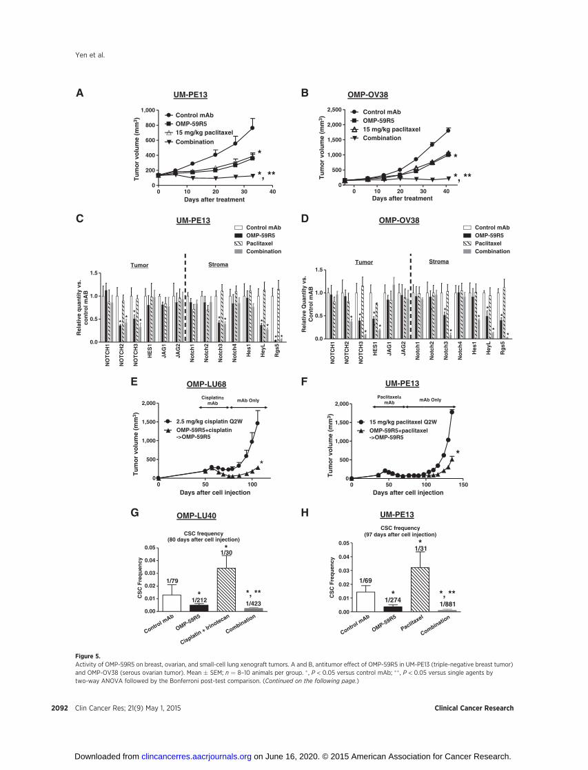
In addition to pancreatic tumors, we also observed antitumorefficacy by anti-Notch2/3 antibody in breast, ovarian, and small-cell lung xenograft tumors. OMP-59R5 was efficacious as a singleagent inUM-PE13 (a triple-negative breast tumor),OMP-OV38 (aserous ovarian tumor), and OMP-LU40 (a small-cell lungcancer; Fig. 5A and B and Supplementary Fig. S7A) and combi-nation activity with the chemotherapeutic agent in UM-PE13,OMP-OV38, and OMP-LU68 (Fig. 5A and B and SupplementaryFig. S7A). qRT-PCR gene expression analysis in UM-PE13 andOMP-OV38 tumors showed that HES1, NOTCH2, and NOTCH3in the tumors andNotch3,Hes1,HeyL, andRgs5 in the stromawererobustly downregulated by OMP-59R5 in these models (Fig. 5Cand D). Immunohistochemistry analysis in UM-PE13 revealedthat the antitumor activity of OMP-59R5 was associated with a 4-fold decrease in Notch3 ICD (Supplementary Fig. S7B). Whenhigh doses of chemotherapy induced tumor regression in UM-PE13 and OMP-LU68 tumors, the combination of OMP-59R5with the chemotherapeutic agents delayed tumor recurrencefollowing discontinuation of the chemotherapeutic agents com-
pared with the rate of tumor recurrence after treatment with thechemotherapeutic agents alone (Fig. 5E and F). In the UM-PE13breast tumor, OMP-59R5 treatment decreased CSC frequency to37% relative to the control, whereas the residual tumor cells afterpaclitaxel treatment were enriched in CSCs exhibiting an approx-imately 2-fold increase in CSC frequency compared with thecontrol (Fig. 5H). Importantly, the combination of anti-Notch2/3 and paclitaxel treatment decreased CSC frequency andtumorigenicity in the residual tumor cells, resulting in a 10-foldreduction in CSC frequency compared with the paclitaxel only-treated group. Similar observations were found in OMP-LU40xenograft tumors (Fig. 5G). Taken together, these findings indi-cate the cross-reactive Notch2/3 targeting antibody OMP-59R5 isefficacious in a wide range of solid tumors, inhibiting Notchsignaling in both tumor and stromal cells while reducing tumor-igenic cell frequency.
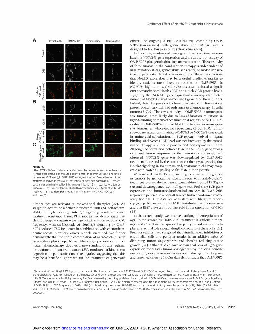
OMP-59R5 alters pericyte coverage in tumor vasculatureIn our experiments, we observed that Rgs5, a marker of devel-
oping pericytes, was consistently downregulated byOMP-59R5 invarious tumors. Pericytes are tightly associated with endothelialcells in normal vasculature to provide structural support to bloodvessels and regulate tissue physiology by modulating vascularstability, whereas pericytes in tumor vasculature exhibit abnormalshapes and are loosely associated with endothelial cells on tumorcapillaries (24). Hamzah and colleagues reported that reductionofRgs5 expression in an endocrine pancreatic tumormodel resultsin pericyte maturation, "normalizes" tumor vasculature, andimproves the delivery of chemotherapeutic agents and theimmune response against the tumor (25). Similarly, patients withbreast cancer with mature pericyte coverage demonstrateimproved disease-free survival and overall survival (26). Toinvestigate the functional significance of OMP-59R5–mediateddownregulation of Rgs5 and its effect on pericytes, we conductedimmunofluorescence staining for desmin, a marker of maturepericytes (24). We found that desmin-positive pericytes in thevasculature were closely associated with vessels in tumors treatedwith OMP-59R5, suggesting pericyte recruitment to endothelialcells and subsequent vascular maturation (Fig. 6A). To determinewhether mature pericytes are linked to a functional vasculature,we visualized plant lectin (tomato)-perfused vessels pretreatedwith pimonidazole. As seen in Supplementary Fig. S8A, pericyteseither overlap or are more closely associated with the endothelialcells in perfused vessels. OMP-59R5 and gemcitabine treatmentincreased perfused blood vessels per area by 15%. An additional20% increase in perfused vessels was seen in tumors after treat-ment with the combination of anti-Notch2/3 and gemcitabine(Fig. 6B and Supplementary Fig. S8B). The increase in perfusion intreated tumors was associated with a reduced tumor hypoxia (Fig.6C), more evident in tumors treated with the combination ofOMP-59R5 and gemcitabine compared with control mAb-treatedtumors. These findings suggest an improved oxygen supply in thetumors as a result of improved pericyte coverage and vascularnormalization by OMP-59R5 treatment.
DiscussionNotch signaling plays an important role in regulating cell fate
decisions in a variety of normal tissues (1). Several lines ofevidence have indicated that dysregulation of the Notch pathwaycan lead to uncontrolled self-renewal of CSCs which generate
www.aacrjournals.org Clin Cancer Res; 21(9) May 1, 2015 2091
Antitumor Effect of Notch2/3 Antagonist (Tarextumab)
on June 16, 2020. © 2015 American Association for Cancer Research. clincancerres.aacrjournals.org Downloaded from
0 50 1000
500
1,000
1,500
2,000
2.5 mg/kg cisplatin Q2W
OMP-59R5+cisplatin->OMP-59R5
Cisplatin±mAb
mAb Only
*
Days after cell injection
0 10 20 30 400
500
1,000
1,500
2,000
2,500 Control mAbOMP-59R515 mg/kg paclitaxelCombination
*
*, **
E
OMP-OV38
B OMP-OV38
Control m
Ab
OMP-59R5
Cisplatin + Iri
notecan
Combination
0.00
0.01
0.02
0.03
0.04
0.05
CSC frequency(80 days after cell injection)
* *, **
1/79
1/212
*1/30
1/423
CS
C F
req
uen
cy
OMP-LU68
C
A UM-PE13
0 10 20 30 400
200
400
600
800
1,000Control mAbOMP-59R515 mg/kg paclitaxel
Combination
*
*, **
Days after treatment Days after treatment
Tu
mo
r vo
lum
e (m
m3 )
Tu
mo
r vo
lum
e (m
m3 )
Tu
mo
r vo
lum
e (m
m3 )
Tu
mo
r vo
lum
e (m
m3 )
D
0 50 100 1500
500
1,000
1,500
2,000
15 mg/kg paclitaxel Q2W
OMP-59R5+paclitaxel->OMP-59R5
Paclitaxel±mAb
mAb Only
*
Days after cell injection
UM-PE13
UM-PE13
CSC frequency(97 days after cell injection)
Control m
Ab
OMP-59R5
Paclitaxel
Combination
0.00
0.01
0.02
0.03
0.04
0.05
1/69
1/274
1/31
1/881*
*
*, **CS
C F
req
uen
cy
F
G H
NO
TC
H1
NO
TC
H2
NO
TC
H3
HE
S1
JAG
1
JAG
2
No
tch
1
No
tch
2
No
tch
3
No
tch
4
Hes
1
Hey
L
Rg
s5
0.0
0.5
1.0
1.5
Control mAbOMP-59R5PaclitaxelCombination
* *
*
*
* *
* *
*
**
**
*
Tumor Stroma
Rel
ativ
e Q
uan
tity
vs.
Co
ntr
ol m
AB
OMP-LU40 UM-PE13
NO
TC
H1
NO
TC
H2
NO
TC
H3
HE
S1
JAG
1
JAG
2
No
tch
1
No
tch
2
No
tch
3
No
tch
4
Hes
1
Hey
L
Rg
s5
0.0
0.5
1.0
1.5
Control mAbOMP-59R5PaclitaxelCombination
**
** * *
*
* *
*
Tumor Stroma
Rel
ativ
e q
uan
tity
vs.
con
tro
l mA
B
Figure 5.Activity of OMP-59R5 on breast, ovarian, and small-cell lung xenograft tumors. A and B, antitumor effect of OMP-59R5 in UM-PE13 (triple-negative breast tumor)and OMP-OV38 (serous ovarian tumor). Mean � SEM; n ¼ 8–10 animals per group. � , P < 0.05 versus control mAb; �� , P < 0.05 versus single agents bytwo-way ANOVA followed by the Bonferroni post-test comparison. (Continued on the following page.)
Clin Cancer Res; 21(9) May 1, 2015 Clinical Cancer Research2092
Yen et al.
on June 16, 2020. © 2015 American Association for Cancer Research. clincancerres.aacrjournals.org Downloaded from
tumors that are resistant to conventional therapies (27). Wesought to determine whether interference with CSC self-renewalability through blocking Notch2/3 signaling would overcometreatment resistance. Using PDX models, we demonstrate thatchemotherapeutic agents were largely ineffective in reducing CSCfrequency, whereas blockade of Notch2/3 signaling by OMP-59R5 reduced CSC frequency in combination with chemothera-peutic agents in various cancer models examined. We furtherdemonstrate that the triple combination of anti-Notch2/3 withgemcitabine plus nab-paclitaxel (Abraxane, a protein-bound pac-litaxel) chemotherapy doublet, a new standard-of-care regimenfor treatment of pancreatic cancer (23), produced striking tumorregression in pancreatic cancer xenografts, suggesting that thismay be a beneficial approach for the treatment of pancreatic
cancer. The ongoing ALPINE clinical trial combining OMP-59R5 (tarextumab) with gemicitabine and nab-paclitaxel isdesigned to test this possibility (clinicaltrials.gov).
In this study, we observed a strong positive correlation betweenbaseline NOTCH3 gene expression and the antitumor activity ofOMP-59R5 plus gemcitabine in pancreatic tumors. The sensitivityof these tumors to the combination therapy is independent ofKras mutation status, gemcitabine sensitivity, or molecular sub-type of pancreatic ductal adenocarcinoma. These data indicatethat Notch3 expression may be a useful predictive marker toidentify patients most likely to respond to OMP-59R5. InNOTCH3 high tumors, OMP-59R5 treatment induced a signifi-cant decrease in bothNotch3 ECDandNotch3 ICDprotein levels,suggesting that NOTCH3 gene expression is an important deter-minant of Notch3 signaling-mediated growth of these tumors.Indeed, Notch3 expression has been associatedwith disease stage,poorer overall survival, and resistance to chemotherapy in solidtumors (5, 7, 9). The low sensitivity to OMP-59R5 in nonrespon-sive tumors is not likely due to loss-of-function mutations inligand-binding domain/other functional regions of NOTCH2/3or due to OMP-59R5–induced Notch1 activation in nonrespon-sive tumors, as whole-exome sequencing of our PDX tumorsshowed no mutations in either NOTCH2 or NOTCH3 that resultin amino acid substitutions in EGF repeats involved in ligandbinding, and Notch1 ICD level was not increased by the combi-nation therapy in either responsive and nonresponsive tumors.Although no correlation between baselineNOTCH2 gene expres-sion and tumor response to the combination therapy wasobserved, NOTCH2 gene was downregulated by OMP-59R5treatment alone and by the combination therapy, suggesting thatNotch2 signaling in the tumors and/or stroma niche may coop-erate with Notch3 signaling to facilitate tumor growth.
We observed that EMT and stem cell gene sets were upregulatedin tumors by gemcitabine. Combination with anti-Notch2/3treatment reversed the increase in gemcitabine-induced EMT genesets and downregulated stem cell gene sets. Real-time PCR geneexpression and immunohistochemical analyses in OMP-59R5responsive pancreatic xenograft tumors further confirmed micro-array findings. Our data are consistent with literature reportssuggesting that acquisition of EMT contributes to drug resistanceand that EMT plays an important role in the generation of CSCs(28).
In the current study, we observed striking downregulation ofRgs5 in the stroma by OMP-59R5 treatment in various tumors.Rgs5 and Notch3 are coexpressed in pericytes and are known toplay an essential role in regulating the functions of these cells (29).Previous studies have suggested that simultaneous inhibition ofendothelial cells and pericytes results in an additive effect ofdisrupting tumor angiogenesis and thereby reducing tumorgrowth (30). Other studies have shown that loss of Rgs5 geneexpression modulates tumor angiogenesis by inducing pericytematuration, vascular normalization, and reducing tumor hypoxiaand vessel leakiness (25). Our data demonstrate that OMP-59R5
Control mAb OMP-59R5 Gemcitabine CombinationD
esm
inC
d31
Mer
ge (red
)C
d31
(gre
en)
Des
min
A
B
Cd3
1Le
ctin
Mer
geC
d31
(red
)(g
reen
)Le
ctin
C
Hyp
oxia
(gr
een)
Cd3
1 (r
ed)
Figure 6.EffectOMP-59R5onmaturepericytes, vascular perfusion, and tumorhypoxia.A, histologic analysis of mature pericyte marker desmin (green), endothelialcell marker Cd31 (red), in OMP-PN17 xenograft tumors. Colocalization of bothmarkers is shown in yellow. B, detection of perfused vasculature. TomatoLectin was administered by intravenous injection 5 minutes before tumorremoval. C, antipimonidazole-labeled hypoxic tumor cells (green) with Cd31(red). N ¼ 3–4 tumors per group. Magnifications: �60 (A), �20 (B),and �4 (C).
(Continued.) C and D, qRT-PCR gene expression in the tumor and stroma in UM-PE13 and OMP-OV38 xenograft tumors at the end of study from A and B.Gene expression was normalized with the housekeeping gene GAPDH and expressed as fold of control mAb-treated tumors. Mean � SD; n ¼ 3–4 per group.� , P < 0.05 versus control mAb by one-way ANOVA followed by the Tukey post-test. E and F, effect of OMP-59R5 on tumor recurrence in OMP-LU68 (small-cell lungtumors) and UM-PE13. Mean � SEM; n ¼ 10 animals per group. � , P < 0.05 versus chemotherapeutic agent alone by the nonparametric t test. G and H, effectof OMP-59R5 on CSC frequency in OMP-LU40 (small-cell lung tumor) and UM-PE13 tumors at the end of study from Supplementary Fig. S6A (OMP-LU40)and F (UM-PE13). Mean� SEM; n¼ 10 animals per group. � , P < 0.05 versus control mAb; �� , P < 0.05 versus gemcitabine by one-way ANOVA followed by the Tukeypost-test.
www.aacrjournals.org Clin Cancer Res; 21(9) May 1, 2015 2093
Antitumor Effect of Notch2/3 Antagonist (Tarextumab)
on June 16, 2020. © 2015 American Association for Cancer Research. clincancerres.aacrjournals.org Downloaded from

treatment decreases Rgs5 gene expression, modulates intratu-moral pericyte localization, and increases vessel perfusion whilereducing tumor hypoxia. OMP-59R5–mediated improvement invasculature functions may increase drug delivery to the tumors,thereby enhancing the antitumor efficacy, particularly in combi-nation with chemotherapeutic agents or other treatment modal-ities. In contrast to anti-Notch2/3, inhibition of DLL4/Notchsignaling using DLL4-specific inhibitors has been shown toinduce dense vasculature network of unperfused and poorlydifferentiated vessels (31, 32). Consistent with these previousstudies, in an small-cell lung cancer xenograft model, we foundthat tumors treatedwith an anti-mouseDLL4 antibody resulted inan increased intratumoral hypoxia and hyperproliferation of theendothelial cells, whereas blocking Notch2/3 signaling by OMP-59R5 produced an opposite effect on tumor vasculatures/stromaversus targeting DLL4/Notch signaling by anti-mouse DLL4 anti-body (Supplementary Fig. S8C). "Normalization" of tumor vas-culature was reported previously in both preclinical tumor mod-els and in patients with cancer receiving anti-VEGF therapy(reviewed in ref. 33).
In summary, our findings provide evidence for the utility oftargeting Notch2 and Notch3 for cancer treatment. Our data alsosuggest that therapeutic approaches targeting pathways importantfor CSCs may improve treatment outcome and overall survival.Our recent phase I data indicate that tarextumab is generally welltolerated and show signs of antitumor efficacy andmodulation ofNotch pathway signaling in the clinic (34). On the basis of thepreclinical studies described here, we are evaluating the utility ofNotch3 gene expression as a predictive biomarker in our ongoingclinical studies and also the testing the ability of tarextumab tomodulate Notch and CSC gene signatures.
Disclosure of Potential Conflicts of InterestNo potential conflicts of interest were disclosed.
Authors' ContributionsConception and design: W.-C. Yen, J. Cain, A. Sato, A.M. Kapoun, J. Lewicki,A. Gurney, T. HoeyDevelopment of methodology: M. Fischer, J. LewickiAcquisition of data (provided animals, acquired and managed patients,provided facilities, etc.): W.-C. Yen, M. Fischer, F. Axelrod, C. Bond, J. Cain,B. Cancilla, J. Shah, T. Tang, B. Wallace, A.M. KapounAnalysis and interpretation of data (e.g., statistical analysis, biostatistics,computational analysis):W.-C. Yen, M. Fischer, F. Axelrod, J. Cain, R. Henner,R. Meisner, J. Shah, T. Tang, B. Wallace, M. Wang, C. Zhang, A.M. Kapoun,T. HoeyWriting, review, and/or revision of the manuscript: W.-C. Yen, M. Fischer,J. Cain, T. Tang, M. Wang, J. Lewicki, A. Gurney, T. HoeyAdministrative, technical, or material support (i.e., reporting or organizingdata, constructing databases): M. FischerStudy supervision: J. Lewicki, T. Hoey
AcknowledgmentsThe authors thank Inkyung Park, Jim Evans, Raymond Tam, Akbar
Currimbhoy, Fiore Cattaruzza, Pete Yeung, Kellie Pickell, Xiaomei Song,and many people at OncoMed Pharmaceuticals, Inc., for their contributionsto this work.
The costs of publication of this article were defrayed in part by thepayment of page charges. This article must therefore be hereby markedadvertisement in accordance with 18 U.S.C. Section 1734 solely to indicatethis fact.
Received November 1, 2014; revised January 13, 2015; accepted January 14,2015; published online May 1, 2015.
References1. Penton AL, Leonard LD, Spinner NB. Notch signaling in human develop-
ment and disease. Semin Cell Dev Biol 2012;23:450–7.2. Lin L, Mernaugh R, Yi F, Blum D, Carbone DP, Dang TP. Targeting specific
regions of the Notch3 ligand-binding domain induces apoptosis andinhibits tumor growth in lung cancer. Cancer Res 2010;70:632–8.
3. Park JT, LiM,NakayamaK,MaoTL,DavidsonB, ZhangZ, et al.Notch3geneamplification in ovarian cancer. Cancer Res 2006;66:6312–8.
4. YamaguchiN,Oyama T, Ito E, SatohH, Azma S,HayashiM, et al. NOTCH3signaling pathway plays crucial roles in the proliferation of ErbB2-negativehuman breast cancer cells. Cancer Res 2008;68:1881–8.
5. Doucas H, Mann CD, Sutton CD, Garcea G, Neal CP, Berry DP, et al.Expression of nuclear Notch3 in pancreatic adenocarcinomas is associatedwith adverse clinical features, and correlates with the expression of STAT3and phosphorylated Akt. J Surg Oncol 2008;97:63–8.
6. Mazur PK, Einwachter H, Lee M, Sipos B, Nakhai H, Rad R, et al. Notch2 isrequired for progression of pancreatic intraepithelial neoplasia and devel-opment of pancreatic ductal adenocarcinoma. Proc Natl Acad Sci U S A2010;107:13438–43.
7. Ozawa T, Kazama S, Akiyoshi T, Murono K, Yoneyama S, Tanaka J, et al.Nuclear Notch3 expression is associated with tumor recurrence in patientswith stage II and III colorectal cancer. Ann Surg Oncol 2014;21:2650–8.
8. Sainson RC, Harris AL. Regulation of angiogenesis by homotypic andheterotypic notch signalling in endothelial cells and pericytes: from basicresearch to potential therapies. Angiogenesis 2008;11:41–51.
9. Nguyen LV, Vanner R, Dirks P, Eaves CJ. Cancer stem cells: an evolvingconcept. Nat Rev 2012;12:133–43.
10. Malik B, Nie D. Cancer stem cells and resistance to chemo and radiotherapy. Front Biosci 2012;4:2142–9.
11. Takebe N, Nguyen D, Yang SX. Targeting Notch pathway in cancer: clinicaldevelopment advances and challenges. Pharmacol Ther 2014;141:140–9.
12. Rothe C, Urlinger S, Lonhning C, Prassler J, Stark Y, Jagur U, et al. Thehuman combinatorial library HuCAL GOLD combines diversification ofall six CDRs according to the natural immune system with a novel displaymethod for efficient selection of high-affinity antibodies. J Mol Biol 2008;376:1182–200.
13. Hoey T, YenWC,Axelrod F, Basi J, Donigian L,Dylla S, et al. DLL4 blockadeinhibits tumor growth and reduces tumor-initiating cell frequency. CellStem Cell 2009;5:168–77.
14. Dalerba P, Guiducci C, Poliani PL, Cifola I, Parenza M, Frattini M, et al.Phenotypic characterization of human colorectal cancer stem cells. ProcNatl Acad Sci U S A 2007;104:10158–63.
15. Baldi P, Long AD. A Bayesian framework for the analysis of microarrayexpression data: regularized t-test and statistical inferences of gene changes.Bioinformatics 2011;17:509–19.
16. Garrido-Laguna I, Uson M, Rajeshkumar NV, Tan AC, de Oiveira E,Karikari C, et al. Tumor engraftment in nude mice and enrichment instroma-related gene pathways predict poor survival and resistance togemitabine in patients with pancreatic cancer. Clin Cancer Res 2011;17:5793–800.
17. HidalgoM, Bruckheimer E, Rajeshkumar NV, Garrido-Laguna I, deOiveiraE, Rubio-Viqueira B, et al. A pilot clinical study of treatment guided bypersonalized tumorgrafts in patients with advanced cancer. Mol CancerTher 2011;10:1311–6.
18. Collisson EA, Sadanandam A, Olson P, Gibb WJ, Truitt M, Gu S, et al.Subtypes of pancreatic ductal adenocarcinoma and their differingresponses to therapy. Nat Med 2011;17:500–4.
19. YenWC, FischerMM,HynesM,Wu J, KimE, Beviglia L, et al. Anti-DLL4 hasbroad spectrum activity in pancreatic cancer dependent on targeting DLL4-Notch signaling in both tumor and vasculature cells. Clin Cancer Res 2012;18:5374–86.
Clin Cancer Res; 21(9) May 1, 2015 Clinical Cancer Research2094
Yen et al.
on June 16, 2020. © 2015 American Association for Cancer Research. clincancerres.aacrjournals.org Downloaded from

20. Ben-Porath I, Thomson MW, Carey VJ, Ge R, Bell GW, Regev A, et al. Anembryonic stem cell-like signature in poorly differentiated aggressivehuman tumors. Nat Genet 2008;40:499–507.
21. Bergers G, Song S. The role of pericytes in blood-vessel formation andmaintenance. Neuro Oncol 2005;7:452–64.
22. Wang JC, Doedens M, Dick JE. Primitive human hematopoietic cells areenriched in cord blood compared with adult bone marrow or mobilizedperipheral blood as measured by the quantitative in vivo SCID-repopulat-ing cell assay. Blood 1997;89:3919–24.
23. Von Hoff DD, Ervin T, Arena FP, Chiorean EG, Infante J, Moore M, et al.Increased survival in pancreatic cancer with nab-paclitaxel plus gemcita-bine. N Engl J Med 2013;369:1691–703.
24. Barlow K, Sahders AM, Soker S, Ergun S, Metheny-Barlow LJ. Pericytes onthe tumor vasculature: Jekyll or Hyde? Cancer microenvironment 2013;6:1–17.
25. Hamzah J, Jugold M, Kiessling F, Rigby P, Manzur M, Marti HH, et al.Vascular normalization in Rgs5-deficient tumours promotes immunedestruction. Nature 2008;453:410–4.
26. CookeVG, LeBleuVS,KeskinD,KhanZ,O'Connell JT, TengY, et al. Pericytedepletion results in hypoxia-associated epithelial-to-mesenchymal transi-tion and metastasis mediated by met signaling pathway. Cancer Cell2012;21:66–81.
27. Wang J, Sullenger BA, Rich JN. Notch signaling in cancer stem cells.Expt Med Biol 2012;727:174–85.
28. Scheel C, Weinberg RA. Cancer stem cells and epithelial-mesenchymaltransition: concepts and molecular links. Semin Cancer Biol 2012;22:396–403.
29. Lovschall H, Mitsiadis TA, Poulsen K, Jensen KH, Kjeldsen AL. Coex-pression of Notch3 and Rgs5 in the pericyte-vascular smoothmuscle cell axis in response to pulp injury. Int J Dev Biol 2007;51:715–21.
30. Kuhnert F, Tam BY, Sennino B, Gray JT, Yuan J, Jocson A, et al. Solublereceptor-mediated selective inhibitionof VEGFR andPDGFRbeta signalingduring physiologic and tumor angiogenesis. Proc Natl Acad Sci U S A2008;105:10185–90.
31. Noguera-Troise I,DalyC, PapadopoulosNJ, Coetzee S, BolandP,GaleNW,et al. Blockade of DLL4 inhibits tumor growth by promoting non-produc-tive angiogenesis. Nature 2006;444:1032–7.
32. Ridgway J, Zhang G, Wu Y, Stawicki S, Liang WC, Chanthery Y, et al.Inhibition of DLL4 signaling inhibits tumor growth by deregulatingangiogenesis. Nature 2006;444:1083–7.
33. Goel S, Duda DG, Xu L, Munn LL, Boucher Y, Fukumura D, et al. Nor-malization of the vasculature for treatment of cancer and other diseases.Physiol Rev 2011;91:1071–121.
34. Smith DC, Chugh R, Patnaik A, Papadopoulos K, Chambers G, Thorpe V,et al. A first-in-human phase 1 study to evaluate the fully human mono-clonal antibodyOMP-59R5 (anti-Notch2/3) administered intravenously topatients with advanced solid tumors. Eur J Cancer 2012;48 Suppl 6:11–2.
www.aacrjournals.org Clin Cancer Res; 21(9) May 1, 2015 2095
Antitumor Effect of Notch2/3 Antagonist (Tarextumab)
on June 16, 2020. © 2015 American Association for Cancer Research. clincancerres.aacrjournals.org Downloaded from
2015;21:2084-2095. Clin Cancer Res Wan-Ching Yen, Marcus M. Fischer, Fumiko Axelrod, et al. Tumor-Initiating Cell Frequency(Tarextumab) Inhibits Tumor Growth and Decreases Targeting Notch Signaling with a Notch2/Notch3 Antagonist
Updated version
http://clincancerres.aacrjournals.org/content/21/9/2084
Access the most recent version of this article at:
Material
Supplementary
http://clincancerres.aacrjournals.org/content/suppl/2015/05/12/21.9.2084.DC1
Access the most recent supplemental material at:
Cited articles
http://clincancerres.aacrjournals.org/content/21/9/2084.full#ref-list-1
This article cites 34 articles, 10 of which you can access for free at:
Citing articles
http://clincancerres.aacrjournals.org/content/21/9/2084.full#related-urls
This article has been cited by 16 HighWire-hosted articles. Access the articles at:
E-mail alerts related to this article or journal.Sign up to receive free email-alerts
Subscriptions
Reprints and
To order reprints of this article or to subscribe to the journal, contact the AACR Publications Department at
Permissions
Rightslink site. Click on "Request Permissions" which will take you to the Copyright Clearance Center's (CCC)
.http://clincancerres.aacrjournals.org/content/21/9/2084To request permission to re-use all or part of this article, use this link
on June 16, 2020. © 2015 American Association for Cancer Research. clincancerres.aacrjournals.org Downloaded from