
Targeting Immune Checkpoints in Hematologic...
12
1521-0081/68/4/1014–1025$25.00 http://dx.doi.org/10.1124/pr.116.012682 PHARMACOLOGICAL REVIEWS Pharmacol Rev 68:1014–1025, October 2016 Copyright © 2016 by The American Society for Pharmacology and Experimental Therapeutics ASSOCIATE EDITOR: MICHAEL G. ROSENBLUM Targeting Immune Checkpoints in Hematologic Malignancies Gheath Alatrash, Naval Daver, and Elizabeth A. Mittendorf Departments of Stem Cell Transplantation and Cellular Therapy (G.A., E.A.M.), Leukemia (N.D.), and Breast Surgical (E.A.M.) Oncology, The University of Texas MD Anderson Cancer Center, Houston, Texas Abstract.................................................................................... 1014 I. Introduction ................................................................................ 1014 II. T Cell Inhibitory Pathways: Cytotoxic-T-lymphocyte Antigen 4, Programmed Death Protein 1, and Programmed Death Protein Ligand 1 ....................................................... 1015 III. Targeting Immune Checkpoint Molecules in Cancer ......................................... 1015 IV. Immune Checkpoint Inhibition in Hematologic Tumors...................................... 1016 A. Lymphoma ............................................................................. 1017 B. Multiple Myeloma ...................................................................... 1019 C. Leukemia .............................................................................. 1020 D. Immune Checkpoint Inhibition after Stem Cell Transplantation: Timing Is Everything. . . 1021 E. Immune Checkpoint Inhibition in the Setting of Engineered T Cell Therapy ............. 1023 VI. Conclusion and Future Directions........................................................... 1023 Acknowledgments .......................................................................... 1023 References ................................................................................. 1023 Abstract——The use of antibodies that target immune checkpoint molecules on the surface of T-lymphocytes and/or tumor cells has revolutionized our approach to cancer therapy. Cytotoxic-T-lymphocyte antigen (CTLA-4) and programmed cell death protein 1 (PD-1) are the two most commonly targeted immune checkpoint molecules. Although the role of antibodies that target CTLA-4 and PD-1 has been established in solid tumor malignancies and Food and Drug Administration approved for melanoma and non-small cell lung cancer, there remains a desperate need to incorporate immune checkpoint inhibition in hematologic malignancies. Unlike solid tumors, a number of considerations must be addressed to appropriately employ immune checkpoint inhibition in hematologic malignancies. For example, hematologic malignancies frequently obliterate the bone marrow and lymph nodes, which are critical immune organs that must be restored for appropriate response to immune checkpoint inhibition. On the other hand, hematologic malignancies are the quintessential immune responsive tumor type, as proven by the success of allogeneic stem cell transplantation (allo-SCT) in hematologic malignancies. Also, sharing an immune cell lineage, malignant hematologic cells often express immune checkpoint molecules that are absent in solid tumor cells, thereby offering direct targets for immune checkpoint inhibition. A number of clinical trials have demonstrated the potential for immune checkpoint inhibition in hematologic malignancies before and after allo-SCT. The ongoing clinical studies and complimentary immune correlatives are providing a growing body of knowledge regarding the role of immune checkpoint inhibition in hematologic malignancies, which will likely become part of the standard of care for hematologic malignancies. I. Introduction Targeting immune checkpoint molecules on the sur- face of tumor cells or immune cells has proven to be a highly effective approach in cancer immunotherapy. A number of clinical trials in a variety of tumor types have been conducted using antibodies that target immune checkpoint molecules. Although there are several im- mune checkpoint pathways that regulate immune cells, to date, the two major approaches to immune check- point blockade that have been investigated clinically have targeted cytotoxic-T-lymphocyte antigen (CTLA-4) ABBREVIATIONS: allo, allogeneic; AML, acute myeloid leukemia; APC, antigen presenting cell; CAR, chimeric antigen receptor; CR, complete response; CTLA-4, cytotoxic-T-lymphocyte antigen; DLBCL, diffuse large B cell lymphoma; FL, follicular lymphoma; GVHD, graft versus host disease; GVL, graft versus leukemia; HL, Hodgkin’s lymphoma; MDS, myelodysplastic syndrome; MM, multiple myeloma; NHL, non-Hodgkin lymphoma; NSCLC, non-small cell lung cancer; PD-1, programmed cell death protein 1; PFS, progression-free survival; pHLA, peptide/human leukocyte antigen complex; PR, partial response; SCT, stem cell transplantation; TCR, T cell receptor; TIL, tumor infiltrating lymphocytes; TReg, regulatory T cells. 1014 by guest on March 27, 2020 Downloaded from
Transcript of Targeting Immune Checkpoints in Hematologic...
1521-0081/68/4/1014–1025$25.00 http://dx.doi.org/10.1124/pr.116.012682PHARMACOLOGICAL REVIEWS Pharmacol Rev 68:1014–1025, October 2016Copyright © 2016 by The American Society for Pharmacology and Experimental Therapeutics
ASSOCIATE EDITOR: MICHAEL G. ROSENBLUM
Targeting Immune Checkpoints in HematologicMalignancies
Gheath Alatrash, Naval Daver, and Elizabeth A. Mittendorf
Departments of Stem Cell Transplantation and Cellular Therapy (G.A., E.A.M.), Leukemia (N.D.), and Breast Surgical (E.A.M.) Oncology,The University of Texas MD Anderson Cancer Center, Houston, Texas
Abstract. . . . . . . . . . . . . . . . . . . . . . . . . . . . . . . . . . . . . . . . . . . . . . . . . . . . . . . . . . . . . . . . . . . . . . . . . . . . . . . . . . . . 1014I. Introduction. . . . . . . . . . . . . . . . . . . . . . . . . . . . . . . . . . . . . . . . . . . . . . . . . . . . . . . . . . . . . . . . . . . . . . . . . . . . . . . . 1014II. T Cell Inhibitory Pathways: Cytotoxic-T-lymphocyte Antigen 4, Programmed Death Protein 1, and
Programmed Death Protein Ligand 1 . . . . . . . . . . . . . . . . . . . . . . . . . . . . . . . . . . . . . . . . . . . . . . . . . . . . . . . 1015III. Targeting Immune Checkpoint Molecules in Cancer . . . . . . . . . . . . . . . . . . . . . . . . . . . . . . . . . . . . . . . . . 1015IV. Immune Checkpoint Inhibition in Hematologic Tumors. . . . . . . . . . . . . . . . . . . . . . . . . . . . . . . . . . . . . . 1016
A. Lymphoma . . . . . . . . . . . . . . . . . . . . . . . . . . . . . . . . . . . . . . . . . . . . . . . . . . . . . . . . . . . . . . . . . . . . . . . . . . . . . 1017B. Multiple Myeloma . . . . . . . . . . . . . . . . . . . . . . . . . . . . . . . . . . . . . . . . . . . . . . . . . . . . . . . . . . . . . . . . . . . . . . 1019C. Leukemia . . . . . . . . . . . . . . . . . . . . . . . . . . . . . . . . . . . . . . . . . . . . . . . . . . . . . . . . . . . . . . . . . . . . . . . . . . . . . . 1020D. Immune Checkpoint Inhibition after Stem Cell Transplantation: Timing Is Everything. . . 1021E. Immune Checkpoint Inhibition in the Setting of Engineered T Cell Therapy . . . . . . . . . . . . . 1023
VI. Conclusion and Future Directions. . . . . . . . . . . . . . . . . . . . . . . . . . . . . . . . . . . . . . . . . . . . . . . . . . . . . . . . . . . 1023Acknowledgments . . . . . . . . . . . . . . . . . . . . . . . . . . . . . . . . . . . . . . . . . . . . . . . . . . . . . . . . . . . . . . . . . . . . . . . . . . 1023References . . . . . . . . . . . . . . . . . . . . . . . . . . . . . . . . . . . . . . . . . . . . . . . . . . . . . . . . . . . . . . . . . . . . . . . . . . . . . . . . . 1023
Abstract——The use of antibodies that target immunecheckpoint molecules on the surface of T-lymphocytesand/or tumor cells has revolutionized our approach tocancer therapy. Cytotoxic-T-lymphocyte antigen (CTLA-4)and programmed cell death protein 1 (PD-1) are the twomost commonly targeted immune checkpoint molecules.Although the role of antibodies that target CTLA-4 andPD-1hasbeenestablished in solid tumormalignanciesandFood and Drug Administration approved for melanomaand non-small cell lung cancer, there remains a desperateneed to incorporate immune checkpoint inhibition inhematologic malignancies. Unlike solid tumors, a numberof considerations must be addressed to appropriatelyemploy immune checkpoint inhibition in hematologicmalignancies. For example, hematologic malignanciesfrequently obliterate the bone marrow and lymph nodes,which are critical immune organs that must be restored
for appropriate response to immune checkpointinhibition. On the other hand, hematologic malignanciesare the quintessential immune responsive tumor type,as proven by the success of allogeneic stem celltransplantation (allo-SCT) in hematologic malignancies.Also, sharing an immune cell lineage, malignanthematologic cells often express immune checkpointmolecules that are absent in solid tumor cells, therebyoffering direct targets for immune checkpoint inhibition.A number of clinical trials have demonstrated thepotential for immune checkpoint inhibition inhematologic malignancies before and after allo-SCT. Theongoing clinical studies and complimentary immunecorrelatives are providing a growing body of knowledgeregarding the role of immune checkpoint inhibition inhematologic malignancies, which will likely become partof the standard of care for hematologic malignancies.
I. Introduction
Targeting immune checkpoint molecules on the sur-face of tumor cells or immune cells has proven to be ahighly effective approach in cancer immunotherapy. Anumber of clinical trials in a variety of tumor types have
been conducted using antibodies that target immunecheckpoint molecules. Although there are several im-mune checkpoint pathways that regulate immune cells,to date, the two major approaches to immune check-point blockade that have been investigated clinicallyhave targeted cytotoxic-T-lymphocyte antigen (CTLA-4)
ABBREVIATIONS: allo, allogeneic; AML, acute myeloid leukemia; APC, antigen presenting cell; CAR, chimeric antigen receptor; CR,complete response; CTLA-4, cytotoxic-T-lymphocyte antigen; DLBCL, diffuse large B cell lymphoma; FL, follicular lymphoma; GVHD, graftversus host disease; GVL, graft versus leukemia; HL, Hodgkin’s lymphoma; MDS, myelodysplastic syndrome; MM, multiple myeloma; NHL,non-Hodgkin lymphoma; NSCLC, non-small cell lung cancer; PD-1, programmed cell death protein 1; PFS, progression-free survival; pHLA,peptide/human leukocyte antigen complex; PR, partial response; SCT, stem cell transplantation; TCR, T cell receptor; TIL, tumorinfiltrating lymphocytes; TReg, regulatory T cells.
1014
by guest on March 27, 2020
Dow
nloaded from
and the programmed cell death pathway. The pro-grammed cell death pathway includes programmed celldeath protein 1 (PD-1) and its ligands programmeddeath ligands 1 (PD-L1) and 2 (PD-L2). To date,melanoma and non-small cell lung cancer (NSCLC)are the two tumor types for which the use of immunecheckpoint inhibition has received Food and DrugAdministration approval. However, there is great in-terest in investigating these agents in hematologicmalignancies, which are known to express immunecheckpoint molecules and to be susceptible to immunemodulation. In addition, there is a desperate need fornovel agents to treat a number of hematologic malig-nancies, because these remain some of the most aggres-sive tumors to afflict adults and children. This reviewwill provide an update on the current state of im-mune checkpoint based approaches in the treatmentof hematologic malignancies, including stem celltransplantation.
II. T Cell Inhibitory Pathways: Cytotoxic-T-lymphocyte Antigen 4, Programmed DeathProtein 1, and Programmed Death Protein
Ligand 1
Upon initial encounter with its antigen in a lymphoidorgan, there are a number of signaling pathways thatmust be triggered within the T cell to achieve adequateactivation. T cells require binding of their T cell receptor(TCR) to the peptide/human leukocyte antigen complex(pHLA) that is expressed on the target, as well asbinding of the T cell costimulatory receptors to theircognate ligands that are expressed by the tumor orantigen presenting cell (APC). CD28 is an importantcostimulatory molecule expressed on the T cell surface.There are two known ligands for CD28, CD80 (B7.1) andCD86 (B7.2), both expressed on APCs. CD80 and CD86are also ligands for CTLA-4, an inhibitory moleculeexpressed on the T cell surface. CTLA-4 binds with ahigher affinity to CD80 and CD86 on the APCs, and ineffect competes with CD28 for binding to these mole-cules (Linsley et al., 1994; Leach et al., 1996; Egen andAllison, 2002; Riley et al., 2002; Schneider et al., 2006).In addition, CTLA-4 activates phosphatases such asSrc-homology 2 domain-containing phosphatase 2,which counteract the phosphorylation steps that ensueafter TCR binding to pHLA and are critical for T cellactivation (Rudd et al., 2009). CTLA-4 is expressed byCD8+ and CD4+ T cells; however, the effects of CTLA-4are primarily seen in the CD4+ T cell population,
including helper T cells and regulatory T cells (TReg).Engagement of CTLA-4 with its ligands results in thedownregulation of helper T cell activities and upregu-lation of TReg cell activities. Together, competition forbinding with CD80 and CD86, the attenuation of helperT cell functions, and the enhancement of TReg activitiesresult in a “break” on effector T cell activation that iscritical for controlling the immune response and main-taining normal immune homeostasis.
Although CTLA-4 plays a major role in regulating theinitial stages of T cell activation, another T cell in-hibitory mechanism, PD-1, plays a critical role inabrogating T cell functions during the later stages ofthe immune response (Nishimura et al., 1999; Freemanet al., 2000; Nishimura et al., 2001). The PD-1 pathway,which involves the T cell inhibitory molecule PD-1 andits ligands PD-L1/PD-L2, modulates the immune re-sponse after T cells exit the circulation and home intoinflamed and tumor tissues. This mechanism regulatesand contains the immune response to prevent tissuedamage and autoimmunity that can be deleterious tothe host. PD-1 and PD-L1/PD-L2 therefore play animportant role in peripheral tolerance. Like CTLA-4,signaling through PD-1 affects phosphatases likeSrc-homology 2 domain-containing phosphatase 2,which offset the activity of the kinases that mediateT cell activation after TCR/pHLA engagement andCD28 activation (Freeman et al., 2000; Yokosukaet al., 2012). PD-1 signaling also promotes TReg
proliferation and immune suppressive functions(Francisco et al., 2009).
III. Targeting Immune Checkpoint Moleculesin Cancer
A number of antibodies that block the interactionbetween immune checkpoint receptors on T cells andtheir ligands on tumor cells have been developed andhave proven to be efficacious in the setting of solidtumor. Several of these are currently being evaluated inhematologic malignancies (Table 1). The rationale for atherapeutic strategy employing antibodies that targetimmune checkpoint molecules stems from the conceptthat impeding the interaction between the immunecheckpoint receptor on the T cell and its ligand on thetumor cell releases the inhibitory brakes that abrogateT cells functions and antitumor immune response.There are a number of critical issues to be consideredwhen employing immune checkpoint blockade in cancerimmunotherapy. The first is that T cells must be
G.A. is supported by a grant from the Leukemia & Lymphoma Society. N.D. is supported by grants from the Ladies Leukemia League andthe Anderson Cancer Center Leukemia SPORE. E.A.M. is an R. Lee Clark Fellow of the University of Texas Anderson Cancer Centersupported by the Jeanne F. Shelby Scholarship Fund.
Address correspondence to: Dr. Gheath Alatrash, DO, Assistant Professor, Department of Stem Cell Transplantation and CellularTherapy, 1400 Holcombe Blvd, Unit 0423, Houston, TX 77030. E-mail: [email protected]
dx.doi.org/10.1124/pr.116.012682.
Checkpoint Blockade for Hematologic Malignancies 1015
present within the tumor microenvironment. This isindeed a critical consideration, because it may dictatethe timing of administration of the immune checkpointblockade in relation to other systemic cancer therapies.The majority of systemic cancer therapies is lymphode-pleting and can affect the number of lymphocytes withinthe tumor microenvironment.The second consideration is that the T cells within
the tumor microenvironment need to possess specificityto distinct antigens expressed by tumor cells. Thecharacteristics of the antigens targeted by tumorinfiltrating lymphocytes (TIL) have been heavily in-vestigated. Antigens expressed by tumor cells generallyfall into two broad categories: 1) mutated antigens thatoftentimes account for neoantigens or 2) tumor-associated antigens that are routinely expressed bynormal tissues but are differentially expressed by thetumor. In one of the original studies using the anti-CTLA-4 antibody ipilimumab, the response in patientswith melanoma directly correlated with a higher num-ber of neoantigens in tumors with a higher mutationalload (Snyder et al., 2014). This was confirmed inNSCLCstudies where PD-1 blockade with pembrolizumab wasused. In that setting, the mutational and neoantigenload, as well as the detection of neoantigen-specificTILs, highly correlated with response to pembrolizu-mab (Rizvi et al., 2015; McGranahan et al., 2016).The third consideration is the expression of immune
checkpoint receptors on TIL and the presence of theircognate ligands on tumor cells or other immune cellswithin the tumor microenvironment. A number ofstudies have demonstrated better efficacy with immunecheckpoint blockade in patients who have high levels ofCTLA-4 and PD-1 on TIL and high expression of CTLA-4 and PD-L1 ligands on the tumor cells (Taube et al.,2014; Van Allen et al., 2015; McGranahan et al., 2016).However, clinical data have also demonstrated theefficacy of immune checkpoint inhibitors in tumors thathave a lower expression of immune checkpoint mole-cules. For example, a clinical trial testing the efficacy ofnivolumab, ipilimumab, and the combination in pa-tients with untreated melanoma, demonstrated clinicalresponse to immune checkpoint blockade even amongpatients with tumors that expressed a low level ofPD-L1, although the response rate was higher in
patients with higher baseline PD-L1 expression(Larkin et al., 2015). Similar results have been observedin a clinical trial in patients with NSCLC (Garon et al.,2015). Although this remains an area of active in-vestigation, the inconsistencies in responses to check-point blockade, based on the expression of the immunecheckpoint molecules, may be attributable to heteroge-neity in the tumor that is not adequately reflected bytumor sampling or to other components of the tumormicroenvironment that regulate response to immunecheckpoint blockade. These data suggest that the pres-ence of PD-L1 expression may not be an accuratebiomarker of response to therapy and many trials nolonger use tumor PD-L1 expression as an eligibilitycriterion.
IV. Immune Checkpoint Inhibition inHematologic Tumors
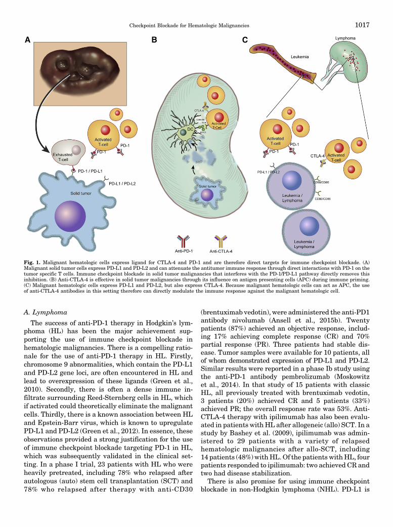
There are a number of factors to be considered withthe use of immune checkpoint blockade in the treatmentof patients with hematologic malignancies, includingleukemia, lymphoma, and multiple myeloma (MM).Because they share a common cell lineage, malignanthematologic tumor cells often express markers typicallyassociated with antigen presenting cells, specificallyCD80 and CD86, hence making them direct targets forantibodies against CTLA-4. This is different from non-hematologic malignancies wherein CTLA-4 targeting isaimed at removing tolerance to the immune primingevents that occur within the lymphoid organs. Inaddition, because they originate and reside withinlymphoid organs, either the bone marrow or lymphnodes, hematologic malignancies could be more suscep-tible to regulation by targeting CTLA-4 (Fig. 1). On theother hand, the timing of the application of immunecheckpoint blockade may be more critical in the settingof hematologic malignancies, especially leukemia,where the tumor itself oftentimes obliterates hostimmunity. Although the clinical application of immunecheckpoint blockade for hematologic malignancies isclearly lagging behind its use in solid tumors, a numberof studies have demonstrated encouraging resultswith immune checkpoint inhibition in hematologicmalignancies.
TABLE 1Immune checkpoint antibodies used in hematologic malignancies
Ipilimumab Nivolumab Pembrolizumab Pidilizumab Atezolizumab
Target CTLA4 PD1 PD1 PD1 PD-L1Class Humanized IgG1 Human IgG4 Humanized IgG4 Humanized IgG1 Humanized IgG1FDA approved indication Unresectable or
metastatic melanomaor in the adjuvantsetting
Unresectable ormetastatic melanoma;metastatic NSCLC;advanced renal cellcarcinoma
Unresectable ormetastatic melanoma;metastatic NSCLC
None at this time.Promising data inDLBCL and FL
None at this time.Ongoing trial inMM
Pharmaceutical Bristol-Myers Squibb Bristol-Myers Squibb Merck Cure Tech/Medivation Genentech/Roche
DLBCL, diffuse B cell lymphoma; FL, follicular lymphoma; MM, multiple myeloma; NSCLC, non-small cell lung cancer.
1016 Alatrash et al.
A. Lymphoma
The success of anti-PD-1 therapy in Hodgkin’s lym-phoma (HL) has been the major achievement sup-porting the use of immune checkpoint blockade inhematologic malignancies. There is a compelling ratio-nale for the use of anti-PD-1 therapy in HL. Firstly,chromosome 9 abnormalities, which contain the PD-L1and PD-L2 gene loci, are often encountered in HL andlead to overexpression of these ligands (Green et al.,2010). Secondly, there is often a dense immune in-filtrate surrounding Reed-Sternberg cells in HL, whichif activated could theoretically eliminate the malignantcells. Thirdly, there is a known association between HLand Epstein-Barr virus, which is known to upregulatePD-L1 and PD-L2 (Green et al., 2012). In essence, theseobservations provided a strong justification for the useof immune checkpoint blockade targeting PD-1 in HL,which was subsequently validated in the clinical set-ting. In a phase I trial, 23 patients with HL who wereheavily pretreated, including 78% who relapsed afterautologous (auto) stem cell transplantation (SCT) and78% who relapsed after therapy with anti-CD30
(brentuximab vedotin), were administered the anti-PD1antibody nivolumab (Ansell et al., 2015b). Twentypatients (87%) achieved an objective response, includ-ing 17% achieving complete response (CR) and 70%partial response (PR). Three patients had stable dis-ease. Tumor samples were available for 10 patients, allof whom demonstrated expression of PD-L1 and PD-L2.Similar results were reported in a phase Ib study usingthe anti-PD-1 antibody pembrolizumab (Moskowitzet al., 2014). In that study of 15 patients with classicHL, all previously treated with brentuximab vedotin,3 patients (20%) achieved CR and 5 patients (33%)achieved PR; the overall response rate was 53%. Anti-CTLA-4 therapy with ipilimumab has also been evalu-ated in patients withHL after allogeneic (allo) SCT. In astudy by Bashey et al. (2009), ipilimumab was admin-istered to 29 patients with a variety of relapsedhematologic malignancies after allo-SCT, including14 patients (48%)withHL.Of the patients withHL, fourpatients responded to ipilimumab: two achieved CR andtwo had disease stabilization.
There is also promise for using immune checkpointblockade in non-Hodgkin lymphoma (NHL). PD-L1 is
Fig. 1. Malignant hematologic cells express ligand for CTLA-4 and PD-1 and are therefore direct targets for immune checkpoint blockade. (A)Malignant solid tumor cells express PD-L1 and PD-L2 and can attenuate the antitumor immune response through direct interactions with PD-1 on thetumor specific T cells. Immune checkpoint blockade in solid tumor malignancies that interferes with the PD-1/PD-L1 pathway directly removes thisinhibition. (B) Anti-CTLA-4 is effective in solid tumor malignancies through its influence on antigen presenting cells (APC) during immune priming.(C) Malignant hematologic cells express PD-L1 and PD-L2, but also express CTLA-4. Because malignant hematologic cells can act as APC, the useof anti-CTLA-4 antibodies in this setting therefore can directly modulate the immune response against the malignant hematologic cell.
Checkpoint Blockade for Hematologic Malignancies 1017
expressed by subtypes of NHL (Green et al., 2010;Andorsky et al., 2011), and immune cell infiltrates inlymphoma tissue have been correlated with clinicaloutcomes (Lippman et al., 1990; Grogan and Miller,1993; Ansell et al., 2001). Based on these observations,immune checkpoint blockade has been tested in NHL,with the most encouraging data in the setting offollicular lymphoma (FL) with the use of the anti-PD-1antibody pidilizumab. In a phase I clinical trial thatenrolled 17 patients with lymphoma and leukemia,including 4 patients with NHL [two diffuse largeB cell lymphoma (DLBCL), 1 FL and 1 acute lympho-cytic cell lymphoma], Berger et al. (2008) showedelimination of tumor masses in the FL patient afterpidilizumab treatment. This observation led to a non-randomized, single center phase II clinical trial in32 patients with relapsed rituximab-sensitive FL(Westin et al., 2014). In that trial, patients were treatedwith the combination of pidilizumab and rituximab.Results from the study showed safety of the combina-tion of pidilizumab and rituximab and activity in29 evaluable patients, which included 15 patients (52%)achieving CR and 4 (14%) achieving PR. Furthermore,the investigators identified immune gene signaturesthat could predict for response to therapy and showedthat the frequency of pre-therapy PD-1 expressingeffector T cells within the tumor correlated positivelywith both tumor response and progression-free survival(PFS). These signatures have not yet been validated.Nevertheless, the identified genes extend beyond im-mune checkpoint molecules, highlighting the complex-ity of modulating the antitumor immune response withcheckpoint antibodies in NHL.There has also been encouraging data with the use of
pidilizumab in the setting of DLBCL after auto-SCT(Armand et al., 2013). In a phase II study, 66 patients
with NHL (49 patients with DLBCL, 4 patients withprimary mediastinal B cell lymphoma and 13 patientswith transformed indolent B cell NHL) were givenpidilizumab within 3 months after auto-SCT. CT andPET scans documented CR in 31 patients (47%) and45 patients (68%), respectively, before administration ofpidilizumab. The overall response rate after pidilizu-mab treatment in the 35 eligible patients who hadmeasurable disease after auto-SCT was 51% and the16-month overall survival and PFS were 0.85 (90% CI,0.74 to 0.92) and 0.72 (90% CI, 0.60 to 0.82), respec-tively, and were not affected by disease status at thetime of administration of pidilizumab. The PFS in thestudy cohort compared favorably with the PFS ofhistorical controls treated in the same institution, 0.52(90% CI, 0.39 to 0.63). Unfortunately, the investigatorsdid not have access to tumor tissue and therefore couldnot provide an analysis of PD-L1 or PD-L2 expression bythe tumor cells, which is critical in NHL, becauseimmune checkpoint molecules are not ubiquitouslyexpressed by malignant NHL cells but are often re-stricted to subgroups of tumors (Green et al., 2010;Andorsky et al., 2011). The investigators did show anincrease in the T cell memory subsets in the peripheralblood over the course of treatment and showed anincrease in PD-L1 expression in subsets of immunecells in the peripheral blood; however, no clear patternsor correlations were identified.
A number of studies have shown expression of CD80andCD86 by lymphoma cells, includingDLBCL andFL,hence providing the rationale for targeting CTLA-4 inNHL (Dorfman et al., 1997; Tsukada et al., 1997;Chaperot et al., 1999). Promising results in NHL havebeen seen with the use of the anti-CTLA-4 antibodyipilimumab. In a phase I study of 18 patients with NHL,including 14 patients with FL, 3 with DLBCL and
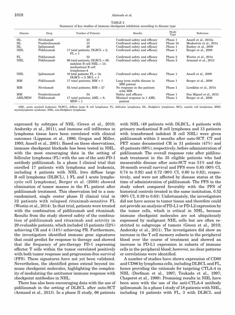
TABLE 2Summary of key studies of immune checkpoint inhibition according to disease type
Disease Drug Number of Patients Results StudyType Reference
HL Nivolumab 23 Confirmed safety and efficacy Phase 1 Ansell et al., 2015aHL Pembrolizumab 15 Confirmed safety and efficacy Phase 1b Moskowitz et al., 2014HL Ipilimumab 29 Confirmed safety and efficacy Phase 1 Bashey et al., 2009NHL Pidilizumab 17 total patients; DLBCL = 2;
FL = 1Confirmed safety and efficacy Phase 1 Berger et al., 2008
FL Pidilizumab 32 Confirmed safety and efficacy Phase 2 Westin et al., 2014NHL Pidilizumab 66 total patients; DLBCL = 49;
indolent B cell NHL = 13;mediastinal B celllymphoma=4
Confirmed safety and efficacy Phase 2 Armand et al., 2013
NHL Ipilimumab 18 total patients; FL = 14;DLBCL = 3; MCL = 1
Confirmed safety and efficacy Phase 1 Ansell et al., 2009
MM Pidilizumab 17 total patients; MM = 1 Long term stable disease inMM patient
Phase 1 Berger et al., 2008
MM Nivolumab 82 total patients; MM = 27 No response in the patientswith MM
Phase 1 Lesokhin et al., 2014
MM Pembrolizumab 34 Safety and efficacy Phase 1 San Miguel et al., 2015AML/MDS Pidilizumab 17 total patients; AML = 8;
MDS = 1Minimal response in 1 AML
patientPhase 1 Berger et al., 2008
AML, acute myeloid leukemia; DLBCL, diffuse large B cell lymphoma; FL, follicular lymphoma; HL, Hodgkin’s lymphoma; MCL, mantle cell lymphoma; MDS,myelodysplastic syndrome; NHL, non-Hodgkin’s lymphoma.
1018 Alatrash et al.

1 with mantle cell lymphoma, clinical responses wereseen in 3 patients, including PR in 1 patientwith FL andCR in 1 patient with DLBCL (Ansell et al., 2009). Asummary of the aforementioned studies is included inTable 2.Furthermore, the investigators demonstrated an in-
crease in T cell proliferation to recall antigens afteripilimumab therapy in five patients (31%). Otherstudies that investigated the use of ipilimumab in thelymphoma setting were conducted after allo-SCT andare discussed in more detail in the following sections.Ongoing clinical trials of immune checkpoint inhibitionin lymphoma are listed in Table 3.
B. Multiple Myeloma
The importance of immunotherapy in MM is exem-plified by the curative potential of allo-SCT in patientswith MM. Despite the potential benefit of allo-SCT, thehigh risk of toxicity has limited its applicability in thesepatients (Mehta and Singhal, 1998; Bensinger et al.,2001; Bruno et al., 2007; Blade et al., 2010; Bjorkstrandet al., 2011; Roddie and Peggs, 2011). Antigen specificT cell clones that target MM cells have been identifiedafter allo-SCT, again highlighting the immunogenicityof MM and the potential to target this disease by
immune modulating agents (Atanackovic et al., 2007;Tyler et al., 2013). Furthermore, studies have shown theexpression of PD-L1 on MM cells and immune cells andexpression of PD-1 on T and natural killer cells withinthe MM microenvironment (Gorgun et al., 2015; Rayet al., 2015). In addition, T cell exhaustion, primarily inthe CD8+ T cell compartment, was demonstrated inpatients with MM after autologous stem cell trans-plantation and correlated with disease relapse. To-gether, these data provide a rationale for targetingimmune checkpoint molecules in patients with MMafter auto-SCT (Chung et al., 2016).
However, to date, there is limited clinical data ofimmune checkpoint blockade in MM. In the phase Istudy of pidilizumab in 17 patients with varioushematologic malignancies discussed in the previoussection, there was one MM patient enrolled who dem-onstrated long-term stable disease after treatment(Berger et al., 2008). However, in an interim analysisof a phase I study that tested nivolumab in patientswith relapsed or refractory lymphoid malignancies,there were no objective responses in any of the 27 MMpatients included (Lesokhin et al., 2014). A number ofhypotheses have been postulated to explain the dis-couraging results of immune checkpoint blockade in
TABLE 3Select ongoing trials of immune checkpoint inhibition in Hodgkin and non-Hodgkin lymphomas
Data compiled from ClinicalTrials.gov (https://clinicaltrials.gov), 7/2016.
Type Therapy Primary Outcome Inclusion Clinicaltrials.govIdentifier
Phase 1 Nivolumab +/2 ipilimumab orlirilumab
Safety Relapsed/refractory NHL and HL NCT01592370
Phase 1 Ipilimumab, nivolumab andbrentuximab vedotin
Safety Relapsed/refractory HL NCT01896999
Phase 1 Ipilimumab or nivolumab postallo-SCT
Safety Relapsed/refractory NHL and HL NCT01822509
Phase 1 Ipilimumab post allo-SCT GVHD and graft rejection Relapsed/refractory NHL and HL NCT00060372Phase 1 Ipilimumab + lenalidomide post
allo- or auto-SCTSafety, including GVHD NHL and HL NCT01919619
Phase 1 Pembrolizumab Safety and objectiveresponse rate
Relapsed/refractory NHL and HL NCT01953692
Phase 1 Pembrolizumab + chemotherapy Safety and TRM Untreated DLBCL NCT02541565Phase 1 Pembrolizumab + dinaciclib Safety Relapsed/refractory NHL NCT02684617Phase 1/2 Nivolumab +/2 Ipilimumab Safety and response rate Relapsed/refractory NHL and HL NCT02304458Phase 1/2 Nivolumab + brentuximab Toxicity and response rate Relapsed/refractory HL NCT02572167Phase 1/2 Nivolumab + urelumab Toxicity and efficacy Relapsed/refractory NHL NCT02253992Phase 1/2 Nivolumab + epacadostat Toxicity, ORR and PFS Relapsed/refractory NHL and HL NCT02327078Phase 1/2 Pembrolizumab + epacadostat Safety and ORR Relapsed/refractory NHL NCT02178722Phase 1/2 Pembrolizumab post CD19 CAR
T cell therapySafety Relapsed/refractory NHL NCT02650999
Phase 2 Nivolumab + brentuximab Metabolic response rate Elderly with untreated HL NCT02758717Phase 2 Nivolumab post auto-SCT Objective and overall
response ratesClassic HL NCT02181738
Phase 2 Pembrolizumab Objective response rate Relapsed/refractory primary mediastinallymphoma or Richter syndrome
NCT02576990
Phase 2 Pembrolizumab Efficacy Recurrent CNS lymphoma NCT02779101Phase 2 Pembrolizumab PFS Relapsed/refractory T-cell NHL NCT02535247Phase 2 Pembrolizumab + rituximab ORR Follicular lymphoma NCT02446457Phase 2 Pembrolizumab + idelalisib or
ibrutinibResponse rate Low grade NHL NCT02332980
Phase 2 Pembrolizumab post auto-SCT PFS Relapsed/refractory NHL and HL NCT02362997Phase 2 Pidilizumab Response Stage III-IV DLBCL in first remission NCT02530125
allo-SCT, allogeneic stem cell transplantation; auto-SCT, autologous stem cell transplantation; CAR, chimeric antigen receptor; CNS, central nervous system; DLBCL,diffuse large b cell lymphoma; GVHD, graft versus host disease; HL, Hodgkin lymphoma; NHL, non-Hodgkin lymphoma; ORR, overall response rate; PFS, progression freesurvival; TRM, treatment related mortality.
Checkpoint Blockade for Hematologic Malignancies 1019
MM. Clonal T cells have been shown to play animportant role in the anti-MM immune response; how-ever, these clonal T cells were shown to have low PD-1expression (Suen et al., 2015). Another study demon-strated that clonalT cells inMMarenot exhausted; ratherthey exhibit a telomere-independent senescent phenotypeor senescence-associated secretory phenotype, whichwould not be expected to respond to immune checkpointblockade (Suen et al., 2014). A summary of these studiesis included in Table 2.Nevertheless, despite this somewhat discouraging
data, a recent study demonstrated a 76% objectiveresponse rate when pembrolizumab was combined withlenalidomide and low-dose dexamethasone for thetreatment of patients (n = 34) with relapsed/refractoryMM (San Miguel et al., 2015). There are currently anumber of clinical trials ongoing evaluating checkpointblockade strategies for MM (Table 4).
C. Leukemia
Even though the majority of clinical studies blockingPD-1 and CTLA-4 using humanized monoclonal anti-bodies have been conducted in solid tumors and lym-phoma, PD-1 and CTLA-4 have also been shown to playa role in leukemia, graft versus leukemia (GVL) andgraft versus host disease (GVHD) (Blazar et al., 1994,1995, 1997; Fevery et al., 2007). Although CD80 andCD86 expression is not expected in solid tumors, bothmolecules have been detected in acute myeloid leuke-mia (AML), chronic myeloid leukemia, and myelodys-plastic syndrome (MDS), owing to a common lineageshared by leukemia cells and APC, which naturallyexpress CD80 and CD86 (Costello et al., 1998; Re et al.,2002; Vollmer et al., 2003; Whiteway et al., 2003; Grafet al., 2005; Yang et al., 2014). In addition, PD-L1expression has also been detected in thesemalignanciesand was shown to be associated with aggressive disease
(Mumprecht et al., 2009; Yang et al., 2014). Further-more, PD-1+ T cells are significantly increased in thebone marrow of patients with relapsed AML comparedwith healthy adult donor bone marrow (Daver et al.,2016).
Previous studies have demonstrated an importantrole for blocking CTLA-4 in leukemia immunity.Fevery et al. (2007) showed that blocking CTLA-4augmented the antileukemia immune response in amurine model. Similarly, blocking the PD-1/PD-L1pathway using anti-PD-L1 antibody enhanced thegraft versus leukemia response in murine models(Zhou et al., 2010; Koestner et al., 2011). The afore-mentioned studies correlating the expression of CTLA-4 and PD-1 ligands with poor outcomes in AML and thepreclinical studies showing improved antileukemiaactivities after blocking CTLA-4 and the PD-1/PD-L1pathway together support the potential role of immunecheckpoint blockade in enhancing the antileukemiaimmunity.
Another interesting concept that is being explored incheckpoint-based therapies for AML and MDS is theability of epigenetic therapy to modulate immunecheckpoint molecule expression on TIL and tumor cells(Zhang et al., 2011; Wrangle et al., 2013). Azacytidine isan epigenetic drug that is approved by the Food andDrug Administration for the treatment of MDS andapproved by the European Medical Agency for thetreatment of MDS and elderly AML. Azacytidine upre-gulates PD-1 and PD-L1 in MDS/AML, and theupregulation of these genes may be associated withemergence of resistance to azacytidine and inferioroverall survival (Yang et al., 2014). These data haveresulted in clinical trials combining epigenetic ther-apy with PD-1/PDL-1 blockade to improve responserates and durability of response in AML and MDS(NCT02397720, NCT02530463).
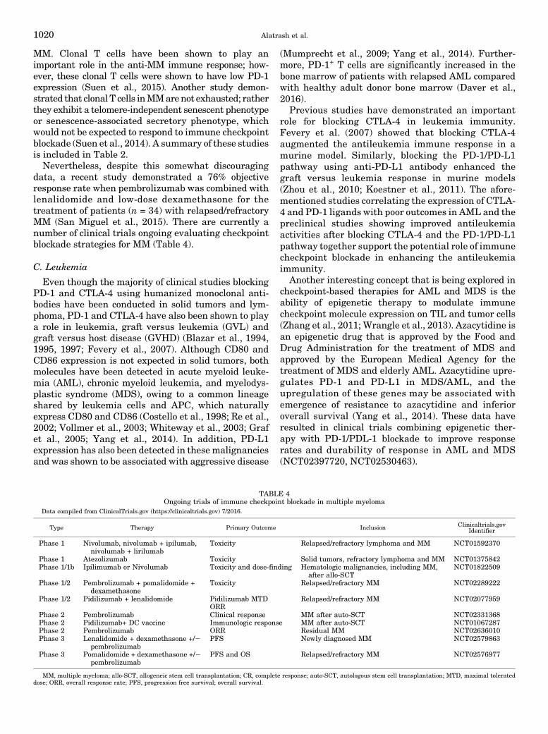
TABLE 4Ongoing trials of immune checkpoint blockade in multiple myeloma
Data compiled from ClinicalTrials.gov (https://clinicaltrials.gov) 7/2016.
Type Therapy Primary Outcome Inclusion Clinicaltrials.govIdentifier
Phase 1 Nivolumab, nivolumab + ipilumab,nivolumab + lirilumab
Toxicity Relapsed/refractory lymphoma and MM NCT01592370
Phase 1 Atezolizumab Toxicity Solid tumors, refractory lymphoma and MM NCT01375842Phase 1/1b Ipilimumab or Nivolumab Toxicity and dose-finding Hematologic malignancies, including MM,
after allo-SCTNCT01822509
Phase 1/2 Pembrolizumab + pomalidomide +dexamethasone
Toxicity Relapsed/refractory MM NCT02289222
Phase 1/2 Pidilizumab + lenalidomide Pidilizumab MTDORR
Relapsed/refractory MM NCT02077959
Phase 2 Pembrolizumab Clinical response MM after auto-SCT NCT02331368Phase 2 Pidilizumab+ DC vaccine Immunologic response MM after auto-SCT NCT01067287Phase 2 Pembrolizumab ORR Residual MM NCT02636010Phase 3 Lenalidomide + dexamethasone +/2
pembrolizumabPFS Newly diagnosed MM NCT02579863
Phase 3 Pomalidomide + dexamethasone +/2pembrolizumab
PFS and OS Relapsed/refractory MM NCT02576977
MM, multiple myeloma; allo-SCT, allogeneic stem cell transplantation; CR, complete response; auto-SCT, autologous stem cell transplantation; MTD, maximal tolerateddose; ORR, overall response rate; PFS, progression free survival; overall survival.
1020 Alatrash et al.

However, the application of immune checkpoint block-ade in the setting of leukemia is more challenging incomparison with solid tumors and lymphoma. One signif-icant obstacle in leukemia is that the underlying diseaseabrogates, and at times may completely obliterate, theimmune system. Also, in the case of acute leukemia, thetumor burden and the rate of tumor proliferation suggestthat the disease may progress before the checkpointantibodies have had sufficient time to activate an immuneresponse, especially if these agents are given alone. Thetiming of checkpoint therapy administration and identi-fication of ideal combinations is critical, and best resultsmay be achieved in themaintenance settingwhen there isminimal residual disease and a fully competent immunesystem that can bemanipulated with immune checkpointblockade or when immune checkpoint agents are com-bined with potentially synergistic standard anti-leukemictherapy. Identification of immune-checkpoint pathwaysbeyond PD-1/PDL-1 and CTLA-4 that dominate in AMLmay further guide the rational selection of specificantibodies for clinical trials. Clinically targetable check-point receptors including PD-1, OX40, and ICOS appearto be overexpressed in the bone marrows of patients withAML (Daver et al., 2016). These findings need to bevalidated in larger studies.In a phase I study of pidilizumab in patients with
various hematologicmalignancies, which included eightpatients with AML and one patient with MDS, minimalresponse was seen in one patient with AML that wasmanifested by a decrease in the blast percentage from50% to 5% (Berger et al., 2008). Four deaths werereported in that study, all of which occurred in AMLpatients andwere attributed to leukemia progression. Asummary of these studies is included in Table 2. Thereare a number of clinical trials currently ongoing to testcheckpoint antibodies as single agents and in combina-tion with standard antileukemia therapies in newlydiagnosed and relapsed leukemia, including AML andMDS, as well as maintenance in AML (Table 5).
D. Immune Checkpoint Inhibition after Stem CellTransplantation: Timing Is Everything
Clinical trials of immune checkpoint blockade havebeen conducted after SCT with promising results. Effec-tive immune reconstitution and the low disease burdenthat are characteristic after SCT provide an ideal settingto enhance the antileukemia/lymphoma immune re-sponse by eliminating the direct immunosuppressiveeffects of the tumor and by providing a microenviron-ment for the emergence of antigen specific cytotoxicT lymphocytes (CTL) (Guillaume et al., 1998; Molldremet al., 2000; Atanackovic et al., 2007; Armand et al., 2013;Tyler et al., 2013; Chung et al., 2016). Studies inlymphoma after auto-SCT are discussed in previoussections and appear to be encouraging. However, im-mune checkpoint blockade in the allo-SCT setting carriesthe potential risk of flaring GVHD (Saha et al., 2013)and, as a result, there have been fewer clinical studiesevaluating immune checkpoint blockade after allo-SCT.The precise timing of T cell reconstitution, includingCD8+ T cells, CD4+ helper T cells, and TReg, within thefirst 2 years after allo-SCT has been correlated withpromoting GVL activity or inciting GVHD (Dutt et al.,2007; Zheng et al., 2009; Alho et al., 2016). Moreover,although the antigens that drive GVL and GVHD arelargely unknown, minor antigens that are common toleukemia and normal tissue have been shown to play acritical role in both processes, and oftentimes patientswith GVHD show no evidence of disease. Therefore, it ispossible that immune checkpoint blockade could elimi-nate the underlying leukemia, albeit at the risk of flaringGVHD. Lastly, most patients who receive allo-SCTs areplaced on immunosuppressive medications to preventGVHD for approximately 6 months after the allo-SCT.Together, the particulars of post-SCT immune reconsti-tution, the target antigens of GVL/GVHD, and the use ofimmunosuppressive medications after allo-SCT high-light the critical role of the timing in implementingimmune checkpoint inhibitors after allo-SCT.
TABLE 5Ongoing trials of immune checkpoint blockade in acute myeloid leukemia
Data were compiled from ClinicalTrials.gov (https://clinicaltrials.gov) 7/2016.
Type Therapy Primary Outcome Inclusion Clinicaltrials.gov Identifier
Phase 1 Ipilimumab Acute GVHD graft rejectionAutoimmune reaction
Solid tumors, lymphoma and leukemia,including relapsed/refractory AMLafter allo-SCT
NCT00060372
Phase 1 Ipilimumab Toxicity and immunologicresponse
Relapsed/refractory AML or CMML orhigh-risk MDS
NCT01757639
Phase 1/1b Ipilimumab or nivolumab Toxicity and MTD Relapsed leukemia, including AML,lymphoma and MM after allo-SCT
NCT01822509
Phase 2 Pidilizumab + DC vaccine Toxicity AML in CR before cell collection for DCgeneration
NCT01096602
Phase 2 Azacytidine + nivolumab Response rate, overallsurvival
Relapsed AML and frontline elderly(.65 years) AML
NCT02397720
Phase 2 Idarubicin and cytarabine+ nivolumab
Event-free survival Induction in newly diagnosed AML,60 years
NCT02464657
Phase 2 Nivolumab Recurrence-Free Survival AML in remission NCT02532231
allo-SCT, allogeneic stem cell transplantation; AML, acute myeloid leukemia; CMML, chronic myelomonocytic leukemia; CR, complete response; DC, dendritic cell; GVHD,graft versus host disease; MDS, myelodysplastic syndrome; MM, multiple myeloma.
Checkpoint Blockade for Hematologic Malignancies 1021
The few reported clinical trials have proven thecomplexity of immune checkpoint inhibition and theGVL/GVHD balance after allo-SCT. In the study byBashey et al. (2009), which enrolled 29 patients withlymphoid and myeloid malignancies, ipilimumabgiven within 125–2368 days (median = 366 days) afterallo-SCT did not precipitate GVHD in any of thepatients. As discussed in the previous sections, re-sponses were noted in five patients, four of whom hadHL and one NHL. There is currently an ongoing studyat the Dana Farber Cancer Institute that is testingincreasing doses of ipilimumab administered to pa-tients with relapsed malignancy after allo-SCT. Re-sults from this phase I/Ib study of 28 patients withrelapsed lymphoid and myeloid malignancies afterallo-SCT who received two different dose levels ofipilimumab (3 or 10 mg/kg) showed efficacy of immunecheckpoint inhibition in the patients treated at thehigher dose level (Davids et al., 2016). Interestingly,patients with extramedullary AML seemed to respondparticularly well to the therapy. Of note, acute (n = 1)and chronic (n = 3) GVHD were observed duringtreatment at the 10 mg/kg dose level. In addition toblocking CTLA-4 with ipilimumab, there is one reportthat shows the safety of blocking PD-1 after allo-SCT.In a case report by Angenendt et al. (2016), one patient
with HD received nivolumab 19 months after allo-SCTwithout inciting GVHD, hence suggesting the possi-bility of using immune checkpoint blockade in thepost-allo-SCT.
In contrast to these encouraging results suggestingthe safety of immune checkpoint blockade after allo-SCT, other studies have confirmed the risk of GVHDafter immune checkpoint inhibition. In the phase I trialby Berger et al. (2008), previously discussed, 4 of17 patients who were treated with pidilizumab hadreceived allo-SCT. One of the four patients had receivedpidilizumab 8 weeks after allo-SCT and subsequentlyexperienced grade 4 GVHD of the gastrointestinal tractand died of persistent AML and GVHD. Because thispatient already had evidence of skin GVHD at studyentry, it was difficult for the investigators to determinewhether the gastrointestinal GVHD was spontaneousor secondary to pidilizumab. Although these studiesprovide a compelling, nonetheless guarded, rationale tofurther evaluate immune checkpoint inhibition afterallo-SCT, the major advance in this area should be todefine the role of immune checkpoint inhibition inpatients with evidence of disease after allo-SCT and todelineate the immune mechanisms that can be modu-lated by immune checkpoint inhibition to favor GVLover GVHD.
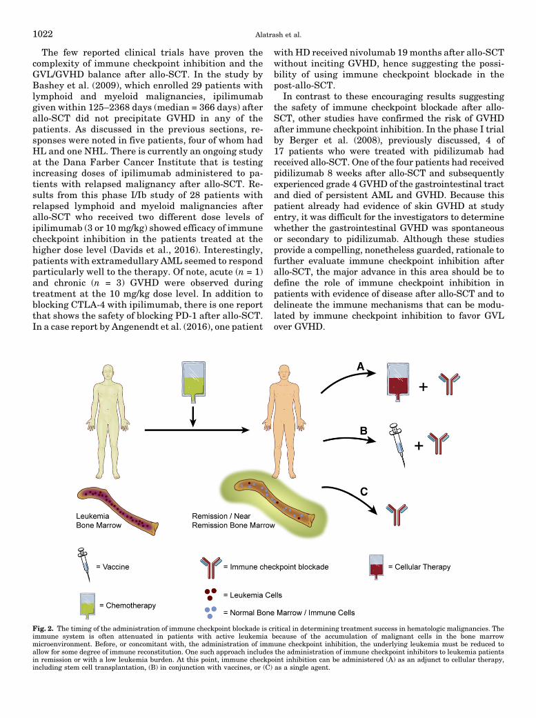
Fig. 2. The timing of the administration of immune checkpoint blockade is critical in determining treatment success in hematologic malignancies. Theimmune system is often attenuated in patients with active leukemia because of the accumulation of malignant cells in the bone marrowmicroenvironment. Before, or concomitant with, the administration of immune checkpoint inhibition, the underlying leukemia must be reduced toallow for some degree of immune reconstitution. One such approach includes the administration of immune checkpoint inhibitors to leukemia patientsin remission or with a low leukemia burden. At this point, immune checkpoint inhibition can be administered (A) as an adjunct to cellular therapy,including stem cell transplantation, (B) in conjunction with vaccines, or (C) as a single agent.
1022 Alatrash et al.
E. Immune Checkpoint Inhibition in the Setting ofEngineered T Cell Therapy
Chimeric antigen receptor (CAR) T cells made theirdebut clinically in the setting of hematologic malignan-cies. A CAR combines a single-chain variable fragmentantigen-specific extracellular region from a monoclonalantibody fused to intracellular domains providing T cellactivation (i.e., CD3-z) and costimulation (i.e., CD28,4-1BB, or OX40). CAR T cells therefore combine thespecificity of monoclonal antibodies with the effectorfunctions of T cells. The CD19 CAR T cell is thequintessential example demonstrating the potential ofthis technology. The efficacy of CD19 CAR T cells wasfirst shown in chronic lymphocytic leukemia (Porteret al., 2011) and recently in acute lymphoblastic leuke-mia (Grupp et al., 2013; Maude et al., 2015). CAR T celltherapy is rapidly advancing for the treatment ofpatients with hematologic malignancies (Porter et al.,2011, 2015; Grupp et al., 2013; Maude et al., 2015);however, there remains room for improving the efficacyand safety of CAR T cell therapy.One approach that could further potentiate the
activity of CAR T cells is to combine CAR T cells withimmune checkpoint blockade. John et al. (2013) dem-onstrated the feasibility of this approach in a HER-2transgenic mouse model. In that study, the combinationof anti-HER-2 CAR T cells and anti-PD-1 therapyenhanced the efficacy of the CAR T cells against HER-2-overexpressing tumors. As expected, mice treatedwith CAR T cells and anti-PD-1 demonstrated higherantitumor activities, but additionally, there was adecrease in myeloid derived suppressor cells in tumorstreated with anti-PD-1. The combination of immunecheckpoint inhibition and CAR T cell therapy usingantibodies or engineered T cells that have modifiedimmune checkpoint receptors (Shin et al., 2012; Ankriet al., 2013) have yet to be tested in preclinical models ofhematologic malignances or in the clinical setting butmay provide an essential synergy that could improvethe outcomes beyond those seen with each individualtherapy. Arguably, hematologic malignancies providethe ideal setting for this approach, because they arecured by immunotherapy, including allo-SCT, CART cells, and immune checkpoint inhibition, and theunderlying disease itself causes major deficiencies inthe immune system, suggesting that an approach thatprovides both an immune system and an immunemodulatory drug may be more effective.
VI. Conclusion and Future Directions
Immune checkpoint inhibition for the treatment ofcancer is undoubtedly a great breakthrough in cancertherapy (Couzin-Frankel, 2013; Dizon et al., 2016). Thefirst clinical trial of immune checkpoint inhibition wasconducted almost 15 years ago (Tchekmedyian et al.,
2002), and the differences these approaches have madein the therapy of previously untreatable solid tumorsand hematologic malignances have been striking.Through the application of immune checkpoint in-hibition, we have learned much about cancer biologyand the way tumors shape the immune response. As wegain a better understanding of the intricacies of thetumor microenvironment and the expression of im-mune checkpoint molecules by the tumor cells and theT cells, beyond CTLA-4 and PD-1 pathways, targetedclinical trials will be designed that take advantage oftherapies that target immune checkpoint moleculescombined with immune-based therapies, chemother-apies, vaccines, and small molecule targeting thera-pies (Fig. 2) to induce synergy with an intent to fullyeradicate the underlyingmalignancy and provide long-lasting cures.
Acknowledgments
Figures were designed by David M. Aten, M.A. (MD AndersonCancer Center).
Author Contributions:
Wrote or contributed to the writing of the manuscript: Alatrash,Mittendorf, and Daver.
ReferencesAlho AC, Kim HT, Chammas MJ, Reynolds CG, Matos TR, Forcade E, Whangbo J,Nikiforow S, Cutler CS, Koreth J, et al. (2016) Unbalanced recovery of regulatoryand effector T cells after allogeneic stem cell transplantation contributes to chronicGVHD. Blood 127:646–657.
Andorsky DJ, Yamada RE, Said J, Pinkus GS, Betting DJ, and Timmerman JM(2011) Programmed death ligand 1 is expressed by non-hodgkin lymphomas andinhibits the activity of tumor-associated T cells. Clin Cancer Res 17:4232–4244.
Angenendt L, Schliemann C, Lutz M, Rebber E, Schulze AB, Weckesser M, Stegger L,Schäfers M, Groth C, Kessler T, et al. (2016) Nivolumab in a patient with refractoryHodgkin’s lymphoma after allogeneic stem cell transplantation. Bone MarrowTransplant 51:443–445.
Ankri C, Shamalov K, Horovitz-Fried M, Mauer S, and Cohen CJ (2013) HumanT cells engineered to express a programmed death 1/28 costimulatory retargetingmolecule display enhanced antitumor activity. J Immunol 191:4121–4129.
Ansell S, Armand P, Timmerman JM, Shipp MA, Bradley-Garelik MB, Zhu L, andLesokhin AM (2015a) Nivolumab in Patients (Pts) with relapsed or refractoryclassical Hodgkin lymphoma (R/R cHL): Clinical outcomes from extended follow-upof a phase 1 study (CA209-039), in American Society of Hematology Annual Meet-ing; 2015 December 5–12; Orlando, FL. Abstract 583, American Society of Hema-tology, Washington, D.C.
Ansell SM, Hurvitz SA, Koenig PA, LaPlant BR, Kabat BF, Fernando D, HabermannTM, Inwards DJ, Verma M, Yamada R, et al. (2009) Phase I study of ipilimumab,an anti-CTLA-4 monoclonal antibody, in patients with relapsed and refractoryB-cell non-Hodgkin lymphoma. Clin Cancer Res 15:6446–6453.
Ansell SM, Lesokhin AM, Borrello I, Halwani A, Scott EC, Gutierrez M, Schuster SJ,Millenson MM, Cattry D, Freeman GJ, et al. (2015b) PD-1 blockade with nivolu-mab in relapsed or refractory Hodgkin’s lymphoma. N Engl J Med 372:311–319.
Ansell SM, Stenson M, Habermann TM, Jelinek DF, and Witzig TE (2001) Cd4+T-cell immune response to large B-cell non-Hodgkin’s lymphoma predicts patientoutcome. J Clin Oncol 19:720–726.
Armand P, Nagler A, Weller EA, Devine SM, Avigan DE, Chen YB, Kaminski MS,Holland HK, Winter JN, Mason JR, et al. (2013) Disabling immune tolerance byprogrammed death-1 blockade with pidilizumab after autologous hematopoieticstem-cell transplantation for diffuse large B-cell lymphoma: results of an in-ternational phase II trial. J Clin Oncol 31:4199–4206.
Atanackovic D, Arfsten J, Cao Y, Gnjatic S, Schnieders F, Bartels K, Schilling G,Faltz C, Wolschke C, Dierlamm J, et al. (2007) Cancer-testis antigens are com-monly expressed in multiple myeloma and induce systemic immunity followingallogeneic stem cell transplantation. Blood 109:1103–1112.
Bashey A, Medina B, Corringham S, Pasek M, Carrier E, Vrooman L, Lowy I,Solomon SR, Morris LE, Holland HK, et al. (2009) CTLA4 blockade with ipilimu-mab to treat relapse of malignancy after allogeneic hematopoietic cell trans-plantation. Blood 113:1581–1588.
Bensinger WI, Maloney D, and Storb R (2001) Allogeneic hematopoietic cell trans-plantation for multiple myeloma. Semin Hematol 38:243–249.
Berger R, Rotem-Yehudar R, Slama G, Landes S, Kneller A, Leiba M, Koren-Michowitz M, Shimoni A, and Nagler A (2008) Phase I safety and pharmacokineticstudy of CT-011, a humanized antibody interacting with PD-1, in patients withadvanced hematologic malignancies. Clin Cancer Res 14:3044–3051.
Checkpoint Blockade for Hematologic Malignancies 1023
Björkstrand B, Iacobelli S, Hegenbart U, Gruber A, Greinix H, Volin L, Narni F,Musto P, Beksac M, Bosi A, et al. (2011) Tandem autologous/reduced-intensityconditioning allogeneic stem-cell transplantation versus autologous trans-plantation in myeloma: long-term follow-up. J Clin Oncol 29:3016–3022.
Bladé J, Rosiñol L, Cibeira MT, Rovira M, and Carreras E (2010) Hematopoietic stemcell transplantation for multiple myeloma beyond 2010. Blood 115:3655–3663.
Blazar BR, Taylor PA, Boyer MW, Panoskaltsis-Mortari A, Allison JP, and ValleraDA (1997) CD28/B7 interactions are required for sustaining the graft-versus-leukemia effect of delayed post-bone marrow transplantation splenocyte infusion inmurine recipients of myeloid or lymphoid leukemia cells. J Immunol 159:3460–3473.
Blazar BR, Taylor PA, Linsley PS, and Vallera DA (1994) In vivo blockade ofCD28/CTLA4: B7/BB1 interaction with CTLA4-Ig reduces lethal murine graft-versus-host disease across the major histocompatibility complex barrier in mice.Blood 83:3815–3825.
Blazar BR, Taylor PA, Panoskaltsis-Mortari A, Gray GS, and Vallera DA (1995)Coblockade of the LFA1:ICAM and CD28/CTLA4:B7 pathways is a highly effectivemeans of preventing acute lethal graft-versus-host disease induced by fully majorhistocompatibility complex-disparate donor grafts. Blood 85:2607–2618.
Bruno B, Rotta M, Patriarca F, Mordini N, Allione B, Carnevale-Schianca F,Giaccone L, Sorasio R, Omedè P, Baldi I, et al. (2007) A comparison of allograftingwith autografting for newly diagnosed myeloma. N Engl J Med 356:1110–1120.
Chaperot L, Plumas J, Jacob MC, Bost F, Molens JP, Sotto JJ, and Bensa JC (1999)Functional expression of CD80 and CD86 allows immunogenicity of malignantB cells from non-Hodgkin’s lymphomas. Exp Hematol 27:479–488.
Chung DJ, Pronschinske KB, Shyer JA, Sharma S, Leung S, Curran SA, LesokhinAM, Devlin SM, Giralt SA, and Young JW (2016) T-cell Exhaustion in MultipleMyeloma Relapse after Autotransplant: Optimal Timing of Immunotherapy.Cancer Immunol Res 4:61–71.
Costello RT, Mallet F, Sainty D, Maraninchi D, Gastaut JA, and Olive D (1998)Regulation of CD80/B7-1 and CD86/B7-2 molecule expression in human primaryacute myeloid leukemia and their role in allogenic immune recognition. Eur JImmunol 28:90–103.
Couzin-Frankel J (2013) Breakthrough of the year 2013. Cancer immunotherapy.Science 342:1432–1433.
Daver N, Basu S, Garcia-Manero G, Cortes J, Ravandi F, Kornblau S, Konopleva M,Andreeff M, Borthakur G, Jain N, et al. (2016) Defining the immune checkpointlandscape of acute myeloid leukemia (AML), in American Association for CancerResearch Annual Meeting; 2016 April 16–20; New Orleans, LA. American Associ-ation for Cancer Research, Philadelphia.
Davids MS, Kim HT, Bachireddy P, Costello C, Liguori R, Savell A, Lukez AP, AviganD, Chen YB, McSweeney P, et al.; Leukemia and Lymphoma Society Blood CancerResearch Partnership (2016) Ipilimumab for Patients with Relapse after AllogeneicTransplantation. N Engl J Med 375:143–153.
Dizon DS, Krilov L, Cohen E, Gangadhar T, Ganz PA, Hensing TA, Hunger S,Krishnamurthi SS, Lassman AB, Markham MJ, et al. (2016) Clinical CancerAdvances 2016: Annual Report on Progress Against Cancer From the AmericanSociety of Clinical Oncology. J Clin Oncol 34:987–1011.
Dorfman DM, Schultze JL, Shahsafaei A, Michalak S, Gribben JG, Freeman GJ,Pinkus GS, and Nadler LM (1997) In vivo expression of B7-1 and B7-2 by follicularlymphoma cells can prevent induction of T-cell anergy but is insufficient to inducesignificant T-cell proliferation. Blood 90:4297–4306.
Dutt S, Tseng D, Ermann J, George TI, Liu YP, Davis CR, Fathman CG, and StroberS (2007) Naive and memory T cells induce different types of graft-versus-hostdisease. J Immunol 179:6547–6554.
Egen JG and Allison JP (2002) Cytotoxic T lymphocyte antigen-4 accumulation in theimmunological synapse is regulated by TCR signal strength. Immunity 16:23–35.
Fevery S, Billiau AD, Sprangers B, Rutgeerts O, Lenaerts C, Goebels J, Landuyt W,Kasran A, Boon L, Sagaert X, et al. (2007) CTLA-4 blockade in murine bonemarrow chimeras induces a host-derived antileukemic effect without graft-versus-host disease. Leukemia 21:1451–1459.
Francisco LM, Salinas VH, Brown KE, Vanguri VK, Freeman GJ, Kuchroo VK,and Sharpe AH (2009) PD-L1 regulates the development, maintenance, andfunction of induced regulatory T cells. J Exp Med 206:3015–3029.
Freeman GJ, Long AJ, Iwai Y, Bourque K, Chernova T, Nishimura H, Fitz LJ,Malenkovich N, Okazaki T, Byrne MC, et al. (2000) Engagement of the PD-1immunoinhibitory receptor by a novel B7 family member leads to negative regu-lation of lymphocyte activation. J Exp Med 192:1027–1034.
Garon EB, Rizvi NA, Hui R, Leighl N, Balmanoukian AS, Eder JP, Patnaik A,Aggarwal C, Gubens M, Horn L, et al.; KEYNOTE-001 Investigators (2015) Pem-brolizumab for the treatment of non-small-cell lung cancer. N Engl J Med 372:2018–2028.
Görgün G, Samur MK, Cowens KB, Paula S, Bianchi G, Anderson JE, White RE,Singh A, Ohguchi H, Suzuki R, et al. (2015) Lenalidomide enhances immunecheckpoint blockade-induced immune response in multiple myeloma. Clin CancerRes 21:4607–4618.
Graf M, Reif S, Hecht K, Pelka-Fleischer R, Kroell T, Pfister K, and Schmetzer H(2005) High expression of costimulatory molecules correlates with low relapse-freesurvival probability in acute myeloid leukemia (AML). Ann Hematol 84:287–297.
Green MR, Monti S, Rodig SJ, Juszczynski P, Currie T, O’Donnell E, Chapuy B,Takeyama K, Neuberg D, Golub TR, et al. (2010) Integrative analysis revealsselective 9p24.1 amplification, increased PD-1 ligand expression, and furtherinduction via JAK2 in nodular sclerosing Hodgkin lymphoma and primary medi-astinal large B-cell lymphoma. Blood 116:3268–3277.
Green MR, Rodig S, Juszczynski P, Ouyang J, Sinha P, O’Donnell E, Neuberg D,and Shipp MA (2012) Constitutive AP-1 activity and EBV infection induce PD-L1in Hodgkin lymphomas and posttransplant lymphoproliferative disorders: impli-cations for targeted therapy. Clin Cancer Res 18:1611–1618.
Grogan TM and Miller TP (1993) Immunobiologic correlates of prognosis in lym-phoma. Semin Oncol 20(5, Suppl 5)58–74.
Grupp SA, Kalos M, Barrett D, Aplenc R, Porter DL, Rheingold SR, Teachey DT,Chew A, Hauck B, Wright JF, et al. (2013) Chimeric antigen receptor-modifiedT cells for acute lymphoid leukemia. N Engl J Med 368:1509–1518.
Guillaume T, Rubinstein DB, and Symann M (1998) Immune reconstitution andimmunotherapy after autologous hematopoietic stem cell transplantation. Blood92:1471–1490.
John LB, Devaud C, Duong CP, Yong CS, Beavis PA, Haynes NM, Chow MT, SmythMJ, Kershaw MH, and Darcy PK (2013) Anti-PD-1 antibody therapy potently en-hances the eradication of established tumors by gene-modified T cells. Clin CancerRes 19:5636–5646.
Koestner W, Hapke M, Herbst J, Klein C, Welte K, Fruehauf J, Flatley A, Vignali DA,Hardtke-Wolenski M, Jaeckel E, et al. (2011) PD-L1 blockade effectively restoresstrong graft-versus-leukemia effects without graft-versus-host disease afterdelayed adoptive transfer of T-cell receptor gene-engineered allogeneic CD8+T cells. Blood 117:1030–1041.
Larkin J, Chiarion-Sileni V, Gonzalez R, Grob JJ, Cowey CL, Lao CD, Schadendorf D,Dummer R, Smylie M, Rutkowski P, et al. (2015) Combined Nivolumab and Ipi-limumab or Monotherapy in Untreated Melanoma. N Engl J Med 373:23–34.
Leach DR, Krummel MF, and Allison JP (1996) Enhancement of antitumor immunityby CTLA-4 blockade. Science 271:1734–1736.
Lesokhin AM, Ansell SM, Armand P, Scott EC, Halwani A, Gutierrez M, MillensonMM, Cohen AD, Schuster SJ, Lebovic D, et al. (2014) Preliminary results of a phaseI study of Nivolumab (BMS-936558) in patients with relapsed or refractory lym-phoid malignancies, in American Society of Hematology Annual Meeting; 2014December 6–9; San Francisco, CA. Abstract 291, American Society of Hematology,Washington, D.C.
Linsley PS, Greene JL, Brady W, Bajorath J, Ledbetter JA, and Peach R (1994)Human B7-1 (CD80) and B7-2 (CD86) bind with similar avidities but distinct ki-netics to CD28 and CTLA-4 receptors. Immunity 1:793–801.
Lippman SM, Spier CM, Miller TP, Slymen DJ, Rybski JA, and Grogan TM (1990)Tumor-infiltrating T-lymphocytes in B-cell diffuse large cell lymphoma related todisease course. Mod Pathol 3:361–367.
Maude SL, Teachey DT, Porter DL, and Grupp SA (2015) CD19-targeted chimericantigen receptor T-cell therapy for acute lymphoblastic leukemia. Blood 125:4017–4023.
McGranahan N, Furness AJ, Rosenthal R, Ramskov S, Lyngaa R, Saini SK, Jamal-Hanjani M, Wilson GA, Birkbak NJ, Hiley CT, et al. (2016) Clonal neoantigenselicit T cell immunoreactivity and sensitivity to immune checkpoint blockade.Science 351:1463–1469.
Mehta J and Singhal S (1998) Graft-versus-myeloma. Bone Marrow Transplant 22:835–843.
Molldrem JJ, Lee PP, Wang C, Felio K, Kantarjian HM, Champlin RE, and DavisMM (2000) Evidence that specific T lymphocytes may participate in the eliminationof chronic myelogenous leukemia. Nat Med 6:1018–1023.
Moskowitz C, Ribrag V, Michot JM, Martinelli G, Zinzani PL, Gutierrez M, DeMaeyer G, Jacob AG, Giallella K, Anderson JW, et al. (2014) PD-1 blockade withthe monoclonal antibody pembrolizumab (MK-3475) in patients with classicalHodgkin lymphoma after brentuximab vedotin failure: Preliminary results from aphase 1b study (KEYNOTE-013), in American Society of Hematology AnnualMeeting; 2014 December 6–9; San Francisco, CA. Abstract 290, American Societyof Hematology, Washington, D.C.
Mumprecht S, Schürch C, Schwaller J, Solenthaler M, and Ochsenbein AF (2009)Programmed death 1 signaling on chronic myeloid leukemia-specific T cells resultsin T-cell exhaustion and disease progression. Blood 114:1528–1536.
Nishimura H, Nose M, Hiai H, Minato N, and Honjo T (1999) Development of lupus-like autoimmune diseases by disruption of the PD-1 gene encoding an ITIM motif-carrying immunoreceptor. Immunity 11:141–151.
Nishimura H, Okazaki T, Tanaka Y, Nakatani K, Hara M, Matsumori A, SasayamaS, Mizoguchi A, Hiai H, Minato N, et al. (2001) Autoimmune dilated cardiomyopathyin PD-1 receptor-deficient mice. Science 291:319–322.
Porter DL, Hwang WT, Frey NV, Lacey SF, Shaw PA, Loren AW, Bagg A, MarcucciKT, Shen A, Gonzalez V, et al. (2015) Chimeric antigen receptor T cells persist andinduce sustained remissions in relapsed refractory chronic lymphocytic leukemia.Sci Transl Med 7:303ra139.
Porter DL, Levine BL, Kalos M, Bagg A, and June CH (2011) Chimeric antigenreceptor-modified T cells in chronic lymphoid leukemia. N Engl J Med 365:725–733.
Ray A, Das DS, Song Y, Richardson P, Munshi NC, Chauhan D, and Anderson KC(2015) Targeting PD1-PDL1 immune checkpoint in plasmacytoid dendritic cellinteractions with T cells, natural killer cells and multiple myeloma cells. Leukemia29:1441–1444.
Re F, Arpinati M, Testoni N, Ricci P, Terragna C, Preda P, Ruggeri D, Senese B,Chirumbolo G, Martelli V, et al. (2002) Expression of CD86 in acute myelogenousleukemia is a marker of dendritic/monocytic lineage. Exp Hematol 30:126–134.
Riley JL, Mao M, Kobayashi S, Biery M, Burchard J, Cavet G, Gregson BP, June CH,and Linsley PS (2002) Modulation of TCR-induced transcriptional profiles by li-gation of CD28, ICOS, and CTLA-4 receptors. Proc Natl Acad Sci USA 99:11790–11795.
Rizvi NA, Hellmann MD, Snyder A, Kvistborg P, Makarov V, Havel JJ, Lee W, YuanJ, Wong P, Ho TS, et al. (2015) Cancer immunology. Mutational landscape deter-mines sensitivity to PD-1 blockade in non-small cell lung cancer. Science 348:124–128.
Roddie C and Peggs KS (2011) Donor lymphocyte infusion following allogeneic he-matopoietic stem cell transplantation. Expert Opin Biol Ther 11:473–487.
Rudd CE, Taylor A, and Schneider H (2009) CD28 and CTLA-4 coreceptor expressionand signal transduction. Immunol Rev 229:12–26.
Saha A, Aoyama K, Taylor PA, Koehn BH, Veenstra RG, Panoskaltsis-Mortari A,Munn DH, Murphy WJ, Azuma M, Yagita H, et al. (2013) Host programmed deathligand 1 is dominant over programmed death ligand 2 expression in regulatinggraft-versus-host disease lethality. Blood 122:3062–3073.
1024 Alatrash et al.
San Miguel J, Mateos M, Shah JJ, Ocio EM, Rodriguez-Otero P, Reece D, MunshiNC, Avigan DE, Ge Y, Balakumaran A, et al. (2015) Pembrolizumab in combina-tion with lenalidomide and low-dose dexamethasone for relapsed/refractory mul-tiple myeloma (RRMM): Keynote-023, in American Society of Hematology AnnualMeeting; 2015 December 5–8; Orlando, FL. Abstract 505, American Society ofHematology, Washington, D.C.
Schneider H, Downey J, Smith A, Zinselmeyer BH, Rush C, Brewer JM, Wei B, HoggN, Garside P, and Rudd CE (2006) Reversal of the TCR stop signal by CTLA-4.Science 313:1972–1975.
Shin JH, Park HB, Oh YM, LimDP, Lee JE, Seo HH, Lee SJ, EomHS, Kim IH, Lee SH,et al. (2012) Positive conversion of negative signaling of CTLA4 potentiates antitumorefficacy of adoptive T-cell therapy in murine tumor models. Blood 119:5678–5687.
Snyder A, Makarov V, Merghoub T, Yuan J, Zaretsky JM, Desrichard A, Walsh LA,Postow MA, Wong P, Ho TS, et al. (2014) Genetic basis for clinical response toCTLA-4 blockade in melanoma. N Engl J Med 371:2189–2199.
Suen H, Brown R, Yang S, Ho PJ, Gibson J, and Joshua D (2015) The failure ofimmune checkpoint blockade in multiple myeloma with PD-1 inhibitors in a phase1 study. Leukemia 29:1621–1622.
Suen H, Joshua DE, Brown RD, Yang S, Barbaro PM, Ho PJ, and Gibson J(2014) Pro-tective cytotoxic clonal T-cells in myeloma have the characteristics of telomere-independent senescence rather than an exhausted or anergic phenotype: Implicationsfor immunotherapy, inAmerican Society of Hematology AnnualMeeting; 2014 December6–9; San Francisco, CA. pp 3367, American Society of Hematology, Washington, D.C.
Taube JM, Klein A, Brahmer JR, Xu H, Pan X, Kim JH, Chen L, Pardoll DM,Topalian SL, and Anders RA (2014) Association of PD-1, PD-1 ligands, and otherfeatures of the tumor immune microenvironment with response to anti-PD-1therapy. Clin Cancer Res 20:5064–5074.
Tchekmedyian S, Glaspy J, Korman A, Keler T, Deo Y, and Davis T (2002) MDX-010(human anti-CTLA4): a phase I trial in malignant melanoma, in Proceedings of theAmerican Society of Clinical Oncology; 2002 May 18–21; Orlando, FL. Abstract 56,American Society of Clinical Oncology, Alexandria, VA.
Tsukada N, Aoki S, Maruyama S, Kishi K, Takahashi M, and Aizawa Y (1997) Theheterogeneous expression of CD80, CD86 and other adhesionmolecules on leukemia andlymphoma cells and their induction by interferon. J Exp Clin Cancer Res 16:171–176.
Tyler EM, Jungbluth AA, O’Reilly RJ, and Koehne G (2013) WT1-specific T-cell re-sponses in high-risk multiple myeloma patients undergoing allogeneic T cell-depleted hematopoietic stem cell transplantation and donor lymphocyte infusions.Blood 121:308–317.
Van Allen EM, Miao D, Schilling B, Shukla SA, Blank C, Zimmer L, Sucker A, HillenU, Geukes Foppen MH, Goldinger SM, et al. (2015) Genomic correlates of responseto CTLA-4 blockade in metastatic melanoma. Science 350:207–211.
Vollmer M, Li L, Schmitt A, Greiner J, Reinhardt P, Ringhoffer M, Wiesneth M,Döhner H, and Schmitt M (2003) Expression of human leucocyte antigens andco-stimulatory molecules on blasts of patients with acute myeloid leukaemia. Br JHaematol 120:1000–1008.
Westin JR, Chu F, Zhang M, Fayad LE, Kwak LW, Fowler N, Romaguera J, HagemeisterF, Fanale M, Samaniego F, et al. (2014) Safety and activity of PD1 blockade bypidilizumab in combination with rituximab in patients with relapsed follicu-lar lymphoma: a single group, open-label, phase 2 trial. Lancet Oncol 15:69–77.
Whiteway A, Corbett T, Anderson R, Macdonald I, and Prentice HG (2003) Expres-sion of co-stimulatory molecules on acute myeloid leukaemia blasts may effectduration of first remission. Br J Haematol 120:442–451.
Wrangle J, Wang W, Koch A, Easwaran H, Mohammad HP, Vendetti F,Vancriekinge W, Demeyer T, Du Z, Parsana P, et al. (2013) Alterations ofimmune response of Non-Small Cell Lung Cancer with Azacytidine. Onco-target 4:2067–2079.
Yang H, Bueso-Ramos C, DiNardo C, Estecio MR, Davanlou M, Geng QR, Fang Z,Nguyen M, Pierce S, Wei Y, et al. (2014) Expression of PD-L1, PD-L2, PD-1 andCTLA4 in myelodysplastic syndromes is enhanced by treatment with hypo-methylating agents. Leukemia 28:1280–1288.
Yokosuka T, Takamatsu M, Kobayashi-Imanishi W, Hashimoto-Tane A, Azuma M,and Saito T (2012) Programmed cell death 1 forms negative costimulatory micro-clusters that directly inhibit T cell receptor signaling by recruiting phosphataseSHP2. J Exp Med 209:1201–1217.
Zhang M, Xiao XQ, Jiang YF, Liang YS, Peng MY, Xu Y, and Gong GZ (2011) DNAdemethylation in PD-1 gene promoter induced by 5-azacytidine activates PD-1expression on Molt-4 cells. Cell Immunol 271:450–454.
Zheng H, Matte-Martone C, Jain D, McNiff J, and Shlomchik WD (2009) Centralmemory CD8+ T cells induce graft-versus-host disease and mediate graft-versus-leukemia. J Immunol 182:5938–5948.
Zhou Q, Munger ME, Highfill SL, Tolar J, Weigel BJ, Riddle M, Sharpe AH, Val-lera DA, Azuma M, Levine BL, et al. (2010) Program death-1 signaling andregulatory T cells collaborate to resist the function of adoptively transferredcytotoxic T lymphocytes in advanced acute myeloid leukemia. Blood 116:2484–2493.
Checkpoint Blockade for Hematologic Malignancies 1025


![Adolescent Alcohol Exposure Persistently Impacts …pharmrev.aspetjournals.org/.../pharmrev/68/4/1074.full.pdfAA019767, AA11605, AA007573, and AA021040], Neurobiology of Adolescent](https://static.fdocuments.in/doc/165x107/5f0f015d7e708231d4420425/adolescent-alcohol-exposure-persistently-impacts-aa019767-aa11605-aa007573-and.jpg)

![INDEX [pharmrev.aspetjournals.org]pharmrev.aspetjournals.org/content/pharmrev/6/4/local/back-matter.… · 546 INDEX Drugallergy-Continued dosage ofdrugaffecting, 395 duration of,408](https://static.fdocuments.in/doc/165x107/5e9d1621ae88997f3e7425b1/index-546-index-drugallergy-continued-dosage-ofdrugaffecting-395-duration-of408.jpg)


![INDEX [pharmrev.aspetjournals.org]pharmrev.aspetjournals.org/content/pharmrev/5/4/local/back-matter.pdf · effectonmyasthenia gravis, 64 ... Anemia, effects ofantibiotics on, 406](https://static.fdocuments.in/doc/165x107/5d45af6088c993f1188d864c/index-effectonmyasthenia-gravis-64-anemia-effects-ofantibiotics-on.jpg)

