Targeting CD22 with the monoclonal antibody epratuzumab ...
18
RESEARCH ARTICLE Open Access Targeting CD22 with the monoclonal antibody epratuzumab modulates human B-cell maturation and cytokine production in response to Toll-like receptor 7 (TLR7) and B-cell receptor (BCR) signaling Natalia V. Giltiay 1,2* , Geraldine L. Shu 2 , Anthony Shock 3 and Edward A. Clark 1,2 Abstract Background: Abnormal B-cell activation is implicated in the pathogenesis of autoimmune diseases, including systemic lupus erythematosus (SLE). The B-cell surface molecule CD22, which regulates activation through the B-cell receptor (BCR), is a potential target for inhibiting pathogenic B cells; however, the regulatory functions of CD22 remain poorly understood. In this study, we determined how targeting of CD22 with epratuzumab (Emab), a humanized anti-CD22 IgG1 monoclonal antibody, affects the activation of human B-cell subsets in response to Toll-like receptor 7 (TLR7) and BCR engagement. Methods: B-cell subsets were isolated from human tonsils and stimulated with F(ab′) 2 anti-human IgM and/or the TLR7 agonist R848 in the presence of Emab or a human IgG1 isotype control. Changes in mRNA levels of genes associated with B-cell activation and differentiation were analyzed by quantitative PCR. Cytokine production was measured by ELISA. Cell proliferation, survival, and differentiation were assessed by flow cytometry. Results: Pretreatment of phenotypically naïve CD19 + CD10 – CD27 – cells with Emab led to a significant increase in IL-10 expression, and in some but not all patient samples to a reduction of IL-6 production in response to TLR7 stimulation alone or in combination with anti-IgM. Emab selectively inhibited the expression of PRDM1, the gene encoding B-lymphocyte-induced maturation protein 1 (Blimp-1) in activated CD10 – CD27 – B cells. CD10 – CD27 – IgD – cells were highly responsive to stimulation through TLR7 as evidenced by the appearance of blasting CD27 hi CD38 hi cells. Emab significantly inhibited the activation and differentiation of CD10 – CD27 – IgD – B cells into plasma cells. Conclusions: Emab can both regulate cytokine expression and block Blimp1-dependent B-cell differentiation, although the effects of Emab may depend on the stage of B-cell development or activation. In addition, Emab inhibits the activation of CD27 – IgD – tonsillar cells, which correspond to so-called double-negative memory B cells, known to be increased in SLE patients with more active disease. These data may be relevant to the therapeutic effect of Emab in vivo via modulation of the production of pro-inflammatory and anti-inflammatory cytokines by B cells. Because Blimp-1 is required by B cells to mature into antibody-producing cells, inhibition of Blimp1 may reduce autoantibody production. Keywords: Antibodies, B cells, B-cell targeted therapy, CD22, IL-6, IL-10, Epratuzumab, Systemic lupus erythematosus, TLR7 * Correspondence: [email protected] 1 Division of Rheumatology, Department of Medicine, University of Washington, Seattle, WA 98109, USA 2 Department of Immunology, University of Washington, Seattle, WA 98109, USA Full list of author information is available at the end of the article © The Author(s). 2017 Open Access This article is distributed under the terms of the Creative Commons Attribution 4.0 International License (http://creativecommons.org/licenses/by/4.0/), which permits unrestricted use, distribution, and reproduction in any medium, provided you give appropriate credit to the original author(s) and the source, provide a link to the Creative Commons license, and indicate if changes were made. The Creative Commons Public Domain Dedication waiver (http://creativecommons.org/publicdomain/zero/1.0/) applies to the data made available in this article, unless otherwise stated. Giltiay et al. Arthritis Research & Therapy (2017) 19:91 DOI 10.1186/s13075-017-1284-2
Transcript of Targeting CD22 with the monoclonal antibody epratuzumab ...
RESEARCH ARTICLE Open Access
Targeting CD22 with the monoclonalantibody epratuzumab modulates humanB-cell maturation and cytokine productionin response to Toll-like receptor 7 (TLR7)and B-cell receptor (BCR) signalingNatalia V. Giltiay1,2*, Geraldine L. Shu2, Anthony Shock3 and Edward A. Clark1,2
Abstract
Background: Abnormal B-cell activation is implicated in the pathogenesis of autoimmune diseases, includingsystemic lupus erythematosus (SLE). The B-cell surface molecule CD22, which regulates activation through the B-cellreceptor (BCR), is a potential target for inhibiting pathogenic B cells; however, the regulatory functions of CD22remain poorly understood. In this study, we determined how targeting of CD22 with epratuzumab (Emab), ahumanized anti-CD22 IgG1 monoclonal antibody, affects the activation of human B-cell subsets in response toToll-like receptor 7 (TLR7) and BCR engagement.
Methods: B-cell subsets were isolated from human tonsils and stimulated with F(ab′)2 anti-human IgM and/or theTLR7 agonist R848 in the presence of Emab or a human IgG1 isotype control. Changes in mRNA levels of genesassociated with B-cell activation and differentiation were analyzed by quantitative PCR. Cytokine production wasmeasured by ELISA. Cell proliferation, survival, and differentiation were assessed by flow cytometry.
Results: Pretreatment of phenotypically naïve CD19+CD10–CD27– cells with Emab led to a significant increase inIL-10 expression, and in some but not all patient samples to a reduction of IL-6 production in response to TLR7stimulation alone or in combination with anti-IgM. Emab selectively inhibited the expression of PRDM1, the geneencoding B-lymphocyte-induced maturation protein 1 (Blimp-1) in activated CD10–CD27– B cells. CD10–CD27–IgD–
cells were highly responsive to stimulation through TLR7 as evidenced by the appearance of blasting CD27hiCD38hi
cells. Emab significantly inhibited the activation and differentiation of CD10–CD27–IgD– B cells into plasma cells.
Conclusions: Emab can both regulate cytokine expression and block Blimp1-dependent B-cell differentiation,although the effects of Emab may depend on the stage of B-cell development or activation. In addition, Emabinhibits the activation of CD27–IgD– tonsillar cells, which correspond to so-called double-negative memory B cells,known to be increased in SLE patients with more active disease. These data may be relevant to the therapeuticeffect of Emab in vivo via modulation of the production of pro-inflammatory and anti-inflammatory cytokines byB cells. Because Blimp-1 is required by B cells to mature into antibody-producing cells, inhibition of Blimp1 mayreduce autoantibody production.
Keywords: Antibodies, B cells, B-cell targeted therapy, CD22, IL-6, IL-10, Epratuzumab, Systemic lupuserythematosus, TLR7
* Correspondence: [email protected] of Rheumatology, Department of Medicine, University ofWashington, Seattle, WA 98109, USA2Department of Immunology, University of Washington, Seattle, WA 98109,USAFull list of author information is available at the end of the article
© The Author(s). 2017 Open Access This article is distributed under the terms of the Creative Commons Attribution 4.0International License (http://creativecommons.org/licenses/by/4.0/), which permits unrestricted use, distribution, andreproduction in any medium, provided you give appropriate credit to the original author(s) and the source, provide a link tothe Creative Commons license, and indicate if changes were made. The Creative Commons Public Domain Dedication waiver(http://creativecommons.org/publicdomain/zero/1.0/) applies to the data made available in this article, unless otherwise stated.
Giltiay et al. Arthritis Research & Therapy (2017) 19:91 DOI 10.1186/s13075-017-1284-2
BackgroundB cells are central mediators of humoral immunity andplay a key role in the protection against pathogens.However, aberrant B-cell activation is a common featureof many autoimmune diseases including systemic lupuserythematosus (SLE), primary Sjögren’s syndrome (pSS),and rheumatoid arthritis (RA). B cells may contributeto the pathogenesis of autoimmune diseases throughseveral mechanisms, including both antibody (Ab)-dependent functions (e.g., secretion of autoantibodies(auto-Abs)) and Ab-independent functions, such asantigen (Ag) presentation to T cells and productionof pro-inflammatory cytokines [1, 2].SLE is a complex, systemic autoimmune disease with
highly diverse clinical manifestations [3]. The hallmarksof the disease are polyclonal B-cell activation, produc-tion of auto-Abs against nuclear-containing Ags, andorgan damage due to immune complex deposition.Abnormalities in various B-cell compartments have beendescribed in SLE patients, including expansion of newlyformed (transitional) cells, as well as an expansion ofAg-experienced CD27+/IgD– switched memory B cellsand CD27–IgD– double-negative (DN) memory B cells[2, 4–6]. Previous studies have shown that the frequencyof circulating CD20–CD27++ plasma cells correlates withSLE disease activity and auto-Ab titers [7]; however, thecellular origins of Ab-producing cells in SLE as well asthe signals that drive their activation are not well de-fined. Alterations in the balance of signals downstreamof the B-cell receptor (BCR) and pro-survival signalsmay contribute to the loss of immune tolerance and tothe survival and activation of autoreactive B cells [8, 9].Dysregulation of BCR signaling, including increasedphosphorylation of Syk and decreased phosphorylationof phosphatase activities, has also been described in SLEpatients [10, 11].B-cell activation via innate immune receptors, including
endosomal Toll-like receptors (TLRs) expressed in B cells,may play a key role in driving autoreactive B cells in SLE[9, 12–15]. In mice, RNA-associated autoantigens activateautoreactive B cells by engaging BCRs and TLR7, anendosomal TLR specialized in the recognition of viralssRNA, and induce the production of auto-Abs [12, 16].Data from murine lupus models further support a role forTLR7 in SLE pathogenesis. Extra copies of the Tlr7 genedrive lupus-like disease [17–19]; whereas lupus-proneTlr7-deficient mice develop attenuated disease symptoms[20, 21]. Importantly, B-cell-intrinsic TLR7 signals candrive cell proliferation and differentiation and amplifyauto-Ab production to further exacerbate SLE disease inmice [22]. In humans, genetic studies have demonstrateda correlation between genetic variations associatedwith dysregulation of TLR7 expression and SLE sus-ceptibility [23–25].
Despite significant evidence implicating TLR7 in SLE,the effects of TLR7 on human B cells have not beenexplored fully. Little is known about the signals that canregulate TLR7-mediated activation of human B cells. IFN-α can promote human B-cell responses to TLR7 ligation,most likely through its ability to upregulate TLR7 receptorlevels [26]. In-vitro stimulation of CD19+CD27– blood Bcells with a synthetic TLR7 ligand induces IgM and IgGproduction as well as secretion of IL-6 and IL-10 [27].TLR7 activation also expands IgM+CD27+ memory B cellsand CD27hi B cells, and a combination of TLR7 plus IFN-α promotes the production of auto-Abs [28]. Thus, TLR7-mediated activation of human B cells, as with mouse Bcells, can induce the production of auto-Abs.While signals from BCR and TLR7 can synergize and
promote inappropriate activation of autoreactive B cells,the engagement of other surface receptors, such asCD19 and CD22, have been proposed to inhibit theiractivation [29, 30]. The CD22 Siglec receptor familymember is expressed predominantly on B cells and bindsvia its extracellular ligand-binding domain to α2-6-linked sialic acids on glycoproteins expressed on thesame cell (cis interactions) or on opposing cells and/orsoluble proteins (trans interactions) [31, 32]. CD22 actsas an adhesion receptor and functions to regulate B-cellmigration [33–35]. Crosslinking of CD22 and the BCRtriggers phosphorylation of the CD22 cytoplasmic tail,leading to the activation of a number of signaling mole-cules, known to either inhibit the BCR signaling or topromote the activation of JNK/SAPK and mitogen acti-vated protein kinase ERK2 [30, 36, 37]. In addition to itsfunction in regulating BCR signaling, CD22 has beenimplicated in the regulation of TLR-mediated signalingin B cells [38]. CD22–/– B cells have hyperactive re-sponses to TLR stimulation compared to wild-type(WT) B cells [38, 39]. Furthermore, studies have shownthat LPS-induced activation of nuclear factor-κB (NF-κB) downstream of TLR4 is inhibited by the expressionof CD22 [38].The expression of both CD22 and its ligands vary
according to the B-cell maturation/activation state. Inthe periphery, CD22 is expressed at maximum densityon human CD27– naïve and transitional B cells, while itis downregulated by plasma cells [40, 41]. CD22 avail-ability on the cell surface is also dependent on maskingor unmasking of CD22 by endogenous (cis) ligand inter-actions [42]. The expression of CD22 ligands on humanB cells is less well explored, but recent studies haveshown that, due to changes in glycosylation, germinalcenter (GC) B cells lose the expression of high-affinityCD22 ligands, leading to CD22 “unmasking” [43].The development of monoclonal Abs designed to tar-
get human CD22 [44] as well recent advances in humanB-cell phenotyping [45] provide new opportunities to
Giltiay et al. Arthritis Research & Therapy (2017) 19:91 Page 2 of 18
explore the effects of CD22 engagement on differentsubsets of human B-cell subsets. Epratuzumab (Emab), ahumanized anti-human CD22 IgG1 Ab, has previouslyshown promising clinical activity both as a single agentand in combination with rituximab in patients with non-Hodgkin’s lymphoma (NHL) [46]. Unlike rituximab,which depletes circulating B cells, Emab does not inducecomplement-dependent cytotoxicity or Ab-dependentcellular cytotoxicity [47]. CD22 ligation by Emab, how-ever, provokes rapid internalization and phosphorylationof CD22, inhibits the phosphorylation of Syk and PLCγ2,and reduces intracellular Ca2+ mobilization after BCRstimulation in vitro [44, 48, 49]. Given the role of CD22in modulating both BCR and TLR signaling, targetingCD22 with Emab has also been explored as a therapy forautoimmune diseases, including SLE and pSS [50, 51].Phase I and IIb clinical trials have demonstrated clinic-ally relevant, sustained improvements in patients withmoderate-to-severe SLE and a good safety profile ofEmab [52, 53]. Emab treatment was found to induce apartial reduction of circulating B cells in SLE patientsaffecting primarily CD27– cells [54], a phenomenon thatlater led to an exploration of the in-vitro effects of Emabon the expression of the adhesion molecules CD62L, β7integrin, and β1 integrin and on B-cell migration towardCXCL12 [41]. CD22 binding by Emab also induces a re-duction of CD19, CD21, and CD79b expression througha process known as trogocytosis (i.e., Fc-mediated recep-tor “shaving” to other effector cells). In line with thesefindings, a decrease in CD19 expression was observed inSLE patients treated with Emab [55]. Recent studies havealso investigated the effects of Emab on cytokine pro-duction. Emab inhibited IL-6 and tumor necrosis factoralpha (TNF-α) production of blood B cells isolated fromhealthy donors and SLE patients in response to BCRcrosslinking alone or in combination with TLR9 ligandCpG [56]. Overall, the available data led to the hypoth-esis that the primary mode of action of Emab is toenhance the normal inhibitory role of CD22 on B-cellactivation [57].Given the importance of TLR7 signaling in activating
autoreactive B cells in SLE, we investigated whetherCD22 crosslinking by Emab might affect B-cell activa-tion in response to BCR and/or TLR7 stimulation. Wefurther aimed to identify which subpopulation of humanB cells might be affected to the greatest extent by Emabin the context of BCR/TLR7 stimulation. Using humantonsillar CD10–CD27– B cells, we found that Emabmodulates cytokine production by inhibiting IL-6, whileat the same time enhancing IL-10 production. Emabdramatically and selectively inhibited levels of PRDM1,the gene encoding Blimp-1 in CD10–CD27– tonsillar Bcells, activated by either TLR7 signaling alone or incombination with BCR stimulation. Tonsillar B-cell
subsets responded differently to BCR/TLR7 stimulation;among them CD10–CD27–IgD– cells, which correspondto DN memory B cells found in the periphery, weremost responsive to TLR7 stimulation, as evidenced bythe appearance of CD27hiCD38hiBlimp1+ plasmablasts.In particular, Emab inhibited the in-vitro differentiationof CD10–CD27–IgD– and CD10+CD27–/+ B cells andreduced the survival of CD10+CD27–/+ B cells but notother B-cell subsets. Thus, Emab can both enhance andinhibit TLR7-driven B-cell responses: depending on the B-cell developmental/maturation state, CD22 crosslinkingby Emab affects B-cell survival, activation, and differenti-ation differently, which may have important implicationsfor the clinical use of Emab and monitoring of CD22-based therapies.
MethodsCell preparation and purificationPost-surgical tonsillar tissue samples were obtained fromValley Medical Day Surgery Center (Renton, WA, USA) inaccordance with an IRB approved protocol. Cell suspen-sions were prepared by gently teasing tissues in R10medium (RPMI 1640 containing 10% FBS (ThermoFisherScientific), 100 U/ml penicillin, 100 mg/ml streptomycin,2 mM L-glutamine, 1 mM sodium pyruvate, and 10 mMHepes) with forceps and scissors, and then separating cellsover Ficoll-Hypaque (GE Healthcare Life Sciences).Tonsillar B cells were enriched by depleting CD2+ T andNK cells using sheep erythrocytes, a technique that relieson the formation of immunorosettes (i.e., rosetting) [58],and then separated again over Ficoll-Hypaque. Samplesafter rosetting were typically ≥90% pure CD20+ B cells. Insome experiments, cells were further enriched for CD27–
CD10– B cells using biotinylated monoclonal Abs (mAbs)against CD3 (G19-4), CD5 (10.2), CD10 (CB-CALLA),and CD27 (O323) (eBioscience) with the StemCellTechnologies Human Biotin Selection Kit for negativeselection. In other experiments, post-rosetted cells werestained with fluorescently labeled mAbs (anti-CD3, CD27,CD10, and IgD antibodies) and then sorted intoCD10–CD27+ (memory), CD10–CD27–IgD+ (naïve), andCD10–CD27–IgD– (IgD–CD27– DN memory) B-cell andCD10–CD27+/– (pre-GC/CG/plasma) cell populationsusing a FACSAria II high-speed cell sorter (BD Pharmin-gen) at 4 °C under sterile conditions. Post-sort analyseswere performed to assess the purity of sorted cells. Periph-eral blood mononuclear cells (PBMCs) from healthydonors (HD) were isolated by density-gradient centrifuga-tion using Ficoll-Hypaque. Written consent was obtainedfrom all blood donors.
In-vitro cell culture and CFSE proliferation assayB cells were cultured in R10 medium at 37 °C and 5%CO2. For gene expression analyses, cells were enriched
Giltiay et al. Arthritis Research & Therapy (2017) 19:91 Page 3 of 18
for CD27–CD10– B cells and plated at 3 × 10⁶ cellsper ml with preincubation for 1 hour at 37 °C withEmab (5 μg/ml) or hIgG1 isotype control (5 μg/ml; Sigma)or R10 medium and then stimulated with TLR7 agonistR848 (50 ng/ml; InvivoGen), F(ab′)2 anti-human IgM(5 μg/ml; Jackson ImmunoResearch Laboratories,Inc.), or a combination of R848 plus anti-IgM. In somecases, cells were pretreated with IFN-α at 1000 U/ml(PBL, Piscataway, NJ, USA). Samples were cultured for 12hours, harvested, and used for RNA isolation. For cellproliferation experiments, cells were loaded with 2.5 μMCFSE (ThermoFisher Scientific) in PBS at 37 °C for 10min, quenched with R10 medium, washed, stimulated,and then cultured for 3 days. For assessing cell survivaland plasma cell differentiation, sorted B-cell subsets wereplaced in 96-well plates at a concentration of 2.5 × 10⁶
cells per ml, treated with different stimuli, and analyzedby flow cytometry after 3–5 days of in-vitro cell culture.
Cytokine productionB cells enriched for CD10–CD27– cells were plated at5 × 106 cells per ml in 96-well plates, preincubated withor without Emab or a human IgG control, andstimulated with R848 and/or F(ab′)2 anti-human IgM.Culture supernatants were collected 3 days post stimula-tion, frozen down, and used to assess cytokine production.Cytokine array (R&D Systems) data showed significant in-duction of IL-6 and IL-10 after R848 and/or anti-IgMstimulation. Further quantification of these cytokines wasperformed using Human Quantikine ELISA Kits (R&DSystems) according to the manufacturer’s instructions.
Flow cytometryFor characterization of tonsillar B-cell subsets, single cellsuspensions were stained with appropriate combinations offluorescently labeled mAbs, including anti-CD10 (CB-CALLA), CD19 (SJ25C1), CD20 (2H7), CD22 (4KB128and S-HCL-1), CD27 (LG.7F9), CD38 (HB7) (eBioscience),IgD (IA6-2), CD3 (SP34-2), and CD95 (DX2) (BD Biosci-ences). Live cells were identified using LIVE/DEAD FixableNear-IR staining (Molecular Probes) according to the man-ufacturer’s instructions. Cultured cells were pelleted,washed, and stained with LIVE/DEAD, followed by surfacestaining with fluorescently labeled mAbs. For Blimp1 intra-cellular staining, cells were first stained with LIVE/DEADfixable dye, washed, stained with appropriate surfacemarkers, washed and then fixed, permeabilized, andstained with PE-conjugated rat IgG2ak anti-Blimp1 Ab(6D3) using the Transcription Factor Buffer Set (BD).CFSE-labeled cells were cultured for 3 days and the levelsof cell proliferation were measured based on CFSEdilution. Multicolor flow cytometry was performed using afive-laser LSRII flow cytometer (BD) and analyzed withFlowJo software (Tree Star).
Imaging flow cytometryEmab anti-CD22 binding and internalization by tonsillar Bcells was assessed by multispectral imaging flow cytometry.Tonsillar B cells were stained with mAb specific for CD10,CD20, CD27, and IgD with or without Emab, conjugatedto Pacific Blue (conjugation was performed using PacificBlue™ Antibody Labeling Kit from Molecular Probes, Ther-moFisher Scientific). Incubation with Pacific Blue-Emabwas performed at either 4 °C on ice in the presence ofNaN3 or at 37 °C for 30 min. CD20+ cells were gated intoCD10–CD27–, CD10–CD27+, and CD10–CD27+/– B-cellsubsets, and Emab binding and receptor-mediated intern-alization was determined for each subset. Then 50,000–100,000 cells were analyzed using 60× camera magnifica-tion using an Image Stream X Mark II instrument and datawere analyzed with IDEAS software (Amnis). The Internal-ization Score (IS) was defined as the ratio of intensity in-side the cell to the intensity of the entire cell.
Quantitative RT-PCRTotal RNA was extracted from cells using an RNeasymini kit with DNase treatment (QIAGEN). First-strandcDNA was generated using 250 ng of total RNA withthe SuperScript III high-capacity cDNA RT-kit usingrandom primers (Invitrogen). Primers, as indicated inAdditional file 1: Table S1, were synthesized (Invitrogen)and diluted to the appropriate concentrations usingmolecular-grade water. Transcript expression was ana-lyzed by quantitative RT-PCR using SYBR® green PCRMaster Mix (Applied Biosystems) on an Applied Biosys-tems StepOnePlus Real Time PCR System using a two-stage cycle of 95 °C for 15 s and 60 °C for 1 min repeatedfor 40 cycles, followed by a dissociation stage. Thresholdcycle (Ct) values were determined by setting a constantthreshold at 0.2. All samples were normalized for the ex-pression of 18S; fold changes in gene expression were cal-culated using the 2−ΔΔCT method and presented asrelative expression to unstimulated controls.
Statistical analysesGraphs and statistical analyses were performed usingPrism 5.0 software (GraphPad, San Diego, CA, USA).Statistical significance between groups was determinedby two-tailed, unpaired Student’s t test or by one-wayANOVA with Turkey post test. Pearson’s correlation wasused to measure the relationship between two variables.Results are reported as mean ± SD or ± SEM. p < 0.05was considered statistically significant.
ResultsCD22 is expressed broadly across tonsillar B-cellsubpopulations and is internalized in response to EmabHuman tonsils contain 50–60% CD19+ cells and thusrepresent a useful source of B cells, including cells with
Giltiay et al. Arthritis Research & Therapy (2017) 19:91 Page 4 of 18
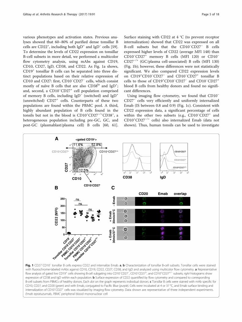
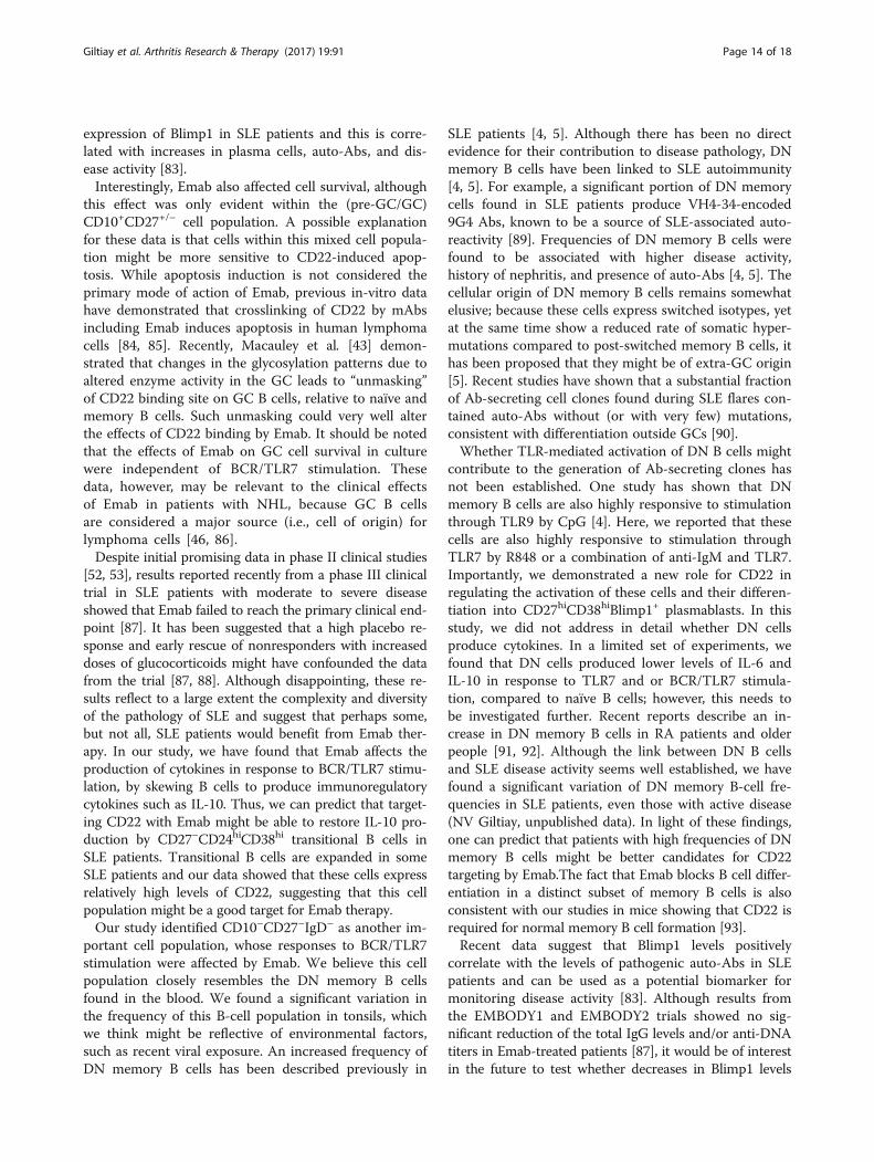
various phenotypes and activation states. Previous ana-lyses showed that 60–80% of purified dense tonsillar Bcells are CD22+, including both IgD+ and IgD– cells [59].To determine the levels of CD22 expression on tonsillarB-cell subsets in more detail, we performed a multicolorflow cytometry analysis, using mAbs against CD19,CD10, CD27, IgD, CD38, and CD22. As Fig. 1a shows,CD19+ tonsillar B cells can be separated into three dis-tinct populations based on their relative expression ofCD10 and CD27: first, CD10–CD27– cells, which consistmostly of naïve B cells that are also CD38lo and IgD+;and, second, a CD10–CD27+ cell population comprisedof memory B cells, including IgD– (switched) and IgD+
(unswitched) CD27+ cells. Counterparts of these twopopulations are found within the PBMC pool. A third,highly abundant population of B cells found in thetonsils but not in the blood is CD10+CD27–/+CD38+, aheterogeneous population including pre-GC, GC, andpost-GC (plasmablast/plasma cell) B cells [60, 61].
Surface staining with CD22 at 4 °C (to prevent receptorinternalization) showed that CD22 was expressed on allB-cell subsets but that the CD10–CD27– B cellsexpressed higher levels of CD22 (average MFI 148) thanCD10–CD27+ memory B cells (MFI 120) or CD10–
CD27–/+ (GC/plasma cell-associated) B cells (MFI 130)(Fig. 1b); however, these differences were not statisticallysignificant. We also compared CD22 expression levelson CD19+CD10–CD27– and CD10–CD27+ tonsillar Bcells to those of CD19+CD10–CD27– and CD10–CD27+
blood B cells from healthy donors and found no signifi-cant differences.Using imaging flow cytometry, we found that CD10–
CD27– cells very efficiently and uniformly internalizedEmab (IS between 0.8 and 0.9) (Fig. 1c). Consistent withCD22 expression data, a significant percentage of cellswithin the other two subsets (e.g., CD10–CD27+ andCD10+CD27–/+ cells) also internalized Emab (data notshown). Thus, human tonsils can be used to investigate
B
CD38 IgD
A
C CD20 Emab overlap
+ E
mab
30m
in/4
° C+
Em
ab30
min
/37°
C-
Em
ab
<gated CD19+>
CD10
CD
27
CD10-CD27-
CD10+CD27-/+CD10-CD27+
Fig. 1 CD27–CD10– tonsillar B cells express CD22 and internalize Emab. a, b Characterization of tonsillar B-cell subsets. Tonsillar cells were stainedwith fluorochrome-labeled mAbs against CD10, CD19, CD22, CD27, CD38, and IgD and analyzed using multicolor flow cytometry. a Representativeflow analysis of gated live CD19+ cells showing B-cell subgating into CD10–CD27–, CD10–CD27+, and CD10+CD27+/– subsets; right histograms showexpression of CD38 and IgD within each population. b Surface expression of CD22 quantified by flow cytometry and compared to correspondingB-cell subsets from PBMCs of healthy donors. Each dot on the graph represents individual donors. c Tonsillar B cells were stained with mAb specific forCD10, CD27, and CD20 (green) and with Emab, conjugated to Pacific Blue (purple). Cells were incubated at 4 or 37 °C, and Emab surface binding andinternalization of CD10–CD27– cells was visualized by imaging flow cytometry. Data shown are representative of three independent experiments.Emab epratuzumab, PBMC peripheral blood mononuclear cell
Giltiay et al. Arthritis Research & Therapy (2017) 19:91 Page 5 of 18
the differential effects of CD22 crosslinking by Emab onthe responses of B-cell subsets from human lymphoidtissue. In particular, the CD19+CD10–CD27– B cells ex-press high levels of CD22 and efficiently internalizedEmab. Because this population likely corresponds to cir-culating CD27– B cells that are significantly decreasedupon Emab administration in vivo [54], we focused ourstudy initially on this B-cell subset. We developed aprotocol for enrichment of CD10–CD27– B cells using atwo-step enrichment process, including rosetting anddepletion of CD3+, CD5+, CD27+, and CD10+ cells bymagnetic bead separation. Using this protocol, we wereable to consistently obtain 70–80% pure “untouched”CD19+CD20+CD10–CD27– B cells not bound bymAbs that might affect their responses (Additionalfile 2: Figure S1).
Emab anti-CD22 does not affect the expression of BCR-inducible genes and genes associated with TLR signalingIn initial experiments, we used CD20+CD10–CD27–
enriched B cells to test the effects of Emab on responsesto BCR and/or TLR7 stimulation. After enrichment, wepreincubated cells for 1 hour at 37 °C with Emab, hIgG1isotype control, or R10 medium and then stimulatedthem with the TLR7 agonist R848, F(ab′)2 anti-humanIgM, or a combination of R848 and anti-IgM for 12hours. Using quantitative RT-PCR, we measured therelative expression of multiple genes known to be in-duced downstream of BCR signaling (C-MYC, BCLXL,TP53) or associated with TLR7 signaling (TLR7, TLR9,MyD88, IRF7, UNC93B, IRAK4, TRAF6). Emab had nosignificant effect on the expression of any of these genes(Additional file 3: Figure S2 and data not shown).
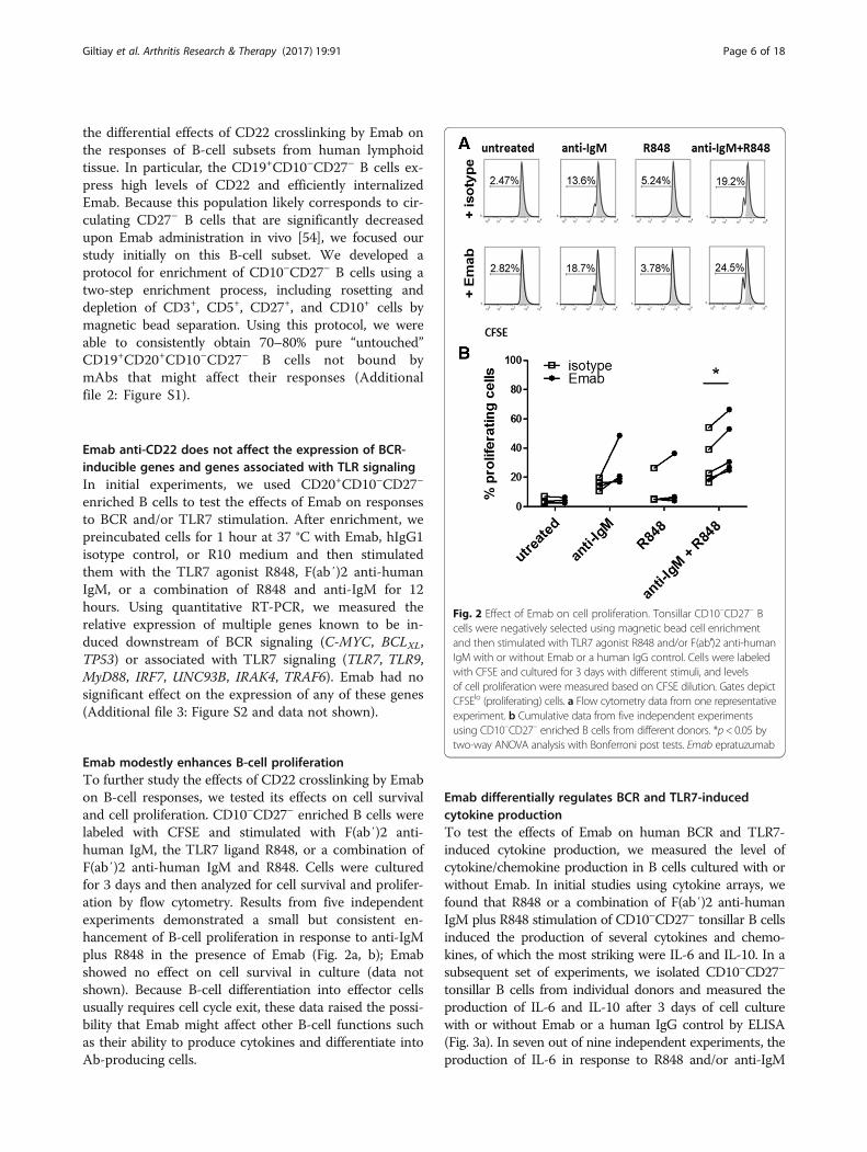
Emab modestly enhances B-cell proliferationTo further study the effects of CD22 crosslinking by Emabon B-cell responses, we tested its effects on cell survivaland cell proliferation. CD10–CD27– enriched B cells werelabeled with CFSE and stimulated with F(ab′)2 anti-human IgM, the TLR7 ligand R848, or a combination ofF(ab′)2 anti-human IgM and R848. Cells were culturedfor 3 days and then analyzed for cell survival and prolifer-ation by flow cytometry. Results from five independentexperiments demonstrated a small but consistent en-hancement of B-cell proliferation in response to anti-IgMplus R848 in the presence of Emab (Fig. 2a, b); Emabshowed no effect on cell survival in culture (data notshown). Because B-cell differentiation into effector cellsusually requires cell cycle exit, these data raised the possi-bility that Emab might affect other B-cell functions suchas their ability to produce cytokines and differentiate intoAb-producing cells.
Emab differentially regulates BCR and TLR7-inducedcytokine productionTo test the effects of Emab on human BCR and TLR7-induced cytokine production, we measured the level ofcytokine/chemokine production in B cells cultured with orwithout Emab. In initial studies using cytokine arrays, wefound that R848 or a combination of F(ab′)2 anti-humanIgM plus R848 stimulation of CD10–CD27– tonsillar B cellsinduced the production of several cytokines and chemo-kines, of which the most striking were IL-6 and IL-10. In asubsequent set of experiments, we isolated CD10–CD27–
tonsillar B cells from individual donors and measured theproduction of IL-6 and IL-10 after 3 days of cell culturewith or without Emab or a human IgG control by ELISA(Fig. 3a). In seven out of nine independent experiments, theproduction of IL-6 in response to R848 and/or anti-IgM
Fig. 2 Effect of Emab on cell proliferation. Tonsillar CD10–CD27– Bcells were negatively selected using magnetic bead cell enrichmentand then stimulated with TLR7 agonist R848 and/or F(ab′)2 anti-humanIgM with or without Emab or a human IgG control. Cells were labeledwith CFSE and cultured for 3 days with different stimuli, and levelsof cell proliferation were measured based on CFSE dilution. Gates depictCFSElo (proliferating) cells. a Flow cytometry data from one representativeexperiment. b Cumulative data from five independent experimentsusing CD10–CD27– enriched B cells from different donors. *p< 0.05 bytwo-way ANOVA analysis with Bonferroni post tests. Emab epratuzumab
Giltiay et al. Arthritis Research & Therapy (2017) 19:91 Page 6 of 18
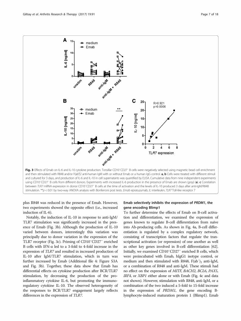
plus R848 was reduced in the presence of Emab. However,two experiments showed the opposite effect (i.e., increasedinduction of IL-6).Notably, the induction of IL-10 in response to anti-IgM/
TLR7 stimulation was significantly increased in the pres-ence of Emab (Fig. 3b). Although the production of IL-10varied between donors, interestingly this variation wasprincipally due to donor variation in the expression of theTLR7 receptor (Fig. 3c). Priming of CD10–CD27– enrichedB cells with IFN-α led to a 3-fold to 4-fold increase in theexpression of TLR7 and resulted in increased production ofIL-10 after IgM/TLR7 stimulation, which in turn wasfurther increased by Emab (Additional file 4: Figure S3Aand Fig. 3b). Together, these data show that Emab hasdifferential effects on cytokine production after BCR/TLR7stimulation, by decreasing the production of the pro-inflammatory cytokine IL-6, but promoting the immuno-regulatory cytokine IL-10. The observed heterogeneity ofthe responses to BCR/TLR7 engagement largely reflectsdifferences in the expression of TLR7.
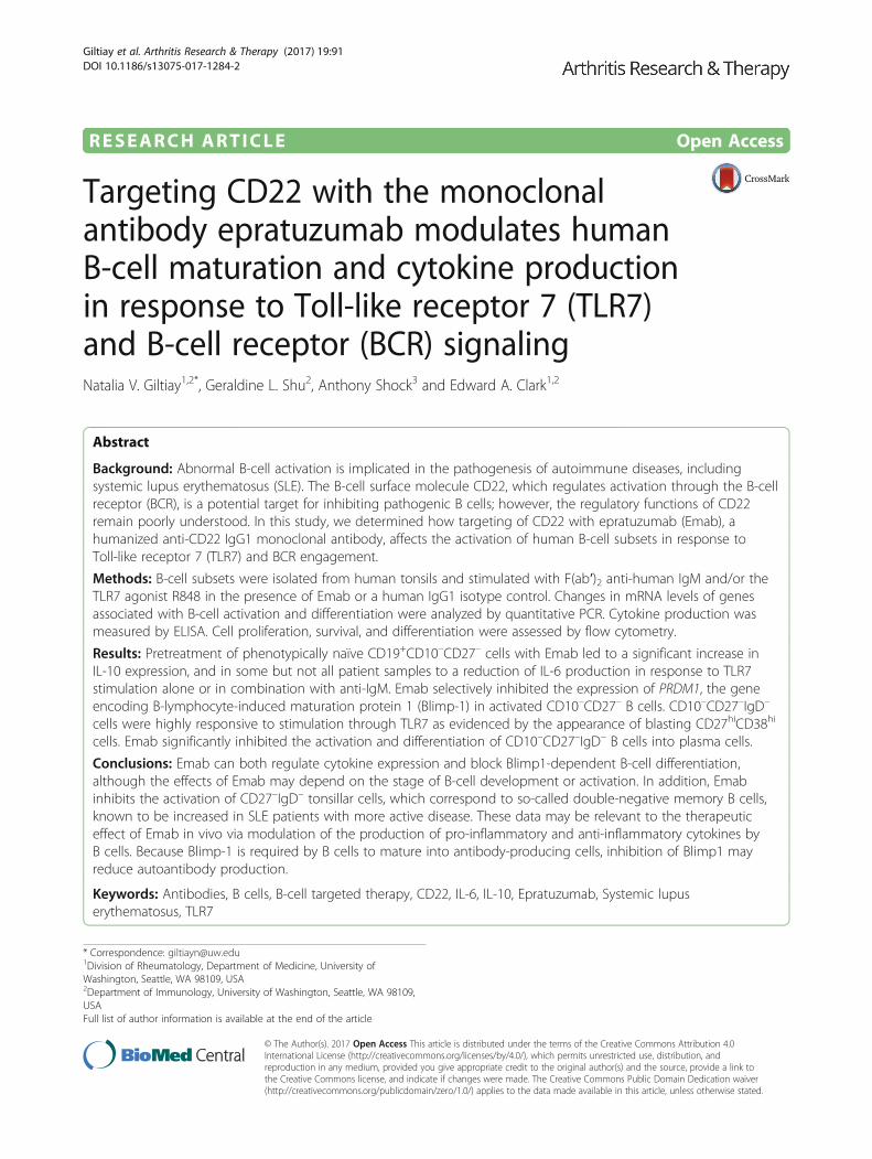
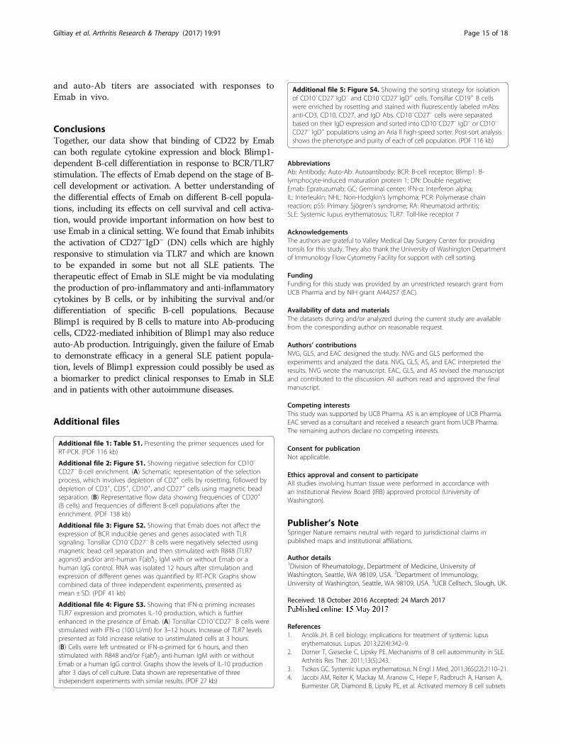
Emab selectively inhibits the expression of PRDM1, thegene encoding Blimp1To further determine the effects of Emab on B-cell activa-tion and differentiation, we examined the expression ofgenes known to regulate B-cell differentiation from naïveinto Ab-producing cells. As shown in Fig. 4a, B-cell differ-entiation is regulated by a complex regulatory network,consisting of transcription factors that regulate the tran-scriptional activation (or repression) of one another as wellas other key genes involved in B-cell differentiation [62].Initially, we examined CD10–CD27– enriched B cells, whichwere preincubated with Emab, hIgG1 isotype control, ormedium and then stimulated with R848, F(ab′)2 anti-IgM,or a combination of R848 and anti-IgM. These stimuli hadno effect on the expression of MITF, BACH2, BCL6, PAX5,IRF4, or XBP1 either alone or with Emab (Fig. 4c and datanot shown). However, stimulation with R848, anti-IgM, or acombination of the two induced a 5-fold to 15-fold increasein the expression of PRDM1, the gene encoding B-lymphocyte-induced maturation protein 1 (Blimp1). Emab
Fig. 3 Effects of Emab on IL-6 and IL-10 cytokine production. Tonsillar CD10–CD27– B cells were negatively selected using magnetic bead cell enrichmentand then stimulated with R848 and/or F(ab′)2 anti-human IgM with or without Emab or a human IgG control. a, b Cells were treated with different stimuliand cultured for 3 days, and production of IL-6 and IL-10 in cell supernatants was quantified by ELISA. Cumulative data from nine independent experimentsusing CD10–CD27– B cells from different donors. Experiments with increased IL-6 production in the presence of Emab are shown (gray) (a). c Correlationbetween TLR7mRNA expression in donor CD10–CD27– B cells at the time of activation and the levels of IL-10 produced 3 days after anti-IgM/R848stimulation. **p < 0.01 by two-way ANOVA analysis with Bonferroni post tests. Emab epratuzumab, IL interleukin, TLR7 Toll-like receptor 7
Giltiay et al. Arthritis Research & Therapy (2017) 19:91 Page 7 of 18
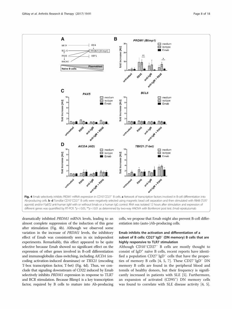
dramatically inhibited PRDM1 mRNA levels, leading to analmost complete suppression of the induction of this geneafter stimulation (Fig. 4b). Although we observed somevariation in the increase of PRDM1 levels, the inhibitoryeffect of Emab was consistently seen in six independentexperiments. Remarkably, this effect appeared to be quiteselective because Emab showed no significant effect on theexpression of other genes involved in B-cell differentiationand immunoglobulin class-switching, including AICDA (en-coding activation-induced deaminase) or TBX21 (encodingT-box transcription factor, T-bet) (Fig. 4d). Thus, we con-clude that signaling downstream of CD22 induced by Emabselectively inhibits PRDM1 expression in response to TLR7and BCR stimulation. Because Blimp1 is a key transcriptionfactor, required by B cells to mature into Ab-producing
cells, we propose that Emab might also prevent B-cell differ-entiation into (auto-)Ab-producing cells.
Emab inhibits the activation and differentiation of asubset of B cells: CD27–IgD– (DN memory) B cells that arehighly responsive to TLR7 stimulationAlthough CD10–CD27– B cells are mostly thought toconsist of IgD+ naïve B cells, recent reports have identi-fied a population CD27–IgD– cells that have the proper-ties of memory B cells [4, 5, 7]. These CD27–IgD– DNmemory B cells are found in the peripheral blood andtonsils of healthy donors, but their frequency is signifi-cantly increased in patients with SLE [5]. Furthermore,an expansion of activated (CD95+) DN memory cellswas found to correlate with SLE disease activity [4, 5].
Naïve B cellsPlasmablast
A B
D
C
Fig. 4 Emab selectively inhibits PRDM1 mRNA expression in CD10–CD27– B cells. a Network of transcription factors involved in B-cell differentiation intoAb-producing cells. b–d Tonsillar CD10–CD27– B cells were negatively selected using magnetic bead cell separation and then stimulated with R848 (TLR7agonist) and/or F(ab′)2 anti-human IgM with or without Emab or a human IgG control. RNA was isolated 12 hours after stimulation and expression ofdifferent genes was quantified by RT-PCR. *p < 0.05, **p< 0.01 as determined by two-way ANOVA with Bonferroni post test. Emab epratuzumab
Giltiay et al. Arthritis Research & Therapy (2017) 19:91 Page 8 of 18
DN memory B cells are also highly responsive to stimu-lation through TLR9 by CpG [4]. This led us tohypothesize that IgD–cells within the CD10–CD27–
population might be a subset that responds to TLR7stimulation by upregulating PRDM1.To address this question, we first assessed whether the
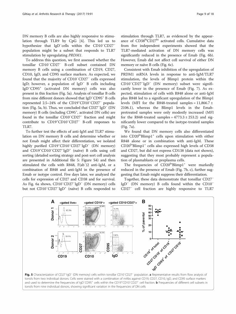
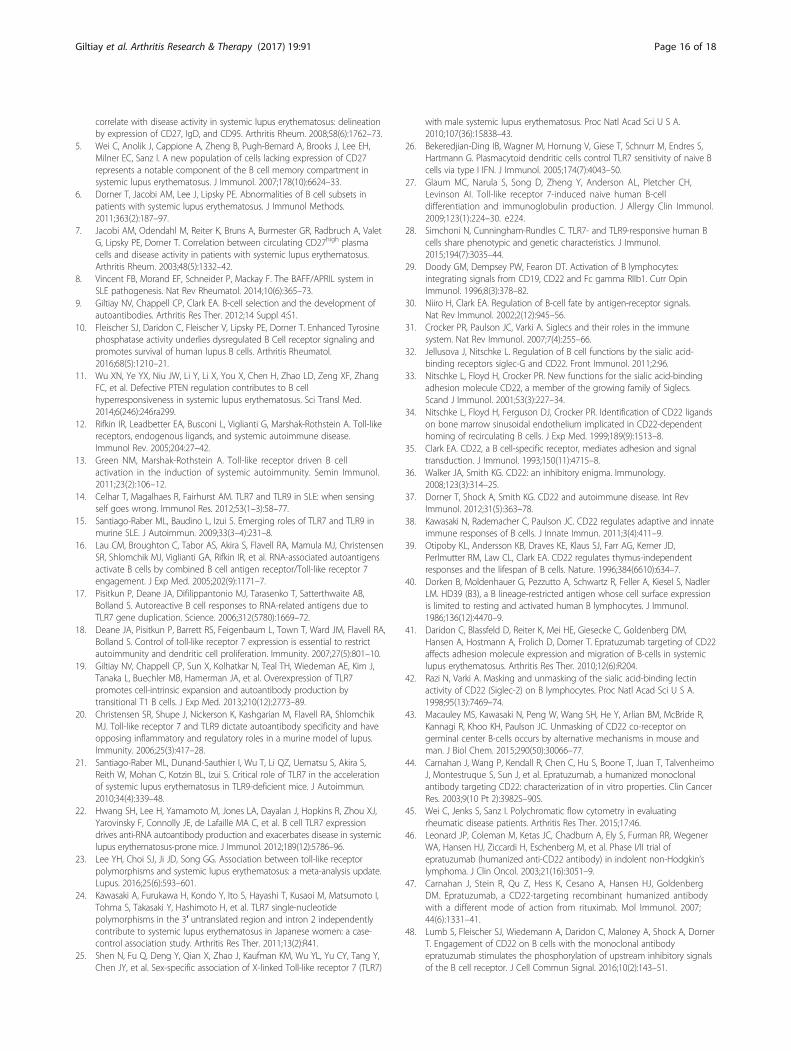
tonsillar CD10–CD27– B-cell subset contained DNmemory B cells using a combination of CD19, CD27,CD10, IgD, and CD95 surface markers. As expected, wefound that the majority of CD10–CD27– cells expressedIgD; however, a population of IgD– B cells includingIgD–CD95+ (activated DN memory) cells was alsopresent in this fraction (Fig. 5a). Analysis of tonsillar B cellsfrom nine different donors showed that IgD–CD95+ B cellsrepresented 2.5–24% of the CD19+CD10–CD27– popula-tion (Fig. 5a, b). Thus, we concluded that CD27–IgD– (DNmemory) B cells (including CD95+, activated DN cells) arefound in the tonsillar CD10–CD27– fraction and mightcontribute to CD19+CD10–CD27– B-cell responses toTLR7.To further test the effects of anti-IgM and TLR7 stimu-
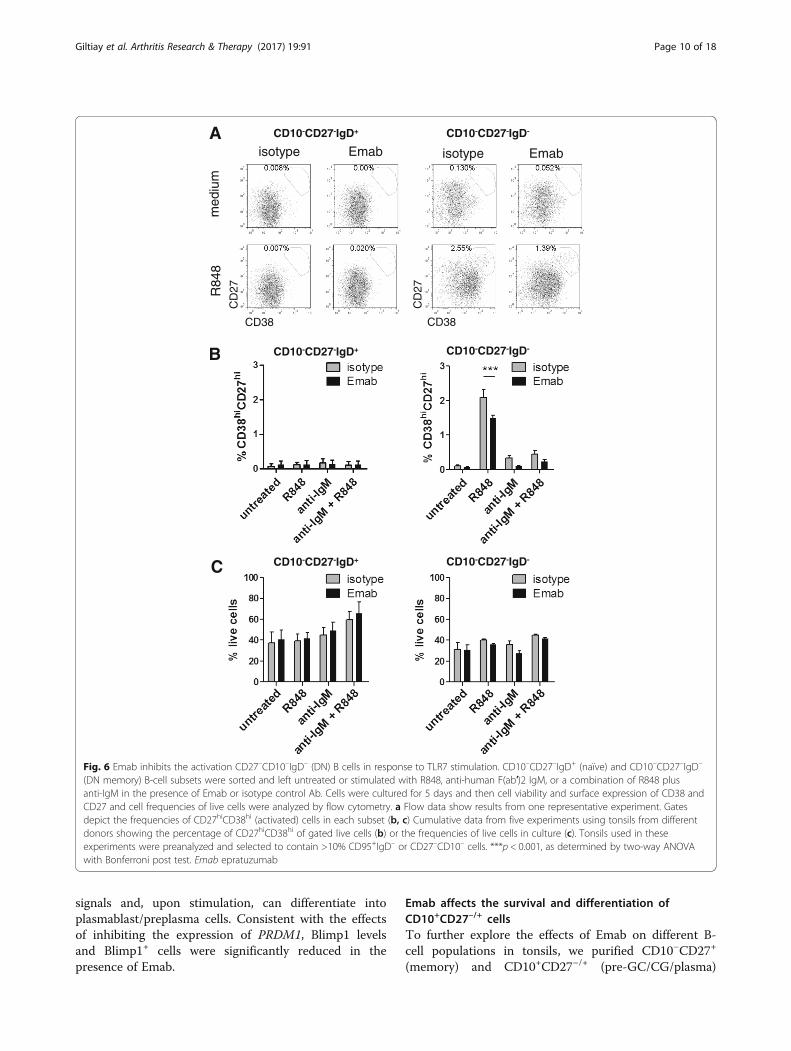
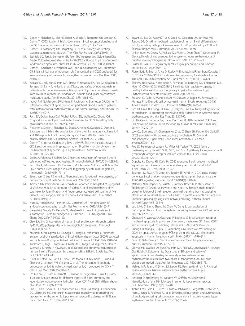
lation on DN memory B cells and determine whether ornot Emab might affect their differentiation, we isolatedhighly purified CD19+CD10–CD27–IgD– (DN memory)and CD19+CD10–CD27–IgD+ (naïve) B cells using cellsorting (detailed sorting strategy and post-sort cell analysisare presented in Additional file 5: Figure S4) and thenstimulated the cells with R848, F(ab′)2 anti-IgM, or acombination of R848 and anti-IgM in the presence ofEmab or isotype control. Five days later, we analyzed thecells for expression of CD27 and CD38 and for survival.As Fig. 6a shows, CD10–CD27–IgD– (DN memory) cellsbut not CD10–CD27–IgD+ (naïve) B cells responded to
stimulation through TLR7, as evidenced by the appear-ance of CD38hiCD27hi activated cells. Cumulative datafrom five independent experiments showed that theTLR7-mediated activation of DN memory cells wassignificantly reduced in the presence of Emab (Fig. 6b).However, Emab did not affect cell survival of either DNmemory or naïve B cells (Fig. 6c).Consistent with Emab inhibition of the upregulation of
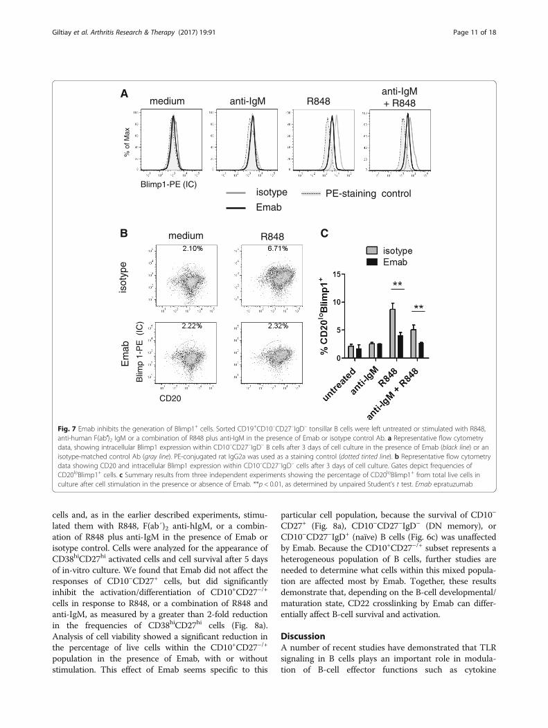
PRDM1 mRNA levels in response to anti-IgM/TLR7stimulation, the levels of Blimp1 protein within theCD10–CD27–IgD– (DN memory) subset were signifi-cantly lower in the presence of Emab (Fig. 7). As ex-pected, stimulation of cells with R848 alone or anti-IgMplus R848 led to a significant upregulation of the Blimp1levels (MFI for the R848-treated samples = 11,866.7 ±2106.1), whereas the Blimp1 levels in the Emab-pretreated samples were only modestly increased (MFIfor the R848-treated samples = 4773.3 ± 253.2) and sig-nificantly lower compared to the isotype-treated samples(Fig. 7a).We found that DN memory cells also differentiated
into CD20loBlimp1+ cells upon stimulation with eitherR848 alone or in combination with anti-IgM. TheseCD20loBlimp1+ cells also expressed high levels of CD38and CD27, but did not express CD138 (data not shown),suggesting that they most probably represent a popula-tion of plasmablasts or preplasma cells.The frequencies of CD20loBlimp1+ were markedly
reduced in the presence of Emab (Fig. 7b, c), further sug-gesting that Emab might suppress their differentiation.Together, these data demonstrate that tonsillar CD27–
IgD– (DN memory) B cells found within the CD10–
CD27– cell fraction are highly responsive to TLR7
A
IgD
CD
95
CD10
CD
27
CD19
<gated live> <gated CD19+> <gated CD10-CD27->
Do
no
r 1
Do
no
r 2
B
Fig. 5 Characterization of CD27–IgD– (DN memory) cells within tonsillar CD10–CD27– population. a Representative results from flow analysis oftonsils from two individual donors. Cells were stained with a combination of mAbs against CD19, CD27, CD10, IgD, and CD95 surface markersand used to determine the frequencies of IgD–CD95+ cells within the CD19+CD10–CD27– cell fraction. b Frequencies of different cell subsets intonsils from nine individual donors, showing significant variation in the frequencies of DN cells
Giltiay et al. Arthritis Research & Therapy (2017) 19:91 Page 9 of 18
signals and, upon stimulation, can differentiate intoplasmablast/preplasma cells. Consistent with the effectsof inhibiting the expression of PRDM1, Blimp1 levelsand Blimp1+ cells were significantly reduced in thepresence of Emab.
Emab affects the survival and differentiation ofCD10+CD27–/+ cellsTo further explore the effects of Emab on different B-cell populations in tonsils, we purified CD10–CD27+
(memory) and CD10+CD27–/+ (pre-GC/CG/plasma)
CD10-CD27-IgD+ CD10-CD27-IgD-
CD10-CD27-IgD+ CD10-CD27-IgD-
med
ium
R
848
isotype Emab isotype Emab
CD38
CD
27
CD38
CD
27
CD10-CD27-IgD+ CD10-CD27-IgD-
B
A
C
Fig. 6 Emab inhibits the activation CD27–CD10–IgD– (DN) B cells in response to TLR7 stimulation. CD10–CD27–IgD+ (naïve) and CD10–CD27–IgD–
(DN memory) B-cell subsets were sorted and left untreated or stimulated with R848, anti-human F(ab′)2 IgM, or a combination of R848 plusanti-IgM in the presence of Emab or isotype control Ab. Cells were cultured for 5 days and then cell viability and surface expression of CD38 andCD27 and cell frequencies of live cells were analyzed by flow cytometry. a Flow data show results from one representative experiment. Gatesdepict the frequencies of CD27hiCD38hi (activated) cells in each subset (b, c) Cumulative data from five experiments using tonsils from differentdonors showing the percentage of CD27hiCD38hi of gated live cells (b) or the frequencies of live cells in culture (c). Tonsils used in theseexperiments were preanalyzed and selected to contain >10% CD95+IgD– or CD27–CD10– cells. ***p < 0.001, as determined by two-way ANOVAwith Bonferroni post test. Emab epratuzumab
Giltiay et al. Arthritis Research & Therapy (2017) 19:91 Page 10 of 18
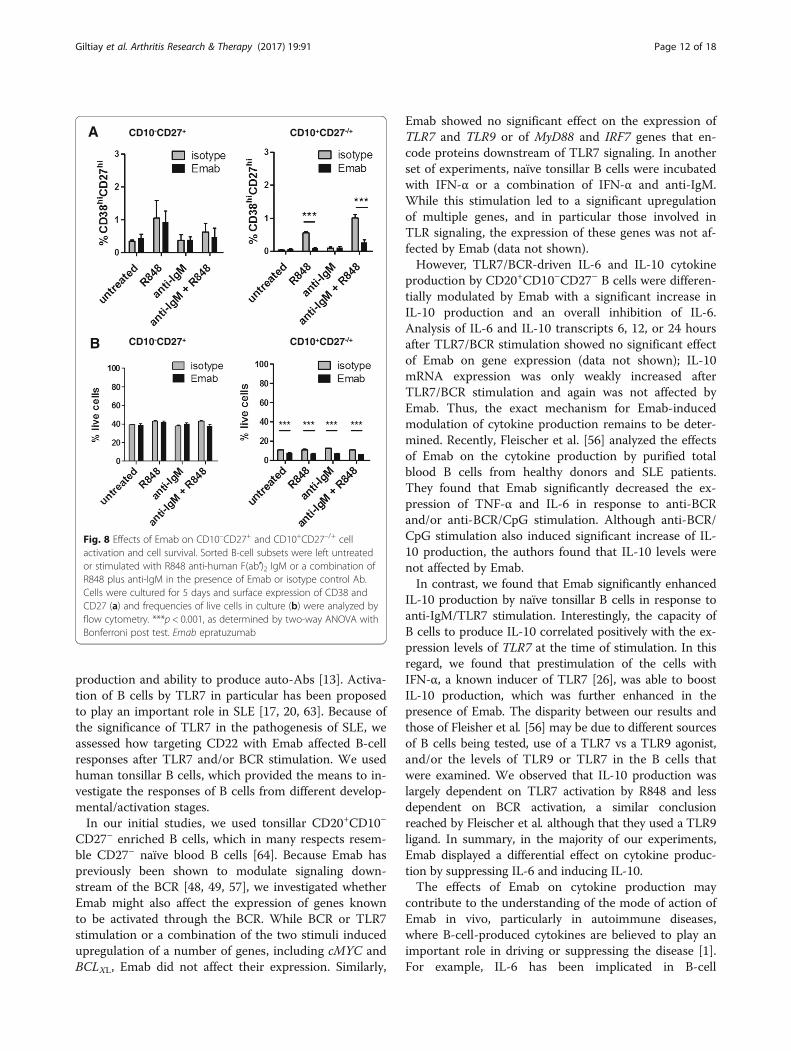
cells and, as in the earlier described experiments, stimu-lated them with R848, F(ab′)2 anti-hIgM, or a combin-ation of R848 plus anti-IgM in the presence of Emab orisotype control. Cells were analyzed for the appearance ofCD38hiCD27hi activated cells and cell survival after 5 daysof in-vitro culture. We found that Emab did not affect theresponses of CD10–CD27+ cells, but did significantlyinhibit the activation/differentiation of CD10+CD27–/+
cells in response to R848, or a combination of R848 andanti-IgM, as measured by a greater than 2-fold reductionin the frequencies of CD38hiCD27hi cells (Fig. 8a).Analysis of cell viability showed a significant reduction inthe percentage of live cells within the CD10+CD27–/+
population in the presence of Emab, with or withoutstimulation. This effect of Emab seems specific to this
particular cell population, because the survival of CD10–
CD27+ (Fig. 8a), CD10–CD27–IgD– (DN memory), orCD10–CD27–IgD+ (naïve) B cells (Fig. 6c) was unaffectedby Emab. Because the CD10+CD27–/+ subset represents aheterogeneous population of B cells, further studies areneeded to determine what cells within this mixed popula-tion are affected most by Emab. Together, these resultsdemonstrate that, depending on the B-cell developmental/maturation state, CD22 crosslinking by Emab can differ-entially affect B-cell survival and activation.
DiscussionA number of recent studies have demonstrated that TLRsignaling in B cells plays an important role in modula-tion of B-cell effector functions such as cytokine
B C
A medium anti-IgM R848
anti-IgM + R848
% o
f Max
Blimp1-PE (IC) PE-staining control isotype
Emab
CD20
Blim
p 1-
PE
(IC
)
medium R848
isot
ype
Em
ab
Fig. 7 Emab inhibits the generation of Blimp1+ cells. Sorted CD19+CD10–CD27–IgD– tonsillar B cells were left untreated or stimulated with R848,anti-human F(ab′)2 IgM or a combination of R848 plus anti-IgM in the presence of Emab or isotype control Ab. a Representative flow cytometrydata, showing intracellular Blimp1 expression within CD10–CD27–IgD– B cells after 3 days of cell culture in the presence of Emab (black line) or anisotype-matched control Ab (gray line). PE-conjugated rat IgG2a was used as a staining control (dotted tinted line). b Representative flow cytometrydata showing CD20 and intracellular Blimp1 expression within CD10–CD27–IgD– cells after 3 days of cell culture. Gates depict frequencies ofCD20loBlimp1+ cells. c Summary results from three independent experiments showing the percentage of CD20loBlimp1+ from total live cells inculture after cell stimulation in the presence or absence of Emab. **p < 0.01, as determined by unpaired Student’s t test. Emab epratuzumab
Giltiay et al. Arthritis Research & Therapy (2017) 19:91 Page 11 of 18
production and ability to produce auto-Abs [13]. Activa-tion of B cells by TLR7 in particular has been proposedto play an important role in SLE [17, 20, 63]. Because ofthe significance of TLR7 in the pathogenesis of SLE, weassessed how targeting CD22 with Emab affected B-cellresponses after TLR7 and/or BCR stimulation. We usedhuman tonsillar B cells, which provided the means to in-vestigate the responses of B cells from different develop-mental/activation stages.In our initial studies, we used tonsillar CD20+CD10–
CD27– enriched B cells, which in many respects resem-ble CD27– naïve blood B cells [64]. Because Emab haspreviously been shown to modulate signaling down-stream of the BCR [48, 49, 57], we investigated whetherEmab might also affect the expression of genes knownto be activated through the BCR. While BCR or TLR7stimulation or a combination of the two stimuli inducedupregulation of a number of genes, including cMYC andBCLXL, Emab did not affect their expression. Similarly,
Emab showed no significant effect on the expression ofTLR7 and TLR9 or of MyD88 and IRF7 genes that en-code proteins downstream of TLR7 signaling. In anotherset of experiments, naïve tonsillar B cells were incubatedwith IFN-α or a combination of IFN-α and anti-IgM.While this stimulation led to a significant upregulationof multiple genes, and in particular those involved inTLR signaling, the expression of these genes was not af-fected by Emab (data not shown).However, TLR7/BCR-driven IL-6 and IL-10 cytokine
production by CD20+CD10–CD27– B cells were differen-tially modulated by Emab with a significant increase inIL-10 production and an overall inhibition of IL-6.Analysis of IL-6 and IL-10 transcripts 6, 12, or 24 hoursafter TLR7/BCR stimulation showed no significant effectof Emab on gene expression (data not shown); IL-10mRNA expression was only weakly increased afterTLR7/BCR stimulation and again was not affected byEmab. Thus, the exact mechanism for Emab-inducedmodulation of cytokine production remains to be deter-mined. Recently, Fleischer et al. [56] analyzed the effectsof Emab on the cytokine production by purified totalblood B cells from healthy donors and SLE patients.They found that Emab significantly decreased the ex-pression of TNF-α and IL-6 in response to anti-BCRand/or anti-BCR/CpG stimulation. Although anti-BCR/CpG stimulation also induced significant increase of IL-10 production, the authors found that IL-10 levels werenot affected by Emab.In contrast, we found that Emab significantly enhanced
IL-10 production by naïve tonsillar B cells in response toanti-IgM/TLR7 stimulation. Interestingly, the capacity ofB cells to produce IL-10 correlated positively with the ex-pression levels of TLR7 at the time of stimulation. In thisregard, we found that prestimulation of the cells withIFN-α, a known inducer of TLR7 [26], was able to boostIL-10 production, which was further enhanced in thepresence of Emab. The disparity between our results andthose of Fleisher et al. [56] may be due to different sourcesof B cells being tested, use of a TLR7 vs a TLR9 agonist,and/or the levels of TLR9 or TLR7 in the B cells thatwere examined. We observed that IL-10 production waslargely dependent on TLR7 activation by R848 and lessdependent on BCR activation, a similar conclusionreached by Fleischer et al. although that they used a TLR9ligand. In summary, in the majority of our experiments,Emab displayed a differential effect on cytokine produc-tion by suppressing IL-6 and inducing IL-10.The effects of Emab on cytokine production may
contribute to the understanding of the mode of action ofEmab in vivo, particularly in autoimmune diseases,where B-cell-produced cytokines are believed to play animportant role in driving or suppressing the disease [1].For example, IL-6 has been implicated in B-cell
A CD10-CD27+ CD10+CD27-/+
CD10-CD27+ CD10+CD27-/+B
Fig. 8 Effects of Emab on CD10–CD27+ and CD10+CD27–/+ cellactivation and cell survival. Sorted B-cell subsets were left untreatedor stimulated with R848 anti-human F(ab′)2 IgM or a combination ofR848 plus anti-IgM in the presence of Emab or isotype control Ab.Cells were cultured for 5 days and surface expression of CD38 andCD27 (a) and frequencies of live cells in culture (b) were analyzed byflow cytometry. ***p < 0.001, as determined by two-way ANOVA withBonferroni post test. Emab epratuzumab
Giltiay et al. Arthritis Research & Therapy (2017) 19:91 Page 12 of 18
differentiation and Ab production [65, 66], and is alsoknown to cooperate with IL-21 to promote the differen-tiation of CD4+ T-follicular helper cells [67–70]. Fur-thermore, increased IL-6 levels have been reported inSLE patients with active disease and more recently IL-6has been identified as a major genetic risk factor for SLE[71]. Emab-mediated IL-6-inhibition could possibly sup-press inflammation associated with SLE. IL-10, on thecontrary, is known to be produced by regulatory B cells[72] and has been proposed to suppress effector Th1and Th17 cell responses [73–75]. Recent studies havesuggested that IL-10 production by B cells might be de-fective in SLE patients [74, 76]. Based on our findings,CD22 engagement by Emab may very well help to repro-gram B cells in SLE patients to restore IL-10 production.Further studies are required to determine how CD22
signaling promotes IL-10 production by TLR7-driven Bcells. Liu et al. [77] have recently shown that activationof STAT3 and ERK is required for TLR-induced IL-10production by human B cells. The authors also foundthat IFN-α enhanced TLR7/8-induced but not TLR9-induced IL-10 production. Although it is well knownthat TLR stimulation elicits IL-10 production, to ourknowledge this study is the first to show a role for CD22in promoting IFN-α/TLR7-induced IL-10 production.Whereas CD22 is often described as a negative regulatorof BCR signaling, it should be noted that previousstudies have shown that CD22 associates with a numberof signaling molecules, such as Syk, PI-3 kinase, Grb2,and phospholipase-Cγ2 [78–80], and that direct CD22engagement can induce activation of ERK2 [81]. In linewith this finding, we have found that Emab inducesincreased ERK phosphorylation in human B cells [82]. Inlight of Liu et al.’s findings [77], this might provide amechanistic explanation of how CD22 crosslinking byEmab promotes IL-10 production.Interestingly, Emab also modestly increases B-cell
proliferation in response to BCR/TLR7 stimulation. Thiseffect of Emab might be selective to the CD27–CD10–
(naïve and DN) cells and also dependent on the particu-lar signals used to activate the cells. In contrast toCD27– (naïve/DN) B cells, Emab did not affect cell pro-liferation of CD27+ (classical memory) cells; however,the rates of cell proliferation were variable between differ-ent donors (data not shown). Previous studies have shownthat Emab can inhibit the proliferation of CD27– andCD27+ blood B cells in response to IL-2, IL-10, F(ab′)2,and/or CD40L and CpG [54]. Thus, the effect of Emab oncell proliferation among various B-cell subsets needs fur-ther investigation. In the context of BCR/TLR7 stimula-tion of the CD27–CD10– subset, the small increase of cellproliferation may be related to our finding that Emab in-hibits B-cell differentiation. Previous studies have shownthat B cells exit the cell cycle once they start to
differentiate into Ab-producing plasma cells and a changein the expression of several transcription factors (e.g., adecrease of BCL6, PAX5, and c-Myc levels and an increaseof Blimp1 expression) has been implicated in the transi-tion/differentiation of naïve B cells into plasma cells [62].In CD10–CD27– B-cell cultures, we found that Emab dra-matically inhibited the levels of PRDM1, the gene encod-ing Blimp1, which was induced in response to TLR7 and/or BCR/TLR7 stimulation. This effect of Emab was highlyspecific to PRDM1 because this Ab did not affect the ex-pression of other genes involved in B-cell differentiationor immunoglobulin class-switching, including MITF,BACH2, BCL6, PAX5, IRF4, XBP1, AICDA, or TBX21. Wedid see a trend toward reduction in the increase TBX21 inresponse to anti-IgM/TLR7 in the presence of Emab, butthe combined results from different experiments did notshow statistical significance.The responses of human B-cell subsets to TLR7 or
anti-BCR/TLR7 stimulation have not been studied in de-tail. Recently, Simchoni et al. [28] showed that TLR7stimulation expands IgM+CD27+ memory B cells andpromotes the generation of CD27hi B cells. In thecurrent study, by comparing four different B-cell popula-tions based on the appearance of CD38hiCD27hi cells,we discovered that the CD10–CD27–IgD– subset was themost responsive to TLR7 stimulation compared to theother B-cell subsets. Notably, the generation ofCD38hiCD27hi cells was inhibited in the presence ofEmab. A portion of CD10+CD27+ and CD10+CD27–/+ Bcells also differentiated into CD38hiCD27hi cells, buttheir frequencies were lower compared to those gener-ated by CD10–CD27–IgD– B cells. Emab clearly de-creased CD38hiCD27hi cell frequencies, particularlywithin the CD10+CD27–/+ cells. CD10–CD27–IgD+
(naïve) B cells did not generate CD38hiCD27hi cells inresponse to TLR7 stimulation. Of note, there were nosignificant differences in TLR7 expression betweenCD10–CD27–IgD– and CD10–CD27–IgD+ B cells,suggesting that their differential responsiveness to TLR7ligation cannot be simply attributed to increased TLR7levels. Furthermore, a comparison between these twocell populations showed no differences in their CD22expression and/or their ability to internalize Emab uponCD22 binding (NV Giltiay, unpublished data). Data frommouse and human studies have indicated that TLR7 canpromote the production of auto-Abs [13, 28]; becauseBlimp1 is required by B cells to mature into Ab-producing cells, we propose that Emab-mediated inhib-ition of Blimp1 may reduce Ab/auto-Ab production.While the mechanisms for Emab-induced inhibition ofB-cell differentiation need further elucidation, our datasuggest that one therapeutic effect of Emab may be viainhibiting the expression of PRDM1 (Blimp1). In thisrespect, it is highly relevant that there is elevated
Giltiay et al. Arthritis Research & Therapy (2017) 19:91 Page 13 of 18
expression of Blimp1 in SLE patients and this is corre-lated with increases in plasma cells, auto-Abs, and dis-ease activity [83].Interestingly, Emab also affected cell survival, although
this effect was only evident within the (pre-GC/GC)CD10+CD27+/– cell population. A possible explanationfor these data is that cells within this mixed cell popula-tion might be more sensitive to CD22-induced apop-tosis. While apoptosis induction is not considered theprimary mode of action of Emab, previous in-vitro datahave demonstrated that crosslinking of CD22 by mAbsincluding Emab induces apoptosis in human lymphomacells [84, 85]. Recently, Macauley et al. [43] demon-strated that changes in the glycosylation patterns due toaltered enzyme activity in the GC leads to “unmasking”of CD22 binding site on GC B cells, relative to naïve andmemory B cells. Such unmasking could very well alterthe effects of CD22 binding by Emab. It should be notedthat the effects of Emab on GC cell survival in culturewere independent of BCR/TLR7 stimulation. Thesedata, however, may be relevant to the clinical effectsof Emab in patients with NHL, because GC B cellsare considered a major source (i.e., cell of origin) forlymphoma cells [46, 86].Despite initial promising data in phase II clinical studies
[52, 53], results reported recently from a phase III clinicaltrial in SLE patients with moderate to severe diseaseshowed that Emab failed to reach the primary clinical end-point [87]. It has been suggested that a high placebo re-sponse and early rescue of nonresponders with increaseddoses of glucocorticoids might have confounded the datafrom the trial [87, 88]. Although disappointing, these re-sults reflect to a large extent the complexity and diversityof the pathology of SLE and suggest that perhaps some,but not all, SLE patients would benefit from Emab ther-apy. In our study, we have found that Emab affects theproduction of cytokines in response to BCR/TLR7 stimu-lation, by skewing B cells to produce immunoregulatorycytokines such as IL-10. Thus, we can predict that target-ing CD22 with Emab might be able to restore IL-10 pro-duction by CD27–CD24hiCD38hi transitional B cells inSLE patients. Transitional B cells are expanded in someSLE patients and our data showed that these cells expressrelatively high levels of CD22, suggesting that this cellpopulation might be a good target for Emab therapy.Our study identified CD10–CD27–IgD– as another im-
portant cell population, whose responses to BCR/TLR7stimulation were affected by Emab. We believe this cellpopulation closely resembles the DN memory B cellsfound in the blood. We found a significant variation inthe frequency of this B-cell population in tonsils, whichwe think might be reflective of environmental factors,such as recent viral exposure. An increased frequency ofDN memory B cells has been described previously in
SLE patients [4, 5]. Although there has been no directevidence for their contribution to disease pathology, DNmemory B cells have been linked to SLE autoimmunity[4, 5]. For example, a significant portion of DN memorycells found in SLE patients produce VH4-34-encoded9G4 Abs, known to be a source of SLE-associated auto-reactivity [89]. Frequencies of DN memory B cells werefound to be associated with higher disease activity,history of nephritis, and presence of auto-Abs [4, 5]. Thecellular origin of DN memory B cells remains somewhatelusive; because these cells express switched isotypes, yetat the same time show a reduced rate of somatic hyper-mutations compared to post-switched memory B cells, ithas been proposed that they might be of extra-GC origin[5]. Recent studies have shown that a substantial fractionof Ab-secreting cell clones found during SLE flares con-tained auto-Abs without (or with very few) mutations,consistent with differentiation outside GCs [90].Whether TLR-mediated activation of DN B cells might
contribute to the generation of Ab-secreting clones hasnot been established. One study has shown that DNmemory B cells are also highly responsive to stimulationthrough TLR9 by CpG [4]. Here, we reported that thesecells are also highly responsive to stimulation throughTLR7 by R848 or a combination of anti-IgM and TLR7.Importantly, we demonstrated a new role for CD22 inregulating the activation of these cells and their differen-tiation into CD27hiCD38hiBlimp1+ plasmablasts. In thisstudy, we did not address in detail whether DN cellsproduce cytokines. In a limited set of experiments, wefound that DN cells produced lower levels of IL-6 andIL-10 in response to TLR7 and or BCR/TLR7 stimula-tion, compared to naïve B cells; however, this needs tobe investigated further. Recent reports describe an in-crease in DN memory B cells in RA patients and olderpeople [91, 92]. Although the link between DN B cellsand SLE disease activity seems well established, we havefound a significant variation of DN memory B-cell fre-quencies in SLE patients, even those with active disease(NV Giltiay, unpublished data). In light of these findings,one can predict that patients with high frequencies of DNmemory B cells might be better candidates for CD22targeting by Emab.The fact that Emab blocks B cell differ-entiation in a distinct subset of memory B cells is alsoconsistent with our studies in mice showing that CD22 isrequired for normal memory B cell formation [93].Recent data suggest that Blimp1 levels positively
correlate with the levels of pathogenic auto-Abs in SLEpatients and can be used as a potential biomarker formonitoring disease activity [83]. Although results fromthe EMBODY1 and EMBODY2 trials showed no sig-nificant reduction of the total IgG levels and/or anti-DNAtiters in Emab-treated patients [87], it would be of interestin the future to test whether decreases in Blimp1 levels
Giltiay et al. Arthritis Research & Therapy (2017) 19:91 Page 14 of 18
and auto-Ab titers are associated with responses toEmab in vivo.
ConclusionsTogether, our data show that binding of CD22 by Emabcan both regulate cytokine expression and block Blimp1-dependent B-cell differentiation in response to BCR/TLR7stimulation. The effects of Emab depend on the stage of B-cell development or activation. A better understanding ofthe differential effects of Emab on different B-cell popula-tions, including its effects on cell survival and cell activa-tion, would provide important information on how best touse Emab in a clinical setting. We found that Emab inhibitsthe activation of CD27–IgD– (DN) cells which are highlyresponsive to stimulation via TLR7 and which are knownto be expanded in some but not all SLE patients. Thetherapeutic effect of Emab in SLE might be via modulatingthe production of pro-inflammatory and anti-inflammatorycytokines by B cells, or by inhibiting the survival and/ordifferentiation of specific B-cell populations. BecauseBlimp1 is required by B cells to mature into Ab-producingcells, CD22-mediated inhibition of Blimp1 may also reduceauto-Ab production. Intriguingly, given the failure of Emabto demonstrate efficacy in a general SLE patient popula-tion, levels of Blimp1 expression could possibly be used asa biomarker to predict clinical responses to Emab in SLEand in patients with other autoimmune diseases.
Additional files
Additional file 1: Table S1. Presenting the primer sequences used forRT-PCR. (PDF 116 kb)
Additional file 2: Figure S1. Showing negative selection for CD10–
CD27– B-cell enrichment. (A) Schematic representation of the selectionprocess, which involves depletion of CD2+ cells by rosetting, followed bydepletion of CD3+, CD5+, CD10+, and CD27+ cells using magnetic beadseparation. (B) Representative flow data showing frequencies of CD20+
(B cells) and frequencies of different B-cell populations after theenrichment. (PDF 138 kb)
Additional file 3: Figure S2. Showing that Emab does not affect theexpression of BCR inducible genes and genes associated with TLRsignaling. Tonsillar CD10–CD27– B cells were negatively selected usingmagnetic bead cell separation and then stimulated with R848 (TLR7agonist) and/or anti-human F(ab′)2 IgM with or without Emab or ahuman IgG control. RNA was isolated 12 hours after stimulation andexpression of different genes was quantified by RT-PCR. Graphs showcombined data of three independent experiments, presented asmean ± SD. (PDF 41 kb)
Additional file 4: Figure S3. Showing that IFN-α priming increasesTLR7 expression and promotes IL-10 production, which is furtherenhanced in the presence of Emab. (A) Tonsillar CD10–CD27– B cells werestimulated with IFN-α (100 U/ml) for 3–12 hours. Increase of TLR7 levelspresented as fold increase relative to unstimulated cells at 3 hours.(B) Cells were left untreated or IFN-α-primed for 6 hours, and thenstimulated with R848 and/or F(ab′)2 anti-human IgM with or withoutEmab or a human IgG control. Graphs show the levels of IL-10 productionafter 3 days of cell culture. Data shown are representative of threeindependent experiments with similar results. (PDF 27 kb)
Additional file 5: Figure S4. Showing the sorting strategy for isolationof CD10–CD27–IgD– and CD10–CD27–IgD+ cells. Tonsillar CD19+ B cellswere enriched by rosetting and stained with fluorescently labeled mAbs:anti-CD3, CD10, CD27, and IgD Abs. CD10–CD27– cells were separatedbased on their IgD expression and sorted into CD10–CD27– IgD– or CD10–
CD27– IgD+ populations using an Aria II high-speed sorter. Post-sort analysisshows the phenotype and purity of each of cell population. (PDF 116 kb)
AbbreviationsAb: Antibody; Auto-Ab: Autoantibody; BCR: B-cell receptor; Blimp1: B-lymphocyte-induced maturation protein 1; DN: Double negative;Emab: Epratuzumab; GC: Germinal center; IFN-α: Interferon alpha;IL: Interleukin; NHL: Non-Hodgkin’s lymphoma; PCR: Polymerase chainreaction; pSS: Primary Sjögren’s syndrome; RA: Rheumatoid arthritis;SLE: Systemic lupus erythematosus; TLR7: Toll-like receptor 7
AcknowledgementsThe authors are grateful to Valley Medical Day Surgery Center for providingtonsils for this study. They also thank the University of Washington Departmentof Immunology Flow Cytometry Facility for support with cell sorting.
FundingFunding for this study was provided by an unrestricted research grant fromUCB Pharma and by NIH grant AI44257 (EAC).
Availability of data and materialsThe datasets during and/or analyzed during the current study are availablefrom the corresponding author on reasonable request.
Authors’ contributionsNVG, GLS, and EAC designed the study. NVG and GLS performed theexperiments and analyzed the data. NVG, GLS, AS, and EAC interpreted theresults. NVG wrote the manuscript. EAC, GLS, and AS revised the manuscriptand contributed to the discussion. All authors read and approved the finalmanuscript.
Competing interestsThis study was supported by UCB Pharma. AS is an employee of UCB Pharma.EAC served as a consultant and received a research grant from UCB Pharma.The remaining authors declare no competing interests.
Consent for publicationNot applicable.
Ethics approval and consent to participateAll studies involving human tissue were performed in accordance withan Institutional Review Board (IRB) approved protocol (University ofWashington).
Publisher’s NoteSpringer Nature remains neutral with regard to jurisdictional claims inpublished maps and institutional affiliations.
Author details1Division of Rheumatology, Department of Medicine, University ofWashington, Seattle, WA 98109, USA. 2Department of Immunology,University of Washington, Seattle, WA 98109, USA. 3UCB Celltech, Slough, UK.
Received: 18 October 2016 Accepted: 24 March 2017
References1. Anolik JH. B cell biology: implications for treatment of systemic lupus
erythematosus. Lupus. 2013;22(4):342–9.2. Dorner T, Giesecke C, Lipsky PE. Mechanisms of B cell autoimmunity in SLE.
Arthritis Res Ther. 2011;13(5):243.3. Tsokos GC. Systemic lupus erythematosus. N Engl J Med. 2011;365(22):2110–21.4. Jacobi AM, Reiter K, Mackay M, Aranow C, Hiepe F, Radbruch A, Hansen A,
Burmester GR, Diamond B, Lipsky PE, et al. Activated memory B cell subsets
Giltiay et al. Arthritis Research & Therapy (2017) 19:91 Page 15 of 18
correlate with disease activity in systemic lupus erythematosus: delineationby expression of CD27, IgD, and CD95. Arthritis Rheum. 2008;58(6):1762–73.
5. Wei C, Anolik J, Cappione A, Zheng B, Pugh-Bernard A, Brooks J, Lee EH,Milner EC, Sanz I. A new population of cells lacking expression of CD27represents a notable component of the B cell memory compartment insystemic lupus erythematosus. J Immunol. 2007;178(10):6624–33.
6. Dorner T, Jacobi AM, Lee J, Lipsky PE. Abnormalities of B cell subsets inpatients with systemic lupus erythematosus. J Immunol Methods.2011;363(2):187–97.
7. Jacobi AM, Odendahl M, Reiter K, Bruns A, Burmester GR, Radbruch A, ValetG, Lipsky PE, Dorner T. Correlation between circulating CD27high plasmacells and disease activity in patients with systemic lupus erythematosus.Arthritis Rheum. 2003;48(5):1332–42.
8. Vincent FB, Morand EF, Schneider P, Mackay F. The BAFF/APRIL system inSLE pathogenesis. Nat Rev Rheumatol. 2014;10(6):365–73.
9. Giltiay NV, Chappell CP, Clark EA. B-cell selection and the development ofautoantibodies. Arthritis Res Ther. 2012;14 Suppl 4:S1.
10. Fleischer SJ, Daridon C, Fleischer V, Lipsky PE, Dorner T. Enhanced Tyrosinephosphatase activity underlies dysregulated B Cell receptor signaling andpromotes survival of human lupus B cells. Arthritis Rheumatol.2016;68(5):1210–21.
11. Wu XN, Ye YX, Niu JW, Li Y, Li X, You X, Chen H, Zhao LD, Zeng XF, ZhangFC, et al. Defective PTEN regulation contributes to B cellhyperresponsiveness in systemic lupus erythematosus. Sci Transl Med.2014;6(246):246ra299.
12. Rifkin IR, Leadbetter EA, Busconi L, Viglianti G, Marshak-Rothstein A. Toll-likereceptors, endogenous ligands, and systemic autoimmune disease.Immunol Rev. 2005;204:27–42.
13. Green NM, Marshak-Rothstein A. Toll-like receptor driven B cellactivation in the induction of systemic autoimmunity. Semin Immunol.2011;23(2):106–12.
14. Celhar T, Magalhaes R, Fairhurst AM. TLR7 and TLR9 in SLE: when sensingself goes wrong. Immunol Res. 2012;53(1–3):58–77.
15. Santiago-Raber ML, Baudino L, Izui S. Emerging roles of TLR7 and TLR9 inmurine SLE. J Autoimmun. 2009;33(3–4):231–8.
16. Lau CM, Broughton C, Tabor AS, Akira S, Flavell RA, Mamula MJ, ChristensenSR, Shlomchik MJ, Viglianti GA, Rifkin IR, et al. RNA-associated autoantigensactivate B cells by combined B cell antigen receptor/Toll-like receptor 7engagement. J Exp Med. 2005;202(9):1171–7.
17. Pisitkun P, Deane JA, Difilippantonio MJ, Tarasenko T, Satterthwaite AB,Bolland S. Autoreactive B cell responses to RNA-related antigens due toTLR7 gene duplication. Science. 2006;312(5780):1669–72.
18. Deane JA, Pisitkun P, Barrett RS, Feigenbaum L, Town T, Ward JM, Flavell RA,Bolland S. Control of toll-like receptor 7 expression is essential to restrictautoimmunity and dendritic cell proliferation. Immunity. 2007;27(5):801–10.
19. Giltiay NV, Chappell CP, Sun X, Kolhatkar N, Teal TH, Wiedeman AE, Kim J,Tanaka L, Buechler MB, Hamerman JA, et al. Overexpression of TLR7promotes cell-intrinsic expansion and autoantibody production bytransitional T1 B cells. J Exp Med. 2013;210(12):2773–89.
20. Christensen SR, Shupe J, Nickerson K, Kashgarian M, Flavell RA, ShlomchikMJ. Toll-like receptor 7 and TLR9 dictate autoantibody specificity and haveopposing inflammatory and regulatory roles in a murine model of lupus.Immunity. 2006;25(3):417–28.
21. Santiago-Raber ML, Dunand-Sauthier I, Wu T, Li QZ, Uematsu S, Akira S,Reith W, Mohan C, Kotzin BL, Izui S. Critical role of TLR7 in the accelerationof systemic lupus erythematosus in TLR9-deficient mice. J Autoimmun.2010;34(4):339–48.
22. Hwang SH, Lee H, Yamamoto M, Jones LA, Dayalan J, Hopkins R, Zhou XJ,Yarovinsky F, Connolly JE, de Lafaille MA C, et al. B cell TLR7 expressiondrives anti-RNA autoantibody production and exacerbates disease in systemiclupus erythematosus-prone mice. J Immunol. 2012;189(12):5786–96.
23. Lee YH, Choi SJ, Ji JD, Song GG. Association between toll-like receptorpolymorphisms and systemic lupus erythematosus: a meta-analysis update.Lupus. 2016;25(6):593–601.
24. Kawasaki A, Furukawa H, Kondo Y, Ito S, Hayashi T, Kusaoi M, Matsumoto I,Tohma S, Takasaki Y, Hashimoto H, et al. TLR7 single-nucleotidepolymorphisms in the 3′ untranslated region and intron 2 independentlycontribute to systemic lupus erythematosus in Japanese women: a case-control association study. Arthritis Res Ther. 2011;13(2):R41.
25. Shen N, Fu Q, Deng Y, Qian X, Zhao J, Kaufman KM, Wu YL, Yu CY, Tang Y,Chen JY, et al. Sex-specific association of X-linked Toll-like receptor 7 (TLR7)
with male systemic lupus erythematosus. Proc Natl Acad Sci U S A.2010;107(36):15838–43.
26. Bekeredjian-Ding IB, Wagner M, Hornung V, Giese T, Schnurr M, Endres S,Hartmann G. Plasmacytoid dendritic cells control TLR7 sensitivity of naive Bcells via type I IFN. J Immunol. 2005;174(7):4043–50.
27. Glaum MC, Narula S, Song D, Zheng Y, Anderson AL, Pletcher CH,Levinson AI. Toll-like receptor 7-induced naive human B-celldifferentiation and immunoglobulin production. J Allergy Clin Immunol.2009;123(1):224–30. e224.
28. Simchoni N, Cunningham-Rundles C. TLR7- and TLR9-responsive human Bcells share phenotypic and genetic characteristics. J Immunol.2015;194(7):3035–44.
29. Doody GM, Dempsey PW, Fearon DT. Activation of B lymphocytes:integrating signals from CD19, CD22 and Fc gamma RIIb1. Curr OpinImmunol. 1996;8(3):378–82.
30. Niiro H, Clark EA. Regulation of B-cell fate by antigen-receptor signals.Nat Rev Immunol. 2002;2(12):945–56.
31. Crocker PR, Paulson JC, Varki A. Siglecs and their roles in the immunesystem. Nat Rev Immunol. 2007;7(4):255–66.
32. Jellusova J, Nitschke L. Regulation of B cell functions by the sialic acid-binding receptors siglec-G and CD22. Front Immunol. 2011;2:96.
33. Nitschke L, Floyd H, Crocker PR. New functions for the sialic acid-bindingadhesion molecule CD22, a member of the growing family of Siglecs.Scand J Immunol. 2001;53(3):227–34.
34. Nitschke L, Floyd H, Ferguson DJ, Crocker PR. Identification of CD22 ligandson bone marrow sinusoidal endothelium implicated in CD22-dependenthoming of recirculating B cells. J Exp Med. 1999;189(9):1513–8.
35. Clark EA. CD22, a B cell-specific receptor, mediates adhesion and signaltransduction. J Immunol. 1993;150(11):4715–8.
36. Walker JA, Smith KG. CD22: an inhibitory enigma. Immunology.2008;123(3):314–25.
37. Dorner T, Shock A, Smith KG. CD22 and autoimmune disease. Int RevImmunol. 2012;31(5):363–78.
38. Kawasaki N, Rademacher C, Paulson JC. CD22 regulates adaptive and innateimmune responses of B cells. J Innate Immun. 2011;3(4):411–9.
39. Otipoby KL, Andersson KB, Draves KE, Klaus SJ, Farr AG, Kerner JD,Perlmutter RM, Law CL, Clark EA. CD22 regulates thymus-independentresponses and the lifespan of B cells. Nature. 1996;384(6610):634–7.
40. Dorken B, Moldenhauer G, Pezzutto A, Schwartz R, Feller A, Kiesel S, NadlerLM. HD39 (B3), a B lineage-restricted antigen whose cell surface expressionis limited to resting and activated human B lymphocytes. J Immunol.1986;136(12):4470–9.
41. Daridon C, Blassfeld D, Reiter K, Mei HE, Giesecke C, Goldenberg DM,Hansen A, Hostmann A, Frolich D, Dorner T. Epratuzumab targeting of CD22affects adhesion molecule expression and migration of B-cells in systemiclupus erythematosus. Arthritis Res Ther. 2010;12(6):R204.
42. Razi N, Varki A. Masking and unmasking of the sialic acid-binding lectinactivity of CD22 (Siglec-2) on B lymphocytes. Proc Natl Acad Sci U S A.1998;95(13):7469–74.
43. Macauley MS, Kawasaki N, Peng W, Wang SH, He Y, Arlian BM, McBride R,Kannagi R, Khoo KH, Paulson JC. Unmasking of CD22 co-receptor ongerminal center B-cells occurs by alternative mechanisms in mouse andman. J Biol Chem. 2015;290(50):30066–77.
44. Carnahan J, Wang P, Kendall R, Chen C, Hu S, Boone T, Juan T, TalvenheimoJ, Montestruque S, Sun J, et al. Epratuzumab, a humanized monoclonalantibody targeting CD22: characterization of in vitro properties. Clin CancerRes. 2003;9(10 Pt 2):3982S–90S.
45. Wei C, Jenks S, Sanz I. Polychromatic flow cytometry in evaluatingrheumatic disease patients. Arthritis Res Ther. 2015;17:46.
46. Leonard JP, Coleman M, Ketas JC, Chadburn A, Ely S, Furman RR, WegenerWA, Hansen HJ, Ziccardi H, Eschenberg M, et al. Phase I/II trial ofepratuzumab (humanized anti-CD22 antibody) in indolent non-Hodgkin’slymphoma. J Clin Oncol. 2003;21(16):3051–9.
47. Carnahan J, Stein R, Qu Z, Hess K, Cesano A, Hansen HJ, GoldenbergDM. Epratuzumab, a CD22-targeting recombinant humanized antibodywith a different mode of action from rituximab. Mol Immunol. 2007;44(6):1331–41.
48. Lumb S, Fleischer SJ, Wiedemann A, Daridon C, Maloney A, Shock A, DornerT. Engagement of CD22 on B cells with the monoclonal antibodyepratuzumab stimulates the phosphorylation of upstream inhibitory signalsof the B cell receptor. J Cell Commun Signal. 2016;10(2):143–51.
Giltiay et al. Arthritis Research & Therapy (2017) 19:91 Page 16 of 18
49. Sieger N, Fleischer SJ, Mei HE, Reiter K, Shock A, Burmester GR, Daridon C,Dorner T. CD22 ligation inhibits downstream B cell receptor signaling andCa(2+) flux upon activation. Arthritis Rheum. 2013;65(3):770–9.
50. Dorner T, Goldenberg DM. Targeting CD22 as a strategy for treatingsystemic autoimmune diseases. Ther Clin Risk Manag. 2007;3(5):953–9.
51. Steinfeld SD, Tant L, Burmester GR, Teoh NK, Wegener WA, Goldenberg DM,Pradier O. Epratuzumab (humanised anti-CD22 antibody) in primary Sjogren’ssyndrome: an open-label phase I/II study. Arthritis Res Ther. 2006;8(4):R129.
52. Dorner T, Kaufmann J, Wegener WA, Teoh N, Goldenberg DM, BurmesterGR. Initial clinical trial of epratuzumab (humanized anti-CD22 antibody) forimmunotherapy of systemic lupus erythematosus. Arthritis Res Ther. 2006;8(3):R74.
53. Wallace DJ, Kalunian K, Petri MA, Strand V, Houssiau FA, Pike M, Kilgallen B,Bongardt S, Barry A, Kelley L, et al. Efficacy and safety of epratuzumab inpatients with moderate/severe active systemic lupus erythematosus: resultsfrom EMBLEM, a phase IIb, randomised, double-blind, placebo-controlled,multicentre study. Ann Rheum Dis. 2014;73(1):183–90.
54. Jacobi AM, Goldenberg DM, Hiepe F, Radbruch A, Burmester GR, Dorner T.Differential effects of epratuzumab on peripheral blood B cells of patientswith systemic lupus erythematosus versus normal controls. Ann Rheum Dis.2008;67(4):450–7.
55. Rossi EA, Goldenberg DM, Michel R, Rossi DL, Wallace DJ, Chang CH.Trogocytosis of multiple B-cell surface markers by CD22 targeting withepratuzumab. Blood. 2013;122(17):3020–9.
56. Fleischer V, Sieber J, Fleischer SJ, Shock A, Heine G, Daridon C, Dorner T.Epratuzumab inhibits the production of the proinflammatory cytokines IL-6and TNF-alpha, but not the regulatory cytokine IL-10, by B cells fromhealthy donors and SLE patients. Arthritis Res Ther. 2015;17:185.
57. Dorner T, Shock A, Goldenberg DM, Lipsky PE. The mechanistic impact ofCD22 engagement with epratuzumab on B cell function: Implications forthe treatment of systemic lupus erythematosus. Autoimmun Rev.2015;14(12):1079–86.
58. Saxon A, Feldhaus J, Robins RA. Single step separation of human T and Bcells using AET treated srbc rosettes. J Immunol Methods. 1976;12(3–4):285–8.
59. Pezzutto A, Rabinovitch PS, Dorken B, Moldenhauer G, Clark EA. Role of theCD22 human B cell antigen in B cell triggering by anti-immunoglobulin.J Immunol. 1988;140(6):1791–5.
60. Sanz I, Wei C, Lee FE, Anolik J. Phenotypic and functional heterogeneity ofhuman memory B cells. Semin Immunol. 2008;20(1):67–82.
61. Kjeldsen MK, Perez-Andres M, Schmitz A, Johansen P, Boegsted M, NyegaardM, Gaihede M, Bukh A, Johnsen HE, Orfao A, et al. Multiparametric flowcytometry for identification and fluorescence activated cell sorting of fivedistinct B-cell subpopulations in normal tonsil tissue. Am J Clin Pathol.2011;136(6):960–9.
62. Nutt SL, Hodgkin PD, Tarlinton DM, Corcoran LM. The generation ofantibody-secreting plasma cells. Nat Rev Immunol. 2015;15(3):160–71.
63. Green NM, Moody KS, Debatis M, Marshak-Rothstein A. Activation ofautoreactive B cells by endogenous TLR7 and TLR3 RNA ligands. J BiolChem. 2012;287(47):39789–99.
64. Clark EA, Shu G. Activation of human B cell proliferation through surfaceBp35 (CD20) polypeptides or immunoglobulin receptors. J Immunol.1987;138(3):720–5.
65. Yoshizaki K, Nakagawa T, Fukunaga K, Tseng LT, Yamamura Y, Kishimoto T.Isolation and characterization of B cell differentiation factor (BCDF) secretedfrom a human B lymphoblastoid cell line. J Immunol. 1984;132(6):2948–54.
66. Kishimoto T, Taga T, Yamasaki K, Matsuda T, Tang B, Muraguchi A, Horii Y,Suematsu S, Hirata Y, Yawata H, et al. Normal and abnormal regulation ofhuman B cell differentiation by a new cytokine, BSF2/IL-6. Adv Exp MedBiol. 1989;254:135–43.
67. Dienz O, Eaton SM, Bond JP, Neveu W, Moquin D, Noubade R, Briso EM,Charland C, Leonard WJ, Ciliberto G, et al. The induction of antibodyproduction by IL-6 is indirectly mediated by IL-21 produced by CD4+ Tcells. J Exp Med. 2009;206(1):69–78.
68. Eto D, Lao C, DiToro D, Barnett B, Escobar TC, Kageyama R, Yusuf I, Crotty S.IL-21 and IL-6 are critical for different aspects of B cell immunity andredundantly induce optimal follicular helper CD4 T cell (Tfh) differentiation.PLoS One. 2011;6(3):e17739.
69. Jain S, Park G, Sproule TJ, Christianson GJ, Leeth CM, Wang H, RoopenianDC, Morse 3rd HC. Interleukin 6 accelerates mortality by promoting theprogression of the systemic lupus erythematosus-like disease of BXSB.Yaamice. PLoS One. 2016;11(4):e0153059.
70. Bryant VL, Ma CS, Avery DT, Li Y, Good KL, Corcoran LM, de Waal MR,Tangye SG. Cytokine-mediated regulation of human B cell differentiationinto Ig-secreting cells: predominant role of IL-21 produced by CXCR5+ Tfollicular helper cells. J Immunol. 2007;179(12):8180–90.
71. Linker-Israeli M, Deans RJ, Wallace DJ, Prehn J, Ozeri-Chen T, Klinenberg JR.Elevated levels of endogenous IL-6 in systemic lupus erythematosus. Aputative role in pathogenesis. J Immunol. 1991;147(1):117–23.
72. Rosser EC, Mauri C. Regulatory B cells: origin, phenotype, and function.Immunity. 2015;42(4):607–12.
73. Flores-Borja F, Bosma A, Ng D, Reddy V, Ehrenstein MR, Isenberg DA, MauriC. CD19 + CD24hiCD38hi B cells maintain regulatory T cells while limitingTH1 and TH17 differentiation. Sci Transl Med. 2013;5(173):173ra123.
74. Blair PA, Norena LY, Flores-Borja F, Rawlings DJ, Isenberg DA, Ehrenstein MR,Mauri C. CD19(+)CD24(hi)CD38(hi) B cells exhibit regulatory capacity inhealthy individuals but are functionally impaired in systemic LupusErythematosus patients. Immunity. 2010;32(1):129–40.
75. Bouaziz JD, Calbo S, Maho-Vaillant M, Saussine A, Bagot M, Bensussan A,Musette P. IL-10 produced by activated human B cells regulates CD4(+)T-cell activation in vitro. Eur J Immunol. 2010;40(10):2686–91.
76. Sim JH, Kim HR, Chang SH, Kim IJ, Lipsky PE, Lee J. Autoregulatory functionof interleukin-10-producing pre-naive B cells is defective in systemic lupuserythematosus. Arthritis Res Ther. 2015;17:190.
77. Liu BS, Cao Y, Huizinga TW, Hafler DA, Toes RE. TLR-mediated STAT3 andERK activation controls IL-10 secretion by human B cells. Eur J Immunol.2014;44(7):2121–9.
78. Law CL, Sidorenko SP, Chandran KA, Zhao Z, Shen SH, Fischer EH, Clark EA.CD22 associates with protein tyrosine phosphatase 1C, Syk, andphospholipase C-gamma(1) upon B cell activation. J Exp Med.1996;183(2):547–60.
79. Poe JC, Fujimoto M, Jansen PJ, Miller AS, Tedder TF. CD22 forms aquaternary complex with SHIP, Grb2, and Shc. A pathway for regulation of Blymphocyte antigen receptor-induced calcium flux. J Biol Chem.2000;275(23):17420–7.
80. Otipoby KL, Draves KE, Clark EA. CD22 regulates B cell receptor-mediatedsignals via two domains that independently recruit Grb2 and SHP-1.J Biol Chem. 2001;276(47):44315–22.
81. Tuscano JM, Riva A, Toscano SN, Tedder TF, Kehrl JH. CD22 cross-linkinggenerates B-cell antigen receptor-independent signals that activate theJNK/SAPK signaling cascade. Blood. 1999;94(4):1382–92.
82. Maloney AHD, Rapecki S, Fossati G, Lumb S, Rosen D, Putta S, Wale N,Spellmeyer D, Cesano A, Hawtin R and Shock A. Epratuzumab inducesbroad inhibition of B cell receptor proximal signaling but has opposingeffects on distal signaling in B cell subsets: a profile of effects on functionalimmune signaling by single cell network profiling. Arthritis Rheum.2014;66(Suppl 10):S1255–6
83. Luo J, Niu X, Liu H, Zhang M, Chen M, Deng S. Up-regulation oftranscription factor Blimp1 in systemic lupus erythematosus. Mol Immunol.2013;56(4):574–82.
84. Chaouchi N, Vazquez A, Galanaud P, Leprince C. B cell antigen receptor-mediated apoptosis. Importance of accessory molecules CD19 and CD22,and of surface IgM cross-linking. J Immunol. 1995;154(7):3096–104.
85. Chang CH, Wang Y, Gupta P, Goldenberg DM. Extensive crosslinking ofCD22 by epratuzumab triggers BCR signaling and caspase-dependentapoptosis in human lymphoma cells. MAbs. 2015;7(1):199–211.
86. Basso K, Dalla-Favera R. Germinal centres and B cell lymphomagenesis.Nat Rev Immunol. 2015;15(3):172–84.
87. Clowse ME, Wallace DJ, Furie RA, Petri MA, Pike MC, Leszczynski P, NeuweltCM, Hobbs K, Keiserman M, Duca L, et al. Efficacy and safety ofepratuzumab in moderately to severely active systemic lupuserythematosus: results from two phase III randomized, double-blind,placebo-controlled trials. Arthritis Rheumatol. 2017;69(2):362–75.
88. Mahieu MA, Strand V, Simon LS, Lipsky PE, Ramsey-Goldman R. A criticalreview of clinical trials in systemic lupus erythematosus. Lupus.2016;25(10):1122–40.
89. Isenberg D, Spellerberg M, Williams W, Griffiths M, Stevenson F.Identification of the 9G4 idiotope in systemic lupus erythematosus.Br J Rheumatol. 1993;32(10):876–82.
90. Tipton CM, Fucile CF, Darce J, Chida A, Ichikawa T, Gregoretti I, Schieferl S,Hom J, Jenks S, Feldman RJ, et al. Diversity, cellular origin and autoreactivityof antibody-secreting cell population expansions in acute systemic lupuserythematosus. Nat Immunol. 2015;16(7):755–65.
Giltiay et al. Arthritis Research & Therapy (2017) 19:91 Page 17 of 18
91. Mahmood Z, Muhammad K, Schmalzing M, Roll P, Dorner T, Tony HP.CD27-IgD- memory B cells are modulated by in vivo interleukin-6 receptor(IL-6R) blockade in rheumatoid arthritis. Arthritis Res Ther. 2015;17:61.
92. Colonna-Romano G, Bulati M, Aquino A, Pellicano M, Vitello S, Lio D,Candore G, Caruso C. A double-negative (IgD-CD27-) B cell population isincreased in the peripheral blood of elderly people. Mech Ageing Dev.2009;130(10):681–90.
93. Craig P. Chappell, Kevin E. Draves, Edward A. Clark. CD22 is required forformation of memory B cell precursors within germinal centers. PLOS ONE.2017;12(3):e0174661.
• We accept pre-submission inquiries
• Our selector tool helps you to find the most relevant journal
• We provide round the clock customer support
• Convenient online submission
• Thorough peer review
• Inclusion in PubMed and all major indexing services
• Maximum visibility for your research
Submit your manuscript atwww.biomedcentral.com/submit
Submit your next manuscript to BioMed Central and we will help you at every step:
Giltiay et al. Arthritis Research & Therapy (2017) 19:91 Page 18 of 18