Targeted loss of the ATR-X syndrome protein in the limb ...
11
Targeted loss of the ATR-X syndrome protein in the limb mesenchyme of mice causes brachydactyly Lauren A. Solomon 1,2,3 , Bailey A. Russell 4 , L. Ashley Watson 1,2,3 , Frank Beier 3,4, ∗ and Nathalie G. Be ´rube ´ 1,2,3, ∗ 1 Department of Paediatrics and 2 Department of Biochemistry, The University of Western Ontario, London, Ontario, Canada N6C 2V5, 3 Children’s Health Research Institute, London, Ontario, Canada N6C 2V5 and 4 Department of Physiology and Pharmacology, The University of Western Ontario, London, Ontario, Canada N6A 5C1 Received June 17, 2013; Revised and Accepted July 22, 2013 ATR-X syndrome is a rare genetic disorder caused by mutations in the ATRX gene. Affected individuals are cog- nitively impaired and display a variety of developmental abnormalities, including skeletal deformities. To investi- gate the function of ATRX during skeletal development, we selectively deleted the gene in the developing forelimb mesenchyme of mice. The absence of ATRX in the limb mesenchyme resulted in shorter digits, or brachydactyly, a defect also observed in a subset of ATR-X patients. This phenotype persisted until adulthood, causing reduced grip strength and altered gait in mutant mice. Examination of the embryonic ATRX-null forelimbs revealed a signifi- cant increase in apoptotic cell death, which could explain the reduced digit length. In addition, staining for the DNA damage markers g-histone 2A family member X (g-H2AX) and 53BP1 demonstrated a significant increase in the number of cells with DNA damage in the embryonic ATRX-null forepaw. Strikingly, only one large bright DNA damage event was observed per nucleus in proliferating cells. These large g-H2AX foci were located in close prox- imity to the nuclear lamina and remained largely unresolved after cell differentiation. In addition, ATRX-depleted forelimb mesenchymal cells did not exhibit hypersensitivity to DNA fork-stalling compounds, suggesting that the nature as well as the response to DNA damage incurred by loss of ATRX in the developing limb fundamentally differs from other tissues. Our data suggest that DNA damage-induced apoptosis is a novel cellular mechanism underlying brachydactyly that might be relevant to additional skeletal syndromes. INTRODUCTION Alpha-thalassemia mental retardation syndrome, X-linked (ATR-X [MIM 301040]) is a rare genetic disorder caused by mutations to the ATRX gene (1). Manifestations of the disease include intellectual disabilities, severe developmental delay, facial dimorphisms, urogenital abnormalities and skeletal de- formities (2,3). The latter include brachydactyly, clinodactyly, tapering of the fingers, overlapping digits, and foot deformities. Approximately two-thirds of patients have short stature (2,3). This X-linked syndrome predominantly affects males, whereas carrier females are mostly asymptomatic, presumably due to skewed X-inactivation (3,4). Many ATR-X patients have a-thalassemia caused by impaired production of the a-globin gene leading to unstable tetramers of b-globin chains, or HbH inclusions, in the blood (1,5). The ATRX protein contains two highly conserved domains where the majority of disease-causing mutations are located. A plant homeodomain-type zinc finger motif, also called the ADD domain, mediates binding to histone H3 trimethylated at lysine 9 and unmethylated at lysine 4 (6 – 8). ATRX also contains a Sucrose non-fermenting 2 (SNF2)-type DNA-dependent ad- enosine triphosphate (ATP)ase domain in the C-terminal portion of the protein (3,9). SNF2 proteins are a family of helicase-like proteins that can hydrolyse ATP to remodel chro- matin or help repair DNA damage (10). The ATRX protein interacts with the heterochromatin protein 1 alpha and the Fas death domain-associated protein (DAXX), proteins located at heterochromatin and promyelocytic leukemia nuclear bodies (11). The DAXX/ATRX complex deposits the histone variant H3.3 at telomeres and at pericentromeric ∗ To whom correspondence should be addressed. Tel: +1 5196858500 (N.G.B.)/ +1 5196612111 (F.B.); Fax: +1 5196858616 (N.G.B.)/ +1 5198502459 (F.B.); Email: [email protected] (N.G.B.)/[email protected] (F.B.) # The Author 2013. Published by Oxford University Press. All rights reserved. For Permissions, please email: [email protected] Human Molecular Genetics, 2013, Vol. 22, No. 24 5015–5025 doi:10.1093/hmg/ddt351 Advance Access published on July 25, 2013 Downloaded from https://academic.oup.com/hmg/article-abstract/22/24/5015/568669 by guest on 15 February 2018
Transcript of Targeted loss of the ATR-X syndrome protein in the limb ...

Targeted loss of the ATR-X syndrome protein in thelimb mesenchyme of mice causes brachydactyly
Lauren A. Solomon1,2,3, Bailey A. Russell4, L. Ashley Watson1,2,3, Frank Beier3,4,∗
and Nathalie G. Berube1,2,3,∗
1Department of Paediatrics and 2Department of Biochemistry, The University of Western Ontario, London, Ontario,
Canada N6C 2V5, 3Children’s Health Research Institute, London, Ontario, Canada N6C 2V5 and 4Department of
Physiology and Pharmacology, The University of Western Ontario, London, Ontario, Canada N6A 5C1
Received June 17, 2013; Revised and Accepted July 22, 2013
ATR-X syndrome is a rare genetic disorder caused by mutations in the ATRX gene. Affected individuals are cog-nitively impaired and display a variety of developmental abnormalities, including skeletal deformities. To investi-gate the function of ATRX during skeletal development, we selectively deleted the gene in the developing forelimbmesenchyme of mice. The absence of ATRX in the limb mesenchyme resulted in shorter digits, or brachydactyly, adefect also observed in a subset of ATR-X patients. This phenotype persisted until adulthood, causing reducedgripstrengthandalteredgait inmutantmice.Examinationof theembryonicATRX-null forelimbsrevealedasignifi-cant increase inapoptotic cell death,which couldexplain the reduceddigit length. Inaddition,staining for the DNAdamage markers g-histone 2A family member X (g-H2AX) and 53BP1 demonstrated a significant increase in thenumber of cells with DNA damage in the embryonic ATRX-null forepaw. Strikingly, only one large bright DNAdamage event was observed per nucleus in proliferating cells. These largeg-H2AX foci were located in close prox-imity to the nuclear lamina and remained largely unresolved after cell differentiation. In addition, ATRX-depletedforelimb mesenchymal cells did not exhibit hypersensitivity to DNA fork-stalling compounds, suggesting thatthe nature as well as the response to DNA damage incurred by loss of ATRX in the developing limb fundamentallydiffers from other tissues. Our data suggest that DNA damage-induced apoptosis is a novel cellular mechanismunderlying brachydactyly that might be relevant to additional skeletal syndromes.
INTRODUCTION
Alpha-thalassemia mental retardation syndrome, X-linked(ATR-X [MIM 301040]) is a rare genetic disorder caused bymutations to the ATRX gene (1). Manifestations of the diseaseinclude intellectual disabilities, severe developmental delay,facial dimorphisms, urogenital abnormalities and skeletal de-formities (2,3). The latter include brachydactyly, clinodactyly,tapering of the fingers, overlapping digits, and foot deformities.Approximately two-thirds of patients have short stature (2,3).This X-linked syndrome predominantly affects males, whereascarrier females are mostly asymptomatic, presumably due toskewed X-inactivation (3,4). Many ATR-X patients havea-thalassemia caused by impaired production of the a-globingene leading to unstable tetramers of b-globin chains, or HbHinclusions, in the blood (1,5).
The ATRX protein contains two highly conserved domainswhere the majority of disease-causing mutations are located.A plant homeodomain-type zinc finger motif, also called theADD domain, mediates binding to histone H3 trimethylated atlysine 9 and unmethylated at lysine 4 (6–8). ATRX also containsa Sucrose non-fermenting 2 (SNF2)-type DNA-dependent ad-enosine triphosphate (ATP)ase domain in the C-terminalportion of the protein (3,9). SNF2 proteins are a family ofhelicase-like proteins that can hydrolyse ATP to remodel chro-matin or help repair DNA damage (10).
The ATRX protein interacts with the heterochromatin protein 1alpha and the Fas death domain-associated protein (DAXX),proteins located at heterochromatin and promyelocytic leukemianuclear bodies (11). The DAXX/ATRX complex depositsthe histone variant H3.3 at telomeres and at pericentromeric
∗To whom correspondence should be addressed. Tel: +1 5196858500 (N.G.B.)/+1 5196612111 (F.B.); Fax: +1 5196858616 (N.G.B.)/+1 5198502459(F.B.); Email: [email protected] (N.G.B.)/[email protected] (F.B.)
# The Author 2013. Published by Oxford University Press. All rights reserved.For Permissions, please email: [email protected]
Human Molecular Genetics, 2013, Vol. 22, No. 24 5015–5025doi:10.1093/hmg/ddt351Advance Access published on July 25, 2013
Downloaded from https://academic.oup.com/hmg/article-abstract/22/24/5015/568669by gueston 15 February 2018

heterochromatin and can modulate transcription from these highlyrepetitive genomic regions (12,13).
Emerging evidence indicates that ATRX is required to main-tain genomic integrity. Depletion of ATRX in human somaticcells by RNA interference caused mitotic defects includingchromosome cohesion, congression and segregation defects(14). In the mouse, conditional inactivation of Atrx in forebrain,muscle and Sertoli cells was reported to induce cell death(15–18). Surprisingly, we showed that deletion of Atrx in chon-drocytes did not result in increased cell death, demonstrating thatthe outcome of ATRX deficiency differs across cell types (19).In mouse embryonic stem cells, ATRX depletion results inreduced histone H3.3 deposition at telomeres and in telomere-dysfunction phenotypes (6). Our group and others have shownthat increased instability at telomeres associated with ATMactivation occurs upon ATRX inactivation (18,20). We demon-strated that combined deletion of the p53 and Atrx genes in thedeveloping nervous system abolishes embryonic cell death,leading to an accumulation of neurons with DNA damage.Thus, loss of ATRX causes DNA damage, which triggersp53-dependent apoptotic cell death (20,21). ATRX-deficientcells are hypersensitive to fork-stalling agents, but not togamma irradiation, suggesting that loss of ATRX specificallypromotes DNA replication stress (20,22). This is supported bythe co-localization of DNA damage foci with the replicationmarker PCNA in ATRX-null cells (18,20). Leung et al. demon-strated that ATRX is recruited to sites of DNA damage with theMRN complex and promotes restart of stalled forks (22). Finally,we were able to show that neuroprogenitors lacking ATRX accu-mulate more DNA damage and display reduced survival upontreatment with telomestatin, a G-quadruplex ligand, suggestingthat replication stress induced by ATRX deficiency is linked toG-quadruplex stability (20).
Given the high frequency of skeletal abnormalities reported inATR-X syndrome patients, especially in hands and feet, wehypothesized that ATRX may protect cells in the developinglimbs from endogenous DNA replication damage or that it
might control pathways required for proper development ofthe skeleton in the distal limbs. We find that conditional deletionof Atrx in limb bud mesenchyme causes a specific and significantshortening of the distal phalanges. Embryonic ATRX-null limbbud cells display one large g-histone 2A family member X(g-H2AX)/53BP1-positive focus adjacent to the nuclear mem-brane in each nucleus that persists upon cell differentiation.While the majority of cells seem to be able to differentiatedespite unrepaired DNA damage, we detected an increase inapoptotic cell death that might explain the reduced digit lengthin the Atrx(Prx1) cKO mice. Our findings suggest that apoptosisin response to DNA damage is a novel mechanism giving rise tobrachydactyly.
RESULTS
Generation of mice lacking Atrx specifically in the limbmesenchyme
We utilized the Cre-LoxP system to generate mice lacking theAtrx gene in early limb bud mesenchyme. Homozygous floxedAtrxloxP femalemice (16) were mated with maleTg(Prx1-cre)1Cjtmice expressing Cre recombinase under the control of the Prx1promoter that drives recombination in early limb bud mesen-chyme (23). Since Atrx is located on the X chromosome, malesresulting from this cross carry one copy of the Atrx gene that con-tains the loxP sites. Cre-positive males are conditionally ATRX-null and are referred to as Atrx(Prx1) cKO. All animals in thisstudy were from the first generation of this cross.
Reverse transcriptase PCR (RT–PCR) analysis of Atrx ex-pression in embryonic day 16.5 forepaws shows a reduction inwild-type Atrx mRNA and the presence of low levels of ashorter transcript resulting from the recombination of exon 18in mutant limb mesenchyme. The amount of this truncatedRNA was greatly reduced, confirming that the RNA is unstableand is equivalent to a null mutation (Fig. 1A), as we have shownpreviously (16). ATRX protein was detected at high levels in the
Figure 1. Atrx is deleted in the forelimbs of Atrx(Prx1) cKO mice. (A) RT–PCR analysis of RNA isolated from embryonic forelimbs of Atrxfl/yPrxcre+males Atrx(Prx1)cKO (KO) and control littermates (Ctrl). Amplification was performed with primers flanking the loxP sites, in introns 17 and 20. Recombined RNA in Atrx(Prx1) cKOis unstableand degraded. (B) Cryosections of E15.5 forelimb tissue were stained for ATRX protein. Atrx(Prx1) cKO mice lack ATRX in the nucleusof all mesenchymetissues. Scale bar: 200 mm (representational high-resolution image: 50 mm) (C) Immunoblot of proteins isolated from E15.5 forelimbs shows loss of ATRX protein inAtrx(Prx1) cKO mice. (D) Atrx(Prx1) cKO mice are born at normal Mendelian ratios, with no associated lethality.
5016 Human Molecular Genetics, 2013, Vol. 22, No. 24
Downloaded from https://academic.oup.com/hmg/article-abstract/22/24/5015/568669by gueston 15 February 2018
control, but was absent in the cartilage and pre-cartilaginous con-densations of the Atrx(Prx1) cKO mice (Fig. 1B and C). ATRXprotein is retained in the nucleus of epithelial cells of the limb,confirming that Cre activity is indeed limited to the limb mesen-chyme when expressed under the control of the Prx1 promoter.Mutant Atrx(Prx1) cKO mice were born at normal Mendelianratios (Fig. 1D) and had a normal birth weight (SupplementaryMaterial, Fig. S1). They lived beyond the age of one year andwere fertile.
Mice lacking Atrx in the forelimb mesenchyme developbrachydactyly
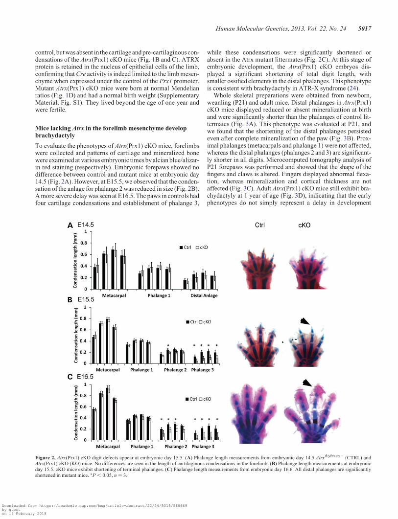
To evaluate the phenotypes of Atrx(Prx1) cKO mice, forelimbswere collected and patterns of cartilage and mineralized bonewere examined at various embryonic times by alcian blue/alizar-in red staining (respectively). Embryonic forepaws showed nodifference between control and mutant mice at embryonic day14.5 (Fig. 2A). However, at E15.5, we observed that the conden-sation of the anlage for phalange 2 was reduced in size (Fig. 2B).A more severe delay was seen at E16.5. The paws in controls hadfour cartilage condensations and establishment of phalange 3,
while these condensations were significantly shortened orabsent in the Atrx mutant littermates (Fig. 2C). At this stage ofembryonic development, the Atrx(Prx1) cKO embryos dis-played a significant shortening of total digit length, withsmaller ossified elements in the distal phalanges. This phenotypeis consistent with brachydactyly in ATR-X syndrome (24).
Whole skeletal preparations were obtained from newborn,weanling (P21) and adult mice. Distal phalanges in Atrx(Prx1)cKO mice displayed reduced or absent mineralization at birthand were significantly shorter than the phalanges of control lit-termates (Fig. 3A). This phenotype was evaluated at P21, andwe found that the shortening of the distal phalanges persistedeven after complete mineralization of the paw (Fig. 3B). Prox-imal phalanges (metacarpals and phalange 1) were not affected,whereas the distal phalanges (phalanges 2 and 3) are significant-ly shorter in all digits. Microcomputed tomography analysis ofP21 forepaws was performed and showed that the shape of thefingers and claws is altered. Fingers displayed abnormal flexa-tion, whereas mineralization and cortical thickness are notaffected (Fig. 3C). Adult Atrx(Prx1) cKO mice still exhibit bra-chydactyly at 1 year of age (Fig. 3D), indicating that the earlyphenotypes do not simply represent a delay in development
Figure 2. Atrx(Prx1) cKO digit defects appear at embryonic day 15.5. (A) Phalange length measurements from embryonic day 14.5 Atrxfl/yPrxcre2 (CTRL) andAtrx(Prx1) cKO (KO) mice. No differences are seen in the length of cartilaginous condensations in the forelimb. (B) Phalange length measurements at embryonicday 15.5. cKO mice exhibit shortening of terminal phalanges. (C) Phalange length measurements from embryonic day 16.6. All distal phalanges are significantlyshortened in mutant mice. ∗P , 0.05, n ¼ 3.
Human Molecular Genetics, 2013, Vol. 22, No. 24 5017
Downloaded from https://academic.oup.com/hmg/article-abstract/22/24/5015/568669by gueston 15 February 2018

but rather a permanent phenotype. The proximal bones of mutantforelimbs (humerus, radius, ulna) did not show any abnormal-ities at any investigated age.
Cell death is increased in the Atrx(Prx1) cKO embryoniclimb bud mesenchyme
The observation that Atrx(Prx1) cKO mice have shorter digitssuggests a reduction in cell numbers during embryogenesis,
which could be explained by reduced proliferation or increasedcell death. Proliferation was assessed using Ki67 staining (amarker of proliferation). Quantification of Ki67-positive cellsin the distal portions of control and Atrx(Prx1) cKO digitsshowed that cell proliferation is not different between genotypesat E13.5 and E15.5, which represent times before and after theappearance of the phenotype, respectively (Fig. 4).
We next assessed the level of apoptosis by the TUNEL assayand staining for activated caspase 3 at E13.5. As expected, we
Figure 3. Atrx(Prx1) cKO mice show brachydactyly. (A) Skeletal stains of control (Ctrl) and Atrx(Prx1) cKO (KO) newborn (P0.5) forelimbs. Cartilage is stained inblue, mineralized tissue is stained in red. Proximal bones (metacarpals and the first phalange) were not affected in the mutant; however, distal phalanges were sig-nificantly shortened and lacked mineralization. ∗P , 0.05, n ¼ 3 (B) Skeletal stains of control (Ctrl) and Atrx(Prx1) cKO (KO) weanling (P21) forelimbs. Significantshortening is observed in all phalanges, excluding the metacarpals ∗P , 0.05, n ¼ 3. (C) MicroCT isosurfaces of P21 digits. Atrx(Prx1) cKO digits lack flexation of theterminal phalanges, and have short, malformed claws. Cortical thickness was unaffected. Scale bar: 1 mm (D) Skeletal stains of control (Ctrl) and Atrx(Prx1) cKO(KO) adult (1 year old) forelimbs. Mineralized tissue is stained in red. Significant shortening is observed in all phalanges. N ¼ 3 for all time points, ∗P , 0.05.
5018 Human Molecular Genetics, 2013, Vol. 22, No. 24
Downloaded from https://academic.oup.com/hmg/article-abstract/22/24/5015/568669by gueston 15 February 2018

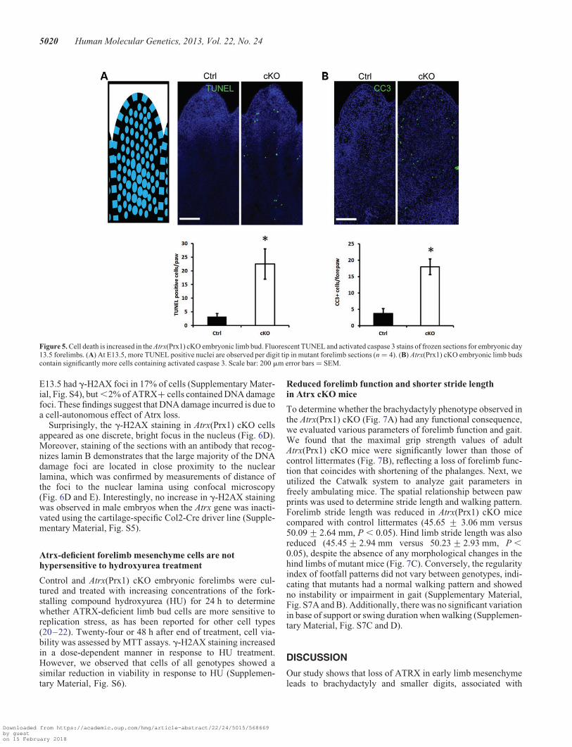
observed clusters of apoptotic cells in the interdigital mesen-chyme in both genotypes. We also observed an increase in thenumber of apoptotic cells in the mutant compared with controldigits. At E13.5, there was a 6.9-fold increase in apoptoticcells in the Atrx(Prx1) cKO (P ¼ 0.038, n ¼ 4), and a 4.8-fold in-crease in the number of cells containing activated caspase 3 (P ¼0.004, n ¼ 4) (Fig. 5). At E15.5 there was a significant 3.8-foldincrease in apoptotic cells in the Atrx(Prx1) cKO (P ¼ 0.028,n ¼ 3) (Supplementary Material, Fig. S2).
Increased level of g-H2AX foci in Atrx-null embryonic limbbud mesenchyme and chondrocytes
As we and others previously reported increased levels of DNAdamage upon loss of Atrx (18,20), we examined Atrx(Prx1)
cKO forelimbs for evidence of DNA damage. We stained embry-onic limb cryosections from E12.5 to E17.5 with an antibodyagainst phosphorylated g-H2AX, a marker of DNA double-strand breaks (DSBs). Confocal imaging and quantification atE13.5 and E15.5 showed a significant increase in the numberof cells harboring g-H2AX foci in the forelimb mesenchymeof Atrx(Prx1) cKO mice (Fig. 6A and B) and cartilaginousdigit rays (Supplementary Material, Fig. S3). To confirm thisresult, we co-stained cryosections with 53BP1, a DNA damageresponse protein that promptly re-localizes to damaged sites.We observed a higher number of 53BP1 foci in mutant forepawsthat largely overlapped with the g-H2AX foci (Fig. 6C). In thelimb buds of heterozygous female embryos, Atrx is deleted in�50% of cells, as expected from a random pattern ofX-inactivation. Forelimb tissue of heterozygote female mice at
Figure 4. Proliferation is unchanged in the Atrx(Prx1) cKO embryonic limb bud. Immunofluorescent stains for the S-phase marker Ki67 in embryonic day 13.5 (A) and15.5 (B) forelimb cryosections. More Ki67+ cells are present in the highly proliferative distal tip when compared with the differentiated digit ray, but no significantchange is observed between genotypes. Scale bar: 20 mm, Error bars ¼ SD, n ¼ 3.
Human Molecular Genetics, 2013, Vol. 22, No. 24 5019
Downloaded from https://academic.oup.com/hmg/article-abstract/22/24/5015/568669by gueston 15 February 2018
E13.5 had g-H2AX foci in 17% of cells (Supplementary Mater-ial, Fig. S4), but ,2% of ATRX+ cells contained DNA damagefoci. These findings suggest that DNA damage incurred is due toa cell-autonomous effect of Atrx loss.
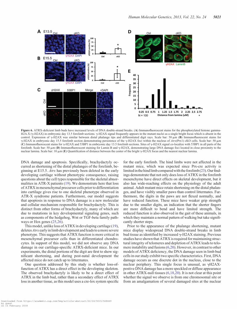
Surprisingly, the g-H2AX staining in Atrx(Prx1) cKO cellsappeared as one discrete, bright focus in the nucleus (Fig. 6D).Moreover, staining of the sections with an antibody that recog-nizes lamin B demonstrates that the large majority of the DNAdamage foci are located in close proximity to the nuclearlamina, which was confirmed by measurements of distance ofthe foci to the nuclear lamina using confocal microscopy(Fig. 6D and E). Interestingly, no increase in g-H2AX stainingwas observed in male embryos when the Atrx gene was inacti-vated using the cartilage-specific Col2-Cre driver line (Supple-mentary Material, Fig. S5).
Atrx-deficient forelimb mesenchyme cells are nothypersensitive to hydroxyurea treatment
Control and Atrx(Prx1) cKO embryonic forelimbs were cul-tured and treated with increasing concentrations of the fork-stalling compound hydroxyurea (HU) for 24 h to determinewhether ATRX-deficient limb bud cells are more sensitive toreplication stress, as has been reported for other cell types(20–22). Twenty-four or 48 h after end of treatment, cell via-bility was assessed by MTT assays. g-H2AX staining increasedin a dose-dependent manner in response to HU treatment.However, we observed that cells of all genotypes showed asimilar reduction in viability in response to HU (Supplemen-tary Material, Fig. S6).
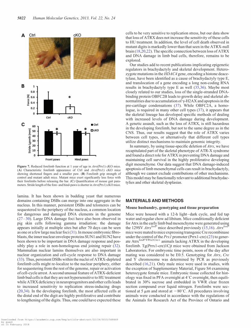
Reduced forelimb function and shorter stride lengthin Atrx cKO mice
To determine whether the brachydactyly phenotype observed inthe Atrx(Prx1) cKO (Fig. 7A) had any functional consequence,we evaluated various parameters of forelimb function and gait.We found that the maximal grip strength values of adultAtrx(Prx1) cKO mice were significantly lower than those ofcontrol littermates (Fig. 7B), reflecting a loss of forelimb func-tion that coincides with shortening of the phalanges. Next, weutilized the Catwalk system to analyze gait parameters infreely ambulating mice. The spatial relationship between pawprints was used to determine stride length and walking pattern.Forelimb stride length was reduced in Atrx(Prx1) cKO micecompared with control littermates (45.65 + 3.06 mm versus50.09+ 2.64 mm, P , 0.05). Hind limb stride length was alsoreduced (45.45+ 2.94 mm versus 50.23+ 2.93 mm, P ,0.05), despite the absence of any morphological changes in thehind limbs of mutant mice (Fig. 7C). Conversely, the regularityindex of footfall patterns did not vary between genotypes, indi-cating that mutants had a normal walking pattern and showedno instability or impairment in gait (Supplementary Material,Fig. S7A and B). Additionally, there was no significant variationin base of support or swing duration when walking (Supplemen-tary Material, Fig. S7C and D).
DISCUSSION
Our study shows that loss of ATRX in early limb mesenchymeleads to brachydactyly and smaller digits, associated with
Figure 5. Cell death is increased in the Atrx(Prx1) cKO embryonic limb bud. Fluorescent TUNEL and activated caspase 3 stains of frozen sections for embryonic day13.5 forelimbs. (A) At E13.5, more TUNEL positive nuclei are observed per digit tip in mutant forelimb sections (n ¼ 4). (B) Atrx(Prx1) cKO embryonic limb budscontain significantly more cells containing activated caspase 3. Scale bar: 200 mm error bars ¼ SEM.
5020 Human Molecular Genetics, 2013, Vol. 22, No. 24
Downloaded from https://academic.oup.com/hmg/article-abstract/22/24/5015/568669by gueston 15 February 2018
DNA damage and apoptosis. Specifically, brachydactyly oc-curred as shortening of the distal phalanges of the forelimb, be-ginning at E15.5. Atrx has previously been deleted in the earlydeveloping cartilage without phenotypic consequence, raisingquestions about the cell types responsible for the skeletal abnor-malities in ATR-X patients (19). We demonstrate here that lossof ATRX in mesenchymal precursor cells prior to differentiationinto cartilage gives rise to one skeletal phenotype observed inATR-X syndrome patients. Furthermore, our model suggeststhat apoptosis in response to DNA damage is a new molecularand cellular mechanism responsible for brachydactyly. This isdistinct from other forms of brachydactyly, many of which aredue to mutations in key developmental signaling genes, suchas components of the hedgehog, Wnt or TGF-beta family path-ways or Hox genes (25,26).
This model, unlike loss of ATRX in developing cartilage (19),deletes Atrx early in limb development and leads to a more severephenotype. This suggests that ATRX function is more critical inmesenchymal precursor cells than in differentiated chondro-cytes. In support of this model, we did not observe any DNAdamage in our cartilage-specific ATRX-deficient mice. In ourexperiments, the distal portions of the digit are first to show sig-nificant shortening, and during post-natal development theaffected mice do not catch up to littermates.
One question addressed by this study is whether loss-of-function of ATRX has a direct effect in the developing skeleton.The observed brachydactyly is likely to be a direct effect ofATRX in the limb bud, rather than a secondary effect of ATRXloss in another tissue, as this model uses a cre-lox system specific
for the early forelimb. The hind limbs were not affected in themutant mice, which was expected since Prx-cre activity islimited in thehind limbcomparedwith the forelimb(23).Our find-ings demonstrate that not only does loss of ATRX in the forelimbmesenchyme have direct effects on skeletal development, but italso has wide-reaching effects on the physiology of the adultanimal. Adult mutant mice retain shortening on the distal phalan-ges, and have visibly smaller paws than control littermates. Fur-thermore, the digits in the paws are not flexed normally, andhave reduced function. These mice have weaker grip strengthdue to the smaller digits, an indication that the shorter fingersare more difficult to bend and have limited strength. Thereduced function is also observed in the gait of these animals, inwhich they maintain a normal pattern of walking but take signifi-cantly shorter steps.
Prior to the appearance of the phalange shortening, mutantmice display widespread DNA double-strand breaks in limbbud tissue as identified by increased g-H2AX staining. Previousstudies have shown that ATRX is required for maintaining struc-tural integrity of telomeres and depletion of ATRX leads to telo-mere instability and fusions (6,20). However, in contrast to othermodels of ATRX deficiency, the DNA damage seen in limb budcells in our study exhibit two specific characteristics. First, DNAdamage occurs as one discrete dot in the nucleus, close to thenuclear periphery. This single focus is unusual, as gH2AX-postive DNA damage has a more speckled or diffuse appearancein other ATRX-null tissues (6,18,20). It is not clear at this pointwhether the signal we observe is from one chromosomal site orfrom an amalgamation of several damaged sites at the nuclear
Figure 6. ATRX-deficient limb buds have increased levels of DNA double-strand breaks. (A) Immunofluorescent stains for the phosphorylated histone gamma-H2A.X (g-H2AX) in embryonic day 13.5 forelimb sections. g-H2AX signal frequently appears in the mutant nuclei as a single bright focus which is absent in thecontrol. Expression of g-H2AX was similar between distal phalange tips and differentiated digit rays. Scale bar: 50 mm (B) Immunofluorescent stains forg-H2AX in embryonic day 15.5 forelimb section demonstrating persistence of the g-H2AX foci within the nucleus of Atrx(Prx1) cKO cells. Scale bar: 50 mm(C) Immunofluorescent stains for g-H2AX and 53BP1 in embryonic day 13.5 forelimb sections. Sites of g-H2AX signal co-localize with 53BP1 in all parts of theforelimb. Scale bar: 50 mm (D) Immunofluorescent staining for Lamin B and g-H2AX, demonstrating large DNA damage foci located in close proximity to thenuclear lamina. Scale bar: 10 mm (E) Quantification of distance between the center of the bright g-H2AX focus and the nearest nuclear lamina.
Human Molecular Genetics, 2013, Vol. 22, No. 24 5021
Downloaded from https://academic.oup.com/hmg/article-abstract/22/24/5015/568669by gueston 15 February 2018
lamina. It has been shown in budding yeast that numerousdomains containing DSBs can merge into one aggregate in thenucleus. In this manner, persistent DSBs and telomeres can besequestered to the periphery of the nucleus, a common locationfor dangerous and damaged DNA elements in the genome(27–30). Large DNA damage foci have also been observed inpig skin cells following gamma irradiation: the damageappears initially at multiple sites but after 70 days can be seenas one or a few large nuclear foci (31). In mouse embryonic fibro-blasts, the inner nuclear envelope proteins SUN1 and SUN2 havebeen shown to be important in DNA damage response and pos-sibly play a role in non-homologous end joining repair (32).Mammalian nuclear lamins themselves are also important innuclear organization and cell-cycle response to DNA damage(33). Thus, persistent DSBs within the nuclei of ATRX-depletedforelimb cells might re-localize to the nuclear periphery, eitherfor sequestering from the rest of the genome, repair or activationof cell-cycle arrest. A second unusual feature of ATRX-deficientlimb bud cells is that they are not hypersensitive to HU treatmentwhile ATRX deficiency in neuroprogenitors and other cells leadsto increased sensitivity to replication stress-inducing drugs(20,34). In the developing forelimb, the most affected cells atthe distal end of the digit are highly proliferative and contributeto lengthening of the digits. Thus, one could have expected these
cells to be very sensitive to replication stress, but our data showthat loss of ATRX does not increase the sensitivity of these cellsto HU treatment. In addition, the level of cell death observed inmutant digits is markedly lower than that seen in the ATRX-nullbrain (18,20,22). The specific connection between loss of ATRXand DNA damage in limb bud cells, therefore, remains to beexplored.
Our studies add to recent publications implicating epigeneticregulators in brachydactyly and skeletal development. Hetero-zygote mutations in the HDAC4 gene, encoding a histone deace-tylase, have been identified as a cause of brachydactyly type E,and translocation of a gene encoding a long non-coding RNAresults in brachydactyly type E as well (35,36). Maybe mostclosely related to our studies, loss of the single-stranded DNA-binding protein OBFC2B leads to growth delay and skeletal ab-normalities due to accumulation ofg-H2AX and apoptosis in thepre-cartilage condensations (37). While OBFC2A, a homo-logue, is required in many other cell types (37), it appears thatthe skeletal lineage has developed specific methods of dealingwith increased levels of DNA damage during development.A genetic assault, such as the loss of ATRX, is still hazardousin the developing forelimb, but not to the same degree as in theCNS. Thus, our results suggest that the role of ATRX variesbetween cell types, or alternatively that different cell typesutilize distinct mechanisms to maintain genomic integrity.
In summary, by using tissue-specific deletion of Atrx, we haverecapitulated part of the skeletal phenotype of ATR-X syndromeand found a direct role for ATRX in preventing DNA damage andmaintaining cell survival in the highly proliferative developingdigit mesenchyme. Our data suggest that DNA damage-inducedapoptosis of limb mesenchymal cells can result in brachydactyly,although we cannot exclude contributions of other mechanisms.This model may be functionally relevant to additional brachydac-tylies and other skeletal dysplasias.
MATERIALS AND METHODS
Mouse husbandry, genotyping and tissue preparation
Mice were housed with a 12-h light–dark cycle, and fed tapwater and regular chow ad libitum. Mice conditionally deficientfor Atrx in the early limb bud mesenchyme were generated usingthe 129SV AtrxloxP mice described previously (15,16). AtrxloxP
mice were mated to mice expressing transgenic Cre recombinaseunder the control of the Prx1 promoter (Prrx1-cre) (23) to gener-ate AtrxloxP/YPrx1Cre+ animals lacking ATRX in the developingforelimb. Tg(Prrx1-cre)1Cjt mice were obtained from JacksonLaboratories. For embryonic time points, noon of the day aftermating was considered to be E0.5. Genotyping for Atrx, Creand Y chromosome was determined by PCR as previouslydescribed (16,21). Only male mice were used in studies, withthe exception of Supplementary Material, Figure S4 examiningheterozygote female mice. Embryonic tissue collected for hist-ology was fixed in PFA overnight at 48C overnight, then equili-brated in 30% sucrose and embedded in VWR clear frozensection compound over liquid nitrogen. Forelimbs were sec-tioned at 5 mm and stored at 2808C. All procedures involvinganimals were conducted in accordance with the regulations ofthe Animals for Research Act of the Province of Ontario and
Figure 7. Reduced forelimb function at 1 year of age in Atrx(Prx1) cKO mice.(A) Characteristic forelimb appearance of Ctrl and Atrx(Prx1) cKO miceshowing shortened fingers and a smaller paw. (B) Forelimb grip strength ofcontrol and mutant adult mice. Mutant mice exert significantly less force withtheir forelimbs before releasing the bar. (C) Quantification of mouse gait para-meters. Stride length of the fore- and hind-paws is shorter in Atrx(Prx1) cKO mice.
5022 Human Molecular Genetics, 2013, Vol. 22, No. 24
Downloaded from https://academic.oup.com/hmg/article-abstract/22/24/5015/568669by gueston 15 February 2018

approved by the University of Western Ontario Animal Careand Use Committee.
Quantification of skeletal defects
Embryonic forepaws were isolated and dehydrated in 95%ethanol for 24 h, followed by acetone for 24 h. Paws were thenstained with 0.015% alcian blue, 0.05% alizarin red and 5%acetic acid in 70% ethanol as described (38). The stained pawswere cleared in glycerol/1% KOH (1:1) and stored in glycerol/ethanol (1:1). Adult skeletons were cleaned and stained asdescribed, then cleared in 2% KOH and stored in glycerol/ethanol (1:1). Embryonic forepaws were cleared and imagedusing a Nikon SMZ1500 dissecting microscope with a Photo-metrics (Tucson, AZ) Coolsnap camera using the ImageMaster5.0 software. Adult phalanges were microdissected andimaged as described. Digit length was measured in ImageJ andanalyzed using Graphpad from at least three independentlitters at each age.
Microcomputed tomography
Mice were euthanized at P21, forelimbs were dissected and fixedin paraformaldehyde overnight followed by immobilization inagarose. Whole forepaws were scanned on a GE Locusscanner at 80 kV and 0.08 mA with a 0.013 mm3 voxel reso-lution with 900 slices per scan. Scans were analyzed usingMicroview 3D visualization and analysis software (MicroView,Version 2.1.2, GE Healthcare Biosciences), as described (39).Whole forepaw isosurface images were generated by applyinga threshold of 932 HU, surface quality factor of 1.00 and asurface decimation factor of 30.
Western blot analysis
Embryonic forelimbs were dissected in PBS into RIPA buffer andhomogenized using a fine-gauge needle. SDS–polyacrylamidegel electrophoresis (PAGE) sample buffer (5 ml) was added tosamples, followed by boiling for 5 min. Samples were separatedby gradient SDS–PAGE (Biorad) and transferred to nitrocellu-lose membranes (Amersham). Membranes were blocked in 2%BSA and incubated with primary antibodies in accordance withthe instructions from the primary antibody supplier. Thefollowing antibodies were used; Anti-ATRX (Santa Cruz, #sc-15408,1:1000), mouse anti-b-actin (Sigma-Aldrich, #A2228,1:10,000), Goat anti-rabbit HRP (Santa Cruz, #sc-2030,1:5000), Goat anti-mouse HRP (Santa Cruz, #sc-2031, 1:5000).Representative blots from three independent experiments areshown.
Immunofluorescence assays
Proliferating cells were visualized using antibodies for Ki67.Frozen sections were rehydrated and antigens retrieved using0.1 M sodium citrate. Sections were permeabilized for 1 h withblocking buffer containing 5% goat serum and 0.1% tween-20in phosphate-buffered saline, followed by overnight incubationwith primary antibodies mouse anti-gamma-H2AX (Millipore,#05-636) and either rabbit anti-53BP1 (Cell Signaling, #4937),rabbit anti-ATRX (Santa Cruz, #sc-15408), or rabbit antiKi67
(Abcam, #ab15580) at a concentration of 1:300 in blockingbuffer. Antibodies were detected using fluorescently conjugatedsecondary antibodies, FITC goat anti-rabbit (Biosource,#ALI0408) and Alexa-594 goat anti-mouse (Invitrogen,#A11032) at dilutions of 1:200. Slides were mounted in mediacontaining DAPI (Vectashield) and confocal images wereacquired on an Olympus FluoView TV1000 coupled to theIX81 Motorized Inverted System Microscope (IX2 Series).Damage was quantified by a blinded observer within a fixedarea of 10 000 mm2 for digit tips and 5000 mm2 for digit rays.Counts were averaged from three independent litters at eachtime point. To assess apoptosis, 5 mm sections from E13.5 andE15.5 mice were used for the TUNEL assay using the RocheIn Situ Cell Death Detection Kit, Fluorescein according to man-ufacturer’s instructions. Briefly, frozen sections were hydratedin PBS and treated with cold citrate for 2 min. Slides werewashed and incubated in the enzyme/label solution mix for 1 hat 378C, then mounted with DAPI-containing mounting media(Vectashield). Sections were imaged on a Leica DMRA2 auto-mated inverted microscope. Signal was quantified from sectionsusing a fixed area over each digit by a blinded observer. Countswere averaged from at least three independent litters.
Activated caspase 3 was visualized in sections using rabbitanti-cleaved caspase-3 (Cell Signaling, #9664, 1:400 dilution).Cryosections were rehydrated in 1XPBS for 5 min and subjectedto antigen retrieval by heating 0.1 M sodium citrate ph6 to 958.Sections were permeabilized with 1X PBS/0.3% Triton X for5 min and incubated with primary antibody diluted in 1XPBS/0.3% Triton-X overnight at 48. Sections were washed 3× with1× PBS/0.3% Triton X for 5 min each and incubated withgoat anti-rabbit Alexa 488 (Molecular Probes, #A-11070,1:800 dilution) diluted in 1XPBS/0.3% Triton-X for 1 h at RT.Sections were washed 2× with 1× PBS/0.3% Triton × for5 min each, incubated with 1 mg/ml DAPI/1XPBS for 5 minand washed 3× with 1× PBS/0.3% Triton × for 5 min each.Slides were then mounted using SlowFade Gold (Invitrogen)and imaged using an inverted fluorescence microscope (Leica,DMI6000b).
Immunofluorescence assays were conducted for g-H2AX andthe nuclear envelope marker Lamin-B as described previously(40,41). Nuclear lamina was detected using a goat anti-Lamin-Bantibody (Abcam, #ab16048) at a concentration of 1:300,whereas DNA double-strand breaks were visualized with mouseanti-g-H2AX (Millipore, #05-636) at a concentration of 1:200in blocking buffer and detected with Alexa-594 donkey anti-mouse (Invitrogen, #A-11005) and FITC donkey anti-goat (Invi-trogen,#A-11055)ata concentrationof 1:200.Three-dimensionalimages of nuclei were collected on an Olympus FluoViewTV1000 coupled to the IX81 Motorized Inverted System Micro-scope (IX2 Series) in image stacks captures at 0.25 um Z-sectionsanddeconvoluted inVolocity (PerkinElmer).Measurementsweretaken from the center of the g-H2AX focus to the closest nuclearperiphery for 100 individual nuclei.
HU treatment and assessment of DNA damage and survival
Primary forelimb mesenchyme was prepared using distal fore-limb tissue dissected from E13.5 embryos. Cells were disso-ciated with collagenase at 378C for 1 h and plated in DMEM/F12 media supplemented with 10% FBS, 0.25% pen/strep and
Human Molecular Genetics, 2013, Vol. 22, No. 24 5023
Downloaded from https://academic.oup.com/hmg/article-abstract/22/24/5015/568669by gueston 15 February 2018

0.25% L-glutamate (Gibco). Forelimb mesenchyme cultureswere treated with increasing doses of HU (0.05–1 mM) for24 h, recovered for 24 or 48 h, and cell viability measured bythe MTT assay as described (42). Briefly, media containingHU was removed and replaced with fresh media for 24 or 48 h,followed by replacement with media containing 0.5 mg/mlMTT for 4 h. Product was stabilized in DMSO and quantifiedon a spectrophotometer at 595 nm.
Gait and grip strength analyses
Behavioral assays were performed at the behavioral core facil-ity at the Robarts Research institute (London, ON). Control andmutant mice at P21 and 1 year of age freely ambulated on anilluminated glass platform, and movements were recordedwith a high speed camera using the Noldus CatWalk system.After an initial training period, at least three un-interruptedruns were collected from each animal. Each run was analyzedusing the Catwalk 7.1 software for classifying foot contacts.Run data for each animal were averaged, and then averages cal-culated within each genotype. Means were analyzed for statis-tical differences between genotypes using Student’s t-test.Mouse forelimb grip strength was assessed using a digitalforce gauge as described previously (43). Briefly, the animalwas grasped by the scruff of the neck in one hand, and thebase of the tail in the other. Measurements were recorded onthe meter as the subject was allowed to grip, and then waspulled away from, the grip bar. Total peak force was deter-mined from an average of five measurements taken fromeach animal, and the means of each genotype werecompared. Statistical analysis of the means was conductedusing Student’s t-test.
SUPPLEMENTARY MATERIAL
Supplementary Material is available at HMG online.
ACKNOWLEDGEMENTS
We thank Drs Douglas Higgs and Richard Gibbons (WeatherallInstitute of Molecular Medicine, John Radcliffe Hospital,Oxford, UK) for kindly providing the AtrxloxP mice used inthis study.
Conflict of Interest statement. None declared.
FUNDING
This work was supported by an operating grant from the Canad-ian Institutes for Health Research to N.G.B. and F.B.(MOP102539), an Ontario Graduate Studentship (OGS) toL.A.S., Paediatrics Graduate Studentship at the University ofWestern Ontario, the Curtis Cadman Studentship and QueenElizabeth II Ontario Graduate Scholarship in Science and Tech-nology (QEIIOGSST) to L.A.W.
REFERENCES
1. Gibbons, R.J., Picketts, D.J., Villard, L. and Higgs, D.R. (1995) Mutations ina putative global transcriptional regulator cause X-linked mental retardationwith alpha-thalassemia (ATR-X syndrome). Cell, 80, 837–845.
2. Gibbons, R.J. and Higgs, D.R. (2000) Molecular-clinical spectrum of theATR-X syndrome. Am. J. Hum. Genet., 97, 204–212.
3. Gibbons, R.J., Brueton, L., Buckle, V.J., Burn, J., Clayton-Smith, J.,Davison, B.C.C., Gardner, R.J.M., Homfray, T., Kearney, L., Kingston,H.M. et al. (1995) Clinical and hematologic aspects of the X-linkeda-thalassemia/mental retardation syndrome (ATR-X). Am. J. Hum. Genet.,55, 288–299.
4. Wada, T., Sugie, H., Fukushima, Y. and Saitoh, S. (2005) Non-skewedX-inactivation may cause mental retardation in a female carrier of X-linkeda-thalassemia/mental retardation syndrome (ATR-X): X-inactivation studyof nine female carriers of ATR-X. Am. J. Hum. Genet., 138A, 18–20.
5. Gibbons, R.J., McDowell, T.J., Raman, S., O’Rourke, D.M., Garrick, D.,Ayyub, H. and Higgs, D.R. (2000) Mutations in ATRX, encoding a SWI/SNF-like protein, cause diverse changes in the pattern of DNA methylation.Nat. Genet., 24, 361–371.
6. Wong, L.H., McGhie, J.D., Sim, M., Anderson, M.A., Ahn, S., Hannan,R.D., George, A.J., Morgan, K.A., Mann, J.R. and Choo, K.H.A. (2010)ATRX Interacts with H3.3 in maintaining telomere structural integrity inpluripotent embryonic stem cells. Genome Res., 20, 351–360.
7. Otani, J., Nankumo, T., Arita, K., Inamoto, S., Ariyoshi, M. and Shirakawa,M. (2009) Structural basis for recognition of H3K4 methylation status by theDNA methyltransferase 3A ATRX-DNMT3-DNMT3L domain. EMBO
Reports, 10, 1235–1241.
8. Dhayalan, A., Tamas, R., Bock, I., Tattermusch, A., Dimitrova, E.,Kudithipudi, S., Ragozin, S. and Jeltsch, A. (2011) The ATRX-ADD domainbinds to H3 tail peptides and reads the combined methylation state of K4 andK9. Hum. Mol. Genet., 20, 2195–2203.
9. Picketts, D.J., Higgs, D.R., Bachoo, S., Blake, D.J., Quarrell, O.W. andGibbons, R.J. (1996) ATRX encodes a novel member of the SNF2 family ofproteins: mutations point to a common mechanism underlying the ATR-Xsyndrome. Hum. Mol. Genet., 5, 1899–1907.
10. Eisen, J.A., Sweder, K.S. and Hanawalt, P.C. (1995) Evolution of the SNF2family of proteins: subfamilies with distinct sequences and functions. Nuc.
Acids Res., 23, 2715–2723.
11. Xue, Y., Gibbons, R., Yan, Z., Yang, D., McDowell, T.L., Sechi, S., Qin, J.,Zhou, S., Higgs, D. and Wang, W. (2003) The ATRX syndrome proteinforms a chromatin-remodeling complex with Daxx and localizes inpromyelocytic leukemia nuclear bodies. Proc. Natl Acad. Sci., 100, 10635–10640.
12. Lewis, P.W., Elsaesser, S.J., Noh, K.-M., Stadler, S.C. and Allis, C.D. (2010)Daxx is an H3.3-specific histone chaperone and cooperates with ATRX inreplication-independent chromatin assembly at telomeres. Proc. Natl Acad.
Sci., 107, 14075–14080.
13. Drane, P., Ouararhni, K., Depaux, A., Shuaib, M. and Hamiche, A. (2010)The death-associated protein DAXX is a novel histone chaperone involvedin the replication-independent deposition of H3.3. Genes Dev., 24, 1253–1265.
14. Ritchie, K., Seah, C., Moulin, J., Isaac, C., Dick, F. and Berube, N.G. (2008)Loss of ATRX leads to chromosome cohesion and congression defects.J. Cell Biol., 180, 315–324.
15. Garrick, D., Sharpe, J.A., Arkell, R., Dobbie, L., Smith, A.J., Wood, W.G.,Higgs, D.R. and Gibbons, R.J. (2006) Loss of Atrx affects trophoblastdevelopment and the pattern of X-inactivation in extraembryonic tissues.PLoS Genet., 2, e58.
16. Berube, N.G., Mangelsdorf, M., Jagla, M., Vanderluit, J., Garrick, D.,Gibbons, R.J., Higgs, D.R., Slack, R.S. and Picketts, D.J. (2005) Thechromatin-remodeling protein ATRX is critical for neuronal survival duringcorticogenesis. J. Clin. Invest., 115, 258–267.
17. Bagheri-Fam, S., Argentaro, A., Svingen, T., Combes, A.N., Sinclair, A.H.,Koopman, P. and Harley, V.R. (2011) Defective survival of proliferatingSertoli cells and androgen receptor function in a mouse model of the ATR-Xsyndrome. Hum. Mol. Genet., 20, 2213–2224.
18. Huh, M.S., Price O’Dea, T., Ouazia, D., McKay, B.C., Parise, G., Parks, R.J.,Rudnicki, M.A. and Picketts, D.J. (2012) Compromised genomicintegrity impedes muscle growth after Atrx inactivation. J. Clin. Invest., 122,4412–4423.
5024 Human Molecular Genetics, 2013, Vol. 22, No. 24
Downloaded from https://academic.oup.com/hmg/article-abstract/22/24/5015/568669by gueston 15 February 2018
19. Solomon, L.A., Li, J.R., Berube, N.G. and Beier, F. (2009) Loss of ATRX inchondrocytes has minimal effects on skeletal development. PLoS ONE, 4,e7106.
20. Watson, L.A., Solomon, L.A., Li, J.R., Jiang, Y., Edwards, M., Shin-Ya, K.,Beier, F. and Berube, N.G. (2013) Atrx deficiency induces telomeredysfunction, endocrine defects, and reduced life span. J. Clin. Invest, 123,2049–2063.
21. Seah, C., Levy, M.A., Jiang, Y., Mokhtarzada, S., Higgs, D.R., Gibbons, R.J.and Berube, N.G. (2008) Neuronal death resulting from targeted disruptionof the Snf2 protein ATRX is mediated by p53. J. Neurosci., 28, 12570–12580.
22. Leung, J.W.-C.,Ghosal, G., Wang,W., Shen,X., Wang, J., Li, L. and Chen, J.(2013) Alpha thalassemia/mental retardation syndrome X-linked geneproduct ATRX is required for proper replication restart and cellularresistance to replication stress. J. Biol. Chem., 288, 6342–6350.
23. Logan, M., Martin, J.F., Nagy, A., Lobe, C., Olson, E.N. and Tabin, C.J.(2002) Expression of Cre recombinase in the developing mouse limb buddriven by a Prxl enhancer. Genesis, 33, 77–80.
24. Gibbons, R.J. (2006) Alpha thalassaemia-mental retardation,×linked.Orphanet. J. Rare Dis., 1, 1–15.
25. Gao, B., Hu, J., Stricker, S., Cheung, M., Ma, G., Law, K.F., Witte, F.,Briscoe, J., Mundlos, S., He, L. et al. (2009) A mutation in Ihh that causesdigit abnormalities alters its signalling capacity and range. Nature, 458,1196–1200.
26. Minami, Y., Oishi, I., Endo, M. and Nishita, M. (2010) Ror-family receptortyrosine kinases in noncanonical Wnt signaling: their implications indevelopmental morphogenesis and human diseases. Dev. Dyn., 239, 1–15.
27. Oza, P., Jaspersen, S.L., Miele, A., Dekker, J. and Peterson, C.L. (2009)Mechanisms that regulate localization of a DNA double-strand break to thenuclear periphery. Genes Dev., 23, 912–927.
28. Soutoglou, E., Dorn, J.F., Sengupta, K., Jasin, M., Nussenzweig, A., Ried, T.,Danuser, G. and Misteli, T. (2007) Positional stability of singledouble-strand breaks in mammalian cells. Nat. Cell Biol., 9, 675–682.
29. Aten, J.A., Stap, J., Krawczyk, P.M., van Oven, C.H., Hoebe, R.A., Essers, J.and Kanaar, R. (2004) Dynamics of DNA double-strand breaks revealed byclustering of damaged chromosome domains. Science, 303, 92–95.
30. Schober, H., Ferreira, H., Kalck, V., Gehlen, L.R. and Gasser, S.M. (2009)Yeast telomerase and the SUN domain protein Mps3 anchor telomeres andrepress subtelomeric recombination. Genes Dev., 23, 928–938.
31. Ahmed, E.A., Agay, D., Schrock, G., Drouet, M., Meineke, V. andScherthan, H. (2012) Persistent DNA damage after high dose in vivo gammaexposure of minipig skin. PLoS ONE, 7, e39521.
32. Lei, K., Zhu, X., Xu, R., Shao, C., Xu, T., Zhuang, Y. and Han, M. (2012)Inner nuclear envelope proteinsSUN1 and SUN2 play a prominent role in theDNA damage response. Curr. Biol., 22, 1609–1615.
33. Johnson, B.R., Nitta, R.T., Frock, R.L., Mounkes, L., Barbie, D.A., Stewart,C.L., Harlow, E. and Kennedy, B.K. (2004) A-type lamins regulateretinoblastoma protein function by promoting subnuclear localization andpreventing proteasomal degradation. Proc. Natl Acad. Sci. USA, 101, 9677–9682.
34. Conte, D., Huh, M., Goodall, E., Delorme, M., Parks, R.J. and Picketts, D.J.(2012) Loss of Atrx sensitizes cells to DNA damaging agents throughp53-mediated death pathways. PLoS ONE, 7, e52167.
35. Maass, P.G., Rump, A., Schulz, H., Stricker, S., Schulze, L., Platzer, K.,Aydin, A., Tinschert, S., Goldring, M.B., Luft, F.C. et al. (2012) A misplacedlncRNA causes brachydactyly in humans. J. Clin. Invest., 122, 3990–4002.
36. Williams, S.R., Aldred, M.A., Der Kaloustian, V.M., Halal, F., Gowans, G.,McLeod, D.R., Zondag, S., Toriello, H.V., Magenis, R.E. and Elsea, S.H.(2010) Haploinsufficiency of HDAC4 causes brachydactyly mentalretardationsyndrome, with brachydactyly type E, developmental delays, andbehavioral problems. Am. J. Hum. Genet., 87, 219–228.
37. Feldhahn, N., Ferretti, E., Robbiani, D.F., Callen, E., Deroubaix, S., Selleri,L., Nussenzweig, A. and Nussenzweig, M.C. (2012) The hSSB1 orthologueObfc2b is essential for skeletogenesis but dispensable for the DNA damageresponse in vivo. EMBO J., 31, 4045–4056.
38. Wang, G., Woods, A., Agoston, H., Ulici, V., Glogauer, M. and Beier, F.(2007) Genetic ablation of Rac1 in cartilage results in chondrodysplasia.Dev. Biol., 306, 612–623.
39. Ulici, V., Hoenselaar, K.D., Agoston, H., McErlain, D.D., Umoh, J.,Chakrabarti, S., Holdsworth, D.W. and Beier, F. (2009) The role of Akt1 interminal stages of endochondral bone formation: angiogenesis andossification. Bone, 45, 1133–1145.
40. Vidakovic, M., Koester, M., Goetze, S., Winkelmann, S., Klar, M.,Poznanovic, G. and Bode, J. (2005) Co-localization of PARP-1 and lamin Bin the nuclear architecture: a halo-fluorescence- and confocal-microscopystudy. J. Cell. Biochem., 96, 555–568.
41. Kuo, L.J. and Yang, L.-X. (2008) g-H2AX - A novel biomarker for DNAdouble-strand breaks. In Vivo, 22, 305–309.
42. Halawani, D., Mondeh, R., Stanton, L.A. and Beier, F. (2004) P38 MAPkinase signaling is necessary for rat chondrosarcoma cell proliferation.Oncogene, 23, 3726–3731.
43. Meyer, O.A., Tilson, H.A., Byrd, W.C. and Riley, M.T. (1979) A method forthe routine assessment of fore- and hindlimb grip strength of rats and mice.Neurobehav. Toxicol. Teratol., 1, 233–236.
Human Molecular Genetics, 2013, Vol. 22, No. 24 5025
Downloaded from https://academic.oup.com/hmg/article-abstract/22/24/5015/568669by gueston 15 February 2018