Targeted imaging of urothelium carcinoma in human … imaging of urothelium carcinoma in human...
6
Targeted imaging of urothelium carcinoma in human bladders by an ICG pHLIP peptide ex vivo Jovana Golijanin a,b , Ali Amin c , Anna Moshnikova b , Joseph M. Brito a , Timothy Y. Tran a , Ramona-Cosmina Adochite b , Gregory O. Andreev b , Troy Crawford b , Donald M. Engelman d,1 , Oleg A. Andreev b , Yana K. Reshetnyak b,1 , and Dragan Golijanin a,1 a Minimally Invasive Urology Institute, Division of Urology, The Miriam Hospital and The Warren Alpert Medical School of Brown University, Providence, RI 02906; b Physics Department, University of Rhode Island, Kingston, RI 02881; c Department of Pathology & Laboratory Medicine, Brown University, The Miriam Hospital, Providence, RI 02906; and d Department of Molecular Biophysics and Biochemistry, Yale University, New Haven, CT 06511 Edited by Owen N. Witte, Howard Hughes Medical Institute, University of California, Los Angeles, CA, and approved July 20, 2016 (received for review June 29, 2016) Bladder cancer is the fifth most common in incidence and one of the most expensive cancers to treat. Early detection greatly improves the chances of survival and bladder preservation. The pH low insertion peptide (pHLIP) conjugated with a near-infrared fluorescent dye [indocyanine green (ICG)] targets low extracel- lular pH, allowing visualization of malignant lesions in human bladder carcinoma ex vivo. Cystectomy specimens obtained after radical surgery were immediately irrigated with nonbuffered saline and instilled with a solution of the ICG pHLIP construct, incubated, and rinsed. Bladders were subsequently opened and imaged, the fluorescent spots were marked, and a standard pathological analysis was carried out to establish the correlation between ICG pHLIP imaging and white light pathological assessment. Accurate targeting of bladder lesions was achieved with a sensitivity of 97%. Specificity is 100%, but reduced to 80% if targeting of necrotic tissue from previous transurethral resections or chemotherapy are considered as false positives. The ICG pHLIP imaging agent marked high-grade urothelial carcinomas, both muscle invasive and nonmuscle invasive. Carcinoma in situ was accurately diagnosed in 11 cases, whereas only four cases were seen using white light, so imaging with the ICG pHLIP peptide offers improved early diagnosis of bladder cancers and may also enable new treatment alternatives. bladder cancer | fluorescence-guided surgery | NIR imaging | acidity | human tissue B ladder cancer is the fifth most common cancer, constituting 4.5% of all new cancer cases in the United States; 76,960 new cases were estimated in 2016, and the death rate currently expected from bladder cancer is 21% (16,390). Approximately 2.4% of men and women will be diagnosed with bladder cancer at some point during their lifetime. In 2012, there were an esti- mated 577,403 individuals living with bladder cancer in the United States. Almost all of these patients require continuous surveil- lance, and occasionally, treatments. For all stages combined, the 5-y relative survival rate is 77%. Survival declines to 70% at 10 y and 65% at 15 y after diagnosis. Bladder cancer can be nonmuscle or muscle invasive. Half of all bladder cancer patients are diagnosed while the tumor is nonmuscle invasive, for which the 5-y survival is 96%. Most (up to 98%) of malignant bladder tumors arise in the epithelium; 90–92% of these bladder cancers are urothelial carci- nomas (1, 2). Less common bladder cancers are squamous cell or adenocarcinomas. Approximately 20–25% of patients have muscle- invasive disease, and of non–muscle-invasive disease, patients will progress to muscle invasive disease at 5-y follow-up depending on intermediate or high risk of the progression (3, 4). An important medical objective is the identification of early stage lesions, such as carcinoma in situ, because it is expected that diagnosis at this stage will decrease the frequency of treatments, increasing patient health and reducing expense. Each type and stage of bladder cancer requires a different type of treatment. High recurrence frequency, procedural costs, and the requirement for prolonged active monitoring make bladder cancer one of the most expensive cancers in the United States, placing a heavy economic burden on the health care system from lifetime endoscopic follow-ups and treatments. Patients suffer from high morbidity and the complications associated with che- motherapy, radiation, and radical surgery (5). Therefore, as noted, timely diagnosis of the tumor and appropriate management pro- tocols are of great significance for decreasing treatment cost and improving a patient’s lifestyle. Advances in the early detection of bladder cancer lesions are likely to increase the chances of timely successful treatment, the prevention of recurrences, and bladder function preservation. Cancers, including urothelial carcinoma, are associated with multiple alterations in the genome, including changes in epige- netic regulation, point mutations, gene deletions, duplications, and chromosomal rearrangements. These changes are hetero- geneous, leading to heterogeneity of the overexpression of par- ticular biomarkers at the surfaces of cancer cells within a tumor and between tumors. Heterogeneity significantly limits success in the use of cell surface biomarkers for the targeted delivery of therapeutics. On other hand, multiple studies have revealed that neoplastic cells produce an acidic environment due to increased metabolic activity (6). Adaptations to the highly acidic micro- environment are critical steps in the transition from an avascular Significance Bladder cancer is the fifth most common cancer. Timely di- agnosis and appropriate early management protocols are of paramount significance for improving patient outcomes. This study shows efficient pH-dependent near-infrared imaging of bladder malignant tumors without targeting of normal tis- sue. Our results demonstrate that the indocyanine green pH low insertion peptide (pHLIP) construct is suitable for use as a predictive clinical marker, specifically staining human bladder tumors after intravesical administration ex vivo. The targeting allows delivery of various imaging probes, which may offer early diagnosis and improve the outcomes of endoscopic and radical surgical resection of urothelial carcinomas. In addition, delivery of therapeutic molecules to cancer cells by pHLIP might open an opportunity for novel targeted treatment of bladder cancers. Author contributions: A.A., O.A.A., Y.K.R., and D.G. designed research; J.G., A.A., A.M., J.M.B., T.Y.T., R.-C.A., T.C., and D.G. performed research; J.G., A.A., G.O.A., O.A.A., Y.K.R., and D.G. analyzed data; and A.A., D.M.E., O.A.A., Y.K.R., and D.G. wrote the paper. Conflict of interest statement: Y.K.R., O.A.A., and D.M.E. are founders of a new company, pHLIP Inc. They have shares in the company, but the company did not fund any part of the work reported in the paper, which was done in their academic laboratories. This article is a PNAS Direct Submission. Freely available online through the PNAS open access option. 1 To whom correspondence may be addressed. Email: [email protected], [email protected], or [email protected]. www.pnas.org/cgi/doi/10.1073/pnas.1610472113 PNAS Early Edition | 1 of 6 APPLIED BIOLOGICAL SCIENCES
Transcript of Targeted imaging of urothelium carcinoma in human … imaging of urothelium carcinoma in human...

Targeted imaging of urothelium carcinoma in humanbladders by an ICG pHLIP peptide ex vivoJovana Golijanina,b, Ali Aminc, Anna Moshnikovab, Joseph M. Britoa, Timothy Y. Trana, Ramona-Cosmina Adochiteb,Gregory O. Andreevb, Troy Crawfordb, Donald M. Engelmand,1, Oleg A. Andreevb, Yana K. Reshetnyakb,1,and Dragan Golijanina,1
aMinimally Invasive Urology Institute, Division of Urology, The Miriam Hospital and The Warren Alpert Medical School of Brown University, Providence, RI02906; bPhysics Department, University of Rhode Island, Kingston, RI 02881; cDepartment of Pathology & Laboratory Medicine, Brown University, TheMiriam Hospital, Providence, RI 02906; and dDepartment of Molecular Biophysics and Biochemistry, Yale University, New Haven, CT 06511
Edited by Owen N. Witte, Howard Hughes Medical Institute, University of California, Los Angeles, CA, and approved July 20, 2016 (received for review June29, 2016)
Bladder cancer is the fifth most common in incidence and one ofthe most expensive cancers to treat. Early detection greatlyimproves the chances of survival and bladder preservation. ThepH low insertion peptide (pHLIP) conjugated with a near-infraredfluorescent dye [indocyanine green (ICG)] targets low extracel-lular pH, allowing visualization of malignant lesions in humanbladder carcinoma ex vivo. Cystectomy specimens obtained afterradical surgery were immediately irrigated with nonbuffered salineand instilled with a solution of the ICG pHLIP construct, incubated,and rinsed. Bladders were subsequently opened and imaged, thefluorescent spots were marked, and a standard pathologicalanalysis was carried out to establish the correlation between ICGpHLIP imaging and white light pathological assessment. Accuratetargeting of bladder lesions was achieved with a sensitivity of97%. Specificity is 100%, but reduced to 80% if targeting of necrotictissue from previous transurethral resections or chemotherapy areconsidered as false positives. The ICG pHLIP imaging agent markedhigh-grade urothelial carcinomas, bothmuscle invasive and nonmuscleinvasive. Carcinoma in situ was accurately diagnosed in 11 cases,whereas only four caseswere seen usingwhite light, so imaging withthe ICG pHLIP peptide offers improved early diagnosis of bladdercancers and may also enable new treatment alternatives.
bladder cancer | fluorescence-guided surgery | NIR imaging | acidity |human tissue
Bladder cancer is the fifth most common cancer, constituting4.5% of all new cancer cases in the United States; 76,960
new cases were estimated in 2016, and the death rate currentlyexpected from bladder cancer is 21% (16,390). Approximately2.4% of men and women will be diagnosed with bladder cancerat some point during their lifetime. In 2012, there were an esti-mated 577,403 individuals living with bladder cancer in the UnitedStates. Almost all of these patients require continuous surveil-lance, and occasionally, treatments. For all stages combined, the 5-yrelative survival rate is 77%. Survival declines to 70% at 10 y and65% at 15 y after diagnosis. Bladder cancer can be nonmuscle ormuscle invasive. Half of all bladder cancer patients are diagnosedwhile the tumor is nonmuscle invasive, for which the 5-y survival is96%. Most (up to 98%) of malignant bladder tumors arise in theepithelium; 90–92% of these bladder cancers are urothelial carci-nomas (1, 2). Less common bladder cancers are squamous cell oradenocarcinomas. Approximately 20–25% of patients have muscle-invasive disease, and of non–muscle-invasive disease, patients willprogress to muscle invasive disease at 5-y follow-up depending onintermediate or high risk of the progression (3, 4). An importantmedical objective is the identification of early stage lesions, suchas carcinoma in situ, because it is expected that diagnosis at thisstage will decrease the frequency of treatments, increasingpatient health and reducing expense.Each type and stage of bladder cancer requires a different type
of treatment. High recurrence frequency, procedural costs, and
the requirement for prolonged active monitoring make bladdercancer one of the most expensive cancers in the United States,placing a heavy economic burden on the health care system fromlifetime endoscopic follow-ups and treatments. Patients sufferfrom high morbidity and the complications associated with che-motherapy, radiation, and radical surgery (5). Therefore, as noted,timely diagnosis of the tumor and appropriate management pro-tocols are of great significance for decreasing treatment cost andimproving a patient’s lifestyle. Advances in the early detection ofbladder cancer lesions are likely to increase the chances of timelysuccessful treatment, the prevention of recurrences, and bladderfunction preservation.Cancers, including urothelial carcinoma, are associated with
multiple alterations in the genome, including changes in epige-netic regulation, point mutations, gene deletions, duplications,and chromosomal rearrangements. These changes are hetero-geneous, leading to heterogeneity of the overexpression of par-ticular biomarkers at the surfaces of cancer cells within a tumorand between tumors. Heterogeneity significantly limits success inthe use of cell surface biomarkers for the targeted delivery oftherapeutics. On other hand, multiple studies have revealed thatneoplastic cells produce an acidic environment due to increasedmetabolic activity (6). Adaptations to the highly acidic micro-environment are critical steps in the transition from an avascular
Significance
Bladder cancer is the fifth most common cancer. Timely di-agnosis and appropriate early management protocols are ofparamount significance for improving patient outcomes. Thisstudy shows efficient pH-dependent near-infrared imaging ofbladder malignant tumors without targeting of normal tis-sue. Our results demonstrate that the indocyanine green pHlow insertion peptide (pHLIP) construct is suitable for use as apredictive clinical marker, specifically staining human bladdertumors after intravesical administration ex vivo. The targetingallows delivery of various imaging probes, which may offerearly diagnosis and improve the outcomes of endoscopic andradical surgical resection of urothelial carcinomas. In addition,delivery of therapeutic molecules to cancer cells by pHLIPmight open an opportunity for novel targeted treatment ofbladder cancers.
Author contributions: A.A., O.A.A., Y.K.R., and D.G. designed research; J.G., A.A., A.M.,J.M.B., T.Y.T., R.-C.A., T.C., and D.G. performed research; J.G., A.A., G.O.A., O.A.A., Y.K.R.,and D.G. analyzed data; and A.A., D.M.E., O.A.A., Y.K.R., and D.G. wrote the paper.
Conflict of interest statement: Y.K.R., O.A.A., and D.M.E. are founders of a new company,pHLIP Inc. They have shares in the company, but the company did not fund any part of thework reported in the paper, which was done in their academic laboratories.
This article is a PNAS Direct Submission.
Freely available online through the PNAS open access option.1To whom correspondence may be addressed. Email: [email protected],[email protected], or [email protected].
www.pnas.org/cgi/doi/10.1073/pnas.1610472113 PNAS Early Edition | 1 of 6
APP
LIED
BIOLO
GICAL
SCIENCE
S

preinvasive tumor to a malignant invasive carcinoma (7–9).Thus, acidity may provide a universal biomarker for tumor tar-geting that is not subject to the selection of resistant cell lines(10). pH low insertion peptides (pHLIPs) are a class of mem-brane-binding peptides that specifically target acidic cells in vitroand in vivo (11) by inserting across cellular membranes when theextracellular pH is low (12). pHLIPs conjugated with fluorescentdyes have been used to differentiate normal from neoplastictissue in various animal tumor models (12–15) and in humanbiopsy head and neck samples (16, 17). This report uses anindocyanine green (ICG) pHLIP conjugate for the diagnosis ofurothelial carcinoma and precancerous lesions in fresh humanradical cystectomy samples ex vivo and points the way toward awide range of diagnostic and therapeutic alternatives.
ResultsWe used a pHLIP labeled with a near-infrared fluorescence(NIRF) dye, ICG, to monitor the targeting of tumors in humanbladders. The absorption spectrum of ICG pHLIP is shown inFig. 1A. The fluorescence of ICG pHLIP increases about 25-foldin the presence of 1-palmitoyl-2-oleoyl-sn-glycero-3-phosphocho-line (POPC) liposomes (Fig. 1B). Thus, binding of ICG pHLIP tothe lipid bilayers of cancerous cell membranes significantly en-hances the emission of ICG.From November 2014 through December 2015, 22 radical
cystectomy patients were included in the study. Patient agesranged from 51 to 84 y (mean age, 67.7 y), and the sex ratio ofM/F was 19/3. Table 1 contains patient demographics, preoper-ative diagnosis, clinical stage of the disease, and the results ofimaging studies. The specimens did not show any adverse mor-phological findings after incubation with ICG pHLIP, and therewas no evidence of damage or degenerative effect in the non-tumoral tissue. The use of ICG pHLIP did not alter the patho-logical assessment of the radical cystectomy tissues. Overall, 29malignant lesions were identified by pathology assessment of the22 bladder specimens stained with ICG pHLIP (3 radical cys-tectomy cases were incubated ex vivo with ICG-Cys as negativecontrols). The frequencies of different pathologies in 29 lesionswere as follows: high-grade muscle invasive urothelial carcinoma(HGI) (Fig. 2 A–C) in 12; high-grade nonmuscle invasive uro-thelial carcinoma (HGN) (Fig. 2 D–F) in 5; carcinoma in situ(CIS) in 11 (Fig. 2G–I), and high-grade dysplasia in 1 (Fig. 2 J–L).In seven cases, NIRF imaging guided the pathologist to CIS notobserved by white light inspection. In case 2, necrotic tissue insidea diverticulum was NIRF positive. For the negative control cases(cases 13, 19, and 20) only the ICG-Cys dye alone was used forinstillation (at concentrations from 8 to 40 μM in an 80-mL vol-ume), and no specific tumor targeting was observed.The tabular results of the sensitivity/specificity tests are shown
in Tables 2–5. The test was performed for cancerous vs. normal
tissue excluding targeting of necrotic and previously treated tis-sue (Tables 2 and 3). The sensitivity and specificity of targetingof cancerous tissue vs. normal were found to be 97% and 100%,respectively. If targeting of necrotic tissue from prior post tran-surethral removal of bladder tumors and previously treated(chemotherapy) necrotic tumors by ICG pHLIP is considered asa false positive, the specificity is reduced from 100% to 80%(Tables 4 and 5).
DiscussionWe used an ICG pHLIP construct to target urothelial carcinomain human bladder specimens immediately after surgical removal.ICG is a US Food and Drug Administration (FDA)-approvedNIRF dye that does not show any independent propensity fortargeting neoplastic tissue as seen in renal cell carcinoma, mostlyby perfusion and diffusion differences or neoplastic and normaltissue (washout). ICG is in clinical use to visualize vasculatureor lymphatics (18–20). ICG has a low level of fluorescence inaqueous solution, whereas its emission increases on binding tohydrophobic pockets of proteins (such as albumin) or cellularmembranes. Targeting by the pHLIP is based on low pH-trig-gered insertion into the lipid bilayers of cancer cell membranes.Thus, the pHLIP tethers the ICG to the membrane, enhancingICG fluorescence by about 25-fold.To avoid/minimize targeting of normal cells by the ICG
pHLIP, the construct was instilled in pH 7.4 PBS supplementedwith 10 mM D-glucose to promote the uptake of the ICG pHLIPby cancer cells. Glycolytic cancer cells exhibit high glucose up-take, which enhances acidification of the extracellular spacein vitro and in vivo (21). Thus, our goal was to selectively pro-mote increased acidity at cancer cell surfaces to enhance pHLIPinsertion and targeting while not affecting normal cells withnormal metabolism.In our study, we had a mixture of different subtypes of uro-
thelial carcinoma, as expected given that the disease had ad-vanced to the point where the bladder had to be removed. Ourcases included typical high-grade urothelial carcinoma but alsohad different variants with prominent squamous cell differenti-ation, micropapillary urothelial carcinoma, adenocarcinoma, andplasmacytoid morphology. It appears that the sensitivity (97%)and specificity (100%) of tumor targeting by ICG pHLIP is ir-relevant to the subtype of tumor. Half of the cystectomy speci-mens in our study revealed evidence of necrosis and effects fromprior treatments, and all revealed evidence of residual tumor(invasive or in situ) adjacent and associated with necrosis,which was targeted by the ICG pHLIP, possibly from entrap-ment or uptake of ICG by necrotic areas. Our previous studiesdid not show targeting of necrotic tissue by pHLIPs in animaltumor models (14). If targeting of necrotic and previouslytreated tissues are considered as false positives, the specificityis decreased to 80%, but no false positives were seen forunperturbed lesions.One lesion gave a positive NIRF imaging signal in the pres-
ence of dysplasia, revealed by subsequent pathology analysis.Urothelial dysplasia is an incidental microscopic finding whereurothelial cells show mild atypical features short of the diagnosisof carcinoma in situ. It is considered a precancerous process, andstudies have shown that up to 19% of urothelial dysplasia casesdevelop urothelial carcinoma (22–24). Although precancerous, itis recommended that patients with dysplasia receive properclinical follow-up for early detection of an imminent carcinoma.Dysplasia has not been clinically detectable, so the ICG pHLIPmay be a useful marker for detection of high-grade dysplasia inurothelium, allowing early detection of precancerous lesions.Bladder tissues are prone to inflammation and infection.
Long-standing inflammation and severe infections can causetransformations in the mucosa-like cystitis cystica and cystitisglandularis that, due to high frequency, are considered normal
Fig. 1. Normalized absorbance (A) and fluorescence (B) spectra are shownof the ICG pHLIP construct measured in PBS, pH 7.4, containing 10 mMD-glucose. The fluorescence (with an excitation wavelength of 790 nm) ofthe ICG pHLIP construct is increased about 25-fold in the presence of POPCliposomes compared with the emission in buffer.
2 of 6 | www.pnas.org/cgi/doi/10.1073/pnas.1610472113 Golijanin et al.

findings in the urothelium. In one case, an area with markeduptake of the ICG pHLIP showed cystitis cystica et glandulariswith chronic inflammation without any evidence of dysplasia ormalignancy. It is noteworthy that almost all 22 cystectomy speci-mens revealed some degree of cystitis cystica et glandularissomewhere in the specimen. Only two lesions revealed cystitiscystica without any other pathology: one lesion (case 9) showed apositive signal with the ICG pHLIP. When the instilled con-centration of the ICG pHLIP was reduced to 4 μM, the cystitiscystica in the second case (case 18) was not stained. Reducingthe concentration of the ICG pHLIP did not affect targeting ofhigh-grade invasive carcinoma and CIS (case 17). We expect thatoptimizing the concentration and shortening the time of the ICGpHLIP instillation will allow a clear signal differentiation amonginflamed, necrotic, and cancerous tissue.
In summary, the ICG pHLIP peptide is a promising tool forthe early detection of urothelial carcinoma, regardless of sub-type, with high sensitivity and specificity. The detection might beused for monitoring the state of disease and/or for marking le-sions for surgical removal. Before the ICG pHLIP imaging agententers clinical trials, toxicology and pharmacology studies will becarried out to establish a safety profile. The ICG pHLIP imagingagent is expected to improve diagnosis and resection of cancer-ous lesions in the bladder. As a result, the recurrence rate mightbe reduced, patient outcomes could be improved, and the cost ofmedical care for bladder cancer will be lowered. In addition,success with targeted imaging could lead to pHLIP delivery oftherapeutic molecules to bladder tumor cells, creating an op-portunity for targeted treatment of bladder cancers. Currently,we are testing novel pHLIP-based therapeutic agents, which
Table 1. Demographic information, pathological stage and diagnosis, lesions seen by white light, and fluorescence imaging
Caseno. Sex/age (y)
Pathologicalstage Pathological diagnosis Grade
Lesionnumber
White lightdiagnosis
Fluorescenceimaging
1 M/63 pT3aN1 Infiltrating high-grade urothelial carcinoma, CIS and necrosis HGI 1 + +2 M/61 pT0N0 Diverticulum with urothelial atypia and treatment effects + +3 F/84 ypT3bN0 Invasive high-grade urothelial carcinoma HGI 2 + +
Invasive high-grade urothelial carcinoma and necrosis HGI 3 + +4 M/51 pT2aN1 Residual infiltrative high-grade urothelial carcinoma
micropapillary features, dysplasia and necrosisHGI 4 + +
5 M/69 pTaN0 Noninvasive high-grade papillary carcinoma HGN 5 + +6 M/65 pT1N0 Residual invasive high-grade urothelial carcinoma, CIS
and necrosisHGI 6 + +
CIS CIS 7 + +CIS CIS 8 + +
7 M/61 pT1N0 Focally invasive high-grade urothelial carcinoma, necrosis HGI 9 + +8 M/79 pT1N0 Dysplasia DIS 10 − +
Treatment effect and CCCG − +9 M/74 pT0N0 CCCG − +10 F/82 pT1N0 Noninvasive high-grade urothelial carcinoma HGN 11 + +
Invasive high-grade urothelial carcinoma HGI 12 + −CIS CIS 13 − +CIS and CCCG CIS 14 − +
11 M/68 pTisN0 CIS and CCCG CIS 15 + +CIS CIS 16 − +
12 M/71 pTisN0 Noninvasive high-grade urothelial carcinoma HGN 17 + +Noninvasive high-grade urothelial carcinoma HGN 18 + +CIS CIS 19 − +CIS CIS 20 − +
13* M/66 pT3N1 Invasive high-grade urothelial carcinoma + ICG-CysUlceration, necrosis, CCCG +
14 M/66 pT1N0 Noninvasive high-grade urothelial carcinoma HGN 21 + +15 M/57 pT1N0 Invasive high-grade urothelial carcinoma HGI 22 + +16 F/77 pTisN0 CIS with early invasion CIS 23 + +
CIS with early invasion CIS 24 − +17† M/57 pT1bN0 Invasive high-grade urothelial carcinoma HGI 25 + +
CIS with early invasion CIS 26 + +Necrosis and treatment effect + +
18† M/72 pT3aN0 Invasive high-grade urothelial carcinoma, CIS HGI 27 + +Necrosis and treatment effect in diverticulum + −
19 M/64 pT2aN0 Invasive high-grade urothelial carcinoma, CIS, Necrosis andtreatment effect
+ ICG-Cys
20 M/63 ypT0N0 CCCG and reactive changes in scar + ICG-Cys21 M/74 pT3aN0 Invasive high-grade urothelial carcinoma in scar HGI 28 + +
Necrosis + +22† M/66 ypT3aN0 Invasive high-grade urothelial carcinoma with neuroendocrine
featuresHGI 29 + +
+, Lesion was detected by white light or fluorescence imaging; −, lesion was not detected by white light or fluorescence imaging.*Forty micromolar of 80 mL construct was used for instillation.†Four micromolar of 80 mL construct was used for instillation.
Golijanin et al. PNAS Early Edition | 3 of 6
APP
LIED
BIOLO
GICAL
SCIENCE
S
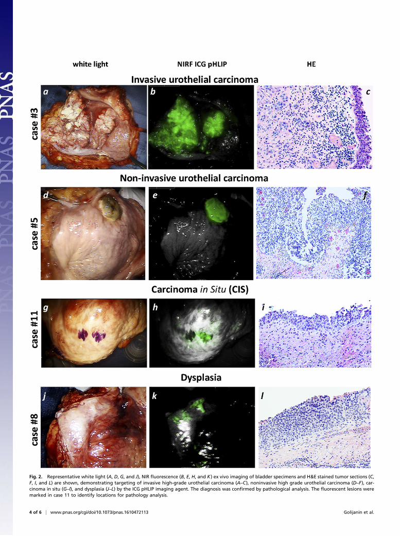
Fig. 2. Representative white light (A, D, G, and J), NIR fluorescence (B, E, H, and K) ex vivo imaging of bladder specimens and H&E stained tumor sections (C,F, I, and L) are shown, demonstrating targeting of invasive high-grade urothelial carcinoma (A–C), noninvasive high grade urothelial carcinoma (D–F), car-cinoma in situ (G–I), and dysplasia (J–L) by the ICG pHLIP imaging agent. The diagnosis was confirmed by pathological analysis. The fluorescent lesions weremarked in case 11 to identify locations for pathology analysis.
4 of 6 | www.pnas.org/cgi/doi/10.1073/pnas.1610472113 Golijanin et al.

demonstrate very promising results. Further, the ICG pHLIPconstruct is a generally applicable imaging agent, because ittargets a general property of the tumor microenvironment, tu-mor acidity. Once approved for clinical use, it could be testedand possibly used on a variety of cancers. We have shown tar-geting of primary tumors and metastatic lesions by fluorescentpHLIPs in more than 15 varieties of human, murine, and rattumors, including lymphoma, melanoma, pancreatic, breast, andprostate transgenic mouse models and human tissue (bladder,kidney, breast, and head/neck stained ex vivo).
Materials and MethodsConjugation of ICG with the pHLIP Peptide. pHLIP variant 3 (Var3) peptide witha single Cys residue at the N terminus, ACDDQNPWRAYLDLLFPTDTLLL-DLLWA, was synthesized and purified by reversed phase chromatography byCS Bio. The near infrared fluorescent dye, ICG maleimide (Intrace Medical),was conjugated to the pHLIP peptide at a ratio of 1:1 in dimethylformamide(DMF). The reaction progress was monitored by the reversed phase (ZorbaxSB-C18 columns, 9.4 × 250 mm, 5 μm; Agilent Technology) HPLCy (HPLC)using a gradient from 5% to 70% acetonitrile in water containing 0.05%trifluoroacetic acid. Also, for the negative control, ICG-malemide was con-jugated with the free amino acid, L-cysteine (Sigma). The concentration oflabeled peptide in buffer was determined by ICG absorption at 800 nm,e800 = 137,000 M−1·cm−1. The purity of the constructs was performed byanalytical HPLC and surface-enhanced laser desorption/ionization time-of-flight(SELDI-TOF) MS, and the amount of free dye in the solution was less than 1%.
Liposome Preparation. Large unilamellar vesicles were prepared by extrusion;2.5 mg POPC (1-palmitoyl-2-oleoyl-snglycero-3-phosphocholine; Avanti PolarLipids) lipids were dissolved in 0.5 mL chloroform, desolvated on a rotaryevaporator, and dried under high vacuum for 3 h. The phospholipid film wasthen rehydrated in pH 7.4 PBS containing 10 mM D-glucose, vortexed for5 min, and repeatedly extruded at least 15 times through a membrane witha 100-nm pore size.
Absorption and Fluorescence Measurements. Absorbance and fluorescencemeasurements were carried out on a Genesys 10S UV-Vis spectrophotometer(Thermo Scientific) and a SpectraMax M2 spectrofluorometer (MolecularDevices), respectively. The absorption spectra were measured in PBS, pH 7.4,containing 10 mM D-glucose from 600 to 850 nm. The fluorescence spectra of10 μM of ICG pHLIP peptide were measured from 810 to 850 nm at 790-nmexcitation wavelength in PBS, pH 7.4, containing 10 mM D-glucose, with orwithout 2 mM of POPC liposomes.
Ex Vivo Imaging of Bladder Specimens. After obtaining institutional reviewboard approval from Rhode Island Hospital, Lifespan, 22 urothelial carci-noma patients that were scheduled for radical cystectomy were selectedover a 12-mo period and appropriate informed consent was obtained. Afterradical cystectomy, bladder specimens were immediately removed and irri-gated three times for 5 min via catheter with nonbuffered saline and in-stilled and incubated with 80 mL of 8 μM or 32.8 μg/mL (unless otherwise isstated, see notes to Table 1) of ICG pHLIP construct or ICG-Cys in PBS, pH 7.4,containing 10 mM D-glucose for 60 min. Then, the unbound constructs wereremoved by rinsing with 80 mL of saline solution three to five times, thebladder was irrigated thoroughly with buffered saline and opened using a Yincision on the anterior wall. Using a da Vinci Si NIRF imaging system(FireflyR), ex vivo fluorescent and white light imaging of the entire bladderand its parts was performed. The fluorescent spots were marked and stan-dard pathological analysis was carried out to explore the correlation be-tween appearance of fluorescent signal and cancer lesions.
Pathological Analysis. The specimen was sectioned and submitted after 24-hfixation in 10% phosphate-buffered formalin according to the standardinstitutional grossing manual, with emphasis on the marked areas of thebladder. The sections were processed for routine histology into paraffin-embedded blocks. Five-micrometer-thick tissue sections were obtained andstained for H&E. Evaluation of pathology was performed by a genitourinarypathologist, and a standard report was prepared based on the AmericanJoint Committee on Cancer Cancer Staging Manual, seventh edition, 2010.
Statistical Analysis. Statistical parameters were calculated according to thefollowing equations:
TRP =TP
TP + FN; SPC =
TNTN+ FP
,
PPV =TP
TP + FP; NPV =
TNFN+ TN
,
FPR=FP
FP + TN; FNR=
FNTP + FN
,
FDR=FP
TP + FP; FOR=
FNFP + TN
,
where TP is the true positive; TN is the true negative; FP is the false positive;FN is the false negative; TRP is the true positive rate or sensitivity; SPC is thetrue negative rate or specificity; PPV is the positive predictive value or pre-cision; NPV is the negative predictive values; FPR is the false positive rate;
Table 4. Tabular results of the sensitivity/specificity test of ICGpHLIP peptide targeting of cancerous lesions in the humanbladder specimens: carcinoma vs. normal including necrotictissue and treatment effects
Receiver operator characteristicscarcinoma vs. normal + necrosis TP + FN FP + TN Sum
TP + FP TP, 28 FP, 5 33FN + TN FN, 1 TN, 20 21Sum 29 25
TP is the true positive; TN is the true negative; FP is the false positive; FN isthe false negative.
Table 3. Descriptive parameters
Measure Results
Sensitivity, TRP 0.966Specificity, SPC 1.000Positive predictive value, PPV 1.000Negative predictive values, NPV 0.950False positive rate, FPR 0.000False negative rate, FNR 0.034False discovery rate, FDR 0.000False omission rate, FOR 0.053
Table 2. Tabular results of the sensitivity/specificity test of ICGpHLIP peptide targeting of cancerous lesions in the humanbladder specimens: carcinoma vs. normal excluding necrotictissue and treatment effects
Receiver operator characteristicscarcinoma vs. normal TP + FN FP + TN Sum
TP + FP TP, 28 FP, 0 28FN + TN FN, 1 TN, 19 20Sum 29 19
TP is the true positive; TN is the true negative; FP is the false positive; FN isthe false negative.
Table 5. Descriptive parameters
Measure Results
Sensitivity, TRP 0.966Specificity, SPC 0.800Positive predictive value, PPV 0.848Negative predictive values, NPV 0.952False positive rate, FPR 0.020False negative rate, FNR 0.034False discovery rate, FDR 0.152False omission rate, FOR 0.040
Golijanin et al. PNAS Early Edition | 5 of 6
APP
LIED
BIOLO
GICAL
SCIENCE
S

FNR is the false negative rate; FDR is the false discovery rate; and FOR is thefalse omission rate.
ACKNOWLEDGMENTS. This work was supported by NIH Grant GM073857 (toD.M.E., O.A.A., and Y.K.R.).
1. Siegel R, Naishadham D, Jemal A (2012) Cancer statistics, 2012. CA Cancer J Clin 62(1):10–29.
2. Pasin E, Josephson DY, Mitra AP, Cote RJ, Stein JP (2008) Superficial bladder cancer:An update on etiology, molecular development, classification, and natural history.Rev Urol 10(1):31–43.
3. Anastasiadis A, de Reijke TM (2012) Best practice in the treatment of nonmuscle in-vasive bladder cancer. Ther Adv Urol 4(1):13–32.
4. Kamat AM, et al. (2014) Defining and treating the spectrum of intermediate risknonmuscle invasive bladder cancer. J Urol 192(2):305–315.
5. Mariotto AB, Yabroff KR, Shao Y, Feuer EJ, Brown ML (2011) Projections of the cost ofcancer care in the United States: 2010-2020. J Natl Cancer Inst 103(2):117–128.
6. Damaghi M, Wojtkowiak JW, Gillies RJ (2013) pH sensing and regulation in cancer.Front Physiol 4:370.
7. Gillies RJ, Verduzco D, Gatenby RA (2012) Evolutionary dynamics of carcinogenesisand why targeted therapy does not work. Nat Rev Cancer 12(7):487–493.
8. Estrella V, et al. (2013) Acidity generated by the tumor microenvironment drives localinvasion. Cancer Res 73(5):1524–1535.
9. Gatenby RA, Gawlinski ET, Gmitro AF, Kaylor B, Gillies RJ (2006) Acid-mediated tumorinvasion: A multidisciplinary study. Cancer Res 66(10):5216–5223.
10. Bailey KM, Wojtkowiak JW, Hashim AI, Gillies RJ (2012) Targeting the metabolicmicroenvironment of tumors. Adv Pharmacol 65:63–107.
11. Andreev OA, Engelman DM, Reshetnyak YK (2014) Targeting diseased tissues bypHLIP insertion at low cell surface pH. Front Physiol 5:97.
12. Weerakkody D, et al. (2013) Family of pH (low) insertion peptides for tumor target-ing. Proc Natl Acad Sci USA 110(15):5834–5839.
13. Reshetnyak YK, et al. (2011) Measuring tumor aggressiveness and targeting meta-static lesions with fluorescent pHLIP. Mol Imaging Biol 13(6):1146–1156.
14. Adochite RC, et al. (2014) Targeting breast tumors with pH (low) insertion peptides.Mol Pharm 11(8):2896–2905.
15. Cruz-Monserrate Z, et al. (2014) Targeting pancreatic ductal adenocarcinoma acidicmicroenvironment. Sci Rep 4:4410.
16. Luo Z, et al. (2014) Widefield optical imaging of changes in uptake of glucose andtissue extracellular pH in head and neck cancer. Cancer Prev Res (Phila) 7(10):1035–1044.
17. Luo Z, Tikekar RV, Samadzadeh KM, Nitin N (2012) Optical molecular imaging ap-proach for rapid assessment of response of individual cancer cells to chemotherapy.J Biomed Opt 17(10):106006.
18. Tobis S, et al. (2012) Robot-assisted and laparoscopic partial nephrectomy with nearinfrared fluorescence imaging. J Endourol 26(7):797–802.
19. Alander JT, et al. (2012) A review of indocyanine green fluorescent imaging in sur-gery. Int J Biomed Imaging 2012:940585.
20. Desmettre T, Devoisselle JM, Mordon S (2000) Fluorescence properties and metabolicfeatures of indocyanine green (ICG) as related to angiography. Surv Ophthalmol45(1):15–27.
21. Kozin SV, Shkarin P, Gerweck LE (2001) The cell transmembrane pH gradient in tu-mors enhances cytotoxicity of specific weak acid chemotherapeutics. Cancer Res61(12):4740–4743.
22. Althausen AF, Prout GR, Jr, Daly JJ (1976) Non-invasive papillary carcinoma of thebladder associated with carcinoma in situ. J Urol 116(5):575–580.
23. Smith G, et al. (1983) Prognostic significance of biopsy results of normal-lookingmucosa in cases of superficial bladder cancer. Br J Urol 55(6):665–669.
24. Zuk RJ, Rogers HS, Martin JE, Baithun SI (1988) Clinicopathological importance ofprimary dysplasia of bladder. J Clin Pathol 41(12):1277–1280.
6 of 6 | www.pnas.org/cgi/doi/10.1073/pnas.1610472113 Golijanin et al.