TAILORING BIOINFORMATICS STRATEGIES FOR THE ...
114
From DEPARTMENT OF CELL AND MOLECULAR BIOLOGY Karolinska Institutet, Stockholm, Sweden TAILORING BIOINFORMATICS STRATEGIES FOR THE CHARACTERIZATION OF THE HUMAN MICROBIOME IN HEALTH AND DISEASE Mauricio Barrientos Somarribas Stockholm 2019
Transcript of TAILORING BIOINFORMATICS STRATEGIES FOR THE ...

From DEPARTMENT OF CELL AND MOLECULAR BIOLOGY Karolinska Institutet, Stockholm, Sweden
TAILORING BIOINFORMATICS STRATEGIES FOR THE
CHARACTERIZATION OF THE HUMAN MICROBIOME IN HEALTH AND DISEASE
Mauricio Barrientos Somarribas
Stockholm 2019

All previously published papers were reproduced with permission from the publisher. Published by Karolinska Institutet. Printed by Eprint AB 2019 © Mauricio Barrientos Somarribas, 2019 ISBN 978-91-7831-508-6
TAILORING BIOINFORMATICS STRATEGIES FOR THE CHARACTERIZATION OF THE HUMAN MICROBIOME IN HEALTH AND DISEASE THESIS FOR DOCTORAL DEGREE (Ph.D.)
By
Mauricio Barrientos Somarribas
Principal Supervisor: Prof. Björn Andersson Karolinska Institutet Department of Cell and Molecular Biology Co-supervisor(s): Tobias Allander, Ph.D Karolinska Institutet Department of Microbiology, Tumor and Cell Biology Stefanie Präst-Nielsen, Ph.D Karolinska Institutet Department of Microbiology, Tumor and Cell Biology Center for Translational Microbiome Research
Opponent: Associate Professor Leo Lahti University of Turku Department of Mathematics and Statistics Examination Board: Associate Professor Tomas Bergström Swedish University of Agricultural Sciences Department of Animal Breeding and Genetics Associate Professor Anders Andersson Kungliga Tekniska Högskolan Department of Gene Technology Science for Life Laboratory Professor Martin Rottenberg Karolinska Institutet Department of Microbiology, Tumor and Cell Biology


To my mother, and everyone who helped me get here

ABSTRACT The human microbiome is a very active area of research due to its potential to explain
health and disease. Advances in high throughput DNA sequencing in the last decade have
catalyzed the growth of microbiome research; DNA sequencing allows for a cost-effective
method to characterize entire microbial communities directly, including unculturable
microbes which were previously difficult to study. 16S rRNA sequencing and shotgun
metagenomics, coupled with bioinformatics methods have powered the characterization of
the human microbiome in different parts of the body. This has led to the discovery of novel
links between the microbiome and diseases such as allergies, cancer, and autoimmune
diseases.
This thesis focuses on the application of both 16S rRNA sequencing and shotgun
metagenomics for the characterization of the human microbiome and its relationship with
health and disease. We established two methodologies to address these questions. The first
methodology is a bench-to-bioinformatics pipeline to discover putative viral pathogens
involved in disease using shotgun metagenomics technology. In paper I, we apply the
proposed pipeline to explore the hypothesis of viral infection as a putative cause of
childhood Acute Lymphoblastic Leukemia. In paper II, we propose a complementary
method to the pipeline to improve the detection of unknown viruses, especially those with
little or no homology to currently known viruses. We applied this method on a collection of
viral-enriched libraries which resulted in the characterization of a new viral-like genome.
The second methodology was developed to explore and generate hypothesis from a human
skin microbiome dataset of Psoriasis and Atopic Dermatitis patients. The results of the
analysis are presented in Paper III and Paper IV. Paper III is a pure data-driven exploration
of the dataset to discover different aspects on how the microbiome is linked to both
diseases. Paper IV follows up from the results of paper III but focuses on characterizing
the skin site microbiome variability in Atopic Dermatitis.

LIST OF SCIENTIFIC PAPERS I. Bogdanovic G, Pou C, Barrientos-Somarribas M, Bjerkner A, Honkaniemi
E, Allander T, et al. Virome characterisation from Guthrie cards in children who later developed acute lymphoblastic leukaemia. Br J Cancer. 2016 Oct 11;115(8):1008–14.
II. Barrientos-Somarribas M*, Messina DN*, Pou C, Lysholm F, Bjerkner A, Allander T, et al. Discovering viral genomes in human metagenomic data by predicting unknown protein families. Sci Rep. 2018;
III. Fyhrquist, N., Muirhead, G., Prast-Nielsen, S., Jeanmougin, M., Olah, P., Skoog, T., Jules-Clement, G., Feld, M., Barrientos-Somarribas, M., Pennino, D., Suomela, S., Tessas, I., Lybeck, E., Baran, A.M., Darban, H., Gangwar, R.S., Gerstel, U., Jahn, K., Karisola, P., Yan, L., Hansmann, B., Katayama, S., Meller, S., Bylesjö, M., Hupé, P., Levi-Schaffer, F., Greco, D., Ranki, A., Schröder, J.M., Barker, J., Kere, J., Tsoka, S., Lauerma, A., Soumelis, V., Nestle, F.O., Homey, B., Andersson, B., Alenius, H. Microbe-host interplay in atopic dermatitis and psoriasis. Unpublished manuscript. 2019.
IV. Barrientos-Somarribas M*, Ottman N*, MAARS Consortium, Andersson B, Alenius H. Microbial and transcriptional differences elucidate atopic dermatitis heterogeneity across skin sites. Unpublished manuscript. 2019

OTHER PUBLICATIONS V. Pou C, Barrientos-Somarribas M, Marin-Juan S, Bogdanovic G, Bjerkner
A, Allander T, et al. Virome definition in cerebrospinal fluid of patients with neurological complications after hematopoietic stem cell transplantation. J Clin Virol. 2018 Nov;108:112–20.

CONTENTS 1 Introduction .................................................................................................................... 1
1.1 The human microbiome ....................................................................................... 1 1.2 An ecological framework to study the microbiome ............................................ 3 1.3 Characterizing the microbiome using DNA sequencing: a conceptual
overview ............................................................................................................... 5 1.4 Shotgun Metagenomics ........................................................................................ 7
1.4.1 Data generation ........................................................................................ 7 1.4.2 Read-based microbiome reconstruction .................................................. 9 1.4.3 Assembly-based microbiome reconstruction ........................................ 14
1.5 Amplicon Sequencing: A cost-effective alternative .......................................... 16 1.5.1 Data generation ...................................................................................... 16 1.5.2 OTU-based profiling .............................................................................. 18 1.5.3 ASV-based profiling .............................................................................. 20
1.6 Tools and techniques to summarize and derive biological insights from the microbiome .................................................................................................. 21 1.6.1 Data distribution .................................................................................... 21 1.6.2 Global analysis – Introduction ............................................................... 23 1.6.3 Global analysis – Alpha diversity .......................................................... 23 1.6.4 Global Analysis - Beta diversity ............................................................ 24 1.6.5 Feature-level analysis ............................................................................ 26 1.6.6 Microbe-microbe interactions ............................................................... 28 1.6.7 Data Integration ..................................................................................... 29
1.7 Challenges in human microbiome characterization .......................................... 30 1.7.1 Virome characterization ......................................................................... 31 1.7.2 The Skin microbiome ............................................................................ 33
2 Aims ............................................................................................................................. 37 3 Results and discussion ................................................................................................. 39
3.1 Papers I & II: Viral discovery ............................................................................ 39 3.1.1 Paper I: Inspecting the roles of viral infections in the pathogenesis
of Acute Lymphoblastic Leukemia ....................................................... 42 3.1.2 Paper II: ORFan protein prediction in viral metagenomics datasets .... 43 3.1.3 Conclusions ............................................................................................ 45
3.2 Papers III & IV: Associating the human skin microbiome to disease .............. 47 3.2.1 Community-level analysis ..................................................................... 48 3.2.2 Population-level analysis ....................................................................... 52 3.2.3 Data integration and functional analysis ............................................... 57 3.2.4 Conclusions ............................................................................................ 58
4 Concluding remarks and future perspectives .............................................................. 61 5 Acknowledgements ..................................................................................................... 63 6 References .................................................................................................................... 69

LIST OF ABBREVIATIONS 16S 16S subunit rRNA gene
AD Atopic Dermatitis / Eczema
ALL Acute Lymphoblastic Leukemia
ASV Amplicon Sequence Variant
CSF Cerebrospinal Fluid
DNA Deoxyribonucleic Acid
EM Expectation Maximization
ESV Exact Sequence Varians
GLM Generalized Linear Model
HMP Human Microbiome Project
ITS Internal Transcribed Spacer
LCA Lowest Common Ancestor
MAE Mean Absolute Error
MAP Maximum a posteriori
MDS Multidimensional Scaling
MSA Multiple Sequence Alignment
OTU Operational Taxonomic Unit
PacBio Pacific Biosciences
PASI Psoriasis Area Severity Index
PCA Principal Component Analysis
PCoA Principal Coordinate Analysis
PSO Psoriasis
RDP Ribosomal Database Project
RNA Ribonucleic Acid

SCORAD SCORing Atopic Dermatitis
SISPA Sequence-Independent, Single Primer Amplification
SNV Single Nucleotide Variation
ssDNA / ssRNA Single stranded DNA or RNA
UMAP Uniform Manifold Approximation and Projection
VLP Viral-like particle

1
1 INTRODUCTION
1.1 The human microbiome
Nearly four decades ago, the bacterium H. pylori was isolated from the stomach, debunking
the common belief that the stomach is a sterile environment. We have now realized that the
human body is host to a vast range of bacteria, fungi, viruses and other eukaryotes that
interact with our organism. The existence of microbial communities in places such as the
gut, the oral cavities or the skin have been well documented for many years now, but recent
studies continue to find microbes residing in unexpected places (Dickson and Huffnagle
2015). For example, the lung and the eye have been recently found to contain microbes
(Hilty et al. 2010; O’Dwyer, Dickson, and Moore 2016; Huffnagle, Dickson, and Lukacs
2017; St. Leger et al. 2017; Shin et al. 2016; Cavuoto et al. 2018). Other studies also
suggest that microbes live in the placenta (Stout et al. 2013), and evidence from
immunosuppressed individuals suggests the existence of viral communities in the blood (L.
Li et al. 2013; Popgeorgiev et al. 2013).
We refer to the collection of microbial communities colonizing the different sites in the
human body as ‘the human microbiome1’ (E. A. Grice and Segre 2012). These communities
are dynamical entities (Gonze et al. 2018; Faust et al. 2015); the composition of any
microbial community in the human body will depend on the physiological conditions (e.g.
temperature, pH, oxygen), resource availability, host-microbe interactions (Gilbert et al.
2018; Virgin 2014) and the interactions within the community (Fredricks 2001; J. Xu
2006). Considering the wide variability of environments in the human body and across
individuals, it is not unexpected that the variability of the human microbiome is huge,
among individuals and between sites. A recent study even suggests microbiota is so unique
that it could identify individuals (Franzosa et al. 2015), although host genetics appear not to
be a strong determinant of the microbiome (Rothschild et al. 2018).
The microbiome plays an important role in maintaining human homeostasis, contributing to
metabolism(LeBlanc et al. 2013; Metges 2000; Flint et al. 2012), training of the immune
system and modulation of the immune response (E. A. Grice and Segre 2012; Ursell et al.
2012; Naik et al. 2012). Consequently, alterations to the healthy resident microbial
1 In this thesis, the term ‘microbiome’ will be used as defined by Ledeberg & McCray(2001): “the ecological community of commensal, symbiotic, and pathogenic microorganisms that literally share our body space and have been all but ignored as determinants of health and disease”. However, it is noted that in the literature the term ‘microbiome’ can also be defined as ‘the collection of genomes from the aforementioned organisms’.

2
communities can have a considerable impact on health. These imbalanced states are termed
‘dysbiosis’ (Petersen and Round 2014) and they have been linked to a wide range of
diseases such as inflammatory bowel disease (Frank et al. 2007; Norman et al. 2015),
asthma (Hilty et al. 2010), atopic dermatitis (Oh et al. 2013; Kong, Oh, Deming, Conlan,
Grice, Beatson, Nomicos, Polley, Komarow, Murray, et al. 2012), and depression (Foster
and McVey Neufeld 2013).
Understanding the role of the microbiome in disease holds the potential for developing new
diagnostic, therapeutic or preventive tools. Examples of this include the fecal transplants
for the treatment of C. difficile infection (Aas, Gessert, and Bakken 2003) and the
development of probiotics (Khalesi et al. 2019). However, further research is required to
elucidate the complexity of the microbiome and to translate these insights gained from the
research into clinical practice.

3
1.2 An ecological framework to study the microbiome
To tackle the complexity of the microbiome and its effect on health and disease, the field
has borrowed a conceptual framework from ecology. Biological complexity is partitioned
into stratified levels of organization: organism, population, community, ecosystem, biome,
and biosphere. However, for the study of the human microbiome, we will only consider the
population, community and ecosystem levels (J. Xu 2006).
At the population level, each species is studied independently from the community. The
goal is to understand a microbial species in isolation and characterize its functions (e.g.
metabolism, replication strategies, virulence, cell biology), patterns of evolution and
variation(J. Xu 2006). Studying the behavior of individual microbial populations forms the
basis to understand how a species will interact with the other members of the community
and with the human body in different environments.
At the community level, the aim is to investigate the composition, behavior and the spatio-
temporal dynamics of microbial populations that share a common environment (Konopka
2009). Challenges at this level include determining the species and strain composition and
profiling the functional and metabolic content of the entire community and the contribution
of each population (Kuczynski et al. 2011).
Another key aim when examining microbial communities is to determine the type and
mechanisms behind microbe-microbe interactions and their spatial distribution (Mah and
O’Toole 2001). Microbial interactions can be classified into 5 broad categories:
1) mutualistic, when both organisms benefit from the interaction 2) commensal, when one
organism benefits from the interaction and the other one remains unaffected,
3) competitive, when one organism will kill or inhibit the growth of another that requires
the same resources; 4) parasitic, when one organism benefits while harming its host, and
5) predatory, when one organism kills another one as part of its life cycle(Lang and
Benbow 2013).
At the ecosystem level, we examine how both individual populations and the entire
community interact with the environment: the human body. The focus of study at this level
include understanding how communities adapt to changes in the environmental conditions
(e.g. nutrients, oxygen, pH) and describing the mechanisms and effects of the crosstalk
between the microbes, the immune system and surrounding cells. Understanding human
health and disease can only happen when the ecosystem is considered (Gilbert et al. 2018).

4
This introduction describes different techniques to extract population and community level
information from microbial communities using DNA sequencing technologies and how to
associate them with health and disease. The following sections (1.3–1.5) describe how
sequencing can be used for characterizing the microbiome and two protocols to achieve
this. Section 1.6 describes statistical techniques and algorithms to summarize population
and community-level information and find ecosystem-level insights through associations
with human information using clinical metadata or other omics datasets. Finally, section 1.7
outlines two challenges in human microbiome research that will be addressed by the work
in this thesis.

5
1.3 Characterizing the microbiome using DNA sequencing: a conceptual overview
Microbiome studies depend on our ability to accurately profile microbial communities. One
way of achieving this goal is to study their metagenome: the collection of genomes from all
microbes in the community. The information from the metagenome enables the
reconstruction of the microbial species profile from all kingdoms, including bacteria,
viruses, archaea and other microscopic eukaryotes. Furthermore, because we are sampling
complete genomes, we can infer the functional potential of communities and individual
populations. Another advantage of metagenomics (i.e. the study of metagenomes), is that it
allows the study of communities without isolating and culturing the organisms (K. Chen
and Pachter 2005).
The concept of an ideal human metagenomics pipeline is presented in Figure 1.3.1. The
data generation follows a single-cell-like protocol, that can be broken down into three steps:
sample processing, nucleic acid extraction and sequencing. The sample processing step
consists of isolating all microbial cells and viral particles from any type of human sample
(e.g. a skin biopsy, stool, or blood). An ideal sample processing step discards human cells
and other molecules since these comprise a sizeable fraction of the original sample but
contain no microbiome information. Purified microbes are then subjected to nucleic acid
extraction, which isolates the genomic content from each microbe and labels them with a
unique barcode that identifies genomic fragments from the same microbial cell or virus.
Then, the barcoded genomic fragments are sequenced, generating error-free digital
representations of the nucleotide composition of each genome.
The resulting sequenced genomes can be used to extract biological information from the
microbial community. Typically, the information we are interested to extract from the
metagenomes falls into three categories (Knight et al. 2018):
• Taxonomical abundance profile: A list of species present in the sample coupled with a
measure of abundance for each species
• Strain-level population information: For microbial populations of interest, inferred
strain variation based on genotypes (SNVs) or gene content.
• Functional profile: The coding potential of the entire community, as well as the
coding content of the different populations. This include gene, gene family and
pathway abundances

6
The biological information can be used to extract insights depending on the question under
study. For example, we can use the taxonomical profile to determine whether certain
populations expand or contract in relation to a clinical condition. We can also examine
population-level variability within the different microbial populations to determine whether
strains with different functional profiles are associated with a phenotype of interest. Finally,
we can also examine the functional potential of the metagenome, both at the global level to
understand what the community is able to synthesize and react to as a whole, and at a per-
clade level to predict mechanisms though which key populations partake in specific
processes.
Unfortunately, the state of technology is far away from this proposed scenario. Sections 1.4
and 1.5 describe the two main protocols for human microbiome profiling using DNA
sequencing technologies: shotgun metagenomics and amplicon sequencing respectively. It
is worth noting that other techniques exist, such as metatranscriptomics, but they will not be
covered in the scope of this document.
Figure 1.3.1 - An ideal metagenomics assay

7
1.4 Shotgun Metagenomics
Shotgun metagenomics is the real-life protocol to obtain metagenomes from a sample. The
first pioneering metagenomics studies surveyed the microbial diversity of the oceans
(Rusch et al. 2007) and it has since been used to characterize soil (Howe et al. 2014),
wastewater (Munck et al. 2015) and even the New York City metro (Afshinnekoo et al.
2015). The human microbiome field has also caught up, with the Human Microbiome
Project utilizing shotgun metagenomics for the latter part of the project (Wylie et al. 2012).
Now it is a common choice for the development of large studies such as MetaHIT
consortium (Qin et al. 2010a).
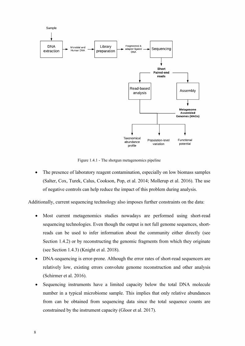
Figure 1.4.1 outlines a typical shotgun metagenomics pipeline. The following subsections
will describe the steps of the process. Section 1.4.1 describes the sequencing data
generation steps. Then Sections 1.4.2 and 1.4.3 describe two alternative but complementary
ways of inferring biological information from the sequences: read-based analysis and
metagenomic assembly.
1.4.1 Data generation
The sample processing steps from the shotgun metagenomics pipeline differ considerably
from those proposed in the conceptual pipeline due to technological limitations. Compared
to the conceptual pipeline, there is no purifying step that isolates and sorts the microbial
cells and viral particles, so the nucleic acid extraction step is performed on the entire
sample. The isolated DNA is then fragmented, amplified and adapters are ligated for
sequencing. This has important implications for the downstream data processing such as:
• The nucleic acid extraction efficiency for each type of microbe will differ depending
on the method, so the extracted genomic DNA concentrations will be biased with
respect to the microbial abundances (Kong et al. 2017).
• If the sample contains human cells, the extracted DNA will be a mixture of human
and microbial DNA. This can impact the sensitivity of the assay if the human-to-
microbial DNA ratio is very high since human fragments will waste part of the
sequencing capacity (Pereira-Marques et al. 2019).
• All DNA is extracted in bulk, meaning it is not possible to track which DNA
fragments come from a particular microbial cell or viral particle. This has to be
inferred computationally downstream.
8
Figure 1.4.1 - The shotgun metagenomics pipeline
• The presence of laboratory reagent contamination, especially on low biomass samples
(Salter, Cox, Turek, Calus, Cookson, Pop, et al. 2014; Mollerup et al. 2016). The use
of negative controls can help reduce the impact of this problem during analysis.
Additionally, current sequencing technology also imposes further constraints on the data:
• Most current metagenomics studies nowadays are performed using short-read
sequencing technologies. Even though the output is not full genome sequences, short-
reads can be used to infer information about the community either directly (see
Section 1.4.2) or by reconstructing the genomic fragments from which they originate
(see Section 1.4.3) (Knight et al. 2018).
• DNA-sequencing is error-prone. Although the error rates of short-read sequencers are
relatively low, existing errors convolute genome reconstruction and other analysis
(Schirmer et al. 2016).
• Sequencing instruments have a limited capacity below the total DNA molecule
number in a typical microbiome sample. This implies that only relative abundances
from can be obtained from sequencing data since the total sequence counts are
constrained by the instrument capacity (Gloor et al. 2017).
9
• High complexity communities require deep sequencing to survey the low abundance
members (Rodriguez-R and Konstantinidis 2014; Cleary et al. 2015)
The resulting sequencing dataset will consist of millions of short sequences. Most
metagenomic sequencing is performed with Illumina technology due to economic reasons,
although other alternatives such as Ion Torrent or long-read technologies like Pacific
Biosciences and Oxford Nanopore exist. This means the output sequences will be paired-
ended (a fragment is sequenced from 5’ to 3’ and also from 3’ to 5’) and read length will
range from 100 to 300 bp depending on the sequencing instrument and kit.
1.4.2 Read-based microbiome reconstruction
One way of reconstructing a microbial community from shotgun metagenomics data is to
leverage the information about existing microbial genomes in public databases to infer what
is contained in the metagenome without explicitly reconstructing the genomes. These
approaches are commonly known as ‘read-based’ or ‘reference-based’. Generally speaking,
read-based analyses yield good results when the microbial diversity of the samples is
relatively well known (Knight et al. 2018), but the final results can be heavily influenced by
the choice of database (Shaiber and Eren 2019; Breitwieser et al. 2019).
Read-based tools can recover different types of microbial information from the reads. The
following subsections describe different types of tools categorized by the microbial
information they recover.
1.4.2.1 Taxonomic profile inference
Taxonomic profile inference tools aim to identify all taxa present in the sample and
estimate the abundance of each taxon. These tools can be broadly classified into three
categories depending on the strategy they use:
• Mapping-based with a strategy to deal with multimapped reads • Mapping-based with Lowest Common Ancestry • Marker gene-based
The first approach is analogous to the transcriptome quantification problem. The problem
can be framed as estimating the abundance of a set of reference genomes in a given dataset.
The general concept consists of mapping the reads to the reference genomes and analyzing
the mapping results to estimate the abundance of each species in the set. One of the main
challenges with the abundance estimation step is to select a model to incorporate the

10
information from multi-mapped reads (reads that are assigned to more than one species) to
obtain accurate abundance measurements. This approach is very reliable if the genomes of
the community members are well characterized. However, if unknown species are present
in the samples, reads from these unknown species can be misclassified and skew the
estimated profiles.
Tools like Pathoscope (Francis et al. 2013) originally implemented this concept, analyzing
the output of traditional mapping tools such as bowtie or bwa and using the Expectation-
Maximization (EM) algorithm to estimate the abundances from all mapped reads. Newer
tools optimize the mapping step by determining if the read is compatible with the reference,
instead of calculating the full alignment. Examples of this include Centrifuge (Kim et al.
2016), which uses an FM-index based genome index for assignment and then implements a
Cufflinks-like EM approach for multimapping read assignment. Pseudo-mapping tools for
RNA-seq such as Salmon (Patro et al. 2017) and Kallisto (N. L. Bray et al. 2016)
incorporate their own models to disambiguate multi-compatible reads in the quantification,
as well as bias estimation. However, due to the large size of the microbial genome
databases compared to a human or mouse transcriptome, RNA-seq tools must be adapted
for metagenomics use, e.g FastViromeExplorer (Tithi et al. 2018), or metakallisto
(Schaeffer et al. 2017). Minhash-based strategies for metagenome profiling could also be
classified in this category (C. T. Brown and Irber 2016; Ondov et al. 2016).
The second approach originates from addressing one of the main causes of read
multimapping in microbial datasets: homologous regions. The lowest common ancestor
(LCA) strategy states that if a sequence originates from a conserved region in different
bacteria, the appropriate classification for the sequence is the taxonomical level where the
region is conserved. For example, if a read maps to a conserved region of the 16S rRNA
gene, it should be classified as ‘Kingdom: Bacteria’, whereas a read from a gene conserved
only in Staphylococcal species should be classified as ‘Genus: Staphylococcus’. In practice,
LCA approaches analyze all the database matches for each read and use the taxonomic
lineage information to determine what is the lowest node in the taxonomical tree that spans
all matches.
The first LCA tools like MEGAN (Huson et al. 2007) analyze blast search results against
nucleotide or protein databases such as NCBI nt or nr, using the NCBI taxonomy to place
reads at the appropriate taxonomic level. However, blast searches are computationally
expensive, and scaling up to millions of sequences becomes prohibitive. Kraken (Wood and
11
Salzberg 2014) pioneered an efficient k-mer based algorithm to implement the LCA.
Kraken’s algorithm depends on building a database of informative k-mers from a set of
reference genomes, where each k-mer is annotated with the LCA of the reference genomes
where it was observed. Then, the database is used to classify reads by annotating each k-
mer of the read and performing an LCA of the k-mer hits to determine the final annotation.
LCA-based methods have both advantages and disadvantages. In principle, these methods
are more robust than other mapping approaches when classifying sequences from genomes
that are not present in the database. However, in practice, the accuracy of most tools suffers
when considering classifications below order (Vollmers, Wiegand, and Kaster 2017;
Lindgreen, Adair, and Gardner 2015). Also, many LCA based tools do not include an
abundance estimation model for the different clades (Schaeffer et al. 2017), so they need to
be complemented with tools like Bracken for accurate abundance estimation (Lu et al.
2017). Finally, modern LCA approaches such as Kraken2 and Clark-S (Ounit and Lonardi
2016) are extremely fast classifiers, but the database construction can be very resource-
consuming.
The last strategy for classification is to use marker genes for profiling. There are two types
of marker genes: universal genes, and ‘clade-specific’ marker genes. Universal marker
genes, such as the 16S rRNA gene, can be used for taxonomical classification if the gene is
conserved in the species of interest and contains sufficient variability to distinguish clades.
A tool that implements this approach is mOTU2 (Milanese et al. 2019), and it uses a set of
40 marker genes for profiling.
On the other hand, “clade-specific” marker genes are present in members of a clade but
absent or too divergent in any other clades. This approach was pioneered by MetaPhlAn
(Segata et al. 2012) and more currently MetaPhlAn2 (Truong et al. 2015). Using the marker
database, clade abundance profiles can be estimated from read-mappings to the marker
gene database, since reads are in general expected to map ‘uniquely’ to markers. For
example, the Metaphlan2 gene database was built by first identifying core genes from all
clades at all taxonomic levels. Then, the suitability of each candidate marker gene was
evaluated by establishing a sequence identity threshold to check if the sequence is divergent
enough from all other potential markers.
Marker-gene based strategies address many limitations from other strategies. Marker gene
databases are considerably smaller than a full genome database, and it streamlines the
abundance estimation by removing the problem of multimapped reads. With appropriate

12
markers, the approach can be highly specific but sacrificing sensitivity (Vollmers,
Wiegand, and Kaster 2017), since only a fraction of the metagenomic data will be used for
the classification and the profile will be tied to the marker selection.
1.4.2.2 Strain-level population inference
Strain-level population analysis aims to recover strain variation from specific populations in
metagenomics samples. Determining strain profiles requires fine-grained analysis, so strain-
calling tools typically work on a per-species basis. The available tools use two main
strategies to infer strain information: analyze gene content or extract single nucleotide
variation(SNV). Ideally, these tools should be able to deconvolute the mixture of strains,
but in practice, most of them focus on characterizing the most abundant strain or computing
some form of a representative strain profile.
Tools that focus on gene content for strain profiling, such as PanPhlAn (Scholz et al. 2016),
are based on the concept of a species pangenome: the complete set of genes present in any
strain of a species. If the pangenome of a species is well known, it is possible to determine
whether different samples contain different strains by examining the gene presence/absence
profiles. The main advantage of this approach is that pangenome profiles enable direct
functional interpretations: the presence or absence of genes with known function can be
directly associated with any phenotype of interest.
On the other hand, SNV-based strain profilers aim to identify informative SNVs that can be
used to distinguish between the strain content of the samples. Some tools like StrainEST
(Albanese and Donati 2017) use a precomputed SNVs database collected from reference
genomes, while other tools like metaSNV (Costea et al. 2017) and StrainPhlAn (Truong et
al. 2017) call the SNVs directly from the sequencing dataset by mapping to a reference
database (metaSNV maps to genomes, while StrainPhlAn uses the MetaPhlAn2 marker
gene database).
Strain-profiling tools are sensitive to sequencing coverage requirements. For example,
PanPhlAn requires a ‘nearly uniform coverage’ across the median number of genes in a
strain to detect a strain in a sample, and imposes further coverage requirements for a gene
to be called as ‘present’ (Scholz et al. 2016). This makes them applicable for high and
medium abundance microbes, but not so effective for low abundance bacteria.
13
1.4.2.3 Functional profile inference
Functional analysis is concerned with the prediction of the coding potential present in the
metagenome. In metagenome analysis, we are interested in identifying the functional
potential of the metagenome as a whole, as well as understanding the coding potential of
individual taxa. Read-based functional profilers mainly focus on characterizing the global
functional potential by quantifying gene families and pathways from the reads. Associating
genes with taxa using reads can be very challenging since the information in short
sequences is often insufficient to distinguish between homologs.
Read-based functional analysis depends on one or more databases of gene and protein
sequences to annotate the reads. Database searches can be performed at the nucleotide level
or using translated searches using blastx or similar tools like Diamond (Buchfink, Xie, and
Huson 2015) or RapSearch2 (Zhao, Tang, and Ye 2012). Once the mappings have been
calculated, gene family level abundances are calculated by aggregating mapping to
sequences in the databases using gene family annotations. Commonly used gene family
annotations come from databases such as COG (Galperin et al. 2015), EggNOG (Huerta-
Cepas et al. 2019) or KEGG (Kanehisa et al. 2016). Alternatively, it is also possible to map
the reads to protein families directly with the HMMER suite (Mistry et al. 2013b) using
databases such as Pfam (Finn et al. 2014) or TIGRfam (Haft, Selengut, and White 2003).
Finally, pathway coverage and abundance can be calculated from the gene family
abundances, using databases such as MetaCyc (Caspi et al. 2018) or KEGG.
In contrast with taxonomical annotation tools, there are fewer tools for read-based
functional profile inference. HUMAnN2 (Abubucker et al. 2012), ShotMAP (Nayfach et al.
2015) and Fun4me (Sharifi and Ye 2017) are some examples of the available command
line- based pipelines for functional characterization. Some online services exist as well,
such as MG-RAST (Keegan, Glass, and Meyer 2016), IMG/M (I.-M. A. Chen et al. 2017)
and EBI metagenomics (Hunter et al. 2014).
14
1.4.3 Assembly-based microbiome reconstruction
An alternative approach to read-based microbiome profiling methods consists of
reconstructing the genomes present in the samples using sequence assembly. Metagenomics
assembly can be considered a special case of the genome assembly problem, with the added
complexity that different genomes are mixed in the same sample and the uneven fragment
coverage due to differences in microbial abundances.
Different metagenomics assemblers have been published in recent years, most of them
based on de Bruijn graphs. Common cited tools include Megahit (D. Li et al. 2015),
SPAdes (Bankevich et al. 2012) or MetaSPAdes (Nurk et al. 2016), IDBA-UD (Peng et al.
2012) and Ray Meta (Boisvert et al. 2012). Although the choice of assembly tool is not
trivial, the Megahit assembler is often recommended as a starting point based on assembly
quality, resource usage and speed compared to other assemblers (Sczyrba et al. 2017;
Ayling, Clark, and Leggett 2019).
Assembly strategies will differ depending on the number of samples, the sequencing depth,
and community complexity (Ghurye, Cepeda-Espinoza, and Pop 2016). Ideally, samples
should be reconstructed individually to maximize the capture of sample-specific variation.
However, to improve the recovery of low-abundance microbes, samples can also be co-
assembled together at the expense of disregarding individual sample variation. Assembly
quality can be assessed by checking statistics such as the contig length distribution,
checking contig read coverage, and measuring the percentage of read incorporation of the
assembly. Reconstructed sequences are typically called ‘contigs’.
The next step after assembly is binning: the aim is to group (or ‘bin’) contigs that belong to
the same genome. Available de novo binning tools rely on two main strategies for grouping
contigs: sequence composition or coverage. Sequence composition-based binning is derived
from the observation that the tetramer composition of fragments of the same genome will
be similar. In contrast, coverage-based binning is based on the assumption that the coverage
of contigs from the same genome will co-vary among different samples. State-of-the-art
binning tools such as CONCOCT (Alneberg et al. 2014), Metabat2 (Kang et al. 2015) and
MaxBin 2.0 (Wu, Simmons, and Singer 2016) use a combination of both strategies.
Recently, a meta-binner called Das-Tool (Sieber et al. 2018) was released, to combines the
results from different binners. Alternatively, genome bins can be inferred by taxonomically
annotating each contig and grouping contigs with the same taxonomical annotation
(Sczyrba et al. 2017).

15
Bins can be evaluated using different tools to determine the quality of the recovered
genomes. Tools like checkM (Parks et al. 2014) or metaQUAST (Mikheenko, Saveliev, and
Gurevich 2016) can be used for this purpose. Common metrics include contamination,
genome completeness, and purity. Bins can be manually refined with the help of annotation
and tools like Anvi’o (Eren et al. 2015). Finally, bins above certain quality criteria can be
labeled taxonomically using tools like Kraken or MEGAN(Wood and Salzberg 2014;
Huson et al. 2007), phylogenetically using PhyloPhlAn (Segata et al. 2013) and run through
gene prediction and annotation pipelines like MetaGeneMark or Prokka. (Rho, Tang, and
Ye 2010; Zhu, Lomsadze, and Borodovsky 2010; Seemann 2014).
Finally, the new genomes and their annotation can be used to infer the taxonomical
abundance profiles of each sample and perform functional analysis both at global and per-
genome level, using read-based tools or ad-hoc strategies similar to the ones implemented
in read-based analysis tools.

16
1.5 Amplicon Sequencing: A cost-effective alternative
An alternative for microbiome profiling is marker gene or amplicon sequencing, also
known as metabarcoding. Unlike shotgun metagenomics, the approach targets conserved
genes in the genomes of microbes which can be used to infer the taxonomic and
phylogenetic structure of the microbial community in a sample. In practice, most human
microbiome studies use the 16S ribosomal RNA subunit (16S rRNA) gene to characterize
bacteria and some types of archaea, since the gene is well conserved and with sufficient
variation to distinguish between subclades (Olsen et al. 1986; Hugenholtz 2002). In this
thesis, I will refer to amplicon sequencing to mean 16S rRNA gene amplicon sequencing
unless noted otherwise. However, the same strategy can be extended to characterize
archaeal (Gantner et al. 2011; Chaban and Hill 2012) and fungal populations (Lindahl et al.
2013) using different conserved genes.
The amplicon sequencing approach was instrumental for the first large scale analysis of
human bacterial communities (E. a. Grice et al. 2009; Costello et al. 2009; Ravel et al.
2011) and for the establishment and execution of the Human Microbiome Project(HMP), a
systematic effort to map the diversity of the microbiota in the body (The Human
Microbiome Project Consortium 2012).
A typical 16S amplicon sequencing pipeline is described in Figure 1.5.1. The following
subsections describe the data generation and community profiling processes.
1.5.1 Data generation
The first step for amplicon sequencing is DNA extraction, similar to shotgun
metagenomics. In the library preparation step, a PCR is used to enrich for the desired
conserved regions and sequencing adapters are ligated to the fragments. Then, the
amplicons are forwarded for sequencing.
The 16S rRNA gene is around 1500bp in bacteria and contains 9 hypervariable regions that
can be targeted for phylogenetic analysis. Due to the constraints of short-read sequencing
length, a selection of hypervariable regions is required. This selection must be performed
carefully, since the resolution of a variable regions to distinguish members of certain clades
vary between variable regions and this impacts directly the downstream interpretation of
results (Graspeuntner et al. 2018; Yang, Wang, and Qian 2016; Teng et al. 2018).

17
Figure 1.5.1 - 16S rRNA amplicon sequencing pipeline
16S rRNA-based microbiome profiling is a cost-effective method for microbiome studies; it
avoids many of the pitfalls from shotgun metagenomics while enabling taxonomic
profiling. The maturity of the technology has even led to its consideration for clinical
diagnostics (Almonacid et al. 2016). For example, human DNA does not waste any
sequencing since the 16S gene is not conserved in humans. Additionally, sequencing depth
requirements are much lower since we are not attempting to reconstruct genomes, and thus
lower abundance species are easier to detect. Bioinformatics analysis of 16S rRNA
sequences is also more standardized compared to shotgun metagenomics (Hillmann et al.
2018).
The main drawback of the 16S rRNA amplicon sequencing approach is that it only yields
taxonomical information, and species and subspecies (strain) resolution can be difficult to
attain (Knight et al. 2018). Another issue with this approach is the dependence on PCR. It
has been shown that the choice of primers and PCR cycles will bias the compositional
estimation, as some primers will favor the amplification of certain clades (Eloe-Fadrosh et
al. 2016). Additionally, PCR amplification carries the risk of chimera formation, which
must be addressed during the analysis steps (Haas et al. 2011). Finally, while the approach
18
can be extended for archaea and fungi, it cannot be extended for universal virome
inspection due to the low level of gene conservation among viruses (Kuczynski et al. 2011).
1.5.2 OTU-based profiling
One way of describing the taxonomical profile of a microbiome sample based on amplicon
data is to create Operational Taxonomic Units or OTUs. OTUs are clusters of amplicon
sequences above a certain similarity threshold and represent the lowest-level “unit” of
analysis.
Amplicon sequencing data processing starts with the quality control and filtering of the
reads, removing low-quality bases and any adapters, primers or linkers used during the
library preparation process. In the case of Illumina sequences, the quality trimming must
take care that the paired-ends still overlap sufficiently so that they can be merged at a later
stage.
The next step is to identify and correct errors in the 16S sequences. Denoising and chimera
removal algorithms typically rely on error models and take advantage of unique sequence
counts and base quality scores to predict which sequences are likely to contain errors. Then,
sequences can be either corrected or discarded if they are PCR artifacts (Quince et al. 2011;
R. C. Edgar et al. 2011).
Denoised sequences are subsequently clustered based on similarity to form the OTUs. An
identity threshold should be selected; a 97% similarity threshold is commonly used in the
literature (Konstantinidis KT 2005), but other thresholds have been suggested (R. C. Edgar
2018; Yarza et al. 2014; Nguyen et al. 2016). Subsequently, a representative sequence is
selected for each OTU for downstream analyses, typically the cluster ‘medoid’.
OTU clustering can be performed in three ways: open-reference, close- reference and de
novo (Caporaso et al. 2010). In closed-reference OTU picking, the clustering is performed
against a set of sequences (known as seeds) from a database like Greengenes (DeSantis et
al. 2006) or Silva (Yilmaz et al. 2014). Sequences that do not cluster over the sequence
identity threshold are discarded. Alternatively, de novo OTU picking compares each
sequence in the dataset to each other and clusters them using the set identity threshold. De
novo OTU picking is computationally expensive since each unique sequence must be
compared to every other. An intermediate solution is to perform open-reference OTU
picking, where sequences are subject to closed-reference OTU picking, and ‘discarded’
sequences are then subject to de novo OTU picking.

19
Once the OTUs have been formed, three main artifacts are produced to enable downstream
analysis (McMurdie and Holmes 2013):
• a feature count matrix (OTUs vs samples) • a phylogenetic tree • taxonomical annotation of the OTUs
The feature count matrix is constructed by counting the reads belonging to each OTU in
each of the samples.
The phylogenetic tree is built from performing a multiple sequence alignment from the
representative sequences of each OTU, and then using the alignment to create a rooted
phylogenetic tree.
Finally, sequences can be taxonomically annotated by comparing them to any database of
sequences with taxonomic annotation. Typically, taxonomical classification is performed
using a Naïve Bayes Classifier.
Clustering sequences into OTUs has several advantages for downstream processing and
analysis. It helps eliminate left-over errors from the denoising process, as the sequences
will most likely end up in the same OTU. Additionally, since the 16S gene is multi-copy,
clustering can collapse paralogs into the same OTU, simplifying interpretation of the
results. It also reduces the computational load of inferring phylogeny, since only the
representative sequences will be considered (R. Edgar 2019).
However, the biological interpretation of OTUs is not straightforward because 1) OTU
formation is sensitive to the choice of clustering algorithm and parameters (Mahé et al.
2014; W. Chen et al. 2013) 2) OTUs are dataset specific, which means they cannot be
easily compared between datasets (Callahan, McMurdie, and Holmes 2017). 3) clustering
can mask relevant biological variation from species and strains (Tikhonov, Leach, and
Wingreen 2015) and 4) OTUs within a single dataset will correspond to different
taxonomical levels, as there is no standardized definition of a bacterial species. 5) The
taxonomical annotation can vary depending on the choice of database (Park and Won
2018).

20
1.5.3 ASV-based profiling
In recent years, the analysis of 16S datasets has shifted from the use of OTUs to the use of
Exact Sequence Variants (ESVs) or Amplicon Sequence Variants (ASVs). ASVs give the
maximal possible taxonomical resolution, allowing some ASVs to reach species or
subspecies resolution. Additionally, it makes it easier to compare between datasets, since
the sequences are directly comparable to each other (Callahan, McMurdie, and Holmes
2017).
The process of inferring ASVs from a dataset is almost identical to creating OTUs, except
for the sequence clustering step. This step is substituted by a more complex denoising step
that attempts to recover all ‘true’ sequence variants in the sample. The resulting ASVs can
be annotated taxonomically, used to construct a feature count table (ASVs vs samples) and
subject to phylogenetic analysis with the same methods as OTUs.
Three denoising pipelines are the most commonly used in the literature to produce ASVs:
DADA2 (Callahan et al. 2016), Deblur (Amir et al. 2017) and UNOISE (R. C. Edgar 2016).
All denoisers are Illumina-specific, although dada2 authors suggest the method can be
applied to 454 sequences as well. The DADA2 denoising strategy relies on learning
parametric error profiles from the sequencing data, and then employ a divisive partitioning
algorithm to infer the true sequence variants. Deblur uses a per-sample approach, using
Hamming pairwise distances calculated from multiple sequence alignments coupled with a
parametric error profile specified by the user to identify noisy sequences. Deblur uses the
UCHIME (R. C. Edgar et al. 2011) algorithm implemented in VSEARCH (Rognes et al.
2016) for chimera removal. The UNOISE3 algorithm is based on predicting ‘zero-radius
OTUs’ or zOTUs. The general idea is to perform one-pass clustering such that the centroids
of the cluster are inferred to be the ‘true sequences. For this, preset values for two
parameters for the clustering have been optimized for different datasets.
A recent benchmark suggests the results from the three pipelines are mostly comparable,
although the total number of predicted ASVs varies with the dataset. From a computational
resource point of view, UNOISE3 is the fastest of the alternatives, but dada2 might be
better at recovering low-abundance organisms(Nearing et al. 2018).

21
1.6 Tools and techniques to summarize and derive biological insights from the microbiome
1.6.1 Data distribution
The biological information obtained from both shotgun metagenomics and amplicon
sequencing datasets after processing can be generally summarized as a set of abundance or
binary matrixes of samples vs. features, with associated metadata for both features and
samples.
In amplicon sequencing, the features will be OTUs or ASVs, and the feature metadata
consists of the taxonomical annotation and phylogenetic tree relating the OTUs/ASVs. In
the case of shotgun metagenomics, it is possible to infer a more varied set of features such
as taxa, genes, gene families, pathways, or strains. The associated metadata varies
depending on the type of data: e.g. a full taxonomical lineage for taxa, or some kind of
categorical annotation for genes and pathways (e.g. Gene Ontology).
Abundance matrixes, as the name implies, store an abundance estimate of each feature in
the samples. The estimates are inferred from some form of sequence counts (e.g. number of
reads mapped). In the case of amplicon sequences, the raw abundance matrix consists of
sequence counts, whereas in shotgun, the abundances can sometimes be already normalized
or transformed by the tool that generated it.
Microbiome data often resembles transcriptome data, where the results are summarized in a
gene or transcript count matrix. However, there are a few key differences. Microbiome data
tends to be sparser, or zero-inflated (Jonsson et al. 2018a; L. Xu et al. 2015; Kaul et al.
2017): not all features are expected to be present in any sample, and different samples can
have feature subsets with little overlap in content. Also, microbiome features are related
along ‘natural hierarchies’ (e.g. taxonomy, phylogeny). This implies that features can be
aggregated at different levels to create different features for downstream analysis.
1.6.1.1 Data normalization
Due to the relative abundance nature of the data, the most common normalization is
performed to adjust the abundances to the sequencing depth of the sample. Some shotgun
tools will output relative abundances (e.g. as percentages) directly. For shotgun data,
abundance estimates can be also adjusted for microbial genome size or gene length.

22
1.6.1.2 Compositional data analysis
Quantification estimates from sequencing data, despite appearing as sequence counts,
should be regarded as relative abundances (Gloor et al. 2017), since the total number of
counts is constrained by the instrument’s capacity. In other words, the abundances within a
sample behave like percentages, where the features are dependent on each other. This has
important implications for data analysis, because standard statistical techniques do not
consider the dependencies between the features. This problem can be overridden if an
estimate of total microbial abundance is available to scale the relative abundances into
absolute abundance, but this is usually not the case.
The key property of relative abundances is that they store information about the ratios
between the features. Ratios, unlike the relative abundances, are amenable to standard
statistical techniques. Thus, different Log-Ratio data transformations have been proposed,
each with different mathematical properties and interpretations. There are three main types:
Additive Log Ratio (ALR), Centered Log Ratio (CLR), and Isoform Log Ratio (ILR)
transformations(Quinn et al. 2018).
• ALR: Every feature is divided by a feature of reference and then log-transformed.
• CLR: Every feature is divided by the geometric mean of all the features.
• ILR: New features are created based on the concept of balances, which are ratios
defined from a sequential binary partition of the features (e.g. a feature dendrogram).
The main issue with log-ratio transformations is the problem of features with zeroes
(Silverman et al. 2018). Typically, zero imputation techniques are applied to deal with this
problem (Quinn et al. 2018).

23
1.6.2 Global analysis – Introduction
To analyze microbiomes at the community level, we require methods to summarize
characteristics of the entire community such as the microbe distribution or find ways to
compare directly entire microbial profiles. Alpha and beta diversity measures accomplish
this. Common packages to perform these analyses include vegan (Oksanen et al. 2019),
microbiome (Lahti, Shetty, and et al 2017), phyloseq (McMurdie and Holmes 2013) and
Qiime (Caporaso et al. 2010).
1.6.3 Global analysis – Alpha diversity
Alpha diversity is a measure describing the ‘local’ species diversity of a site. Although the
use of ‘local’ can vary between ecologists, in human microbiome research one estimate of
an alpha diversity index is calculated per sample. The distribution of the index is then
analyzed according to the relevant sample groups (e.g. disease condition, anatomical site).
Alpha diversity indexes typically consider the number of species (i.e. richness) and the
distribution of species abundances (i.e. evenness) (Knight et al. 2018).
Species richness can be estimated directly from the number of observed species in a
sample. However, due to limited sequencing depth, we expect the number of observed
species to be an underestimate of the real richness value (Hughes et al. 2001). Therefore,
some indexes like Chao1 and ACE aim to estimate species richness as the “effective
number of species”. For example, Chao1 estimates the number of missing species based on
the number of species with low counts (such as singletons and doubletons) (Chao 1984).
ACE type estimators rely on the inverse of the singleton count (Hughes et al. 2001).
Rarefaction curves can also be used to estimate richness, but it has been suggested to be
suboptimal (McMurdie and Holmes 2014).
As mentioned previously, alpha diversity measures can also account for evenness or species
abundance. In general, more ‘even’ samples (samples in which every species has similar
numbers) are more diverse than samples where few species have high numbers and the rest
are low abundant. The most commonly used index is the Shannon-Wiener index, based on
Shannon’s entropy which measures the information entropy (or uncertainty) in a sample
(Shannon 1948). Intuitively, the Shannon entropy measures the difficulty of guessing what
species would come out if a random microbe was drawn from a community. A similar
measure is the Simpson diversity index, which is formulated as the probability that a

24
random sample of 2 species will result in both individuals being of the same species
(Simpson 1949).
Alpha diversity indexes are useful proxies to identify differences or changes in the
distribution of microbes between groups of samples. The measures can help identify total
changes in the number of species in the samples, or differences in microbial abundances.
1.6.4 Global Analysis - Beta diversity
Beta diversity was formally defined as the ratio between local (alpha) diversity and total
diversity (of the ecosystem under study). However, in human microbiome research, beta
diversity indexes are used in practice as pairwise distance or dissimilarity measures to
explore the variability within and between sample groups (e.g. clinical condition, sample
type). Similar to alpha diversity indexes, different beta diversity measures make different
assumptions and therefore have different biological interpretations. Some examples of
commonly used beta diversity indexes include:
• Jaccard index: a metric that considers only the binary presence/absence of each taxon. It is calculated as the ratio of common species between both samples divided by the total number of unique species.
• Bray-Curtis dissimilarity: a semi-metric (does not necessarily satisfy triangle inequality) which considers not only the presence but also the abundance of the common taxa between the samples (J. R. Bray and Curtis 1957)
• Yue-Clayton is a similarity measure that considers both the abundance of common species and also the abundances of species unique to one of the samples (Yue and Clayton 2005).
• Aitchison distance, a compositional data analysis concept defined the Euclidean distance of Centered or Isometric Log Ratio transformed vectors can also be used as a beta diversity measure (Gloor et al. 2017).
The aforementioned indexes assume each taxon is equally distinct from each other.
However, beta diversity indexes can also consider the phylogenetic distance between
community members in the calculation. Indexes based on phylogenetic distances include:
• UniFrac: Considers only presence/absence of taxa, similar to the Jaccard index
(Lozupone and Knight 2005).
• Weighted and Generalized UniFrac: Considers phylogenetic distance but also
considers taxon abundance (J. Chen et al. 2012).

25
The beta diversity index should be carefully selected depending on the question under
study. Indexes like the Jaccard or unweighted UniFrac weight low and high abundance
microbes equally, whereas weighted UniFrac or Bray Curtis will highlight communities
where the dominant microbes are similar. Similarly, the choice between phylogenetic-based
and ‘standard’ indexes depends on what functions are the focus of the study. In the case of
the human microbiome, closely related microbes can have very different roles in disease
(e.g. Staphylococci in AD), so a phylogenetic-based measure could obscure this difference.
However, when studying processes that are more likely to be conserved between related
species, phylogenetic-based distance measure will identify better groups with similar core
functions.
1.6.4.1 Statistical testing
Distances calculated from beta diversity indexes can be used to compare groups using a
statistical test called Permutational Multivariable Analysis of Variance or
PERMANOVA(Tang, Chen, and Alekseyenko 2016). The PERMANOVA tests for the null
hypothesis that the centroids and dispersion of groups are equal. Other alternatives include
the Analysis of Similarities (ANOSIM) test, which uses ranks to test whether similarities
between two groups are different from the similarities within the groups, and the Mantel
test (Anderson and Walsh 2013).
1.6.4.2 Ordination
Ordination is the process of projecting samples into a low-dimensional space for
visualization. The most well-known ordination method is Principal Component Analysis
(PCA), but the method is not appropriate for compositional data. It is however possible to
apply PCA to CLR- or ILR-transformed data (Section 1.6.1.2).
In microbiome studies, distance-based ordination methods are quite common. Methods like
Metric Multidimensional Scaling or (m)MDS can be applied to beta diversity metrics (i.e.
not appropriate for Bray Curtis). Other methods like Non-metric Multidimensional Scaling,
and more recently t-SNE (Van Der Maaten and Hinton 2008) and UMAP (McInnes et al.
2018) can also be used for ordination, but should be used with caution since they can
introduce distortion(Cooley et al. 2019).

26
1.6.4.3 Clustering
Beta diversity indexes can also be used for cluster analysis: automatically find sample
groups based on the features. Different clustering algorithms exist, with different
requirements and complexity. Examples include k-means and its variants, mixture-model
based clustering and density-based clustering such as DBSCAN. Another clustering
technique commonly used in the microbiome research is hierarchical agglomerative
clustering (HAC), which is used in conjunction with heatmap visualizations to relate
features to hierarchical groupings.
Briefly, hierarchical agglomerative clustering is an iterative clustering algorithm that begins
by placing each sample in an individual cluster. Then, at each step, the two clusters with the
closest distance are merged into a new cluster. This is repeated until all clusters are merged
into a single one, and this process generates a binary-tree structure which can be easily
visualized with a dendrogram. An important consideration for HAC based clustering is the
choice of cluster-to-cluster distances a.k.a linkage: alternatives include single-linkage,
complete-linkage, weighted or unweighted average linkage clustering (UPGMA or
WPGMA) among others.
1.6.5 Feature-level analysis
Another way to approach microbiome data is to examine the association between individual
features (microbes or genes) and phenotypes or sample groups (like disease condition).
Typically, the problem is framed as a comparison between groups (i.e. an association with a
categorical variable), so it is also known as differential abundance analysis. For continuous
measurements, it is possible to extend some models for continuous variables; alternatively,
the discretization of values is an option.
Many different approaches and models have been used in the literature for differential
abundance testing. The following subsections summarize the most common choices.
1.6.5.1 Non-parametric or normalization-based approaches
Many studies perform differential abundance analysis using non-parametric statistical tests
such as Kruskal-Wallis or Wilcoxon test, as they make no data distribution assumptions.
Another method is to arcsin-transform the relative abundance data and use multivariate
linear regression for testing. This approach is implemented in tools such as MaAsLin
(Morgan et al. 2012).

27
1.6.5.2 Count-based models - GLMMs
The RNA-seq differential expression problem is analogous to the microbiome differential
abundance problem, so many authors have chosen to apply state-of-the-art tools of the
transcriptomics field such as DESeq2 (Love, Huber, and Anders 2014) and edgeR
(Robinson, McCarthy, and Smyth 2010) for microbiome differential abundance analysis. A
recent benchmark suggests these methods perform well for microbiome data (Weiss et al.
2017a).
Transcript abundance data is commonly modeled as a negative binomial distribution of
counts, but as discussed in section 1.6.1, microbial data tends to be sparser than
transcriptome data. To address the excess zeroes that might not be captured by the
overdispersion parameter of the negative binomial model, different models have been
proposed for analysis: zero-inflated gaussian(ZIG) (Paulson et al. 2013), zero-inflated
Poisson (ZIP) (Jonsson et al. 2018b), zero-inflated negative binomial (ZINB) (J. Chen et al.
2018) and the use of hurdle models (a two part model for 0’s and a normal model for
values) (L. Xu et al. 2015).
1.6.5.3 Compositional Data-based testing
The compositional data analysis field has developed methods for differential abundance
testing as well. For example, the Analysis of Composition of Microbiomes (ANCOM)
method (Mandal et al. 2015), derives a pseudo F statistic that can be used to determine
statistical significance per feature. ANCOM makes two strong assumptions to simplify the
problem with compositionality: 1) at least two of the tested features are not differentially
expressed and 2) features are not all differentially expressed by the same amount. Another
method for differential abundance analysis is ALDEx2, which performs classical statistical
tests on CLR or ALR transformed data (Fernandes et al. 2013). In this case, the differential
abundance result should be interpreted with respect to the chosen reference (e.g. geometric
mean, or a linear combination of features). Finally, a very recent paper from the Knight lab
describes a method based on the ranking of differentials (i.e. ratios of relative features
between two groups) (Morton et al. 2019).
1.6.5.4 Machine learning
A less traditional alternative to identify features associated with metadata variables or
phenotypes is to use machine learning models. One possibility is to use models from which
a measure of variable importance for classification or regression can be calculated, such as
28
decision trees or random forests. Another possibility is to combine machine learning
techniques with classical statistical testing. For example, MaAsLin uses gradient boosting
machines for feature selection but performs GLM based testing (Morgan et al. 2012).
1.6.6 Microbe-microbe interactions
Another task in the analysis of microbiome datasets is to infer relationships between
microbial populations (or genes) in a community. From a data analysis perspective, it is
possible to capture patterns of co-variation and use these to hypothesize about the
relationships.
1.6.6.1 Network analysis
Microbial co-abundance networks are built with the same methods applied to create
transcriptional co-expression networks. To create a network, the analyst can define one or
more criteria to determine if two microbes share an edge. Common criteria include setting a
minimum similarity threshold between the microbe abundance profiles. Examples of
similarity measures include the Pearson or Spearman correlation, Euclidean distance, or the
mutual information criteria. The compositional data analysis field has contributed the
SparCC (Friedman and Alm 2012) and proportionality (Lovell et al. 2015) similarity
measures as well.
Once the network has been constructed, different network analysis techniques can be
applied depending on the question. For example, calculating network statistics, such as
node degree and centrality, are helpful to identify microbes with a strong influence in the
community. Additionally, community discovery algorithms can help identify subsets of
microbes that depend on each other. Network visualizations can be constructed using force-
directed layouts.
1.6.6.2 Frequent Pattern mining
Frequent Pattern Mining algorithms are designed to search efficiently for sets of items that
co-occur together often in a set of transactions. For microbiome data, by defining items as
microbes, and the transactions as samples, we can adapt these algorithms to identify all the
sets of microbes that occur together ‘frequently’ in samples. In contrast with network
analysis, pattern mining only considers the presence (and not the abundance) of the items
(i.e. microbes). Additionally, these algorithms are not ideal to identify microbes with
antagonistic relationships, since this can imply the microbes will often not co-occur.

29
Commonly used algorithms for pattern mining include a priori, ECLAT and FP-growth
(Chee et al. 2018).
1.6.7 Data Integration
The questions relating the human microbiome to health and disease are becoming
increasingly more complex. To answer these, the analysis requires the integration of
different datasets, both microbial and human.
Data integration is a complex subject, and it typically requires large sample sizes for
statistical power. In the case of microbiome studies, integration methods are developed ad
hoc for the data and questions at hand. For example, Morgan et al. 2015 implemented a
strategy to associate human transcriptome data to microbial data in inflammatory bowel
disease. Another study, based on the LifeLinesDeep cohort, examined the relationship
between host genetics and the microbiome integrating 16S data, shotgun metagenomics,
genomics and other phenotype variables like fasting glucose levels (Rothschild et al. 2018).
However, there are also efforts to develop general tools to aid with general data integration.
A promising recent development is Hierarchical All-against-All significance testing or
HALLA (huttenhower.sph.harvard.edu/halla), which attempts to reduce the multiple testing
burden by performing statistical testing based on a hierarchical aggregation of features.
Other examples include tools like MVDA (Serra et al. 2015) which employs a multiview
clustering approach, and the framework proposed by Pedersen et al. 2018.
30
1.7 Challenges in human microbiome characterization
The methods discussed in the previous sections have enabled the characterization of the
diversity of the human microbiome in general, especially in the gut. A PubMed search on
July 2019 revealed more than 15000 hits for gut microbiome studies compared to less than
10 000 hits for oral, skin and lung microbiome together. In the gut, diverse aspects of the
microbiome have been explored, like the characterization of novel bacterial (Wylie et al.
2012) and viral (Dutilh et al. 2014) taxa, temporal dynamics (Caporaso et al. 2011) and
bacterial genomic variation (Schloissnig et al. 2013). A human gut gene catalog has also
been established (J. Li et al. 2014b) by combining data from other gut microbiome studies
(Qin et al. 2010b, 2012).
However, many studies have also explored other body sites, like the respiratory tract
(Dickson et al. 2016; O’Dwyer, Dickson, and Moore 2016; Huffnagle, Dickson, and Lukacs
2017) and the biogeographical and temporal dynamics of skin (Oh et al. 2014, 2016). The
latest report from the Human Microbiome Project (HMP) focused on sampling the nose, the
mouth and the vagina(Lloyd-Price et al. 2017). Data from 48 different body sites is
available in the HMP data portal (https://portal.hmpdacc.org/search)
Other microbiome studies have focused on characterizing the human virome in different
conditions. For example, (Minot et al. 2013) described the variability and temporal
dynamics of the human virome in the gut, and Norman et al. 2015 examined the role of the
virome in inflammatory bowel disease. Other studies have explored the virome composition
in the lower respiratory tract (Lysholm et al. 2012) skin (Hannigan et al. 2015) and the
oropharynx(Yolken et al. 2014).
Shotgun metagenomics and 16s rRNA sequencing have been instrumental in associating the
microbiome to a wide range of diseases as well. Examples include obesity, diabetes and
inflammatory bowel disease in gut (Gevers et al. 2014; Frank et al. 2007; Hartstra et al.
2015), urinary tract infections (Stapleton 2016), atopic dermatitis and psoriasis in skin
(Kobayashi et al. 2015; Gong et al. 2006; Waldman et al. 2001; Alekseyenko et al. 2013)
and neurological disorders like depression and anxiety(Foster and McVey Neufeld 2013;
Zheng et al. 2016). The microbiome has also been linked to cancer (Neto, Whitaker, and
Pei 2016; Guma et al. 2016).
However, there are still many open questions and challenges regarding the role of the
human microbiome in health and disease. The following subsections summarize current

31
challenges in two areas of human microbiome research that are addressed in this thesis:
virome exploration and the association between skin microbiome and disease.
1.7.1 Virome characterization
From the different components of the human microbiome, the virome is the most
understudied (Virgin 2014). This fact is not surprising if we examine the numbers of viral
genomes available in public databases. To July 2017, the NCBI genome database contains
205 692 entries for prokaryotic genomes compared to only 32 212 viral genome entries.
Moreover, it has been estimated that we have only explored about 1% of the global virome
at the sequence level (Mokili, Rohwer, and Dutilh 2012).
We have yet to decode most of the variety of the human virome. One effort in this direction
is viral discovery projects, which aim to discover novel viruses by studying diseases with
unknown etiology. Clinically relevant viruses such as the Human Bocavirus (Allander et al.
2005), old-world arenavirus (Palacios et al. 2008), and the Merkel cell polyomavirus
(Spurgeon and Lambert 2013) have been discovered with these approaches. However,
careful result validation is paramount in these kinds of studies, since they can often result in
false positives due to contamination (Naccache et al. 2013).
Viral discovery projects hold great potential, since many complex diseases of unknown
etiology are hypothesized to be caused or mediated by infections (Kraszewska-Głomba,
Matkowska-Kocjan, and Szenborn 2015; Virtanen and Jacobson 2012; Pou et al. 2018).
One example of such diseases is childhood acute lymphoblastic leukemia or ALL. There is
evidence of pre-leukemic clones in utero developing into ALL later in life (Wiemels et al.
2002; Maia et al. 2004). DNA viral infections have been hypothesized to play a role in
generating the aberrant clones that give rise to ALL, but attempts to identify a candidate
virus using traditional methods have yielded no results (Gustafsson and Carstensen 1999;
Isa et al. 2004).
Another challenge in the study of human viruses is to understand the link of many
‘asymptomatic’ viruses to health and disease. Most individuals are estimated to carry at
least 10 asymptomatic viral infections during their lifetime (Virgin, Wherry, and Ahmed
2009). One hypothesis is that these viruses play an important role in immunomodulation.
For example, it has been observed that GBV-C, an asymptomatic chronic infection, confers
some protection in the presence of HIV infection (Lanteri et al. 2015). Characterizing and
studying the interactions between asymptomatic viruses, such as the giant marseille-like
32
virus (Popgeorgiev et al. 2013), anelloviruses and human endogenous retroviruses (L. Li et
al. 2013), with our immune system might help us elucidate mechanisms of immune system
development, maturation and their potential involvement in immune pathologies such as
allergies or autoimmunity.
Also, the human body is colonized by bacteriophages (Dutilh et al. 2014; Minot et al. 2013;
Hannigan et al. 2015) which could have a direct impact on human health (Barr et al. 2013;
Kernbauer, Ding, and Cadwell 2014; Hsu et al. 2019) , and disease (Tetz and Tetz 2016).
Interestingly, the human prokaryotic virome appears to be stable across time. Oh et al. 2016
observed that while the human skin virome is quite variable, the phageome is more stable.
This is consistent with observations in saliva (Pride et al. 2012) and gut (Minot et al. 2013),
which found an 80% conservation of viral diversity during a 2.5 year follow up period.
Although it has been suggested that this stability is intrinsically linked to the stability of the
bacterial microbiome, the specific mechanisms behind phage modulation of microbiota are
still mostly unknown.
Shotgun metagenomics projects should theoretically facilitate the study of the entire viral
fraction of any sample, but practical limitations hamper its effectiveness. For instance, viral
fragments are often orders of magnitude lower in abundance compared to host and bacterial
fragments after library preparation, rendering them hard to detect. To address this
shortcoming, many virome studies opt for viral particle enrichment. The most common
enrichment procedures are based on filtration, centrifugation and homogenization. These
protocols are effective at reducing the host and bacterial load, but they can also bias the
viral composition (Conceição-Neto et al. 2015).
Virome characterization also presents specific challenges at the data analysis level which
are usually not accounted for by more general microbiome analysis tools (Rose et al. 2016).
The main challenge is the recovery and characterization of unknown viruses with no
homology to known species (Hurwitz, U’Ren, and Youens-Clark 2016). Other challenges
include low coverage and high within-species and within-population genomic
heterogeneity, which complicate the accurate reconstruction and annotation of viral
fragments. For this reason, researchers have developed specialized methods, such as
assemblers that address the variability of viral populations (Hunt et al. 2015), and methods
for viral haplotype reconstruction (Prosperi and Salemi 2012; Giallonardo et al. 2014). A
comprehensive review of these challenges has already been addressed in Rose et al. 2016.

33
1.7.2 The Skin microbiome
The skin microbiome plays an important role in maintaining homeostasis. For example, the
microbiota is involved in processes like perspiration (Natsch, Schmid, and Flachsmann
2004), immune system modulation (Lai et al. 2010) and in the maintenance of barrier
function of the skin, where the microbial communities exert defense against pathogens
through colonization resistance mechanisms (Otto 2010).
One of the most salient characteristics of the skin microbiome is that it is not a global
homogeneous community, but rather a heterogeneous collection of communities in balance
with their microenvironment (E. a. Grice et al. 2009; Costello et al. 2009). These
microenvironments are divided into three categories: dry, sebaceous and moist (E. a. Grice
et al. 2009), and are characterized by different physiological conditions such as humidity,
temperature, pH, nutrients, and antimicrobial peptide composition (Schommer and Gallo
2013). The microenvironments, in conjunction with the immune system, help determine the
composition of the microbial community residing in any given site. In fact, body sites have
been found to be very different in terms of bacterial species richness, diversity, stability and
species composition (Oh et al. 2014, 2016) Moreover, body site diversity is so striking that
diversity between body sites within the same individual is greater than diversity in the same
body site between individuals (Oh et al. 2014) , but stable over time (Oh et al. 2016).
Designing and executing a good skin microbiome study is challenging, despite being easily
accessible for sampling. The selection of the appropriate sampling strategy is paramount.
Besides skin site variation, both the sampling method and level of skin under study have
been shown to influence the final characterization (E. A. Grice et al. 2008; Nakatsuji et al.
2013). In addition, many studies also control for external factors that could influence the
microbiota and therefore confound the analysis. Examples of these include antibiotic use
(E. a. Grice et al. 2009; Costello et al. 2009), topical medications and emollients (Kong,
Oh, Deming, Conlan, Grice, Beatson, Nomicos, Polley, Komarow, Mullikin, et al. 2012)
and washing or hygiene habits(Oh et al. 2014). Another factor to consider when designing a
study is that skin microbiome is low-biomass, so there is a high risk of contamination
(Kong et al. 2017).
The skin microbiome is very rich, hosting not only a wide range of bacteria but also
viruses, fungi and other eukaryotes (Oh et al. 2014, 2016; Lacey, Ní Raghallaigh, and
Powell 2011). From the bacterial side, most studies suggest there are 4 dominating bacterial
phyla on healthy skin: Actinobacteria, Firmicutes, Proteobacteria and Bacteroidetes. It has

34
also been observed that the Propionibacterium, Staphylococcus and Corynebacterium genus
are common in most skin sites(E. a. Grice et al. 2009; Costello et al. 2009).
Fungal and viral species have also been observed in the skin. The most common fungus is
Malassezia spp. (Oh et al. 2014, 2013), but other fungi such as Candida spp.(Waldman et
al. 2001) and dermatophytes (Findley et al. 2013) are commonly associated with skin.
Other eukaryotes such as Demodex mites (Lacey, Ní Raghallaigh, and Powell 2011) have
also been associated with the skin. Human viruses such as papillomavirus and
polyomavirus are frequently detected in skin (Foulongne et al. 2012; Hannigan et al. 2015;
Schowalter et al. 2010; Oh et al. 2016), although Poxviridae and Circoviridae have also
been observed (Oh et al. 2016; Foulongne et al. 2012). Polyomaviruses are of particular
scientific interest, since two previously unknown strains are shed chronically from healthy
skin, but are yet to be linked with any disease, while another member of the family has been
associated with cancer (Schowalter et al. 2010). Phages like Staphylococcus and
Streptococcus phages were found ubiquitously in Oh et al. 2016, but their relevance is still
unclear.
The skin microbiome has long been considered to play a role in many skin pathologies.
With the microbiome revolution, we have begun to elucidate its effect in diseases such
atopic dermatitis (Kong, Oh, Deming, Conlan, Grice, Beatson, Nomicos, Polley, Komarow,
Mullikin, et al. 2012), psoriasis (Fry et al. 2013) and other diseases like acne and rosacea
(Fredricks 2001). In this thesis, we focused on atopic dermatitis and psoriasis, since these
diseases can be used as models to study the allergy and autoimmunity.
1.7.2.1 Atopic dermatitis
Atopic dermatitis (AD), is a chronic allergic disease associated with genetic defects in the
skin barrier function. This is supported by evidence that 10 to 30% of patients suffer from
mutations in the FILAGGRIN gene (S. J. Brown and Mclean 2012). The skin microbiome is
suspected to influence the development and severity of AD (Williams and Gallo 2015). For
instance, Kong et al. 2012 observed a link between disease severity and lower bacterial
diversity, and a corresponding increased diversity post-treatment, supporting the
hypothesis.
Most microbiome studies in AD have focused on Staphylococcus aureus, which colonizes
90% of patients (Rudikoff and Lebwohl 1998). S. aureus is believed to be the main driver
of inflammation and it is closely linked to the severity of the flares (Gong et al. 2006;

35
Pascolini et al. 2011). Proposed mechanisms are S. aureus virulence factors such as the
alpha- and delta- hemolysins (Brauweiler, Goleva, and Leung 2014; Nakamura et al. 2013).
Interestingly, Kobayashi et al. 2015 demonstrated a dysbiosis-mediated mechanism for S.
aureus colonization that results in inflammation, highlighting the role of other local
microbial players.
Other suspected players in AD are Staphylococcus epidermidis and Malassezia spp. S.
epidermidis is a skin commensal involved in immune modulation (Lai et al. 2009; Naik et
al. 2012, 2015). In the context of AD, S. epidermidis has been shown to inhibit S. aureus
growth and biofilm formation, through the production of antimicrobial peptides (Sugimoto
et al. 2013; Cogen et al. 2010) and immune modulation (Lai et al. 2010). However, its role
in the disease is still unclear, as Kong et al. 2012 observed a corresponding increase in S.
epidermidis during flares. Whether S. epidermidis has a commensal relation with S. aureus
or grows as a counter-response to the increase of S aureus is an open question. The
involvement of Malassezia spp. in AD is also unclear, as there is conflicting evidence in
favor and against its implication (Jo, Kennedy, and Kong 2016).
Another open question in AD microbiome research is the role of skin site with respect to
the skin microbiome. Skin site is known to be an important determinant of the healthy skin
microbiome (Oh et al. 2014; E. a. Grice et al. 2009), but skin variability in AD is not well
studied. A recent study by Baurecht et al. 2018, suggest that there is a decrease in diversity
between skin sites compared to healthy skin.
1.7.2.2 Psoriasis
Psoriasis is an autoimmune disease characterized by keratinocyte hyperproliferation and
immune cell infiltration. There are 5 main types of psoriasis based on the clinical
presentation, and while genetic predisposition factors have been identified, the causes
remain elusive (Parisi et al. 2012).
The role of the microbiome in psoriasis is less understood compared to AD, but some
associations have been identified. For example, psoriasis is generally characterized by a
lower bacterial diversity on the skin (Fahlén et al. 2012; Gao et al. 2008; Alekseyenko et al.
2013) but increased fungal diversity compared to healthy skin (Takemoto et al. 2015).
Statnikov et al. 2013 proposed that the skin microbiome could be used as an accurate
marker for psoriasis diagnosis. Several studies also linked the guttate form of psoriasis to
Group A Streptococcal infection (Leung et al. 1995). The role of fungal microbiota in

36
psoriasis is of special interest, as lesions can resemble the ones caused by some fungal
infections. Several studies focusing on Malassezia spp. and Candida spp. exist, but no
definite association has been proven (L. C. Paulino et al. 2006; Luciana C. Paulino, Tseng,
and Blaser 2008; Waldman et al. 2001). A recent shotgun metagenomics study suggests that
key insights about the influence of the microbiome in psoriasis can be obtained by studying
strain-level differences in the microbiota (Tett et al. 2017).
The skin microbiome field is growing rapidly, and the increasing evidence of its
involvement in diseases such as atopic dermatitis and psoriasis call for increased efforts to
characterize the microbiome profiles in-depth, which in turn might result in better
diagnostics and treatment for these maladies.

37
2 AIMS
The overall aim of this thesis is to explore ways to improve the state-of-the-art methods
used to investigate the human microbiome and its role in health and disease. The specific
aims of the constituent studies are as follows:
• To propose a bench-to-bioinformatics viral discovery pipeline to explore hypotheses
related to unknown etiological agents causing disease.
• To utilize the viral discovery pipeline to explore as a way to explore viral-mediated
pathogenesis hypothesis of childhood Acute Lymphoblastic Leukemia (Paper I)
• To propose a method for the discovery of novel viruses in virome datasets (Paper II)
• To establish a methodology for the exploration and hypothesis generation of human
skin microbiome datasets
• To apply our exploration methodology on a human skin microbiome dataset from
atopic dermatitis and psoriasis patients (Papers III & IV)

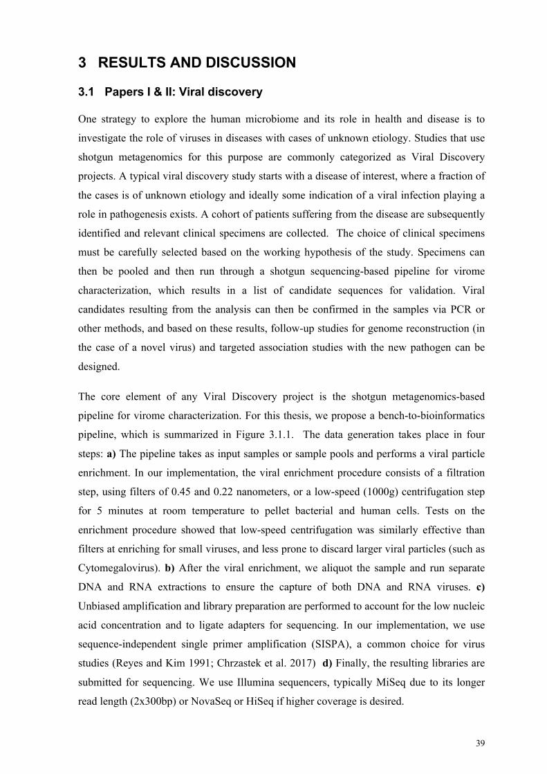
39
3 RESULTS AND DISCUSSION
3.1 Papers I & II: Viral discovery
One strategy to explore the human microbiome and its role in health and disease is to
investigate the role of viruses in diseases with cases of unknown etiology. Studies that use
shotgun metagenomics for this purpose are commonly categorized as Viral Discovery
projects. A typical viral discovery study starts with a disease of interest, where a fraction of
the cases is of unknown etiology and ideally some indication of a viral infection playing a
role in pathogenesis exists. A cohort of patients suffering from the disease are subsequently
identified and relevant clinical specimens are collected. The choice of clinical specimens
must be carefully selected based on the working hypothesis of the study. Specimens can
then be pooled and then run through a shotgun sequencing-based pipeline for virome
characterization, which results in a list of candidate sequences for validation. Viral
candidates resulting from the analysis can then be confirmed in the samples via PCR or
other methods, and based on these results, follow-up studies for genome reconstruction (in
the case of a novel virus) and targeted association studies with the new pathogen can be
designed.
The core element of any Viral Discovery project is the shotgun metagenomics-based
pipeline for virome characterization. For this thesis, we propose a bench-to-bioinformatics
pipeline, which is summarized in Figure 3.1.1. The data generation takes place in four
steps: a) The pipeline takes as input samples or sample pools and performs a viral particle
enrichment. In our implementation, the viral enrichment procedure consists of a filtration
step, using filters of 0.45 and 0.22 nanometers, or a low-speed (1000g) centrifugation step
for 5 minutes at room temperature to pellet bacterial and human cells. Tests on the
enrichment procedure showed that low-speed centrifugation was similarly effective than
filters at enriching for small viruses, and less prone to discard larger viral particles (such as
Cytomegalovirus). b) After the viral enrichment, we aliquot the sample and run separate
DNA and RNA extractions to ensure the capture of both DNA and RNA viruses. c)
Unbiased amplification and library preparation are performed to account for the low nucleic
acid concentration and to ligate adapters for sequencing. In our implementation, we use
sequence-independent single primer amplification (SISPA), a common choice for virus
studies (Reyes and Kim 1991; Chrzastek et al. 2017) d) Finally, the resulting libraries are
submitted for sequencing. We use Illumina sequencers, typically MiSeq due to its longer
read length (2x300bp) or NovaSeq or HiSeq if higher coverage is desired.

40
Figure 3.1.1 Bench-to-Bioinformatics viral discovery pipeline
The sequencing data is analyzed through our bioinformatics pipeline, which can be split
into two ‘conceptual’ modules: preprocessing and discovery. The preprocessing module
starts by removing adapters, both from Illumina and SISPA amplification and trimming
low-quality bases, which typically occur at the end of Illumina reads. Then, the pipeline
performs a filtering step to remove human sequences by mapping the quality-filtered reads
against the human genome reference. This step yields a set of reads we label as “microbial
reads” and an estimate of microbial-to-human ratio, which varies greatly with the type of
clinical specimen, the condition (disease vs. healthy) and the viral enrichment procedure.
The human filtering step reduces the downstream processing load and removes the
identifiable human sequences to ensure patient anonymity.

41
Figure 3.1.2 – MEGAN output of the validation of the viral discovery pipeline with a mock sample
The discovery module takes the filtered microbial reads and processes them using two
different strategies: 1) reads are analyzed directly with read-based taxonomical inference
tools, or 2) reads are through metagenomics assembly pipelines, and the resulting contigs
annotated downstream. Earlier iterations of the pipeline (used in papers I & II) relied
heavily on blast-like searches for annotation and LCA tools such as MEGAN for
taxonomical annotation. However, method developments for efficient taxonomical profiling
and taxonomical sequence annotation have become available, and these have been
incorporated into the latest versions of the pipeline: https://github.com/maubarsom/VD_pipeline
We validated the suitability of our pipeline for viral discovery by assembling a mock
sample and processing it through our method. The mock was created by combining four
clinical specimens positive for DNA and RNA pathogens: parvovirus (plasma, ssDNA),
adenovirus (nasopharyngeal aspirate, dsDNA) and cytomegalovirus (CMV or HHV-5,
serum, dsDNA), respiratory syncytial virus type A (RSV, tracheal secretion, ssRNA) and
enterovirus (nasopharyngeal aspirate, ssRNA). The sample containing CMV(HHV-5) was
merged in a lower proportion to assess the sensitivity of the integrative approach to detect
low copy viruses. The pipeline was able to recover sequences from four out of the five
viruses including CMV which was merged at a lower proportion (Figure 3.1.2).

42
3.1.1 Paper I: Inspecting the roles of viral infections in the pathogenesis of Acute Lymphoblastic Leukemia
Paper 1 is an application of our pipeline to examine the possible role of a viral infection in
the pathogenesis of childhood acute lymphoblastic leukemia (ALL). In this study, we
inspected the virome of neonatal bloodspots (NBS) taken from newborns who later
developed ALL. The virome of ALL children was then compared to the virome of healthy
children. Viruses from the normal human flora were identified, and sequences belonging to
pathogenic viruses such as human herpesvirus 6 and parvovirus B19 were detected.
To validate the results, we designed primers specific to the detected viruses and tested the
sequenced pools and the individual clinical specimens by PCR. The presence of HHV-6
was confirmed in 2 patients and 3 controls, while 1 patient and 0 controls were positive for
parvovirus B19. These findings, despite being potentially interesting candidates, do not
suggest the existence of an association between the observed viruses and the disease.
Paper I illustrates many of experimental design and implementation challenges that arise in
viral discovery projects. The goal of the study is to find evidence of a viral infection in
children that developed ALL, to serve as a basis for subsequent studies to determine
possible causality and elucidate mechanisms. This study chose to inspect neonatal
bloodspots, since they are sampled close to the date of birth and being convenient for
research purposes. Neonatal bloodspots could in principle contain traces of the infection,
but depending on the time of the event, evidence of the causal viral infection is likely to be
cleared. Better sampling strategies such as periodically collecting maternal blood or
amniotic fluid over the course of a pregnancy in a prospective study could be envisioned.
However, without other evidence, a project of this scale would be hard to justify.
Another challenge, which is common to most viral discovery projects, is the choice of
pooling strategy and sequencing depth. Appropriate choices for these variables will depend
greatly on the disease under study, the number of patients and the type of samples. Factors
to consider in the decision include the 1) expected prevalence of the virus in the population
of interest (from which we will obtain our samples), 2) the virus load and especially in
relation to human and bacterial load in the clinical specimens of interest, and 3) whether the
pathogen is completely unknown or at least a close relative to known ones. Since many of
these are impossible to know or estimate beforehand, these choices commonly made based
on a combination of previous experience and economical constraints.
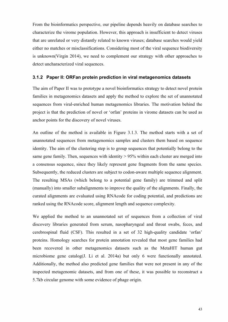
43
From the bioinformatics perspective, our pipeline depends heavily on database searches to
characterize the virome population. However, this approach is insufficient to detect viruses
that are unrelated or very distantly related to known viruses; database searches would yield
either no matches or misclassifications. Considering most of the viral sequence biodiversity
is unknown(Virgin 2014), we need to complement our strategy with other approaches to
detect uncharacterized viral sequences.
3.1.2 Paper II: ORFan protein prediction in viral metagenomics datasets
The aim of Paper II was to prototype a novel bioinformatics strategy to detect novel protein
families in metagenomics datasets and apply the method to explore the set of unannotated
sequences from viral-enriched human metagenomics libraries. The motivation behind the
project is that the prediction of novel or ‘orfan’ proteins in virome datasets can be used as
anchor points for the discovery of novel viruses.
An outline of the method is available in Figure 3.1.3. The method starts with a set of
unannotated sequences from metagenomics samples and clusters them based on sequence
identity. The aim of the clustering step is to group sequences that potentially belong to the
same gene family. Then, sequences with identity > 95% within each cluster are merged into
a consensus sequence, since they likely represent gene fragments from the same species.
Subsequently, the reduced clusters are subject to codon-aware multiple sequence alignment.
The resulting MSAs (which belong to a potential gene family) are trimmed and split
(manually) into smaller subalignments to improve the quality of the alignments. Finally, the
curated alignments are evaluated using RNAcode for coding potential, and predictions are
ranked using the RNAcode score, alignment length and sequence complexity.
We applied the method to an unannotated set of sequences from a collection of viral
discovery libraries generated from serum, nasopharyngeal and throat swabs, feces, and
cerebrospinal fluid (CSF). This resulted in a set of 32 high-quality candidate ‘orfan’
proteins. Homology searches for protein annotation revealed that most gene families had
been recovered in other metagenomics datasets such as the MetaHIT human gut
microbiome gene catalog(J. Li et al. 2014a) but only 6 were functionally annotated.
Additionally, the method also predicted gene families that were not present in any of the
inspected metagenomic datasets, and from one of these, it was possible to reconstruct a
5.7kb circular genome with some evidence of phage origin.

44
Figure 3.1.3 - ORFan protein prediction method summary
Our results suggest that the method is effective to recover ORFan proteins from viral
metagenomics datasets. However, the implementation of the method requires further work
to become widely applicable in practice. For example, the main clustering step with MCL
together with the single-linkage subclustering on each cluster to reduce redundancy were
very time-consuming. Reimplementing the method with state-of-the-art methods for
clustering such as VSEARCH (Rognes et al. 2016) or MMSeqs2 (Steinegger and Söding
2017) could improve the speed and throughput of the method without compromising cluster
quality. The other main limitation lies in the curation of high-quality multiple sequence
alignments. The choice of a codon-aware alignment tool such as MACSE was key, but
without automatized methods for alignment curation, the scalability of the method is
severely limited. The development of methods in this area would improve the scalability of
this method greatly.

45
From a viral discovery perspective, these results also suggest that predicting novel ORFan
can be effectively used to recover unknown viral genomes. The recovery of the
bacteriophage-like genome in paper II was based on one of the 32 predicted ORFan
proteins. The presence of the fragment in the fecal sample pool was confirmed by PCR and
subsequently tested in the individual fecal specimens. From these, only one out of 10
individuals tested positive for the fragment. Interestingly, this observation provides further
evidence about the efficacy of our viral discovery pipeline, despite possible limitations
imposed by the pooling and sequencing depth.
The genome reconstruction follow-up was performed using an inverse PCR, using primers
designed from the new protein family sequences. This resulted in an amplicon of around 7
kbps, suggesting the genomic fragment was circular. We then sequenced and assembled the
fragment using a combination of Illumina and Sanger sequencing, with the help of the IVA
assembler(Hunt et al. 2015). Annotation of the circular fragment was performed using ab
initio gene prediction and homology searches with HMMER3 (Mistry et al. 2013a).
Phylogenetic placement was based on one ORF with Pfam (Finn et al. 2014) annotation. An
alternative for genome recovery in other viral discovery projects could involve pooling
positive-testing samples and performing deep shotgun sequencing together with viral
enrichment strategies.
The role of bacteriophages in human health is not well understood, but studies in recent
years have revived the interest in the field. For example, Hsu et al. 2019 recently
demonstrated the impact of phage predation on the microbiome and its impact on the
metabolome in a mouse model, suggesting that the phages could actively play a similar role
in humans. Additionally, phage therapy is regaining traction as an alternative to antibiotics,
with recent studies suggesting it can fend off bacterial infections (Hu et al. 2018; Schooley
et al. 2017; Chan et al. 2018). Thus, the characterization of phages could have important
repercussions in the fight against antibiotic-resistant bacteria in the near future (Kortright et
al. 2019).
3.1.3 Conclusions
In conclusion, paper I and II showcase the development of effective methods to execute
viral discovery projects. However, continuous research is needed to improve the quality of
virome characterization, both from the data generation and bioinformatics angles. For
example, the development of efficient methods for unbiased viral enrichment would
drastically impact the quality of virome characterization and novel virus discovery. From

46
the bioinformatics side, novel strategies for the characterization and annotation of unknown
viruses are required. Tools based on machine learning techniques like VirFinder (Ren et al.
2017) and Metavir2 (Roux et al. 2014) are pushing the boundaries for novel virome
characterization, but there is plenty of room for development.

47
3.2 Papers III & IV: Associating the human skin microbiome to disease
Another way to study the influence of the microbiome in health and disease is to sample
and characterize human microbial communities of interest and study the community
variation in relation to the phenotype of interest (e.g. a comparison between communities
from healthy individuals and patients suffering from a disease). In this thesis, we studied a
cohort of Atopic Dermatitis (AD), Psoriasis (PSO) and healthy controls to understand the
role of the skin microbiome in allergy and autoimmunity, using AD and PSO as model
diseases. One lesional skin sample and one non-lesional skin sample from matching skin
locations were collected from each patient and every sample was processed with 16S rRNA
sequencing, shotgun metagenomics for microbiome data and microarrays for human
transcriptome profiling. Extensive clinical data from each study participant was also
collected for analysis.
The results of the analysis of the cohort are presented in papers III and IV. Paper III
describes the results from an open-ended exploratory analysis to examine the role of the
skin microbiome in AD and psoriasis versus healthy skin. In this study, lesional samples
from both diseases were compared to the samples from healthy individuals. Paper IV
focused on understanding the microbial and transcriptional variability in atopic dermatitis
across skin sites. This study was motivated by the observation that few studies had analyzed
diseased skin-site variability as thoroughly as it has been studied in healthy skin (Segre
2014, 2016). In paper IV, we analyzed both lesional and non-lesional sample from Atopic
Dermatitis patients and the healthy controls samples.
The bacterial taxonomical profile for both papers was obtained from the V3-V4 16S rRNA
454 pyrosequencing data using the QIIME pipeline. Open OTU picking was performed
against the Greengenes database (13.8) with a similarity threshold of 99.3%. The choice of
threshold was selected to optimize distinguishing S. aureus from other Staphylococcus
species. The 16S rRNA processing pipeline choice is now outdated by 2019 standards, but
the choices were state-of-the-art at the time of data generation, processing and analysis. In
any case, most ASV-based methods are not compatible with the data since they are specific
for Illumina data. Shotgun metagenomics data was not used for taxonomical profiling due
to issues with data quality that made the taxonomical profiles unreliable (data not shown).

48
To analyze this dataset, we established a general methodology for microbiome analytics,
focused on exploration and hypothesis generation. The methodology follows a top-down
approach based on the ecological levels of complexity from Section 1.2. The methodology
can be summarized as follows:
• Community-level analysis
o Identify community-level variation associated with the condition under study
o Identify clades driving the observed community-level variation
o Deduce microbial interactions between the clades that drive variation
o Study associations with other disease-related variables (disease subtypes, risk
factors, severity)
• Population-level analysis
o Explore strain-level variation on key microbial populations
• Mechanisms:
o Use functional data and data integration to elucidate mechanisms
The following sections will describe each of the six steps of the methodology and
exemplify how these were applied to explore biological questions in both paper III and IV.
3.2.1 Community-level analysis
The first step in the analysis of the skin microbiome dataset was to identify community-
level associations with the variables of interest. Since communities are typically high-
dimensional vectors of values, finding global associations requires statistical techniques to
summarize some characteristics of the community. In microbiome analysis, alpha and beta
diversity indexes are commonly used for this purpose.
In paper III, we were interested in understanding the variability in the microbial
communities among the two diseases and healthy skin. For this, we compared the Shannon
index between AD, psoriasis, and healthy skin and observed decreased that AD samples
had a decreased alpha diversity with respect to the controls and psoriasis samples. Beta
diversity ordinations based on the Bray-Curtis dissimilarity show a segregation between the
AD and healthy populations, and similarly between PSO and healthy. In paper IV, the same
diversity measures allowed us to identify varying ‘microbial’ dynamics between the thigh
and the upper back in AD samples. Consistent with other studies, we observed decreased
inter-site variability in AD with respect to healthy using beta diversity (Baurecht et al.
2018).

49
The choice of alpha and beta diversity measures should be tuned to the phenomenon under
study. In the context of the human skin microbiome, indexes like the Shannon or Simpson
diversity are useful summaries to identify dysbiosis, since they can reflect changes in the
composition (both in number and abundance) of the microbiome. However, alpha diversity
measures interpreted in isolation can be misleading. In paper IV, the Shannon diversity
index from the upper back samples does not change significantly between the groups
(Figure 3.2.1), but further investigation with beta diversity measures and taxonomical
barplots reveal that the healthy upper back community has a very different bacterial
composition compared to AD upper back (Figure 3.2.2).
Identify clades driving global variation
Once patterns in global community variation have been identified, a natural follow-up is to
identify which microbes drive these patterns. In paper III, we employed two different
strategies to identify OTUs driving the differences between the diseases. First, we
performed a differential abundance analysis, comparing the arcsin transformed abundance
between the three diseases while accounting for possible confounding factors with a GLM.
The second strategy consisted of training Random Forest classifiers to discriminate between
the conditions (healthy vs PSO, healthy vs AD, and AD vs PSO), and using the variable
importance Z-score to identify the OTUs that most reliably distinguished between the
healthy and diseased groups. Both analyses yielded coherent results, with differentially
abundant microbes having high Z-scores. Important microbes in AD include S. aureus, S.
epidermidis, Burkholderia sp., Staphylococcus sp. and in psoriasis, C. simulans, C.
Kroppenstedtii and Lactobacillus spp.
In paper IV, we performed differential abundance analysis between healthy controls,
lesional and non-lesional for each skin site (thigh and upper back). For this, we applied a
compositionally-aware method called ANCOM (Mandal et al. 2015). The analysis
identified S. aureus and a Staphylococcus spp. as being differential abundant between the
groups in the thigh. Two other OTUs were identified as differentially abundant in upper
back, but these are known kit contaminants (Glassing et al. 2016; Salter, Cox, Turek, Calus,
Cookson, Moffatt, et al. 2014). A possible explanation for this is that upper back controls
are lower-biomass with respect to AD upper back and therefore more likely to be
contaminated during the sample processing. The choice of ANCOM was due to its effective
control of the false discovery rate (FDR) compared to other methods (Weiss et al. 2017b).

50
Figure 3.2.1 - Shannon diversity index of AD and healthy samples between skin sites (thigh and upper back)
Figure 3.2.2 - Beta diversity plot of AD and healthy samples based on the Bray Curtis dissimilarity using
UMAP (Paper IV)
●
**
****
****
Kruskal−Wallis, p = 1.4e−12
ns
ns
ns
Kruskal−Wallis, p = 0.079
Thigh Upper back
Lesional Non−lesional Control Lesional Non−lesional Control
0
1
2
3
4
5Sh
anno
n di
vers
ity in
dex
thigh upper_back
−5 0 5 −5 0 5−15
−10
−5
0
5
10
UMAP1
UMAP
2
statusLesional
Non lesional
Control
lesionalLES
NON_LES
clinical_groupAD
CTRL

51
Microbe-microbe interactions
Once key microbial players have been identified, we can use the abundance data to infer to
understand dependencies between the microbial key players and the rest of the community.
In paper III, we constructed two microbial co-abundance networks using SparCC
(Friedman and Alm 2012) using all OTUs present in more than 5% of the samples, one for
AD and one for psoriasis. The resulting AD co-abundance network resulted in a single
connected component. Salient features of the network include that S. aureus correlations
with most bacteria were negative, and the existence of a clique between S. epidermidis, two
Corynebacterium sp. OTUs and Finegoldia sp., with links to P. acnes (now C. acnes). In
psoriasis, the co-abundance network yielded a set of five independent connected
components. The network suggests that C. simulans, which is a strong determinant for
psoriasis is associated with two OTUs: Streptococcus spp. and P. anaerobius. Another
notable feature of the network is that the node with the highest degree is an OTU labeled as
C. kroppenstedtii.
Association with disease-related variables
Another way of understanding how the community is involved in the disease is to examine
the association with measures of disease variability. Examples include disease severity, risk
factors, comorbidities, or the presence of specific symptoms. In paper III, we examined the
association between microbial features and disease severity for both AD and PSO. We
employed a forward selection strategy to train regression models containing 5 to 50
microbial features (in increments of 5) to predict SCORAD (AD severity index) or PASI
(PSO severity index) scores. Microbial features were ranked according to their individual
correlation with the severity score. The best model was achieved using 35 OTUs to predict
SCORAD with a Mean Absolute Error (MAE) of 9.84.
In paper IV, we re-examined the microbial association with severity with respect to skin
site. In this analysis, we focused only on S. aureus association to severity, based on our
results from paper III and previous studies (Kong, Oh, Deming, Conlan, Grice, Beatson,
Nomicos, Polley, Komarow, Murray, et al. 2012). Interestingly, the association between S.
aureus and severity could only be observed in the thigh but not in the upper back.

52
3.2.2 Population-level analysis
Strain analysis can help provide insights about the results derived from the community-
level analysis. For example, we hypothesized that the differences in the association between
S. aureus and severity observed in paper IV could be partially explained by the presence of
different S. aureus strains. To investigate this, we profiled S. aureus strains using
StrainEST (Albanese and Donati 2017) from the available shotgun metagenomics data. The
tool outputs relative abundance strain profiles for S. aureus for each sample based on the
available genomes from NCBI. Figure 3.2.3 summarizes the number of detected S. aureus
strains per sample. These results suggest that most samples have more than one S. aureus
strain co-colonizing the skin, both in lesions and non-lesions.
The S. aureus strain data allowed us to examine different questions. Firstly, we inspected
the S. aureus strain profiles at the level of each individual: are the strains that colonize
nonlesional skin the same that are present in lesional skin? Analyzing subjects with both a
lesional and a nonlesional sample, we observed that the most dominant strain matches in 19
out of 21 patients sampled in the thigh and 7 out of 7 patients sampled in the upper back.
However, the Bray-Curtis dissimilarity of the profiles is not very high for most samples
(Figure 3.2.4). Another observation is that thigh lesional samples tend to have more
detected strains than nonlesional (Figure 3.2.5).
We can generate different hypotheses from these observations. One possibility is that the
dominant S. aureus strain present in unaffected skin, plays a role in exacerbating severity
when external factors (e.g. inflammation) together with other microbiome changes (e.g. loss
of ‘protective’ species) trigger its virulence. Another possible hypothesis is that the thigh
lesional ‘microenvironment’ is more favorable than the upper back for S. aureus strains
colonization. Another hypothesis could be that the thigh lesional microenvironment is more
favorable for the recruitment of virulent S. aureus strains.

53
Figure 3.2.3 Distribution of number of strains per sample
Figure 3.2.4 Bray-Curtis dissimilarity between lesional and nonlesional samples per individual
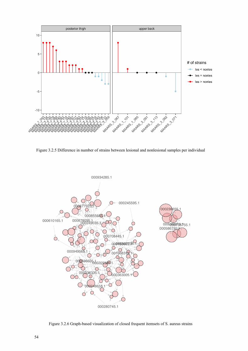
54
Figure 3.2.5 Difference in number of strains between lesional and nonlesional samples per individual
Figure 3.2.6 Graph-based visualization of closed frequent itemsets of S. aureus strains

55
We also examined the co-occurrence of S. aureus strains. To accomplish this, we applied
the ECLAT algorithm to find closed frequent itemsets with a minimum support of 6
(arbitrary choice). This resulted in a list of strain-sets represented in a graph-based layout
in Figure 3.2.6. The most notable feature of these results was a set of four strains which co-
occur very frequently, but only with each other. In particular, one of the strains
(000248915.1) appears to be central to the other three, since it appears together with them
21,18 and 16 samples, which are the closed itemsets with the highest support in the data.
We further investigated which strains are associated with AD severity in our samples. We
used a simple Bayesian model to estimate the posterior likelihood of a strain to occur in a
patient with severe AD as opposed to a patient with moderate AD. Assuming a probability
p of a strain colonizing a severe patient (with probability 1-p of colonizing a moderate
patient), the likelihood of observing a number of severe patients carrying the strain from a
set of N patients is distributed as a Bernoulli(N,p). We set a Beta(2,2) prior for the
parameter p, to enforce the belief that by default, strains do not show a preference between
severe and moderate patients. The posteriors for the 12 strains with the highest MAP
estimates for p are shown in Figure 3.2.7. Interestingly, the group of 4 highly co-occurring
strains (000239655.1, 000248915.1, 000586755.1 and 000735755.1) are all among the
strains with the highest MAP estimates for probability of occurring in severe patients. We
asked also if these severity-associated strains showed any distinct preference for any of the
sites. Figure 3.2.8 reveals that most of these strains appear in both sites.
The strain analysis helped refine some hypotheses concerning the site-specific differences
in severity, but some open questions remain. For example, we identified a set of strains that
are associated to severe disease, but these are present in both skin sites. A possible follow-
up study could focus on S. aureus strain variability across skin sites. Another caveat about
these results is the reliability of the strain inference algorithm and the database. StrainEST
uses NCBI genomes as ‘strains’, so it is possible that the algorithm misclassifies a SNP
profile that is not present in the database as combinations of close strains. However, the fact
that the most dominant strain matches across lesional and non-lesional samples of the same
individual suggests the algorithm works correctly to some degree. Further work is required
to validate and characterize these strains.

56
Figure 3.2.7 Posterior distribution of the probability of a given strain appearing in a severe sample (vs. a
moderate sample). The figure shows the 12 strains with the highest MAP estimate.
Figure 3.2.8 Number of samples where each of the severe-associated strains was detected

57
3.2.3 Data integration and functional analysis
The final aim of microbiome studies is to find possible mechanisms that drive the microbial
and phenotype variation identified in previous analyses. Shotgun metagenomics data allows
the identification of gene families and pathways present in the microbial community.
Mechanisms can be also inferred by integrating human-level data, such as direct
measurements of physiological conditions (oxygen, pH), molecules (lipid content,
antibacterial peptides), or omics datasets. In the case of papers III and IV, we used both
shotgun metagenomics and the skin transcriptome data for this purpose.
In AD, we wanted to understand the effects of S. aureus colonization on the human skin.
For this, we divided the lesional samples based on their S. aureus abundance profile into
two categories: samples with ‘high’ (> 87% relative abundance) or ‘low’ abundance of the
OTU. Based on this partition we explored 1) which microbial functions were associated
with the groups, and 2) the effect of S. aureus dominance on the skin transcriptome.
For (1), we used PiCRUST (Langille et al. 2013) to perform an initial prediction of
functions that are differentially represented between the S. aureus high and low groups. The
analysis resulted in gene families from different KEGG pathways like “Bacterial toxins”,
the “Two-component system” and “Glycolysis / Gluconeogenesis” enriched in the ‘high’
samples. PiCRUST predictions were then validated using a subset of shotgun
metagenomics samples. Gene family abundance prediction was obtained using HUMAnN2,
and statistical analysis confirmed the enrichment of the genes in the predicted pathways.
Notable examples include the alpha and delta-toxin S.aureus genes.
PiCRUST is a useful hypothesis generation for microbial mechanisms from 16S data.
However, results should be interpreted with care, since the taxonomical resolution of 16S is
not sufficient to make inferences about strain-level functional variation. Shotgun
metagenomics data or targeted experiments are good alternatives to validate the predictions
test the predictions made by PiCRUST. However, validation approaches must be planned
carefully: merging annotations between PiCRUST and HUMAnN2 was problematic since
they employ different databases: KEGG and UniRef with COG annotations respectively.
We chose to match the annotations based on gene name but recognize this is far from ideal.

58
To explore (2), the association between S. aureus and the skin transcriptome, we performed
a differential expression analysis between S. aureus high and low groups, which resulted in
a set of 256 DEGs (referred in the manuscript as “S. aureus signature”). Annotation of the
DEGs using Ingenuity Pathway Analysis observed an enrichment of different categories,
such as Keratinocyte differentiation and different components of the kynurenine pathway.
Based on these results, we hypothesized that the host could employ a tryptophan
‘starvation’ strategy to control S. aureus colonization. Follow-up experiments based on this
hypothesis revealed that AD S. aureus strains have low dependence on tryptophan, so the
hypothesis was discarded. We performed a similar analysis in paper IV but focusing on the
AD thigh samples. Results like ‘keratinocyte differentiation’ were also present, but a
surprising association with ‘Circadian Rhythm signaling’ were also predicted. Further
research is required to determine the significance of this finding.
To elucidate mechanisms in psoriasis, we selected a different strategy since there were no
bacterial OTU candidates with a strong association with the disease as S. aureus in AD.
Therefore, we decided to test the associations between data-defined gene groups and the top
differentially abundant OTUs using MaAsLin. Gene groups were inferred from the results
of module detection on the Psoriasis-versus-healthy DEG co-expression network. We
performed association tests of 12 gene modules against the 25 most differentially abundant
OTUs, accounting for body site, age, gender and institution. This resulted in 6 associations
after multiple testing correction, three of which related an OTU annotated as
Corynebacterium spp. to genes enriched for cell cycle, and inflammatory pathways. This
strategy is similar to the one used by (Morgan et al. 2015).
3.2.4 Conclusions
The work on papers III and IV illustrate the use of our established methodology for the
exploration of a human skin microbiome dataset. This allowed us to draw new insights,
generate and test hypothesis, and confirm previous observations from other studies about
the relationship between the host and the skin microbiome in psoriasis and atopic
dermatitis. The methodology proved to be flexible enough to allow the use of a broad range
of statistical techniques and algorithms, while providing enough structure to facilitate
reasoning about the results and connecting insights drawn from different analyses.

59
Many choices in technology, data processing and analysis for this skin microbiome project
do not reflect the current state of the art. The project was conceived and executed when 454
pyrosequencing was the standard for 16S rRNA sequencing (circa 2011). Nevertheless, the
cohort is still (to our knowledge) one of the largest studies of the skin microbiome in both
diseases. Also, solid study design choices, e.g. the use of negative controls, were
implemented in the study when they were not standard practice. Nevertheless, the results of
the analysis are congruent with the literature.
A modern redesign of the study would be developed using shotgun metagenomics.
However, ensuring good data quality and sufficient sequencing depth to characterize skin
communities is still a challenge due to the low biomass of the samples and the high
proportion of human DNA. In the case of designing a similar amplicon-based study,
sequencing the V1-V3 regions would be a better choice than V3-V4, as the V1-V3 region
has a better resolution for Staphylococcus species (Meisel et al. 2016).
However, the analysis strategies and most of the methods we used are still widely used in
current microbiome papers, and many modern analysis techniques like machine learning
and compositional-based data analysis were applied in this work. The observations and
generated hypothesis will lead to interesting biological insights with the appropriate follow
up studies. Future work will include further mining of the shotgun metagenomics dataset
for non-bacterial microbes, and bench work to validate some of the observations.
An important limitation of this study was the choice of anatomical locations. During the
patient sampling phase, a second AD skin site (upper back) was included in the design. The
measure enabled the study in paper IV, since the number of AD samples from each site was
balanced (~40 per group). However, the collection of healthy controls for the upper back
site was overlooked, so only few healthy controls are available for the upper back. This
implies that the interpretation of results where samples are compared to upper back controls
must account for this sample size unbalance.
In conclusion, the results of paper III and paper IV will serve as a basis for the design and
development of future studies to further understand the role of skin microbiome in atopic
dermatitis and psoriasis.


61
4 CONCLUDING REMARKS AND FUTURE PERSPECTIVES
The human microbiome field has been growing at an accelerated pace for the past decade
due to the developments in high throughput sequencing and bioinformatics methods. This
has resulted in numerous studies looking for possible associations between the human
microbiome and every disease. However, the lack of standardized practices has hampered
reproducibility and the ability to compare between studies due to the relative novelty of the
field.
The field has started to reach an inflection point: the cumulative knowledge has finally
started to push towards a new stage where we are demanding more stringency about
experimental design, data generation and analysis methodologies. Examples of these trends
include big efforts like the CAMI challenge (Vollmers, Wiegand, and Kaster 2017) and
reviews for conducting high quality microbiome research (Knight et al. 2018; Kong et al.
2017). As human microbiome research evolves into a more rigorous state, we will be able
to distinguish true associations between the microbiome with disease and better
characterize the mechanisms behind these associations, hopefully leading to translational
research that impacts human health.
The work in this thesis has sought to contribute to the state of human microbiome research
by developing methods and executing analysis following the best practices in the field. We
proposed a bench to bioinformatics pipeline for executing viral discovery projects, together
with a method prototype for unknown virus discovery. The methods helped explore the role
of the virome in acute lymphoblastic leukemia and also enabled the discovery of a new
bacteriophage genome. Additionally, we analyzed one of the largest skin microbiome
datasets regarding atopic dermatitis and psoriasis. This resulted in many insights and
testable hypotheses that can potentially form the basis of further studies to understand the
relationship between the microbiome and these diseases.
From the technology development perspective, the microbiome field will surely be
impacted by developments in single-molecule long-read sequencing. Currently, Pacific
Biosciences and Oxford Nanopore have already revolutionized the way we approach
genome sequencing projects(Koren et al. 2013), helped monitor the spread of viruses in an
outbreak(Quick et al. 2016), and coupled with tools like What’s In My Pot(WIMP) enable
real-time profiling of any metagenomics sample(Juul et al. 2015). Although throughput,
error rates and costs are currently limiting, it is only a matter of time before they become

62
routinely employed for metagenomics projects. In parallel, developments in single-cell
technologies will provide an alternative method to perform metagenomics studies that
address many of the limitations we have with current techniques (Rinke et al. 2013). New
bioinformatics methods will surely accompany the fast-paced developments in technology.
The progress in methodology and technology coupled with improved study designs should
allow the field to solve many open questions. Hopefully, these answers will impact human
health during the coming decades.

63
5 ACKNOWLEDGEMENTS
First of all, I would like to thank my main supervisor Björn Andersson for giving me the
opportunity to do my PhD in the lab and for all the scientific and academic discussions. To
Hamid for all the life advice and the chats and all the data (maybe you went a bit too crazy
on the data!). To all present and former lab members, for the nice environment and for
being very quiet in the office specially during the last two years. Talavera: maje, estoy acá
no se si gracias a vos o por tu culpa (lol). Gracias por todo voj, los taquitos, el kebab, las
habladas, las birras, las obscenidades, los modelos probabilísticos, la vida y todo lo demás
que es básicamente innumerable, me vale muy pero muy v*** si no te parece, vos te leés la
tesis y me decís que te gustó. Al Pou, merci per tot tíooo, si a alguien le debo literalmente
el tener los requisitos para estarme graduando es a vos <3. Como dijo un Poueta alguna
vez: "no existe más muerte que la de nuestros sueños". Also, I would like to thank my co-
supervisors Tobias and Stefanie for all the scientific discussions.
I would also like to thank all my collaborators, with whom I have had the pleasure to work
and to learn from during these years. In particular, I would like to thank Filipe, Helen and
Alan in KCL for all the great work and support in the MAARS project. I really learned a lot
from you and had a lot of fun hanging out too! Also, the Alenius lab, the Ranki lab and the
MAARS consortium, and in particular Noora, Hanna, Elina and Annamari for all the
great work with the skin projects! A Andre gracias por toda la paciencia con el proyecto
de EB y suerte con tu tésis que ya casi estás! To Anna for being the best student one could
ever wish for! To the metagenomics JC: Luisa, Fredrik, Johannes, Yue, Paulo, Steffi,
Justine, John for giving me a platform to discuss microbiome research with such a smart
bunch of people, for the grilling (I hope) and all the fika!
To all the friends I found during these years in these Nordic lands, I would like to thank
you for keeping me grounded, for all your support, fun times and for helping me maintain a
little bit of sanity. It is crazy the amount of people I have met during all these years; too
many people have come and gone, and a piece of my heart has certainly gone with each of
you. Despite my best efforts, I'll for sure miss many of you here, but your mark in my life
will linger on :).
Chicki, thanks for being my best friend during these years I've gotten to know you. Even
despite how different we are, you were always there for me, and I'll be forever thankful for
this even if I might not have fully appreciated it at the time. Thanks for always helping me

64
see things from a different perspective <3. Tati, mi panchi poooorc favorita, crec que non
hi ha paraules per explicar-te l'important que has estat (i encara ets) per mi durant tots
aquets anys; con todo y todo me alegro de aún tenerte en mi vida. Gracias por abrir mi
mente, por todo el apoyo y cariño, las vaciladas, los viajes y todo lo que me has enseñado.
Sandrita, basicamente empezamos el doctorado casi juntos, y casi que sos PI y yo aqui
intentando terminar jaja. Gracias por la energía, la actitud y por siempre encontrar tiempo
para vernos (aunque sea 3 horas después de las acordadas jajaja). Vale, grazie per tutte le
serate in giro, la musica, i concerti, tutto il funk, per prendere tutto sciallo, e per le storie di
tutti i casini e belinate in questa vita. Anche per il bellisimo viaggio a Londra di break di
tesi :D (sei di fianco a me in aereo mentre scrivo questo). Noora, my first Nordic friend
after sooo many years, it was lots of fun (also a bit intense hehe) to work together. Thanks
for all the fun, the beers and brunches, talks about life and specially paper IV in this thesis!
Paula, thanks for all the chats and the support, and the awesome movie and singing
evenings too. Konrad, probably my most ADHD friend ever :D thanks for everything, the
advice, the geekness, the help the music, the jams, everything. Come back to Stockholm
soon, so many future projects to do! Carlos Mata, por todo el chingue, las birritas, los
taquitos y las pachucadas de los barrios del sur jaja.
Sarah, thanks for being the best awkward friend one might ask for (lololol) For all the
never-ending talks about everything, the swears, the posh English, the football (you're
s***), all the intense married-couple-like discussions and never giving up on me. Yildiz,
thanks for being a voice of wisdom and for helping me realize there are other people with a
similar kind of crazy as mine. Thanks for all our discussions both deep and light-hearted,
for the all the interesting articles, books, ALL the help editing this document, fikas, board
games and everything else. Rafa, thanks for all the games, music, concerts, random talks
and for meeting up even if meant for you to come out of the cave into the light! Also,
thanks specially for screwing up all our Hanabi games we didn't finish, and for telling me
the wrong dress code for your wedding ahueahueahue. Vero, thanks for the support, for all
the pizzas, for giving me a second home for Christmas and all the awesome lazy days.
To my swedish girls Frida and Amanda: thank you for helping me reconnect with this
complicated piece of frozen land I found myself living in for longer than expected. Fri(dita
hehe), thank you for becoming a very unlikely friend in a crazy time: the writing of this
document. For always telling me things straight yet being one of the most caring and warm-
hearted persons I've met here. Also, for the badminton matches, teaching me a bit of
football vision, and ... I'll get my nutmeg revenge sooner or later! Amanda, thanks for

65
being as random, happy, sweet and awesome as you are. Also, for the awesome basketball
skills, for getting me to do random front-flips in Rålis and constantly inviting me to swim
despite my incapability.
To all my friends from KI, thank you for making this 'interesting' place a bit easier to
withstand. I would like to thank specially: Alberto: gracias por todas las fiestas y la buena
vibra que siempre está con vos. Shahul (paki!), thanks for feeding me and the fun, the
fooseball, the deep talks, and the advice (even if unsolicited sometimes). Daniela: thanks
for being my big sister, all the advice the fikas and the cakes! To Irene, thank you for
always being there, for staying in touch, for the help with the thesis editing, and for your
constant stream of energy and positivity (hehe <3). Joanne, for being a true hardliner, for
the awesome karaoke and fun times. Alča, thanks for having such an open heart and infinite
energy, for the best laser tag session ever and your czechness. To Alek for the awesome
music making. A Ale Fernandez, gracias por tenerme en tu lab de intern hace millones de
años, y por las habladas y consejos a lo largo de estos años. To Alba, infinite kudos and
thousand thanks for the amazing thesis cover!
To all my friends from the CMB times: Gonçalo , Simona, Katrin, Giuseppe, Mehdi,
Marion, Laure, Eric, Ashwini, Philine, Lisa, Laura, Riccardo ... thanks for all the fun,
pubs, parties and fika's :D. To Olov Andersson's group, Dodo, Christos, Jeremy, Nicki
thanks for adopting me for lunch when I first got left with no group hehe.
To the 9D mitochondria gang: Sarah, Roby, Mara, Jelena, Miriam, Rodolfo, Laura,
Diana, David(s) thanks for being my foster lunch group once we moved to biomedicum
and for all the fun outside work too (and filling up our concert!). To the people in 9C,
thanks for taking about 3 months to start talking to the guy alone in the office (thus forcing
me to socialize with other people in the building hehe) , but also for being very nice to me
(and sharing your fikas) after you finally got to it :D , specially, Nuno, Vero, Ju, Graciela,
Leo, Kathy , Cassandra, Cajsa. Cagla and Pontus (to be fair, you probably were the first
to talk to me after a week). Amir, Juancito, Pierre, Hassan and Christian, for all the
fooseball matches and shitty goals! I have the feeling I will still be waiting for the
fooseball tournament by the time I'm done. And of course, so many cool people around the
rest of KI: Theresa, Jorge Correia, Nuria, Jennine, Magali, Emilio, Sindhu, Polina, Jill
, Anders, Paulo, Milana, Alvaro & Rafa, Maria Azorin, Paola P., Tanya, Roberta ,
Walter, Elena, Edu, Shady, Sharesta, Tina, Varsha, Florian and everyone else my

66
chicken thesis-fried brain has forgotten to mention (sorry), for all the lunches, dinners,
parties, bbqs, events, beers and everything else.
To everyone in KEFF and in the SSIF basketball, for all the years of fun and friendly-
spirited football and basketball, for accepting (and helping me partially recover from) my
addiction to ballhogging. In particular, thanks to Xico, David, Rob, André, Andreia,
Moutinho, Frida, Emma & Teresa for the fun in and out of the football pitch, BBQs,
badminton, beers, randomness. Fede, maejtro!, por darme todos los pases , la magia en el
terreno de juego , las recomendaciones musicales y consejos de vida. A la KEFF
comunidad latina (Edu, Juan, Moises, Joao) por llevarme al colombiano a comer bandejita
paisa calidad . Andreia for introducing me to badminton and paddel tennis (I'll kick your
ass soon enough!) A Eloy por todos las discusiones de baloncesto y vida post-partido.
Amanda Ainali, for helping me with the swedish and the beers (the basketball too)!
To the bands I've played along the years, All covers (L-G, Daniel and crew, in loving
memory of Annika and Anders), as well as everyone in Divans orkester (Lars, Jerry 1
& 2, Siv, and Åke) for the music, and for taking me in to play with you, as different a
member as I might have been. To present and former members of Bossa Academica:
Konrad, Markel, Ana, Abraham, Marcelo and Jorge, for teaching me so much about
music, for helping me take my musicianship to another level, for all the gigs and rehearsals,
for not hating me everytime I use 'beautiful cadenzas' and go too 'jazzy', or swing around in
the rhythm randomly in any given song(hehe).
To Shady & Asa, for being the best corridor mates. Mahmoud and Anders, for being my
concert-going friends, without which most of my concerts would have been very boring and
keep the metal alive \m/. To the people in Magiska: Sara, Keo, Lorenzone, Ste, Teo,
thanks for making me feel at home there. To the CR crew: Andre, Luis Carlos, Carlos,
Yani, Marce y Antea (tica honoraria): PV mooops, gracias por darme un pedazo de
tiquicia por acá, las birritas y el bullying por no saber cuales son las Julieta jajaja.
To my friends abroad: Gabo y Ale, por ser mis senpai con esto del doctorado, no estaría
aca si no fuera por uds de fijo. A Gabo también por los comentarios de la tesis! A Oscar,
por muy conocedoramente convencerme de no ir a gringoland y venir a Europa.
Posiblemente una de las mejores decisiones que he tomado. To Priyata, for staying in my
life throughout all these years, for all the visits, for all the talks, and for being my lifeline
abroad. Marcia, obrigado! such a random friendship that happened to go through the years
as nothing, and for meeting in real life quite regularly. Looking forward to meeting you

67
around soon, it's been a while now :) Mateja, hvala, for being one of my first friends in
Sweden, and having me over in Croatia as if it hadn't been 5 years since we last met <3.
Sephora, for being my favorite nurse, for teaching me French and all the visits and talks.
To Mo(nica) for all the postcards! To my friends from the year in Finland: Tin, Monica,
Selam, Dish, Amjad, Julia, Chiara: for staying around in one way or another <3. To
Maddie, for coming to visit and giving me a place to crash in Marseille and taking me
around!
A la gente de tiquicia, gracias porque a pesar de los años aún han seguido estado conmigo
de un modo u otro a lo largo de estos 8 años desde que jalé. Gracias por sacar un rato para
verme cuando bajo (diría Leo, "por qué no subir?"). En especial a la Flaca, por ser una
constante en mi vida <3, a Peto y Leo por las habladas profundas, las estupideces y la
música calidad. A Jose por las smash mejengas (pero no por usar las pestes), a Andrés por
ordenar las pizzas cada año que vuelvo, a Ali por ser la única de "miau" en venir a
visitarme, a la China por caerte a estocolmo, y siempre llegar tarde pero igual venir a
verme jaja, a Katha por sacar el rato de tu ocupada vida para verme y llevarme a comer por
ahi. A July por ser un conocedor, a Luiiish por ser mi compañero de doctorado a la
distancia y a Naty por llevarme a pasear con los peludos y ser una peleona. Al resto de mis
amigos: Las Anitas (Moreno y Chevez), Chebas, Rebe, Carito Padilla, Luisk, Desiree,
Iosi, Chan por tenerme presente siempre con uds de un modo u otro. A Cindy y Rodrigo,
por cuidar a mi madre y llevarme a pasear cuando ando por allá!
Last but not least, a mis padres, gracias por todas las oportunidades que me dieron en esta
vida sin las cuales obviamente no estaría aquí. Gracias por enseñarme como vivir, y por
apoyarme en todas las decisiones que he tomado, aunque estas me hayan llevado a vivir a
10 000 km de uds. Ma, gracias por estar siempre ahi cuando te necesito y por la paz de
todos estos años aunque no esté por allá. Dad, gracias por siempre estar pendiente, y los
paseos a la playa para agarrar sol que aca hace falta. Y al resto de mi familia por siempre
recibirme con los brazos abiertos cuando voy a visitar.


69
6 REFERENCES
Aas, Johannes, Charles E Gessert, and Johan S Bakken. 2003. “Recurrent Clostridium
Difficile Colitis: Case Series Involving 18 Patients Treated with Donor Stool
Administered via a Nasogastric Tube.” Clinical Infectious Diseases : An Official
Publication of the Infectious Diseases Society of America 36 (5): 580–85.
https://doi.org/10.1086/367657.
Abubucker, Sahar, Nicola Segata, Johannes Goll, Alyxandria M. Schubert, Jacques Izard,
Brandi L. Cantarel, Beltran Rodriguez-Mueller, et al. 2012. “Metabolic Reconstruction
for Metagenomic Data and Its Application to the Human Microbiome.” PLoS
Computational Biology 8 (6). https://doi.org/10.1371/journal.pcbi.1002358.
Afshinnekoo, Ebrahim, Cem Meydan, Shanin Chowdhury, Dyala Jaroudi, Collin Boyer,
Nick Bernstein, Julia M. Maritz, et al. 2015. “Geospatial Resolution of Human and
Bacterial Diversity with City-Scale Metagenomics.” Cell Systems 1 (1): 97-97.e3.
https://doi.org/10.1016/j.cels.2015.07.006.
Albanese, Davide, and Claudio Donati. 2017. “Strain Profiling and Epidemiology of
Bacterial Species from Metagenomic Sequencing.” Nature Communications 8 (1):
2260. https://doi.org/10.1038/s41467-017-02209-5.
Alekseyenko, Alexander V, Guillermo I Perez-Perez, Aieska De Souza, Bruce Strober,
Zhan Gao, Monika Bihan, Kelvin Li, Barbara A Methé, and Martin J Blaser. 2013.
“Community Differentiation of the Cutaneous Microbiota in Psoriasis.” Microbiome 1
(1): 31. https://doi.org/10.1186/2049-2618-1-31.
Allander, Tobias, Martti T Tammi, Margareta Eriksson, Annelie Bjerkner, Annika
Tiveljung-Lindell, and Björn Andersson. 2005. “Cloning of a Human Parvovirus by
Molecular Screening of Respiratory Tract Samples.” Proceedings of the National
Academy of Sciences of the United States of America 102 (36).
https://doi.org/10.1073/pnas.0504666102.
Almonacid, Daniel E, Laurens Kraal, Francisco J Ossandon, Yelena V Budovskaya, Juan
Pablo Cardenas, Jessica Richman, and Zachary S Apte. 2016. “16S RRNA Gene
Sequencing as a Clinical Diagnostic Aid for Gastrointestinal-Related Conditions.”
BioRxiv.

70
Alneberg, Johannes, Brynjar Smári Bjarnason, Ino de Bruijn, Melanie Schirmer, Joshua
Quick, Umer Z Ijaz, Leo Lahti, Nicholas J Loman, Anders F Andersson, and
Christopher Quince. 2014. “Binning Metagenomic Contigs by Coverage and
Composition.” Nature Methods 11 (11): 1144–46. https://doi.org/10.1038/nmeth.3103.
Amir, Amnon, Daniel McDonald, Jose A. Navas-Molina, Evguenia Kopylova, James T.
Morton, Zhenjiang Zech Xu, Eric P. Kightley, et al. 2017. “Deblur Rapidly Resolves
Single-Nucleotide Community Sequence Patterns.” Edited by Jack A. Gilbert.
MSystems 2 (2). https://doi.org/10.1128/mSystems.00191-16.
Anderson, Marti J., and Daniel C. I. Walsh. 2013. “PERMANOVA, ANOSIM, and the
Mantel Test in the Face of Heterogeneous Dispersions: What Null Hypothesis Are
You Testing?” Ecological Monographs 83 (4): 557–74. https://doi.org/10.1890/12-
2010.1.
Ayling, Martin, Matthew D Clark, and Richard M Leggett. 2019. “New Approaches for
Metagenome Assembly with Short Reads.” Briefings in Bioinformatics, February.
https://doi.org/10.1093/bib/bbz020.
Bankevich, Anton, Sergey Nurk, Dmitry Antipov, Alexey A. Gurevich, Mikhail Dvorkin,
Alexander S. Kulikov, Valery M. Lesin, et al. 2012. “SPAdes: A New Genome
Assembly Algorithm and Its Applications to Single-Cell Sequencing.” Journal of
Computational Biology 19 (5): 455–77. https://doi.org/10.1089/cmb.2012.0021.
Barr, Jeremy J, Rita Auro, Mike Furlan, Katrine L Whiteson, Marcella L Erb, Joe Pogliano,
Aleksandr Stotland, et al. 2013. “Bacteriophage Adhering to Mucus Provide a Non-
Host-Derived Immunity.” Proceedings of the National Academy of Sciences of the
United States of America 110 (26): 10771–76.
https://doi.org/10.1073/pnas.1305923110.
Baurecht, Hansjörg, Malte C. Rühlemann, Elke Rodríguez, Frederieke Thielking, Inken
Harder, Anna-Sophie Erkens, Dora Stölzl, et al. 2018. “Epidermal Lipid Composition,
Barrier Integrity, and Eczematous Inflammation Are Associated with Skin
Microbiome Configuration.” The Journal of Allergy and Clinical Immunology 141 (5):
1668-1676.e16. https://doi.org/10.1016/j.jaci.2018.01.019.
Boisvert, Sébastien, Frédéric Raymond, Élénie Godzaridis, François Laviolette, and
Jacques Corbeil. 2012. “Ray Meta: Scalable de Novo Metagenome Assembly and

71
Profiling.” Genome Biology 13 (12): R122. https://doi.org/10.1186/gb-2012-13-12-
r122.
Brauweiler, Anne M, Elena Goleva, and Donald Y M Leung. 2014. “Th2 Cytokines
Increase Staphylococcus Aureus Alpha Toxin-Induced Keratinocyte Death through the
Signal Transducer and Activator of Transcription 6 (STAT6).” The Journal of
Investigative Dermatology 134 (8): 2114–21. https://doi.org/10.1038/jid.2014.43.
Bray, J. Roger, and J. T. Curtis. 1957. “An Ordination of the Upland Forest Communities
of Southern Wisconsin.” Ecological Monographs 27 (4): 325–49.
https://doi.org/10.2307/1942268.
Bray, Nicolas L, Harold Pimentel, Páll Melsted, and Lior Pachter. 2016. “Near-Optimal
Probabilistic RNA-Seq Quantification.” Nature Biotechnology 34 (5): 525–27.
https://doi.org/10.1038/nbt.3519.
Breitwieser, Florian P, Mihaela Pertea, Aleksey Zimin, and Steven L Salzberg. 2019.
“Human Contamination in Bacterial Genomes Has Created Thousands of Spurious
Proteins.” Genome Research 29 (6): gr.245373.118.
https://doi.org/10.1101/GR.245373.118.
Brown, C Titus, and Luiz Irber. 2016. “Sourmash: A Library for MinHash Sketching of
DNA.” The Journal of Open Source Software. https://doi.org/10.21105/joss.00027.
Brown, Sara J, and W H Irwin Mclean. 2012. “One Remarkable Molecule: Filaggrin.” J
Invest Dermatol 132 (3 Pt 2): 751–62. https://doi.org/10.1038/jid.2011.393.One.
Buchfink, Benjamin, Chao Xie, and Daniel H Huson. 2015. “Fast and Sensitive Protein
Alignment Using DIAMOND.” Nature Methods 12 (1): 59–60.
https://doi.org/10.1038/nmeth.3176.
Callahan, Benjamin J, Paul J McMurdie, and Susan P Holmes. 2017. “Exact Sequence
Variants Should Replace Operational Taxonomic Units in Marker-Gene Data
Analysis.” The ISME Journal 11 (12): 2639–43.
https://doi.org/10.1038/ismej.2017.119.
Callahan, Benjamin J, Paul J McMurdie, Michael J Rosen, Andrew W Han, Amy Jo A
Johnson, and Susan P Holmes. 2016. “DADA2: High-Resolution Sample Inference
from Illumina Amplicon Data.” Nature Methods 13 (7): 581–83.

72
https://doi.org/10.1038/nmeth.3869.
Caporaso, J. Gregory, Christian L. Lauber, Elizabeth K. Costello, Donna Berg-Lyons,
Antonio Gonzalez, Jesse Stombaugh, Dan Knights, et al. 2011. “Moving Pictures of
the Human Microbiome.” Genome Biology 12 (5): R50. https://doi.org/10.1186/gb-
2011-12-5-r50.
Caporaso, J Gregory, Justin Kuczynski, Jesse Stombaugh, Kyle Bittinger, Frederic D
Bushman, Elizabeth K Costello, Noah Fierer, et al. 2010. “QIIME Allows Analysis of
High-Throughput Community Sequencing Data.” Nature Methods.
https://doi.org/10.1038/nmeth.f.303.
Caspi, Ron, Richard Billington, Carol A Fulcher, Ingrid M Keseler, Anamika Kothari,
Markus Krummenacker, Mario Latendresse, et al. 2018. “The MetaCyc Database of
Metabolic Pathways and Enzymes.” Nucleic Acids Research 46 (D1): D633–39.
https://doi.org/10.1093/nar/gkx935.
Cavuoto, Kara M, Santanu Banerjee, Darlene Miller, and Anat Galor. 2018. “Composition
and Comparison of the Ocular Surface Microbiome in Infants and Older Children.”
Translational Vision Science & Technology 7 (6): 16.
https://doi.org/10.1167/tvst.7.6.16.
Chaban, Bonnie, and Janet E Hill. 2012. “A ‘universal’ Type II Chaperonin PCR Detection
System for the Investigation of Archaea in Complex Microbial Communities.” The
ISME Journal 6 (2): 430–39. https://doi.org/10.1038/ismej.2011.96.
Chan, Benjamin K, Paul E Turner, Samuel Kim, Hamid R Mojibian, John A Elefteriades,
and Deepak Narayan. 2018. “Phage Treatment of an Aortic Graft Infected with
Pseudomonas Aeruginosa.” Evolution, Medicine, and Public Health 2018 (1): 60–66.
https://doi.org/10.1093/emph/eoy005.
Chao, Anne. 1984. “Nonparametric Estimation of the Number of Classes in a Population.”
Scandinavian Journal of Statistics. WileyBoard of the Foundation of the Scandinavian
Journal of Statistics. https://doi.org/10.2307/4615964.
Chee, Chin-Hoong, Jafreezal Jaafar, Izzatdin Abdul Aziz, Mohd Hilmi Hasan, and William
Yeoh. 2018. “Algorithms for Frequent Itemset Mining: A Literature Review.”
Artificial Intelligence Review, March, 1–19. https://doi.org/10.1007/s10462-018-9629-

73
z.
Chen, I-Min A., Victor M. Markowitz, Ken Chu, Krishna Palaniappan, Ernest Szeto, Manoj
Pillay, Anna Ratner, et al. 2017. “IMG/M: Integrated Genome and Metagenome
Comparative Data Analysis System.” Nucleic Acids Research 45 (D1): D507–16.
https://doi.org/10.1093/nar/gkw929.
Chen, Jun, Kyle Bittinger, Emily S Charlson, Christian Hoffmann, James Lewis, Gary D
Wu, Ronald G Collman, Frederic D Bushman, and Hongzhe Li. 2012. “Associating
Microbiome Composition with Environmental Covariates Using Generalized UniFrac
Distances.” Bioinformatics (Oxford, England) 28 (16): 2106–13.
https://doi.org/10.1093/bioinformatics/bts342.
Chen, Jun, Emily King, Rebecca Deek, Zhi Wei, Yue Yu, Diane Grill, Karla Ballman, and
Oliver Stegle. 2018. “An Omnibus Test for Differential Distribution Analysis of
Microbiome Sequencing Data.” Edited by Oliver Stegle. Bioinformatics 34 (4): 643–
51. https://doi.org/10.1093/bioinformatics/btx650.
Chen, Kevin, and Lior Pachter. 2005. “Bioinformatics for Whole-Genome Shotgun
Sequencing of Microbial Communities.” PLoS Computational Biology.
https://doi.org/10.1371/journal.pcbi.0010024.
Chen, Wei, Clarence K. Zhang, Yongmei Cheng, Shaowu Zhang, and Hongyu Zhao. 2013.
“A Comparison of Methods for Clustering 16S RRNA Sequences into OTUs.” Edited
by Maurizio Casiraghi. PLoS ONE 8 (8): e70837.
https://doi.org/10.1371/journal.pone.0070837.
Chrzastek, Klaudia, Dong-hun Lee, Diane Smith, Poonam Sharma, David L. Suarez, Mary
Pantin-Jackwood, and Darrell R. Kapczynski. 2017. “Use of Sequence-Independent,
Single-Primer-Amplification (SISPA) for Rapid Detection, Identification, and
Characterization of Avian RNA Viruses.” Virology 509 (September): 159–66.
https://doi.org/10.1016/j.virol.2017.06.019.
Cleary, Brian, Ilana Lauren Brito, Katherine Huang, Dirk Gevers, Terrance Shea, Sarah
Young, and Eric J Alm. 2015. “Detection of Low-Abundance Bacterial Strains in
Metagenomic Datasets by Eigengenome Partitioning.” Nature Biotechnology 33 (10):
1053–60. https://doi.org/10.1038/nbt.3329.

74
Cogen, Anna L, Kenshi Yamasaki, Jun Muto, Katheryn M Sanchez, Laura Crotty
Alexander, Jackelyn Tanios, Yuping Lai, Judy E Kim, Victor Nizet, and Richard L
Gallo. 2010. “Staphylococcus Epidermidis Antimicrobial Delta-Toxin (Phenol-Soluble
Modulin-Gamma) Cooperates with Host Antimicrobial Peptides to Kill Group A
Streptococcus.” PloS One 5 (1): e8557. https://doi.org/10.1371/journal.pone.0008557.
Conceição-Neto, Nádia, Mark Zeller, Hanne Lefrère, Pieter De Bruyn, Leen Beller, Ward
Deboutte, Claude Kwe Yinda, et al. 2015. “Modular Approach to Customise Sample
Preparation Procedures for Viral Metagenomics: A Reproducible Protocol for Virome
Analysis.” Scientific Reports 5 (January): 16532. https://doi.org/10.1038/srep16532.
Cooley, Shamus M., Timothy Hamilton, Eric J. Deeds, and J. Christian J. Ray. 2019. “A
Novel Metric Reveals Previously Unrecognized Distortion in Dimensionality
Reduction of ScRNA-Seq Data.” BioRxiv, July, 689851.
https://doi.org/10.1101/689851.
Costea, Paul Igor, Robin Munch, Luis Pedro Coelho, Lucas Paoli, Shinichi Sunagawa, and
Peer Bork. 2017. “MetaSNV: A Tool for Metagenomic Strain Level Analysis.” PLoS
ONE 12 (7). https://doi.org/10.1371/journal.pone.0182392.
Costello, Elizabeth K, Christian L Lauber, Micah Hamady, Noah Fierer, Jeffrey I Gordon,
and Rob Knight. 2009. “Bacterial Community Variation in Human Body Habitats
across Space and Time.” Science 326 (5960): 1694–97.
https://doi.org/10.1126/science.1177486.Bacterial.
DeSantis, T. Z., P. Hugenholtz, N. Larsen, M. Rojas, E. L. Brodie, K. Keller, T. Huber, D.
Dalevi, P. Hu, and G. L. Andersen. 2006. “Greengenes, a Chimera-Checked 16S
RRNA Gene Database and Workbench Compatible with ARB.” Applied and
Environmental Microbiology 72 (7): 5069–72. https://doi.org/10.1128/AEM.03006-05.
Dickson, Robert P., John R. Erb-Downward, Fernando J. Martinez, and Gary B. Huffnagle.
2016. “The Microbiome and the Respiratory Tract.” Annual Review of Physiology 78
(1): 481–504. https://doi.org/10.1146/annurev-physiol-021115-105238.
Dickson, Robert P, and Gary B Huffnagle. 2015. “The Lung Microbiome: New Principles
for Respiratory Bacteriology in Health and Disease.” PLoS Pathogens 11 (7):
e1004923. https://doi.org/10.1371/journal.ppat.1004923.

75
Dutilh, Bas E., Noriko Cassman, Katelyn McNair, Savannah E. Sanchez, Genivaldo G. Z.
Silva, Lance Boling, Jeremy J. Barr, et al. 2014. “A Highly Abundant Bacteriophage
Discovered in the Unknown Sequences of Human Faecal Metagenomes.” Nature
Communications 5 (July). https://doi.org/10.1038/ncomms5498.
Edgar, Robert. 2019. “Defining and Interpreting OTUs.” 2019.
Edgar, Robert C. 2016. “UNOISE2: Improved Error-Correction for Illumina 16S and ITS
Amplicon Sequencing.” BioRxiv, October, 081257. https://doi.org/10.1101/081257.
Edgar, Robert C., Brian J. Haas, Jose C. Clemente, Christopher Quince, and Rob Knight.
2011. “UCHIME Improves Sensitivity and Speed of Chimera Detection.”
Bioinformatics 27 (16): 2194–2200. https://doi.org/10.1093/bioinformatics/btr381.
Edgar, Robert C. 2018. “Updating the 97% Identity Threshold for 16S Ribosomal RNA
OTUs.” Edited by Alfonso Valencia. Bioinformatics (Oxford, England) 34 (14): 2371–
75. https://doi.org/10.1093/bioinformatics/bty113.
Eloe-Fadrosh, Emiley A., Natalia N. Ivanova, Tanja Woyke, and Nikos C. Kyrpides. 2016.
“Metagenomics Uncovers Gaps in Amplicon-Based Detection of Microbial Diversity.”
Nature Microbiology 1 (4): 15032. https://doi.org/10.1038/nmicrobiol.2015.32.
Eren, A. Murat, Özcan C. Esen, Christopher Quince, Joseph H. Vineis, Hilary G. Morrison,
Mitchell L. Sogin, and Tom O. Delmont. 2015. “Anvi’o: An Advanced Analysis and
Visualization Platform for ‘omics Data.” PeerJ 3 (October): e1319.
https://doi.org/10.7717/peerj.1319.
Fahlén, Annika, Lars Engstrand, Barbara S. Baker, Anne Powles, and Lionel Fry. 2012.
“Comparison of Bacterial Microbiota in Skin Biopsies from Normal and Psoriatic
Skin.” Archives of Dermatological Research 304 (1): 15–22.
https://doi.org/10.1007/s00403-011-1189-x.
Faust, Karoline, Leo Lahti, Didier Gonze, Willem M de Vos, and Jeroen Raes. 2015.
“Metagenomics Meets Time Series Analysis: Unraveling Microbial Community
Dynamics.” Current Opinion in Microbiology 25 (June): 56–66.
https://doi.org/10.1016/j.mib.2015.04.004.
Fernandes, Andrew D., Jean M. Macklaim, Thomas G. Linn, Gregor Reid, and Gregory B.
Gloor. 2013. “ANOVA-Like Differential Expression (ALDEx) Analysis for Mixed
76
Population RNA-Seq.” Edited by John Parkinson. PLoS ONE 8 (7): e67019.
https://doi.org/10.1371/journal.pone.0067019.
Findley, Keisha, Julia Oh, Joy Yang, Sean Conlan, Clayton Deming, Jennifer A Meyer,
Deborah Schoenfeld, et al. 2013. “Topographic Diversity of Fungal and Bacterial
Communities in Human Skin.” Nature 498 (7454): 367–70.
https://doi.org/10.1038/nature12171.
Finn, Robert D, Alex Bateman, Jody Clements, Penelope Coggill, Ruth Y Eberhardt, Sean
R Eddy, Andreas Heger, et al. 2014. “Pfam: The Protein Families Database.” Nucleic
Acids Research 42 (Database issue): D222-30. https://doi.org/10.1093/nar/gkt1223.
Flint, Harry J., Karen P. Scott, Sylvia H. Duncan, Petra Louis, and Evelyne Forano. 2012.
“Microbial Degradation of Complex Carbohydrates in the Gut.” Gut Microbes 3 (4):
289–306. https://doi.org/10.4161/gmic.19897.
Foster, Jane A, and Karen-Anne McVey Neufeld. 2013. “Gut-Brain Axis: How the
Microbiome Influences Anxiety and Depression.” Trends in Neurosciences 36 (5):
305–12. https://doi.org/10.1016/j.tins.2013.01.005.
Foulongne, Vincent, Virginie Sauvage, Charles Hebert, Olivier Dereure, Justine Cheval,
Meriadeg Ar Gouilh, Kevin Pariente, et al. 2012. “Human Skin Microbiota: High
Diversity of DNA Viruses Identified on the Human Skin by High Throughput
Sequencing.” Edited by Amanda Ewart Toland. PLoS ONE 7 (6): e38499.
https://doi.org/10.1371/journal.pone.0038499.
Francis, O. E., M. Bendall, S. Manimaran, C. Hong, N. L. Clement, E. Castro-Nallar, Q.
Snell, et al. 2013. “Pathoscope: Species Identification and Strain Attribution with
Unassembled Sequencing Data.” Genome Research 23 (10): 1721–29.
https://doi.org/10.1101/gr.150151.112.
Frank, D. N., A. L. St. Amand, R. A. Feldman, E. C. Boedeker, N. Harpaz, and N. R. Pace.
2007. “Molecular-Phylogenetic Characterization of Microbial Community Imbalances
in Human Inflammatory Bowel Diseases.” Proceedings of the National Academy of
Sciences 104 (34): 13780–85. https://doi.org/10.1073/pnas.0706625104.
Franzosa, Eric A, Katherine Huang, James F Meadow, Dirk Gevers, Katherine P Lemon,
Brendan J M Bohannan, and Curtis Huttenhower. 2015. “Identifying Personal

77
Microbiomes Using Metagenomic Codes.” Proceedings of the National Academy of
Sciences 112 (22): E2930–38. https://doi.org/10.1073/pnas.1423854112.
Fredricks, David N. 2001. “Microbial Ecology of Human Skin in Health and Disease.”
Journal of Investigative Dermatology Symposium Proceedings 6 (3): 167–69.
https://doi.org/10.1046/j.0022-202x.2001.00039.x.
Friedman, Jonathan, and Eric J. Alm. 2012. “Inferring Correlation Networks from Genomic
Survey Data.” Edited by Christian von Mering. PLoS Computational Biology 8 (9):
e1002687. https://doi.org/10.1371/journal.pcbi.1002687.
Fry, L., B. S. Baker, A. V. Powles, A. Fahlen, and L. Engstrand. 2013. “Is Chronic Plaque
Psoriasis Triggered by Microbiota in the Skin?” British Journal of Dermatology.
https://doi.org/10.1111/bjd.12322.
Galperin, Michael Y., Kira S. Makarova, Yuri I. Wolf, and Eugene V. Koonin. 2015.
“Expanded Microbial Genome Coverage and Improved Protein Family Annotation in
the COG Database.” Nucleic Acids Research 43 (D1): D261–69.
https://doi.org/10.1093/nar/gku1223.
Gantner, Stephan, Anders F. Andersson, Laura Alonso-Sáez, and Stefan Bertilsson. 2011.
“Novel Primers for 16S RRNA-Based Archaeal Community Analyses in
Environmental Samples.” Journal of Microbiological Methods 84 (1): 12–18.
https://doi.org/10.1016/j.mimet.2010.10.001.
Gao, Zhan, Chi-hong Tseng, Bruce E. Strober, Zhiheng Pei, and Martin J. Blaser. 2008.
“Substantial Alterations of the Cutaneous Bacterial Biota in Psoriatic Lesions.” Edited
by Niyaz Ahmed. PLoS ONE 3 (7): e2719.
https://doi.org/10.1371/journal.pone.0002719.
Gevers, Dirk, Subra Kugathasan, Lee A. Denson, Yoshiki Vázquez-Baeza, Will
Van Treuren, Boyu Ren, Emma Schwager, et al. 2014. “The Treatment-Naive
Microbiome in New-Onset Crohn’s Disease.” Cell Host & Microbe 15 (3): 382–92.
https://doi.org/10.1016/j.chom.2014.02.005.
Ghurye, Jay S., Victoria Cepeda-Espinoza, and Mihai Pop. 2016. “Metagenomic Assembly:
Overview, Challenges and Applications.” The Yale Journal of Biology and Medicine
89 (3): 353. https://www.ncbi.nlm.nih.gov/pmc/articles/PMC5045144/.

78
Giallonardo, Francesca Di, Armin Töpfer, Melanie Rey, Sandhya Prabhakaran, Yannick
Duport, Christine Leemann, Stefan Schmutz, et al. 2014. “Full-Length Haplotype
Reconstruction to Infer the Structure of Heterogeneous Virus Populations.” Nucleic
Acids Research 42 (14): e115. https://doi.org/10.1093/nar/gku537.
Gilbert, Jack A., Martin J. Blaser, J. Gregory Caporaso, Janet K. Jansson, Susan V. Lynch,
and Rob Knight. 2018. “Current Understanding of the Human Microbiome.” Nature
Medicine 24 (4): 392–400. https://doi.org/10.1038/nm.4517.
Glassing, Angela, Scot E. Dowd, Susan Galandiuk, Brian Davis, and Rodrick J. Chiodini.
2016. “Inherent Bacterial DNA Contamination of Extraction and Sequencing Reagents
May Affect Interpretation of Microbiota in Low Bacterial Biomass Samples.” Gut
Pathogens 8 (1): 24. https://doi.org/10.1186/s13099-016-0103-7.
Gloor, Gregory B., Jean M. Macklaim, Vera Pawlowsky-Glahn, and Juan J. Egozcue. 2017.
“Microbiome Datasets Are Compositional: And This Is Not Optional.” Frontiers in
Microbiology 8 (November): 2224. https://doi.org/10.3389/fmicb.2017.02224.
Gong, J.Q., L. Lin, T. Lin, F. Hao, F.Q. Zeng, Z.G. Bi, D. Yi, and B. Zhao. 2006. “Skin
Colonization by Staphylococcus Aureus in Patients with Eczema and Atopic
Dermatitis and Relevant Combined Topical Therapy: A Double-Blind Multicentre
Randomized Controlled Trial.” British Journal of Dermatology 155 (4): 680–87.
https://doi.org/10.1111/j.1365-2133.2006.07410.x.
Gonze, Didier, Katharine Z Coyte, Leo Lahti, and Karoline Faust. 2018. “Microbial
Communities as Dynamical Systems.” Current Opinion in Microbiology 44 (August):
41–49. https://doi.org/10.1016/j.mib.2018.07.004.
Graspeuntner, Simon, Nathalie Loeper, Sven Künzel, John F. Baines, and Jan Rupp. 2018.
“Selection of Validated Hypervariable Regions Is Crucial in 16S-Based Microbiota
Studies of the Female Genital Tract.” Scientific Reports 8 (1): 9678.
https://doi.org/10.1038/s41598-018-27757-8.
Grice, Elizabeth a., Heidi H. Kong, Sean Conlan, Clayton B. Deming, Joie Davis, Alice C.
Young, Nisc Comparative Sequencing Program, et al. 2009. “Topographical and
Temporal Diversity of the Human Skin.” Science 324 (5931): 1190–92.
https://doi.org/10.1126/science.1171700.Topographical.

79
Grice, Elizabeth A., Heidi H. Kong, Gabriel Renaud, Alice C. Young, Gerard G. Bouffard,
Robert W. Blakesley, Tyra G. Wolfsberg, Maria L. Turner, and Julia A. Segre. 2008.
“A Diversity Profile of the Human Skin Microbiota.” Genome Research 18 (7): 1043–
50. https://doi.org/10.1101/gr.075549.107.
Grice, Elizabeth A, and Julia A Segre. 2012. “The Human Microbiome: Our Second
Genome.” Annual Review of Genomics and Human Genetics 13: 151–70.
https://doi.org/10.1146/annurev-genom-090711-163814.
Guma, Sergei, Remegio Maglantay, Ryan Lau, Rosemary Wieczorek, Jonathan Melamed,
Fang-Ming Deng, Ming Zhou, et al. 2016. “Papillary Urothelial Carcinoma with
Squamous Differentiation in Association with Human Papilloma Virus: Case Report
and Literature Review.” American Journal of Clinical and Experimental Urology 4
(1): 12–16. http://www.ncbi.nlm.nih.gov/pubmed/27069958.
Gustafsson, B, and J Carstensen. 1999. “Evidence of Space-Time Clustering of Childhood
Acute Lymphoblastic Leukaemia in Sweden.” British Journal of Cancer 79 (3–4):
655–57. https://doi.org/10.1038/sj.bjc.6690103.
Haas, Brian J, Dirk Gevers, Ashlee M Earl, Mike Feldgarden, Doyle V Ward, Georgia
Giannoukos, Dawn Ciulla, et al. 2011. “Chimeric 16S RRNA Sequence Formation and
Detection in Sanger and 454-Pyrosequenced PCR Amplicons.” Genome Research 21
(3): 494–504. https://doi.org/10.1101/gr.112730.110.
Haft, Daniel H, Jeremy D Selengut, and Owen White. 2003. “The TIGRFAMs Database of
Protein Families.” Nucleic Acids Research 31 (1): 371–73.
https://doi.org/10.1093/nar/gkg128.
Hannigan, Geoffrey D., Jacquelyn S. Meisel, Amanda S. Tyldsley, Qi Zheng, Brendan P.
Hodkinson, Adam J. Sanmiguel, Samuel Minot, Frederic D. Bushman, and Elizabeth
A. Grice. 2015. “The Human Skin Double-Stranded DNA Virome: Topographical and
Temporal Diversity, Genetic Enrichment, and Dynamic Associations with the Host
Microbiome.” MBio 6 (5). https://doi.org/10.1128/mBio.01578-15.
Hartstra, Annick V., Kristien E.C. Bouter, Fredrik Bäckhed, and Max Nieuwdorp. 2015.
“Insights Into the Role of the Microbiome in Obesity and Type 2 Diabetes.” Diabetes
Care 38 (1): 159–65. https://doi.org/10.2337/dc14-0769.

80
Hillmann, Benjamin, Gabriel A. Al-Ghalith, Robin R. Shields-Cutler, Qiyun Zhu, Daryl M.
Gohl, Kenneth B. Beckman, Rob Knight, and Dan Knights. 2018. “Evaluating the
Information Content of Shallow Shotgun Metagenomics.” Edited by John F. Rawls.
MSystems 3 (6): e00069-18. https://doi.org/10.1128/mSystems.00069-18.
Hilty, Markus, Conor Burke, Helder Pedro, Paul Cardenas, Andy Bush, Cara Bossley, Jane
Davies, et al. 2010. “Disordered Microbial Communities in Asthmatic Airways.”
Edited by Olivier Neyrolles. PLoS ONE 5 (1): e8578.
https://doi.org/10.1371/journal.pone.0008578.
Howe, Adina Chuang, Janet K Jansson, Stephanie A Malfatti, Susannah G Tringe, James M
Tiedje, and C Titus Brown. 2014. “Tackling Soil Diversity with the Assembly of
Large, Complex Metagenomes.” Proceedings of the National Academy of Sciences of
the United States of America 111 (13): 4904–9.
https://doi.org/10.1073/pnas.1402564111.
Hsu, Bryan B., Travis E. Gibson, Vladimir Yeliseyev, Qing Liu, Lorena Lyon, Lynn Bry,
Pamela A. Silver, and Georg K. Gerber. 2019. “Dynamic Modulation of the Gut
Microbiota and Metabolome by Bacteriophages in a Mouse Model.” Cell Host &
Microbe 25 (6): 803-814.e5. https://doi.org/10.1016/j.chom.2019.05.001.
Hu, Yue O O, Luisa W Hugerth, Carina Bengtsson, Arlisa Alisjahbana, Maike Seifert,
Anaga Kamal, Åsa Sjöling, et al. 2018. “Bacteriophages Synergize with the Gut
Microbial Community To Combat Salmonella.” Edited by Katrine L. Whiteson.
MSystems 3 (5). https://doi.org/10.1128/mSystems.00119-18.
Huerta-Cepas, Jaime, Damian Szklarczyk, Davide Heller, Ana Hernández-Plaza, Sofia K
Forslund, Helen Cook, Daniel R Mende, et al. 2019. “EggNOG 5.0: A Hierarchical,
Functionally and Phylogenetically Annotated Orthology Resource Based on 5090
Organisms and 2502 Viruses.” Nucleic Acids Research 47 (D1): D309–14.
https://doi.org/10.1093/nar/gky1085.
Huffnagle, G B, R P Dickson, and N W Lukacs. 2017. “The Respiratory Tract Microbiome
and Lung Inflammation: A Two-Way Street.” Mucosal Immunology 10 (2): 299–306.
https://doi.org/10.1038/mi.2016.108.
Hugenholtz, Philip. 2002. “Exploring Prokaryotic Diversity in the Genomic Era.” Genome
Biology 3 (2): REVIEWS0003. http://www.ncbi.nlm.nih.gov/pubmed/11864374.

81
Hughes, J B, J J Hellmann, T H Ricketts, and B J Bohannan. 2001. “Counting the
Uncountable: Statistical Approaches to Estimating Microbial Diversity.” Applied and
Environmental Microbiology 67 (10): 4399–4406.
https://doi.org/10.1128/aem.67.10.4399-4406.2001.
Hunt, Martin, Astrid Gall, Swee Hoe Ong, Jacqui Brener, Bridget Ferns, Philip Goulder,
Eleni Nastouli, Jacqueline A Keane, Paul Kellam, and Thomas D Otto. 2015. “IVA:
Accurate de Novo Assembly of RNA Virus Genomes.” Bioinformatics (Oxford,
England) 31 (14): 2374–76. https://doi.org/10.1093/bioinformatics/btv120.
Hunter, Sarah, Matthew Corbett, Hubert Denise, Matthew Fraser, Alejandra Gonzalez-
Beltran, Christopher Hunter, Philip Jones, et al. 2014. “EBI Metagenomics—a New
Resource for the Analysis and Archiving of Metagenomic Data.” Nucleic Acids
Research 42 (D1): D600–606. https://doi.org/10.1093/nar/gkt961.
Hurwitz, Bonnie L., Jana M. U’Ren, and Ken Youens-Clark. 2016. “Computational
Prospecting the Great Viral Unknown.” Edited by Andrew Millard. FEMS
Microbiology Letters 363 (10): fnw077. https://doi.org/10.1093/femsle/fnw077.
Huson, Daniel H, Alexander F Auch, Ji Qi, and Stephan C Schuster. 2007. “MEGAN
Analysis of Metagenomic Data.” Genome Research 17 (3): 377–86.
https://doi.org/10.1101/gr.5969107.
Isa, Adiba, Peter Priftakis, Kristina Broliden, and Britt Gustafsson. 2004. “Human
Parvovirus B19 DNA Is Not Detected in Guthrie Cards from Children Who Have
Developed Acute Lymphoblastic Leukemia.” Pediatric Blood & Cancer 42 (4): 357–
60. https://doi.org/10.1002/pbc.20001.
Jo, Jay-Hyun, Elizabeth A. Kennedy, and Heidi H. Kong. 2016. “Topographical and
Physiological Differences of the Skin Mycobiome in Health and Disease.” Virulence,
October, 00–00. https://doi.org/10.1080/21505594.2016.1249093.
Jonsson, Viktor, Tobias Österlund, Olle Nerman, and Erik Kristiansson. 2018a. “Modelling
of Zero-Inflation Improves Inference of Metagenomic Gene Count Data.” Statistical
Methods in Medical Research, November, 096228021881135.
https://doi.org/10.1177/0962280218811354.
Juul, Sissel, Fernando Izquierdo, Adam Hurst, Xiaoguang Dai, Amber Wright, Eugene

82
Kulesha, Roger Pettett, and Daniel J. Turner. 2015. “What’s in My Pot? Real-Time
Species Identification on the MinIONTM.” BioRxiv, November, 030742.
https://doi.org/10.1101/030742.
Kanehisa, Minoru, Yoko Sato, Masayuki Kawashima, Miho Furumichi, and Mao Tanabe.
2016. “KEGG as a Reference Resource for Gene and Protein Annotation.” Nucleic
Acids Research 44 (D1): D457–62. https://doi.org/10.1093/nar/gkv1070.
Kang, Dongwan D., Jeff Froula, Rob Egan, and Zhong Wang. 2015. “MetaBAT, an
Efficient Tool for Accurately Reconstructing Single Genomes from Complex
Microbial Communities.” PeerJ 3. https://doi.org/10.7717/peerj.1165.
Kaul, Abhishek, Siddhartha Mandal, Ori Davidov, and Shyamal D Peddada. 2017.
“Analysis of Microbiome Data in the Presence of Excess Zeros.” Frontiers in
Microbiology 8: 2114. https://doi.org/10.3389/fmicb.2017.02114.
Keegan, Kevin P., Elizabeth M. Glass, and Folker Meyer. 2016. “MG-RAST, a
Metagenomics Service for Analysis of Microbial Community Structure and Function.”
In Methods in Molecular Biology (Clifton, N.J.), 1399:207–33.
https://doi.org/10.1007/978-1-4939-3369-3_13.
Kernbauer, Elisabeth, Yi Ding, and Ken Cadwell. 2014. “An Enteric Virus Can Replace the
Beneficial Function of Commensal Bacteria.” Nature 516 (7529): 94–98.
https://doi.org/10.1038/nature13960.
Khalesi, Saman, Nick Bellissimo, Corneel Vandelanotte, Susan Williams, Dragana Stanley,
and Christopher Irwin. 2019. “A Review of Probiotic Supplementation in Healthy
Adults: Helpful or Hype?” European Journal of Clinical Nutrition 73 (1): 24–37.
https://doi.org/10.1038/s41430-018-0135-9.
Kim, Daehwan, Li Song, Florian P Breitwieser, and Steven L Salzberg. 2016. “Centrifuge:
Rapid and Sensitive Classification of Metagenomic Sequences.” Genome Research 26
(12): 1721–29. https://doi.org/10.1101/gr.210641.116.
Knight, Rob, Alison Vrbanac, Bryn C. Taylor, Alexander Aksenov, Chris Callewaert,
Justine Debelius, Antonio Gonzalez, et al. 2018. “Best Practices for Analysing
Microbiomes.” Nature Reviews Microbiology 16 (7): 410–22.
https://doi.org/10.1038/s41579-018-0029-9.

83
Kobayashi, Tetsuro, Martin Glatz, Keisuke Horiuchi, Hiroshi Kawasaki, Haruhiko
Akiyama, Daniel H. Kaplan, Heidi H. Kong, Masayuki Amagai, and Keisuke Nagao.
2015. “Dysbiosis and Staphyloccus Aureus Colonization Drives Inflammation in
Atopic Dermatitis.” Immunity 42 (4): 756–66.
https://doi.org/10.1016/j.immuni.2015.03.014.
Kong, Heidi H., Julia Oh, Clay Deming, Sean Conlan, Elizabeth A. Grice, Melony A.
Beatson, Effie Nomicos, Eric C. Polley, Hirsh D. Komarow, Jim Mullikin, et al. 2012.
“Temporal Shifts in the Skin Microbiome Associated with Disease Flares and
Treatment in Children with Atopic Dermatitis.” Genome Research 22 (5): 850–59.
https://doi.org/10.1101/gr.131029.111.
Kong, Heidi H., Julia Oh, Clay Deming, Sean Conlan, Elizabeth A. Grice, Melony A.
Beatson, Effie Nomicos, Eric C. Polley, Hirsh D. Komarow, Patrick R. Murray, et al.
2012. “Temporal Shifts in the Skin Microbiome Associated with Disease Flares and
Treatment in Children with Atopic Dermatitis.” Genome Research 22 (5): 850–59.
https://doi.org/10.1101/gr.131029.111.
Kong, Heidi H, Björn Andersson, Thomas Clavel, John E Common, Scott A Jackson,
Nathan D Olson, Julia A Segre, and Claudia Traidl-Hoffmann. 2017. “Performing
Skin Microbiome Research: A Method to the Madness.” The Journal of Investigative
Dermatology 137 (3): 561–68. https://doi.org/10.1016/j.jid.2016.10.033.
Konopka, Allan. 2009. “What Is Microbial Community Ecology?” The ISME Journal 3
(11): 1223–30. https://doi.org/10.1038/ismej.2009.88.
Koren, Sergey, Gregory P Harhay, Timothy P L Smith, James L Bono, Dayna M Harhay,
Scott D Mcvey, Diana Radune, Nicholas H Bergman, and Adam M Phillippy. 2013.
“Reducing Assembly Complexity of Microbial Genomes with Single-Molecule
Sequencing.” Genome Biology 14 (9): R101. https://doi.org/10.1186/gb-2013-14-9-
r101.
Kortright, Kaitlyn E, Benjamin K Chan, Jonathan L Koff, and Paul E Turner. 2019. “Phage
Therapy: A Renewed Approach to Combat Antibiotic-Resistant Bacteria.” Cell Host &
Microbe 25 (2): 219–32. https://doi.org/10.1016/j.chom.2019.01.014.
Kraszewska-Głomba, Barbara, Agnieszka Matkowska-Kocjan, and Leszek Szenborn. 2015.
“The Pathogenesis of Periodic Fever, Aphthous Stomatitis, Pharyngitis, and Cervical

84
Adenitis Syndrome: A Review of Current Research.” Mediators of Inflammation
2015: 563876. https://doi.org/10.1155/2015/563876.
Kuczynski, Justin, Christian L. Lauber, William A. Walters, Laura Wegener Parfrey, José
C. Clemente, Dirk Gevers, and Rob Knight. 2011. “Experimental and Analytical Tools
for Studying the Human Microbiome.” Nature Reviews Genetics 13 (1): 47–58.
https://doi.org/10.1038/nrg3129.
Lacey, Noreen, Síona Ní Raghallaigh, and Frank C Powell. 2011. “Demodex Mites--
Commensals, Parasites or Mutualistic Organisms?” Dermatology 222 (2): 128–30.
https://doi.org/10.1159/000323009.
Lahti, Leo, Sudarshan Shetty, and et al. 2017. “Tools for Microbiome Analysis in R.”
http://microbiome.github.com/microbiome.
Lai, Yuping, Anna L. Cogen, Katherine A. Radek, Hyun Jeong Park, Daniel T. MacLeod,
Anke Leichtle, Allen F. Ryan, Anna Di Nardo, and Richard L. Gallo. 2010.
“Activation of TLR2 by a Small Molecule Produced by Staphylococcus Epidermidis
Increases Antimicrobial Defense against Bacterial Skin Infections.” Journal of
Investigative Dermatology 130 (9): 2211–21. https://doi.org/10.1038/jid.2010.123.
Lai, Yuping, Anna Di Nardo, Teruaki Nakatsuji, Anke Leichtle, Yan Yang, Anna L Cogen,
Zi-Rong Wu, et al. 2009. “Commensal Bacteria Regulate Toll-like Receptor 3–
Dependent Inflammation after Skin Injury.” Nature Medicine 15 (12): 1377–82.
https://doi.org/10.1038/nm.2062.
Lang, Jennifer M., and M Eric Benbow. 2013. “Species Interactions and Competition.”
Nature Education Knowledge 4 (8).
https://www.nature.com/scitable/knowledge/library/species-interactions-and-
competition-102131429.
Langille, Morgan G I, Jesse Zaneveld, J Gregory Caporaso, Daniel McDonald, Dan
Knights, Joshua A Reyes, Jose C Clemente, et al. 2013. “Predictive Functional
Profiling of Microbial Communities Using 16S RRNA Marker Gene Sequences.”
Nature Biotechnology 31 (9): 814–21. https://doi.org/10.1038/nbt.2676.
Lanteri, M. C., F. Vahidnia, S. Tan, J. T. Stapleton, P. J. Norris, J. Heitman, X. Deng, et al.
2015. “Downregulation of Cytokines and Chemokines by GB Virus C After

85
Transmission Via Blood Transfusion in HIV-Positive Blood Recipients.” Journal of
Infectious Diseases 211 (10): 1585–96. https://doi.org/10.1093/infdis/jiu660.
LeBlanc, Jean Guy, Christian Milani, Graciela Savoy de Giori, Fernando Sesma, Douwe
van Sinderen, and Marco Ventura. 2013. “Bacteria as Vitamin Suppliers to Their Host:
A Gut Microbiota Perspective.” Current Opinion in Biotechnology 24 (2): 160–68.
https://doi.org/10.1016/J.COPBIO.2012.08.005.
Leger, Anthony J. St., Jigar V. Desai, Rebecca A. Drummond, Abirami Kugadas, Fatimah
Almaghrabi, Phyllis Silver, Kumarkrishna Raychaudhuri, et al. 2017. “An Ocular
Commensal Protects against Corneal Infection by Driving an Interleukin-17 Response
from Mucosal Γδ T Cells.” Immunity 47 (1): 148-158.e5.
https://doi.org/10.1016/j.immuni.2017.06.014.
Leung, D. Y M, J. B. Travers, R. Giorno, D. A. Norris, R. Skinner, J. Aelion, L. V. Kazemi,
et al. 1995. “Evidence for a Streptococcal Superantigen-Driven Process in Acute
Guttate Psoriasis.” Journal of Clinical Investigation 96 (5): 2106–12.
https://doi.org/10.1172/JCI118263.
Li, D., C.-M. Liu, R. Luo, K. Sadakane, and T.-W. Lam. 2015. “MEGAHIT: An Ultra-Fast
Single-Node Solution for Large and Complex Metagenomics Assembly via Succinct
de Bruijn Graph.” Bioinformatics 31 (10): 1674–76.
https://doi.org/10.1093/bioinformatics/btv033.
Li, Junhua, Huijue Jia, Xianghang Cai, Huanzi Zhong, Qiang Feng, Shinichi Sunagawa,
Manimozhiyan Arumugam, et al. 2014a. “An Integrated Catalog of Reference Genes
in the Human Gut Microbiome.” Nature Biotechnology 32 (8): 834–41.
https://doi.org/10.1038/nbt.2942.
Li, Linlin, Xutao Deng, Piyada Linsuwanon, David Bangsberg, Mwebesa Bosco Bwana,
Peter Hunt, Jeffrey N Martin, Steven G Deeks, and Eric Delwart. 2013. “AIDS Alters
the Commensal Plasma Virome.” Journal of Virology 87 (19): 10912–15.
https://doi.org/10.1128/JVI.01839-13.
Lindahl, B D, R H Nilsson, L Tedersoo, K Abarenkov, T Carlsen, R Kjoller, U Koljalg, et
al. 2013. “Fungal Community Analysis by High-Throughput Sequencing of Amplified
Markers - a User’s Guide.” New Phytologist 199 (1): 288–99.
https://doi.org/10.1111/nph.12243.

86
Lindgreen, Stinus, Karen L Adair, and Paul Gardner. 2015. “An Evaluation of the Accuracy
and Speed of Metagenome Analysis Tools.” BioRxiv. Cold Spring Harbor Labs
Journals. https://doi.org/10.1101/017830.
Lloyd-Price, Jason, Anup Mahurkar, Gholamali Rahnavard, Jonathan Crabtree, Joshua
Orvis, A. Brantley Hall, Arthur Brady, et al. 2017. “Strains, Functions and Dynamics
in the Expanded Human Microbiome Project.” Nature 550 (7674): 61–66.
https://doi.org/10.1038/nature23889.
Love, Michael I, Wolfgang Huber, and Simon Anders. 2014. “Moderated Estimation of
Fold Change and Dispersion for RNA-Seq Data with DESeq2.” Genome Biology 15
(12): 550. https://doi.org/10.1186/s13059-014-0550-8.
Lovell, David, Vera Pawlowsky-Glahn, Juan José Egozcue, Samuel Marguerat, and Jürg
Bähler. 2015. “Proportionality: A Valid Alternative to Correlation for Relative Data.”
Edited by Roland L. Dunbrack Jr. PLOS Computational Biology 11 (3): e1004075.
https://doi.org/10.1371/journal.pcbi.1004075.
Lozupone, C., and R. Knight. 2005. “UniFrac: A New Phylogenetic Method for Comparing
Microbial Communities.” Applied and Environmental Microbiology 71 (12): 8228–35.
https://doi.org/10.1128/AEM.71.12.8228-8235.2005.
Lu, Jennifer, Florian P. Breitwieser, Peter Thielen, and Steven L. Salzberg. 2017. “Bracken:
Estimating Species Abundance in Metagenomics Data.” PeerJ Computer Science 3
(January): e104. https://doi.org/10.7717/peerj-cs.104.
Lysholm, Fredrik, Anna Wetterbom, Cecilia Lindau, Hamid Darban, Annelie Bjerkner,
Kristina Fahlander, A. Michael Lindberg, Bengt Persson, Tobias Allander, and Björn
Andersson. 2012. “Characterization of the Viral Microbiome in Patients with Severe
Lower Respiratory Tract Infections, Using Metagenomic Sequencing.” Edited by
Sarah K. Highlander. PloS One 7 (2): e30875.
https://doi.org/10.1371/journal.pone.0030875.
Maaten, Laurens Van Der, and Geoffrey Hinton. 2008. “Visualizing Data Using T-SNE.”
Journal of Machine Learning Research.
Mah, Thien-Fah C, and George A O’Toole. 2001. “Mechanisms of Biofilm Resistance to
Antimicrobial Agents.” Trends in Microbiology 9 (1): 34–39.
87
https://doi.org/10.1016/S0966-842X(00)01913-2.
Mahé, Frédéric, Torbjørn Rognes, Christopher Quince, Colomban de Vargas, and Micah
Dunthorn. 2014. “Swarm: Robust and Fast Clustering Method for Amplicon-Based
Studies.” PeerJ 2 (September): e593. https://doi.org/10.7717/peerj.593.
Maia, Ana Teresa, Roxane Tussiwand, Giovanni Cazzaniga, Paolo Rebulla, Susan Colman,
Andrea Biondi, and Mel Greaves. 2004. “Identification of Preleukemic Precursors of
Hyperdiploid Acute Lymphoblastic Leukemia in Cord Blood.” Genes, Chromosomes
and Cancer 40 (1): 38–43. https://doi.org/10.1002/gcc.20010.
Mandal, Siddhartha, Will Van Treuren, Richard A White, Merete Eggesbø, Rob Knight,
and Shyamal D Peddada. 2015. “Analysis of Composition of Microbiomes: A Novel
Method for Studying Microbial Composition.” Microbial Ecology in Health &
Disease 26 (May): 27663. https://doi.org/10.3402/mehd.v26.27663.
McInnes, Leland, John Healy, Nathaniel Saul, and Lukas Großberger. 2018. “UMAP:
Uniform Manifold Approximation and Projection.” Journal of Open Source Software
3 (29): 861. https://doi.org/10.21105/joss.00861.
McMurdie, Paul J., and Susan Holmes. 2013. “Phyloseq: An R Package for Reproducible
Interactive Analysis and Graphics of Microbiome Census Data.” Edited by Michael
Watson. PLoS ONE 8 (4): e61217. https://doi.org/10.1371/journal.pone.0061217.
———. 2014. “Waste Not, Want Not: Why Rarefying Microbiome Data Is Inadmissible.”
Edited by Alice Carolyn McHardy. PLoS Computational Biology 10 (4): e1003531.
https://doi.org/10.1371/journal.pcbi.1003531.
Meisel, Jacquelyn S., Geoffrey D. Hannigan, Amanda S. Tyldsley, Adam J. SanMiguel,
Brendan P. Hodkinson, Qi Zheng, and Elizabeth A. Grice. 2016. “Skin Microbiome
Surveys Are Strongly Influenced by Experimental Design.” Journal of Investigative
Dermatology 136 (5). https://doi.org/10.1016/j.jid.2016.01.016.
Metges, Cornelia C. 2000. “Contribution of Microbial Amino Acids to Amino Acid
Homeostasis of the Host.” The Journal of Nutrition 130 (7): 1857S-1864S.
https://doi.org/10.1093/jn/130.7.1857S.
Mikheenko, Alla, Vladislav Saveliev, and Alexey Gurevich. 2016. “MetaQUAST:
Evaluation of Metagenome Assemblies.” Bioinformatics (Oxford, England) 32 (7):

88
1088–90. https://doi.org/10.1093/bioinformatics/btv697.
Milanese, Alessio, Daniel R Mende, Lucas Paoli, Guillem Salazar, Hans-Joachim
Ruscheweyh, Miguelangel Cuenca, Pascal Hingamp, et al. 2019. “Microbial
Abundance, Activity and Population Genomic Profiling with MOTUs2.” Nature
Communications 10 (1): 1014. https://doi.org/10.1038/s41467-019-08844-4.
Minot, Samuel, Alexandra Bryson, Christel Chehoud, Gary D Wu, James D Lewis, and
Frederic D Bushman. 2013. “Rapid Evolution of the Human Gut Virome.”
Proceedings of the National Academy of Sciences of the United States of America 110
(30): 12450–55. https://doi.org/10.1073/pnas.1300833110.
Mistry, Jaina, Robert D. Finn, Sean R. Eddy, Alex Bateman, and Marco Punta. 2013a.
“Challenges in Homology Search: HMMER3 and Convergent Evolution of Coiled-
Coil Regions.” Nucleic Acids Research 41 (12). https://doi.org/10.1093/nar/gkt263.
Mokili, John L, Forest Rohwer, and Bas E Dutilh. 2012. “Metagenomics and Future
Perspectives in Virus Discovery.” Current Opinion in Virology 2 (1): 63–77.
https://doi.org/10.1016/j.coviro.2011.12.004.
Mollerup, Sarah, Jens Friis-Nielsen, Lasse Vinner, Thomas Arn Hansen, Stine Raith
Richter, Helena Fridholm, Jose Alejandro Romero Herrera, et al. 2016.
“Propionibacterium Acnes - Disease Causing Agent or Common Contaminant?
Detection in Diverse Patient Samples by next Generation Sequencing.” Journal of
Clinical Microbiology, January, JCM.02723-15-. https://doi.org/10.1128/JCM.02723-
15.
Morgan, Xochitl C, Boyko Kabakchiev, Levi Waldron, Andrea D Tyler, Timothy L Tickle,
Raquel Milgrom, Joanne M Stempak, et al. 2015. “Associations between Host Gene
Expression, the Mucosal Microbiome, and Clinical Outcome in the Pelvic Pouch of
Patients with Inflammatory Bowel Disease.” Genome Biology 16 (1): 67.
https://doi.org/10.1186/s13059-015-0637-x.
Morgan, Xochitl C, Timothy L Tickle, Harry Sokol, Dirk Gevers, Kathryn L Devaney,
Doyle V Ward, Joshua A Reyes, et al. 2012. “Dysfunction of the Intestinal
Microbiome in Inflammatory Bowel Disease and Treatment.” Genome Biology 13 (9):
R79. https://doi.org/10.1186/gb-2012-13-9-r79.

89
Morton, James T., Clarisse Marotz, Alex Washburne, Justin Silverman, Livia S. Zaramela,
Anna Edlund, Karsten Zengler, and Rob Knight. 2019. “Establishing Microbial
Composition Measurement Standards with Reference Frames.” Nature
Communications 10 (1): 2719. https://doi.org/10.1038/s41467-019-10656-5.
Munck, Christian, Mads Albertsen, Amar Telke, Mostafa Ellabaan, Per Halkjær Nielsen,
and Morten O A Sommer. 2015. “Limited Dissemination of the Wastewater Treatment
Plant Core Resistome.” Nature Communications 6: 8452.
https://doi.org/10.1038/ncomms9452.
Naccache, Samia N, Alexander L Greninger, Deanna Lee, Lark L Coffey, Tung Phan,
Annie Rein-Weston, Andrew Aronsohn, John Hackett, Eric L Delwart, and Charles Y
Chiu. 2013. “The Perils of Pathogen Discovery: Origin of a Novel Parvovirus-like
Hybrid Genome Traced to Nucleic Acid Extraction Spin Columns.” Journal of
Virology 87: 11966–77. https://doi.org/10.1128/JVI.02323-13.
Naik, Shruti, Nicolas Bouladoux, Jonathan L. Linehan, Seong-Ji Han, Oliver J. Harrison,
Christoph Wilhelm, Sean Conlan, et al. 2015. “Commensal–Dendritic-Cell Interaction
Specifies a Unique Protective Skin Immune Signature.” Nature 520 (7545): 104–8.
https://doi.org/10.1038/nature14052.
Naik, Shruti, Nicolas Bouladoux, Christoph Wilhelm, Michael J Molloy, Rosalba Salcedo,
Wolfgang Kastenmuller, Clayton Deming, et al. 2012. “Compartmentalized Control of
Skin Immunity by Resident Commensals.” Science (New York, N.Y.) 337 (6098):
1115–19. https://doi.org/10.1126/science.1225152.
Nakamura, Yuumi, Jon Oscherwitz, Kemp B. Cease, Susana M. Chan, Raul Muñoz-
Planillo, Mizuho Hasegawa, Amer E. Villaruz, et al. 2013. “Staphylococcus δ-Toxin
Induces Allergic Skin Disease by Activating Mast Cells.” Nature 503 (7476): 397–
401. https://doi.org/10.1038/nature12655.
Nakatsuji, Teruaki, Hsin-I Chiang, Shangi B Jiang, Harish Nagarajan, Karsten Zengler, and
Richard L Gallo. 2013. “The Microbiome Extends to Subepidermal Compartments of
Normal Skin.” Nature Communications 4: 1431. https://doi.org/10.1038/ncomms2441.
Natsch, Andreas, Joachim Schmid, and Felix Flachsmann. 2004. “Identification of
Odoriferous Sulfanylalkanols in Human Axilla Secretions and Their Formation
through Cleavage of Cysteine Precursors by a C-S Lyase Isolated from Axilla

90
Bacteria.” Chemistry and Biodiversity 1 (7): 1058–72.
https://doi.org/10.1002/cbdv.200490079.
Nayfach, Stephen, Patrick H Bradley, Stacia K Wyman, Timothy J Laurent, Alex Williams,
Jonathan A Eisen, Katherine S Pollard, and Thomas J Sharpton. 2015. “Automated
and Accurate Estimation of Gene Family Abundance from Shotgun Metagenomes.”
PLoS Computational Biology 11 (11): e1004573.
https://doi.org/10.1371/journal.pcbi.1004573.
Nearing, Jacob T, Gavin M Douglas, André M Comeau, and Morgan G.I. Langille. 2018.
“Denoising the Denoisers: An Independent Evaluation of Microbiome Sequence Error-
Correction Approaches.” PeerJ 6 (August): e5364. https://doi.org/10.7717/peerj.5364.
Neto, Antonio Galvao, April Whitaker, and Zhiheng Pei. 2016. “Microbiome and Potential
Targets for Chemoprevention of Esophageal Adenocarcinoma.” Seminars in Oncology
43 (1): 86–96. https://doi.org/10.1053/j.seminoncol.2015.09.005.
Nguyen, Nam-Phuong, Tandy Warnow, Mihai Pop, and Bryan White. 2016. “A Perspective
on 16S RRNA Operational Taxonomic Unit Clustering Using Sequence Similarity.”
Npj Biofilms and Microbiomes 2 (1): 16004.
https://doi.org/10.1038/npjbiofilms.2016.4.
Norman, Jason M, Scott A Handley, Megan T Baldridge, Lindsay Droit, Catherine Y Liu,
Brian C Keller, Amal Kambal, et al. 2015. “Disease-Specific Alterations in the Enteric
Virome in Inflammatory Bowel Disease.” Cell 160 (3): 447–60.
https://doi.org/10.1016/j.cell.2015.01.002.
Nurk, Sergey, Dmitry Meleshko, Anton Korobeynikov, and Pavel Pevzner. 2016.
“MetaSPAdes: A New Versatile de Novo Metagenomics Assembler,” April.
http://arxiv.org/abs/1604.03071.
O’Dwyer, David N., Robert P. Dickson, and Bethany B. Moore. 2016. “The Lung
Microbiome, Immunity, and the Pathogenesis of Chronic Lung Disease.” The Journal
of Immunology 196 (12): 4839–47. https://doi.org/10.4049/jimmunol.1600279.
Oh, Julia, Allyson L. Byrd, Clay Deming, Sean Conlan, Heidi H. Kong, and Julia A. Segre.
2014. “Biogeography and Individuality Shape Function in the Human Skin
Metagenome.” Nature 514 (7520): 59–64. https://doi.org/10.1038/nature13786.

91
Oh, Julia, Allyson L. Byrd, Morgan Park, Heidi H. Kong, and Julia A. Segre. 2016.
“Temporal Stability of the Human Skin Microbiome.” Cell 165 (4): 854–66.
https://doi.org/10.1016/j.cell.2016.04.008.
Oh, Julia, Alexandra F. Freeman, Morgan Park, Robert Sokolic, Fabio Candotti, Steven M.
Holland, Julia A. Segre, and Heidi H. Kong. 2013. “The Altered Landscape of the
Human Skin Microbiome in Patients with Primary Immunodeficiencies.” Genome
Research 23 (12): 2103–14. https://doi.org/10.1101/gr.159467.113.
Oksanen, Jari, F. Guillaume Blanchet, Michael Friendly, Roeland Kindt, Pierre Legendre,
Dan McGlinn, Peter R. Minchin, et al. 2019. “The Vegan Package.”
Olsen, G J, D J Lane, S J Giovannoni, N R Pace, and D A Stahl. 1986. “Microbial Ecology
and Evolution: A Ribosomal RNA Approach.” Annual Review of Microbiology 40 (1):
337–65. https://doi.org/10.1146/annurev.mi.40.100186.002005.
Ondov, Brian D., Todd J. Treangen, Páll Melsted, Adam B. Mallonee, Nicholas H.
Bergman, Sergey Koren, and Adam M. Phillippy. 2016. “Mash: Fast Genome and
Metagenome Distance Estimation Using MinHash.” Genome Biology 17 (1): 132.
https://doi.org/10.1186/s13059-016-0997-x.
Otto, Michael. 2010. “Staphylococcus Colonization of the Skin and Antimicrobial
Peptides.” Expert Review of Dermatology 5 (2): 183–95.
https://doi.org/10.1586/edm.10.6.
Ounit, Rachid, and Stefano Lonardi. 2016. “Higher Classification Sensitivity of Short
Metagenomic Reads with CLARK-S-S.” Bioinformatics 32 (24): 3823–25.
https://doi.org/10.1093/bioinformatics/btw542.
Palacios, Gustavo, Julian Druce, Lei Du, Thomas Tran, Chris Birch, Thomas Briese, Sean
Conlan, et al. 2008. “A New Arenavirus in a Cluster of Fatal Transplant-Associated
Diseases.” The New England Journal of Medicine 358 (10): 991–98.
https://doi.org/10.1056/NEJMoa073785.
Parisi, Rosa, Deborah P M Symmons, Christopher E M Griffiths, and Darren M Ashcroft.
2012. “Global Epidemiology of Psoriasis: A Systematic Review of Incidence and
Prevalence.” Journal of Investigative Dermatology 133 (2): 377–85.
https://doi.org/10.1038/jid.2012.339.

92
Park, Sang-Cheol, and Sungho Won. 2018. “Evaluation of 16S RRNA Databases for
Taxonomic Assignments Using Mock Community.” Genomics & Informatics 16 (4):
e24. https://doi.org/10.5808/GI.2018.16.4.e24.
Parks, Donovan H, Michael Imelfort, Connor T Skennerton, Philip Hugenholtz, and Gene
W Tyson. 2014. “CheckM: Assessing the Quality of Microbial Genomes Recovered
from Isolates, Single Cells, and Metagenomes.” Genome Research, October.
https://doi.org/10.7287/peerj.preprints.554v1.
Pascolini, Chiara, Jolinda Sinagra, Simone Pecetta, Valentina Bordignon, Alessandra De
Santis, Laura Cilli, Viviana Cafiso, et al. 2011. “Molecular and Immunological
Characterization of Staphylococcus Aureus in Pediatric Atopic Dermatitis:
Implications for Prophylaxis and Clinical Management.” Clinical & Developmental
Immunology 2011: 718708. https://doi.org/10.1155/2011/718708.
Patro, Rob, Geet Duggal, Michael I Love, Rafael A Irizarry, and Carl Kingsford. 2017.
“Salmon Provides Fast and Bias-Aware Quantification of Transcript Expression.”
Nature Methods 14 (4): 417–19. https://doi.org/10.1038/nmeth.4197.
Paulino, L. C., C.-H. Tseng, B. E. Strober, and M. J. Blaser. 2006. “Molecular Analysis of
Fungal Microbiota in Samples from Healthy Human Skin and Psoriatic Lesions.”
Journal of Clinical Microbiology 44 (8): 2933–41.
https://doi.org/10.1128/JCM.00785-06.
Paulino, Luciana C., Chi-Hong Tseng, and Martin J. Blaser. 2008. “Analysis of Malassezia
Microbiota in Healthy Superficial Human Skin and in Psoriatic Lesions by Multiplex
Real-Time PCR.” FEMS Yeast Research 8 (3): 460–71. https://doi.org/10.1111/j.1567-
1364.2008.00359.x.
Paulson, Joseph N, O Colin Stine, Héctor Corrada Bravo, and Mihai Pop. 2013.
“Differential Abundance Analysis for Microbial Marker-Gene Surveys.” Nature
Methods 10 (12): 1200–1202. https://doi.org/10.1038/nmeth.2658.
Pedersen, Helle Krogh, Sofia K. Forslund, Valborg Gudmundsdottir, Anders Østergaard
Petersen, Falk Hildebrand, Tuulia Hyötyläinen, Trine Nielsen, et al. 2018. “A
Computational Framework to Integrate High-Throughput ‘-Omics’ Datasets for the
Identification of Potential Mechanistic Links.” Nature Protocols 13 (12): 2781–2800.
https://doi.org/10.1038/s41596-018-0064-z.

93
Peng, Y., H. C. M. Leung, S. M. Yiu, and F. Y. L. Chin. 2012. “IDBA-UD: A de Novo
Assembler for Single-Cell and Metagenomic Sequencing Data with Highly Uneven
Depth.” Bioinformatics 28 (11): 1420–28.
https://doi.org/10.1093/bioinformatics/bts174.
Pereira-Marques, Joana, Anne Hout, Rui M. Ferreira, Michiel Weber, Ines Pinto-Ribeiro,
Leen-Jan van Doorn, Cornelis Willem Knetsch, and Ceu Figueiredo. 2019. “Impact of
Host DNA and Sequencing Depth on the Taxonomic Resolution of Whole
Metagenome Sequencing for Microbiome Analysis.” Frontiers in Microbiology 10
(June): 1277. https://doi.org/10.3389/fmicb.2019.01277.
Petersen, Charisse, and June L. Round. 2014. “Defining Dysbiosis and Its Influence on
Host Immunity and Disease.” Cellular Microbiology 16 (7): 1024–33.
https://doi.org/10.1111/cmi.12308.
Popgeorgiev, Nikolay, Mickaël Boyer, Laura Fancello, Sonia Monteil, Catherine Robert,
Romain Rivet, Claude Nappez, et al. 2013. “Marseillevirus-like Virus Recovered from
Blood Donated by Asymptomatic Humans.” The Journal of Infectious Diseases 208
(7): 1042–50. https://doi.org/10.1093/infdis/jit292.
Pou, C., M. Barrientos-Somarribas, S. Marin-Juan, G. Bogdanovic, A. Bjerkner, T.
Allander, B. Gustafsson, and B. Andersson. 2018. “Virome Definition in
Cerebrospinal Fluid of Patients with Neurological Complications after Hematopoietic
Stem Cell Transplantation.” Journal of Clinical Virology 108 (November): 112–20.
https://doi.org/10.1016/j.jcv.2018.09.014.
Pride, David T, Julia Salzman, Matthew Haynes, Forest Rohwer, Clara Davis-Long,
Richard A White, Peter Loomer, Gary C Armitage, and David A Relman. 2012.
“Evidence of a Robust Resident Bacteriophage Population Revealed through Analysis
of the Human Salivary Virome.” The ISME Journal 6 (5): 915–26.
https://doi.org/10.1038/ismej.2011.169.
Prosperi, Mattia C F, and Marco Salemi. 2012. “QuRe: Software for Viral Quasispecies
Reconstruction from next-Generation Sequencing Data.” Bioinformatics 28 (1): 132–
33. https://doi.org/10.1093/bioinformatics/btr627.
Qin, Junjie, Ruiqiang Li, Jeroen Raes, Manimozhiyan Arumugam, Kristoffer Solvsten
Burgdorf, Chaysavanh Manichanh, Trine Nielsen, et al. 2010a. “A Human Gut

94
Microbial Gene Catalogue Established by Metagenomic Sequencing.” Nature 464
(7285): 59–65. https://doi.org/10.1038/nature08821.
———. 2010b. “A Human Gut Microbial Gene Catalogue Established by Metagenomic
Sequencing.” Nature 464 (7285): 59–65. https://doi.org/10.1038/nature08821.
Qin, Junjie, Yingrui Li, Zhiming Cai, Shenghui Songgang Li, Jianfeng Zhu, Fan Zhang,
Suisha Liang, et al. 2012. “A Metagenome-Wide Association Study of Gut Microbiota
in Type 2 Diabetes.” Nature 490 (7418): 55–60. https://doi.org/10.1038/nature11450.
Quick, Joshua, Nicholas J. Loman, Sophie Duraffour, Jared T. Simpson, Ettore Severi,
Lauren Cowley, Joseph Akoi Bore, et al. 2016. “Real-Time, Portable Genome
Sequencing for Ebola Surveillance.” Nature 530 (7589): 228–32.
https://doi.org/10.1038/nature16996.
Quince, Christopher, Anders Lanzen, Russell J Davenport, and Peter J Turnbaugh. 2011.
“Removing Noise From Pyrosequenced Amplicons.” BMC Bioinformatics 12 (1): 38.
https://doi.org/10.1186/1471-2105-12-38.
Quinn, Thomas P., Ionas Erb, Greg Gloor, Cedric Notredame, Mark F. Richardson, and
Tamsyn M. Crowley. 2018. “A Field Guide for the Compositional Analysis of Any-
Omics Data.” BioRxiv, December, 484766. https://doi.org/10.1101/484766.
Ravel, J., P. Gajer, Z. Abdo, G. M. Schneider, S. S. K. Koenig, S. L. McCulle, S.
Karlebach, et al. 2011. “Vaginal Microbiome of Reproductive-Age Women.”
Proceedings of the National Academy of Sciences 108 (Supplement_1): 4680–87.
https://doi.org/10.1073/pnas.1002611107.
Ren, Jie, Nathan A. Ahlgren, Yang Young Lu, Jed A. Fuhrman, and Fengzhu Sun. 2017.
“VirFinder: A Novel k-Mer Based Tool for Identifying Viral Sequences from
Assembled Metagenomic Data.” Microbiome 5 (1): 69.
https://doi.org/10.1186/s40168-017-0283-5.
Reyes, G R, and J P Kim. 1991. “Sequence-Independent, Single-Primer Amplification
(SISPA) of Complex DNA Populations.” Molecular and Cellular Probes 5 (6): 473–
81. http://www.ncbi.nlm.nih.gov/pubmed/1664049.
Rho, Mina, Haixu Tang, and Yuzhen Ye. 2010. “FragGeneScan: Predicting Genes in Short
and Error-Prone Reads.” Nucleic Acids Research 38 (20).

95
https://doi.org/10.1093/nar/gkq747.
Robinson, M. D., D. J. McCarthy, and G. K. Smyth. 2010. “EdgeR: A Bioconductor
Package for Differential Expression Analysis of Digital Gene Expression Data.”
Bioinformatics 26 (1): 139–40. https://doi.org/10.1093/bioinformatics/btp616.
Rodriguez-R, Luis M, and Konstantinos T Konstantinidis. 2014. “Estimating Coverage in
Metagenomic Data Sets and Why It Matters.” The ISME Journal 8 (11): 2349–51.
https://doi.org/10.1038/ismej.2014.76.
Rognes, Torbjørn, Tomáš Flouri, Ben Nichols, Christopher Quince, and Frédéric Mahé.
2016. “VSEARCH: A Versatile Open Source Tool for Metagenomics.” PeerJ 4
(October): e2584. https://doi.org/10.7717/peerj.2584.
Rose, Rebecca, Bede Constantinides, Avraam Tapinos, David L Robertson, and Mattia
Prosperi. 2016. “Challenges in the Analysis of Viral Metagenomes.” Virus Evolution 2
(2): 1–11. https://doi.org/10.1093/ve/vew022.
Rothschild, Daphna, Omer Weissbrod, Elad Barkan, Alexander Kurilshikov, Tal Korem,
David Zeevi, Paul I. Costea, et al. 2018. “Environment Dominates over Host Genetics
in Shaping Human Gut Microbiota.” Nature 555 (7695): 210–15.
https://doi.org/10.1038/nature25973.
Roux, Simon, Jeremy Tournayre, Antoine Mahul, Didier Debroas, and François Enault.
2014. “Metavir 2: New Tools for Viral Metagenome Comparison and Assembled
Virome Analysis.” BMC Bioinformatics 15 (1): 76. https://doi.org/10.1186/1471-
2105-15-76.
Rudikoff, Donald, and Mark Lebwohl. 1998. “Atopic Dermatitis.” The Lancet 351 (9117):
1715–21. https://doi.org/10.1016/S0140-6736(97)12082-7.
Rusch, Douglas B., Aaron L. Halpern, Granger Sutton, Karla B. Heidelberg, Shannon
Williamson, Shibu Yooseph, Dongying Wu, et al. 2007. “The Sorcerer II Global
Ocean Sampling Expedition: Northwest Atlantic through Eastern Tropical Pacific.”
PLoS Biology 5 (3): 0398–0431. https://doi.org/10.1371/journal.pbio.0050077.
Salter, Susannah J, Michael J Cox, Elena M Turek, Szymon T Calus, William O Cookson,
Miriam F Moffatt, Paul Turner, et al. 2014. “Reagent and Laboratory Contamination
Can Critically Impact Sequence-Based Microbiome Analyses.” BMC Biology 12 (1):

96
87. https://doi.org/10.1186/s12915-014-0087-z.
Salter, Susannah J, Michael J Cox, Elena M Turek, Szymon T Calus, William O Cookson,
Miriam F Moffatt, Paul Turner, Julian Parkhill, Nicholas J Loman, and Alan W
Walker. 2014. “Reagent and Laboratory Contamination Can Critically Impact
Sequence-Based Microbiome Analyses.” BMC Biology 12 (1): 87.
https://doi.org/10.1186/s12915-014-0087-z.
Schaeffer, L, H Pimentel, N Bray, P Melsted, and L Pachter. 2017. “Pseudoalignment for
Metagenomic Read Assignment.” Edited by Bonnie Berger. Bioinformatics 33 (14):
2082–88. https://doi.org/10.1093/bioinformatics/btx106.
Schirmer, Melanie, Rosalinda D’Amore, Umer Z. Ijaz, Neil Hall, and Christopher Quince.
2016. “Illumina Error Profiles: Resolving Fine-Scale Variation in Metagenomic
Sequencing Data.” BMC Bioinformatics 17 (1): 125. https://doi.org/10.1186/s12859-
016-0976-y.
Schloissnig, Siegfried, Manimozhiyan Arumugam, Shinichi Sunagawa, Makedonka
Mitreva, Julien Tap, Ana Zhu, Alison Waller, et al. 2013. “Genomic Variation
Landscape of the Human Gut Microbiome.” Nature 493 (7430): 45–50.
https://doi.org/10.1038/nature11711.
Scholz, Matthias, Doyle V Ward, Edoardo Pasolli, Thomas Tolio, Moreno Zolfo, Francesco
Asnicar, Duy Tin Truong, Adrian Tett, Ardythe L Morrow, and Nicola Segata. 2016.
“Strain-Level Microbial Epidemiology and Population Genomics from Shotgun
Metagenomics.” Nature Methods 13 (5): 435–38. https://doi.org/10.1038/nmeth.3802.
Schommer, Nina N., and Richard L. Gallo. 2013. “Structure and Function of the Human
Skin Microbiome.” Trends in Microbiology. https://doi.org/10.1016/j.tim.2013.10.001.
Schooley, Robert T, Biswajit Biswas, Jason J Gill, Adriana Hernandez-Morales, Jacob
Lancaster, Lauren Lessor, Jeremy J Barr, et al. 2017. “Development and Use of
Personalized Bacteriophage-Based Therapeutic Cocktails To Treat a Patient with a
Disseminated Resistant Acinetobacter Baumannii Infection.” Antimicrobial Agents
and Chemotherapy 61 (10). https://doi.org/10.1128/AAC.00954-17.
Schowalter, Rachel M, Diana V Pastrana, Katherine A Pumphrey, Adam L Moyer, and
Christopher B Buck. 2010. “Merkel Cell Polyomavirus and Two Previously Unknown

97
Polyomaviruses Are Chronically Shed from Human Skin.” Cell Host & Microbe 7 (6):
509–15. https://doi.org/10.1016/j.chom.2010.05.006.
Sczyrba, Alexander, Peter Hofmann, Peter Belmann, David Koslicki, Stefan Janssen,
Johannes Dröge, Ivan Gregor, et al. 2017. “Critical Assessment of Metagenome
Interpretation—a Benchmark of Metagenomics Software.” Nature Methods 14 (11):
1063–71. https://doi.org/10.1038/nmeth.4458.
Seemann, T. 2014. “Prokka: Rapid Prokaryotic Genome Annotation.” Bioinformatics 30
(14): 2068–69. https://doi.org/10.1093/bioinformatics/btu153.
Segata, Nicola, Daniela Börnigen, Xochitl C Morgan, and Curtis Huttenhower. 2013.
“PhyloPhlAn Is a New Method for Improved Phylogenetic and Taxonomic Placement
of Microbes.” Nature Communications 4 (1): 2304.
https://doi.org/10.1038/ncomms3304.
Segata, Nicola, Levi Waldron, Annalisa Ballarini, Vagheesh Narasimhan, Olivier Jousson,
and Curtis Huttenhower. 2012. “Metagenomic Microbial Community Profiling Using
Unique Clade-Specific Marker Genes.” Nature Methods 9 (8): 811–14.
https://doi.org/10.1038/nmeth.2066.
Serra, Angela, Michele Fratello, Vittorio Fortino, Giancarlo Raiconi, Roberto Tagliaferri,
and Dario Greco. 2015. “MVDA: A Multi-View Genomic Data Integration
Methodology.” BMC Bioinformatics 16 (1): 261. https://doi.org/10.1186/s12859-015-
0680-3.
Shaiber, Alon, and A Murat Eren. 2019. “Composite Metagenome-Assembled Genomes
Reduce the Quality of Public Genome Repositories.” MBio 10 (3): e00725-19.
https://doi.org/10.1128/mBio.00725-19.
Shannon, C. E. 1948. “A Mathematical Theory of Communication.” Bell System Technical
Journal 27 (3): 379–423. https://doi.org/10.1002/j.1538-7305.1948.tb01338.x.
Sharifi, Fatemeh, and Yuzhen Ye. 2017. “From Gene Annotation to Function Prediction for
Metagenomics.” In Methods in Molecular Biology (Clifton, N.J.), 1611:27–34.
https://doi.org/10.1007/978-1-4939-7015-5_3.
Shin, Hakdong, Kenneth Price, Luong Albert, Jack Dodick, Lisa Park, and Maria Gloria
Dominguez-Bello. 2016. “Changes in the Eye Microbiota Associated with Contact

98
Lens Wearing.” MBio 7 (2): e00198. https://doi.org/10.1128/mBio.00198-16.
Sieber, Christian M. K., Alexander J. Probst, Allison Sharrar, Brian C. Thomas, Matthias
Hess, Susannah G. Tringe, and Jillian F. Banfield. 2018. “Recovery of Genomes from
Metagenomes via a Dereplication, Aggregation and Scoring Strategy.” Nature
Microbiology 3 (7): 836–43. https://doi.org/10.1038/s41564-018-0171-1.
Silverman, Justin D., Kimberly Roche, Sayan Mukherjee, and Lawrence A. David. 2018.
“Naught All Zeros in Sequence Count Data Are the Same.” BioRxiv, November,
477794. https://doi.org/10.1101/477794.
Simpson, E. H. 1949. “Measurement of Diversity.” Nature 163 (4148): 688–688.
https://doi.org/10.1038/163688a0.
Spurgeon, Megan E, and Paul F Lambert. 2013. “Merkel Cell Polyomavirus: A Newly
Discovered Human Virus with Oncogenic Potential.” Virology 435 (1): 118–30.
https://doi.org/10.1016/j.virol.2012.09.029.
Stapleton, Ann E. 2016. “The Vaginal Microbiota and Urinary Tract Infection.”
Microbiology Spectrum 4 (6). https://doi.org/10.1128/microbiolspec.UTI-0025-2016.
Statnikov, A, A V Alekseyenko, Z Li, M Henaff, G I Perez-Perez, M J Blaser, and C F
Aliferis. 2013. “Microbiomic Signatures of Psoriasis: Feasibility and Methodology
Comparison.” Sci Rep 3 (September): 2620. https://doi.org/srep02620
[pii]\r10.1038/srep02620.
Steinegger, Martin, and Johannes Söding. 2017. “MMseqs2 Enables Sensitive Protein
Sequence Searching for the Analysis of Massive Data Sets.” Nature Biotechnology 35
(11): 1026–28. https://doi.org/10.1038/nbt.3988.
Stout, Molly J., Bridget Conlon, Michele Landeau, Iris Lee, Carolyn Bower, Qiuhong
Zhao, Kimberly A. Roehl, et al. 2013. “Identification of Intracellular Bacteria in the
Basal Plate of the Human Placenta in Term and Preterm Gestations.” American
Journal of Obstetrics and Gynecology 208 (3): 226.e1-226.e7.
https://doi.org/10.1016/j.ajog.2013.01.018.
Sugimoto, Shinya, Takeo Iwamoto, Koji Takada, Ken-Ichi Okuda, Akiko Tajima, Tadayuki
Iwase, and Yoshimitsu Mizunoe. 2013. “Staphylococcus Epidermidis Esp Degrades
Specific Proteins Associated with Staphylococcus Aureus Biofilm Formation and

99
Host-Pathogen Interaction.” Journal of Bacteriology 195 (8): 1645–55.
https://doi.org/10.1128/JB.01672-12.
Takemoto, Akemi, Otomi Cho, Yuka Morohoshi, Takashi Sugita, and Masahiko Muto.
2015. “Molecular Characterization of the Skin Fungal Microbiome in Patients with
Psoriasis.” The Journal of Dermatology 42 (2): 166–70. https://doi.org/10.1111/1346-
8138.12739.
Tang, Zheng-Zheng, Guanhua Chen, and Alexander V. Alekseyenko. 2016.
“PERMANOVA-S: Association Test for Microbial Community Composition That
Accommodates Confounders and Multiple Distances.” Bioinformatics 32 (17): 2618–
25. https://doi.org/10.1093/bioinformatics/btw311.
Teng, Fei, Sree Sankar Darveekaran Nair, Pengfei Zhu, Shanshan Li, Shi Huang, Xiaolan
Li, Jian Xu, and Fang Yang. 2018. “Impact of DNA Extraction Method and Targeted
16S-RRNA Hypervariable Region on Oral Microbiota Profiling.” Scientific Reports 8
(1): 16321. https://doi.org/10.1038/s41598-018-34294-x.
Tett, Adrian, Edoardo Pasolli, Stefania Farina, Duy Tin Truong, Francesco Asnicar,
Moreno Zolfo, Francesco Beghini, et al. 2017. “Unexplored Diversity and Strain-
Level Structure of the Skin Microbiome Associated with Psoriasis.” Npj Biofilms and
Microbiomes 3 (1): 14. https://doi.org/10.1038/s41522-017-0022-5.
Tetz, George, and Victor Tetz. 2016. “Bacteriophage Infections of Microbiota Can Lead to
Leaky Gut in an Experimental Rodent Model.” Gut Pathogens, 1–4.
https://doi.org/10.1186/s13099-016-0109-1.
Tikhonov, Mikhail, Robert W Leach, and Ned S Wingreen. 2015. “Interpreting 16S
Metagenomic Data without Clustering to Achieve Sub-OTU Resolution.” The ISME
Journal 9 (1): 68–80. https://doi.org/10.1038/ismej.2014.117.
Tithi, Saima Sultana, Frank O. Aylward, Roderick V. Jensen, and Liqing Zhang. 2018.
“FastViromeExplorer: A Pipeline for Virus and Phage Identification and Abundance
Profiling in Metagenomics Data.” PeerJ 6 (January): e4227.
https://doi.org/10.7717/peerj.4227.
Truong, Duy Tin, Eric a Franzosa, Timothy L Tickle, Matthias Scholz, George Weingart,
Edoardo Pasolli, Adrian Tett, Curtis Huttenhower, and Nicola Segata. 2015.

100
“MetaPhlAn2 for Enhanced Metagenomic Taxonomic Profiling.” Nature Methods 12
(10): 902–3. https://doi.org/10.1038/nmeth.3589.
Truong, Duy Tin, Adrian Tett, Edoardo Pasolli, Curtis Huttenhower, and Nicola Segata.
2017. “Microbial Strain-Level Population Structure and Genetic Diversity from
Metagenomes.” Genome Research 27 (4): 626–38.
https://doi.org/10.1101/gr.216242.116.
Ursell, Luke K, Jessica L Metcalf, Laura Wegener Parfrey, and Rob Knight. 2012.
“Defining the Human Microbiome.” Nutrition Reviews 70 Suppl 1 (Suppl 1): S38-44.
https://doi.org/10.1111/j.1753-4887.2012.00493.x.
Virgin, Herbert W. 2014. “The Virome in Mammalian Physiology and Disease.” Cell.
https://doi.org/10.1016/j.cell.2014.02.032.
Virgin, Herbert W, E John Wherry, and Rafi Ahmed. 2009. “Redefining Chronic Viral
Infection.” Cell 138 (1): 30–50. https://doi.org/10.1016/j.cell.2009.06.036.
Virtanen, Jussi Oskari, and Steve Jacobson. 2012. “Viruses and Multiple Sclerosis.” CNS &
Neurological Disorders Drug Targets 11 (5): 528–44.
http://www.ncbi.nlm.nih.gov/pubmed/22583435.
Vollmers, John, Sandra Wiegand, and Anne Kristin Kaster. 2017. “Comparing and
Evaluating Metagenome Assembly Tools from a Microbiologist’s Perspective - Not
Only Size Matters!” PLoS ONE. https://doi.org/10.1371/journal.pone.0169662.
Waldman, A., A. Gilhar, L. Duek, and Israela Berdicevsky. 2001. “Incidence of Candida in
Psoriasis - A Study on the Fungal Flora of Psoriatic Patients.” Mycoses 44 (3–4): 77–
81. https://doi.org/10.1046/j.1439-0507.2001.00608.x.
Weiss, Sophie, Zhenjiang Zech Xu, Shyamal Peddada, Amnon Amir, Kyle Bittinger,
Antonio Gonzalez, Catherine Lozupone, et al. 2017a. “Normalization and Microbial
Differential Abundance Strategies Depend upon Data Characteristics.” Microbiome 5
(1): 27. https://doi.org/10.1186/s40168-017-0237-y.
Wiemels, J. L., B. C. Leonard, Y. Wang, M. R. Segal, S. P. Hunger, M. T. Smith, V.
Crouse, X. Ma, P. A. Buffler, and S. R. Pine. 2002. “Site-Specific Translocation and
Evidence of Postnatal Origin of the t(1;19) E2A-PBX1 Fusion in Childhood Acute
Lymphoblastic Leukemia.” Proceedings of the National Academy of Sciences 99 (23):

101
15101–6. https://doi.org/10.1073/pnas.222481199.
Williams, Michael R., and Richard L. Gallo. 2015. “The Role of the Skin Microbiome in
Atopic Dermatitis.” Current Allergy and Asthma Reports 15 (11): 65.
https://doi.org/10.1007/s11882-015-0567-4.
Wood, Derrick E, and Steven L Salzberg. 2014. “Kraken: Ultrafast Metagenomic Sequence
Classification Using Exact Alignments.” Genome Biology 15: R46.
https://doi.org/10.1186/gb-2014-15-3-r46.
Wu, Yu-Wei, Blake A. Simmons, and Steven W. Singer. 2016. “MaxBin 2.0: An
Automated Binning Algorithm to Recover Genomes from Multiple Metagenomic
Datasets.” Bioinformatics 32 (4): 605–7.
https://doi.org/10.1093/bioinformatics/btv638.
Wylie, Kristine M., Rebecca M. Truty, Thomas J. TJ Sharpton, Kathie A.
Mihindukulasuriya, Yanjiao Zhou, Hongyu Gao, Erica Sodergren, George M.
Weinstock, and Katherine S. Pollard. 2012. “Novel Bacterial Taxa in the Human
Microbiome.” Edited by Sarah K. Highlander. PLoS ONE 7 (6): e35294.
https://doi.org/10.1371/journal.pone.0035294.
Xu, Jianping. 2006. “Microbial Ecology in the Age of Genomics and Metagenomics:
Concepts, Tools, and Recent Advances.” Molecular Ecology 15 (7): 1713–31.
https://doi.org/10.1111/j.1365-294X.2006.02882.x.
Xu, Lizhen, Andrew D. Paterson, Williams Turpin, and Wei Xu. 2015. “Assessment and
Selection of Competing Models for Zero-Inflated Microbiome Data.” Edited by
Yinglin Xia. PLOS ONE 10 (7): e0129606.
https://doi.org/10.1371/journal.pone.0129606.
Yang, Bo, Yong Wang, and Pei-Yuan Qian. 2016. “Sensitivity and Correlation of
Hypervariable Regions in 16S RRNA Genes in Phylogenetic Analysis.” BMC
Bioinformatics 17 (1): 135. https://doi.org/10.1186/s12859-016-0992-y.
Yarza, Pablo, Pelin Yilmaz, Elmar Pruesse, Frank Oliver Glöckner, Wolfgang Ludwig,
Karl-Heinz Schleifer, William B. Whitman, Jean Euzéby, Rudolf Amann, and Ramon
Rosselló-Móra. 2014. “Uniting the Classification of Cultured and Uncultured Bacteria
and Archaea Using 16S RRNA Gene Sequences.” Nature Reviews Microbiology 12

102
(9): 635–45. https://doi.org/10.1038/nrmicro3330.
Yilmaz, Pelin, Laura Wegener Parfrey, Pablo Yarza, Jan Gerken, Elmar Pruesse, Christian
Quast, Timmy Schweer, Jörg Peplies, Wolfgang Ludwig, and Frank Oliver Glöckner.
2014. “The SILVA and ‘All-Species Living Tree Project (LTP)’ Taxonomic
Frameworks.” Nucleic Acids Research 42 (D1). https://doi.org/10.1093/nar/gkt1209.
Yolken, R. H., L. Jones-Brando, D. D. Dunigan, G. Kannan, F. Dickerson, E. Severance, S.
Sabunciyan, et al. 2014. “Chlorovirus ATCV-1 Is Part of the Human Oropharyngeal
Virome and Is Associated with Changes in Cognitive Functions in Humans and Mice.”
Proceedings of the National Academy of Sciences 111 (45): 16106–11.
https://doi.org/10.1073/pnas.1418895111.
Yue, Jack C., and Murray K. Clayton. 2005. “A Similarity Measure Based on Species
Proportions.” Communications in Statistics - Theory and Methods 34 (11): 2123–31.
https://doi.org/10.1080/STA-200066418.
Zhao, Yongan, Haixu Tang, and Yuzhen Ye. 2012. “RAPSearch2: A Fast and Memory-
Efficient Protein Similarity Search Tool for next-Generation Sequencing Data.”
Bioinformatics 28 (1): 125–26. https://doi.org/10.1093/bioinformatics/btr595.
Zheng, P, B Zeng, C Zhou, M Liu, Z Fang, X Xu, L Zeng, et al. 2016. “Gut Microbiome
Remodeling Induces Depressive-like Behaviors through a Pathway Mediated by the
Host’s Metabolism.” Molecular Psychiatry 21 (6): 786–96.
https://doi.org/10.1038/mp.2016.44.
Zhu, Wenhan, Alexandre Lomsadze, and Mark Borodovsky. 2010. “Ab Initio Gene
Identification in Metagenomic Sequences.” Nucleic Acids Research 38 (12).
https://doi.org/10.1093/nar/gkq275.