Taenia spp..
29
Cestodes Taenia spp Hawler Medical University College of Medicine Prepared by: Aram Majeed Ibrahim Hussein
-
Upload
ibrahim-hussein -
Category
Health & Medicine
-
view
117 -
download
2
Transcript of Taenia spp..

Cestodes
Taenia sppHawler Medical University
College of Medicine
Prepared by:
Aram Majeed
Ibrahim Hussein

Medically important spp. of Taenia.
T. saginata (beef tapeworm)
T. solium (pork tapeworm)
Taenia asiatica (Asian tapeworm)
Taenia multiceps

Name of diseases caused by each spp.
Both species T. saginata and T. solium cause of Taeniasis.
Taenia solium pork tapeworm infections can lead to cysticercosis which is a disease that can cause seizures.

Hosts
Species Intermediate host Definitive host
T. saginata cow, cattle human
T. Solium pig, human human
HabitatAdult worms of both species live in the small intestine (upper jejunum) of man.

Intermediate hosts and definitive hosts of T. asiatica and T. multiceps
Intermediate hosts …
• T. asiatica : pig.
• T. multiceps : sheep, cattle, horse other herbivores animals
Definitive hosts …..
• T. asiatica : Human
• T. Multiceps : Human, dog, wolf and fox

Morphology of Taenia saginata
• T. saginata is a long flattened ribbon-like tapeworm that is white in color. It is about 6 to 7 milimeters in width.
• The adult T. saginata usually grows to be about 4 to 8 meters in length, with about 1000 segments called proglottids.
• Unlike the T. solium, the scolex is "unarmed" because it has 4 suckers but no hooks.

Morphological differences
Aspect Taenia saginata Taenia solium
• Adult size Longer (4-8 m) Smaller (4-2 m)
• Scolex 2 mm in diameter with 1 mm in diameter with 4 suckers
4 suckers but no hooklets and hooklets
• Number of segments 1000 – 2000 700 – 1000

Taenia scolex
Taenia saginata Taenia solium

Taenia Adult worm
Taenia soliumTaenia saginata
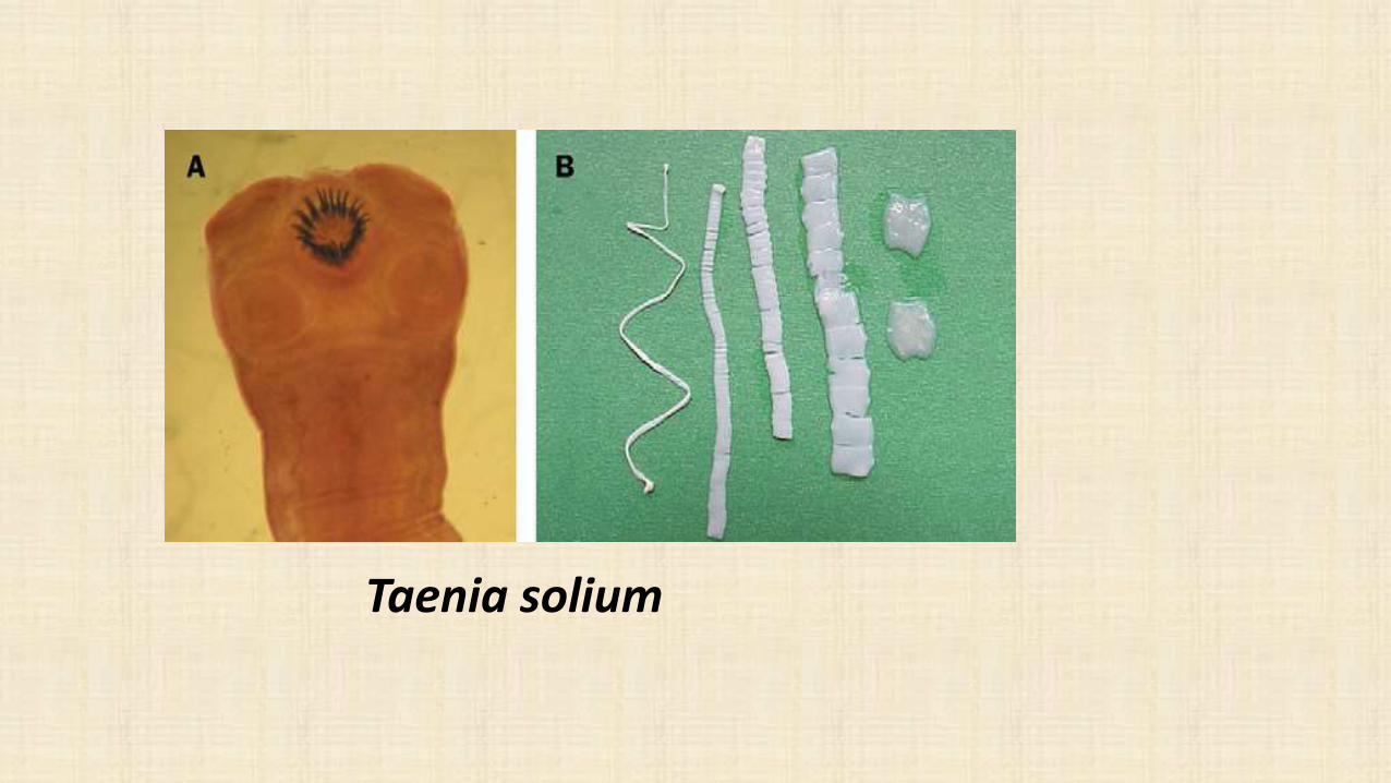
Taenia solium

Taenia saginata

Aspect Taenia saginata Taenia solium
Mature segment 2 ovary lobes 3 ovary lobes
Gravid segment 15 - 30 uterine branches 7 -13 uterine branches
Larvae Cysticercus bovis only in cattle, no scolex no hooklets
Cysticercus cellulosae in pig and man,scolex with hooklets
Egg in both species are indistinguiable
Morphological differences
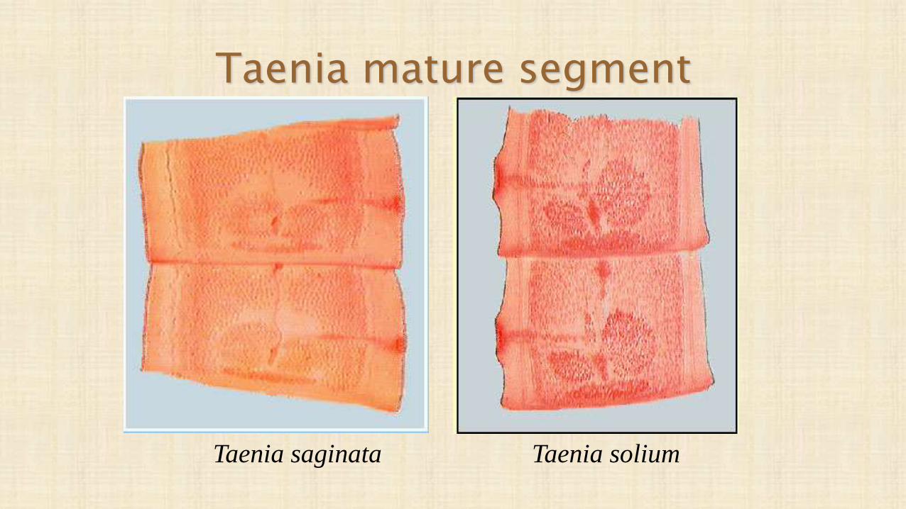
Taenia mature segment
Taenia soliumTaenia saginata

Taenia Gravid Proglotid
Taenia soliumTaenia saginata

Egg
• The eggs of Taenia saginata
and T. solium are indistinguishable
morphologically.
• The eggs are spherical, diameter
31 to 43 µm, with a thick radially
striated brown embryophore.
• Inside each is an oncosphere with
6 hooklets.

Life cycle of Taenia spp

Life cycle of Taenia solium

Neurocysticercosis
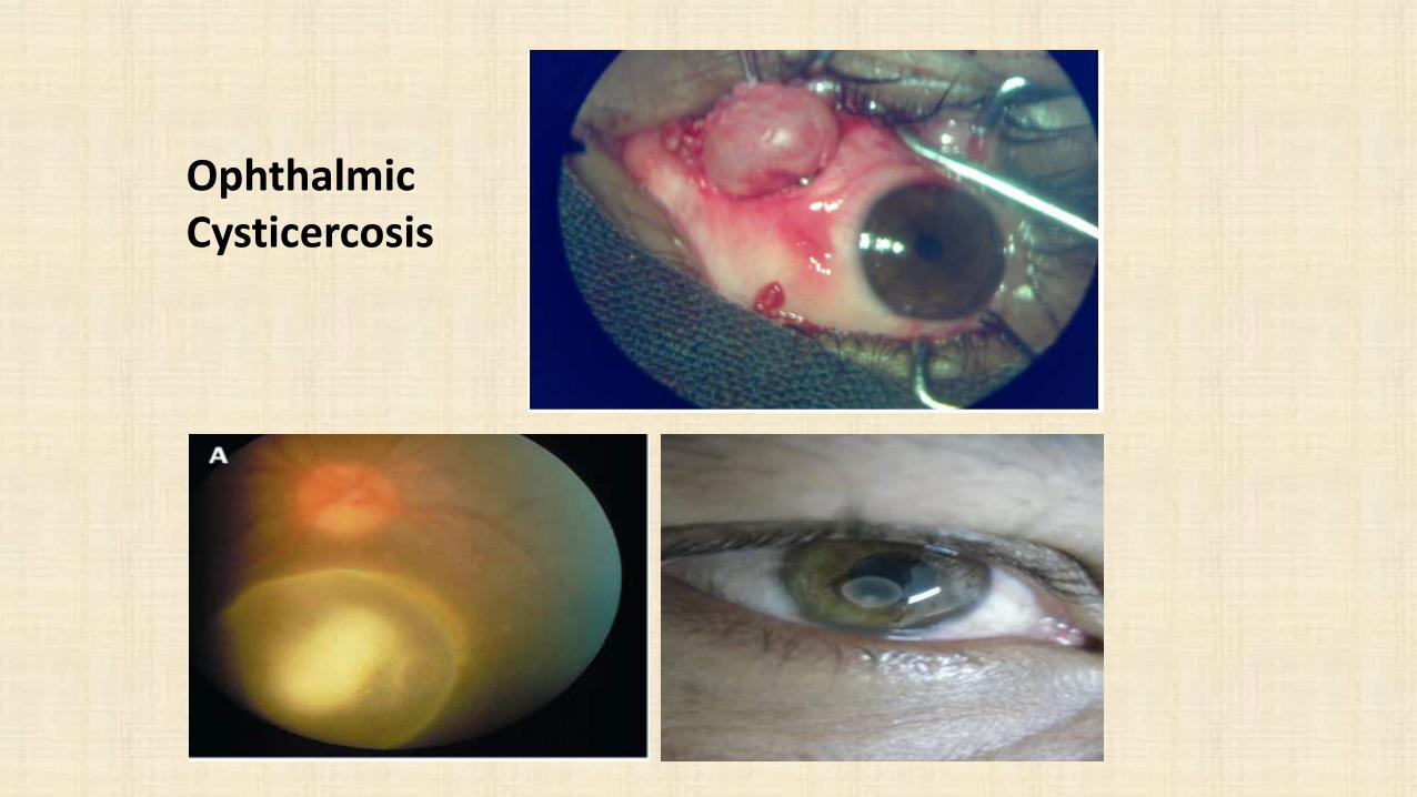
Ophthalmic Cysticercosis
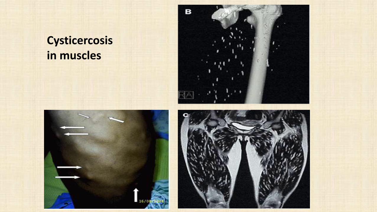
Cysticercosisin muscles

Mode of transmission
Taenia saginata
Man acquires infection by eating raw or undercooked beef containing encysted larval stage (Cysticercus bovis).
Mode of transmission
Taenia solium; man acquires infection by:
1- Eating raw or undercooked pork containing encysted larval stage (Cysticercus cellulosae).
2- Ingesting the egg with contaminated food or water.
3- A man harbouring adult worms may auto-infect either by unhygienic personal habits or by reverse peristaltic movements of the intestine whereby the gravid segments are thrown into the stomach.

Infective stage Taenia saginata Cysticercus bovis
Taenia solium Cysticercus cellulosae and egg
Diagnostic stageTaenia saginata gravid proglottids and egg
Taenia solium gravid proglottids and egg
T. asiatica …… cysticercus
T. multiceps …. Eggs

Pathogenesis
• Usually no symptoms, may cause abdominal discomfort, indigestion, diarrhea alternating with constipation and loss of appetite.
• Cysticercosis (by cysticercus cullulosae ) may occur in any organ.
• May develop in subcutaneous tissue and form visible nodules or in brain forming epileptic attacks, and interior vitreous chamber of the eye.

Laboratory diagnosis
1- Stool microscopy; same for both Taenia, by demonstration of eggs or gravid proglottides in stool by:
A- Direct fecal smear
B- Brine floatation technique
C- Cellophane-tape technique
NOTE; acid fast stain is used to distinguish the eggs, T.saginata is acid fast positive (red) while T.solium is negative (blue) or by detection of proglottids & scolex in feces.
Laboratory diagnosis cont..
2. Detection of Taenia Ag in faeces; Coproantigen is the most sensitive method (95% sensitivity and 99% specificity) to detect Taenia specific Ag in the stool but does not differentiate between species.
3- Molecular diagnosis; Includes DNA probes & PCR

Diagnosis of cysticercosis
1- Biopsy of subcutaneous nodule (cysticerci)
2- X-ray (calcified cysticerci), CT scan & MRI for neurocysticercosis.
3- Histopathological diagnosis (cysticercus in autopsy tissue).
4- Serological tests (IHA, IFA & ELISA anticysticercus Ab).
5- Differential leucocyte count (eosinophilia)
Treatment
• Praziquantel and niclosamide single dose of 4 tablets of 500 mg for T. saginata and T. solium.
• Praziquantel high dose over 3-7 days for T. solium.