Table of Contents SMN ELISA kit - Enzo Life Sciences
20
Product Manual SMN ELISA kit Catalog # ADI-900-209 For use with cell and tissue extracts of human and mouse origin The development of the SMN ELISA Kit was a collaborative effort between Enzo Life Sciences International, Inc. and the SMA Foundation. New Standard Formulation
Transcript of Table of Contents SMN ELISA kit - Enzo Life Sciences

Product Manual
SMN ELISA kit Catalog # ADI-900-209
For use with cell and tissue extracts of human and mouse origin
The development of the SMN ELISA Kit was a collaborative effort between Enzo Life Sciences International, Inc. and the SMA Foundation.
New Standard
Formulation
Product Manual
USE FOR RESEARCH PURPOSES ONLY
Unless otherwise specified expressly on the packaging, all products sold hereunder are intended for and may be used for research purposes only and may not be used for food, drug, cosmetic or household use or for the diagnosis or treatment of human beings. Purchase does not include any right or license to use, develop or otherwise exploit these products com-mercially. Any commercial use, development or exploitation of these prod-ucts or development using these products without the express written au-thorization of Enzo Life Sciences, Inc. is strictly prohibited. Buyer assumes all risk and liability for the use and/or results obtained by the use of the products covered by this invoice whether used singularly or in combination with other products.
LIMITED WARRANTY; DISCLAIMER OF WARRANTIES
These products are offered under a limited warranty. The products are guaranteed to meet all appropriate specifications described in the package insert at the time of shipment. Enzo Life Sciences’ sole obligation is to re-place the product to the extent of the purchasing price. All claims must be made to Enzo Life Sciences, Inc., within five (5) days of receipt of order. THIS WARRANTY IS EXPRESSLY IN LIEU OF ANY OTHER WARRAN-TIES OR LIABILITIES, EXPRESS OR IMPLIED, INCLUDING WARRAN-TIES OF MERCHANTABILITY, FITNESS FOR A PARTICULAR PUR-POSE, AND NON- INFRINGEMENT OF THE PATENT OR OTHER IN-TELLECTUAL PROPERTY RIGHTS OF OTHERS, AND ALL SUCH WARRANTIES (AND ANY OTHER WARRANTIES IMPLIED BY LAW) ARE EXPRESSLY DISCLAIMED.
TRADEMARKS AND PATENTS
Several Enzo Life Sciences products and product applications are covered by US and foreign patents and patents pending. Enzo is a trademark of Enzo Life Sciences, Inc.
ABOUT THE SMA FOUNDATION
Founded in 2003, the Spinal Muscular Atrophy Foundation is a nonprofit organization dedicated to accelerating progress towards a treatment and cure for spinal muscular atrophy. Since its inception, the Foundation has committed over $60 million to SMA research. In addition, the Foundation works to raise awareness and generate support for increased efforts in SMA among the leaders of industry and government. For more information about the Spinal Muscular Atrophy Foundation, visit www.smafoundation.org or call (646) 253-7100.
FOR RESEARCH USE ONLY.
NOT FOR USE IN DIAGNOSTIC PROCEDURES.
Product Manual
TABLE OF CONTENTS
Introduction ................................................................................................ 2
Principle ................................................................................................... .3
Materials Supplied ...................................................................................... 4
Storage ...................................................................................................... 5
Materials Needed but Not Supplied ............................................................ 5
Sample Handling ........................................................................................ 6
Reagent Preparation ................................................................................ 12
Assay Procedure ...................................................................................... 13
Calculation of Results .............................................................................. 14
Typical Results ......................................................................................... 15
Performance Characteristics .................................................................... 19
References ............................................................................................... 17
Contact Information .................................................................................. 18
Check our website for ad-ditional proto-cols, technical notes and FAQs.
Reagents re-quire separate storage condi-tions.

Catalog number: ADI-900-209 Revision: 01/30/2015 2
Product Manual
INTRODUCTION
The SMN ELISA kit is a complete kit for the quantitative de-termination of SMN in samples of human and mouse origin. The kit was developed in collaboration with the SMA Founda-tion (New York, NY). Please read the complete kit insert before performing this assay.
Survival Motor Neuron (SMN) is a ~38 kDa protein produced chiefly by the SMN1 gene, located on the telomeric portion of chromosome 5q1-4. A nearly identical centromeric copy of the gene (SMN2) also produces a small amount of full-length SMN protein, but due to a translationally silent CT transition that results in alternative splicing of the pre-mRNA, most of the re-sulting SMN is truncated, causing reduced protein stability and lower overall SMN levels5-6. Deletion or mutation of the SMN1 gene results in a reduced level of full-length SMN protein and manifests as a range of neuromuscular phenotypes in humans as the disease spinal muscular atrophy (SMA). SMA is charac-terized by muscle weakness and atrophy, functional disability and is the most common lethal genetic disease of infants and toddlers. Approximately one in 35 adults is a carrier of the SMN1 mutation. The incidence of SMA is 1 in 6,000 to 1 in 10,000 live births7.
SMN protein is present in the cell cytoplasm, and also in the nucleus where it is concentrated in “gem” structures associat-ed with Cajal bodies8-9. SMN protein is a constituent of Gemin-containing complexes, and is thought to participate in many aspects of RNA metabolism. SMN complexes have been shown to mediate the assembly of uridine-rich small nuclear ribonucleoproteins (snRNPs), which in turn act as critical com-ponents of spliceosomes10.
Protected by US Patent no. US. 6,080,577.

Catalog number: ADI-900-209 Revision: 01/30/2015 3
Product Manual
PRINCIPLE
1. Samples and standards are added to wells coated with a monoclonal antibody specific for SMN. The plate is then incubated.
2. The plate is washed, leaving only bound SMN on the plate. A yellow solution of rabbit polyclonal antibody to human SMN is then added. This binds the SMN captured on the plate. The plate is then incubated.
3. The plate is washed to remove excess antibody. A blue solution of HRP conjugate is added to each well that binds to the rabbit polyclonal SMN antibody. The plate is again incubated.
4. The plate is washed to remove excess HRP conjugate. TMB Substrate solution is added. An HRP-catalyzed reac-tion generates a blue color in the solution.
5. Stop solution is added to stop the substrate reaction. The resulting yellow color is read at 450nm. The amount of signal is directly proportional to the level of SMN in the sample.

Catalog number: ADI-900-209 Revision: 01/30/2015 4
Product Manual
MATERIALS SUPPLIED
1. SMN Microtiter Plate, One Plate of 96 Wells, Catalog No. 80-2311 A plate using break-apart strips coated with a mono-clonal antibody specific to SMN.
2. SMN Antibody, 10ml, Catalog No. 80-2313 A yellow solution of rabbit polyclonal antibody to human SMN.
3. Assay Buffer 13, 100ml, Catalog No. 80-1627 Tris buffered saline containing proteins and detergents.
4. SMN Conjugate, 10ml, Catalog No. 80-2314 A blue solution of goat anti-rabbit IgG conjugated to horseradish peroxidase.
5. Wash Buffer Concentrate, 100ml, Catalog No. 80-1287 Tris buffered saline containing detergents.
6. SMN (human) Standard, , Catalog No. 80-2312 Two vials each containing 3200pg of recombinant hu-man SMN.
7. TMB Substrate, 10ml, Catalog No. 80-0350 A solution of 3,3’,5,5’ tetramethylbenzidine (TMB) and hydrogen peroxide.
8. Stop Solution 2, 10ml, Catalog No. 80-0377 A 1N solution of hydrochloric acid in water. Keep tightly capped.
9. Extraction Reagent 4, 100ml, Catalog No. 80-2315
Tris buffered saline containing salts, detergents, enzyme and glycerol.
10. SMN Assay Layout Sheet, 1 each, Catalog No. 30-0291
11. Plate Sealer, 3 each, Catalog No. 30-0012
The standard should be han-dled with care due to the known and unknown ef-fects of the anti-gen.
Do not mix com-ponents from dif-ferent kit lots or use reagents be-yond the kit’s ex-piration date.
Stop solution is caustic. Keep tightly capped.
Protect sub-strate from pro-longed expo-sure to light.
Activity of conju-gate is affected by nucleophiles such as azide, cyanide, and hy-droxylamine.

Catalog number: ADI-900-209 Revision: 01/30/2015 5
Product Manual
STORAGE
All components of this kit, except the Standards, are stable at 4°C until the kit’s expiration date. The Standards should be stored at or below -20°C upon receipt.
MATERIALS NEEDED BUT NOT SUPPLIED
1. Deionized or distilled water.
2. Precision pipets for volumes between 10µl and 1,000µl.
3. Disposable polypropylene test tubes for dilution of sam-ples and standards.
4. Repeater pipets for dispensing 100µl.
5. Disposable beakers for diluting buffer concentrates.
6. Graduated cylinders.
7. A microplate shaker.
8. Adsorbent paper for blotting.
9. Microplate reader capable of reading at 450nm.
10. Graph paper for plotting the standard curve or, prefera-bly, an immunoassay software package capable of per-forming a 4 parameter logistic curve fit.
11. Hemocytometer for cell counts.
12. Cover slip for hemocytometer.
13. Trypan Blue 0.4%, Sigma #T8154 or equivalent.
14. Mechanical homogenizer or manual dounce homogeniz-er
15. Phosphate buffered saline.
16. Protease inhibitor cocktail (PIC), Sigma P8340 or equiva-lent.
17. Phenylmethlysulphonyl fluoride (PMSF), Sigma #P7626 or equivalent.
Reagents re-quire separate storage condi-tions.
Catalog number: ADI-900-209 Revision: 01/30/2015 6
Product Manual
SAMPLE HANDLING
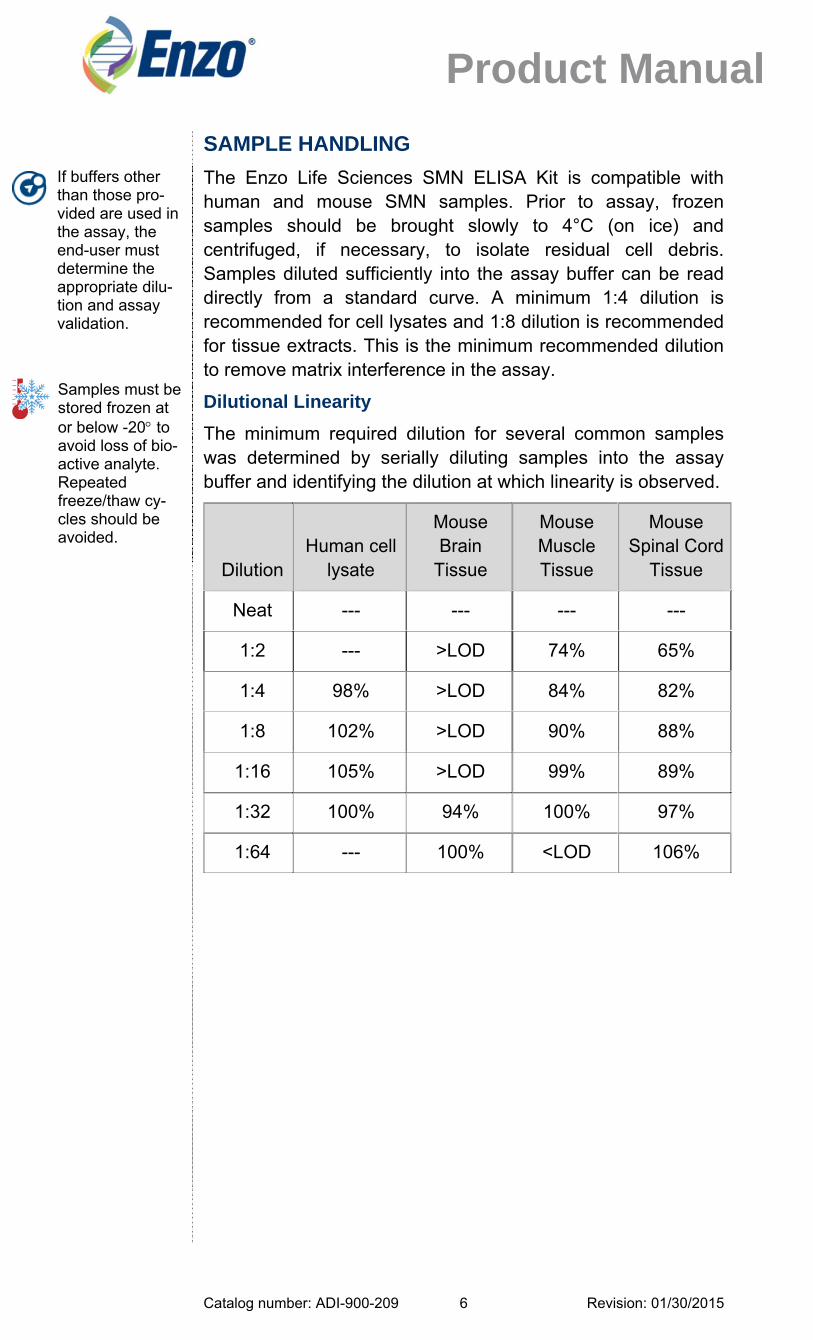
The Enzo Life Sciences SMN ELISA Kit is compatible with human and mouse SMN samples. Prior to assay, frozen samples should be brought slowly to 4°C (on ice) and centrifuged, if necessary, to isolate residual cell debris. Samples diluted sufficiently into the assay buffer can be read directly from a standard curve. A minimum 1:4 dilution is recommended for cell lysates and 1:8 dilution is recommended for tissue extracts. This is the minimum recommended dilution to remove matrix interference in the assay.
Dilutional Linearity
The minimum required dilution for several common samples was determined by serially diluting samples into the assay buffer and identifying the dilution at which linearity is observed.
Dilution
Human cell lysate
Mouse Brain
Tissue
Mouse Muscle Tissue
Mouse Spinal Cord
Tissue
Neat --- --- --- ---
1:2 --- >LOD 74% 65%
1:4 98% >LOD 84% 82%
1:8 102% >LOD 90% 88%
1:16 105% >LOD 99% 89%
1:32 100% 94% 100% 97%
1:64 --- 100% <LOD 106%
If buffers other than those pro-vided are used in the assay, the end-user must determine the appropriate dilu-tion and assay validation.
Samples must be stored frozen at or below -20 to avoid loss of bio-active analyte. Repeated freeze/thaw cy-cles should be avoided.

Catalog number: ADI-900-209 Revision: 01/30/2015 7
Product Manual
Spike and Recovery
After diluting each sample matrix to its minimum required dilu-tion, recombinant human SMN was spiked at high, medium, and low concentrations. The recovery of the standard in spiked samples was compared to the recovery of identical spikes in the assay buffer. The mean and range of percent recovery at the three concentrations are indicated below for each matrix.
Mean Spike & Recovery Results
Sample Matrix (# of samples)
Minimum Required dilution
Spike Concentration
(pg/mL)
Avg %Recovery
(Range)
Human PBMC lysate (n=5)
1:4
1667 100% (88-116%)
667 100% (88-116%)
267 99% (79-134%)
Mouse brain extract (n=2)
≥1:8a
1250 84% (83-84%)
250 86% (85-87%)
50 104% (96-112%)
Mouse muscle extract (n=2)
≥1:8b
1250 79% (76-81%)
250 88% (85-90%)
50 125% (103-146%)
Mouse spinal cord extract (n=2)
≥1:8c
1250 68% (66-69%)
250 68% (67-68%)
50 44% (39-48%)
a – Dilute mouse brain tissue extract such that the final protein concentration in the assay sample is 25µg/mL with a minimum dilution of 1:8.
b – Dilute mouse muscle tissue extract such that the final pro-tein concentration in the assay sample is 50µg/mL with a min-imum dilution of 1:8.
c – Dilute mouse spinal cord tissue extract such that the final protein concentration in the assay sample is 100µg/mL with a minimum dilution of 1:8.
Catalog number: ADI-900-209 Revision: 01/30/2015 8
Product Manual
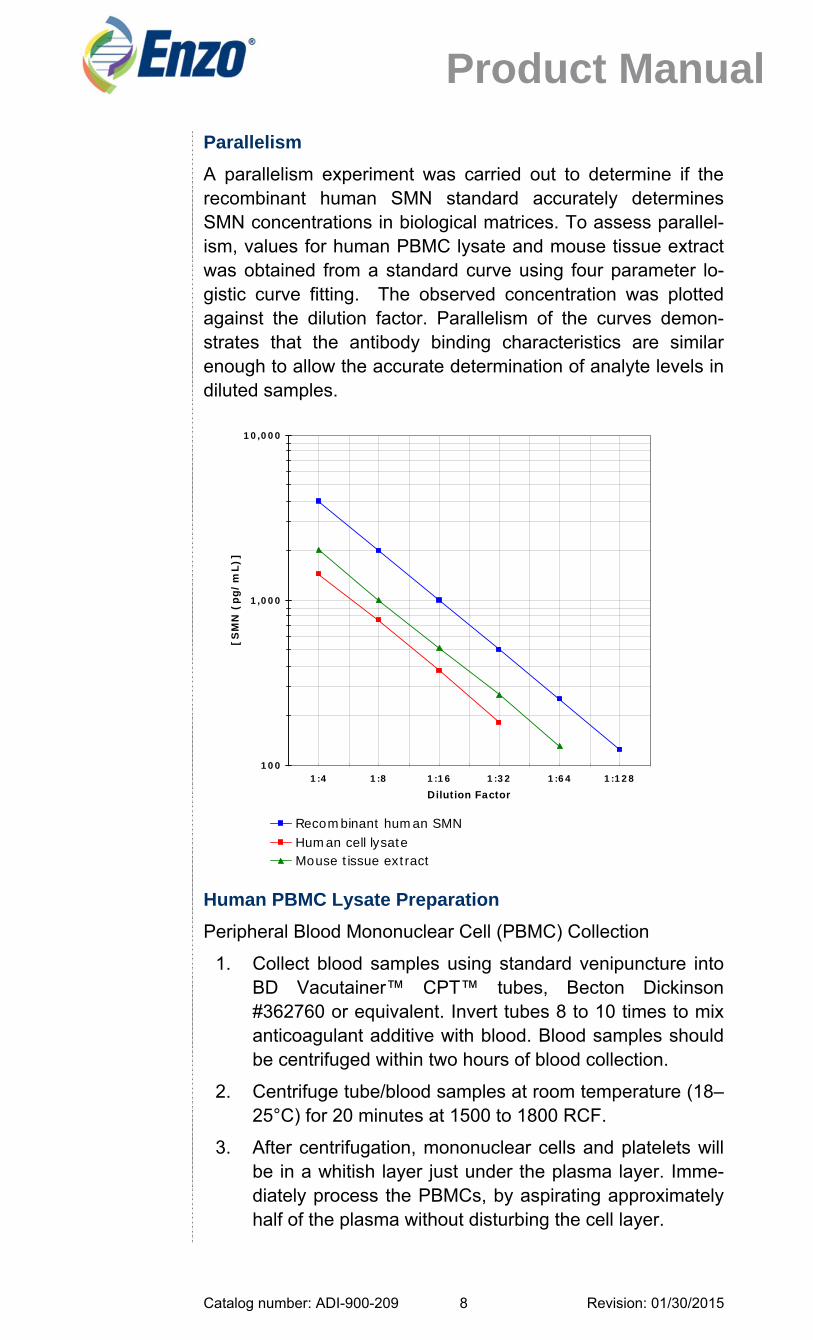
Parallelism
A parallelism experiment was carried out to determine if the recombinant human SMN standard accurately determines SMN concentrations in biological matrices. To assess parallel-ism, values for human PBMC lysate and mouse tissue extract was obtained from a standard curve using four parameter lo-gistic curve fitting. The observed concentration was plotted against the dilution factor. Parallelism of the curves demon-strates that the antibody binding characteristics are similar enough to allow the accurate determination of analyte levels in diluted samples.
Human PBMC Lysate Preparation
Peripheral Blood Mononuclear Cell (PBMC) Collection
1. Collect blood samples using standard venipuncture into BD Vacutainer™ CPT™ tubes, Becton Dickinson #362760 or equivalent. Invert tubes 8 to 10 times to mix anticoagulant additive with blood. Blood samples should be centrifuged within two hours of blood collection.
2. Centrifuge tube/blood samples at room temperature (18–25°C) for 20 minutes at 1500 to 1800 RCF.
3. After centrifugation, mononuclear cells and platelets will be in a whitish layer just under the plasma layer. Imme-diately process the PBMCs, by aspirating approximately half of the plasma without disturbing the cell layer.
100
1,000
10,000
1:4 1:8 1:16 1:32 1:64 1:128
Dilution Factor
[SM
N (
pg/m
L)]
Recombinant human SMNHuman cell lysateMouse tissue extract

Catalog number: ADI-900-209 Revision: 01/30/2015 9
Product Manual
4. Collect the cell layer and transfer to a 15ml conical cen-trifuge tube with cap.
5. Add PBS to the PBMCs to bring the volume to 15ml. Cap tube and invert to mix cells.
6. Centrifuge tube for 15 minutes at 300 RCF, 4°C. Aspirate supernatant without disturbing the cell pellet.
7. Resuspend cell pellet in residual PBS by gently vortexing or tapping tube with index finger.
8. Add PBS to resuspended pellet to bring volume to 10ml. Cap tube and invert to mix cells.
9. Centrifuge tube for 15 minutes at 300 RCF, 4°C. Aspirate supernatant without disturbing the cell pellet.
10. Repeat washing steps 8 and 9 for a total of 3 washes.
11. Assay immediately or freeze down in freezing media and store in liquid nitrogen. We recommend using the Nalgene® “Mr. Frosty” Container (or equivalent) and the manufacturer’s instruction manual.
PBMC Thawing
1. Remove vials containing frozen cells from liquid nitrogen and place in 37°C water bath.
2. Remove vials from water bath when no ice crystals re-main.
3. Transfer cell solution to 15ml conical centrifuge tube with cap.
4. Add PBS to the PBMCs to bring the volume to 15ml. Cap tube and invert to mix cells.
5. Centrifuge tube for 15 minutes at 300 RCF, 4°C. Aspirate supernatant without disturbing the cell pellet.
6. Resuspend cell pellet in 2ml PBS for performing cell counts.
Cell Counts with Hemocytometer
1. Transfer 50µl of cell suspension to a solution containing 75µl PBS and 125µl Trypan blue. Vortex the trypan-blue cell solution.
2. With the cover slip in place, transfer a small amount of trypan blue-cell suspension to a chamber on the hemo-cytometer. Ensure that the entire area under the cover slip contains the staining solution before removing any excess staining solution from the edge of the cover slip.

Catalog number: ADI-900-209 Revision: 01/30/2015 10
Product Manual
3. Both chambers of the hemocytometer must contain staining solution before performing cell counts.
4. Place hemocytometer on the microscope and count the number of trypan-blue excluding (viable) cells in the 4 outer squares. If there are less than 10 cells or more than 100 cells per square, repeat the procedure adjust-ing to an appropriate dilution factor.
5. Calculate the cell concentration as follows:
Cell concentration per milliliter = Total cell count in 4 squares x 2500 x 5 (dilution factor)
Total cell count = Cell concentration per milliliter x 2.0ml (cell suspension)
6. Centrifuge cell suspension for 10 minutes at 300 RCF, 4°C. Aspirate supernatant without disturbing cell pellet.
7. Proceed to cell lysis immediately.
Cell Lysis
1. Resuspend cell pellet in Extraction Reagent 4, containing protease inhibitors. Add 1ml of extraction reagent per 108 cells. See Reagent Preparation Section for addition of protease inhibitors to Extraction Reagent 4.
2. Incubate cell suspension on ice for 30 minutes for complete lysis.
3. Transfer cell lysis to 1.5ml centrifuge tube. Centrifuge cell lysates for 10 minutes at 14,000 RCF, 4°C.
4. Clarified lysates may be assayed immediately, or aliquoted and stored at -70°C.
5. No degradation of SMN in cell lysate was observed after 2 freeze-thaw cycles.

Catalog number: ADI-900-209 Revision: 01/30/2015 11
Product Manual
Cell Lysate Sample Handling
1. If cell lysates were frozen prior to assay, the frozen lysate samples should be brought slowly to 4°C (on ice) and, if residual precipitate is present, centrifuge to isolate residual cell debris. Samples diluted sufficiently into the assay buffer can be read directly from a standard curve. A minimum 1:4 dilution is recommended for cell lysates to remove matrix interference in the assay.
Mouse Tissue Homogenization
1. Prepare Extraction Reagent 4 with protease inhibitors. Recommended protease inhibitors are 0.5µl of PIC8340 per ml of reagent and PMSF to a final concentration of 1mM.
2. Transfer tissue sample to appropriate sized tube for ho-mogenization with 1mL of prepared Extraction Reagent 4.
3. For mechanical homogenizer, disrupt the tissue with three pulses of 3-4 seconds each. For manual dounce homogenizer, complete a minimum of 5 passes of the pestle past the buffer/tissue volume, or until tissue ap-pears completely homogenized. Keep samples on ice while completing all preparations.
4. Pellet out tissue/cellular debris via centrifugation at 14000g for 10 minutes at 4°C and transfer supernatant to a clean tube.
5. Measure the protein content of the supernatant using the biorad DC protein assay (with reagent S).
6. Prepare tissue homogenates for use in the SMN assay by diluting the extracted samples in assay buffer. Sam-ples must be diluted at least 1:8. Dilute brain, muscle, and spinal cord tissue samples to final assay protein concentrations of 25µg/mL, 50µg/mL and 100µg/mL, re-spectively.

Catalog number: ADI-900-209 Revision: 01/30/2015 12
Product Manual
REAGENT PREPARATION
1. Wash Buffer
Prepare the Wash Buffer by diluting 50ml of the supplied concentrate with 950ml of deionized water. This can be stored at room temperature until the kit expiration, or for 3 months, whichever is earlier.
2. Addition of Inhibitors to Extraction Reagent 4
Add protease inhibitors to extraction reagent prior to use. Add 0.5µl of PIC per ml of extraction reagent and add PMSF to a final concentration of 1mM. Do not store ex-traction reagent with protease inhibitors.
3. Human SMN Standards
Allow the 3200pg human SMN standard to warm to room temperature. Reconstitute one vial of standard with 1mL of assay buffer for a 3200pg/mL stock vial. Vortex thoroughly, wait 5 minutes and vortex again prior to use. Label the vial standard#1.
Label six disposable 12 x 75 mm polypropylene plastic tubes, or equivalent, #2 through #7. Standards can be prepared at room temperature and are stable up to 60 minutes. . Pipet 250µl assay buffer into tubes #2 through #7.. Remove 250µl from standard #1 and add to tube #2. Vortex thoroughly. Continue this for tubes #3 through #7.
Standards can be used at room temperature and should be used within 60 minutes of preparation. The concentration of SMN in the tubes is labeled above.
Bring all reagents to room tempera-ture for at least 30 minutes prior to opening.
Standards must be made up in plastic tubes.

Catalog number: ADI-900-209 Revision: 01/30/2015 13
Product Manual
ASSAY PROCEDURE
Refer to the Assay Layout Sheet to determine the number of wells to be used and put any remaining wellstrips with the desiccant back into the foil pouch and seal the bag. Store unused wellstrips at 4°C.
1. Pipet 100µl of assay buffer into the S0 (0pg/mL standard) wells.
2. Pipet 100µl of Standards #1 through #7 into the appropriate wells.
3. Pipet 100µl of the Samples into the appropriate wells.
4. Seal the plate. Incubate at room temperature on a plate shaker for 1 hour at ~500rpm.
5. Empty the contents of the wells and wash by adding 300µl of wash solution to every well. Repeat the wash 3 more times for a total of 4 washes. After the final wash, empty or aspirate the wells and firmly tap the plate on a lint free paper towel to remove any remaining wash buffer.
6. Pipet 100µl of yellow Antibody solution into each well, except the Blank.
7. Seal the plate. Incubate at room temperature on a plate shaker for 1 hour at ~500rpm.
8. Wash as above (Step 5).
9. Add 100µl of blue Conjugate solution to each well, except the Blank.
10. Seal the plate. Incubate at room temperature on a plate shaker for 30 minutes at ~500rpm.
11. Wash as above (Step 5). Pipet 100µl of Substrate Solution into each well.
12. Seal the plate. Incubate at room temperature on a plate shaker for 30 minutes at ~500rpm.
13. Pipet 100µl Stop Solution to each well.
14. Zero the plate reader against the Blank wells, read the optical density at 450nm. If the plate reader is not able to automatically subtract blank well values from each well, manually subtract the mean optical density of the Blank wells from all the readings.
Pipet the rea-gents to the sides of the wells to avoid possible contamination.
Pipet the stand-ards and sam-ples to the bot-tom of the wells.
Pre-rinse each pipet tip with re-agent. Use fresh pipet tips for each sample, standard, and reagent.

Catalog number: ADI-900-209 Revision: 01/30/2015 14
Product Manual
CALCULATION OF RESULTS
Several options are available for the calculation of the concen-tration of SMN in the samples. We recommend that the data be handled by an immunoassay software package utilizing a 4 parameter logistic curve fitting program. If data reduction soft-ware is not readily available, the concentration of SMN can be calculated as follows:
1. Calculate the average net Optical Density (OD) bound for each standard and sample by subtracting the average Blank OD from the average OD for each standard and sample.
Average Net OD = Average OD - Average Blank OD
2. Plot the Average Net OD for each standard versus SMN (human) concentration in each standard. Approximate a straight line through the points. The concentration of SMN in the unknowns can be determined by interpola-tion.
Samples with concentrations outside of the standard curve range will need to be re-analyzed using a different dilution.
Make sure to mul-tiply sample con-centrations by the dilution factor used during sample preparation.
Catalog number: ADI-900-209 Revision: 01/30/2015 15
Product Manual
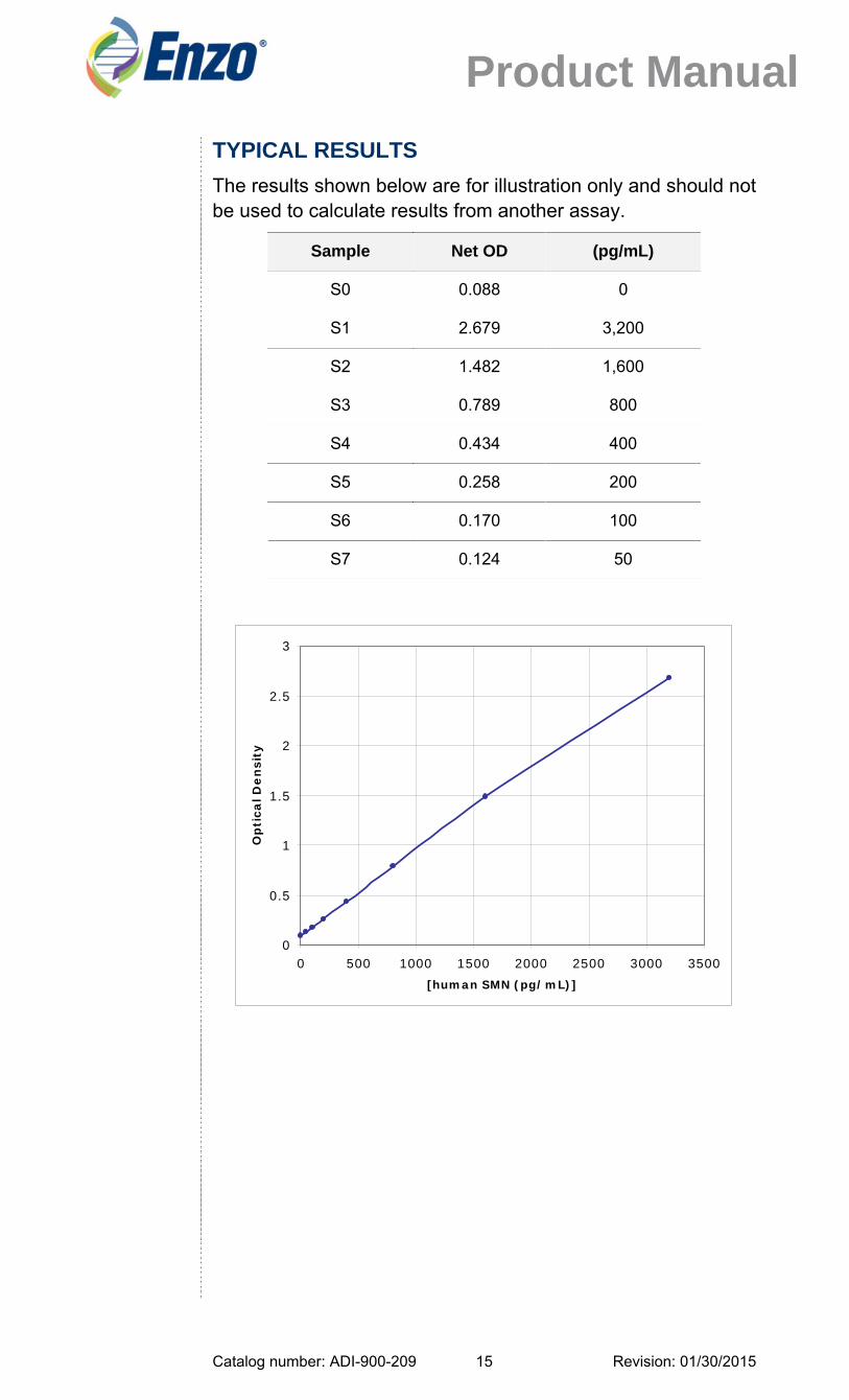
TYPICAL RESULTS
The results shown below are for illustration only and should not be used to calculate results from another assay.
Sample Net OD (pg/mL)
S0 0.088 0
S1 2.679 3,200
S2 1.482 1,600
S3 0.789 800
S4 0.434 400
S5 0.258 200
S6 0.170 100
S7 0.124 50
0
0.5
1
1.5
2
2.5
3
0 500 1000 1500 2000 2500 3000 3500[human SMN (pg/mL)]
Op
tica
l De
nsi
ty

Catalog number: ADI-900-209 Revision: 01/30/2015 16
Product Manual
PERFORMANCE CHARACTERISTICS
Specificity
This kit detects SMN protein of both human and mouse origin. Other species have not been tested.
Sensitivity
The sensitivity of the assay, defined as the concentration of SMN measured at 2 standard deviations from the mean of 20 replicates of zero standard along the standard curve, was de-termined to be 50pg/mL.
Precision
Intra-assay precision was determined by assaying 20 repli-cates of three buffer controls containing SMN in a single as-say.
pg/mL %CV
928 0.8
322 1.0
122 3.2
Inter-assay precision was determined by measuring buffer controls (n=12) of varying SMN concentrations in multiple as-says over several days.
pg/mL %CV
983 7.1
378 8.9
134 11.4

Catalog number: ADI-900-209 Revision: 01/30/2015 17
Product Manual
REFERENCES
1. Sumner, C.J. (2007) J Child Neurol. 22, 979-989.
2. Brzustowicz, L.M., et al. (1990) Nature 344, 540-541.
3. Melki, J., et al. (1990) Lancet 336, 271-273.
4. Lefebvre, S., et al. (1995) Cell 80, 155-165.
5. Lorson, C., et al. (1998) Nat Genet 19, 63-6.
6. Burnett, B.G., et al. (2009) Mol Cell Biol. 29, 1107-1115.
7. Pearn, J. (1978) J Med Genet. 15, 409-413.
8. Liu, Q., et al. (1997) Cell 90, 1013-1021.
9. Cavalho T., et al. (1999) J Cell Bio 147, 715-28.
10. Fischer, U., et al. (1997) Cell 90, 1023-9.
Catalog number: ADI-900-209 Revision: 01/30/2015 18
Product Manual
GLOBAL HEADQUARTERS Enzo Life Sciences Inc. 10 Executive Boulevard Farmingdale, NY 11735 Toll-Free:1.800.942.0430 Phone:631.694.7070 Fax: 631.694.7501 [email protected]
EUROPE/ASIA Enzo Life Sciences (ELS) AG Industriestrasse 17 CH-4415 Lausen Switzerland Phone:+41/0 61 926 89 89 Fax:+41/0 61 926 89 79 [email protected]
For local distributors and detailed product information visit us online: www.enzolifesciences.com