Table of Contents - nationalcprassociation.com · Updates to ACLS in 2015 As we learn more about...
28
Table of Contents Overview of Advanced Cardiovascular Life Support .............................................................. 2 Updates to ACLS in 2015................................................................................................. 3 Chain of Survival .............................................................................................................4 Adult BLS Chain of Survival ........................................................................................... 4 BLS and ACLS Surveys ....................................................................................................5 The BLS Survey........................................................................................................... 5 Solo Provider Adult BLS .......................................................................................... 6 Team Adult BLS ............................................................................................................ 7 Cardiac Arrest.................................................................................................................8 Ventricular Fibrillation and Pulseless Ventricular Tachycardia .............................................. 9 Ventricular Fibrillation and Pulseless Ventricular Tachycardia Algorithm ................................ 10 Pulseless Electrical Activity and Asystole ........................................................................ 11 PEA and Asystole Algorithm ............................................................................................ 12 Respiratory Arrest ......................................................................................................... 13 Airway Management ..................................................................................................14 Return of Spontaneous Circulation (ROSC) and Post Arrest Care ........................................ 16 Rapid Differential Diagnosis of Cardiac Arrest .................................................................. 17 Bradycardia ................................................................................................................. 18 Bradycardia Algorithm ................................................................................................ 18 Tachycardia ................................................................................................................. 19 Tachycardia Algorithm ................................................................................................ 20 Atrioventricular (Heart) Block ......................................................................................... 21 Acute Coronary Syndrome............................................................................................. 22 Acute Coronary Syndrome Algorithm............................................................................ 23 Acute Stroke ................................................................................................................ 24 Time is Brain! Stroke Time Goals for Evaluation and Therapy .......................................... 25 Team Dynamics ........................................................................................................... 26 Resuscitation and Life Support Medications ...................................................................... 27
Transcript of Table of Contents - nationalcprassociation.com · Updates to ACLS in 2015 As we learn more about...
Table of Contents
Overview of Advanced Cardiovascular Life Support .............................................................. 2
Updates to ACLS in 2015 ................................................................................................. 3
Chain of Survival ............................................................................................................. 4
Adult BLS Chain of Survival ........................................................................................... 4
BLS and ACLS Surveys .................................................................................................... 5
The BLS Survey ........................................................................................................... 5
Solo Provider Adult BLS .......................................................................................... 6
Team Adult BLS ............................................................................................................ 7
Cardiac Arrest ................................................................................................................. 8
Ventricular Fibrillation and Pulseless Ventricular Tachycardia .............................................. 9
Ventricular Fibrillation and Pulseless Ventricular Tachycardia Algorithm ................................ 10
Pulseless Electrical Activity and Asystole ........................................................................ 11
PEA and Asystole Algorithm ............................................................................................ 12
Respiratory Arrest ......................................................................................................... 13
Airway Management ..................................................................................................14
Return of Spontaneous Circulation (ROSC) and Post Arrest Care ........................................ 16
Rapid Differential Diagnosis of Cardiac Arrest .................................................................. 17
Bradycardia ................................................................................................................. 18
Bradycardia Algorithm ................................................................................................ 18
Tachycardia ................................................................................................................. 19
Tachycardia Algorithm ................................................................................................ 20
Atrioventricular (Heart) Block ......................................................................................... 21
Acute Coronary Syndrome ............................................................................................. 22
Acute Coronary Syndrome Algorithm ............................................................................ 23
Acute Stroke ................................................................................................................ 24
Time is Brain! Stroke Time Goals for Evaluation and Therapy .......................................... 25
Team Dynamics ........................................................................................................... 26
Resuscitation and Life Support Medications ...................................................................... 27

Overview of Advanced Cardiovascular Life Support
Advanced Cardiac Life Support, or ACLS, is a system of algorithms and best practice
recommendations intended to provide the best outcome for patients in cardiopulmonary crisis.
ACLS protocols are based on basic and clinical research, patient case studies, clinical studies, and
reflect the consensus opinion of experts in the field. While the term Advanced Cardiovascular Life
Support was coined by the American Heart Association, the content contained in this manual is
based on the most recent guidelines published by the American Heart Association, the American
College of Cardiology, the American Red Cross, and The European Society of Cardiology.
Support (BLS). Once you become certified in ACLS, the certification is valid for two years. However,
we encourage you to regularly login back in to your account to check for updates on resuscitation
science advances.
This Advanced Life Support provider manual includes:
➢ Updates to ACLS in 2015
➢ Solo and Team BLS
➢ The ACLS Survey
➢ Cardiac and Respiratory Arrest
➢ Return of Spontaneous Circulation (ROSC) and Post Arrest Care
➢ Bradycardia and Tachycardia Management
➢ ECG Rhythm Recognition
➢ Atrioventricular Blocks
➢ Acute Coronary Syndrome and Acute Stroke Management
➢ Resuscitation Medications
➢ Team Dynamics and Systems of Care
Prior to taking ACLS, it is assumed that you are proficient and currently certified in Basic Life

Updates to ACLS in 2015
As we learn more about resuscitation science and medicine, physicians and researchers realize
what works best and what works fastest in a critical, life-saving situation. Therefore, it is
necessary to periodically update life-support techniques and algorithms. If you have previously
certified in advanced cardiovascular life support, then you will probably be most interested in
what has changed since the latest update in 2010. The table below also includes changes
proposed since the last AHA manual was published. These changes will likely appear in future
editions of the provider manual.
Chain of Survival
Advanced Cardiovascular Life Support continues to emphasize the Chain of Survival. The Chain
of Survival is a sequence of steps or links that, when followed to its completion, increases the
likelihood that a victim of a life-threatening event will survive. The adult and pediatric chains of
survival are slightly different. The person who is providing BLS is only responsible for the early
links, that is, making sure the person is cared for by emergency personnel. The emphasis on
early care is to reinforce that time is a critical factor in life support care. The 2015 standards
include the concept of out of hospital care versus in-hospital care.
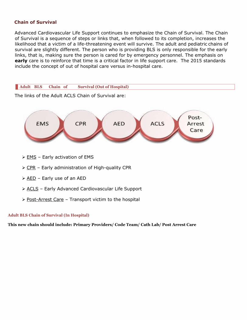
Adult BLS Chain of Survival (Out of Hospital)
The links of the Adult ACLS Chain of Survival are:
➢ EMS – Early activation of EMS
➢ CPR – Early administration of High-quality CPR
➢ AED – Early use of an AED
➢ ACLS – Early Advanced Cardiovascular Life Support
➢ Post-Arrest Care – Transport victim to the hospital
Adult BLS Chain of Survival (In Hospital) This new chain should include: Primary Providers/ Code Team/ Cath Lab/ Post Arrest Care

BLS and ACLS Surveys
ACLS draws heavily on Basic Life Support (BLS). In fact, it is assumed that all people who are
pursuing ACLS will be competent in the techniques of BLS—so much so that it is considered a
prerequisite to ACLS.
The first step in any resuscitation is to make sure the
rescuers (you!) and the victim are safe. Therefore, if
your victim is in the middle of the highway or in a
burning building, the first step is to move the victim
to safety.
Assuming you and the victim are in a safe location,
the next step is to assess whether the patient is
responsive
If patient is not responsive, move to BLS survey
If patient is responsive, move to ACLS survey
The BLS Survey
The BLS Survey
1. Responsive? Shake and Shout! Don’t be afraid to make noise.
Check for effective breathing for 5 to 10 seconds.
2. Activate EMS/Call Code In the hospital, you can call a “code” and send someone to get a
defibrillator. In the community, call 911 and send for an AED
3. Circulation Simultaneously, check the carotid pulse for no more than 10
seconds while checking for respiratory effort. If no pulse, begin
high quality CPR.
4. Defibrillation If there is a shockable rhythm, pulseless ventricular tachycardia or
ventricular fibrillation, provide a shock
Adult BLS is slightly different if there is one provider (solo) or more than one provider (team)
present. The difference between solo provider BLS and team BLS is that responsibilities are
shared when more than one person is present. These will be detailed in Solo and Team Adult
BLS.
For healthcare providers, the difference between a witnessed cardiac arrest and a victim who is
found down is the order of the initial steps.
➢ If you are alone and witness a victim suddenly collapse: Assume cardiac arrest with
a shockable rhythm. If you can get an AED quickly, you may activate EMS, leave the
victim to get an AED, provide CPR for 2 minutes, and use the AED.
➢ If you are alone and find an unresponsive adult: Tailor response to the prospective
cause of injury.
➢ If you suspect cardiac arrest: Activate EMS, get AED, 2 min of CPR, use AED
➢ If you suspect asphyxia: 2 min of CPR, Activate EMS, get AED, use AED

➢ Always make sure that you are safe and the
victim is safe before you start BLS.
➢ Check to see if the victim is responsive. Shake
and shout! Is the victim breathing effectively?
Does the victim have a pulse in the carotid
artery?
➢ If you witnessed the victim suddenly collapse,
assume cardiac arrest with a shockable
rhythm. If you can get an AED quickly, you
may activate EMS, leave the victim to get an
AED, CPR for 2 minutes, and use AED.
➢ If you find an unresponsive adult, tailor
response to the presumed cause of injury.
➢ If you suspect cardiac arrest: Activate
EMS, get AED, 2 min of CPR, use AED
➢ If you suspect asphyxia: 2 min of CPR,
Activate EMS, get AED, use AED
➢ High Quality CPR includes
➢ Fast and deep compressions, 100
to 120 compressions per minute
➢ Two inches deep, complete rebound
➢ If you can provide respiration, 2
breaths for 30 comps
➢ If you cannot provide respiration,
just give chest comps
➢ Check for a pulse and cardiac rhythm every two minutes. Follow directions on the AED.
After providing a shock, immediately resume CPR. Keep going until EMS arrives or the
victim regains circulation.

➢ Always make sure that your team is safe and the
victim is safe before you start BLS.
➢ Check to see if the victim is responsive. Shake and
shout! Is the victim breathing effectively? Does
the victim have a pulse in the carotid artery?
➢ One provider activates EMS and retrieves an AED.
The other provider(s) stays with the victim.
➢ Provide High Quality CPR includes
➢ Fast and deep compressions, 100
to 120 compressions per minute
➢ Two inches deep, complete rebound
➢ If you can provide breaths, 2 breaths
for 30 comps
➢ If you cannot provide breaths, just
give chest comps
➢ The provider who retrieved the AED applies the
AED and follows directions given by the device.
The provider that stayed with the victim provides
CPR until the AED is ready.
➢ Check for a pulse and cardiac rhythm every two
minutes. Follow directions on the AED. If
a shock is indicated, clear everyone and administer a shock. After providing a shock,
immediately resume Team CPR.
➢ In Team CPR, the provider giving chest compressions changes every 2 minutes
➢ Keep going until EMS arrives or the victim regains spontaneous circulation.

Cardiac arrest is the sudden sensation cessation of blood flow to the tissues in brain the results
from a heart that is not pumping effectively. Four rhythms may occur during cardiac arrest:
ventricular fibrillation, pulseless ventricular tachycardia, pulseless electrical activity, and
asystole. The primary intervention for ventricular fibrillation and pulseless ventricular tachycardia
is unsynchronized cardioversion, more commonly known as a “shock.” The primary intervention
for pulseless electrical activity and asystole is pharmacological, beginning with the administration
of epinephrine.
While ACLS provides algorithms for each of these cardiac arrest rhythms, in the real world a
patient may move between these rhythms during a single instance of cardiac arrest. Therefore,
the provider must be able to accurately assess and adapt to changing circumstances. After every
2 minutes of CPR, check for a pulse and check the cardiac rhythm. If the rhythm has switched
from shockable or to shockable, then switch algorithms.
Cardiac Arrest

In ventricular fibrillation or pulseless ventricular tachycardia, the heart's conduction system
exhibits a disordered rhythm that can sometimes be corrected by applying energy to it. This
energy may come in the form of an automated external defibrillator (AED) defibrillator paddles,
or defibrillator pads. VFib and VTach are treated with unsynchronized cardioversion, since there
is no way for the defibrillator to decipher the disordered waveform. In fact, it is important not to
provide synchronized shock for these rhythms.
Ventricular fibrillation is recognized by a disordered waveform, appearing as rapid peaks and
valleys as shown in this ECG rhythm strip:
Ventricular tachycardia may provide waveform similar to any other tachycardia; however, the
biggest difference in cardiac arrest is that the patient will not have a pulse and, consequently,
will be unconscious and unresponsive. Two examples of ventricular tachycardia are shown in this
ECG rhythm strips. The first is narrow complex tachycardia and the second is wide complex
tachycardia:
Ventricular Fibrillation and Pulseless Ventricular Tachycardia

Ventricular Fibrillation and Pulseless Ventricular Tachycardia Algorithm
Once you have determined that
a patient has a shockable
rhythm, immediately provide an
unsynchronized shock.
If you are using biphasic energy,
use recommended settings on
the device. If you do not know
what that setting is, use the
highest available setting, (120 to
200 J).
If you are using a monophasic
energy source, administer 360 J.
Resume CPR immediately after a
shock. Minimize interruptions of
chest compressions. Provide 2
rescue breaths for each 30
compressions.
Epinephrine (1 mg IV/IO) is
given every 3 to 5 minutes (two
2 minute cycles of CPR)
Vasopressin (40 units IV/IO) can
be used instead of the first or
second dose of epinephrine
Amiodarone (IV/IO)
o First dose 300 mg
o Second dose 150 mg
Lidocaine may replace amiodarone when amiodarone is not available.
o First dose: 1-1.5 mg/kg IV
o Second dose: 0.5-0.75 mg/kg IV every 5 to 10 min
If the arrest rhythm is no longer shockable, move to PEA/Asystole algorithm
If the patient regains consciousness, move to ROSC algorithm for him and him
➢
➢
➢
➢
➢
➢
➢
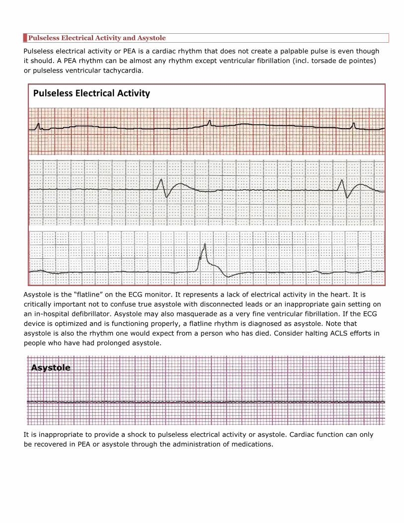
Pulseless electrical activity or PEA is a cardiac rhythm that does not create a palpable pulse is even though
it should. A PEA rhythm can be almost any rhythm except ventricular fibrillation (incl. torsade de pointes)
or pulseless ventricular tachycardia.
Asystole is the “flatline” on the ECG monitor. It represents a lack of electrical activity in the heart. It is
critically important not to confuse true asystole with disconnected leads or an inappropriate gain setting on
an in-hospital defibrillator. Asystole may also masquerade as a very fine ventricular fibrillation. If the ECG
device is optimized and is functioning properly, a flatline rhythm is diagnosed as asystole. Note that
asystole is also the rhythm one would expect from a person who has died. Consider halting ACLS efforts in
people who have had prolonged asystole.
It is inappropriate to provide a shock to pulseless electrical activity or asystole. Cardiac function can only
be recovered in PEA or asystole through the administration of medications.
Pulseless Electrical Activity and Asystole

As long as the patient is in PEA
or asystole, the rhythm is not
shockable.
Chest compressions/high-quality
CPR should be interrupted as
little as possible during
resuscitation.
After 2 min. of high-quality CPR,
give 1 mg of epinephrine IV/IO.
Consider advanced airway and
waveform capnography.
Remember, chest compressions
are a means of artificial
circulation, which should deliver
the epinephrine to the heart.
Without chest compressions,
epinephrine is not likely to be
effective.
Chest compressions should be
continued while epinephrine is
administered.
Rhythm checks every 2 min.
Epinephrine (1 mg IV/IO) is given every 3 to 5 minutes (after two, 2 minute cycles of CPR)
Vasopressin (40 units IV/IO) can be used instead of the first or second dose of epinephrine
If the arrest rhythm becomes shockable, move to VFib/Pulseless VTach algorithm
If the patient regains circulation, move to ROSC algorithm
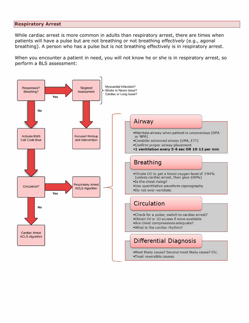
While cardiac arrest is more common in adults than respiratory arrest, there are times when
patients will have a pulse but are not breathing or not breathing effectively (e.g., agonal
breathing). A person who has a pulse but is not breathing effectively is in respiratory arrest.
When you encounter a patient in need, you will not know he or she is in respiratory arrest, so
perform a BLS assessment:
Respiratory Arrest
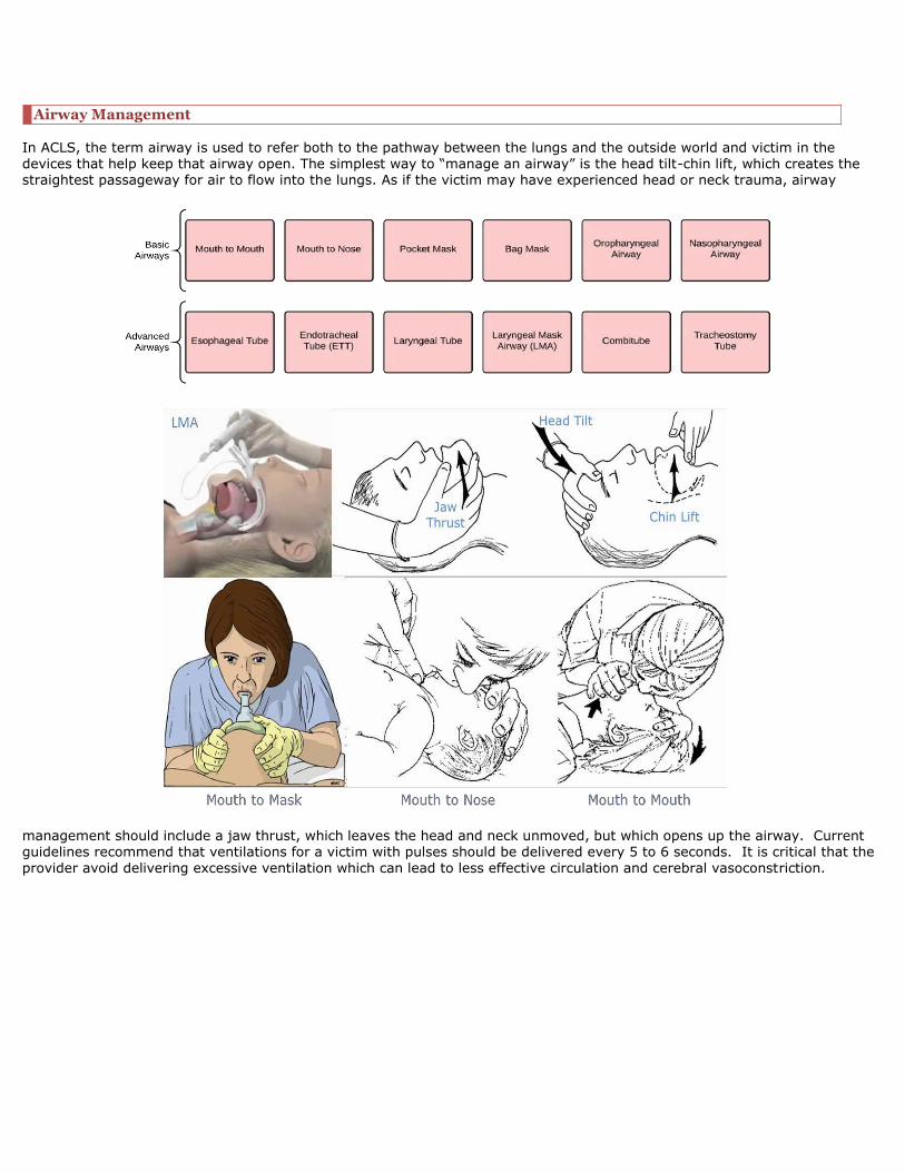
In ACLS, the term airway is used to refer both to the pathway between the lungs and the outside world and victim in the devices that help keep that airway open. The simplest way to “manage an airway” is the head tilt-chin lift, which creates the straightest passageway for air to flow into the lungs. As if the victim may have experienced head or neck trauma, airway
management should include a jaw thrust, which leaves the head and neck unmoved, but which opens up the airway. Current guidelines recommend that ventilations for a victim with pulses should be delivered every 5 to 6 seconds. It is critical that the
provider avoid delivering excessive ventilation which can lead to less effective circulation and cerebral vasoconstriction.
Airway Management

If one is to use a pocket mask or a bag mask to perform ventilations, it is important to make a tight seal with a mask
on the victim's face. Proper use of these masks may require the rescuer to use one or even two hands to secure the
mask to the victim's face.
A nasopharyngeal airway, which extends from the nose to the pharynx, can be used in both conscious and unconscious
patients. An oropharyngeal airway can only be used in unconscious patients because it may stimulate the gag reflex.
Advanced airways such as endotracheal tubes (ET tubes) and laryngeal mask airways (LMAs) usually require specialized
training, but are useful in-hospital resuscitations (especially LMAs).

While nasopharyngeal and oropharyngeal airways are basic airways, they do require a bit of
preparation and skill to use correctly.
•Select an airway that is the correct size for the patient
•Place the device at the side of the patient’s face. Choose the device that extends from the tip of the nose to the earlobe. Use the largest diameter device that will fit.
•Lubricate the airway with a water-soluble lubricant
•Insert the device slowly, straight into the face (not toward the brain!)
•It should feel snug; do not force
the device. If it feels stuck,
remove it and try the other
nostril.
•Adequate suctioning usually requires negative pressures of - 80 to -120 mmHg. Wallmounted suction can deliver this, but portable devices may not.
•When suctioning the oropharynx, do not insert the catheter too deeply. Extend the catheter to the maximum safe depth and suction as you withdraw.
•When suctioning an endotracheal tube, remember that the tube is within the trachea and you may be suctioning near the bronchi/lung. Therefore sterile technique should be used.
•Each suction attempt should be for no longer than 10 seconds. Prior to suctioning, give a brief period of 100% oxygen— remember that the patient will get no oxygen during suctioning.
•Monitor vital signs during
suctioning and stop suctioning
immediately if the patient
experiences hypoxemia (O2 sats <94%), has a new arrhythmia, or
becomes cyanotic.
Inserting an
Oropharyngeal
Airway
Inserting an
Nasopharyngeal
Airway
Tips on Suctioning
•Select an airway that is the correct size for the patient
•Too big and it will damage the throat
•Too small and it will press the tongue into the airway
•Place the device at the side of the patient’s face. Choose the device that extends from the corner of the mouth to the earlobe
•Clear the mouth or blood or secretions with suction, if possible.
•Insert the device so that the point is toward the roof of the mouth or parallel to the teeth
•Do not press the tongue back into the throat
•Once the device is almost fully
inserted, turn it so that the
tongue is cupped by the interior
curve of the device.

Return of Spontaneous Circulation (ROSC) and Post Arrest Care
The patient who has been successfully resuscitated
will regain spontaneous circulation.
You can detect spontaneous circulation by feeling a
palpable pulse at the carotid artery.
Even after Return of Spontaneous Circulation (ROSC),
the patient still needs close attention and support. The
patient is at risk for reentering cardiac arrest at any
time. Therefore, the patient should be moved to an
intensive care unit.
➢ Titrate the patient's blood oxygen levels to ≥94%
Does the person need an advanced airway? If so, it
should be placed.
Add quantitative waveform capnography
Titrate the patient's systolic blood pressure to at least
90 mmHg. this may require the addition of fluids
and/or vasopressors o Epinephrine IV 0.1-0.5
mcg/kg/min o Dopamine IV 5-10 mcg/kg/min o
Norepinephrine IV 0.1-0.5 mcg/kg/min o 1-2 liters of
fluid resuscitation
➢ Does the person follow verbal commands? If not, there
may be neurological compromise. Consider inducing
therapeutic hypothermia with 4°C fluids during fluid
resuscitation.
Does the person have signs of myocardial infarction by
ECG? Move to ACS algorithm.
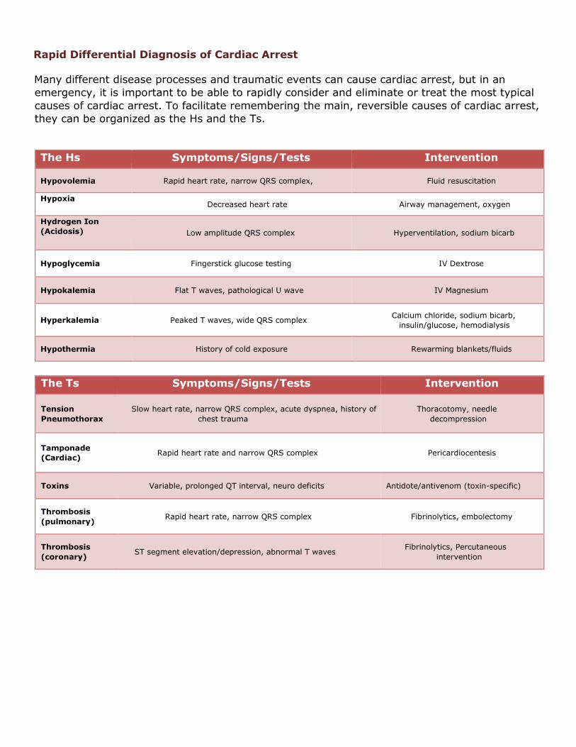
Rapid Differential Diagnosis of Cardiac Arrest
Many different disease processes and traumatic events can cause cardiac arrest, but in an
emergency, it is important to be able to rapidly consider and eliminate or treat the most typical
causes of cardiac arrest. To facilitate remembering the main, reversible causes of cardiac arrest,
they can be organized as the Hs and the Ts.
The Hs Symptoms/Signs/Tests Intervention
Hypovolemia Rapid heart rate, narrow QRS complex, Fluid resuscitation
Hypoxia Decreased heart rate Airway management, oxygen
Hydrogen Ion
(Acidosis)
Low amplitude QRS complex
Hyperventilation, sodium bicarb
Hypoglycemia Fingerstick glucose testing IV Dextrose
Hypokalemia Flat T waves, pathological U wave IV Magnesium
Hyperkalemia
Peaked T waves, wide QRS complex
Calcium chloride, sodium bicarb,
insulin/glucose, hemodialysis
Hypothermia History of cold exposure Rewarming blankets/fluids
The Ts Symptoms/Signs/Tests Intervention
Tension
Pneumothorax
Slow heart rate, narrow QRS complex, acute dyspnea, history of
chest trauma
Thoracotomy, needle
decompression
Tamponade
(Cardiac)
Rapid heart rate and narrow QRS complex
Pericardiocentesis
Toxins Variable, prolonged QT interval, neuro deficits Antidote/antivenom (toxin-specific)
Thrombosis
(pulmonary)
Rapid heart rate, narrow QRS complex
Fibrinolytics, embolectomy
Thrombosis
(coronary)
ST segment elevation/depression, abnormal T waves
Fibrinolytics, Percutaneous
intervention

Bradycardia
Bradycardia is any heart rate less than
60 bpm. In practice, however,
bradycardia is only a concern if it is
unusual or abnormal for the patient or
causing symptoms.
New cases of bradycardia should be
evaluated, but most will not require
specific treatment.
Evaluation of bradycardia includes
cardiac and blood oxygen
monitoring and a 12 lead ECG if
available.
Bradycardia may be treated by providing
supplemental oxygen and supporting the
patient's airway if needed.
Unstable bradycardia (i.e., an
abnormally slow heart rate that causes
altered mental status, hypotension,
symptoms of shock, cardiac chest pain,
or new signs and symptoms of heart
failure) should be treated immediately.
Unstable bradycardia is first treated
with intravenous atropine at a dose of
0.5 mg. Additional doses can be given
every 3 to 5 min. up to a maximum of 3
mg. Pulseless bradycardia is considered
PEA.
If atropine is unsuccessful in treating
symptomatic, unstable bradycardia,
consider transcutaneous pacing, dopamine or epinephrine infusion, or transvenous pacing. An
intensive or cardiologist may need to be consulted for these interventions and the patient
may need to be moved to the intensive care unit

Tachycardia
Atrial fibrillation is the most common arrhythmia. It is diagnosed by electrocardiogram,
specifically the RR intervals follow no repetitive pattern. Some leads may show P waves while
most leads do not. Atrial contraction rates may exceed 300 bpm. The ventricular rate often
range is between 100 to 180 bpm. The pulse may be “irregularly irregular.”
Atrial flutter is a cardiac arrhythmia that generates rapid, regular atrial depolarizations at a rate
of about 300 bpm. This often translates to a regular ventricular rate of 150 bpm, but may be far
less if there is a 3:1 or 4:1 conduction. By electrocardiogram, or atrial flutter is recognized by a
sawtooth pattern sometimes called F waves. These waves are most notable in leads II, III, and
aVF.
Narrow QRS complex tachycardias include several different tachyarrhythmias. A narrow QRS
complex tachycardia is distinguished by a QRS complex of less than 120 ms. One of the more
common narrow complex tachycardias is supraventricular tachycardia, shown below.
Wide complex tachycardias are difficult to distinguish from ventricular tachycardia. Ventricular
tachycardia leading to cardiac arrest should be treated using the ventricular
tachycardia algorithm. A wide complex tachycardia in a conscious person should be treated using
the tachycardia algorithm.
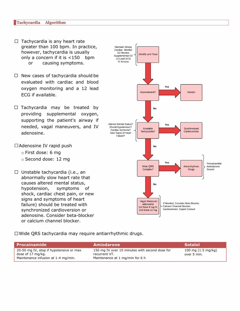
Tachycardia is any heart rate
greater than 100 bpm. In practice,
however, tachycardia is usually
only a concern if it is <150 bpm
or causing symptoms.
New cases of tachycardia should be
evaluated with cardiac and blood
oxygen monitoring and a 12 lead
ECG if available.
Tachycardia may be treated by
providing supplemental oxygen,
supporting the patient's airway if
needed, vagal maneuvers, and IV
adenosine.
Adenosine IV rapid push
o First dose: 6 mg
o Second dose: 12 mg
Unstable tachycardia (i.e., an
abnormally slow heart rate that
causes altered mental status,
hypotension, symptoms of
shock, cardiac chest pain, or new
signs and symptoms of heart
failure) should be treated with
synchronized cardioversion or
adenosine. Consider beta-blocker
or calcium channel blocker.
Wide QRS tachycardia may require antiarrhythmic drugs.
Procainamide Amiodarone Sotalol
20-50 mg IV, stop if hypotensive or max dose of 17 mg/kg.
Maintenance infusion at 1-4 mg/min.
150 mg IV over
recurrent VT. Maintenance at
10 minutes with
1 mg/min for 6 h
second dose for 100
over
mg (1.5
5 min.
mg/kg)
Algorithm

Atrioventricular block or heart block is a failure of the heart's electrical system to properly coordinate conduction.
There are four main types of atrioventricular block: first degree, second degree type I, second degree type II, and
third degree heart block. The types of second degree heart block are referred to as Mobitz type I and Mobitz type II.
Second degree heart block Mobitz type I is also known as the Wenckebach phenomenon.
Atrioventricular blocks may be acute or chronic. Chronic heart block may be treated with pacemaker devices. From the
perspective of ACLS assessment and intervention, heart block is important because it can cause hemodynamic
instability and can evolve into cardiac arrest. In ACLS, heart block is often treated as a bradyarrhythmia.
Atrioventricular (Heart) Block
The PR interval is a
consistent size, but
longer or larger than it
should be in first degree
heart block.
The PR interval increases
in size until a QRS
complexes dropped,
resulting in missed
“beat.”
A QRS wave will
occasionally drop,
though the PR
interval is the
same size.
Complete dissociation
between P waves and
the QRS complex. No
atrial impulses reach the
ventricle.
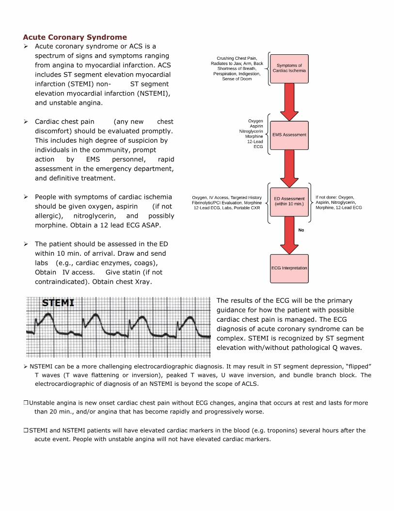
Acute Coronary Syndrome
Acute coronary syndrome or ACS is a
spectrum of signs and symptoms ranging
from angina to myocardial infarction. ACS
includes ST segment elevation myocardial
infarction (STEMI) non- ST segment
elevation myocardial infarction (NSTEMI),
and unstable angina.
Cardiac chest pain (any new chest
discomfort) should be evaluated promptly.
This includes high degree of suspicion by
individuals in the community, prompt
action by EMS personnel, rapid
assessment in the emergency department,
and definitive treatment.
People with symptoms of cardiac ischemia
should be given oxygen, aspirin (if not
allergic), nitroglycerin, and possibly
morphine. Obtain a 12 lead ECG ASAP.
The patient should be assessed in the ED
within 10 min. of arrival. Draw and send
labs (e.g., cardiac enzymes, coags),
Obtain IV access. Give statin (if not
contraindicated). Obtain chest Xray.
The results of the ECG will be the primary
guidance for how the patient with possible
cardiac chest pain is managed. The ECG
diagnosis of acute coronary syndrome can be
complex. STEMI is recognized by ST segment
elevation with/without pathological Q waves.
➢ NSTEMI can be a more challenging electrocardiographic diagnosis. It may result in ST segment depression, “flipped”
T waves (T wave flattening or inversion), peaked T waves, U wave inversion, and bundle branch block. The
electrocardiographic of diagnosis of an NSTEMI is beyond the scope of ACLS.
Unstable angina is new onset cardiac chest pain without ECG changes, angina that occurs at rest and lasts for more
than 20 min., and/or angina that has become rapidly and progressively worse.
STEMI and NSTEMI patients will have elevated cardiac markers in the blood (e.g. troponins) several hours after the
acute event. People with unstable angina will not have elevated cardiac markers.
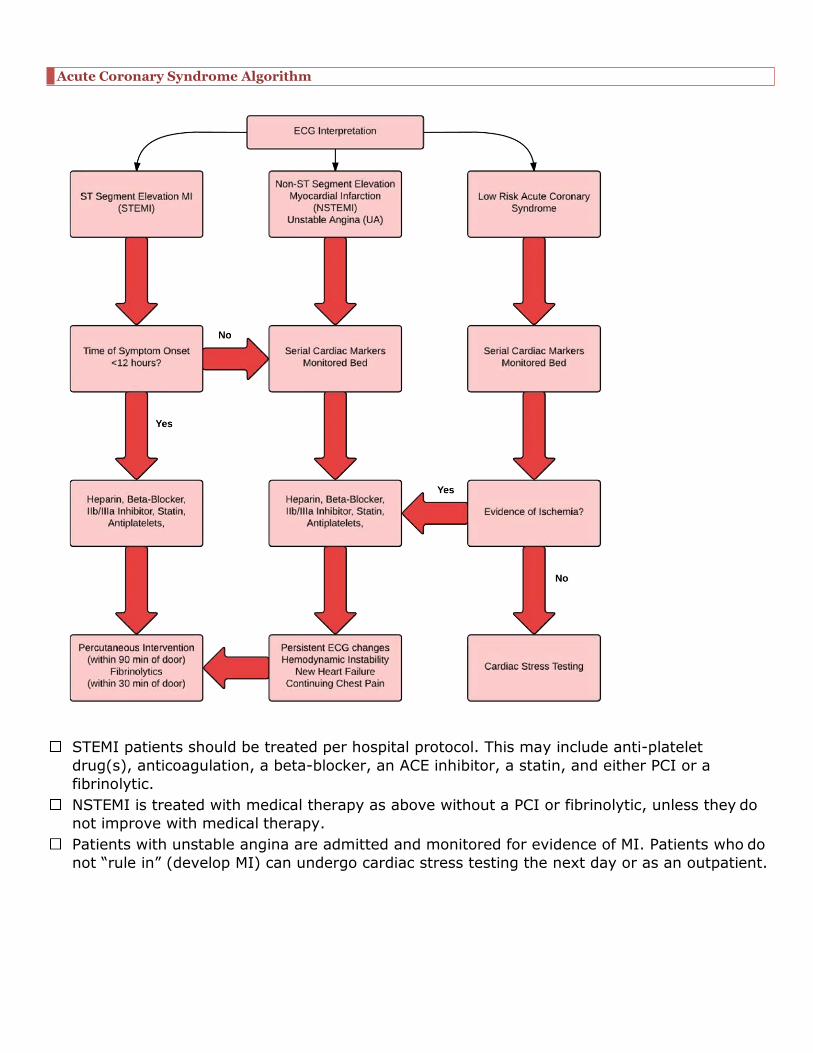
STEMI patients should be treated per hospital protocol. This may include anti-platelet
drug(s), anticoagulation, a beta-blocker, an ACE inhibitor, a statin, and either PCI or a
fibrinolytic.
NSTEMI is treated with medical therapy as above without a PCI or fibrinolytic, unless they do
not improve with medical therapy.
Patients with unstable angina are admitted and monitored for evidence of MI. Patients who do
not “rule in” (develop MI) can undergo cardiac stress testing the next day or as an outpatient.
Acute Coronary Syndrome Algorithm

Acute Stroke
The Cincinnati Prehospital Stroke Scale
Task Abnormal
Ask the patient to smile Face droops on one side
Ask the patient to say
“you can't teach an old dog
new tricks”
Speech slurs
Ask patient to close eyes One arm drifts lower and turns and
extend both arms inward (pronator drift) straight out, palms up
If one of these tasks is abnormal, stroke probability is 72% If all
three tasks are abnormal, stroke probability is >85%
The EMS team should take patients with suspected
stroke to a stroke center. While in transit, the EMS
team should try to determine the time at which the
patient was last normal, which is considered the onset
of symptoms. EMS administer oxygen via nasal cannula
or face mask, obtain a fingerstick glucose
measurement, and alert the stroke center.
Within 10 min. of the patient's arrival at the hospital,
personnel should assess the patient. They should obtain
vital signs and IV access, draw and send labs (e.g.
coags), obtain a 12-lead ECG, order CT, and perform a
general assessment.
Within 25 min. of the patient's arrival at the hospital,
the stroke team should determine symptom onset,
perform and narrow exam including the NIH stroke scale
or equivalent, perform the fibrinolytic checklist, have the
results of the CT scan of the brain.
Within 45 min. of the patient's arrival at the hospital,
the CT scan of the brain should be read for the presence
of ischemic or hemorrhagic stroke.
Within 60 min. of the patient's arrival at the hospital,
fibrinolytic therapy should be administered in cases of
ischemic stroke if the patient is a candidate. If the
patient with an ischemic stroke is not a candidate for
fibrinolytic, administer aspirin if the patient is not
allergic. If the patient is having a hemorrhagic stroke,
neurosurgery should be consulted.
Within three hours of the patient's arrival at the
hospital, the patient should be moved to the
neurology/neurosurgery intensive care unit, stroke unit,
or med/surg intensive care unit.

In people who are candidates for fibrinolytics, the goal is to administer the agent within 3 hours
of the onset of symptoms.
Fibrinolytic Checklist
Inclusion Criteria Exclusion Criteria
Ischemic stroke with neurological deficit Stroke/ head trauma in last 3 months
Onset of symptoms <3 hours History of brain
Age >18 years old Brain tumor, arteriovenous malformation, or aneurysm
Brain or spine surgery in last
Arterial line or blood draw in last week
Possible subarachnoid hemorrhage
Systolic ≥ 185 mmHg or diastolic ≥ 110 mmHg consistently
Serum glucose <50 mg/dL (<2.8 mmol/L)
Currently bleeding internally or bleeding diathesis
Platelet <100,000/mm (if known)
INR >1.7 or PT >15 seconds (if known)
Elevated aPTT (if known)
Currently taking anticoagulants
Hemorrhage on CT
Stroke includes > 1/3 of entire cerebrum
Relative Exclusion Criteria
Minor neurologic deficits
Rapidly improving neurologic deficits
Major surgery/serious trauma in last 2 weeks
Gastrointestinal/urinary tract bleeding in last 3 weeks
Heart attack in last 3 months
Seizure with stroke with postictal period
Pregnant
In select individuals, the window for fibrinolytics can be extended to 4.5 hours.
Fibrinolytic Checklist for 3 to 4.5 Hours from Symptom Onset
Meets all criteria for fibrinolytic use at 3 hours plus these criteria:
Inclusion Criteria Exclusion Criteria
Ischemic stroke with neurological deficit Currently taking anticoagulants
Onset of symptoms 3 to 4.5 hours Severe stroke; NIH Stroke Scale score >25
Age 18 to 79 years old Previous ischemic stroke and diabetes
Time is Brain!Stroke Time Goals for Evaluation and Therapy

Team Dynamics
The 2015 edition of the AHA ACLS guidelines continues to highlight the importance of effective
team dynamics during resuscitation. ACLS in the hospital will be performed by several providers.
These individuals must provide coordinated, organized care. Providers must organize themselves
rapidly and efficiently. The AHA recommends establishing a Team Leader and several Team
Members.
The Team Leader is usually a physician, ideally the provider with the most experience in leading
ACLS codes. Resuscitation demands mutual respect, knowledge sharing, and constructive
criticism, after the code.
Team Leader Responsibilities Team Member Responsibilities
Usually stands at the foot of the bed Stands in a position dictated by role
Competent in all ACLS duties Competent in specific role (at least)
Directs Team Members in a professional, calm voice Responds with eye contact and voice affirmation
Assigns roles Clearly states when he/she cannot perform a role
Listens for confirmation from Team Member Informs Team Leader when task is complete
Ask for ideas from Team Members when needed Openly share suggestions if it does not disrupt flow
Critiques Team Performance after code Provides constructive feedback after code
Documents resuscitation in patient chart Provides information for documentation as needed
When performing a resuscitation, the Team
Leader and Team Members should assort
themselves around the patient so they can be
maximally effective and have sufficient room
to perform their role.

Drug Use(s) Dosage/Route Contraindications/Warnings
Adenosine Supraventricular Tachycardia; May be
used for other tachycardia rhythms
First dose: 6 mg IV push Second
dose: 12 mg IV
Second or third degree heart block;
tachycardia due to poisoning
Amiodarone Pulseless ventricular tachycardia
Ventricular fibrillation
First dose: 300 mg bolus
Second dose: 150 mg
Max: 2.2 grams/day
Second or third degree heart block;
hypotension may result with rapid infusion or multiple doses
Atropine Symptomatic bradycardia
(No longer recommended for PEA or
asystole)
0.5 mg IV every 3-5 min Max: 3
mg max dose
Higher doses may be required
for organophosphate poisoning
Doses < 0.5 mg may worsen
bradycardia
Toxins, poisons, and overdoses Up to 2 to 4 mg until symptoms resolve
Dopamine Symptomatic bradycardia (if atropine
fails)
Pressor for shock/hypotension
2 to 20 mcg/kg IV per min
Titrate to blood pressure response
Do not mix with sodium bicarbonate or
alkalis/bases
Correct hypovolemia before
administration
Epinephrine Cardiac arrest
Anaphylaxis
Symptomatic bradycardia
instead of dopamine
1 mg (1:10000) IV OR
2-2.5 mg (1:1000) ETT q3 to 5 min
Then infuse 0.1-0.5 mcg/kg/min titrated to
blood pressure response
Cocaine-induced ventricular
tachycardia
May increase oxygen demand
Symptomatic bradycardia (if
atropine fails)
Pressor for hypotension
2-10 mcg/minute infusion titrated to blood
pressure response
Lidocaine Cardiac arrest VFib/VTach First dose: 1-1.5 mg/kg IV
Second dose: 0.5-0.75 mg/kg IV every
5 to 10 min
Max: 3 mg/kg
Infuse 1-4 mg IV per min
Wide complex bradycardia
Should not be used in cases of acute
myocardial infarction
Observe for signs of toxicity
Wide complex tachycardia Wide complex tachycardia with pulse:
0.5-1.5 mg/kg IV; may repeat twice at half
dose in 5-10 minutes to total of 3mg/kg;
followed with infusion of 1-4 mg per minute
infusion
Wide complex bradycardia
Magnesium
Sulfate
Torsades de pointes Pulseless Torsades: 1-2 gram IV bolus
Torsades with a pulse: 1-2 gram IV over
5-60 minutes followed by infusion at
0.5-1 gram per hour IV
Rapid bolus may cause hypotension
and bradycardia; Can also be used to
reverse digitalis poisoning
Hypomagnesemia with cardiac arrest 1-2 gram IV bolus
Vasopressin Ventricular fibrillation
Pulseless ventricular tachycardia
Asystole
PEA
40 units IV instead of epinephrine Deliver through central line Peripheral
IV administration can cause tissue
necrosis
Shock Infuse 0.02-0.04 units/min IV
Resuscitation and Life Support Medications


![Mismanagement of a Resuscitation and Other Concerns at the ... · BCLS [Basic Cardiac Life Support]/ACLS [Advanced Cardiac Life Support] until training is current unless a waiver](https://static.fdocuments.in/doc/165x107/5e0595c0e8e7eb392436c687/mismanagement-of-a-resuscitation-and-other-concerns-at-the-bcls-basic-cardiac.jpg)