Table of Contents Contents - BioLegend Table of Contents Toll Free 1.877.BIOLEGEND (246.5343)...
47
10 Table of Contents Toll Free 1.877.BIOLEGEND (246.5343) Contents Technical Charts and Protocols ..................................................................................................................................... 11 Brilliant Violet™ Fluorophores ........................................................................................................................................................................................................... 12 Fluorophores for Flow Cytometry ................................................................................................................................................................................................... 14 Human CD Molecules, HLDA9 Workshop ..................................................................................................................................................................................... 16 Biofunctional Antibodies - LEAF™ and Ultra-LEAF™ ................................................................................................................................................................. 30 Cross-Reactivity Chart, Non-Human Primate .............................................................................................................................................................................. 40 Cross-Reactivity Chart, Other Species............................................................................................................................................................................................ 43 TNF Receptor and Ligand Superfamilies ...................................................................................................................................................................................... 45 Cell Surface Immunofluorescence Staining Protocol ............................................................................................................................................................... 47 Intracellular Cytokine Staining Protocol ....................................................................................................................................................................................... 48 Th17 Polarization of Mouse CD4 Cells ........................................................................................................................................................................................... 49 FOXP3 Intracellular Staining Protocol ............................................................................................................................................................................................ 49 Western Blotting Protocol .................................................................................................................................................................................................................. 50 Immunoprecipitation Protocol......................................................................................................................................................................................................... 51 General Sandwich ELISA Protocol ................................................................................................................................................................................................... 52 ELISPOT Protocol ................................................................................................................................................................................................................................... 54 Immunofluorescence Protocol ......................................................................................................................................................................................................... 55 Immunocytochemistry and Immunohistochemistry ............................................................................................................................................................... 56
Transcript of Table of Contents Contents - BioLegend Table of Contents Toll Free 1.877.BIOLEGEND (246.5343)...

10
Table of Contents
Toll Free 1.877.BIOLEGEND (246.5343)
ContentsTechnical Charts and Protocols .....................................................................................................................................11
Brilliant Violet™ Fluorophores ........................................................................................................................................................................................................... 12Fluorophores for Flow Cytometry ................................................................................................................................................................................................... 14Human CD Molecules, HLDA9 Workshop ..................................................................................................................................................................................... 16Biofunctional Antibodies - LEAF™ and Ultra-LEAF™ ................................................................................................................................................................. 30Cross-Reactivity Chart, Non-Human Primate .............................................................................................................................................................................. 40Cross-Reactivity Chart, Other Species ............................................................................................................................................................................................ 43TNF Receptor and Ligand Superfamilies ...................................................................................................................................................................................... 45Cell Surface Immunofluorescence Staining Protocol ............................................................................................................................................................... 47Intracellular Cytokine Staining Protocol ....................................................................................................................................................................................... 48Th17 Polarization of Mouse CD4 Cells ........................................................................................................................................................................................... 49FOXP3 Intracellular Staining Protocol ............................................................................................................................................................................................ 49Western Blotting Protocol .................................................................................................................................................................................................................. 50Immunoprecipitation Protocol ......................................................................................................................................................................................................... 51General Sandwich ELISA Protocol ................................................................................................................................................................................................... 52ELISPOT Protocol ................................................................................................................................................................................................................................... 54Immunofluorescence Protocol ......................................................................................................................................................................................................... 55Immunocytochemistry and Immunohistochemistry ............................................................................................................................................................... 56

12Toll Free 1.877.BIOLEGEND (246.5343)
Brilliant Violet™ Fluorophores
Brilliant Violet™ antibody conjugates, proudly co-developed by BioLegend and Sirigen, are an innovative class of novel research reagents, providing more options for your multicolor flow cytometry panels and getting you better results. Maximize the capacity of your violet laser with our large selection of directly labeled Brilliant Violet™ antibody conjugates.
The Brilliant Violet™ family of fluorescent molecules are organic polymers with an extraordinary capacity to absorb energy (extinction coefficient) and a high efficiency with which to convert that absorbed energy to an emitted signal (quantum yield). When conjugated to antibodies, this results in high intensity brightness on labeled cells. Optimally excited by the violet laser at 405 nm, the Brilliant Violet™ family of fluorophores spans a wide range of emissions, allowing them to be used together in multicolor panels. Unlike quantum dots, conjugated polymers have discrete excitation spectra, similar to that of organic dyes, which minimizes potential issues with cross-beam compensation.

13biolegend.com | [email protected]
Brilliant Violet™ Fluorophores
Easy to Use and Trouble-FreeBrilliant Violet™ antibodies are simple to use, compatible with standard staining buffers, and stable to fixation. Provided in convenient 5 μL test sizes at optimal ready-to-use concentrations, our Brilliant Violet™ conjugates can easily be added to your multi-color panels. Mouse products are now offered in microgram (µg) size for exceptional value. To compare fluorescence spectra data of Brilliant Violet™ conjugates with our other fluorophores, use our Fluorescence Spectra Analyzer at: biolegend.com/spectraanalyzer.
Extensive Antibody SelectionBioLegend provides an expansive selection of antibody specificities for all our Brilliant Violet™ fluorophores. All products are manufactured by our expert chemists in San Diego, CA, and are supported by our 100% satisfaction guarantee. For the latest product updates, a brief video tutorial on Brilliant Violet™ products, and seminar for using Brilliant Violet™ products in multicolor panels, visit: biolegend.com/brilliantviolet.
CharacteristicsFluorophore EX max (nm) EM max (nm) Recommended Filter Brightness Index*
Brilliant Violet 421™ 405 421 450/50 5
Brilliant Violet 510™ 405 510 510/50 3
Brilliant Violet 570™ 405 570 585/42 2-3
Brilliant Violet 605™ 405 603 610/20 4
Brilliant Violet 650™ 405 645 660/20 3
Brilliant Violet 711™ 405 711 710/50 3
Brilliant Violet 785™ 405 785 780/60 3
*Brightness Index is a relative scale of fluorophore brightness in the context of conjugated antibodies; 1 is dim, 5 is brightest.
Brilliant Violet™ Information for Mobile DevicesDownload our Brilliant Violet™ Antibodies app for iPhone and iPad. Find fluorophore descriptions, testing results, example data, stain index values, spectra data, a fluorophore equivalency tool, and considerations for multicolor flow cytometry on the go, at any time. You can also view our helpful tutorials. Visit: biolegend.com/bviossupport.

14Toll Free 1.877.BIOLEGEND (246.5343)
Fluorophores for Flow Cytometry

16Toll Free 1.877.BIOLEGEND (246.5343)
Human CD Molecules, HLDA9 WorkshopCD Antigen Other Names
Molecular Weight (kD) Cellular Expression Ligand/Receptor/Association Functions Intracellular Interaction
CD1a R4, T6, HTA1 49 cortical thy-c, Langerhans, DC CD1-restricted TCR lipid Ag presentation β2m, CD8
CD1b R1, T6 45 cortical thy-c, Langerhans, DC CD1-restricted TCR lipid Ag presentation β2m, CD8
CD1c BDCA-1, R7, T6 43 cortical thy-c, Langerhans, DC, B sub CD1-restricted TCR lipid Ag presentation β2m, CD8
CD1d R3 43–49 intestinal epi, B sub, DC CD1-restricted TCR lipid Ag presentation β2m
CD1e R4 28 DC (mainli in Gogi) lipid Ag presentation β2m
CD2 T11, LFA-2, SRBC-R 50 T, thy-c, NK, B sub, mono sub CD58, CD59 and CD15 adh, T activ Fyn, Lck, SH3KBP1, Sp1, MAD, PTPR-C
CD3γ T3 20–26 mature T, different levels on thy-c Ag complex signaling T activ, regulates TCR expression CD3/TCR complex
CD3δ T3 21 mature T, different levels on thy-c Ag complex signaling T activ, regulates TCR expression CD3/TCR complex
CD3ε T3 20 mature T, different levels on thy-c Ag complex signaling T activ, regulates TCR expression
CD3/TCR complex, Syk, ZAP-70, Lck, SHC, Grb4, PI3Kα, NCK1
CD4 T4, Leu-3, L3T4, W3/25 55 thy-c sub, T helper/inducer, Treg, mono/
mac MHC class II, HIV gp120, IL-16 T activ, thymic differen, HIV-R Lck
CD5 T1, Tp67, Leu-1, Ly-1 67 thy-c, T, B sub, B-CLL CD72 regulates T-B interaction, T activ TCR or BCR, Fyn, Lck, ZAP-70, PKCα,
PKCβ1, PKCγ, PI3Kα, SHP1
CD6 T12 105–130 thy-c, T, B sub, neuron sub CD166, 3A11 thy-c dev, T activ
CD7 gp40 40 thy-c, T, NK, myeloid progenitor CD7 ligand (K-12) T costim PI3Kα, PI4Kα
CD8a T8, CD8, Leu-2, Ly-2 32–34 thy-c sub, cytotoxic T, NK, DC sub MHC class I coreceptor for MHC class I Lck, KAT
CD8b CD8, Lyt3 30–32 thy-c sub, cytotoxic T MHC class I coreceptor for MHC class I
CD9 p24, MRP-1, DRAP-27 22–27 pt, pre-B, eosino, baso, act T, endo, epi,
stem PSG17 adh and migration, pt activ CD63, CD81, CD82, CD315, and CD316, PKCα
CD10 CALLA, NEP, gp100 100 B and T precursors, fibro, neutro peptidase, regulates B growth Lyn, SHC, PI3Kα, PI3Kβ
CD11a LFA-1, integrin αL 180 all leuko CD54, CD102, CD50, and CD242 adh and costim CD18, RANBP9
CD11b Mac-1, integrin αM 170 gran, mono/mac, NK, T sub, B sub, DC CD54, iC3b and ECM , CD242 adh, chemotaxis, apoptosis CD18, IRAK1
CD11c p150, 95, CR4, integrin αX 150 mono/mac, NK, gran, T and B sub, DC iC3b, ECM adh CD18
CDw12 90–120 mono, gran, pt, NK unknown
CD13 APN, gp150 150–170 gran, mono and their precursors, endo, epi, DC, fibro, placenta
coronavirus, CMV, L-leucyl-β-napthylamine aminopeptidase N, adh, coronavirus R TM4SF1
CD14 LPS R 53–55 mono, mac, Langerhans, gran (low) LPS R for complex of LPS and LBP, innate immune response LTF, TLR2, TLR3, TLR4, LBP
CD15 Lewis X (Lex), SSEA-1, 3-FAL gran, transient in brain CD62E adh, gran activ
CD15s Sialyl Lewis X gran, mono, endo, memory helper T, act T and B, NK, HEV, endo CD62E adh
CD15u 3’ sulfated Lewis X gran, mono, T sub, B sub, NK, endo CD62P adh
CD15su 6’ sulfated Lewis X gran, mono, T sub, B sub, NK, endo CD62L adh
CD16 FcγRIIIΑ, CD16a 50–65 neutro, NK, act mono, mac, DC aggregated IgG, IgG-Ag complex low affinity Fcg R, mediates phagocytosis and ADCC, degranulation Lck, ZAP-70, SHC
act: ................. activatedactiv: .............. activationADCC: ............ Antibody-dependent
cell-mediated cytotoxicityadh: ................ adhesionAg: .................. AntigenALL: ................ acute lymphoid leukemiaB: ..................... B lymphocytesβ2m: .............. beta-2-microglobulinbaso: .............. basophilsBCR:................ B cell receptorBM: ................. bone marrowCD:.................. Cluster of Differentiation
CNS: ............... central nervous systemC’: .................... complementcostim: .......... costimulationDC:.................. dendritic cellsdev: ................ developmentdifferen: ........ differentiationECM:............... extracellular matrixendo: ............. endothelial cellseosino: .......... eosinophilsepi: ................. epithelial cellseryth: ............. erythrocytesFDC: ............... follicular dendritic cellsfibro: .............. fibroblasts
gran: .............. granulocyteshemato: ........ hematopoietichepato: ......... hepatocytesHSC: ............... hematopoietic stem cellsIEL: .................. intraepithelial
lymphocytesinhib: ............. inhibitionLangerhans: Langerhans cellsLDL: ................ low density lipoproteinleuko: ............ leukocyteslympho: ........ lymphocytesmac: ............... macrophagesmast: .............. mast cells
mega: ............ megakaryocytesMHC: .............. major histocompatibility
complexmono:............ monocytesneutro: .......... neutrophilsNK: .................. natural killer cellspDC: ............... plasmacytoid dendritic
cells pt: ................... plateletsprolif: ............. proliferationR: ..................... receptorstem: .............. stem cellssub: ................ subset
T: ..................... T lymphocytesTCR: ................ T cell receptorthy-c: ............. thymocytesTNFRSF: ........ TNF receptor superfamilyTNFSF: ........... TNF superfamilytransd: ........... transduction Treg: ............... T regulatory cellsupreg:............ upregulatedvWF: ............... von Willebrand factorw/: ................. associated with

17biolegend.com | [email protected]
Human CD Molecules, HLDA9 WorkshopCD Antigen Other Names
Molecular Weight (kD) Cellular Expression Ligand/Receptor/Association Functions Intracellular Interaction
CD16b FcγRIIIB 48 neutro IgG phagocytosis, ADCC SAP
CD17 LacCer, lactosylceramide mono, pt, B sub, gran, DC, T glycolipid metabolism, angiogenesis, apoptosis
CD18 β2 integrin 95 leuko ICAMs, ECM adh CD11a, b, c, Syk, FAK, ILK, RACK1, RANBP9, PKCα, PKCδ, PKCε, PYK2
CD19 B4 95 B (not on plasma), FDC BCR coreceptor, signaling CD21, CD81, CD225, BCR, Fyn, Lyn, Syk, BTK, VAV1, VAV2, PLCγ2, Grb2
CD20 B1, Bp35, Ly-44 33–37 B, T sub B activ and prolif Fyn, CK2A1, CK2A2
CD21 CR2, EBV-R, C3dR 145, 110 mature B, FDC, T sub C3d, EBV, CD23 signaling, B activ CD19, CD81, CD225, BCRp53
CD22 BL-CAM, Siglec-2 130 BCD22, IgM, CD45RO, sialoglyco-conjugate NeuGcα2-6Galβ1-4GlcNAc, CD75
adh, signaling Lyn, Syk, SHIP1, SLP76, Grb2, PI3Kα, PLCγ1, SHP1
CD23 FcεRII, BLAST-2 45 B (upreg upon activ), act mac, eosino, FDC, pt, intestinal epi IgE, CD21, CD11b, CD11c low affinity R for IgE, allergic response,
activ Fyn
CD24 BA-1, HAS 35–45 B, gran, epi, mono, T sub CD62P, CD24 cell prolif and differen Fgr, Lyn
CD25 Tac, p55, IL-2Rα 55 act T, B, and mono; DC sub, Treg IL-2 IL-2R α chain, w/ β and γ chains to form high affinity IL-2R CD122, CD132, STAT3, NFκB1, STAT5b
CD26Dipeptidyl peptidase IV (DPP IV)
110 mature thy-c, T (upreg upon activ), B sub, NK, mac, epi adenosine deaminase, collagen exoprotease, HIV pathogenesis, costim ADA, CD4, FAP
CD27 T14, S152, TNFRSF7 50–55 T, medullary thy-c, B sub, NK CD70 costim TRAF2, TRAF3, TRAF5, Siva
CD28 Tp44, T44 44 most T, thy-c, plasma, NK CD80 (B7-1), CD86 (B7-2) costim ITK, Grb2, PI3Kα, PI3Kβ, PI3Kγ, PLC-γ1
CD29 integrin β1, gpIIa 130 T, B, mono, gran (low), pt, mast, fibro, endo, NK VCAM-1, MAdCAM-1, ECM adh, activ, embryogenesis integrin α chains, PKCα, PKCε, RACK1,
FAK, 14-3-3β, ILK, RhoGAP5, PIK4α
CD30 Ki-1, Ber-H2, TNFRSF8 105 act T, act B, mono, act NK, Reed-Sternberg
cells CD153 lympho prolif and death ALK, TRAF1, TRAF2, TRAF3, TRAF5
CD31 PECAM-1, endocam 130–140 mono, pt, gran, endo, lympho sub CD31, CD38 adh Lck, Fyn, c-Src, Hck, c-Yes, Csk, PI3Kα,
PLC-γ1
CD32 FcγRII 40 B, mono/mac, gran, DC, pt, endo aggregated IgG, phosphatases B dev and activ, phagocytosis and mediators release, ADCC Fyn, Hck, Lyn, Syk, LAT, BLK, SHC
CD33 p67, Siglec-3 67 mono, gran, mast, myeloid progenitors certain sialic acids-linked carbohydrates
lectin activity for sugar chains containing sialic acid, adh c-Src, SHP1, SHP2
CD34 gp105-120, Mucosialin 105–120 HSC and progenitors, endo L-selectin adh PKCδ, CRKL
CD35 CR1, C3b/C4b-R 190–280 eryth, B, mono, neutro, eosino, FDC, T sub C3b, C4b, iC3, iC4 adh, phagocytosis CD55
CD36 GPIV, gpIIIb 85 pt, mono/mac, endo, erythroid precursors oxidized LDL, thrombospondin, collagen scavenger R, adh and phagocytosis Fyn, Lyn, c-Yes, c-Src
CD37 gp 52-40 40–52 mature B, low on T, gran, mono, DC CD53, CD81, CD82, MHC class II adh, signaling
CD38 T10, ADP-ribosyl cyclase 42 variable levels on majority of hemato and
some non-hemato cells, high on plasma CD31 cell activ, prolif and adh Lck
CD39 Entpd1, NTPDase-1 78 mac, Langerhans, DC, act B, NK, microglia,
endo, Treg ADP/ATP ATP and ADP degradation, modulates pt activ, immune response
CD40 Bp50, TNFRSF5 45–48 B, mono/mac, DC, endo, fibro, epi, keratinocytes CD154 costim, differen and isotype-switching JAK3, PI3Kα, TRAF1-3, TRAF5-6, Ku80
CD41 gpIIb, αIIb integrin 120/23 dimer pt, mega fibrinogen, fibronectin, vWF and
thrombospondin pt activ and aggregation CD61
CD42a gpIX 17–22 pt, mega vWF and thrombin pt adh and activ CD42b, c and d
CD42b gpIbα 145 pt, mega vWF and thrombin pt adh and activ CD42a, c and d, Grb2
CD42c gpIbβ 24 pt, mega vWF and thrombin pt adh and activ CD42a, b and d, 14-3-3 ζ, PKACA
CD42d gpV 82 pt, mega vWF and thrombin, complexed with CD42a, b and c pt adh and activ CD42a, b and c, 14-3-3 ζ
CD43sialophorin, leukosialin, gpL115
115–135 leuko, except resting B, pt (low) CD54, CD62E, CD169 adh and anti-adh Fyn

18Toll Free 1.877.BIOLEGEND (246.5343)
Human CD Molecules, HLDA9 WorkshopCD Antigen Other Names
Molecular Weight (kD) Cellular Expression Ligand/Receptor/Association Functions Intracellular Interaction
CD44 H-CAM, Pgp-1, EMCR III, CD44s 85 hemato and non-hemato cells, except pt hyaluronan leuko rolling, homing, and aggregation Csk, Fyn, Lck, Rho, PKN
CD44R CD44v 85–250heterogeneous with different isoforms, constitutively expressed on epi, mono, upreg on act leuko
hyaluronan leuko rolling, homing, and aggregation
CD45 LCA, T200 180–240 hemato cells, except eryth and pt galectin-1 activ, signaling CD2, CD3, CD4 and CD45AP, Fyn, Lck, Lyn, RasGAP, SHP-1, SLP76, Grb2
CD45RA 205–220 B, T sub (naive T), mono, medullary thy-c isoform of CD45 containing the A exon activ, signaling
CD45RB 190–220 T sub, B, mono, mac, gran, DC, NK isoform of CD45 containing the B exon activ, signaling
CD45RC 220 B, NK, CD8+ T, sub of CD4+ T, medullary thy-c, mono, DC
isoform of CD45 containing the C exon activ, signaling
CD45RO UCHL-1 180 act T and memory T, B sub, act mono, mac, gran, cortical thy-c, DC sub
isoform of CD45 containing none of the A, B and C exons, CD22 activ, signaling
CD46 MCP 64–68 leuko, pt, endo, epi, placental trophoblasts, sperm and variety of tumor cells
C3b, C4b, measles virus, factor I co-factor for factor I, C’ activ, fertilization, R for measles virus c-Yes, c-Src
CD47 IAP 50–55 hemato cells, epi, endo, fibro, brain, mesenchymal cells CD172a, CD172g adh, activ, apoptosis FAK, BNIP3
CD48 Blast-1, BCM1, Sgp-60, SLAMF2 45 leuko CD244, CD2 (low) adh, costim Lck
CD49a VLA-1α, α1 integrin 200 act T, mono, melanoma cells, endo collagen, laminin-1 adh, embryo dev CD29, Martilin 1, Talin
CD49b VLA-2α, gpIa, α2 integrin 160 pt, NK sub, B, mono, act T, epi, endo, mega collagen, echovirus 1 adh, pt aggregation, R for echovirus 1 CD29, CD9, Perlecan
CD49c VLA-3α, α3 integrin 125 most adh cell lines, low on B and T collagen, laminin-5, fibronectin adh, signaling CD29, CD9, Galectin 8
CD49d VLA-4α, α4 integrin 150 T, B, thy-c, mono, eosino, baso, NK, mast,
DC, erythroblastic precursors VCAM-1, MAdCAM-1, fibronectin, CD242 adh, cell migration, homing and activ CD29, β7 integrin, PRKACA, HIC5
CD49e VLA-5α, α5 integrin 135, 25 thy-c, T, mono, pt, early and act B, endo,
epi fibronectin, invasin adh, cell survival and apoptosis CD29, RhoGAP5
CD49f VLA-6α, α6 integrin 125 memory T, thy-c, mono, pt, mega, epi,
endo, cytotrophoblasts laminins, invasin adh, cell migration, embryogenesis CD29, CD104, Fyn, SHC, PKCδ, Grb2
CD50 ICAM-3 120–140 leuko, thy-c, Langerhans, endo CD11a/CD18 adh and costim PKCθ
CD51 vitronectin R, integrin αV
125, 24 dimer
pt, act T, endo, osteoblasts, melanoma cells, mega, endo
fibrinogen, thrombospondin, vWF, CD242 adh, signal transd integrin b1, b3, b5, b6 or b8, Fyn, FAK
CD52 CAMPATH-1, HE5 25–29 thy-c, lympho, mono/mac, epi, sperm, mast
costim, antibodies are useful for lysis of target cells, costim
CD53 OX-44 32–42 leuko, DC, osteoblasts, osteoclasts VLA-4, MHC class II signal transduction CD9, CD81, CD82
CD54 ICAM-1 90–95 endo, epi, mono, low on resting lympho (upreg upon activ)
CD11a/CD18, CD11b/CD18, Rhinovirus, CD43
extravasation of leuko from blood vessels, regulates T activ Moesin, Ezrin
CD55 DAF 60–70 hemato and non-hemato cells C3b, C4b, CD97, Coxsackie virus, Echovirus
C’ activ, ligand or protective molecule in fertilization, signal transd Fyn, Lck
CD56 NCAM, Leu-19, NKH-1 175–220 neural tissue, NK, , NKT, T sub, small-cell
lung carcinomas NCAM, heparin sulfate homophilic and heterophilic adh Fyn, FAK
CD57 HNK-1, Leu-7 110 NK sub, NKT, T sub, some B cell lines CD62L, CD62P adh
CD58 LFA-3 55–70 leuko, eryth, epi, endo, fibro CD2 adh, costim
CD59 Protectin, H19, 1F-5Ag 19–25 hemato and non-hemato cells C8α, C9 prevents C’ polymerization, protects
cells from C’-mediated lysis c-Src, lck, fyn
CD60a GD3 T sub, thy-c, melanocytes, glial cells, pt, gran apoptosis, costim
CD60b 9-O-sialyl GD3 T sub, act B, melanomas costim
CD60c 7-O-sialyl GD3 T sub activ

19biolegend.com | [email protected]
Human CD Molecules, HLDA9 WorkshopCD Antigen Other Names
Molecular Weight (kD) Cellular Expression Ligand/Receptor/Association Functions Intracellular Interaction
CD61 gpIIIa, β3 integrin 110 w/ CD41 on pt, w/ CD51 on mac, endo, pt, fibro, osteoclasts, mast
fibrinogen, vitronectin, fibronectin, vWF
mediates cell adh to diverse matrix proteins
CD41, CD51, c-Src, SHC1, AKT1, Talin, FAK, Paxillin
CD62E E-selectin, ELAM-1, LECAM-2 97–115 act endo CD15s, ESL-1, CD162, CD43,
CD65, CD66a, e, CD147leuko rolling, tumor cell adh, angiogenesis PLC-γ1, SHP2, FAK, Paxillin
CD62L L-selectin, LECAM-1, LAM-1 74– 95 B, T sub, mono, gran, NK, thy-c CD34, MAdCAM-1, GlyCAM-1,
CD57, CD15su leuko rolling and homing Grb2
CD62P P-selectin, GMP-140, PADGEM 140 act pt, endo CD162, CD24, CD57, CD15u leuko tethering and rolling AP47, CD24, SNX17
CD63 LIMP, MLA1, LAMP-3 40–60 act pt, mono, mac, degranulated neutro,
fibro, osteoclasts VLA-3, VLA-4, CD9, CD81 regulates cell motility PI4Kα
CD64 FcγRI, FcR I 72 mono, mac, DC, IFN-γ or G-CSF act gran IgG high affinity R for IgG, mediates phagocytosis, Ag capture, and ADCC Hck, Syk, CRKL, LAT
CD65 Ceramide-dodecasaccharide
gran and some mono, myeloid leukemia cells CD62E unknown
CD65s Sialylated-CD65 gran, mono, myeloid leukemia cells phagocytosis
CD66a BGP, NCA-160 140–180 gran, epi, colon, liver, hemato tissues CD66a, c, e, CD62E, Opa homophilic and heterophilic adh, neutro activ
c-Src, SHP2, SHP1, MAP3K10, SHC, Paxillin
CD66b CD67, CGM6, NCA-95 95–100 gran CD66c adh, neutro activ
CD66c NCA 90 neutro, epi, colon carcinoma CD66a, b, c, e adh, neutro activ
CD66d CGM1 35 neutro Opa I neutro activ, phagocytosis c-Src, CKI-alpha-like
CD66e CEA 180–200 adult colon epi, colon cancer CD66a, c, e, Opa, CD62E homophilic and heterophilic adh
CD66f PSG, Sp-1 54–72 epi, placental syncytiotrophoblasts, fetal liver
immune reg and protection of fetus from maternal immune system
CD68 macrosialin, gp110 94–110 mac, neutro, baso, DC, myeloid
progenitors, act mono oxidized LDL phagocytosis
CD69 AIM, VEA 28, 32 act leuko, NK, thy-c sub, pt, Langerhans signaling, costim
CD70 Ki-24, CD27L, TNFSF7 50 act B, act T CD27 costim
CD71 TfR, T9 95 proliferating cells, reticulocytes, erythroid precursors transferrin transferrin R, iron uptake Rab5B, TCRζ
CD72 Lyb-2 39 B (except plasma), mac, FDC, T sub CD5, CD100 B activ and prolif Grb2, SHP1, BLNK
CD73 Ecto-5’-nucleotidase 70 T sub, B sub, FDC, epi, endo AMP dephosphorylation, costim, adh β-Actin, Fibronectin 1, Laminin A
CD74 Ii, invariant chain 33, 35, 41 B, act T, mac, Langerhans, DC, act endo and epi CD44, MHC class II, MIF intracellular sorting of MHC class II,
B activ Cathepsin L, CD1d
CD75 lactosamines B sub, T sub, eryth CD22 adh
CD75sCDw76, α2, 6-sialylated lactosamines
34, 37, 43, 200 majority B, T sub, endo, epi sub CD22 adh
CD77 Pk Ag, BLA, CTH, Gb3
germinal center B, high expression on Burkitt’s lymphomas Shiga Toxin (Verotoxin) apoptosis
CD79a Igα, MB1 33, 45 B subunit of BCR complex, signaling Ig/CD5/CD19/CD22/CD79b , Lck, Fyn, Lyn, Syk, SHP1, BLK
CD79b Igβ, B29 36–40 B subunit of BCR; signaling Ig/CD5/CD19/CD22/CD79a , Lck, Fyn, Lyn, FAK, Syk, ZAP70, SHP1, BLK
CD80 B7, B7-1, BB1 60 act B, act T, mac, DC CD28, CD152 costim
CD81 TAPA-1 26 T, B, NK, mono, thy-c, DC, endo, fibro activ, costim, differen CD19, CD21, CD225, CD315 and CD316, SHC
CD82 R2, 4F9, C33, Kai1 45–90 leuko, pt, epi, endothelial, fibro KITENIN Signaling, adh, tumor metastasis, inhibition
MHC molecules, CD53, CD37, CD81, integrins, EGFR
CD83 HB15 43 mature DC, act T, act B, Langerhans CD83L costim
CD84 GR6 72–86 mature B, T sub, mono/mac, pt, thy-c CD84 adh, activ EAT2, SAP
CD85a LIR-3, ILT5, LILRB3 mono/mac, gran, DC, T sub inhibits NK cytotoxicity MHC class I

20Toll Free 1.877.BIOLEGEND (246.5343)
Human CD Molecules, HLDA9 WorkshopCD Antigen Other Names
Molecular Weight (kD) Cellular Expression Ligand/Receptor/Association Functions Intracellular Interaction
CD85b ILT8 mono, DC, B, NK, T sub activates NK cytotoxicity FcεRIγ
CD85c LIR-8, LILRB5 mono, DC, B, NK, T sub activates NK cytotoxicity FcεRIγ
CD85d LIR-2, ILT4, LILRB2 110 mono, DC inhibits NK cytotoxicity MHC class I, SHP1
CD85e LIR-4, ILT6, LILRA3 mono, DC, B, NK, T sub activates NK cytotoxicity FcεRIγ
CD85f LIT11 mono, DC, B, NK, T sub activates NK cytotoxicity
CD85g ILT7 mono, pDC, B, NK, T sub CD317 activates NK cytotoxicity FcεRIγ
CD85h LIR-7, ILT1, LILRA2 mono, DC, B, NK, T sub, gran activates NK cytotoxicity FcεRIγ
CD85i LIR-6, LILRA1 mono, DC, T sub activates NK cytotoxicity FcεRIγ
CD85j LIR-1, ILT2, LILRB1 110 lympho, mono/mac, DC, NK sub inhibits NK cytotoxicity MHC class I
CD85k LIR-5, ILT3, LILRB4 60 gran, mono/mac, DC inhibits NK cytotoxicity MHC class I, SHP1, SHP2
CD85l ILT9 NK, T sub, mono/mac, DC, B FcεRIγ
CD85m ILT10 T sub, mono/mac, DC, B FcεRIγ
CD86 B70, B7-2 80 mono, act B, act T, DC, endo CD28, CD152 costim of T activ and prolif
CD87 uPAR 32–66 gran, mono, NK, T, endo, fibro, hepato uPA, vitronectin cell chemotaxis, adh Fyn, Hck, JAK1, TYK2
CD88 C5aR 43 gran, mono, DC, astrocytes C5a gran activ β-arrestin, C5L2
CD89 FcaR, IgA R 45–100 mono/mac, gran IgA1, IgA2 phagocytosis, degranulation, respiratory burst Lyn
CD90 Thy-1 25–35 HSC, neurons, fibro, stromal cells, HEV endo, thy-c CD51/CD61, Mac-1 adh, signaling Fyn, Lck
CD91 α2M-R, LRP 600 mono, mac, neurons, fibro α2M, LDLs, HSP96, lipoprotein metabolism, phagocytosis, Ag presentation c-Src, Rap, JIP, SHC, PRKACA
CD92 CDw92, CTL1 70 neutro, mono, lympho, endo, epi, fibro, DC choline choline transporter
CD93 CDw93, C1qRp 120 mono, gran, endo, pt, DC C1q phagocytosis, adh RANBP1, ARHGAP15
CD94 Kp43 43, 39, 70 NK, NKT, T sub HLA-E CD94/NKG2A inhibits NK function, CD94/NKG2C activates NK NKG-2, DAP12
CD95 Fas, APO-1 45 mono, neutro, lympho (upreg upon activ), fibro Fas ligand (CD95L, CD178) induces apoptosis Fyn, Lck, FADD, Daxx, RIP, FAF1, PKCα,
SHP1
CD96 TACTILE 160 NK and T (upreg upon activ) CD155 adh
CD97 EMR1 28, 74–85 gran, mono, low on lympho (upreg upon activ), mac, DC CD55, chondroitin sulfates neutro migration, adh
CD98 4F2, FRP-1, RL-388 80, 45 mono, lympho and NK (upreg upon activ), gran, some non-hemato CD29, CD147, tropomyosin, actin activ, adh actin, EGFR
CD99 MIC2, E2 32 lympho, NK, mono, gran, endo, epi, some tumor cells cyclophilin A leuko migration, activ, adh
CD99R E2 32 T, NK, myeloid cells isoform of CD99
CD100 SEMA4D 150 leuko, oligodendrocytes CD45, CD72, plexin B monocyte migration, T, B activ, angiogenesis, anti-apoptosis
CD101 V7, p126, IGSF2 120 mono, gran, DC, act T, Langerhans T activ and prolif
CD102 ICAM-2 55–65 lympho, mono, pt, endo LFA-1, Mac-1 adh, costim, lympho recirculation Moesin
CD103 HML-1, αE integrin, αIEL 150, 25 IEL, some peripheral blood lympho, act
lympho E-cadherin lympho retention, activ β7 integrin,
CD104 β4 integrin, TSP-180 205 epi, endo, Schwann cells, keratinocytes laminins, plectin cell adh, migration, tumor metastasis CD49f, Fyn, c-Yes, FAK, Grb2, PKCα, PKCδ,
14-3-3β, 14-3-3τ
CD105 Endoglin 90 endo, mesenchymal stem, erythroid precursors, act mono, mac TGF-β1, TGF-β3 adh, angiogenesis, modulates cellular
response to TGF-β1 TGF-β RI, TGF-β RII
CD106VCAM-1, INCAM-110, V-CAM
110 act endo, FDC, mesenchymal stem, stromal cells CD49d/CD29, α9/β1 integrin leuko adh, transmigration and costim Moesin, Ezrin
CD107a LAMP-1 100–120 act pt, act T, act endo, act gran possible role in cell adh
CD107b LAMP-2 100–120 act pt, act T, act endo possible role in cell adh

21biolegend.com | [email protected]
Human CD Molecules, HLDA9 WorkshopCD Antigen Other Names
Molecular Weight (kD) Cellular Expression Ligand/Receptor/Association Functions Intracellular Interaction
CDw108 SEMA7A, JMH 80 act T, eryth CD232 negative reg of T function, axon growth Plexin C1
CD109 8A3, 7D1, E123 170 act T, act pt, HSC, mesenchymal stem, endo TGF-β negative regulation
CD110 MPL, TPO-R 85–92 HSC and progenitors, mega, pt, endo sub thrombopoietin TPO R, mega dev, hematopoiesis IRS2, SHC, SHP2, SOCS1, JAK2
CD111 PRR1, Nectin-1, PVRL1 75 stem sub, neurons, endo, epi, fibro nectin-3, Afadin, HSVgD homophilic and intercellular adh, HSV R AF6, PARD3
CD112 PRR2, Nectin-2, HveB 72, 64 mono, neutro, sub of CD34+ cells, endo,
epi CD226, Nectin-3, Afadin adh AF6
CD113 PVRL3, Nectin-3, PRR3, CDw113 83 epi, testis, placenta, liver nectin-1, nectin-2, PVR adh AF6
CD114 G-CSFR, CSF-3R 130/100 myeloid progenitor cells, endo, trophoblastic cells G-CSF myeloid cell differen Lck, Lyn, Hck, Syk, Grb2, SHIP1, Jak1, Jak2
CD115 M-CSFR, c-fms, CSF-1R, FMS 150 mono, mac, monocytic progenitors,
neurons, osteoclasts M-CSF monocytic cell survival, prolif, and differen
Fyn, c-Yes, Lyn, Cbl, Grb2, RasGAP, SHIP1, SHIP2
CD116 GM-CSFRα 80 mono/mac, gran, DC, endo, fibro GM-CSF myeloid and DC differen CDw131, Lyn, IKKα, IKKβ
CD117 c-kit, SCFR 145 HSC and progenitors, mast cells, melanocytes, hepatocytes SCF (c-kit ligand) signaling, crucial for HSC, gonadal and
pigment stem cell growth and dev Lck, Fyn, Lyn, c-Src, c-Yes, HCK, Tec, BTK
CD118 LIFR, gp190 190 mono, fibro, embryonic stem, liver, placenta LIF, OSM, CNTF, CT1 LIF R, cell differen, prolif CD130, PLC-γ1, SHP1, SHP2, ERK2
CD119 IFNγR, IFNγRα, CDw119 90–100 lympho, NK, mono/mac, gran, endo,
epi, fibro IFN-γ w/ IFNγAF-1, involved in host defense and immunopathological process Jak1, Jak2, SOCS1, STAT1, SHP2
CD120a TNFR-I, p55 50–60 low level on leuko and most non-hemato cells TNF-α and TNF-β cell differen, apoptosis, necrosis; anti-
bacterial, viral and parasitic infectionFAK, Jak1, Jak2, SHP1, SHP2, STAT1, TRAF1, TRAF2,
CD120b TNFR-II, p80 75–85 leuko and non-hemato cells TNF-α and TNF-β cell differen, apoptosis, necrosis; anti-bacterial, viral and parasitic infection CK1, STAT1, TRAF1-3
CD121a IL-1R type I, IL-1RI 80 low level on fibro, lympho, mono/mac, gran, DC, epi, neural cells IL-1α, IL-1β, IL-1RA w/ IL-1R AcP, mediates IL-1 signaling MyD88, IRAK2
CD121b IL-1R type II, IL-1RII 60–70 B, mono/mac, some T, keratinocytes IL-1α, IL-1β, IL-1RA mediates negative signaling
CD122 IL-2Rβ 75 NK, T, B, mono IL-2, IL-15 IL-2 and IL-15 R β chain, signaling CD25, CD132, Lck, JAK1, JAK3, STAT1, STAT3, STAT5A, STAT5B, SOCS1
CD123 IL-3Rα 70baso, eosino, hemato progenitors, mac, DC, endo, small sub of lympho, mast, mega
IL-3 IL-3 R α chain CD131, Vav1, Tec, CISH
CD124 IL-4Rα 140 low level on lympho and their progenitors, mono, endo, epi, fibro IL-4, IL-13 w/ CD132 or IL-13Rα chain, R for IL-4
and IL-13CD132, CD213a, SHC, SHP1, SHP2, RACK1, SHIP, JAK1, IRS1, IRS2
CD125 IL-5Rα, CDw125 60 eosino, baso, act B, mast IL-5 w/ CD131, R for IL-5 CD131, JAK1, JAK2
CD126 IL-6Rα 80 act B and plasma, T, mono, gran, epi, fibro IL-6 binds IL-6, then w/ signaling subunit CD130 CD130, c-Src, STAT3, WWP1, WWP2
CD127 IL-7R, IL-7Rα 65–90 B precursors, majority of T, thy-c IL-7 IL-7 R α chain CD132, Fyn, Lyn, JAK1, PTK2B
CDw129 IL-9R 57 mast, mac, act gran, thy-c, erythroid and myeloid progenitors IL-9 w/ CD132, IL-9 receptor CD132, 14-3-3 ζ, JAK1
CD130 gp130 130–140 T, act B, plasma, mono, endo, mast, epi transducing biological activities of IL-6, IL-11, LIF, CNF and Oncostatin M
CD126, IL-11R, LIF-R, OSMRb, TYK2, Vav1, SHP1, SHP2, JAK1, JAK3, SOCS3, STAT3
CD131 IL-3Rβ, common β chain, CDw131 120–140 mono, gran, early B, HSC, endo, fibro signaling for IL-3R, IL-5R and GM-CSFR,
cell activ and growthCD123, CD125, CD116, Lck, Syk, Lyn, Fyn, JAK1, JAK2, STAT1, STAT3
CD132 common γ chain, IL-2Rγ 64–70 T, B, NK, mono/mac, gran, DC signaling
CD25, CD122, CD124, CD127, CD129, IL15Ra, JAK1, JAK3, SHB, SHC, STAT1, STAT5A
CD133 AC133, Prominin-1 120 HSC sub, epi and endo precursors, neural precursors
CD134 OX-40, TNFRSF4 48–50 act T, Treg CD134 ligand T activ, prolif, differen, and apoptosis; cell adh TRAF1-5, Siva

22Toll Free 1.877.BIOLEGEND (246.5343)
Human CD Molecules, HLDA9 WorkshopCD Antigen Other Names
Molecular Weight (kD) Cellular Expression Ligand/Receptor/Association Functions Intracellular Interaction
CD135 Flt3/Flk2, STK-1 155, 160 HSC, myelomonocytic, and primitive B progenitors, thy-c sub flt3 ligand R tyrosine kinase, hemato progenitors
growth SHC, NICK1, Grb2, SHP1, FLT3, SOCS1
CD136 MSP-R, RON, CDw136 150, 40 mac, epi, some hemato and carcinoma
cell lines MSP, HGFI induction of migration, morphological change and prolif, anti-apoptosis
c-Src, c-Yes, PI3Kα, PLCγ1, Grb2, 14-3-3 proteins (β, ε, σ, ζ, θ, η), JAK2
CD137 4-1BB, TNFRSF9 39 act T, FDC, mono, act B, epi 4-1BB ligand costim, T activ, survival, DC development TRAF 1-3
CD138 syndecan-1 85–92 plasma, pre-B, epi, neural cells, breast cancer cells extracellular matrix, FGF adh, cell growth CASK
CD139 209, 228 B, mono, gran, DC, eryth
CD140a PDGF-Rα 180 fibro, mesenchymal cells, pt, glial cells and chondrocytes PDGF-A, PDGF-B, PDGF-C cell prolif, differen, and survival PLCγ1, CRK, Grb2, STAT1, STAT3, STAT5A,
STAT5B, JAK1, CD140b
CD140b PDGF-Rβ 180 fibro, mesenchymal cells, pt, glial cells and chondrocytes PDGF-B, PDGF-D cell prolif, differen, and survival CD140a
CD141 Thrombomodulin 105 mono, neutro, pt, endo thrombin initiation of protein C anti-coagulant pathway
CD142 Tissue Factor (TF), Factor III 45–47 mono, epi, astrocytes, Schwann cells,
endo, smooth muscle initiates blood clotting factor VIIa
CD143 ACE 90, 170 endo, epi, DC, neurons, fibro, act mac angiotensin I, bradykinin angiotensin converting enzyme, controls blood pressure
CD144 VE-Cadherin, Cadherin-5 130 endo, stem sub CD144, β catenin adh c-Src, Csk, SHC, SHP2
CDw145 90, 110 endo, some stromal cells
CD146MUC18, S-endo, MCAM, Mel-CAM, Endo-CAM
130 endo, melanoma cells, FDC, act T, fibro, stromal heterotypic adh Fyn
CD147 Neurothelin, basigin, EMMPRIN 55–65 broadly on hemato and non-hemato CD62E adh, T activ, embryonic dev
CD148 HPTP-eta, p260, DEP-1 200–260 endo, epi, gran, mono, DC, pt, B, act T,
fibro p120(ctn) tyrosine phosphatase, adh, angiogenesis, T activ LAT, PLC-γ1, SRC
CD150 SLAM, IPO-3 75–95 T (upreg upon activ), Treg, B, DC, endo, HSC
CD150, measles virus, tyrosine phosphatase CD45
adh, costim, signaling, measles virus infection
Fyn, Fgr, SHP2, SLAM, EAT2, SH2D1A, PTPN11
CD151 PETA-3 32 endo, mega, pt, epi adh, signaling α3β1, α6β1, and α6β4
CD152 CTLA-4 33 act T, act B CD86, CD80 negative reg of T activ Fyn, Lck, Lyn, STAT5A, STAT5B, Jak2, PI3Kα, SHP2
CD153 CD30L, TNFSF8 40 act T, act mac, act neutro, act B CD30 costim of T activ
CD154CD40L, T-BAM, gp39, TRAP, TNFSF5
32–39 act T, act pt, act mono CD40 costim αIIbβ3, α5β1 integrin, CD80, CD86, NFATC2, AICDA, p53
CD155 PVR, Necl-5 60–90 mono, mac, some tumor cells poliovirus, vitronectin, CD226, CD96, αVβ3, CD111, CD112
cell migration and adh, polio virus infection CD113, AP1M2
CD156a ADAM8, MS2, CD156 69 mono, gran, neuron, oligodendrocytes α9β1 integrin adh, metalloproteases
CD156b TACE, ADAM 17 100–120 lympho, mono, gran, DC, endo, epi TNF-α, TGF-α, NgR, p75NTR cleavage of TNF-α, TGF-α, NgR, p75NTR SH3, TIMP-3
CD156c ADAM10 98, 85, 58, 56 articular chondrocytes, leuko, brain, tumor cells pro-TNF-α, APP, Notch metalloprotease; cell-cell, cell-matrix
interaction Notch, Ephrin A1, A2, A3
CD157 BST-1, Bp3 42–45 gran, mono, B progenitors, endo, T sub ADP-ribosyl-cyclic ADP-ribose hydrolase, pre-B growth, signaling CD38, Caveolin
CD158a KIR2DL1, p58.1 58/50 most NK, T sub HLA-Cw2, Cw4, Cw5, Cw6 inhibits NK cytotoxicity CD159a, CD159c
CD158b1 KIR2DL2, p58.2 58/50 most NK, T sub HLA-Cw1, Cw3, Cw7, Cw8 inhibits NK cytotoxicity
CD158b2 KIR2DL3, p58 58/50 most NK, T sub HLA-Cw1, Cw3, Cw7, Cw8 inhibits NK cytotoxicity Lck
CD158c KIR3DP1, KIR2DS6, KIRX most NK, T sub
CD158d KIR2DL4, KIR103AS 41 NK, some T HLA-G activates NK cytotoxicity CD159a, PTPN6, PTPN11

23biolegend.com | [email protected]
Human CD Molecules, HLDA9 WorkshopCD Antigen Other Names
Molecular Weight (kD) Cellular Expression Ligand/Receptor/Association Functions Intracellular Interaction
CD158e1 KIR3DL1, NKB1, NKB1B, p70 70 NK, some T HLA-Bw4 inhibits NK cytotoxicity CDK3
CD158e2 KIR3DS1, NKAT10 70 NK, some T HLA-Bw4 activates NK cytotoxicity CDK3
CD158f KIR2DL5A NK, some T HLA-B inhibits NK cytotoxicity SHP1, SHP2
CD158g KIR2DS5 NK, some T activates NK cytotoxicity
CD158h KIR2DS1 NK, some T HLA-C activates NK cytotoxicity
CD158i KIR2DS4 50 NK, some T HLA-C activates NK cytotoxicity
CD158j KIR2DS2 NK, some T HLA-C activates NK cytotoxicity DAP12
CD158k KIR3DL2 70 NK, some T HLA-A inhibits NK cytotoxicity
CD158z KIR3DL3, KIRC1 NK, some T inhibits NK cytotoxicity
CD159a NKG2A 43 NK, some T HLA-E negative reg of NK activ CD94, CD158d, SHP1, SHP2
CD159c NKG2C, KLRC2 40 NK, CD8+ T sub HLA-E activates NK cytotoxicity CD94, CD158d, CD158k, DAP12
CD160 BY55 27 NK sub, CTL, IEL HLA-C costim LCK
CD161 NKR-P1A 40 most NK, NK-T, memory T, thy-c LLT1 NK cytotoxicity, induces immature thy-c prolif SHP1
CD162 PSGL-1 110–120 mono, gran, most T, B, HSC, endo CD62P, CD62L, CD62E adh, leuko rolling Syk
CD162R PEN-5 140 NK CD62L
CD163 M130, GHI/61 100 mono, mac hemoglobin/hepatoglobin complex endocytosis PKCα, CSNK2B
CD164 MGC-24, MUC-24, Endolyn 80 epi, mono, lympho, stromal cells, hemato
progenitors adh, HSC homing MYO5B
CD165 AD2, gp37 42 lympho sub, mono, immature thy-c, pt, epi, neuron adh TPST1
CD166 ALCAM 100–105 act T, mono, epi, fibro, neurons, mesenchymal stem/progenitor cells CD6, CD166 adh, T activ PTPRZ1
CD167a DDR1 52–62 epi, DC, inducible in leuko collagen collagen R Grb4, SHP2, PLCγ1, SHC
CD168 RHAMM 80, 84, 88 mono, T sub, thy-c sub, act lympho CD44 hyaluronic acid R, cell adh ERK1
CD169 sialoadhesin, Siglec-1 200 tissue mac CD227, CD206, CD43 adh
CD170 Siglec-5 140 mono, mac, neutro, DC ganglioside adh
CD171 L1CAM, N-CAM L1, L1 antigen 190–220 T sub, B sub, DC, mono, neurons L1, CD56, CD24, CD9, CD166 adh CSNK2A1, RANBP9
CD172a SIRPα 110 mono, DC, gran, stem CD47 adh SHP1, SHP2, JAK2
CD172b SIRPβ, SIRPβ1 90, 60 mono, gran, DC, brain, kidney, testis phagocytosis, cell activ DAP12
CD172g SIRPγ, SIRPβ2 45–55 majority of T, act NK, B sub CD47 cell adh, costim
CD173 Blood group H 2, blood group O eryth, HSC sub, pt
CD174 Lewis Y HSC sub, epi
CD175 Tn HSC sub, epi TFRA
CD175s Sialyl-TN erythroblasts, endo, epi TFRA
CD176 T-F Ag HSC sub, eryth, endo TFRA
CD177 NB1, HNA-2a 56–64 neutro sub, baso, NK, T sub, mono, mega, endo
CD178 FasL, CD95L, TNFSF6 27–40 act T, testis, DC, tumor cells CD95 induces apoptosis Fyn, Lck, FADD, Daxx, FAF1, c-FLIP. PKCα,
SHP1
CD179a V pre B 16–18 pro- and early pre-B CD179b, Igμ chain early B differen
CD179b λ 5 22 pro- and early pre-B CD179a, Igμ chain early B differen
CD180 RP105 95–105 B sub, mono, DC LPS LPS recognition and signaling, B activ MD-1
CD181 CXCR1, CDw128A, IL-8RA 67–70 neutro, baso, NK, T sub, mono, endo, mast IL-8, NAP-2, GCP-2, GRO-α neutro chemotaxis and activ,
neoangiogenesis
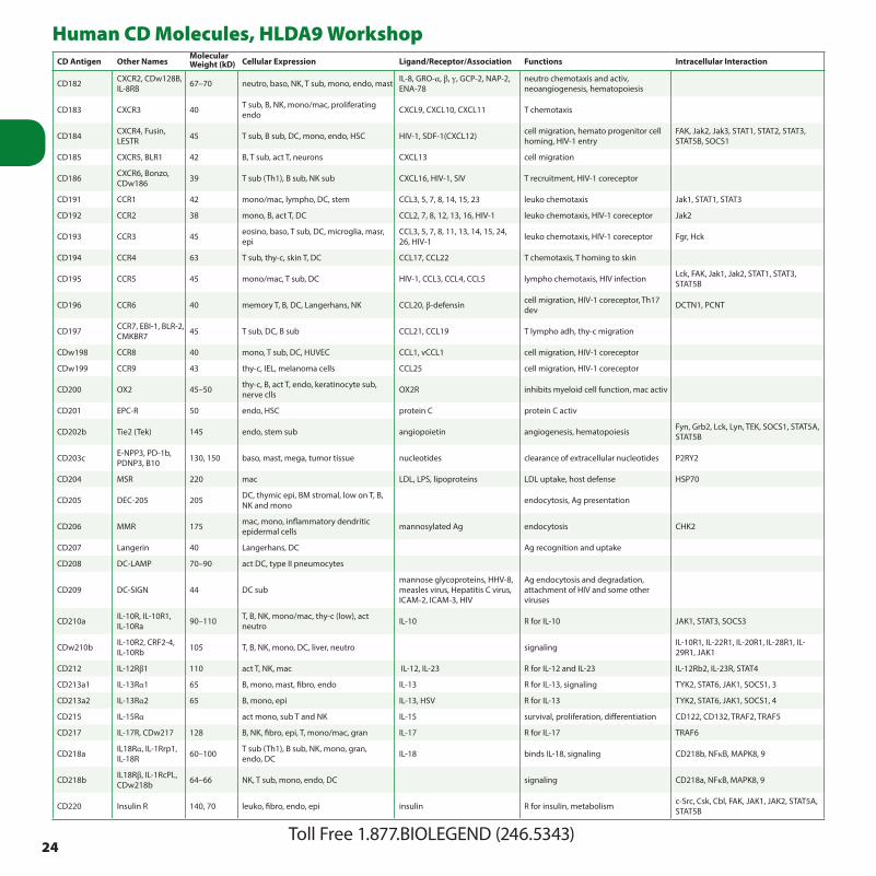
24Toll Free 1.877.BIOLEGEND (246.5343)
Human CD Molecules, HLDA9 WorkshopCD Antigen Other Names
Molecular Weight (kD) Cellular Expression Ligand/Receptor/Association Functions Intracellular Interaction
CD182 CXCR2, CDw128B, IL-8RB 67–70 neutro, baso, NK, T sub, mono, endo, mast IL-8, GRO-α, β, γ, GCP-2, NAP-2,
ENA-78neutro chemotaxis and activ, neoangiogenesis, hematopoiesis
CD183 CXCR3 40 T sub, B, NK, mono/mac, proliferating endo CXCL9, CXCL10, CXCL11 T chemotaxis
CD184 CXCR4, Fusin, LESTR 45 T sub, B sub, DC, mono, endo, HSC HIV-1, SDF-1(CXCL12) cell migration, hemato progenitor cell
homing, HIV-1 entryFAK, Jak2, Jak3, STAT1, STAT2, STAT3, STAT5B, SOCS1
CD185 CXCR5, BLR1 42 B, T sub, act T, neurons CXCL13 cell migration
CD186 CXCR6, Bonzo, CDw186 39 T sub (Th1), B sub, NK sub CXCL16, HIV-1, SIV T recruitment, HIV-1 coreceptor
CD191 CCR1 42 mono/mac, lympho, DC, stem CCL3, 5, 7, 8, 14, 15, 23 leuko chemotaxis Jak1, STAT1, STAT3
CD192 CCR2 38 mono, B, act T, DC CCL2, 7, 8, 12, 13, 16, HIV-1 leuko chemotaxis, HIV-1 coreceptor Jak2
CD193 CCR3 45 eosino, baso, T sub, DC, microglia, masr, epi
CCL3, 5, 7, 8, 11, 13, 14, 15, 24, 26, HIV-1 leuko chemotaxis, HIV-1 coreceptor Fgr, Hck
CD194 CCR4 63 T sub, thy-c, skin T, DC CCL17, CCL22 T chemotaxis, T homing to skin
CD195 CCR5 45 mono/mac, T sub, DC HIV-1, CCL3, CCL4, CCL5 lympho chemotaxis, HIV infection Lck, FAK, Jak1, Jak2, STAT1, STAT3, STAT5B
CD196 CCR6 40 memory T, B, DC, Langerhans, NK CCL20, β-defensin cell migration, HIV-1 coreceptor, Th17 dev DCTN1, PCNT
CD197 CCR7, EBI-1, BLR-2, CMKBR7 45 T sub, DC, B sub CCL21, CCL19 T lympho adh, thy-c migration
CDw198 CCR8 40 mono, T sub, DC, HUVEC CCL1, vCCL1 cell migration, HIV-1 coreceptor
CDw199 CCR9 43 thy-c, IEL, melanoma cells CCL25 cell migration, HIV-1 coreceptor
CD200 OX2 45–50 thy-c, B, act T, endo, keratinocyte sub, nerve clls OX2R inhibits myeloid cell function, mac activ
CD201 EPC-R 50 endo, HSC protein C protein C activ
CD202b Tie2 (Tek) 145 endo, stem sub angiopoietin angiogenesis, hematopoiesis Fyn, Grb2, Lck, Lyn, TEK, SOCS1, STAT5A, STAT5B
CD203c E-NPP3, PD-1b, PDNP3, B10 130, 150 baso, mast, mega, tumor tissue nucleotides clearance of extracellular nucleotides P2RY2
CD204 MSR 220 mac LDL, LPS, lipoproteins LDL uptake, host defense HSP70
CD205 DEC-205 205 DC, thymic epi, BM stromal, low on T, B, NK and mono endocytosis, Ag presentation
CD206 MMR 175 mac, mono, inflammatory dendritic epidermal cells mannosylated Ag endocytosis CHK2
CD207 Langerin 40 Langerhans, DC Ag recognition and uptake
CD208 DC-LAMP 70–90 act DC, type II pneumocytes
CD209 DC-SIGN 44 DC submannose glycoproteins, HHV-8, measles virus, Hepatitis C virus, ICAM-2, ICAM-3, HIV
Ag endocytosis and degradation, attachment of HIV and some other viruses
CD210a IL-10R, IL-10R1, IL-10Ra 90–110 T, B, NK, mono/mac, thy-c (low), act
neutro IL-10 R for IL-10 JAK1, STAT3, SOCS3
CDw210b IL-10R2, CRF2-4, IL-10Rb 105 T, B, NK, mono, DC, liver, neutro signaling IL-10R1, IL-22R1, IL-20R1, IL-28R1, IL-
29R1, JAK1
CD212 IL-12Rβ1 110 act T, NK, mac IL-12, IL-23 R for IL-12 and IL-23 IL-12Rb2, IL-23R, STAT4
CD213a1 IL-13Rα1 65 B, mono, mast, fibro, endo IL-13 R for IL-13, signaling TYK2, STAT6, JAK1, SOCS1, 3
CD213a2 IL-13Rα2 65 B, mono, epi IL-13, HSV R for IL-13 TYK2, STAT6, JAK1, SOCS1, 4
CD215 IL-15Rα act mono, sub T and NK IL-15 survival, proliferation, differentiation CD122, CD132, TRAF2, TRAF5
CD217 IL-17R, CDw217 128 B, NK, fibro, epi, T, mono/mac, gran IL-17 R for IL-17 TRAF6
CD218a IL18Rα, IL-1Rrp1, IL-18R 60–100 T sub (Th1), B sub, NK, mono, gran,
endo, DC IL-18 binds IL-18, signaling CD218b, NFκB, MAPK8, 9
CD218b IL18Rβ, IL-1RcPL, CDw218b 64–66 NK, T sub, mono, endo, DC signaling CD218a, NFκB, MAPK8, 9
CD220 Insulin R 140, 70 leuko, fibro, endo, epi insulin R for insulin, metabolism c-Src, Csk, Cbl, FAK, JAK1, JAK2, STAT5A, STAT5B

25biolegend.com | [email protected]
Human CD Molecules, HLDA9 WorkshopCD Antigen Other Names
Molecular Weight (kD) Cellular Expression Ligand/Receptor/Association Functions Intracellular Interaction
CD221 IGF-IR 135 leuko and variety of non-hemato cells insulin, IGF-I, IGF-II signaling, cell prolif/ differen c-Src, ASK1, CRK, Csk, Jak1, Jak2, CRKL, STAT3
CD222 M6P-R, IGF-IIR 230, 250 lympho, mono, gran, fibro, myocytes, embryonic tissue
plasminogen, proliferin, LAP, IGFII, M6P, LIF, HSV
activates latent TGF-β, cell adh, migration, angiogenesis CD87
CD223 LAG-3 70 act T, act NK MHC class II negative reg of T expansion and homeostasis CBF, CENP-J
CD224γ-glutamyl transpeptidase, GGT
27, 62–68 T sub, B sub, mac, endo, HSC, renal tubular cells, pancreas
glutathione, leukotriene C4, glutathione S-conjugates
inhibits apoptosis, cellular detoxification and leukotriene biosynthesis CD19, CD21, CD37, CD53, CD81, CD82
CD225 Leu-13 17 leuko, endo lympho activ and dev CD81, CD19, CD21
CD226 DNAM-1, PTA-1, TLiSA1 65 T sub, NK, mono, pt, B sub, thy-c, act
HUVEC CD155, CD112 costim, adh LFA-1, Fyn
CD227 MUC-1, EMA, mucin 1 300 epi, stem sub, mono, act T, DC CD54, CD169, selectins cell adh and signaling Lck, c-Src, ZAP70, Grb2, SOS1
CD228 MTf, p97 80–97 melanoma cells, epi, brain, skeletal and heart muscle iron Fe transport PLG
CD229 Ly-9, SLAMF3 100, 120 T, B, thy-c, DC, NK CD229 adh, costim SH2D1A, EAT2
CD230 Prion protein, PrP 35 hemato and non-hemato cells, neurons prevents cells from apoptosis, stem cell renewal BIP, Grb2
CD231 TALLA-1, A15 28–45 T-ALL and neuroblastoma cells, neurons
CD232 VESP-R 200 B, mono, gran, NK, act T, DC CDw108, poxvirus A39R cell differen and migration
CD233 Band3 95–110 eryth, kidney anion exchanger Lyn, Syk
CD234 DARC, Fy, Duffy 35–45 eryth, endo, neurons, epi, cerebellum CXCL1, 5, 8; CCL2, 5, 7, malaria parasite chemokine decoy R, malarial parasite R FUZ
CD235a Glycophorin A 36 eryth hepatitis A virus, babesia, P. falciparum parasite R, cell aggregation
CD235ab Glycophorin A/B 20 eryth hepatitis A virus, babesia, P. falciparum parasite R, cell aggregation
CD235b Glycophorin B 20 eryth babesia, P. falciparum parasite R, cell aggregation
CD236 Glycophorin C/D 30–40 eryth, stem sub Gerbich Antigen
CD236R Glycophorin C 40 eryth, stem sub P. falciparum BAEBL Gerbich Antigen
CD238 Kell 93 eryth, hemato progenitors endothelin-3 endothelin-3 converting enzyme
CD239 B-CAM 90 eryth, fibro, inducible in epi laminin α5 eryth differen and trafficking
CD240CE Rh30CE 30–32 eryth
CD240D Rh30D, RhD 30–32 eryth
CD240DCE Rh30D/CE 30 eryth
CD241 Rhesus 50 50 eryth Rh antigens complex with CD47, LW, glycophorin B
CD242 ICAM-4 37–43 eryth CD11a, b; CD18; CD49b, d, e, CD51 adh, LW blood group
CD243 MDR-1, P-gp, gp170, ABC-B1 170 stem, multi-drug resistant tumor cells toxic xenobiotics, many anti-
cancer drugsion pump, regulates drugs uptake, distribution, and elimination TP53, NR112
CD244 2B4 66 NK, T sub, baso, mono CD48 stimulates NK activ, costimulates T cells LAT, EAT2, SH2D1A
CD245 p220/240 215–245 T, B, NK, mono, gran, pt signal transd, costim
CD246ALK, Ki-1, Anaplastic lymphoma kinase
200 some T lymphomas, endo, some neural cells midkine R for tyrosine kinase, regulates cell
growth and apoptosis ALK, SHC, JAK3, PLCγ1, IRS1, STAT3
CD247 CD3z, Zeta chain, CD3ζ 16 T, NK Ag recognition and signal transd CD3/TCR complex, Lck, Fyn, ZAP-70, Csk,
SHP1, JAK3, STAT5A, STAT5B
CD248 TEM1, Endosialin 165 embryonic endo, tumor endo angiogenesis RASD2
CD249 APA, ENPEP, gp160, EAP 160 epi, endo converts angiotensin II to angiotensin III angiotensin II and cholecystokinin-8
CD252 OX40L, TNFSF4 34 DC, act B, endo, mast OX40 costim TRAF2

26Toll Free 1.877.BIOLEGEND (246.5343)
Human CD Molecules, HLDA9 WorkshopCD Antigen Other Names
Molecular Weight (kD) Cellular Expression Ligand/Receptor/Association Functions Intracellular Interaction
CD253 TRAIL, APO2L, TNFSF10 40 act T, NK, B DR4, DR5, DcR1, DcR2 apoptosis CASP8, CASP3, BCL2
CD254 TRANCE, RANKL, TNFSF11, OPGL 40 act T, stromal cells, osteoclasts RANK T-B and T-DC interaction, bone dev c-Src, AKT1, ERK1, ERK2, TRAF6
CD255 TWEAK, TNFSF12 18, 30–35 act mono, endo, fibro FN14, APO3 activ, prolif, apoptosis
CD256 APRIL, TALL-2, TNFSF13 38 leuko, pancreas, colon TACI, BCMA T and B prolif Fas, Furin
CD257 BLyS, BAFF, TNFSF13b, TALL-1 34 act mono, DC TACI, BCMA, BAFF-R T, B growth and dev, costim TRAF3, NFκB
CD258 LIGHT, TNFSF14 29 act T, act mono HVEM, LTβR costim of T cells, induces apoptosis TRAF2, TRAF3, SMAC
CD261 TRAIL-R1, DR4, TNFRSF10a 56 act T, some tumor cells TRAIL (CD253) induces apoptosis BCL10, Caspase 8, Caspase 10, FADD,
c-FLIP, BTK
CD262TRAIL-R2, DR5, KILLER, TNFRSF10b
55 leuko, heart, placenta, liver, tumor cells TRAIL (CD253) induces apoptosis BCL10, Caspase 8, Caspase 10, FADD, c-FLIP, BTK
CD263 TRAIL-R3, DcR1, TRID, TNFRSF10c 65 low level in most tissues, negative in most
tumor tissues TRAIL (CD253) inhibits TRAIL-induced apoptosis Rap1α , FADD
CD264 TRAIL-R4, DcR2, TNFSF10d 35 low level in most tissues, negative in most
tumor tissues TRAIL (CD253) inhibits TRAIL-induced apoptosis FADD, CASP10, p53
CD265 TRANCE-R, RANK, TNFRSF11a 97 DC, act mono TRANCE (CD254) R for TRANCE c-Src, Cbl, CblB, Grb2, MAP3K7, TRAF1-3,
TRAF5-6, TAB2
CD266 TWEAK-R, Fn14, TNFRSF12a 14 endo, epi, keratinocytes TWEAK (CD255) regulates apoptosis, prolif, angiogenesis TRAF1-3
CD267 TACI, TNFRSF13b 32 B, myeloma cells BAFF, APRIL inhibits B prolif TRAF2, TRAF5-6
CD268 BAFFR, BR3, TNFRSF13c 19 B, T sub BAFF B survival and maturation, T activ TRAF3, PMPCB, NFκB
CD269 BCMA, TNFRSF17 B, plasma cells BAFF, APRIL plasma cell survival TRAF1-3
CD270 HVEM, HVEA, TR2, TNFRSF14, ATAR
T, B, NK, mono/mac, gran, endo, epi, neuron, fibro
LIGHT, LTa, BTLA, CD160, Herpes Virus costimulation, inhibition TRAF1, TRAF2, TRAF3, TRAF5
CD271NGFR (p75), p75NGFR, p75NTR, TNFRSF16
75 neurons, Schwann cells, melanocytes, B, mono, keratinocytes NGF, BDNF, NT-3, NT-4 low affinity R for NGF, induces apoptosis,
embryogenesis, hair growthERK1, ERK2, Grb2, PRKACB, SHC, TRAF2, TRAF4, TRAF6
CD272 BTLA 33 T (upreg upon act), B, mac, DC HVEM inhib of T cell function SHP1, SHP2
CD273 B7DC, PDL2, PD-L2 25 DC, act mono/mac PD-1 costim, inhib RPL17
CD274 B7H1, PDL1, PD-L1 40 T, B, NK, DC, mac, epi PD-1 costim, inhib RPL17
CD275 B7H2, ICOSL, B7RP1, B7h 40 act mono, mac, DC ICOS costim
CD276 B7H3, B7RP-2 45, 110 DC, act mono, act T, act B, act NK, epi may play role in costim or inhib
CD277 BT3.1, BTN3A1 T, B, NK, mono, DC, endo BT3.1-R T activ
CD278 ICOS 47–57 act T, thy-c sub B7H2 (CD275) T costim
CD279 PD1 50–55 act T, act B, thy-c sub CD273, CD274 T tolerance, negative reg TRAF2, RUNX1, PTPase-6
CD280 TEM22, ENDO180, uPARAP 180 fibro, endo, mac, osteoclasts, osteocytes,
chondrocytes collagen remodeling, uptake and degradation of collagen uPAR
CD281 TLR1 90 mono, mac, DC, keratinocytes lipoproteins regulates TLR2 function TLR2, MyD88
CD282 TLR2 86 mono, gran, mac, DC, keratinocytes, epi lipoproteins, glycansinnate immune response to some bacteria and mycoplasma pathogens, signaling
TLR1, TLR6, RIP2, TOLLIP, PI3Kα, H-Ras, MYD88
CD283 TLR3 120 DC, fibro, epi dsRNA innate immune response to viral pathogens TRIAD3, MYD88, TRAF6, MAP3K7, TAB2
CD284 TLR4 110 mono, mac, endo, epi, gran (low), DC (low) LPS innate immune response to gram-
negative bacteriaMD2, CD14; Syk, BTK, IRAK2, MYD88, RIP2, TOLLIP, TRIAD3, MAPK8IP1
CD286 TLR6 90 mono, mac, gran, DC, epi lipoproteins, glycans innate immune response to some bacteria and mycoplasma pathogens TLR2, CD36, BTK, MyD88

27biolegend.com | [email protected]
Human CD Molecules, HLDA9 WorkshopCD Antigen Other Names
Molecular Weight (kD) Cellular Expression Ligand/Receptor/Association Functions Intracellular Interaction
CD288 TLR8 110 mono, mac, DC, neurons, axons ssRNA anti-viral immune response, brain dev, hematopoiesis BTK, MyD88, IRAK4
CD289 TLR9 110–119 pDC, B, mono CpG DNA innate immune response to bacteria or virus, DC maturation BTK, TRIAD3, H-Ras, MAPK8IP3, MyD88
CD290 TLR10 90 B, pDC coreceptor with TLR2 TLR1, 2 and 10
CD292 BMPR1A, ALK3 55, 80mesenchymal cells, epi, bone progenitor, neurons, chondrocytes, skeletal muscle, cardiac myocytes
BMP2, 4, 7, GDF-5 kinase, hair morphogenesis, anti-apoptosis, embryogenesis TAB1, SMAD4, SMAD6
CDw293 BMPR1B, ALK6 55, 80 mesenchymal cells, bone progenitor, chondrocytes, epi, heart, kidney BMP2, 4, 7, GDF-5 kinase, regulates cartilage formation PAK1, RhoD, SH3KBP1, SOCS6, SMAD6,
SMAD6, SMAD7
CD294 CRTH2, DP2, GPR44 55–70 Th2 cells, baso, eosino PGD2
regulates immune and inflammatory response, induces Th2 cells, eosino and baso migration
TFDP1
CD295 LEPR, OB-R 132 hemato cells, heart, placenta, liver, kidney, pancreas leptin
regulates fat metabolism, proliferative/anti-apoptotic T cells and hemato precursors
CD296 ART1 36 neutro, heart, skeletal muscle PDGFβ, integrins, defensin transfers ADP-ribose to target proteins, regulates cellular metabolism
CD297 ART4 36 Dombrock+ RBC, mono, mac, baso, endo, intestine, ovary
metabolism, dombrock blood group antigen
CD298 ATP1B3 32 broad, highly expressed in CNS, testis dimerizes with ATPα1, 2 or 3non-catalytic component of ATPase coupling exchange of sodium and potassium ions
CD299 DC-SIGNR, L-SIGN 45 endo of liver and lymph nodes ICAM-3, HIV gp120, Hepatitis C virus, ebolavirus
T cell trafficking, HCV, EBOV and HIV infection
CD300a CMRF35H, CMRF-35-H9, IRp60 34 mono/mac, neutro, DC, NK, mast, T and
B sub inhib
CD300b IREM-3 26–32 myeloid cells activ DAP-12, NFAT/AP-1
CD300c CMRF35A, LIR 25 mono/mac, neutro, DC, NK, T and B sub, mast
CD300e CMRF35L1, IREM2, CLM-2 mono/mac, DC sub
CD300f IREM-1, IREM1, CLM, MAIR-V mono, gran inhib PI3K
CD301 MGL1, CLECSF14 42 mac, immature DC carbohydrate with Gal/GalNAc structure
cell adh, mac migration, cellular recognition
CD302 DCL1 30 mono/mac, DC
CD303 BDCA2 38 plasmacytoid DC Ag capture, inhib of interferon α/β production
CD304BDCA4, neuropilin-1, NRP1
103 DC, T, neurons, endo VEGF165, SEMA3A, complexed with plexin-1
angiogenesis, DC-T cell interaction, neuritogenesis SEMA3A, B, C, F, G
CD305 LAIR1 31 T, B, NK, DC, mono/mac Collagen inhibits cellular activ and inflammation Csk, SHP2
CD306 LAIR2 16 T, mono Collagen inhibits cellular activ and inflammation
CD307a FCRL1, FCRH, IFGP1, IRTA5 55 B B activation, B development
CD307bFCRL2, SPAP1, FCRH2, IFGP4, IRTA4
B
CD307c FCRL3, IFGP3, IRTA3, SPAP2 B, subset Treg
CD307d FCRL4, FCRH4, IGFP2, IRTA1 57 subset B inhibition SHP-1, SHP-2
CD307eFCRL5, BXMAS1, FCRH5, IRTA2, CD307
106 plasma cell, Germinal Center B IgG, IgA (weak) inhibition

28Toll Free 1.877.BIOLEGEND (246.5343)
Human CD Molecules, HLDA9 WorkshopCD Antigen Other Names
Molecular Weight (kD) Cellular Expression Ligand/Receptor/Association Functions Intracellular Interaction
CD309 VEGFR2, KDR 195/235 endo, primitive stem, some tumor cells VEGF-A, E, C and D angiogenesis Fyn, c-Yes, c-Src, Grb2, Grb10, NCK1, Cbl, STAT1
CD312 EMR2 90 mac, act mono, DC, liver, lung chondroitin sulfates, glycosaminoglycan, CD55
involved in immune and inflammatory response
CD314 NKG2D 42 NK, CD8+ T sub, γ/δ T, mac MICA, MICB, ULBP-1, 2, 3 NK activ DAP10, DAP12
CD315 CD9P-1, FPRP, EWI-F 135 mega, hepato, epi, endo, weak on B and
mono CD9 and CD81
CD316 EWI-2, PGRL 62–78 T, B, NK, hepato inhibits tumor cell metastasis CD9, CD81, KAI1/CD82, and CD85g
CD317 BST2, Tetherin 29–33plasma cells, act T, mono, gran, lymphoplasmacytoid cells, stromal cells, fibro, pDC
CD85g anti-retrovirus, may play a role in pre-B cell growth GATA1
CD318 CDCP1, SIMA135 80/140 HSC, epi, some tumor cells adh c-Src, PKCδ
CD319 CRACC, CS1 66 NK, most T, act B, mature DC CD319 activates NK cytotoxicity, adh
CD320 8D6, NG29 FDC stimulates B growth
CD321 JAM1, JAM, JAM-A, F11R 35–40 epi, endo, leuko, pt, eryth, lung, placenta,
kidney PAR3, LFA-1, reovirus cell adh, leuko migration, epithelial barrier maintenance, pt activ PKCα, CSNK2A1, CASK, PTPB
CD322 JAM2, VE-JAM 48 endo, HEV in tonsils, heart PAR-3, α4β1 integrin, JAM3, JAM-2 cell adh, lympho homing
CD324 E-Cadherin, CDH1 120 epi, keratinocytes, trophoblasts, pt CD103, E-cadherin, PS1, catenins, internalin
cell-cell, cell-matrix adh; tumor suppression; cell growth and differen GSK3β, CSNK2A1, RICS, IRS1
CD325 N-Cadherin, CDH2 CDw325 130 neurons, skeletal and cardiac myocytes,
fibro, epi, pancreas, liver N-cadherin, catenins, FGFR, PS1 cell-cell, cell-matrix adh; cell growth and differen PI3Kα, RICS
CD326Ep-CAM, MK-1, KSA, EGP40, TROP1
40 epi, low on thy-c and T, tumor cells LAIR-1, LAIR-2, Ep-CAM inhibits cellular activ and inflammation Claudin 7, TACC1
CD327 Siglec-6, OB-BP-1, CD33L1, CDw327 B, placenta trophoblasts, gran leptin, sialyl-Tn adh
CD328 Siglec-7, p75/AIRM 65–75 NK, T sub, mono, gran GD3, LSTb inhibits NK and T activity Grb2, SHP2, TRAF4
CD329 Siglec-9 mono; sub of NK, B, T and neutro; hepato, myeloid leukemia GD1a, LSTc inhibits immune response
CD331 FGFR1, FLG, FLT2, N-SAM 60, 110–160 broad, epi, endo, fibro, mesenchymal,
cardiac myocytes, fetal tissue aFGF, bFGF, K-FGF cell growth, limb dev CRK, Grb2, Grb4, Grb14, PI3Kα, PI3Kβ, PLCγ1, SOS1
CD332 FGFR2, BEK, K-SAM 135 brain, liver, prostate, kidney, lung, spinal
cord, fetal tissues aFGF, bFGF, K-FGF, FGF-6 limb induction, craniofacial dev Lyn, Fyn, Cbl, PLCγ1, PAK1, PTK2B
CD333 FGFR3, ACH 115–135 brain, kidney, testis in adult, small intestine in fetus aFGF limb induction, craniofacial dev Grb2, PTK2B, STAT1, STAT3
CD334 FGFR4, TKF 88–110 liver, kidney, lung, pancreas, lympho, mac aFGF, FGF19 bone and muscle dev, cancer progression/metastasis PLCγ1, STAT1, STAT3
CD335 NKp46, NCR1 46 NK viral hemagglutinins, heparan sulfate proteoglycans NK activ
CD336 NKp44, NCR2 44 act NK viral hemagglutinins NK activ DAP12
CD337 NKp30, NCR3 30 NK heparan sulfate proteoglycans NK activ
CD338 ABCG2, ABCP, MXR, BCRP, Brcp1 72
liver, kidney, intestine, lung, endo, melanoma, placenta, side population of stem, certain drug resistant tumors
some xenobiotics absorption and excretion of certain xenobiotics
CD339 Jagged-1, Jag1 134BM stromal, thymic epi, endo, Schwann cells, keratinocytes, ovary, prostate, pancreas, placenta, heart
Notch1, 2, and 3 cell-fate decisions in hematopoiesis, cardiovascular dev
CD340 Her-2, Neu, erb-B2 185
epi, endo, keratinocytes, CD34+ stem sub, fetal mesodermal and extraembryonic tissues, over-expressed on many malignant cells
gp30 cell growth and differen, tumor cell metastasis
CD344 Frizzled-4, FZD4, EVR1, FEVR 48–53
epi, endo, mesenchymal, myeloid progenitors, neuronal progenitors, intestinal neurons
Norrin, Wnt proteins cell prolif and differen, embryonic dev, retinal angiogenesis PLC-γ1, STAT1, STAT3

29biolegend.com | [email protected]
Human CD Molecules, HLDA9 WorkshopCD Antigen Other Names
Molecular Weight (kD) Cellular Expression Ligand/Receptor/Association Functions Intracellular Interaction
CD349 Frizzled-9, FZD9, FZD3, FZ9 64
mesenchymal stem, neural precursor, mammary epi, adult brain, fetal brain, testis, kidney, eye and pancreas, some tumors
Wnt-2, Wnt-7a B dev, hippocampal dev, tissue morphogenesis
CD350 Frizzled-10, FZD10, FZ10 65
brain, embyro, kidney, liver, pancreas, placenta, mammary and lung epi, some tumors
Wnt7b dev of limb, nervous system
CD351 Fcα/µR 58 B, DC, mono, T (low) IgM, IgA antigen presentation?
CD352 SLAMF6, NTB-A, KAL1, Ly108 60 NK, T, B CD352 costim, activ EAT-2, SAP
CD353 SLAMF8, BLAME, SBBI42 mac, DC, B regulation macrophage function
CD354 TREM-1 30 mono, gran activation, costimulation DAP-12
CD355 CRTAM act CD8 Tcells, act NK/NKT cells Necl2 adhesion
CD357 TNFRSF18, GITR, AITR 30 T (low), act T, Treg GITRL suppress Treg function TRAF1, TRAF2, TRAF3
CD358 TNFRSF21, DR6 40 broad tissue distribution, overexpressed by tumor cells APP apoptosis
CD360 IL-21R, NILR B, low on T, NK, mono, and DC IL-21 T, NK activation, proliferation and development, B apoptosis CD132, JAK1, JAK3, STAT1, STAT3, STAT5
CD361 EVI2b, EVDB broad in hemato and some non-hemato cells
CD362Syndecan-2, SDC2.1.186, Fibroglycan
48 mac, epi, endo, osteoblast fibronectin, TACI, IL-8 adhesion, B cell activation, apoptosis
CD363 S1PR1, EDG-1, CHEDG1, S1P1 endo, subset B, NK S1P adhesion, endothelial differentiation GNAI1, GNAI3

30Toll Free 1.877.BIOLEGEND (246.5343)
Mouse Biofunctional Antibodies - LEAF™ and Ultra-LEAF™Specificity Clone Applications LEAF™ Cat. No. Ultra-LEAF™ Cat. No.
4-1BB Ligand (CD137L) TKS-1 FC, Block 107108 -
α-GalCer:CD1d complex L363 FC, IF, Block 140504 -
Allergin-1 TX83 FC, Block 141804 -
Asialo-GM1 Poly21460 FC, Depletion - 146002
B7-H4 (B7S1) HMH4-5G1 FC 139404 -
CD1d (CD1.1, Ly-38) 1B1 FC, IHC, IP, Block 123504, 123515 -
CD1d K253 FC, Block, ELISA, IF 140804 -
CD2 RM2-5 FC, IP, Block, Costim 100110 -
CD3 17A2 FC, IHC, Activ, CMCD, Depletion 100207, 100208, 100223 100238
CD3ε 145-2C11 FC, IHC, IP, WB, CMCD, Block, Apop, Depletion, Activ, IF 100313, 100314, 100331 100339, 100340
CD4 GK1.5 FC, Block, Costim, Depletion, IHC, IP 100415, 100416, 100435 100441, 100442
CD4 RM4-5 FC, Depletion, Block, IHC, CYTOF® 100520 -
CD8a 53-6.7 FC, IHC, IP, Depletion, Block, CYTOF® 100715, 100716, 100735 100745, 100746
CD11a M17/4 FC, IHC, IP, Block 101109, 101110, 101115 101118
CD11b M1/70 FC, IP, Block, Depletion, IHC, IF, CYTOF® 101213, 101214, 101231 101248
CD14 Sa14-2 FC 123304 -
CD16/32 93 FC, IP, Block 101309, 101310, 101321 101330
CD18 M18/2 FC, IP, WB, Block, Stim, IHC 101409, 101410 -
CD19 6D5 FC, IF, CYTOF® 115514 -
CD21/CD35 (CR2/CR1) 7E9 FC, FA 123404 -
CD24 M1/69 FC, WB, IHC, IF, Activ, CMCD 101810 -
CD25 3C7 FC, IHC, Block, CYTOF® 101906 -
CD25 PC61 FC, IP, IHC, Block, Depletion 102013, 102014, 102031 102039, 102040
CD27 LG.3A10 FC, IHC, IP, FA 124204 -
CD28 37.51 FC, IP, IHC, Costim, Block 102111, 102112 102115, 102116
CD28 E18 FC, Costim, Block 122003, 122004 -
CD29 HMß1-1 FC, IP, Block, IHC 102209, 102210 -
CD31 390 FC, IP, Block, IHC 102412 -
CD31 MEC13.3 FC, IP, Block, IHC 102511, 102512 -
CD40 1C10 FC, IP, Costim, Block 102809, 102810 102812
CD40 3/23 FC, IHC, FA 124604, 124619 124628
CD40 HM40-3 FC, Block, Costim, IHC 102907, 102908 -
CD41 MWReg30 FC, FA 133909, 133910 -
CD44 IM7 FC, ELISA, IHC, IP, CMCD, Stim, CYTOF® 103013, 103014, 103033 103046
CD45 30-F11 FC, IP, CMCD, IHC, WB, CyTOF® 103119, 103120 -
CD45R (B220) RA3-6B2 FC, IP, Activ, IHC, CYTOF® 103216 -
CD47 miap301 FC, Block 127511, 127512 -
CD48 HM48-1 FC, IP, Costim, Block 103408 103429, 103430

31biolegend.com | [email protected]
Mouse Biofunctional Antibodies - LEAF™ and Ultra-LEAF™Specificity Clone Applications LEAF™ Cat. No. Ultra-LEAF™ Cat. No.
CD49b HMα2 FC, IP, Block 103507 -
CD49d 9C10 (MFR4.B) FC, Block, IHC 103707, 103708 -
CD49d R1-2 FC, IP, Block, IHC 103610 -
CD49e 5H10-27(MFR5) FC, Block, Costim, IHC 103807, 103808 -
CD49f GoH3 FC, IF, Block, IHC, IP 313613, 313614 -
CD51 RMV-7 FC, IP, Block 104107, 104108 -
CD54 YN1/1.7.4 FC, Block, IHC, IP, WB 116109, 116110 -
CD61 2C9.G2 (HMß3-1) FC, Block, Activ, IHC 104309, 104310 -
CD62L MEL-14 FC, IP, CMCD, Block, IHC, CYTOF® 104416 -
CD66a (CEACAM1a) MAb-CC1 FC, WB, IP, IHC, ELISA, Block 134504 -
CD70 FR70 FC, Block, IHC, IP 104608 -
CD80 16-10A1 FC, IHC, IP, Block 104710 -
CD81 Eat-2 FC, IP, WB, Activ, Stim 104908 -
CD86 GL-1 FC, Block, IF, IHC, IP 105010 -
CD88 (C5aR) 20/70 FC, Block 135804 -
CD90 G7 FC, Activ, Apop, IHC 105203 -
CD90.2 30-H12 FC, Depletion, Costim, IHC 105309, 105310 -
CD96 (TACTILE) 3.3 FC, Block 131704 -
CD106 429 (MVCAM.A) FC, IP, Block, IHC 105708 -
CD115 (CSF-1R) AFS98 FC, Block 135503, 135504 -
CD117 (c-Kit) ACK2 FC, Block, Depletion 135104 135114
CD120a (TNFR Type I/p55) 55R-170 FC, IP, Block 112902 -
CD120a (TNFR Type I/p55) 55R-593 FC, IP, Block 113102 -
CD120b (TNFR Type II/p75) TR75-32.4 FC, IP, Block 113202 -
CD120b (TNFR Type II/p75) TR75-54.7 FC, Block, IP, ELISA Capture, WB 113302 -
CD122 (IL-2Rβ) TM-β1 FC, IP, Block, Depletion 123203, 123204 -
CD126 (IL-6Rα chain) D7715A7 FC, IP, Block, Neut 115807, 115808, 115809 -
CD127 (IL-7Rα) A7R34 FC, IP, WB, Block, IHC, IF 135004 -
CD132 (common γ chain) TUGm2 FC, Block 132304 -
CD134 (OX-40) OX-86 FC, IHC, Costim 119407, 119408 -
CD137 17B5 FC, Block 106107 -
CD140b APB5 FC, Block 136004 -
CD144 (VE-cadherin) BV13 FC, WB, IF, Block 138003 -
CD150 (SLAM) TC15-12F12.2 FC, IP, Costim, Block 115905 -
CD152 9H10 ELISA, FC, Block, Costim 106204 -
CD152 UC10-4B9 FC, Block, IP, ELISA 106308 -
CD154 MR1 FC, IHC, Block 106508 -
CD155 (PVR) 4.24.1 FC 132204 -

32Toll Free 1.877.BIOLEGEND (246.5343)
Mouse Biofunctional Antibodies - LEAF™ and Ultra-LEAF™Specificity Clone Applications LEAF™ Cat. No. Ultra-LEAF™ Cat. No.
CD172a (SIRPα) P84 FC, Block, IHC, IF, IP 144003, 144004 -
CD178 (FasL) MFL3 FC, Block, Apop, IF 106608 -
CD178 (FasL) MFL4 FC, Block 106706 -
CD178.1 (FasL) Kay-10 FC, Block, IHC 106808 -
CD183 (CXCR3) CXCR3-173 FC, Block 126509, 126517 126525, 126526
CD193 (CCR3) J073E5 FC 144503, 144504 -
CD200R (OX2R) OX-110 FC, FA 123912 -
CD200R3 Ba13 FC, Activ 142203, 142204 -
CD200R3 Ba160 FC 142304 -
CD210 (IL-10R) 1B1.3a FC, Block 112707, 112708 112709, 112710
CD223 (LAG-3) C9B7W FC, FA, IP, ELISA 125204 -
CD226 (DNAM-1) 480.1 FC 132003, 132004 -
CD252 (OX40L) RM134L FC, Block, IHC 108808 -
CD253 (TRAIL) N2B2 FC, Block 109308 -
CD254 (TRANCE, RANKL) IK22/5 FC, IP, WB, Block 510007, 510008 -
CD255 (TWEAK) MTW-1 FC, Block 120008 -
CD258 (LIGHT) 15B2 ELISA, Block, IP 137104 -
CD262 (DR5, TRAIL-R2) MD5-1 FC, Cyt 119907 -
CD272 (BTLA) 6A6 FC, IP, Block 139104 -
CD273 (B7-DC, PD-L2) TY25 FC, WB, IP, Block, IHC 107208 -
CD274 (B7-H1, PD-L1) 10F.9G2 FC, IF, Block 124303, 124304, 124309 124318
CD275 (B7-H2, B7-RP1, ICOSL) HK5.3 FC, Block 107407, 107408 107410
CD276 (B7-H3) MIH35 FC, FA 135604 -
CD278 (ICOS) 7E.17G9 FC, Block 117407, 117408 -
CD278 (ICOS) C398.4A FC, IHC, IP, Costim 313511, 313512 -
CD279 (PD-1) 29F.1A12 FC, Block 135203, 135204 -
CD279 (PD-1) RMP1-14 FC, IHC, WB, FA 114107, 114108 -
CD284 (TLR4)/MD2 Complex MTS510 FC, IHC, ICFC, IF, IP, Block 117607, 117608 117612
CD314 (NKG2D) C7 FC, Block 115710 -
CD314 (NKG2D) CX5 FC, Block 130204 -
CD317 (BST2, PDCA-1) 927 FC, IF, IP, FA, Depletion 127004 -
CD335 (NKp46) 29A1.4 FC, IHC, Activ 137613, 137614 -
CD351 (Fc α/μ receptor) TX61 FC, Block 137303 -
CD355 (CRTAM) 11-5/CRTAM FC, WB 142004 -
CD357 (GITR) DTA-1 FC, IP, FA 126303, 126304 -
CXCR7 8F11-M16 FC 331110 -
Delta-like 4 (DLL4) HMD4-1 FC, IHC, FA 130804 -
DR3 (TNFRSF25) 4C12 Agonist, FC 144403, 144404 -

33biolegend.com | [email protected]
Mouse Biofunctional Antibodies - LEAF™ and Ultra-LEAF™Specificity Clone Applications LEAF™ Cat. No. Ultra-LEAF™ Cat. No.
F4/80 BM8 FC, IHC, WB 123103 -
FcεRIα MAR-1 FC, Depletion, IHC 134312 -
FR4 (Folate Receptor 4) TH6 FC, Cyt 125102 -
Galectin-9 RG9-35 ICFC, Block 136106 -
GM-CSF MP1-22E9 ELISA Capture, ELISPOT Capture, ICC, IHC, Neut 505407, 505408 -
I-A/I-E M5/114.15.2 FC, IP, IHC, Block 107610 -
Integrin β7 FIB27 FC, IP, Block 121003 -
Integrin β7 FIB504 FC, Block 321218 -
IFNAR-1 MAR1-5A3 FC, IP, WB, ELISA, Block 127303, 127304 127321, 127322
IFN-β MIB-5E9.1 IP, Neut, WB 508103, 508104 -
IFN-γ AN-18 ELISPOT Capture, ELISA Capture, IP, Neut 517903, 517904 -
IFN-γ H22 WB, Neut, ELISA, IF, IP 513205, 513206 -
IFN-γ R4-6A2 ELISA Capture, ELISPOT Capture, IHC, Neut 505705, 505706, 505707 -
IFN-γ XMG1.2 ELISA Capture, ELISPOT Capture, ICFC, IHC, Neut, WB, CyTOF® 505811, 505812, 505827 505833, 505834
IL-1α ALF-161 ELISA Capture, ELISPOT Capture, ICFC, IP, Neut, WB 503206 -
IL-1β B122 ELISA Capture, ELISPOT Capture, Depletion, IHC, IP, Neut, WB 503504 -
IL-2 JES6-1A12 ELISA Capture, ELISPOT Capture, Neut, WB 503704 503705, 503706
IL-2 JES6-5H4 ELISA, Neut, ICFC, IP, IHC 503812 -
IL-3 MP2-8F8 ELISA Capture, ELISPOT Capture, Neut 503906 -
IL-4 11B11 ELISA Capture, ELISPOT Capture, ICC, IHC, IP, Neut, CyTOF® 504107, 504108, 504115 504121, 504122
IL-5 TRFK5 ELISA Capture, ELISPOT Capture, ICFC, IHC, Neut, WB, CyTOF® 504307, 504308 -
IL-6 MP5-20F3 ELISA Capture, ELISPOT Capture, ICFC, IHC, Neut, CyTOF® 504505, 504506 -
IL-9 D8402E8 ELISA Capture 504704 -
IL-9 D9302C12 Neut, WB 504802 -
IL-10 JES5-2A5 ELISA Capture, ELISPOT Capture, Neut, 504903, 504904 -
IL-10 JES5-16E3 ELISA Capture, ELISPOT Capture, ICFC, IHC, Neut, CyTOF® 505011, 505012 -
IL-12/IL-23 p40 (monomer, dimer, heterodimer) C15.6 ELISA Capture, ELISPOT Capture, ICFC, IHC, IP, WB 505207, 505208 -
IL-12/IL-23 p40 (monomer, dimer, heterodimer) C17.8 ELISA, Neut, ICFC, IP, WB 505303, 505304, 505305 505307, 505308
IL-17A TC11-18H10.1 ELISA Capture, ELISPOT Capture, Neut, WB, CyTOF® 506905, 506906, 506923 -
IL-17A TC11-8H4 ELISA, WB 507004 -
IL-23 (p19) MMp19B2 Block 513805, 513806 -
IL-25 35B ELISA, Neut 514403, 514404 -
IL-27 p28 MM27-7B1 ICFC, Neut - 516912
Jagged 2 HMJ2-1 FC, IHC 131004 -
LAP (TGF-β1) TW7-20B9 FC, IP, Neut, WB 141303, 141304 -
Ly-6C HK1.4 FC, Activ, IHC 128020 -
Ly-6G 1A8 FC, Depletion, IHC 127620 127631, 127632
Ly-6G/Ly-6C (Gr-1) RB6-8C5 FC, IP, CMCD, Depletion, IHC, WB 108413, 108414 108435, 108436

34Toll Free 1.877.BIOLEGEND (246.5343)
Mouse Biofunctional Antibodies - LEAF™ and Ultra-LEAF™Specificity Clone Applications LEAF™ Cat. No. Ultra-LEAF™ Cat. No.
Ly49H 3D10 FC, IP, Block 144703, 144704 -
MAdCAM-1 MECA-367 FC, IP, WB, IHC, Block 120704 -
MCP-1 2H5 ELISA Capture, ELISPOT Capture, IHC, Neut, WB 505905, 505906 -
NK-1.1 PK136 FC, IP, CMCD, Depletion, Block, Activ, IHC, IF, CYTOF® 108711, 108712 -
PD-1H (VISTA) MH5A FC, Block, WB, IHC 143703, 143704 -
TCR β chain H57-597 FC, IP, Stim, Depletion, IHC, CYTOF® 109214 -
TCR γ/δ GL3 FC, IHC, IP 118114 -
TCR γ/δ UC7-13D5 FC, IP, Stim, Depletion 107506 -
TER-119/Erythroid Cells TER-119 FC, IP, WB, IHC, CMCD 116214, 116229 -
TGF-β1 19D8 Block 521703, 521704 -
Tim-4 RMT4-54 FC, FA 130004 -
TNF-α 6B8 ELISA Capture, ELISPOT Capture 510803 -
TNF-α MP6-XT22 ELISA, ICFC, Neut, WB, IHC, CyTOF®, IF 506309, 506310, 506325 506331, 506332
TNF-α TN3-19.12 ELISA Capture, ELISPOT Capture, ICFC, IP, Neut, WB 506105, 506106 -
VEGF-A 2G11-2A05 Neut, WB, ELISA, IHC, FA 512807, 512808 -
VEGFR-3 (FLT-4) AFL4 IHC, IP, WB, ELISA, Block 140903, 140904 -

35biolegend.com | [email protected]
Human Biofunctional Antibodies - LEAF™ and Ultra-LEAF™Specificity Clone Applications LEAF™ Cat. No. Ultra-LEAF™ Cat. No.
Asialo-GM1 Poly21460 FC, Depletion - 146002
C3a/C3a(desArg)/C3 K13/16 ELISA Capture, WB, IP, Neut 518103, 518104 -
C5a/C5a(desArg)/C5 G25/2 ELISA Capture, WB, IP, Neut 518303, 518304 -
CD1d 51.1 FC, WB, IP, IHC, Stim 350304 -
CD2 RPA-2.10 FC, IHC, Block 300212 -
CD2 TS1/8 FC, Block 309212 -
CD3 HIT3a FC, IHC, IP, Activ 300313, 300314 300331, 300332
CD3 OKT3 FC, IHC, Activ 317303, 317304, 317315 317325, 317326
CD3 UCHT1 FC, IHC, IP, Activ, WB, CyTOF® 300413, 300414, 300432 300437, 300438
CD4 OKT4 FC, IHC 317403, 317404 -
CD4 RPA-T4 FC, IHC, Block, CyTOF® 300515, 300516 -
CD8a RPA-T8 FC, IHC, Costim, CyTOF® 301018 -
CD11a HI111 FC, Block, IHC, WB 301213, 301214 -
CD11b (activated) CBRM1/5 FC, IP, Block 301407 -
CD11b ICRF44 FC, IHC, Block, IF, CyTOF® 301311, 301312 -
CD11b M1/70 FC, IP, Block, Depletion, IHC, IF, CyTOF® 101213, 101214, 101231 101248
CD11c 3.9 FC, IHC, Block, CyTOF® 301616 301632
CD13 WM15 FC, IHC, Block 301708 -
CD14 M5E2 FC, IF, IHC, Block, CyTOF® 301809, 301810 -
CD16 3G8 FC, IHC, IP, Stim, Block, CyTOF® 302013, 302014, 302033 302050
CD18 TS1/18 FC, Block 302111, 302112 302116
CD19 HIB19 FC, IHC, Block, CyTOF® 302214 -
CD27 LG.3A10 FC, IP, IHC, FA 124204 -
CD28 CD28.2 FC, IHC, IP, Activ, Costim 302913, 302914, 302923 302933, 302934
CD29 TS2/16 FC, IF, IHC, IP, Activ 303009, 303010 -
CD31 WM59 FC, IF, IHC, Block 303108 -
CD33 WM53 FC, IP, WB, Costim, CyTOF® 303410 -
CD40 5C3 FC, Block, Activ 334303, 334304 -
CD40 G28.5 FC, FA, WB, IF 303607, 303608, 303611 303614
CD40 HB14 FC, Costim 313009, 313010 -
CD41 HIP8 FC, IHC, Block, CyTOF® 303712 -
CD42b HIP1 FC, IHC, WB, Block 303908 -
CD44 IM7 FC, ELISA, IHC, IP, CMCD, Stim, CyTOF® 103013, 103014, 103033 103046
CD45R/B220 RA3-6B2 FC, IHC, IP, Activ, CyTOF® 103216 -
CD49b P1E6-C5 FC, Block, IP, ELISA 359303, 359304 -
CD49d 9F10 FC, IHC, Costim 304309, 304310 -
CD49e NKI-SAM-1 FC, IP, ELISA, Block 328004 -
CD49f GoH3 FC, IF, Block, IHC, IP 313613, 313614 -

36Toll Free 1.877.BIOLEGEND (246.5343)
Human Biofunctional Antibodies - LEAF™ and Ultra-LEAF™Specificity Clone Applications LEAF™ Cat. No. Ultra-LEAF™ Cat. No.
CD51 NKI-M9 FC, ELISA, Block, IP 327904 -
CD51/61 23C6 FC, IHC, Block, IF, IP 304413 -
CD54 HA58 FC, IHC 353103, 353104 -
CD54 HCD54 FC, IHC, Block 322703, 322704 -
CD56 (NCAM) HCD56 FC, FA, CyTOF® 318324 -
CD56 (NCAM) MEM-188 FC, IHC, IP, WB 304622 -
CD58 TS2/9 FC, IP, IHC, Block 330911, 330912 -
CD62E HAE-1f FC, FA 336004 -
CD62L DREG-56 FC, WB, Block 304811, 304812 -
CD62P (P-Selectin) AK4 FC, IHC, Block 304911 -
CD63 H5C6 FC, IF, IP, WB 353013, 353014 -
CD64 10.1 FC 305015, 305016 -
CD80 2D10 FC, IHC, IP, WB, Block 305211, 305212 -
CD85d (ILT4) 42D1 FC, FA 338704 -
CD85g (ILT7) 17G10.2 FC, FA 326403 -
CD85j (ILT2) GHI/75 FC, FA 333704 -
CD86 IT2.2 FC, IHC, WB, Block 305409, 305410 -
CD95 (Fas) DX2 FC, IF, IHC, Apop 305613, 305614 -
CD95 (Fas) EOS9.1 FC, Apop 305703, 305704 -
CD96 NK92.39 FC, FA 338404 -
CD117 (c-kit) A3C6E2 FC, Block 323404 -
CD119 (IFN-γ R α chain) GIR-208 FC, IHC, IP, WB, Block 308604 -
CD122 (IL-2Rβ) TU27 FC, FA 339004 -
CD129 (IL-9 R) AH9R7 FC, Block, ELISA 310408 -
CD132 (common γ chain) TUGh4 FC, FA 338604 -
CD137 (4-1BB) 4B4-1 FC, IP, ELISA, FA 309811 -
CD147 HIM6 FC, IHC, WB, Block 306206 -
CD150 (SLAM) A12 (7D4) FC, IHC, IP, Costim 306310 -
CD151 (PETA-3) 50-6 FC, Block, WB, ICC 350404 -
CD152 (CTLA-4) L3D10 FC, Block 349903, 349904 -
CD154 24-31 FC, IF, Block 310811, 310812 310827, 310828
CD158d (KIR2DL4) mAb 33 (33) FC, Activ 347003 -
CD178 (Fas-L) NOK-1 FC, ICC, IF, IP, Block, WB 306408, 306409 -
CD180 (RP105) MHR73-11 FC, ICFC, IHC, IP, Stim 312907 -
CD183 (CXCR3) G025H7 Block, FC 353717, 353718 -
CD184 (CXCR4) 12G5 FC, ICC, IF, IHC, Block, CyTOF® 306512 -
CD197 (CCR7) G043H7 FC, Block 353221, 353222 -
CD206 (MMR) 15-2 FC, IF, IHC, WB, Block 321111, 321112 -

37biolegend.com | [email protected]
Human Biofunctional Antibodies - LEAF™ and Ultra-LEAF™Specificity Clone Applications LEAF™ Cat. No. Ultra-LEAF™ Cat. No.
CD210 (IL-10 R) 3F9 FC, IP, Block 308805, 308806, 308807 308810
CD218a (IL-18Rα) H44 FC, IHC 313802 -
CD221 (IGF-1R) 1H7/CD221 FC, Block, IHC, IP, WB 351804 -
CD229 (Ly-9) HLy-9.1.25 FC, IP, ICC, FA 326104 -
CD243 (MDR-1) UIC2 FC, Block, IP, IHC 348603 -
CD253 (TRAIL) RIK-2 FC, Block 308207, 308208 -
CD255 (TWEAK) CARL-1 FC, Neut, IHC 308307 -
CD258 (LIGHT) 15B2 ELISA, Block, IP 137104 -
CD272 (BTLA) MIH26 FC, Agonist 344503, 344504 -
CD273 (B7-DC, PD-L2) 24F.10C12 FC, IHC, FA 329610 -
CD273 (B7-DC, PD-L2) MIH18 FC, IHC, IF, Block 345504 -
CD274 (B7-H1, PD-L1) 29E.2A3 FC, IHC, FA 329709, 329710, 329711 329715, 329716
CD276 (B7-H3) MIH35 FC, FA 135604 -
CD278 (ICOS) C398.4A FC, IHC, IP, Costim 313511, 313512 -
CD279 (PD-1) EH12.2H7 FC, Block 329911, 329912 329925, 329926
CD282 (TLR2) TL2.1 FC, IHC, IP, WB, Block 309709, 309710 309716
CD284 (TLR4) HTA125 FC, IHC, IF, Block 312807, 312808 312814
CD290 (TLR10) 3C10C5 FC 354601, 354602 -
CD314 (NKG2D) 1D11 FC, IP, FA 320809, 320810 320814
CD325 (N-Cadherin) 8C11 FC, IF, Block, WB 350804 -
CD335 (NKp46) 9E2 FC, FA 331904 -
CD336 (NKp44) P44-8 FC, Block 325104 -
CD337 (NKp30) P30-15 FC, ELISA, Block 325203, 325204 -
CD351 (Fc α/μ receptor) TX61 FC, Block 137303 -
CD352 (NTB-A) NT-7 FC, Block 317204 -
CD354 (TREM-1) TREM-26 FC, WB, Activ 314907 -
CD355 (CRTAM) Cr24.1 FC, FA 339104 -
CD360 (IL-21R) 17A12 FC, IF 359503, 359504 -
CXCR7 8F11-M16 FC 331110 -
Delta-like protein 4 (DLL4) MHD4-46 FC 346504 -
FcεRIα AER-37 (CRA-1) FC, Activ 334604 -
Galectin-9 9M1-3 ICFC, FC, Block 348903, 348904 -
G-CSF BVD13-3A5 ELISA Capture, ELISPOT Capture, IHC, Neut 502104 -
GM-CSF BVD2-23B6 ELISA Capture, ELISPOT Capture, IP, Neut, WB 502203, 502204 -
HLA-A,B,C W6/32 FC, IHC, IP, WB, Block, Activ 311411, 311412, 311423 311428
HLA-DR L243 FC, IHC, IP, WB, Block, CyTOF® 307611, 307612 307648
HLA-G 87G FC, IHC, ELISA, FA 335904 -
IFN-β IFNb/A1 WB, ELISA, Neut 514003, 514004 -

38Toll Free 1.877.BIOLEGEND (246.5343)
Human Biofunctional Antibodies - LEAF™ and Ultra-LEAF™Specificity Clone Applications LEAF™ Cat. No. Ultra-LEAF™ Cat. No.
IFN-γ B27 ELISA, ICFC, IP, IHC, Neut, WB, CyTOF® 506512, 506513 -
IFN-γ MD-1 ELISA Capture, ELISPOT Capture, ICFC, IHC, Neut, WB 507513 -
IFN-γ NIB42 ELISA Capture, ELISPOT Capture, Neut 502403, 502404 -
Integrin α9β1 Y9A2 FC, IP, IF, Block 351603 -
Integrin β7 FIB27 FC, IP, Block 121003 -
Integrin β7 FIB504 FC, Block 321218 -
IL-1α 364-3B3-14 ELISA Capture, ELISPOT Capture, ICFC, IHC 500102 -
IL-1β H1b-27 ELISA Capture, ELISPOT Capture, Block 511604 -
IL-2 MQ1-17H12 ELISA Capture, ELISPOT Capture, ICFC, IHC, IP, CyTOF® 500313 -
IL-4 8D4-8 ELISA Capture, ELISPOT Capture, ICFC, IHC, IP, Neut 500706, 500707 -
IL-4 MP4-25D2 ELISA, ICFC, IHC, IP, Neut, WB, CyTOF® 500814, 500815 -
IL-5 JES1-39D10 ELISA Capture, ELISPOT Capture, ICFC, IHC, Neut, WB 500907 -
IL-5 JES1-5A10 ELISA Capture, ELISPOT Capture, Neut 501004 -
IL-5 TRFK5 ELISA Capture, ELISPOT Capture, ICFC, IHC, Neut, WB, CyTOF® 504307, 504308 -
IL-6 MQ2-13A5 ELISA Capture, ELISPOT Capture, ICFC, Neut, CyTOF® 501109, 501110 -
IL-7 BVD10-40F6 ELISA Capture, ELISPOT Capture, Neut, WB 501303 -
IL-9 MH9A3 ELISA, Neut 507704 -
IL-9 MH9D1 ELISA, Neut 512004 -
IL-10 JES3-19F1 ELISA Capture, ELISPOT Capture, ICFC, IHC, Neut 506810 -
IL-10 JES3-9D7 ELISA Capture, ELISPOT Capture, ICFC, IHC, Neut 501406, 501407 -
IL-12/IL-23 p40 (monomer, homodimer) C8.3 ELISA Capture, ELISPOT Capture 501704 -
IL-12/IL-23 p40 (monomer, homodimer, heterodimer) C8.6 ICFC, IP, Neut, WB 508803, 508804 -
IL-12/IL-23 p40 (monomer, homodimer, heterodimer) C11.5 ICFC, Block, IHC, Neut, WB 501813 -
IL-13 JES10-5A2 ELISA Capture, ELISPOT Capture, ICFC, IHC, Neut, WB 501909, 501910 -
LT-α (TNF-β) 359-238-8 ELISA Capture, ELISPOT Capture, Neut 503004 -
Lymphotoxin β R (LT-βR) 31G4D8 FC, IP, WB, Apop 322004 -
MICA/MICB 6D4 FC, IHC, Block, IP 320909, 320910 -
MCP-1 2H5 ELISA Capture, ELISPOT Capture, IHC, Neut, WB 505905, 505906 -
MCP-1 5D3-F7 ELISA Capture, ELISPOT Capture, ICFC, IHC, IP, Neut, WB 502606, 502607 -
NKp80 5D12 FC, IP, Block 346703 -
Notch 1 MHN1-519 FC, Block 352103, 352104 -
TCR γ/δ B1 FC, FA, IHC 331203, 331204 -
TGF-β1 19D8 Block 521703, 521704 -
Tim-3 F38-2E2 FC, Costim, Block 345003, 345004 345009, 345010
TNF-α MAb1 ELISA, Neut, WB 502803, 502804 -
TNF-α MAb11 ELISA Capture, ELISPOT Capture, ICFC, ICC, IHC, Neut, CyTOF® 502922 -
TSLP 15B11.3 ELISA Capture, ELISPOT Capture, Neut 512203, 512204 -

39biolegend.com | [email protected]
Rat Biofunctional Antibodies - LEAF™ and Ultra-LEAF™Specificity Clone Applications LEAF™ Cat. No. Ultra-LEAF™ Cat. No.
Asialo-GM1 Poly21460 FC, Depletion - 146002
CD3 1F4 FC 201410 -
CD8a OX-8 FC, IP, FA 201714 -
CD27 LG.3A10 FC, IP, IHC, FA 124204 -
CD29 HMß1-1 FC, IHC, IP, Block 102209, 102210 -
CD61 2C9.G2 (HMß3-1) FC, IHC, Block, Activ 104309, 104310 -
CD81 Eat-2 FC, IP, WB, Activ, Stim 104908 -
CD126 (IL-6Rα chain) D7715A7 FC, IP, Block, Neut 115807, 115808, 115809 -
CD178 (FasL) MFL4 FC, Block 106706 -
CD278 (ICOS) C398.4A FC, IHC, IP, Costim 313511, 313512 -
IFN-γ DB-1 ELISA Capture, Neut, ICFC, IHC, WB, ELISPOT Capture 507808 -
IL-1β B122 ELISA Capture, ELISPOT Capture, Depletion, IHC, IP, Neut, WB 503504 -
MCP-1 2H5 ELISA Capture, ELISPOT Capture, ICFC, IHC, Neut, WB 505905, 505906 -
TNF-α TN3-19.12 ELISA Capture, ELISPOT Capture, ICFC, IP, Neut, WB 506105, 506106 -
Isotype ControlsSpecificity Clone Applications LEAF™ Cat. No. Ultra-LEAF™ Cat. No.
Mouse IgG1, κ Isotype control MG1-45 FC, ICFC, WB, IP, ICC, IF, FA 401403, 401404, 401405 401407, 401408
Mouse IgG1, κ Isotype control MOPC-21 FC, ICFC, WB, IP, ICC, IF, FA 400123, 400124, 400153 400165, 400166
Mouse IgG2a, κ Isotype control MG2a-53 FC, ICFC, WB, IP, ICC, IF, IHC, FA 401503, 401504 -
Mouse IgG2a, κ Isotype control MOPC-173 FC, WB, IP, ICFC, ICC, IF, IHC, FA 400223, 400224 400263, 400264
Mouse IgG2b, κ Isotype control MG2b-57 FC, ICFC, WB, IP, ICC, IF, FA 401212 -
Mouse IgG2b, κ Isotype control MPC-11 FC, ICFC, WB, IP, ICC, IF, FA 400323, 400324, 400339 400348
Mouse IgG3, κ Isotype control MG3-35 FC, ICFC, IP, WB, ICC, IF, FA 401310, 401313 -
Mouse IgM, κ Isotype control MM-30 FC, ICFC, ICC, IF, FA 401604 -
Rat IgG1, κ Isotype control RTK2071 FC, ICFC, WB, IP, ICC, IF, IHC, FA 400413, 400414, 400427 400431, 400432
Rat IgG2a, κ Isotype control RTK2758 FC, ICFC, WB, IP, ICC, IF, IHC, FA 400515, 400516, 400533 400543, 400544
Rat IgG2b, κ Isotype control RTK4530 FA, FC, ICC, ICFC, IF, IHC, IP, WB 400621, 400622, 400637 400643, 400644
Rat IgG2c, κ Isotype control RTK4174 FC, ICFC, WB, IP, ICC, IF, FA 400709, 400710 -
Arm Ham IgG Isotype control HTK888 FA, FC, ICC, ICFC, IF, IP, WB 400915, 400916, 400933 400939, 400940
Arm Ham IgM Isotype control HTK204 FC, ICFC, WB, IP, ICC, IF, FA 401005, 401006 -
Syrian Ham IgG Isotype control SHG-1 FC, ICFC, WB, IP, ICC, IF, IHC, FA 402014 -

40Toll Free 1.877.BIOLEGEND (246.5343)
Cross-Reactivity Chart, Non-Human Primate
Specificity CloneAfrican Green Baboon
Capuchin Monkey Chimpanzee
Common Marmoset
Cotton-topped Tamarin
Cynomolgus Macaque
Pigtailed Macaque
Rhesus Macaque
Sooty Mangabey
Squirrel Monkey
CCR10 6588-5 Yes
CD2 RPA-2.10 Yes Yes Yes No No Yes Yes Yes
CD3 UCHT1 Yes No No No No
CD3 SK7 Yes No No
CD3 OKT3 No No
CD4 RPA-T4 No Yes No
CD4 OKT4 Yes Yes Yes
CD5 UCHT2 No Yes Yes Yes No No
CD8 SK1 Yes Yes No No Yes Yes Yes Yes
CD8a HIT8a No Yes Yes No No No
CD8a RPA-T8 Yes Yes No Yes Yes Yes Yes
CD10 HI10a Yes Yes Yes Yes Yes
CD11a HI111 Yes Yes Yes No No
CD11b M1/70 Yes Yes Yes Yes
CD11b ICRF44 Yes Yes Yes Yes Yes
CD11c Bu15 No No
CD11c 3.9 Yes Yes Yes Yes Yes Yes
CD13 WM15 Yes Yes Yes No No No
CD14 M5E2 No Yes Yes Yes Yes Yes Yes Yes Yes
CD15 HI98 No Yes No
CD16 3G8 Yes Yes Yes Yes Yes Yes Yes Yes Yes Yes
CD19 HIB19 No Yes No No No
CD20 2H7 Yes Yes Yes Yes Yes Yes Yes
CD22 HIB22 Yes No No
CD24 ML5 Yes Yes No No
CD25 BC96 Yes Yes Yes Yes Yes
CD25 M-A251 Yes Yes Yes
CD27 M-T271 Yes Yes Yes
CD27 O323 Yes Yes Yes Yes
CD28 CD28.2 Yes Yes Yes Yes Yes Yes Yes Yes
CD31 WM59 Yes Yes Yes Yes No
CD33 HIM3-4 No Yes No No
CD33 WM53 Yes No
CD34 581 Yes
CD34 561 Yes Yes
CD38 HIT2 Yes No No
CD39 A1 Yes Yes
CD40 5C3 Yes Yes Yes Yes Yes
CD40 HB14 Yes Yes
CD41 HIP8 Yes Yes Yes Yes Yes
CD42b HIP1 Yes
CD44 IM7 Yes Yes Yes Yes Yes
CD45 HI30 Yes No No No
CD45RA HI100 No Yes No No
CD45RO UCHL1 No Yes Yes No No
CD49b AK-7 Yes Yes Yes Yes Yes
CD49d 9F10 Yes Yes Yes Yes Yes Yes

41biolegend.com | [email protected]
Cross-Reactivity Chart, Non-Human Primate
Specificity CloneAfrican Green Baboon
Capuchin Monkey Chimpanzee
Common Marmoset
Cotton-topped Tamarin
Cynomolgus Macaque
Pigtailed Macaque
Rhesus Macaque
Sooty Mangabey
Squirrel Monkey
CD49e NKI-SAM1 Yes
CD49f GoH3 Yes Yes Yes Yes Yes
CD50 CBR-IC3C1 No No
CD53 HI29 No Yes No No
CD54 MEM-111 No No
CD54 HCD54 Yes No
CD57 HCD57 No No
CD59 p282(H19) Yes Yes Yes Yes
CD61 VI-PL2 Yes Yes Yes
CD62L DREG-56 No Yes No No No
CD62P AK4 No Yes No No
CD64 10.1 Yes Yes Yes Yes Yes Yes
CD66b G10F5 Yes No
CD69 FN50 Yes Yes Yes Yes Yes No
CD74 LN2 Yes Yes Yes
CD79b CB3-1 No
CD80 2D10 Yes
CD81 5A6 Yes
CD83 HB15e Yes Yes Yes Yes Yes
CD86 IT2.2 Yes Yes Yes Yes Yes Yes Yes
CD89 A59 Yes
CD90 5E10 Yes Yes Yes Yes
CD95 DX2 Yes Yes Yes Yes Yes Yes Yes Yes Yes Yes No
CD97 VIM3b No
CD100 A8 Yes
CD103 Ber-ACT8 Yes
CD107a H4A3 Yes Yes Yes Yes Yes
CD117 104D2 Yes
CD123 6H6 No Yes
CD134 ACT35 No Yes
CD137 4B4-1 Yes Yes Yes Yes
CD147 HIM6 Yes Yes Yes No No
CD150 A12(7D4) Yes No Yes Yes
CD154 24-31 Yes Yes
CD161 HP-3G10 Yes Yes Yes Yes
CD162 KPL-1 Yes
CD163 GHI/61 Yes Yes
CD177 MEM-166 Yes Yes
CD180 MHR73-11 Yes Yes Yes
CD182 5E8/CXCR2 No No
CD183 G025H7 Yes Yes
CD184 12G5 Yes Yes Yes Yes Yes Yes
CD193 5E8 No No
CD195 (CCR5) T21/8 No No
CD195 (CCR5) HEK/1/85a Yes Yes
CD196 G034E3 Yes Yes
CD197 (CCR7) G043H7 Yes Yes

42Toll Free 1.877.BIOLEGEND (246.5343)
Cross-Reactivity Chart, Non-Human Primate
Specificity CloneAfrican Green Baboon
Capuchin Monkey Chimpanzee
Common Marmoset
Cotton-topped Tamarin
Cynomolgus Macaque
Pigtailed Macaque
Rhesus Macaque
Sooty Mangabey
Squirrel Monkey
CD207 4C7 Yes
CD207 10E2 Yes
CD210 3F9 Yes Yes
CD218 H44 Yes Yes
CD226 DX11 Yes Yes Yes Yes
CD274 (B7, PDL-1) 29E.2A3 Yes Yes Yes No No Yes
CD278 (ICOS) C398.4A Yes
CD279 (PD-1) EH12.2H7 Yes Yes Yes Yes Yes
CD284 HTA125 No No
CD290 3C10C5 Yes No
CD317 RS38E Yes Yes Yes
CD335 (NKp46) 9E2 No No
CD360 (IL-21R) 2G1-K12 Yes Yes
CXCL10 (IP-10) J034D6 Yes
CX3CR1 2A9-1 No No
CX3CR1 K0124E1 Yes Yes
FOXP3 150D Yes Yes Yes
FOXP3 206D Yes Yes Yes Yes
FOXP3 259D Yes Yes Yes Yes
GM-CSF BVD2-21C11 Yes Yes
HLA-A, B, C W6/32 Yes Yes Yes Yes
HLA-DR L243 Yes Yes Yes Yes Yes Yes Yes Yes Yes
HLA-DR LN3 Yes
IFN-γ 4S.B3 Yes Yes Yes Yes
IFN-γ B27 Yes Yes Yes Yes Yes Yes Yes
IFN-γ MD-1 Yes Yes Yes Yes
IL-1α 364-3B3-14 Yes
IL-2 MQ1-17H12 Yes Yes Yes Yes Yes
IL-4 8D4-8 Yes Yes Yes Yes
IL-4 MP4-25D2 Yes
IL-5 JES1-39D10 Yes Yes
IL-10 JES3-9D7 Yes Yes Yes
IL-16 14.1 Yes
IL-21 3A3-N2 Yes
Integrin β7 FIB504 Yes Yes Yes Yes
MCP-1 2H5 Yes
MCP-1 5D3-F7 Yes Yes
Phosphotyrosine PY20 Yes Yes Yes Yes Yes Yes Yes Yes Yes Yes Yes
RANTES (CCL5) VL1 Yes
TCR α/β R73 No Yes Yes
TCR Vα24-Jα18 6B11 Yes Yes Yes
TCR γ/δ B1.1(B1) Yes Yes Yes Yes Yes
TCR Vγ9 B3.1(B3) Yes Yes No
TCR Vδ2 B6 Yes Yes
TNF-α MAb11 Yes Yes Yes Yes Yes Yes
Tubulin-α TU-01 Yes Yes Yes Yes Yes Yes Yes Yes Yes Yes Yes
Tubulin-α 10D8 Yes Yes Yes Yes Yes Yes Yes Yes Yes Yes Yes
Ubiquitin P4D1 Yes Yes Yes Yes Yes Yes Yes Yes Yes Yes Yes
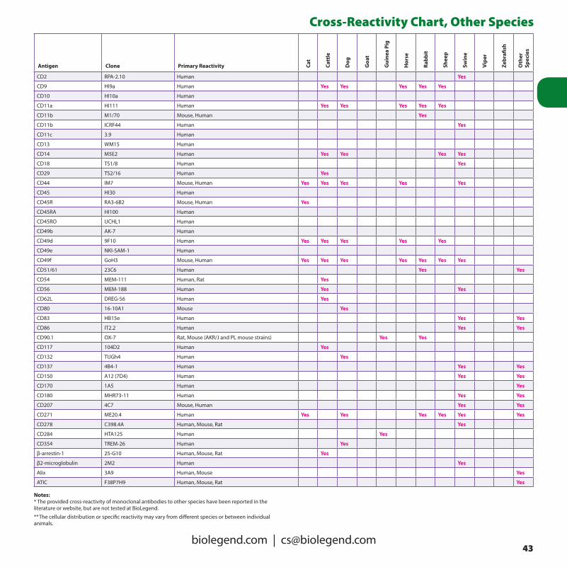
43biolegend.com | [email protected]
Cross-Reactivity Chart, Other Species
Antigen Clone Primary Reactivity Cat
Catt
le
Dog
Goa
t
Gui
nea
Pig
Hor
se
Rabb
it
Shee
p
Swin
e
Vipe
r
Zebr
afish
Oth
er
Spec
ies
CD2 RPA-2.10 Human Yes
CD9 HI9a Human Yes Yes Yes Yes Yes
CD10 HI10a Human
CD11a HI111 Human Yes Yes Yes Yes Yes
CD11b M1/70 Mouse, Human Yes
CD11b ICRF44 Human Yes
CD11c 3.9 Human
CD13 WM15 Human
CD14 M5E2 Human Yes Yes Yes Yes
CD18 TS1/8 Human Yes
CD29 TS2/16 Human Yes
CD44 IM7 Mouse, Human Yes Yes Yes Yes Yes
CD45 HI30 Human
CD45R RA3-6B2 Mouse, Human Yes
CD45RA HI100 Human
CD45RO UCHL1 Human
CD49b AK-7 Human
CD49d 9F10 Human Yes Yes Yes Yes Yes
CD49e NKI-SAM-1 Human
CD49f GoH3 Mouse, Human Yes Yes Yes Yes Yes Yes Yes
CD51/61 23C6 Human Yes Yes
CD54 MEM-111 Human, Rat Yes
CD56 MEM-188 Human Yes Yes
CD62L DREG-56 Human Yes
CD80 16-10A1 Mouse Yes
CD83 HB15e Human Yes Yes
CD86 IT2.2 Human Yes Yes
CD90.1 OX-7 Rat, Mouse (AKR/J and PL mouse strains) Yes Yes
CD117 104D2 Human Yes
CD132 TUGh4 Human Yes
CD137 4B4-1 Human Yes Yes
CD150 A12 (7D4) Human Yes Yes
CD170 1A5 Human Yes
CD180 MHR73-11 Human Yes Yes
CD207 4C7 Mouse, Human Yes Yes
CD271 ME20.4 Human Yes Yes Yes Yes Yes Yes
CD278 C398.4A Human, Mouse, Rat Yes
CD284 HTA125 Human Yes
CD354 TREM-26 Human Yes
β-arrestin-1 25-G10 Human, Mouse, Rat Yes
β2-microglobulin 2M2 Human Yes
Alix 3A9 Human, Mouse Yes
ATIC F38P7H9 Human, Mouse, Rat Yes
Notes:* The provided cross-reactivity of monoclonal antibodies to other species have been reported in the literature or website, but are not tested at BioLegend.
** The cellular distribution or specific reactivity may vary from different species or between individual animals.

44Toll Free 1.877.BIOLEGEND (246.5343)
Cross-Reactivity Chart, Other Species
Antigen Clone Primary Reactivity Cat
Catt
le
Dog
Goa
t
Gui
nea
Pig
Hor
se
Rabb
it
Shee
p
Swin
e
Vipe
r
Zebr
afish
Oth
er
Spec
ies
Bax 6A7 Human, Mouse, Rat Yes
BCL-XS/L 2H12 Human, Mouse, Rat Yes
Bcl11b 25B6 Human, Mouse Yes Yes
Biotin 1D4-C5 All species Yes Yes Yes Yes Yes Yes Yes Yes Yes Yes Yes Yes
c-Myc 9.00E+10 All species Yes Yes Yes Yes Yes Yes Yes Yes Yes Yes Yes Yes
cdc2 (p34) POH-1 Human Yes Yes
Cyclin A E23.1 Human, Mouse Yes
Cytochrome c 7H8.2C12 Most Species Yes
Cytokeratin (pan reactive) C-11 All species Yes Yes Yes Yes Yes Yes Yes Yes Yes Yes Yes Yes
Cytokeratin 7/17 C-46 Human Yes Yes
FOXP3 150D Human, Mouse, Rat Yes Yes
FOXP3 206D Human Yes Yes
FOXP3 259D Human Yes Yes
GAPDH Poly6314 Human, Mouse, Rat Yes
GFAP 2E1.E9 Human, Mouse, Rat Yes
HLA-A,B,C W6/32 Human Yes Yes Yes Yes
HLA-DR L243 Human Yes
hnRNPK F45P9C7 Human, Mouse, Rat Yes Yes
IFN-γ 4S.B3 Human Yes Yes
IFN-γ MD-1 Human Yes Yes
IgG Fc HP6017 Human Yes Yes Yes Yes
IL-1β JK1B-1 Human Yes Yes
IL-2 MQ1-17H12 Human Yes Yes
IL-2 Poly5111 Human Yes
IL-4 8D4-8 Human Yes Yes
IL-4 MP4-25D2 Human Yes
IL-5 TRFK5 Mouse, Human Yes
IL-10 JES3-9D7 Human Yes Yes
Integrin α9 β1 Y9A2 Human Yes
Integrin β7 FIB504 Mouse, Human Yes Yes
Ki-67 Ki-67 Human Yes
MAP2ab MT-08 Human, Mouse Yes
Neurofilament heavy protein NF-01 All species Yes Yes Yes Yes Yes Yes Yes Yes Yes Yes Yes Yes
Neurofilament medium protein NF-09 All species Yes Yes Yes Yes Yes Yes Yes Yes Yes Yes Yes Yes
NGF JKhNGF-1 Human Yes Yes
NGF JKhNGF-2 Mouse, Human Yes Yes
p53 BP53-12 Human Yes
PCNA PC10 Human, Mouse, Rat Yes
Perforin dG9 Human Yes
Phosphotyrosine PY20 All species Yes Yes Yes Yes Yes Yes Yes Yes Yes Yes Yes Yes
TCP-1 beta F39P7F11 Human, Mouse, Rat Yes
TIMP-1 F31P2A5 Human Yes
TIMP-2 F27P3A4 Human, Mouse Yes Yes Yes Yes
TNF-α MAb11 Human Yes Yes
TNF-α TN3-19.12 Mouse, Rat Yes
Tubulin-α 10D8 Human, Mouse, Rat Yes Yes Yes Yes Yes Yes Yes Yes Yes Yes Yes Yes
Ubiquitin P4D1 All species Yes Yes Yes Yes Yes Yes Yes Yes Yes Yes Yes Yes
Tubulin-α TU-01 All species Yes Yes Yes Yes Yes Yes Yes Yes Yes Yes Yes Yes

45biolegend.com | [email protected]
TNF Receptor and Ligand Superfamilies
Nomenclature of the TNF SuperfamilyTNFSF Nomenclature Molecular Aliases Gene
TNFSF1 TNF-β, LT, LT-α 6p21.3
TNFSF2 TNF-α, TNF, cachectin, necrosin, cytotoxin 6p21.3
TNFSF3 LT-β, TNFC, LTB 6p21.3
TNFSF4 OX-40 ligand (OX-40L), gp34, TXGP-1, CD252 1q25
TNFSF5 CD40 ligand (CD40L), CD154, gp39, TRAP Xq26
TNFSF6 Fas ligand (FasL), CD95L, CD178, ApoI L 1q23
TNFSF7 CD70, CD27 ligand (CD27L), Ki-24 19p13
TNFSF8 CD30 ligand (CD30L), CD153 9q33
TNFSF9 4-1BB ligand (4-1BBL), CDw137L 19p13.3
TNFSF10 TRAIL, Apo-2 ligand (Apo-2L), TL2, CD253 3q26
TNFSF11 RANK ligand (RANKL), TRANCE, OPGL, ODF, CD254 13q14
TNFSF12 TWEAK, Apo-3 ligand (Apo3L), CD255 17p13
TNFSF12-13 TWE-PRIL 17p13.1
TNFSF13 APRIL, TALL2, TRDL-1, , ZTNF2, TNFSF13A, CD256 17p13.1
TNFSF13B BAFF, TALL1, zTNF4, THANK, Blys, DTL, CD257 13q32-34
TNFSF14 LIGHT, LTg, TR2, HVEM-L, CD258 19p13.3
TNFSF15 TL1,TL1A,VEGI 9q32
TNFSF18 AITRL, TL6, hGITRL 1q23
The TNF receptor superfamily (TNFRSF) consists of a group of glycoproteins characterized by cysteine-rich motifs in the extracellular domain (CRDs). These receptors share about 20–30% overall homology. Most TNFRSF are type I membrane proteins (with an extracellular N-terminus and an intracellular C-terminus). TNFRSF members can be divided into death receptors, decoy receptors, and activating receptors. Most activating receptors mediate intracellular signals through adaptor proteins, i.e., TNF receptor-associated factors (TRAFs), while the death receptors mediate intracellular signals through death domain (DD) associated proteins (FADD, TRADD). Members of the TNFRSF can also be proteolytically cleaved to form soluble receptors that act as “slow release” depots for ligands.Ligands that bind TNFRSF are members of the TNF superfamily (TNFSF). TNFSF ligands are either soluble or membrane-bound
glycoproteins. Soluble TNFSF proteins usually have biological activities similar to other cytokines. Membrane-bound TNFSF ligands are type II transmembrane proteins (extracellular C-terminus) with short cytoplasmic domains and longer extracellular regions. Most TNFSF molecules are homotrimeric and share a common structural motif, the TNF homology domain (THD), which binds to the CRDs of TNFRs. The interaction of TNFSF ligands with their receptors plays divergent and important roles in the regulation of cell growth, survival, and death. Abnormal expression of some family members results in a variety of pathophysiological states, including immune deficiency, autoimmune diseases, and enhanced tumor growth and metastasis. TNFRSF/TNFSF molecules have proven to be effective clinical therapeutic targets.

46Toll Free 1.877.BIOLEGEND (246.5343)
TNF Receptor and Ligand SuperfamiliesNomenclature of the TNF Receptor Superfamily
TNFRSF Nomenclature Molecular Aliases Signaling Adaptors Gene
TNFRSF1A TNFR type I, CD120a, TNFAR, p55TNFR, TNFR60 TRADD 12p13.2-p13.31
TNFRSF1B TNFR type II, CD120b, TNFR80, p75TNFR, TNFBR TRAF1,2,5 1p36.3-p36.2
TNFRSF3 TNFR III, LTBR, TNFCR, TNFR-RP, TNFR2-RP, CD18 TRAF3,4,5 12p13
TNFRSF4 OX-40, ACT35, TXGP1L, CD134 TRAF1,2,3,5 1p36
TNFRSF5 CD40, Bp50, p50 TRAF1,2,3,5,6 20q12-q13.2
TNFRSF6 Fas, CD95, APO-1, APT1, TNFRSF6A FADD 10q24.1
TNFRSF6B DcR3, TR6, M68 secreted 20q13
TNFRSF7 CD27, S152, Tp55, T14 TRAF2, SIVA 12p13
TNFRSF8 CD30, Ki-1 TRAF1,2,3,5 1p36
TNFRSF9 4-1BB, CDw137, ILA TRAF1,2,3 1p36
TNFRSF10A DR4, TRAIL-R1, APO-2, CD261 FADD 8p22
TNFRSF10B DR5, TRAIL-R2, KILLER, CD262, TRICK2A, TRICKB FADD 8p22-p21
TNFRSF10C DcR1, TRAIL-R3, LIT, TRID, CD263 8p22-p21
TNFRSF10D DcR2, TRAIL-R4, TRUNDD, CD264 8p21
TNFRSF11A RANK, ODFR, TRANCE-R, CD265 TRAF1,2,3,5,6 18q22.1
TNFRSF11B OPG, TR1, OCIF secreted 8q24
TNFRSF12 DR3, TRAMP, APO-3, TRS, WSL-1, LARD, DDR3, WSL-LR TRADD 1p36.3
TNFRSF12A TWEAK-R, Fn14, FGF-inducible 14, CD266 TRAF1,2,3,5 16p13.3
TNFRSF12L DR3L 1p36.2
TNFRSF13B TACI, CD267 TRAF2,5,6 17p11
TNFRSF13C BAFF-R, CD268, BR3 TRAF3 22q13.1-q13.31
TNFRSF14 HVEM, TR2, LIGHT-R, ATAR, HVEA TRAF1,2,3,5 1p36.3-p36.2
TNFRSF16 NGF-R, NTR, p75NGFR, CD271 TRAF2,4,6 17q21-q22
TNFRSF17 BCMA, BCM, TNFRSF13, TNFRSF13a, CD269 TRAF1,2,3,5,6 16p13.1
TNFRSF18 AITR, GITR TRAF1,2,3 1p36.3
TNFRSF19 TROY, TAJ, TAJ-α, TRADE TRAF1,2,3,5 13q12.11-q12.3
TNFRSF19L RELT TRAF1 11q13.4
TNFRSF21 DR6, Death receptor 6 TRADD 6p21.1-12.2
TNFRSF22 SOBa; Tnfrh2, Tnfrsf1al2, mDcTrailr2
TNFRSF23 mSOB, Tnfrh1, mDcTrailr1

47biolegend.com | [email protected]
Cell Surface Immunofluorescence Staining ProtocolHarvest Tissue or Cells:1. Obtain desired tissue (e.g., spleen, lymph node, thymus, bone
marrow) and prepare a single cell suspension in Cell Staining Buffer (Cat. No. 420201) by gently mashing. Debris can be removed by filtration of the cell suspension through 70–100 μm nylon mesh strainer. If using in vitro stimulated cells, simply resuspend previously activated cultures in Cell Staining Buffer and proceed to Step 2.
2. Add Cell Staining Buffer up to ~15 mL and centrifuge at 350 × g for 5 minutes, discard supernatant, then proceed to step 6.
Lyse Red Blood Cells:3. If necessary (e.g., spleen), dilute 10× Red Blood Cell (RBC) Lysis
Buffer (Cat. No. 420301) to 1× working concentration with DI water and resuspend pellet in 3 mL 1× RBC Lysis Buffer. Incubate on ice for 5 minutes.
4. Stop cell lysis by adding 10 mL Cell Staining Buffer to the tube. Centrifuge for 5 minutes at 350 × g and discard supernatant.
5. Repeat wash as in step 2.6. Count viable cells and resuspend in Cell Staining Buffer at 5–10 ×
106 cells/mL and distribute 100 μL/tube of cell suspension into 12 × 75 mm plastic tubes.
Block Fc-Receptors:7. Reagents that block Fc receptors may be useful for reducing FcR-
mediated nonspecific staining. In the mouse, TruStain fcX™(anti-mouse CD16/32) (Cat. No. 101319/20) can be used to block nonspecific staining of antibodies. In this case, block Fc receptors by pre-incubating cells with 1.0 μg Mouse TruStain fcX™ per 106
cells in a 100 μL volume for 5-10 minutes. To block Fc receptors on human cells, Human TruStain FcX™ (Cat. No. 422301/2) is recommended.
Cell-Surface Staining with Antibody:8. Add appropriately conjugated fluorescent, biotinylated, or purified
primary antibodies (e.g., anti-CD3-FITC, anti-CD4-Biotin, and anti-CD8-APC) at predetermined optimum concentrations and incubate on ice for 15–20 minutes in the dark.
9. Wash 2× with at least 2 mL of Cell Staining Buffer by centrifugation at 350 × g for 5 minutes, discard supernatant.
10. If using a purified primary antibody, resuspend pellet in residual buffer and add previously determined optimum concentrations of anti-species immunoglobulin fluorochrome conjugated secondary antibody (e.g., FITC anti-mouse Ig) and incubate in the dark for 15–20 minutes.
If using a biotinylated primary antibody, resuspend cell pellet in residual buffer and add previously determined optimum concentrations of fluorochrome conjugated Streptavidin (SAv) reagent (e.g., SAv-PE, Cat. No. 405204) and incubate on ice for 15–20 minutes in the dark.
11. Repeat step 9.
12. Resuspend cell pellet in 0.5 mL of Cell Staining Buffer and add 10 μL (0.25 µg)/million cells of 7-AAD Viability Staining Solution (Cat. No. 420403/4) to exclude dead cells. We recommend utilizing our Fluorescence Spectra Analyzer (biolegend.com/spectraanalyzer) to determine compatibility of 7-AAD with other fluorochromes.
13. Incubate on ice for 3–5 minutes in the dark.14. Analyze under a Flow Cytometer with appropriate machine
settings.
Immunofluorescent Staining of Whole Blood:1. Add predetermined optimal concentrations of desired
fluorochrome conjugated, biotinylated, or purified primary antibodies to 100 μL of anti-coagulated whole blood.
2. Incubate at room temperature for 15–20 minutes in the dark.3. Dilute 10× Red Blood Cell (RBC) Lysis Buffer (Cat. No. 420301) to
1× working concentration with DI water. Warm the buffer to room temperature prior to use. Add 2 mL of 1× RBC lysis solution to whole blood/antibody mixture. Incubate at room temperature for 10 minutes.
4. Centrifuge at 350 × g for 5 minutes, discard the supernatant.5. Wash 2× with at least 2 mL of Cell Staining Buffer by centrifugation
at 350 × g for 5 minutes.6. If using a purified primary antibody, resuspend pellet in residual
buffer and add a previously determined optimum concentration of anti-species immunoglobulin fluorochrome conjugated secondary antibody (e.g., FITC anti-mouse Ig) and incubate in the dark for 15–20 minutes.
If using a biotinylated primary antibody, resuspend cell pellet in residual buffer and add a previously determined optimum concentration of fluorochrome conjugated Streptavidin (SAv) reagent (e.g., SAv-PE, Cat. No. 405204) and incubate for 15–20 minutes in the dark.
7. Repeat step 5.8. Resuspend cells in 0.5 mL Cell Staining Buffer or 0.5 mL FluoroFix™
buffer (Cat. No. 422101).9. Analyze under a Flow Cytometer with appropriate machine
settings.
Key Reference:1. Current Protocols in Cytometry (John Wiley & Sons, New York),
Unit 6 Phenotypic Analysis.
Reagent List:1. Cell Staining Buffer (Cat. No. 420201)2. Red Cell Lysis Buffer (Cat. No. 420301)3. 7-AAD Viability Staining Solution (Cat. No. 420401)4. TruStain fcX™(anti-mouse CD16/32) (Cat. No. 101319/20)5. Human TruStain FcX™ (Cat. No. 422301/2)
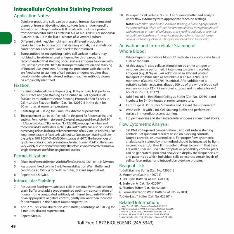
48Toll Free 1.877.BIOLEGEND (246.5343)
Intracellular Cytokine Staining ProtocolApplication Notes:1. Cytokine-producing cells can be prepared from in vivo-stimulated
tissues or from in vitro-stimulated cultures (e.g., antigen-specific activation or mitogen-induced). It is critical to include a protein transport inhibitor such as brefeldin A (Cat. No. 420601) or monensin (Cat. No. 420701) in the last 4–6 hours of in vitro cell culture.
2. Different cytokines/chemokines have different production peaks. In order to obtain optimal staining signals, the stimulation conditions for each stimulant need to be optimized.
3. Some antibodies recognizing native cell surface markers may not bind to fixed/denatured antigens. For this reason, it is recommended that staining of cell surface antigens be done with live, unfixed cells PRIOR to fixation/permeabilization and staining of intracellular cytokines. Altering the procedure such that cells are fixed prior to staining of cell surface antigens requires that paraformaldehyde-denatured antigen-reactive antibody clones be empirically identified.
Fixation:1. If staining intracellular antigens (e.g., IFN-γ or IL-4), first perform
cell surface antigen staining as described in BioLegend’s Cell Surface Immunofluorescence Staining Protocol, then fix cells in 0.5 mL/tube Fixation Buffer (Cat. No. 420801) in the dark for 20 minutes at room temperature.
2. Centrifuge at 350 × g for 5 minutes, discard supernatant.3. The experiment can be put “on hold” at this point for future staining and
analysis. For short-term storage (<2 weeks), resuspend the cells in 0.5-1 mL/tube Cyto-Last™ Buffer (Cat. No. 422501), mix, cap the tubes, and then store at 4°C in the dark (Note: Cyto-Last™ Buffer can also be used for preserving cells in bulk at a cell concentration of 0.5-2.0 x 10⁶ cells/mL). For long-term storage of fixed cells without surface antigen staining, dilute the cells in 90% FCS/10% DMSO for storage at -80°C. The frequencies of cytokine-producing cells present in activated human PBMC cultures can vary widely due to donor variability. Therefore, cryopreserved cells from a single donor are useful for longitudinal studies.
Permeabilization:4. Dilute 10× Permeabilization Wash Buffer (Cat. No. 421001) to 1× in DI water.5. Resuspend fixed cells in 2 mL Permeabilization Wash Buffer and
centrifuge at 350 × g for 5–10 minutes, discard supernatant.6. Repeat step 5 twice.
Intracellular Staining:7. Resuspend fixed/permeabilized cells in residual Permeabilization
Wash Buffer and add a predetermined optimum concentration of fluorochrome conjugated antibody of interest (e.g., anti-IFN-γ-PE) or an appropriate negative control, gently mix and then incubate for 20 minutes in the dark at room temperature.
8. Add 2 mL of Permeabilization Wash Buffer, centrifuge at 350 × g for 5 minutes, discard supernatant.
9. Repeat Step 8.
10. Resuspend cell pellet in 0.5 mL Cell Staining Buffer and analyze under flow cytometry with appropriate machine settings.
Note: To confirm specific anti-cytokine staining, a blocking experiment is recommended in which cells are fixed/permeabilized then preincubated with an excess amount of unlabeled anti-cytokine antibody and/or the recombinant cytokine of interest is preincubated with fluorochrome-conjugated anti-cytokine antibody before its addition to the cells.
Activation and Intracellular Staining of Whole Blood:1. Dilute heparinized whole blood 1:1 with sterile appropriate tissue
culture medium.2. At this stage, in vitro cellular stimulation by either antigen or
mitogen can be performed. If intending to stain intracellular antigens (e.g., IFN-γ or IL-4), addition of an efficient protein transport inhibitor such as brefeldin A (Cat. No. 420601) or monensin (Cat. No. 420701) is critical. After addition of a suitable cellular activator, aliquot 200 μL of the whole blood cell suspension into 12 × 75 mm plastic tubes and incubate for 4–6 hours in 5% CO2 at 37°C.
3. Add 2 mL of 1× Red Blood Cell Lysis Buffer (Cat. No. 420301) and incubate for 5–10 minutes at room temperature.
4. Centrifuge at 350 × g for 5 minutes and discard the supernatant.5. Wash cells 1× with 2 mL Cell Staining Buffer and perform cell
surface immunofluorescent staining.6. Fix, permeabilize and stain intracellular antigens as described above.
Flow Cytometric Analysis:1. Set PMT voltage and compensation using cell surface staining
controls. Set quadrant markers based on blocking controls, isotype controls, or unstained cells. For proper flow cytometric analysis, cells stained by this method should be inspected by light microscopy and/or flow light scatter pattern to confirm that they are well dispersed. Bivariate dot plots or probability contour plots can be generated upon data analysis to display the frequencies of and patterns by which individual cells co-express certain levels of cell surface antigen and intracellular cytokine proteins.
Reagent List:1. Cell Staining Buffer (Cat. No. 420201)2. Monensin (Cat. No. 420701)3. RBC Lysis Buffer (Cat. No. 420301)4. Brefeldin A (Cat. No. 420601)5. Fixation Buffer (Cat. No. 420801)6. Permeabilization Wash Buffer (Cat. No. 421001)7. Cyto-Last™ Buffer (Cat. No. 422501)
Related Information:1. Jung T, et al. 1993. J. Immunol. Methods 159:197.2. Vikingsson A., et al. 1994. J. Immunol. Methods 173:219.3. Prussin C., et al. 1995. J. Immunol. Methods 188:117.4. Elson, L.H., et al. 1995. J. Immunol. 1995. 154:4294.5. Assenmacher, M., et al. 1994. Eur. J. Immunol. 24:1097.

49biolegend.com | [email protected]
FOXP3 Intracellular Staining Protocol BioLegend’s FOXP3 Fix/Perm Buffer Set (Cat. No. 421403) is
designed and formulated for intracellular staining of FOXP3 with minimal effects on surface marker staining. Highly recommended for optimal FOXP3 intracellular immuno fluorescent staining.
1. Stain cell surface markers as described in Cell Surface Immunofluorescence Staining Protocol, page 47.
2. Add 1 mL of 1× FOXP3 Fix/Perm solution (Cat. No. 421401) to each tube, vortex and incubate at room temperature in the dark for 20 minutes. Pellet cells by centrifugation and discard the supernatant.
3. Wash once with cell staining buffer (Cat. No. 420201) by centrifugation at 350 × g for 5 minutes and discard the supernatant.
4. Wash once by resuspension and centrifugation with 1 mL 1× FOXP3 Perm buffer (Cat. No. 421402).
5. Re-suspend cells in 1 mL 1× FOXP3 Perm buffer, incubate at room temperature in the dark for 15 minutes, pellet cells and discard supernatant. Resuspend cells in 100 μL of 1× FOXP3 Perm buffer.
6. Add appropriate amount of fluorochrome conjugated anti-FOXP3 antibody and incubate at room temperature in the dark for 30 minutes.
7. Wash twice with cell staining buffer by resuspension and centrifugation, and resuspend in 0.5 mL cell staining buffer for analysis by flow cytometry with appropriate instrument setting.
Related Information:1. Roncador, G., et al. 2005 Eur. J. Immunol. 35:1681.
Reagent List:1. Cell Staining Buffer (Cat. No. 420201)
2. FOXP3 Fix/Perm Buffer Set (Cat. No. 421403)
Th17 Polarization of Mouse CD4 CellsIntroductionT helper 17 (Th17) cells are a subset of CD4 T helper cells characterized by their production of IL-17, particularly IL-17A and IL-17F. They are involved in autoimmune diseases such as multiple sclerosis, psoriasis, autoimmune uveitis, rheumatoid arthritis, and juvenile diabetes, but are also important in anti-microbial defenses at epithelial/ mucosal barriers, particularly against Candida and Staphylococcus. In addition to IL-17, Th17 cells also produce IL-21 and IL-22 that contribute to pro-inflammatory responses. The transcription factors RORα and RORγ have been identified as important factors in the differentiation of these cells. Th17 cells can be induced in vitro by a variety of cytokines including TGF-β, IL-6, IL-21 and IL-23. Here we provide a effective protocol for the generation of mouse IL-17 producing cells in vitro.
Isolation of CD4 cells from lymph nodes1. Harvest lymph nodes (superfical cervical, madibular, axillary,
inguinal, and mesenteric) from mice.2. Tease lymph nodes through a sterile 70-μm nylon cell strainer to
achieve single-cell suspensions in RPMI 1640 contain 10% FCS (complete medium).
3. Resuspend cells in complete medium and use your favorite method to isolate CD4 cells. Check out Biocompare.com to find useful kits.
Th17 Polarization of CD4 cells1. Coat 60 × 15 mm of plastic petri dishes with anti-mouse CD3ε, clone
145-2C11 (2 μg/mL). Incubate at 37°C for 2 hours or 4°C overnight. Aseptically decant antibody solution from the plate. Wash plate 3 times with sterile PBS. Discard liquid.
2. Plate CD4 cells at 1 x 10⁶/mL. Culture cells for 3 days in presence of anti-mouse CD28, clone 37.51 (5 μg/mL), IL-6 (50 ng/mL), TGF-β1 (1ng/mL), anti-mouse IL-4 (10 μg/mL), anti-mouse IFN-γ (10 μg/mL), and 300 nM of FICZ. Alternatively, mouse IL-23 (5 ng/mL) can be used in place of FICZ.
3. On day 3, wash cells once and then restimulate in complete medium with 500 ng/mL PdBU, and 500 ng/mL ionomycin, in the presence of Brefeldin A for 4 - 5 hours.
4. After harvesting, the cells are ready for staining.
Materials• Sterile PBS• Cellculturemedium(RPMI1640supplementedwith10%FBS)• Sterileplasticpetridishes• RBCLysisBuffer(Cat.No.420301)• Anti-mouseCD3ε,clone145-2C11(LEAF™format,Cat.No.100314)• Anti-mouseCD28,clone37.51,(LEAF™format,Cat.No.102112)• Anti-mouseIL-4(LEAF™format,Cat.No.504108)• Anti-mouseIFN-γ(LEAF™format,Cat.No.505812)• RecombinantmouseIL-6(carrier-free)(Cat.No.575702)• RecombinanthumanTGF-β1(carrier-free)(Cat.No.580702)• RecombinantmouseIL-23(carrier-free)(Cat.No.589002)• BrefeldinA(Cat.No.420601)• PdBu(Phorbol12,13-dibutyrate)(Cat.No.P1269fromSigma)• FICZ(6-formylindolo[3,2-b]carbazole)(Cat.No.BML-GR206-0100
from Enzo Life Sciences)
References1. Cua DJ, et al. 2010. Nat Rev Immunol. 10: 479. ✦ 2. Pappu R, et al. 2010. J Clin Immunol. 30:185. ✦ 3. Kennedy, J., et al. 1996. J. Interferon Cytokine Res. 16: 611. ✦ 4. Schubert, D., et al. 2004. J. Immunol. 172: 4503. ✦5. Infante-Duarte, C., et al. 2000. J. Immunol. 165: 6107. ✦ 6. Harrington, L. E., et al. 2005. Nature Immunol. 6:1123. ✦ 7. Nekrasova, T., et al. 2005. J. Immunol. 175: 2734. ✦ 8. Yen, D., et al. 2006. J. Clin. Invest. 116: 1310.✦ 9. Ehirchiou, D., et al. 2007. J. Exp. Med. 204:1519. ✦ 10. Kang, S. G., et al. 2007. J. Immunol. 179:3724. ✦ 11. Harrington, L. E., et al. 2005. Nature Immunol. 6:1123.

50Toll Free 1.877.BIOLEGEND (246.5343)
Western Blotting ProtocolSample Preparation:1. Place cells in a centrifuge tube and centrifuge to collect cell pellet.
Wash cells with 1× PBS, aspirate.2. Lyse cell pellet with 100 μL lysis buffer/1 × 106 cells on ice for 30 min.3. Centrifuge at 14,000 rpm (16,000 × g) for 10 min at 4°C.4. Transfer supernatant to a new tube and discard pellet. Remove 20
μL supernatant and mix with 20 μL of 2× sample buffer.5. Boil for 5 min. Cool to RT for 5 min. Centrifuge for 5 min.6. Load 20 µg - 30 µg extract per well of a 1.5 mm gel.7. Set gel running conditions according to the manufacturer’s
instructions. Transfer proteins to a nitrocellulose or PVDF membrane with variable power settings according to manufacturer’s instructions.
Membrane Blocking:1. Remove the nitrocellulose or PVDF membrane from the transfer
apparatus and immediately place in blocking buffer consisting of 5% nonfat dry milk in TBS-T*.
2. Incubate the blot for 1 hr at room temperature, or overnight at 4°C with agitation.
Antibody Incubation:1. Dilute primary antibody to the recommended concentration in 5%
nonfat dry milk/TBS-T* (usually at 1 µg/mL). Place the membrane in the primary antibody solution and incubate 2 hrs at room temperature, or overnight at 4°C with agitation.
2. Wash five times for 5-10 min each with TBS -T.3. Incubate the membrane for 1-2 hr at room temperature with
BioLegend’s horseradish peroxidase-conjugated secondary antibody, diluted with 5% nonfat dry milk in TBS-T*.
4. Wash 5 times for 10 min each with TBS-T, and once for 2 min with PBS.
Protein Detection:1. Incubate membrane (protein side up) with 10 mL ECL (enhanced
chemiluminescence) substrate for 1–2 min. The final volume required is 0.125 mL/cm2.
2. Drain off excess detection reagent, wrap blots in plastic wrap and gently smooth out any air bubbles.
3. Place membrane, protein side up, in a film cassette and expose to film. Exposures can vary from 5 seconds to 60 min.
* If phosphorylation-specific antibodies are used, the membrane blocking buffer and antibody dilution buffer should not contain milk.
Tips:High Background1. Transfer buffers may have become contaminated. Contamination
can be transferred to the blots from electrophoresis and related equipment used in blot preparation.
2. Post-antibody washes may not have been performed for a sufficient period of time or with large enough volume. Wash buffers may not have been thoroughly removed.
3. Blocking and incubation agents used were not freshly prepared or were too dilute.
4. Membrane may have dried.
No Signal or Poor Signal1. Transfer efficiency may have been poor. Monitor protein transfer by
staining the gel and by including prestained MW marker.2. Incorrect storage of antibodies or ECL western blotting detection
reagents may result in poor signal.3. Insufficient protein may have been loaded on the gel. Depending
on the location of the target protein, membrane or nuclear preparations may be required (instead of whole cell lysates).
4. Film exposure time may have been too short.
Solutions and Reagents:Cell Lysis Buffer:• 20 mM Tris-HCl, pH 7.5• 150 mM NaCl• 1% NP-40• 2 mM EDTA• 1 µg/mL leupeptin• 1 µg/mL aprotinin• 1 mM Na3PO4
• 1 mM PMSF• 5 mM NaF• 3 mM Na4P2O4
5× SDS Sample Buffer:• 312.5 mM Tris-HCl (pH 6.8)• 10% SDS (w/v)• 250 mM DTT• 50% Glycerol• 0.05% Bromophenol Blue
(w/v)• Use at 1×
Transfer Buffer:• 2.25 g Tris base• 10.5 g Glycine• 200 mL Methanol• Add distilled water to 1.0 L
TBS-T (Tris-buffered saline containing 0.1% Tween-20):• Dissolve 8 g of NaCl, 0.2 g
of KCl, 3 g of Tris base and 1 mL Tween-20 in 800 mL of distilled H2O. Adjust the pH to 7.4 with HCl. Add distilled H2O to 1 liter.
Blocking Buffer:• TBS-T with 5% nonfat dry
milk. Milk will acidify TBS-T.• Re-pH after dissolving milk.
Prepare fresh milk solution and adjust pH to 7.4 after addition of milk.
Primary and Secondary Antibody Dilution Buffer:• TBS-T with 5% nonfat dry milkNote: If phosphorylation-specific
antibodies are used, the membrane blocking buffer and antibody dilution buffer should not contain milk.
Alternate Blocking Buffer:• TBS-T with 4% Bovine Serum
Albumin (BSA)
Alternate Primary and Secondary Antibody Dilution Buffer:• TBS-T with 4% Bovine Serum
Albumin (BSA)

51biolegend.com | [email protected]
Immunoprecipitation ProtocolSample Preparation:1. Collect cells and transfer to a centrifuge tube and centrifuge at 3,000
rpm for 5 min at 4°C.2. Discard supernatant and store cell pellet at -80°C or process
immediately as described below.
Immunoprecipitation:1. Remove samples from -80°C freezer and place on ice.2. Immediately add 800 μL ice-cold lysis buffer to the samples and
vortex, then incubate for 30 min on ice.3. Spin lysates at 14,000 rpm in a pre-cooled centrifuge for 10 min.4. Transfer 720 μL supernatant to a fresh pre-cooled microcentrifuge
tube containing washed protein A or G agarose bead slurry (30 μL packed beads) and rock the mixture for 30 min at 4°C.
5. Remove the agarose beads by pulsing in a microcentrifuge (5 seconds at 14,000 rpm, 4°C).
6. Transfer supernatant and place in a fresh pre-cooled microcentrifuge tube containing ~4 µg antibody and rock gently at 4°C for 2 hrs. Capture the immune complex by adding 50 μL of washed protein A or G agarose bead slurry (30 μL packed beads) and rock the mixture for 1 hr at 4°C.
7. Collect the agarose beads by spinning in a centrifuge (30 seconds at 3,000 rpm, 4° C). Aspirate and discard supernatant. Wash beads 3 times with cold cell lysis buffer.
8. After final wash, remove supernatant and add 40 μL 2× SDS sample buffer. Boil for 5 min at 95°C and pellet beads at maximum speed in a microcentrifuge for 5 min at room temperature. Carefully collect supernatant.
9. Load up to 30 μL of sample per well of a 1.5 mm thick gel. Run gel according to manufacturer’s recommendations and continue with immunoblotting using BioLegend’s Western Blotting Protocol, p. 50 (alternatively, radiolabeled proteins prepared from target cells can be used to directly visualize the immunoprecipitated protein).
Tips:1. The choice of lysis buffer depends on the location of the protein
(membrane, cytosolic, nuclear).2. Immunoprecipitates allowed to incubate overnight may have a
higher background than ones processed for shorter periods of time due to time-dependent aggregation or denaturation of cellular proteins.
3. Always use an isotype-matched irrelevant control antibody (monoclonal) or same-species serum from a non-immunized animal to remove non-specific antibody binding in cellular lysates.
Solutions and Reagents:Cell Lysis Buffer:• 20mMTris-HCl,pH7.5 • 1 µg/mL aprotinin• 150mMNaCl • 1 mM Na3PO4
• 1%NP-40 • 1 mM PMSF• 2mMEDTA • 5 mM NaF• 1µg/mLleupeptin • 3 mM Na4P2O4
Pretreatment of Protein A/G sepharose: Transfer 30 μL protein A or G sepharose per sample in a
microcentrifuge tube, wash twice in cell lysis buffer to remove preservative (add cell lysis buffer up to 800 μL and spin down in benchtop centrifuge). Resuspend protein A or G sepharose in cell lysis buffer at 100 μL per sample.
5× SDS Sample Buffer: See page 50Transfer Buffer:• 2.25 g Tris base• 10.5 g Glycine• 1 g SDS• 200 mL Methanol• Add distilled water to 1.0 L
Relative Affinity of Protein A and Protein G Agarose for various IgG subclasses1:
AntibodyAffinity for protein A–Agarose
Affinity for protein G–Agarose
Mouse IgG1 + ++++
Mouse IgG2a ++++ ++++
Mouse IgG2b +++ +++
Mouse IgG3 ++ +++
Rat IgG1 +/++ +
Rat IgG2a - ++++
Rat IgG2b - ++
Rat IgG2c ++/++++ ++
Rabbit polyclonal ++++ +++
References:1. Harlow, E. and Lane, D. eds. (1988). Antibodies, A Laboratory Manual.
Cold Spring Harbor Laboratory, N.Y., 617 - 618.

52Toll Free 1.877.BIOLEGEND (246.5343)
General Sandwich ELISA ProtocolCoat the Plate:1. Dilute unlabeled capture antibody to a final concentration of
0.5–8 µg/mL in ELISA Coating Buffer (Cat. No. 421701) or PBS without a carrier protein and transfer 100 μL to each well of a high protein-binding ELISA plate (e.g., Nunc™ Maxisorp™).
2. Seal the plate to prevent evaporation and incubate overnight (16-18 hours) at 2-8°C.
Block the Plate:3. Bring the plate to room temperature, discard the capture antibody
solution, wash the plate 3 times with Wash Buffer (Cat. No. 421601), and block non-specific binding sites by adding 200 μL of Assay Diluent (Cat. No. 421203) to each well.
4. Seal the plate and incubate at room temperature for ≥ 1 hour. 5. Wash 3 times with Wash Buffer. Firmly blot plate facing down against
clean paper towels.
Add Standards and Samples:6. Dilute standards and samples to desired concentrations in Assay
Diluent in polypropylene tubes and add 100 μL to wells. 7. Seal the plate and incubate at room temperature for 2 hours or at
4°C overnight, with or without shaking, depending on the assay.8. Wash ≥ 3 times with Wash Buffer. Washes can be effectively
accomplished by filling wells with either a squirt bottle, carboy, manifold dispenser, multi-channel pipettor or automatic plate washer. For increased stringency, after each wash, let the plate stand briefly, flick off the buffer, and blot plates on paper towels before refilling.
Add Detection Antibody:9. Dilute the biotin-labeled detection antibody in Assay Diluent.
Add 100 μL of diluted antibody to each well. 10. Seal the plate and incubate at room temperature for 1-2 hours.11. Wash ≥ 3 times with wash buffer.
Add Avidin-Horseradish Peroxidase (Avidin-HRP):12. Dilute the Avidin-HRP conjugate (Cat. No. 405103) or other enzyme
conjugate to its pre-determined optimal concentration in Assay Diluent (usually between 1/500 – 1/2000). Add 100 μL per well.
13. Seal the plate and incubate at room temperature for 30 min.14. Wash ≥ 5 times with Wash Buffer.
Add Substrate (TMB for faster color development): 15. For each plate, mix 6 mL of TMB Reagent A with 6 mL TMB Reagent B
(Bio Legend TMB Substrate Reagent Set, Cat. No. 421101) immediately prior to use. Alternatively, use ready-to-use TMB High Sensitivity Substrate Solution (Cat. No. 421501). Transfer 100 μL into each well. Incubate at room temperature (10 – 30 min) for color development.
16. To stop the color reaction, add 100 μL of Stop Solution for TMB Substrate (Cat. No. 423001).
17. Read the optical density (OD) for each well with a microplate reader set to 450 nm.
OR:
Add Substrate (ABTS for slower color development): 15. Thaw ABTS Substrate Solution within 20 min of use. Add 11 μL
of 30% H2O2 per 11 mL of substrate and vortex. Immediately dispense 100 μL into each well and incubate at room temperature (20–60 min) for color development.
16. To stop the color reaction, add 50 μL of an ABTS Stop Solution.17. Read the optical density (OD) for each well with a microplate reader
set to 405 nm.

53biolegend.com | [email protected]
General Sandwich ELISA ProtocolSolutions and Buffers:Note: Do not use sodium azide in any buffers or solutions, as sodium azide
inactivates the horseradish-peroxidase enzyme.
Carbonate Coating Buffer:• 8.4 g NaHCO3
• 3.56 g Na2CO3
• Add deionized water up to 1.0L, pH to 9.5
Phosphate Buffered Saline (PBS):• 80.0 g NaCl• 14.4 g Na2HPO4
• 2.4 g KH2PO4
• 2.0 g KCl• Add deionized water up to 10 L, pH to 7.4 with HCL
Wash Buffer (BioLegend Cat. No. 421601 recommended):• 0.5 mL of Tween-20 in 1 L PBS
Assay Diluent (BioLegend Cat. No. 421203 recommended):
• 10% fetal bovine serum or 1% BSA in PBS• Filter before use to remove particulates.
ABTS Substrate Solution:• 150 mg 2,2’-Azino-bis-(3-ethylbenzthiazoline-6-sulfonic acid)
(Sigma, Cat. No. A-1888)• Add to 500 mL of 0.1M citric acid in deionized water• Adjust pH to 4.35 with NaOH• Aliquot 11 mL per vial and store at -20°C.
Avoid light exposure during preparation and storage.
ABTS Stop Solution:• Combine 50 mL dimethylformamide (DMF; Pierce, Cat. No. 20672)
with 50 mL deionized water• Add 20 g sodium dodecyl sulfate.
TMB (tetramethylbenzidine) Substrate Reagent Set: • BioLegend Cat. No. 421101
TMB High Sensitivity Substrate Solution: • BioLegend Cat. No. 421501
TMB Stop Solution:• BioLegend Cat. No. 423001
General References:1. Davies, C. 1994. The Immunoassay Handbook. D. Wild, Ed. Stockton Press, New York.2. Abrams, J.S. 1995. Immunoenzymetric assay of mouse and human cytokines using NIP-labeled
anti-cytokine antibodies. Current Protocols in Immunology (J. Coligan, A. Kruisbeek, D. Margulies, E. Shevach, W. Strober, Eds). John Wiley and Sons, New York. Unit 6.20.
3. Sander, B., et al. 1993. J. Immunol. Meth. 166:201.4. Abrams, J.S., et al. 1992. Immunol. Rev. 127:5.
Cytokine ELISA Troubleshooting TipsPoor Signal-to-Noise Ratio• Titrate capture antibody and detection antibody against each other
to obtain optimal dilutions.• Try capture antibody at 0.5 – 8 µg/mL (generally 2 µg/mL).• Try detection antibody at 0.25 – 2 µg/mL (generally 1 µg/mL).
Low Sensitivity• Try overnight incubations of standards and samples at 4°C.
Poor Signal• If using HRP, avoid sodium azide in wash buffers and diluents, as
sodium azide inhibits HRP enzyme activity.• Verify that appropriate antibody pairs were used and that samples
and/or standards are expected to react with the antibody pairs.• Check activity of enzyme and substrate by coating with Detection
Antibody (1 µg/mL), adding Avidin-HRP and developing with appropriate substrate. If enzyme/substrate is active, a strong signal should be observed.
Poor Standard Curve• Recombinant protein vials should be quick-spun for maximum
recovery. • BioLegend suggests that cytokines be stored in a concentrated
format (>100 ng/mL) at -70°C. Do not repeatedly freeze-thaw the recombinant protein standard, as loss of activity may occur.
High Background• Increase stringency of washing steps by soaking plates for ~1
minute between washes.• Determine optimum Capture and Detection Antibody dilutions.• Increase the dilution of the Detection Antibody and/or increase the
number of washes after Avidin-HRP incubation.

54Toll Free 1.877.BIOLEGEND (246.5343)
ELISPOT ProtocolCoat the Plate:1. Dilute Low-Endotoxin/Azide-Free sterile unlabeled capture antibody
(Bio Legend’s LEAF™ format antibodies are specifically designed for this assay) to a final concentration of 0.5 – 10 µg/mL in sterile Carbonate Coating Buffer or PBS without carrier protein and transfer 100 μL/well to a high affinity binding PVDF membrane ELISPOT plate (e.g., Millipore; Cat. No. MAIPS-4510).
2. Seal the plate and incubate overnight at 4°C.
Block the Plate: 3. Wash the plate 3 times with sterile PBS, 200 μL/well. 4. Add 200 μL/well of sterile Blocking Buffer. 5. Incubate at room temperature for ≥ 1 hour. 6. Wash the plate 3 times with sterile PBS, 200 μL/well.
Set-Up Tissue Culture and Add Antigen or Mitogen: 7. Add appropriate sterile antigen or mitogen solution diluted in
appropriate sterile tissue culture medium (TC) to ELISPOT plate, 100 μL/well.
8. Add cells diluted in sterile tissue culture medium, 100 μL/well. Use 50,000–500,000 cells/well (the minimum number of cells should be determined in preliminary experiments).
9. Incubate at 37°C with 5% CO2 in humidified atmosphere for a pre-optimized stimulation period. BioLegend recommends a 24 hour incubation for IFN-γ, IL-2, and TNF-α, and a 48 hour incubation period for IL-4, IL-5, and IL-10 for most activation conditions.
Add Detection Antibody:10. Wash the plate 3 times with PBS, 200 μL/well.11. Wash the plate 3 times with PBS-Tween, 200 μL/well.12. Add 100 μL/well of diluted biotinylated detection antibody at
0.25–2 µg/mL in PBS-Tween-BSA. 13. Seal the plate and incubate at 4°C overnight, or 2 hours at room
temperature.
Add Avidin-Horseradish Peroxidase (Avidin-HRP): 14. Wash plate 4 times with PBS-Tween, 200 μL/well.15. Add 100 μL per well of the Avidin-HRP conjugate (Cat. No. 405103)
or other enzyme conjugate diluted to its pre-determined optimal concentration in PBS-Tween-BSA (usually between 1/500 – 1/2000).
16. Seal the plate and incubate at room temperature for 1–2 hours.17. Wash the plate 3 times with PBS-Tween, 200 μL/well.18. Wash the plate 3 times with PBS, 200 μL/well.
Add Substrate: 19. Add 200 μL/well of fresh Substrate solution.20. Monitor spot/color development at room temperature and stop the
reaction by rinsing the plate with tap water and vigorously flicking the plate over a waste container or sink, followed by blotting on paper towels or other absorbent materials.
21. Air dry plate overnight, until it is completely dry.22. Count spots manually with a dissecting microscope or using an
automated image acquisition/analysis unit (plates can be analyzed for up to 3 months).
Solutions and Buffers: Note: Do not use sodium azide in any buffers or solutions as sodium
azide inactivates the horseradish-peroxidase enzyme.
Phosphate Buffered Saline (PBS):• 80.0 g NaCl• 14.4 g Na2HPO4
• 2.4 g KH2PO4
• 2.0 g KCl• Add ddH2O up to 10 L; pH to 7.2 with HCl, 0.22 µM sterile filter.
Carbonate Coating Buffer:• 8.4 g Na HCO3
• 3.56 g Na2CO3
• Add ddH2O up to 1.0 L, pH to 9.5, 0.22 µM sterile filter.
PBS-Tween:• 0.05% Tween-20 in PBS (500 μL Tween-20 in 1L PBS)
Blocking Buffer (PBS-BSA):• 1% BSA in PBS, 0.22 µM sterile filter. (10 g BSA-Fraction V in 1L PBS)
PBS-Tween-BSA:• 1% BSA in PBS-Tween (10 g BSA-Fraction V in 1L PBS-Tween)
Tissue Culture (TC) Medium:• As appropriate for cells being analyzed
AEC Solution:• 100 mg AEC (3-amino-9-ethyl-carbazole) in 10 mL DMF (N,N,
Dimethylformamide)• Solution should be prepared in a glass tube in a fume hood.
AEC Buffer:• (0.1 M Acetate): 148 mL 0.2 M acetic acid (11.55 mL glacial acetic acid
per liter of water) and 352 mL of 0.2 M sodium acetate (27.2 g per liter of water)
• Bring up to 1L with water and adjust to pH 5.0 if required.
Substrate Solution:• 800 μL AEC solution in 24 mL AEC buffer• Filter with 0.45 μm filter and add 12 μL 30% H2O2
• Prepare the Substrate Solution immediately prior to use.

55biolegend.com | [email protected]
Immunofluorescence ProtocolSample Preparation:1. Grow cultured cells on chamber slides overnight, or add appropriate
amount of cells to poly-L-lysine/fibronectin coated chamber slides and incubate at least 30 min at 37°C, at the time of fixation cells should be ~50% confluent.
2. Rinse cells briefly in PBS.3. Fix cells by incubation with neutral buffered 4%
Paraformaldehyde (or Bio Legend Fixation Buffer Cat. No. 420801) for 15 minutes at room temperature.
4. Wash the cells once with PBS.5. Permeabilize the cells with 0.01-0.2% Triton X-100/PBS solution
for 2-10 minutes at room temperature.6. Wash the cells three times with PBS.
Sample Blocking:7. Block samples in 5% normal serum from the same species as
the secondary antibody in 5% normal serum for 1 hour at room temperature, or overnight at 4°C.
Sample Staining:8. Dilute primary antibody to recommended concentration/dilution in
2% FBS/PBS. 9. Add 200 μL of the above diluted primary antibody to each well in
an 8 well chamber slide and incubate 2 hrs at room temperature, or overnight at 4°C.
10. Rinse three times in PBS, 3 minutes each.
Note: If using primary antibodies directly conjugated with FITC or Alexa Fluor® 488, then skip steps 9–11.
11. Prepare fluorochrome-conjugated secondary antibody antibodies in 2% normal serum according to the recommended manufacturer specification data sheet and add 200 μL per well (8 wells) to the chamber slides.
12. Incubate the slides for 1 hour at room temperature in the dark.13. Rinse three times in PBS, 3 minutes each.14. Coverslip with anti-fade mounting medium. 15. Prepare a mixture of 1:1:1 of vaseline: paraffin: lanolin (VALAP) in a
glass jar. Unlike nail polish, VALAP is an inert substance that does not contain organic solvents. Heat up the mixture until you can effectively seal the coverslip using a paintbrush.
Solutions and Buffers:Phosphate Buffered Saline (PBS):• 8.0g NaCl• 1.44 g Na2HPO4
• 0.24 g KH2PO4
• 0.2 g KCl• Add ddH2O up to 1 L, pH to 7.2 with HCl

56Toll Free 1.877.BIOLEGEND (246.5343)
Immunocytochemistry and ImmunohistochemistryTips for Successful Immunodetection1. Conditions for fixation and permeabilization will depend on whether
the specimen is a well of cultured cells or mounted tissue sections of varying thickness. The most common method of fixation is incubation in 2-4% paraformaldehyde (PFA), anywhere from 15 minutes at room temperature to overnight at 4°C. Paraformaldehyde fixes the specimen by covalently cross-linking neighboring primary-amine containing proteins with one another. This preserves cellular morphology and allows storage of the specimen. Fixation is followed by washing in a serum-containing wash buffer to quench the cross-linking action of the PFA and incubation in a permeabilization buffer of Triton X-100 or Tween-20 (check out http://www.labome.com/method/Detergents-Triton-X-100-Tween-20-and-More.html for questions on how detergents differ). Since Triton X-100 solubilizes the membrane to create irreversible holes, it is the most commonly used permeabilization agent, utilized anywhere from 0.01-0.2% in PBS. In order to allow open access to the antigen, adequate holes in the cellular membrane (and any sub-cellular compartments like the nuclear membrane) are required. If the intent is to detect antigens contained in these compartments, a harsher fixation and permeabilization may be required than a condition that had been suitable for detection of cytoplasmic targets in cultured cell specimens.
2. Antigen retrieval can be required for tissues that are paraffin embedded. For fluorescence microscopy, frozen tissue preservation is preferred to reduce the contribution of the paraffin and subsequent processing of deparaffination to the creation of unnecessary autofluorescent background signal. Frozen tissue preparations also do not require the more extensive sample preparation and antigen retrieval typically required for paraffin-embedded specimens. The conditions for antigen retrieval need to be optimized for each protein, since changes in temperature, duration of incubation and pH of the citrate buffer used in the antigen retrieval can affect the conformation of the antigen and thus, the ability of the antibody to detect the appropriate epitope. A general protocol to begin optimization can be found at: biolegend.com/IHC_Paraffin
3. Blocking buffers need to be included in a protocol in response to the complexity of the specimen and the combination of reagents being used in the detection.
a. Endogenous peroxidase quenching is included in a protocol when performing chromogenic IHC and using a horseradish peroxidase (HRP) enzyme conjugated to the primary or secondary antibody that will convert the chromogenic substrate (DAB) into a detectable signal. Since peroxidase is endogenous to many cells and tissues, applying a peroxide solution to the biological specimen, prior to the application of any antibodies or any protein blocking solutions, will exhaust that peroxidase activity, so as to prevent any unnecessary background when using the HRP/DAB combination.
b. Endogenous biotin-binding is also a step required when using a biotin-streptavidin detection in tissue slices or cell culture. Biotin is an endogenous metabolic precursor found localized largely in the mitochondria of all cells to varying degrees. You can purchase endogenous biotin blocking kits that can walk you step by step through this process. Biotin-blocking would occur after peroxidase quenching, but prior to protein blocking.
c. Blocking non-specific protein/antibody binding involves incubating samples with complex protein solutions in order to neutralize otherwise chemically “sticky” sites, which can be located either within the tissue or as the antibodies denature and become chemically “sticky” themselves. In some instances, a solution of 2% bovine serum albumin (BSA) will suffice, though normally only in less complex biological specimens like cell culture. For primary cultures and tissue sections, much of the unwanted background can be prevented by using FCS (fetal calf serum) or NDS (normal donkey serum). Serum isn’t purified to a single protein like BSA and therefore confers better heterogeneity in blocking sticky targets.
4. Amplification is often required in imaging applications. The lower limits of detection of an antigen are not solely dependent on the abundance of that antigen in imaging applications. What more often influences “detectability” of a target is the sensitivity or efficiency of the microscope you will use. One way to ensure the likelihood of success when imaging a target is to amplify the primary intended signal without increasing the background signal at the same time. There are immunologic methods of amplification. For example, simply using a fluorescent secondary antibody to detect an unconjugated primary is an amplification of the signal one would have gotten if using directly a fluorescently conjugated primary antibody. There are also methods of enzymatic amplification of a fluorescent signal using an HRP-conjugated primary or secondary antibody that converts an indeterminate number of fluorescently labeled tyramide substrates. However, as a general rule, with every additional reagent used in amplification of a signal, a certain amount of background will increase as well, and at some point, a threshold will be met where the increase in signal does not outpace the increase in background.
5. Photoprotection is required for any fixed cell imaging application. All organic fluorophores are susceptible to irreversible photobleaching, resulting from the production of reactive oxygen by-products associated with the excitation of the fluorescent molecules in an aqueous environment. Antifade reagents are rich in antioxidants in an attempt to scavenge those ROS products before they can destroy the integrity of the fluorophore. Antifade is most commonly found in mounting media. Whether a glycerol or curing mounting media should be used is dependent on the application. The quality of the antifade used in different mounting media can vary significantly, so it’s advisable to test out different brands to find the best reagent for your application.