T3a Mullally- Concussions and Head Trauma - oppcm2019.orgoppcm2019.org/Tuesday/T3 Mullally-...
79
William J. Mullally M.D. Associate Chief of Clinical Neurology Brigham and Women’s Faulkner Hospital Director of Sports Neurology and Concussion Department of Neurology Brigham and Women’s Hospital CONCUSSION
Transcript of T3a Mullally- Concussions and Head Trauma - oppcm2019.orgoppcm2019.org/Tuesday/T3 Mullally-...
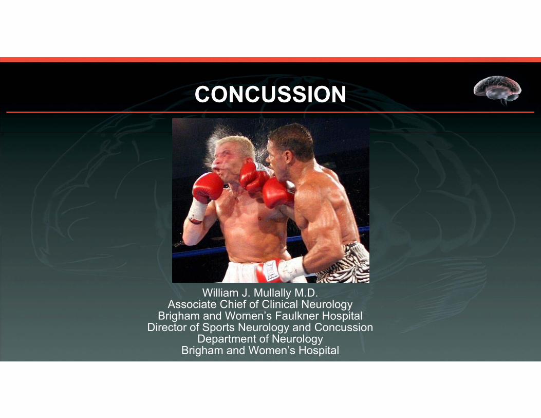
William J. Mullally M.D.Associate Chief of Clinical Neurology
Brigham and Women’s Faulkner HospitalDirector of Sports Neurology and Concussion
Department of NeurologyBrigham and Women’s Hospital
CONCUSSION

LEARNING OBJECTIVES • Define concussion
• Recognizing symptoms of post concussion syndrome
• Approach to the management of post traumatic headaches and post concussion syndrome
• Basic understanding of Chronic Traumatic Encephalopathy
2

DISCLOSURES
NONE
3

Marciano vs. Walcott 9/23/1952• http://www.youtube.com/watch?v=W4fWf4jtQWU
4
https://www.youtube.com/watch?feature=player_detailpage&v=Tyv4du7BTOc
FOOTBALL CONCUSSION

INCIDENCE
• 1.7 million ER visits / yr. for TBI- over $60 billion! Most patient are released to home indicating that the injuries are not severe
• Estimates of sports-related mild traumatic brain injury (mTBI) range from 1.6–3.8 million affected individuals annually in the United States, many of whom do not obtain immediate medical attention.
• One survey of a collegiate cohort revealed that 43% of concussed athletes knowingly hid symptoms

HISTORY
• First descriptions of concussion over 2400 years ago by Hippocrates
• “Falls down immediately, loses speech, cannot see or hear.”• Constellation of symptoms a mystery
• Derived from the Latin “concussus” meaning “to shake violently”

8
HISTORY
• 10th Century Persia- Rhazes - Distinction between abnormal physiologic state and structural brain injury
• 14th Century Milan- Lanfrancus - “Commotio Cerebri” Transient paralysis of cerebral function as opposed to “Contusio Cerebri”
• 1928- Martland “ Punch Drunk” syndrome, modified in 1937 to Dementia Pugilistica by Millspaugh
• 1949 – Critchley- “Punch Drug Syndromes- Chronic Traumatic Encephalopathy”
• 2002- Omalu described the 1st football case of CTE with extensive tau deposition
• 2009- McKee et al- 68 of 85 brains with a history of repetitive mild TBI revealed evidence of CTE

CONCUSSION CONUNDRUM
• What is a concussion?
• What is mild traumatic brain injury?
• Does every blow to the head that results in symptoms constitute a concussion?

AMERICAN ACADEMY OF NEUROLOGY2013
Concussion is defined as a clinical syndrome of biomechanically induced alteration of brain function typically affecting memory and orientation, which may involve loss of consciousness.

ZURICH INTERNATIONAL SYMPOSIUMON CONCUSSION IN SPORTS - 2012
The definition from the Zurich International Symposium placed emphasis on functional changes following a head injury rather than loss of consciousness or posttraumatic amnesia.

12
ZURICH CONCUSSION CRITERIA
a) Symptoms – somatic ( headache, dizziness), cognitive (mental fogginess), and/or emotional symptoms (lability)
b) Physical signs- loss of consciousness, amnesiac) Behavioral changes – irritabilityd) Cognitive impairment- Slow reaction time, impaired memory.e) Sleep disturbance- insomnia
If any one or more of these components is present then a concussion should be suspected.

AMERICAN ACADEMY OF NEUROLOGY 1997
• Concussion was defined as a trauma induced alteration in mental status that may or may not result in loss of consciousness.
• From a clinical standpoint this is the most useful definition

CONCUSSION• Mildest form of mild traumatic brain injury• “See Stars” or momentarily “Dazed and Confused”• Loss of consciousness- inconvertible evidence that a TBI has
occurred but observed in < 10% of concussions. • Duration of LOC may be an indication of the severity but does not
always predict the outcome.• Retrograde and anterograde amnesia
14

CASE 1- SOCCER
• 21 year old male who while playing soccer was tripped while going for the ball and scraped the right side of his head against the ground. The patient did not lose consciousness or experience a change in mental status. There was swelling of the area with an abrasion and discomfort at the site. No other symptoms and neurologic exam was normal.

CASE 2- SOCCER
• 16 year old female who while attempting to head the ball after a corner kick bumped her head against the head of another player. She did not lose consciousness or experience a change in mental status. There was no period of confusion or amnesia but following the trauma she experienced a global headache with slight nausea and photophobia. Neurologic exam was normal including cognitive function. The pain gradually lessened after several days then resolved.

CASE 3 - FOOTBALL
• 26 year old male who while playing football was struck on the left side of his head by the helmet of another player. He immediately “saw stars” and felt dazed. He did not lose consciousness or experience anterograde amnesia. Following he had a global headache with nausea and photophobia. He felt as if he was in a “fog”. Neurologic exam was normal.

CASE 4- RECREATION
• 45 year old male who fell from his bicycle. He sustained trauma to the right side of his head and his helmet was cracked. According to his companion he was unconsciousness for 5 minutes then slightly confused for 20 to 30 minutes. He was taken to a hospital emergency room where he complained of a headache but neurologic exam was normal with the exception of slight unsteadiness of gait. An MRI scan was performed.

SUBDURAL HEMATOMA

20
BLOOD BIOMARKERS IN THE DIAGNOSIS OF TRAUMATIC BRAIN INJURY
GLIAL FIBRILLARY ACIDIC PROTEIN (GFAP)- ASTROGLIAL PROTEIN FOUND IN WHITE AND GRAY MATTER.
UBIQUITIN C-TERMINALHYDROLASE (UCH-1)- NEURONS
TOTAL TAU- NEURONS
S- 100 CALCIUM-BINDING PROTEIN B- ASTROGLIAL PROTEIN
Neurofilament light

TRAUMATIC BRAIN INJURY • Trauma from direct skull impact and/or penetrating injury to brain
• Cervical flexion extension injury
• Blast injury

Mechanical shearing of endothelial cells in small cells
Mechanical stretching and disruption of axonal plasma membranes
Impaired regulation of blood-brain
barrier and cerebral blood flow.
Release of excitatory amino acids
Deregulated ion flux with efflux of
potassium and influx of calcium
Calpain activation by increased intracellular
calcium
Mechanical breakage of microtubules
CONCUSSION
Acute failure in neuronal function
Glucose Consumption
Increased membrane pump activity to
restore ionic balance.
Energy crisis in damaged neurons
Proteolytic breakdown of
neurofilament proteins
Microtubule disassembly
Axonal transport defect
Binding of glutamate to NMDA receptors
Neuronal depolarization
Focal ischemia and blood-brain barrier
damage Acute widespread
suppression of neurons
ROTATIONAL BRAIN INJURY
PATHOPHYSIOLOGY OF CONCUSSION

• Head injury , as a result of direct head trauma, a blast injury or forcible flexion extension of the cervical spine, stimulates the trigeminal sensory afferents directly and/or as a result of sterile inflammation with the release of cytokines resulting in sequential activation of second and third order neurons in the pain pathways in the brainstem, hypothalamus and thalamus causing a headache often with migrainous features and accompanying symptoms. Input from the upper cervical nerve roots converge on the trigeminal nucleus caudalis and may contribute to the activation process.
• This does not necessarily denote a TBI.- The “ LUGE HEADACHE” as an example.
PROPOSED ETIOLOGY OF THE POST TRAUMATIC HEADACHE

24
DEMOGRAPHICS- Incidence is higher in females- anatomical, endocrinologic and physiologic
- Children ages 0-4 have the highest rates
- Adults – Falls and MVAs ( fall related injuries increasing in the elderly)
- 15- 24 age group- Sports are second to MVAs
- Age < 15 are more susceptible

25
EVALUATION
- Neurologic exam, Sports Concussion Assessment Tool (SCAT 3), Standardized Assessment of Concussion (SAC), Balance Error Scoring System (BESS).
- Immobilize the cervical spine
- If there is LOC, seizure or a severe injury is suspected transfer to the hospital
- Glasgow coma scale- Patients with concussion score 13-15

Neuroimaging
• CT is sufficient to rule out a severe injury. Diffusion tensor MRI may detect white matter injuries
– Usually contributes little to the mild head injury - evaluation
– Abnormal neurologic exam including testing of gait – Progressive headache– Recurrent vomiting– Loss of consciousness of > 1 minute– Prolonged anterograde amnesia– Seizure– Skull fracture– Age > 60– Alcohol or drug intoxication– Coagulopathy– GCS score of < 12, 2 hours post injury

SPORTS CONCUSSION• 170 million adults and 44 million children participate in sports• Due to more males studied, total number of concussions is greater for
males( football, hockey, rugby) than females for all sports combined but incidence is higher in females (soccer and basketball)
• 5% of athletes will suffer a concussion• 20% of athletes in contact sports• 15% of all injuries are concussions
Giza CC., et al. Summary of evidence-based guideline update: Evaluation and management of concussion in sports. Report of the Guideline Development Subcommittee of the American Academy of Neurology. Neurology June 11, 2013 vol. 80 no. 24 2250-2257

INJURY RATE PER 1000 ATHLETES EXPOSED
• Football (2.34) • Men’s Ice Hockey (1.47) • Women’s Soccer (1.42) • Wrestling (1.27)• Men’s Soccer (1.08) • Women’s Lacrosse (0.70) • Field Hockey (0.57) • Women’s Basketball (0.50) • Men’s Basketball (0.32) • Softball (0.25) • Baseball (0.19) • Volleyball (0.15)
The vast majority of head injuries do not result in loss of consciousness (more than 90%)

FOOTBALL• Body mass index greater than 27 kg/m2
and training time less than 3 hours weekly likely increase the risk of concussion
• Player to player contact
• Playing on artificial turf is possibly a risk factor
• Risk on kickoffs 4X higher
• 25% are the result of prohibited activity
• 2/3 of concussions occur during competition
• History of prior concussion
Certain positions may be at greater risk

SIGNS AND SYMPTOMS
• Headache
• Dizziness/Vertigo- disequilibrium and imbalance
• Cognitive impairment (e.g. feeling like “in a fog”, memory and concentration impaired, slow reaction time)
• Confusion, orthostatic lightheadedness, vision blurring/eye fatigue, tinnitus, photophobia, phonophobia, bad taste in the mouth, fatigue/lethargy, anosmia
• Depression, anxiety, personality changes, mood lability, irritability, aggression, acting out, and social inappropriateness
• Sleep disturbance (e.g. drowsiness, insomnia)
• Neck pain

POST TRAUMATIC HEADACHE

HEADACHE ATTRIBUTED TO HEAD AND/OR NECK TRAUMA
Acute headache attributed to traumatic injury to the head (mild; moderate or severe) or whiplash
Persistent headache attributed to traumatic injury to the head (mild; moderate or severe) or whiplash
Headaches begin within 7 days of the traumatic insult (only 37% of soldiers report a headache in first 7 days)

PHENOTYPE OF POST TRAUMATIC HEADACHE• Migraine headache- most common type - >60%
• Tension type headache - 21%
• Cervicogenic headache – 10%
• Trigeminal autonomic cephalgia - cluster headache
• Occipital neuralgia
• Supraorbital and infraorbital neuralgia
• Low pressure headache due to CSF leak
• Secondary headache due to structural lesions

INCIDENCE OF POST TRAUMATIC HEADACHE • 25- 90% of patients who have suffered head trauma• 15 – 78% continue for more than 3 months• 6.8 % in Pediatric age group have headaches at 6 months• 20% persistent headaches at 4 years• 4% of all symptomatic headaches• 20% of soldiers, most commonly due to blast injuries.• PTH can occur after mild, moderate or severe TBI but more common
after a mild injury.

PERSISTENT POSTTRAUMATIC HEADACHE
• History of headaches specifically migraine• History of prior TBI• Rebound from abortive medication• Comorbid psychiatric disorders• Effect of litigation and return to work
35

POST CONCUSSION SYNDROME• 30 – 80 %
• Symptoms usually resolve in a few weeks
• Young athletes ( < 15 ) have a more prolonged recovery
• 20% have persistent symptoms- psychosocial problems, psychiatric disorders, litigation, compensation, malingering may contribute.
• Neurofilament light may predict prolonged PCS
• Schrader’s study – Lithuania
• Prussian railway compensation system for work related injuries – 19th century
• 80% of soldiers do not return to combat
36

TRADITIONAL MANAGEMENT
Initial period of physical restNo training, playing, exercise, weightsBeware of exertion with activities of daily living
Initial period of cognitive restAvoid television, extensive reading, video games, telephone, textingCaution re: daytime sleep
Rest is for wimps…
Is it 5th down yet?

• Is there data to support complete cognitive and physical rest?
•Can cognitive or physical activities delay recovery or potentially do harm?

PHYSICAL REST• 88 patients ages 11-22 within 24 hours of concussion were
randomized to strict rest for 5 days (45) vs 1-2 days of rest followed by stepwise return to activity (43).
• There was no clinically significant difference in neurocognitive or balance outcomes.
• Strict rest resulted in more daily postconcussive symptoms and slower symptoms resolution.
• Pediatrics February 2015 Thomas et al
39

COGNITIVE REST• 335 patients ages 8-23 (mean age 15, 62% male; 19% suffered LOC
and 39% with a prior concussion) after suffering a concussion were placed in one of 4 groups: cognitive rest with minimal activity; moderate cognitive activity; significant cognitive activity; full Cognitive activity.
• The highest quartile took approximately 100 days to recover• The lower 3 quartiles took approximately 20 - 50 days and patients in
those quartiles had similar duration of symptoms.• Pediatrics February 2014- Brown et al
40

• The emerging concept of cognitive and physical deconditioning as a cause of persistent symptoms
• “Cogniphobia”• Are ongoing headaches a contraindication to resuming unrestricted
vigorous physical exercise?

MANAGEMENT
• Expect gradual resolution of symptoms within 7-10 days but occasionally symptoms persist for weeks to months.
• Gradual return to school, social and physical activities that does not result in significant exacerbation of symptoms
• When symptoms resolve or return to pre-injury baseline proceed through step-wise return to sport / play (RTP) protocol after receiving medical clearance

CONCUSSION TREATMENT -MULTIDISCIPLANARY
• Speech/language therapy - cognitive rehabilitation therapy if symptoms persist for longer than 1 month
• Vestibular therapy if dizziness persists for more than 2 weeks• Physical therapy for neck pain that persists• Neuropsychology/Psychiatry for persistent symptoms accompanied
by depression or anxiety• Pharmacotherapy if symptoms (headache, dizziness, insomnia,
anxiety, depression) persist for more than 2 weeks

MEDICATION- ABORTIVE• Simple analgesics• NSAIDS• Triptans• Muscle relaxants• Midrin• AVOID NARCOTICS and BUTALBITAL COMPOUND

MEDICATION- PREVENTATIVE• Tricyclic antidepressant medication – amitriptyline, nortriptyline,
imipramine• Beta blockers- propranolol, timolol, atenolol• Topiramate

ALTERNATIVE TREATMENT• Occipital and trigeminal nerve blocks• Trigger point injections• Botulinum toxin injections• Acupuncture• Relaxation therapy and biofeedback• TENS• Cognitive behavioral therapy

RECOVERY
• May take longer in children and adolescents• Pre-existing medical problems such as migraine, ADD,
mood disorders, learning disability may delay recovery• History of prior traumatic brain injury• Females may recover at a slower rate • Higher post concussion symptom score• Prominent dizziness may portend a slower recovery

RECOVERED?
• History- Free of symptoms or back to baseline
• Neurologic exam- May include BESS (Balance Error Scoring System) balance assessment.
• Neurocognitive testing – (ImPACT – Immediate Post Concussion Assessment and Cognitive Testing. A computerized test that is only helpful when it can be compared to a pre-injury baseline test- 6 neuropsychological test modules reported in 5 fields- Verbal Memory, Visual Memory, Reaction Time, Processing Speed, Impulse Control).
PREPARTICIPATION EVALUATION
• History:– Type of sport?– Previous symptoms of head injury?/length of recovery (recall
unreliable from teammates, coaches) – Prior head, maxillofacial, spine injuries?– Non-sporting head injuries?– Type of player (“physical”?)– Ability to “take a hit”– Age
• Opportunity to Educate!

SAME DAY RETURN TO PLAY?
•NO!• Unanimously agreed that no RTP should occur
on the day of a suspected traumatic brain injury

SECOND IMPACT SYNDROME
Fact or Fiction

PREVENTION
• Protective equipment• Mouth guards have benefit in prevention oral injury, but no
evidence of TBI reduction
• Head gear and helmets show reduction in biomechanical forces reducing the incidence of serious head and facial injury, but have not effected a reduction in concussion

CHRONIC TRAUMATIC ENCEPHALOPATHY (CTE)
• Acknowledge potential for long-term problems in all athletes• CTE is a pathologic diagnosis with unknown incidence in athletic
populations. The clinical syndrome that is a result of the pathologic changes is referred to as “Traumatic Encephalopathy Syndrome”
• Progressive tauopathy

54
TRAUMATIC ENCEPHALOPATHY SYNDROME
Progressive neurodegenerative disease that is the cumulative effect of repetitive head trauma
1)Cognitive and/or behavioral impairment2)Cerebellar dysfunction3)Pyramidal tract or extrapyramidal disease4)Persistence of symptoms for more than 2 years5)No other neurologic disorder to account for all of the clinical
features
55
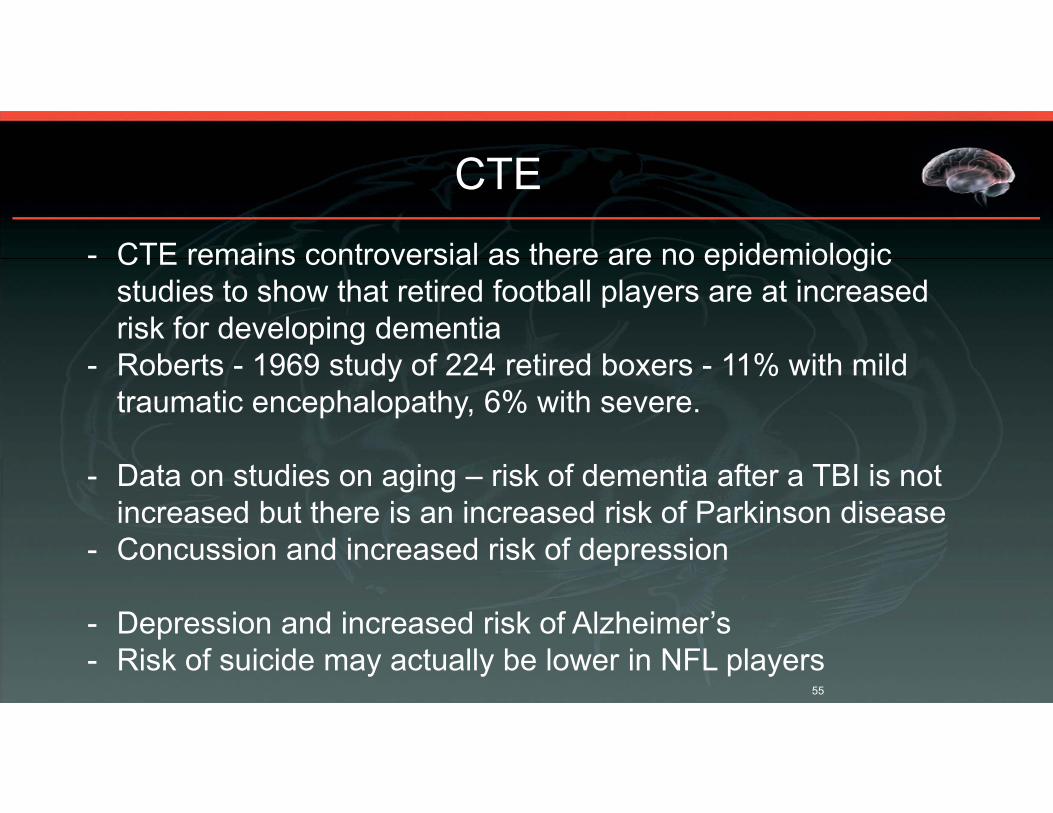
- CTE remains controversial as there are no epidemiologic studies to show that retired football players are at increased risk for developing dementia
- Roberts - 1969 study of 224 retired boxers - 11% with mild traumatic encephalopathy, 6% with severe.
- Data on studies on aging – risk of dementia after a TBI is not increased but there is an increased risk of Parkinson disease
- Concussion and increased risk of depression
- Depression and increased risk of Alzheimer’s- Risk of suicide may actually be lower in NFL players
CTE

56
Do repetitive concussions place one at risk for CTE and ALS?
Is it the number of concussions or insufficient time for recovery?
Are certain individuals more susceptible?
Are other factors such as depression, contributing?(Tau levels increase with exercise)
QUESTIONS THAT MUST BE ANSWERED
CTE

KEY POINTS• Concussion has reached epidemic proportions and clinicians must be
able diagnose, provide guidance in the days and weeks following the injury and manage common symptoms.
• Chronic Traumatic Encephalopathy is a potential complication of repetitive mild traumatic brain injury but longitudinal studies are required to gain a better understanding of the incidence and additional risk factors.
57

NEXT BEST STEPS1) Biomechanical force induced change in mental status is likely a concussion and should be managed accordingly
2) Athletes must be immediately removed from competition and should not return for a minimum of 10-14 days
3) A reduction in cognitive activity, but not absolute rest, is important and an early return to physical exercise as tolerated should be encouraged
4) Athletes who have suffered 5 definite concussions should probably be advised to retire from their sport
5) CTE appears to be a real but relatively uncommon complication of repetitive concussion
58

REFERENCES• Mullally, WJ. Concussion. The American Journal of Medicine. 2017; 130, Issue 8:
885-892.
• Giza CC, Kutcher JS, Ashwal S, Barth J, Getchius TS, Gioia GA, Gronseth GS, Guskiewicz K, mandel S, Manley G, et al., “Summary of evidence-based guideline update: Evaluation and management of concussion in sports: report of the Guideline Development Subcommittee of the American Academy of Neurology,” Neurology, vol. 80, no. 24, pp. 2250-2257, 2013.
• Howell DR, Mannix RC, Quinn B, Taylor A, Tan CO, Meehan WP, Physical Activity Level and Symptom Duration Are Not Associated After Concussion. Am J Sports Med April 2016, vol. 44, no. 4, 1040-1046.
59

REFERENCES• Brown N, Mannix RC, O’Brien MJ, Gostine D, Collins MW, Meehan WP, Effect of Cognitive
Activity Level on Duration of Post-Concussion Symptoms. Pediatrics. 2014: 133 (2): e299-e304.• Thomas DJ, Apps JN et al. Benefits of Strict Rest After Acute Concussion: A Randomized
Controlled Trial. Pediatrics. 2015: 135(2): 1-11• Randolph C, Is Chronic Traumatic Encephalopathy a Real Disease? Current Sports Medicine
Reports. 2014: 13 (1): 33-37.• McKee AC, Stein TD, Kiernan PT, Alvarez VE, The neuropathology of chronic traumatic
encephalopathy. Brain Pathol. 2015:25(3): 350-364.• Mez J, Daneshvar DH, Kiernan PT, et al. Clinicopathological Evaluation of Chronic Traumatic
Encephalopathy in Players of American Football. JAMA 2017: 318(4): 360-370.• Maroon JC, Winkelman R, Bost J, et al. Chronic Traumatic Encephalopathy in Contact Sports: A
Systematic Review of All Reported Pathologic Cases. PLoS One 2015: 10(2): e0117338. doi: 10.1371/journal.pone.0117338
60

ADVANCES IN LIFE SCIENCES AND HEALTH
In Press
ADVANCES IN LIFE SCIENCES AND HEALTH
The Grading of Head and Brain Injury in Sportswith Return to Play Guidelines Eliminating theConfusion of ConcussionWilliam Mullally1*, Kathryn Hall21 Brigham and Women’s Hospital, Harvard Medical School, USA.2 Massachusetts General Hospital, Clinical Research Center, USA.
*Corresponding author: [email protected]
Abstract:Context: Sports are a common cause of head injury in today’s society. The term concussion has nouniversally accepted definition and rather than continue to use an epithet that can be confusing our focusshould be on the diagnosis of a traumatic brain injury. It is imperative that athletes who suffer a traumaticbrain injury are identified with alacrity and protected from further injury. We propose a grading system andreturn to play guidelines to assist sports medicine practitioners in the diagnosis of the severity of a headinjury and a practical timetable for the safe return to sports participation.Evidence acquisition: The PubMed database was searched (1991-2014) for articles related to concussionand traumatic brain injury in sports. Grading systems were reviewed.Study Design: Clinical review.Level of Evidence: Level 3Results: There is limited data to support the grading systems of traumatic brain injury in sports. One of thereasons for that is the lack of a clear definition of the word concussion. There is also a paucity of judiciousguidelines to assist sports medicine practitioners in making a determination as to when an athlete canresume sports participation with reasonable safety.Conclusion: We present a new grading system of head and brain injury in sports with return to playguidelines and have eliminated the confusion and dissenting views that accompany the term concussion.We have also attempted to clearly separate out injuries that are not indicative of a traumatic brain injury,per se. Our goal is to protect athletes by providing the medical professionals who treat them with a basicframework for diagnosis and a set of guidelines to assist in making recommendations with regard to returnto sport participation.
Keywords:Concussion; Traumatic Brain Injury; Posttraumatic Headache; Grading of Head Injury; Return to PlayGuidelines
1. INTRODUCTION
In the United States, the Center for Disease Control has estimated that 1.7 million patients each year are evaluated
at a hospital emergency room for a traumatic brain injury (TBI) and the vast majority does not require hospital
MEET OUR MASTER CLINICIANS: CONCUSSIONS AND HEAD TRAUMA

ADVANCES IN LIFE SCIENCES AND HEALTH
admission indicating that the injuries are not severe. Sports and many recreational activities are associated with an
increased risk of head injury and in the 15-24 age group, sports are second only to motor vehicle accidents as the
most frequent cause of head injury. The full magnitude of the problem remains elusive, as many patients who have
suffered a mild TBI (mTBI) do not receive medical attention. TBI has become a major public health concern and it is
the responsibility of the medical community to protect the athlete from suffering permanent brain injury [1–4].
2. CONCUSSION CONUNDRUM
In the field of sports medicine there continues to be pervasive confusion regarding the definition of concussion.
The term concussion is derived from the Latin concussus, which means, “ to shake violently”[5]. Concussion implies
a transient disruption in neurologic function secondary to mechanical forces. The Quality Standard Subcommittee
of the American Academy of Neurology in 1997 formally defined concussion as “a trauma induced alteration in
mental status that may or may not result in loss of consciousness”[6]. In 2013 that was updated and concussion
was described as “a clinical syndrome of biomechanically induced alteration of brain function typically affecting
memory and orientation, which may involve loss of consciousness”[7]. The updated definition is not particularly
helpful from a clinical standpoint and there is still no consensus in the medical community as to what actually
constitutes a concussion. Concussion is often used interchangeably with mTBI or mild/minor closed head injury
[8, 9]. When the term concussion is used it immediately conjures up a vivid picture of a significant blow to the
head that unquestionably results in an injury to the brain. Impaired functional status occurs as a result of the injury
and many have therefore altered the definition, as it is not uncommon for patients to develop symptoms that are at
times disabling, after a blow to the head that does not in any way alter mental status. The Zurich definition from the
International Symposia on Concussion in Sports places emphasis on functional changes following a concussion rather
than loss of consciousness or post-traumatic amnesia [10]. The question then becomes, does every blow to the head
constitute a concussion or mTBI if the subject experiences symptoms? We know that rapid acceleration-deceleration
of the head can result in an injury to the brain but does that imply that every “whiplash” injury that is followed by a
headache constitutes a concussion/mTBI.
To properly diagnose and manage athletes who have sustained a head injury, multiple attempts have been made to
classify concussion severity [11]. The most commonly used systems are shown in Figure 1 [6, 9, 10, 12–16]. The
problem with each of these grading systems is that they are wedded to a term that has no clearly accepted definition.
Redefining the term does not solve the problem and in essence, we are attempting to place a “square peg into a round
hole”. We would be better served to eliminate the term concussion from the medical glossary of diagnoses, relegating
it to the status of terms such as “knockout” or “stinger”. A single classification system, grading head and brain injury,
is required to render appropriate treatment and make standardized return to play recommendations.
3. HEAD AND BRAIN INJURY
In its mildest form, direct head trauma can result in an abrasion, laceration, or a contusion. An abrasion consists of
superficial damage to the skin while a laceration is an actual tear. A contusion occurs when capillaries are damaged
resulting in a hematoma. Localized pain as a result of the injury can occur and resolves as the swelling recedes and
the traumatized area heals. These traumatic insults need not result in a brain injury. Mild head trauma may also
stimulate the trigeminal sensory afferents resulting in sequential activation of the second and third order neurons
in the pain pathways in the brain stem, hypothalamus and thalamus. The trauma may be the result of a direct blow
to the head or occur indirectly as a result of forced flexion and extension of the cervical spine. Stimulation of the
upper cervical sensory nerve roots that converge on the trigeminal nucleus caudalis may contribute to the activation
MEET OUR MASTER CLINICIANS: CONCUSSIONS AND HEAD TRAUMA

The Grading of Head and Brain Injury in Sports with Return to Play Guidelines Eliminating the Confusion of Concussion
Figure 1. Comparison of primary concussion grading scales.
process. A headache, often with migrainous features, may then develop and occasionally neurologic symptoms
consistent with an aura can occur when direct mechanical irritation precipitates cortical spreading depression [17–20].
This activation of the trigeminovascular system does not, in itself, necessarily denote a traumatic brain injury. The
headache that occurs usually lasts hours but can persist, and individuals with a past history of migraine are likely to
be more susceptible [21].
When trauma induces an alteration in neurologic function as evidenced by a change in mental status, with or
without loss consciousness, a series of neurochemical changes occur that are transient but depending on the severity
of the insult, can cause axonal injury that results in permanent damage. The traumatic injury may be a direct blow to
the head resulting in coup or contrecoup insults, or the result of mechanical forces imparted by a flexion extension
injury of the cervical spine causing acceleration and rapid deceleration of the brain within the skull. The mechanical
and chemical changes are the result of the torsional and shearing forces affecting the neural tissue. In animal studies
the neurochemical changes develop over a period of hours. A release of excitatory neurotransmitters, particularly
glutamate binding to the NMDA receptor, precipitates the outflow of potassium into the extracellular space and an
influx of calcium into the cell. This initially results in a hypermetabolic glycolytic state as membrane pumps become
activated to restore homeostasis. Lactate is produced impairing neuronal function and there is a reduction in cerebral
blood flow, diminished glucose utilization and axonal injury. This diminished metabolic state can last from days to
weeks and during this period of time the neural tissue is more susceptible to further injury. The average recovery time
is 7-10 days but occasionally the reduced regional blood flow and metabolic abnormalities can persist for months
[22–24].
Severe brain injuries can occur with or without skull fracture and include focal or diffuse axonal injury with edema
and increased intracranial pressure, cerebral contusion, cerebral shearing, intraventricular hemorrhage, subarachnoid
hemorrhage, intracerebral hemorrhage, subdural hematoma and epidural hematoma. The consequences of these
MEET OUR MASTER CLINICIANS: CONCUSSIONS AND HEAD TRAUMA

ADVANCES IN LIFE SCIENCES AND HEALTH
injuries are devastating and often result in death.
From the standpoint of assessing the severity of the typical sports related head injury it is often difficult to
differentiate mild from moderate, after a disturbance of mental status has occurred. Attempts have been made to
grade the severity based on the onset of confusion, the presence and duration of anterograde amnesia, whether or not
there was loss of consciousness and the duration of post concussion/traumatic symptoms [5, 25–27]. The Glasgow
Coma Scale (GCS) is commonly used, in a hospital emergency room setting, to grade the severity of traumatic
brain injuries based on verbal, motor, and eye-opening reactions to stimuli. The assessment is done in the first few
hours following the injury and cannot be completed retrospectively. The duration of posttraumatic amnesia is not
assessed nor are post traumatic/concussive symptoms considered. While the GCS is useful in the initial assessment
of the patient’s level of consciousness it is not particularly helpful in the subsequent grading of head and brain
injuries that occur typically in sports with the goal of providing a guideline for recommendations regarding return to
play [28]. The duration of post traumatic amnesia is likely important, however, but is extremely difficult to assess,
retrospectively, as we rely on the history obtained from the patient which, in the context of the brain insult, may
not be accurate and may also be influenced by what was related to the patient by witnesses, family, coaches and
teammates[24, 25, 29]. While loss of consciousness may not always correlate with the outcome of a traumatic
brain injury [29], it does provide inconvertible evidence that a significant injury has occurred and that there will
be neurochemical and potentially structural changes in the brain as a result of that injury [18, 22]. The importance
of loss of consciousness including duration should not be ignored as it may also signify that a serious brain injury
has occurred [30, 31]. Another parameter to consider when assessing the severity of a head and brain injury is the
constellation of somatic, neurobehavioral and cognitive symptoms that patients experience. Symptoms can include
headache, dizziness, insomnia, photophobia, irritability, fatigue, insomnia, difficulty with concentration and memory
and patients often report the sensation that they feel as if they are “in a fog” [22, 32]. These symptoms are referred to
as part of a “post concussion syndrome”, however they are often observed in patients who did not suffer a defined
traumatic brain injury and did not experience a change in mental status or loss of consciousness as a result of their
injury. From a clinical standpoint using an “alteration in brain function typically affecting memory and orientation”
[7] as a way to determine if a concussion/TBI has occurred is essentially useless.
Neurocognitive testing, in particular computerized neuropsychological tests such as the Immediate Post-Concussion
Assessment and Cognitive Testing (ImPACT), can be a useful screen in assessing cognitive functioning related to a
mild TBI [29, 33–35]. Baseline testing is becoming a standard in sports programs in schools throughout the country
and a comparison of an athlete’s performance to the pre-injury test can be beneficial in making the decision regarding
return to play. Without a baseline, however, the testing is not particularly helpful in assessing the status of an injured
athlete. Neurocognitive testing is only a guide to assist in the clinical management of the patient. A diagnosis and
recommendations cannot be made solely based on the results and test retest reliability can be variable [35–38].
4. GRADING SYSTEM
The American Academy of Neurology now recommends that each athlete who has suffered a concussion be
assessed and managed individually without relying on a grading system or immutable return to play guidelines [7].
From a practical standpoint, however, there is a wide variation in the level of training and experience of the licensed
professionals who care for athletes who have suffered sports related injuries. With the goal of protecting athletes
from suffering serious and permanent injury, we think that it is important that a clear and defined grading system
be established to assist the clinician in identifying the severity of the injury and providing a uniform and logical
timetable for a return to sports participation. The grading system and RTP guidelines also minimize the pressure that
can potentially be placed on the health care professional, by coaches, agents, parents, and fellow athletes to allow the
injured athlete to return to competition prematurely.
MEET OUR MASTER CLINICIANS: CONCUSSIONS AND HEAD TRAUMA

The Grading of Head and Brain Injury in Sports with Return to Play Guidelines Eliminating the Confusion of Concussion
Unfortunately prospective randomized controlled trials regarding the assessment and management of the different
grades of brain injury, in sports, have provided limited information. The exception is in the management of the
severe head injuries such as those that result in an intracranial hemorrhage which fortunately are quite rare. We
must, therefore, make clinical decisions based on retrospective data, limited prospective data, experimental models,
consensus statements and personal experience. After reviewing the best available information in the medical literature
we have constructed a grading scale of head and brain injury (Figure 2) in sports that can be used in the clinical
setting to make cogent decisions regarding return to play. We have only recently entered the era where our primary
objectives are to recognize when a potential brain injury has occurred, assess the severity of that injury and then
formulate a return to play strategy that protects the athlete from sustaining permanent injury. With the publication of
the landmark paper by McKee et al, revealing that a potential consequence of repetitive mild traumatic brain injury is
a progressive tauopathy, which we now refer to as chronic traumatic encephalopathy, [39] our level of vigilance has
been heightened. Athletes must be protected from the potential long-term consequences of repeated mild insults to
the brain while at the same time respecting their right to participate in their sport.
Figure 2. Grading scale of head and brain injury in sports.
Athletes in the Grade 1 category have not suffered a traumatic brain injury and are only restricted from play by
the extent of the local insult. They may be cleared to return to play once the affected area has been addressed and
treated by a medical professional. A Grade 2 injury also does not specifically denote brain injury but the athlete
should not be allowed to resume physical activities until the headache and accompanying symptoms have resolved.
Neurocognitive testing should be performed as a safeguard and if the results are acceptable and compare favorably to
baseline testing then the athlete can be cleared for the return to play protocol. Grade 3 and 4 injuries constitute what
has historically been referred to as a concussion. The alteration in mental status, with or without a disturbance of
consciousness, indicates that a brain injury has occurred with the resultant mechanical and neurochemical changes
[22]. The distinction between the two grades is somewhat arbitrary and based on the available information in the
medical literature and the consensus of those specializing in sports injuries. Head injuries are not uniform and cases
MEET OUR MASTER CLINICIANS: CONCUSSIONS AND HEAD TRAUMA

ADVANCES IN LIFE SCIENCES AND HEALTH
will never be identical. The estimation of the period of amnesia is just that, an estimate, as we must rely on the history
obtained from the individual who suffered the injury. There is some information in the medical literature that suggests
that loss of consciousness and amnesia are not accurate indicators of the severity of an injury but both signify, without
any question, that a brain injury has definitely occurred and provide tangible evidence to the health care clinician who
is performing the clinical evaluation, thus facilitating the ultimate determination of when an athlete can safely return
to play [29, 32, 40, 41]. Grading is inherently relative and for the purpose of classification we consider anterograde
amnesia of less than 30 minutes and/or LOC of less than 1 minute to indicate a mild brain injury. We classify this
as a Grade 3 injury. With Grade 4, the patients have experienced amnesia for more than 30 minutes, but less than
24 hours, or LOC for longer than 1 minute but less than 5 minutes, denoting a moderately severe brain injury. For
the purpose of protecting the athlete we have included patients who have suffered amnesia for more than 24 hours
or LOC of longer than 5 minutes in the Grade 5, severe brain injury category. Intracranial hemorrhage, cerebral
contusion, shearing injury and increased intracranial pressure are also included in the Grade 5 category.
This grading system is not useful in the immediate post injury time period and should not be used until the
athlete has had a medical evaluation to insure that there is no evidence of a catastrophic injury, such as intracranial
hemorrhage, as severe injuries can occur following seemingly mild trauma, without, or with only brief, LOC. A
persistently abnormal elementary neurologic exam excludes Grade 1-4 categories and can only be observed in grade
5. Imaging of the brain is usually normal in patients who have suffered an apparent mTBI but should be performed if
the medical professional who has evaluated the patient is concerned about the possibility of a structural injury. If an
abnormality secondary to trauma is detected that immediately imputes a Grade 5 designation. Injuries to the cervical
spine may occur in association with head trauma and must be diagnosed and treated appropriately.
Following traumatic brain injuries that are graded as 3 or above, patients usually experience multiple symptoms in
addition to headache, including fatigue, insomnia, irritability, vertigo, nausea, visual blurring and cognitive difficulty.
Attempts have been made to grade brain injury/concussion based on the duration of symptoms [5, 25, 40]. In Grades
3 and 4, symptoms will usually resolve within 10 days but in some patients, especially adolescents, symptoms may
persist and this does not necessarily correlate with the severity of the injury [30–32, 42–44]. Female athletes may be
more susceptible to mTBI [45]. Factors that may contribute to the chronicity of symptoms include a prior history
of headache, depression, chronic fatigue syndrome and chronic pain syndrome [30]. To grade the severity of an
injury based solely on lingering subjective symptoms would be capricious. Unlike individuals who have suffered an
injury in a motor vehicle accident or a work related accident where secondary gain is a potential cause of persistent
symptoms, athletes may, on occasion, minimize or deny symptoms to expedite their release from medical care.
Neurocognitive testing can be extremely important, as poor performance may parallel the duration of posttraumatic
symptoms including memory difficulty, but it is only an adjunct to the clinical assessment [38, 40]. Cognitive
dysfunction occurs commonly in patients suffering from chronic nonmalignant pain and it would be a sophistry to
conclude that poor performance on a neurocognitive test, per se, signifies that a traumatic brain injury has occurred [?]. A patient with a Grade 2 injury may perform poorly on the testing purely because of the posttraumatic headache.
5. RETURN TO PLAY GUIDELINES
While a grading system may provide inferences with regard to prognosis, the main utility is in establishing a
guideline for return to play (RTP). The guidelines should not preempt the clinical judgment of the medical professional
and should only be used as an adjunct in making the determination of when an athlete can resume full participation
in their sport with reasonable safety. We present RTP guidelines (Figure 3) using the information available in the
medical literature. Evidence from prospective studies to define each and every step in the decision making process is
not yet available. The goal of RTP guidelines is to protect the athlete from serious and permanent brain injury while
at the same time respecting the right of the athlete to participate in the sport of his or her choice. When releasing
MEET OUR MASTER CLINICIANS: CONCUSSIONS AND HEAD TRAUMA

The Grading of Head and Brain Injury in Sports with Return to Play Guidelines Eliminating the Confusion of Concussion
an athlete to RTP the medical professional must take into consideration the specific sport and the inherent risk of
sustaining a recurrent injury in that particular endeavor. Sports such as boxing and full contact martial arts present a
unique challenge as the ultimate goal of the sport is to inflict damage rendering one of the combatants incapable of
continuing in the contest. Neutralizing an opponent through physical force is inherent in sports such as football and
rugby but inflicting injury is not the specific goal. Major changes are ongoing in college and professional football to
reduce the number of head injuries.
Figure 3. Return to play guidelines.
In the absence of a defined brain injury, Grade 1 and 2, an athlete can return to sports participation immediately
after their symptoms have resolved and they have been evaluated and cleared by a medical professional. Grade 1
injuries do not require the RTP progression protocol as defined by the 2012 Zurich Consensus statement (Figure 4)
[10]. A Grade 2 injury must be confirmed by a medical evaluation which documents that a change in mental status
did not occur at the time of the injury and there was no period of amnesia. If there is any question then the athlete
should be managed as a Grade 3 injury. We recommend, however, that the RTP protocol be followed for athletes who
have sustained a Grade 2 injury after a health care practitioner has cleared them to play.
In animal studies, the metabolic abnormalities that occur after a traumatic brain injury return to normal in 7 - 10
days [22]. In humans this may take longer and the decrease in glucose metabolism can last from 2 - 4 weeks. The
symptoms after a mild TBI, however, usually resolve within 7 - 10 days and cognitive function returns to normal
[22, 32]. We, therefore, do not think that an athlete with a grade 3 injury should be subjected to any type of contact
for 10 days. The athlete can begin the process to RTP when they are symptom free and neurocognitive function has
returned to baseline but contact must be assiduously avoided until they are 10 days post injury. Sports medicine
practitioners must be cognizant of the fact that young athletes in high school and in youth sports require a longer
recovery period when compared to college and professional athletes and the effects of a TBI on a developing brain
have not been explicitly defined [44, 46, 47].
Catastrophic brain swelling has been reported in case studies when an athlete who has sustained a brain injury
MEET OUR MASTER CLINICIANS: CONCUSSIONS AND HEAD TRAUMA

ADVANCES IN LIFE SCIENCES AND HEALTH
Figure 4. Progression protocol for return to play.
suffers a second brain injury before the symptoms from the first injury have full resolved. This is referred to as the
second impact syndrome but there is no good scientific data to support this theory [11, 48–50]. We think that it is
better to err on the side of caution, however, and our guidelines require that an athlete be symptom free before a RTP
protocol can be initiated.
While the distinction between grade 3 and 4 injuries is empiric, the prolonged amnesia or longer period of
unconsciousness that occurs with a grade 4 injury suggests intuitively that a more severe injury has occurred. To
protect the athlete a minimum of 30 days must transpire, after the first such injury, before the athlete can begin the
RTP progression protocol.
All grade 5 injuries are severe and only differ in the degree. Athletes who have suffered structural damage of the
brain as indicated by abnormal brain imaging or a persistently abnormal elementary neurologic exam should never
return to play. If a comprehensive neurologic evaluation does not reveal evidence of a structural brain injury, then the
athlete can potentially return to play at 180 days if they receive neurologic clearance. This would include those who
have sustained uncomplicated skull fractures.
Athletes who have suffered a TBI are at increased risk to suffer a subsequent injury [43]. Recovery from
posttraumatic symptoms is also slower. In our guidelines we provide a schedule to assist the medical professional in
making a decision regarding RTP. The goal of the recommendations is to allow a longer period of time to recover
from the injury and also to protect the brain from another injury before adequate healing has occurred. With
repeated injuries there is an increase risk of developing chronic traumatic encephalopathy and that must be a prime
consideration when the treating clinician is asked to clear an athlete to return to play [39]. When an athlete has
suffered more than one TBI, Grade 3 and above, the RTP guidelines for the highest grade of injury that they have
sustained should be followed.
Many of the symptoms that occur following a traumatic head and brain injury, such as headache, dizziness,
depression etc., are prevalent in the general population and when evaluating athletes who have preexisting problems
for RTP, a return to their baseline should be considered the equivalent of becoming symptom free.
When an athlete who has suffered a grade 2, 3, 4 or 5 injury has been medically cleared to RTP they must first
enter and successfully complete the graduated RTP protocol before they can return to full participation in their sport
without restriction. Athletes must remain asymptomatic through each step or they must return to the previous level
and then continue progression until they complete the protocol without experiencing a relapse. Athletes who have
sustained grade 1 injury do not require the graduated protocol and may return to full participation after a medical
evaluation. If they develop symptoms such as headache after resuming exercise, however, they should be placed at
rest. When they are again asymptomatic they must then enter and complete the RTP protocol before they can resume
MEET OUR MASTER CLINICIANS: CONCUSSIONS AND HEAD TRAUMA

The Grading of Head and Brain Injury in Sports with Return to Play Guidelines Eliminating the Confusion of Concussion
full sport participation.
6. CONCLUSION
In conclusion, we present a new grading system of head and brain injury in sports and a RTP guideline. We have
eliminated the confusion and dissenting views that accompany the term concussion and have attempted to clearly
separate out injuries that are not indicative of a traumatic brain injury, per se. Our goal is to protect athletes by
providing the medical professionals who treat them with a basic framework for diagnosis and a paradigm to assist in
the RTP determination.
References
[1] C. for Disease Control, Prevention(CDC), et al., “Rates of hospitalization related to traumatic brain injury–nine
states, 2003,” MMWR: Morbidity and Mortality Weekly Report, vol. 56, no. 8, pp. 167–170, 2007.
[2] C. for Disease Control, Prevention, et al., “Nonfatal traumatic brain injuries related to sports and recreation
activities among persons aged ¡/=19 years–United States, 2001-2009,” MMWR: Morbidity and mortality weeklyreport, vol. 60, no. 39, pp. 1337–1342, 2011.
[3] S. R. Laker, “Epidemiology of concussion and mild traumatic brain injury,” PMR, vol. 3, no. 10, pp. S354–S358,
2011.
[4] J. A. Langlois, W. Rutland-Brown, and M. M. Wald, “The epidemiology and impact of traumatic brain injury: a
brief over view,” The Journal of Head Trauma Rehabilitation, vol. 21, no. 5, pp. 375–378, 2006.
[5] R. C. Cantu, “Posttraumatic Retrograde and Anterograde Amnesia: Pathophysiology and Implications in
Grading and Safe Return to Play,” Journal of Athletic Training, vol. 36, no. 3, pp. 244–248, 2001.
[6] A. A. of Neurology et al., “Practice Parameter: the management of concussion in sports (summary statement):
report of the Quality Standards Sub Committee,” Neurology, vol. 48, no. 3, pp. 581–585, 1997.
[7] C. C. Giza, J. S. Kutcher, S. Ashwal, J. Barth, T. S. Getchius, G. A. Gioia, G. S. Gronseth, K. Guskiewicz,
S. Mandel, G. Manley, et al., “Summary of evidence-based guideline update: Evaluation and management
of concussion in sports: report of the Guideline Development Subcommittee of the American Academy of
Neurology,” Neurology, vol. 80, no. 24, pp. 2250–2257, 2013.
[8] T. R. Snedden, “Concept analysis of concussion,” Journal for Specialists in Pediatric Nursing, vol. 18, no. 3,
pp. 211–220, 2013.
[9] C. H. Tator, “Let’s standardize the definition of concussion and get reliable incidence data,” The CanadianJournal of Neurological Sciences, vol. 36, no. 04, pp. 405–406, 2009.
[10] P. McCrory, W. H. Meeuwisse, M. Aubry, R. C. Cantu, J. Dvorak, R. J. Echemendia, L. Engebretsen, K. Johnston,
J. S. Kutcher, M. Raftery, et al., “Consensus statement on concussion in sport: the 4th International Conference
on Concussion in Sport, Zurich, November 2012,” Journal of Athletic Training, vol. 48, no. 4, pp. 554–575,
2013.
[11] R. C. Cantu, “Second-impact syndrome,” Clinics in Sports Medicine, vol. 17, no. 1, pp. 37–44, 1998.
[12] Definition and Classification of Concussion.
[13] R. C. Cantu, “Return to play guidelines after a head injury,” Clinics in Sports Medicine, vol. 17, no. 1, pp. 45–60,
1998.
[14] C. M. Society, “Report of the Sports Medicine Committee: Guidelines for the Management of Concussions in
Sport,” 1991.
[15] K. M. Johnston, P. McCrory, N. G. Mohtadi, and W. Meeuwisse, “Evidence-Based review of sport-related
MEET OUR MASTER CLINICIANS: CONCUSSIONS AND HEAD TRAUMA

ADVANCES IN LIFE SCIENCES AND HEALTH
concussion: clinical science,” Clinical Journal of Sport Medicine, vol. 11, no. 3, pp. 150–159, 2001.
[16] P. McCrory, K. M. Johnston, N. G. Mohtadi, and W. Meeuwisse, “Evidence-based review of sport-related
concussion: basic science,” Clinical Journal of Sport Medicine, vol. 11, no. 3, pp. 160–165, 2001.
[17] K. Eikermann-Haerter, A. Negro, and C. Ayata, “Spreading depression and the clinical correlates of migraine,”
Reviews in the Neurosciences, vol. 24, no. 4, pp. 353–363, 2013.
[18] C. L. Mayer, B. R. Huber, and E. Peskind, “Traumatic brain injury, neuroinflammation, and post-traumatic
headaches,” Headache: The Journal of Head and Face Pain, vol. 53, no. 9, pp. 1523–1530, 2013.
[19] T. S. Monteith and D. Borsook, “Insights and advances in post-traumatic headache: research considerations,”
Current Neurology and Neuroscience Reports, vol. 14, no. 2, pp. 0428–013–0428–2, 2014.
[20] D. Pietrobon and M. A. Moskowitz, “Pathophysiology of migraine,” Annual Review of Physiology, vol. 75,
pp. 365–391, 2013.
[21] W. C. Walker, J. H. Marwitz, A. R. Wilk, J. M. Ketchum, J. M. Hoffman, A. W. Brown, and S. Lucas, “Prediction
of headache severity (density and functional impact) after traumatic brain injury: A longitudinal multicenter
study,” Cephalalgia, vol. 33, no. 12, pp. 998–1008, 2013.
[22] C. C. Giza and D. A. Hovda, “The Neurometabolic Cascade of Concussion,” Journal of Athletic Training,
vol. 36, no. 3, p. 228, 2001.
[23] B. Willer and J. J. Leddy, “Management of concussion and post-concussion syndrome,” Current TreatmentOptions in Neurology, vol. 8, no. 5, pp. 415–426, 2006.
[24] J. J. Bazarian, B. Blyth, and L. Cimpello, “Bench to bedside: evidence for brain injury after concussionlooking
beyond the computed tomography scan,” Academic Emergency Medicine, vol. 13, no. 2, pp. 199–214, 2006.
[25] R. C. Cantu, K. Guskiewicz, and J. K. Register-Mihalik, “A retrospective clinical analysis of moderate to severe
athletic concussions,” PMR, vol. 2, no. 12, pp. 1088–1093, 2010.
[26] S. Slobounov, “Concussion classification: Historical perspectives and current trends,” Injuries in Athletics:Causes and Consequences, pp. 399–414, 2008.
[27] J. Van der Naalt, A. Van Zomeren, W. Sluiter, and J. Minderhoud, “One year outcome in mild to moderate head
injury: the predictive value of acute injury characteristics related to complaints and return to work,” Journal ofNeurology, Neurosurgery & Psychiatry, vol. 66, no. 2, pp. 207–213, 1999.
[28] M. D. Barker, J. Whyte, C. R. Pretz, M. Sherer, N. Temkin, F. M. Hammond, Z. Saad, and T. Novack,
“Application and Clinical Utility of the Glasgow Coma Scale Over Time: A Study Employing the NIDRR
Traumatic Brain Injury Model Systems Database,” The Journal of Head Trauma Rehabilitation, vol. 29, no. 5,
pp. 400–406, 2014.
[29] M. R. Lovell, G. L. Iverson, M. W. Collins, D. McKeag, and J. C. Maroon, “Does loss of consciousness
predict neuropsychological decrements after concussion?,” Clinical Journal of Sport Medicine, vol. 9, no. 4,
pp. 193–198, 1999.
[30] S. R. McCauley, C. Boake, H. S. Levin, C. F. Contant, and J. X. Song, “Postconcussional disorder following mild
to moderate traumatic brain injury: anxiety, depression, and social support as risk factors and comorbidities,”
Journal of Clinical and Experimental Neuropsychology, vol. 23, no. 6, pp. 792–808, 2001.
[31] M. P. McClincy, M. R. Lovell, J. Pardini, M. W. Collins, and M. K. Spore, “Recovery from sports concussion in
high school and collegiate athletes,” Brain Injury, vol. 20, no. 1, pp. 33–39, 2006.
[32] J. P. Kelly, “Loss of Consciousness: Pathophysiology and Implications in Grading and Safe Return to Play,”
Journal of Athletic Training, vol. 36, no. 3, p. 249, 2001.
[33] C. Boake, S. R. McCauley, H. S. Levin, C. Pedroza, C. F. Contant, J. X. Song, S. A. Brown, H. Goodman, S. I.
Brundage, and P. J. Diaz-Marchan, “Diagnostic criteria for postconcussional syndrome after mild to moderate
traumatic brain injury,” J Neuropsychiatry Clin Neurosci, vol. 17, no. 3, pp. 350–356, 2005.
[34] B. Lau, M. R. Lovell, M. W. Collins, and J. Pardini, “Neurocognitive and symptom predictors of recovery in
high school athletes,” Clinical Journal of Sport Medicine, vol. 19, no. 3, pp. 216–221, 2009.
[35] P. Schatz and N. Sandel, “Sensitivity and specificity of the online version of ImPACT in high school and
MEET OUR MASTER CLINICIANS: CONCUSSIONS AND HEAD TRAUMA

The Grading of Head and Brain Injury in Sports with Return to Play Guidelines Eliminating the Confusion of Concussion
collegiate athletes,” The American Journal of Sports Medicine, vol. 41, no. 2, pp. 321–326, 2013.
[36] S. P. Broglio, M. S. Ferrara, S. N. Macciocchi, T. A. Baumgartner, and R. Elliott, “Test-retest reliability of
computerized concussion assessment programs,” Journal of Athletic Training, vol. 42, no. 4, p. 509, 2007.
[37] J. Bruce, R. Echemendia, W. Meeuwisse, P. Comper, and A. Sisco The Clinical Neuropsychologist, vol. 28,
no. 1, pp. 14–25, 2014. 1 year test-retest reliability of ImPACT in professional ice hockey players.
[38] W. P. Meehan, P. dHemecourt, C. L. Collins, A. M. Taylor, and R. D. Comstock, “Computerized neurocognitive
testing for the management of sport-related concussions,” Pediatrics, vol. 129, no. 1, pp. 38–44, 2012.
[39] A. C. McKee, T. D. Stein, C. J. Nowinski, R. A. Stern, D. H. Daneshvar, V. E. Alvarez, H.-S. Lee, G. Hall, S. M.
Wojtowicz, C. M. Baugh, et al., “The spectrum of disease in chronic traumatic encephalopathy,” Brain, vol. 136,
no. 1, pp. 43–64, 2013.
[40] D. Erlanger, T. Kaushik, R. Cantu, J. T. Barth, D. K. Broshek, J. R. Freeman, and F. M. Webbe, “Symptom-based
assessment of the severity of a concussion,” Journal of Neurosurgery, vol. 98, no. 3, pp. 477–484, 2003.
[41] M. MOREHEAD, R. T. BARTUS, R. L. DEAN, J. A. MIOTKE, S. MURPHY, J. SALL, and H. GOLDMAN,
“Histopathologic consequences of moderate concussion in an animal model: correlations with duration of
unconsciousness,” Journal of Neurotrauma, vol. 11, no. 6, pp. 657–667, 1994.
[42] E. J. Pellman, M. R. Lovell, D. C. Viano, and I. R. Casson, “Concussion in professional football: recovery of
NFL and high school athletes assessed by computerized neuropsychological testing–Part 12,” Neurosurgery,
vol. 58, no. 2, pp. 263–274, 2006.
[43] K. A. Scopaz and J. R. Hatzenbuehler, “Risk modifiers for concussion and prolonged recovery,” Sports Health:A Multidisciplinary Approach, vol. 5, no. 6, pp. 537–541, 2013.
[44] A. Sim, L. Terryberry-Spohr, and K. R. Wilson, “Prolonged recovery of memory functioning after mild traumatic
brain injury in adolescent athletes,” J Neurosurg, vol. 108, no. 3, pp. 511–551, 2008.
[45] R. Dick, “Is there a gender difference in concussion incidence and outcomes?,” British Journal of SportsMedicine, vol. 43, no. Suppl 1, pp. i46–i50, 2009.
[46] P. P. Mularoni, “Sports medicine in children: sports-related concussion,” FP Essentials, vol. 417, pp. 11–21,
2014.
[47] R. Zafonte, “Diagnosis and management of sports-related concussion: a 15-year-old athlete with a concussion,”
JAMA, vol. 306, no. 1, pp. 79–86, 2011.
[48] P. McCrory, G. Davis, and M. Makdissi, “Second impact syndrome or cerebral swelling after sporting head
injury,” Current Sports Medicine Reports, vol. 11, no. 1, pp. 21–23, 2012.
[49] P. McCrory, “Does second impact syndrome exist?,” Clinical Journal of Sport Medicine, vol. 11, no. 3, pp. 144–
149, 2001.
[50] P. R. McCrory and S. F. Berkovic, “Second impact syndrome,” Neurology, vol. 50, no. 3, pp. 677–683, 1998.
MEET OUR MASTER CLINICIANS: CONCUSSIONS AND HEAD TRAUMA

ConcussionWilliam J. Mullally, MDDepartment of Neurology, Brigham and Women’s Faulkner Hospital, Boston, Mass.
ABSTRACT
Concussion has been recognized as a clinical entity for more than 1000 years. Throughout the 20th centuryit was studied extensively in boxers, but it did not pique the interest of the general population because it isthe accepted goal of the boxer to inflict such an injury on their opponent. In 2002, however, the possibilitythat repetitive concussions could result in chronic brain damage and a progressive neurologic disorder wasraised by a postmortem evaluation of a retired player in the most popular sports institution in the UnitedStates, the National Football League. Since that time concussion has been a frequent topic of conversationin homes, schools, and on television and has become a major focus of sports programs in communities andschools at all levels. Now all 50 states, the District of Columbia, and the National Collegiate AthleticAssociation have enacted laws and rules to protect the athlete.� 2017 Elsevier Inc. All rights reserved. � The American Journal of Medicine (2017) 130, 885-892
KEYWORDS: Chronic traumatic encephalopathy; Concussion; Mild traumatic brain injury; Post-concussion syn-drome; Posttraumatic headache
SEE RELATED EDITORIAL, p877.
Concussion has been recognized as a clinical entity for morethan 1000 years. Throughout the 20th century it was studiedextensively in boxers,1-3 but it did not pique the interest of thegeneral population because it is the accepted goal of the boxerto inflict such an injury on their opponent. In 2002, however,the possibility that repetitive concussions could result inchronic brain damage and a progressive neurologic disorderwas raised by a postmortem evaluation of a retired player inthe most popular sports institution in the United States, theNational Football League.4,5 Since that time concussion hasbeen a frequent topic of conversation in homes, schools, andon television and has become a major focus of sports pro-grams in communities and schools at all levels.
In the Unites States, according to the Centers for DiseaseControl and Prevention, each year 1.7 million people pre-sent to a hospital emergency room with a traumatic braininjury (TBI), and 1.365 million are treated and released,indicating that their injuries are not severe. In adults the
leading causes of head injury are falls and motor vehicleaccidents, but in the 15- to 24-year age group sports aresecond only to motor vehicle accidents. It has been esti-mated that 1.6 to 3.8 million sports-related mild TBIs occurin athletes annually, but the exact number is unknown.6-9
Despite the National Collegiate Athletic Associationmandatory concussion education program for student ath-letes, an anonymous survey of a cohort of college athletesrevealed that 43% of athletes who had suffered a concussiondeliberately concealed their symptoms, indicating the degreeto which underreporting may take place.10 It is essential thatwe are able to accurately and expeditiously diagnose aconcussion, provide effective treatment, and developstrategies to prevent recurrent injuries.
DIAGNOSISThe term “concussion” is derived from the Latin “con-cussus,” which means “to shake violently.”11 Although thefirst recorded description of concussion has been attributedto Hippocrates approximately 2400 years ago, it was notuntil the 10th century AD that a Persian physician, Rhazes,made the distinction between concussion as an abnormalphysiologic state as opposed to a brain injury. In the 13thcentury a European physician, Lanfrancus, referred toconcussion as “commotio cerebri” in contrast to “contusiocerebri,” and that term is still used today in Europe.12
Funding: None.Conflict of Interest: None.Authorship: WJM was the sole author of the manuscript.Requests for reprints should be addressed to William J. Mullally, MD,
Department of Neurology, Brigham and Women’s Faulkner Hospital, 1153Centre Street, Boston, MA 02130.
E-mail address: [email protected]
REVIEW
MEET OUR MASTER CLINICIANS: CONCUSSIONS AND HEAD TRAUMA

Concussion is often used interchangeably with “mildtraumatic brain injury”; however, it is actually a descriptiveterm for the mildest form. In 2013 the American Academy ofNeurology defined concussion as a “clinical syndrome ofbiomechanically induced alteration of brain function typi-cally affecting memory and orientation, which may involveloss of consciousness.”13 Althoughthis description is accurate, it is notparticularly helpful from a clinicaldiagnostic standpoint. The ZurichSymposium on Concussion inSport in 2012 placed emphasis onphysical signs and symptomsfollowing head trauma; however,the presence of symptoms does notnecessarily denote that a braininjury, per se, has occurred.14 Forthe clinician making a diagnosticdetermination after evaluating apatient, in 1997 the AmericanAcademy of Neurology providedthe most useful definition,describing concussion as a “traumainduced alteration in mental statusthat may or may not result in loss ofconsciousness.”15
Concussion occurs as a result of direct trauma, rapidaccelerationedeceleration of the head such as a “whiplash”injury, or a blast injury commonly seen in military personnelserving in a war zone. With the change in mental status, inits mildest form, the patient is dazed or “sees stars” and maybe momentarily confused. Depending on the severity of theinjury, loss of consciousness may occur, followed by a briefperiod of amnesia, but loss of consciousness is seen in lessthan 10% of patients who suffer a concussion. The loss ofconsciousness is precipitated by the rotational forces at thejunction of the midbrain and thalamus, resulting in a tran-sient disruption of the reticular activating system.14,16
Although loss of consciousness does not always predictthe outcome of a traumatic brain injury, it does provideincontrovertible evidence that a significant injury hasoccurred, and prolonged loss of consciousness suggests asevere traumatic brain injury rather than a concussion. Theduration of posttraumatic amnesia, both anterograde (aninability to assimilate new memory) and retrograde (mem-ory of events preceding the injury), may be an indication ofthe severity of the injury; however, amnesia is often difficultto assess because the patient’s recollection may reflect whatwas related to them after the event by witnesses, family,friends, teammates, and coaches.17-21
Although the diagnosis of concussion is subjective, based onclinical assessment, there is exciting new research on bloodbiomarkers associated with astroglial (glial fibrillary acidicprotein, S100 calcium binding protein B) and axonal (tau,ubiquitin C-terminal hydrolase 1) injury. The biomarkers havebeen demonstrated to accurately detectwhenaTBI has occurredand may be able to predict the severity of an injury.22-24
PATHOPHYSIOLOGYWhen head trauma induces an alteration in neurologicfunction, a series of neurochemical changes develop overhours, as observed in animal studies. There is a suddenrelease of excitatory neurotransmitters, particularly gluta-mate binding to N-methyl-D-aspartate receptors, causing the
sudden release of potassium intothe extracellular space, followedby an influx of calcium into thecell. This results in a transient hy-permetabolic glycolytic state asmembrane pumps become acti-vated to restore homeostasis.Lactate is produced, impairingneuronal function, and there is areduction of blood flow anddiminished glucose utilization.Axonal injury may occur ifcalcium remains in the mitochon-dria. Although the average recov-ery time is 7-10 days, the alteredmetabolic state and diminishedblood flow may last for weeks, andduring this period the neural tissueis more susceptible to furtherinjury.25-27
The pathophysiologic process that results in corticalspreading depression following head trauma is also observedin migraine and may be the underlying mechanism of thedysfunction that occurs in transient global amnesia.28-31
EVALUATIONWhen a patient suffers a head injury the initial concern iswhether the traumatic insult has caused an epidural orsubdural hematoma, parenchymal hemorrhage, or increasedintracranial pressure. An accompanying neck injury must beconsidered, and while the patient is being evaluated thecervical spine should be immobilized. If there is loss ofconsciousness or a severe injury is suspected then the patientshould be taken to the hospital emergency room, and it is ofparamount importance to remember that a severe injury canoccur without loss of consciousness. A brief generalizedtonic clonic seizure may be precipitated by direct braintrauma, and although immediate seizures have a goodprognosis they are an indication for an emergent evalua-tion.16,32 The Glasgow Coma Scale, which tests eye, verbal,and motor response to stimuli, is useful in the initialassessment of a traumatic brain injury, and patients whohave suffered a concussion score 13 to 15 on the 15-pointscale.33,34
Conventional neuroimaging of the brain with computedtomography (CT) and magnetic resonance imaging scansusually contributes little to the evaluation of concussionbecause it does not detect microscopic axonal injury.Diffusion tensor imaging is a magnetic resonance imagingtechnique that assesses white matter microstructural
CLINICAL SIGNIFICANCE
� More than 50% of our population par-ticipates in sports, and there areapproximately 3.8 million concussionsper year.
� Concussion is the mildest form of atraumatic brain injury and occurs when ahead injury causes a sudden change inmental status.
� Headache and dizziness are the mostcommon post-concussion symptoms.
� Chronic traumatic encephalopathy, aprogressive tauopathy, has been attrib-uted to repetitive concussions.
886 The American Journal of Medicine, Vol 130, No 8, August 2017
MEET OUR MASTER CLINICIANS: CONCUSSIONS AND HEAD TRAUMA

integrity and has been shown to detect white matter injuriesin mild traumatic brain injury, but whether the findings areclinically useful remains controversial.14,35-37 Indicationsfor a head CT scan in the acute evaluation are listed inFigure 1. A CT scan of the cervical spine should beperformed if a spine injury is suspected.
A patient who is evaluated at the emergency room isusually observed for a few hours, and if their examinationresults are normal and there are no worrisome symptomsthey are discharged to home to the care of a responsibleparty with a written list of instructions, including symptomsthat would warrant a re-evaluation at the hospital. After athorough evaluation has been performed there are no data tosupport waking the patient up at regular intervals during thenight.16,36
POST-CONCUSSION SYNDROMEHeadache is the most common post-concussive symptom,followed by dizziness, which is more often a sense ofdisequilibrium and imbalance than objective vertigo.Patients may report a feeling of mental “fogginess” withmild cognitive difficulty affecting memory and concentra-tion. Common symptoms are listed in Figure 2.13,14,16,27,36
Headache following head trauma has been reported tooccur in 25%-90% of patients,38,40-43 with 15%-75% ofposttraumatic headaches still present at 3 months and 20% at4 years. In the pediatric age group the incidence of chronicheadache is much lower, and one prospective study reported6.8% at 6 months.40,44,45 It is interesting that chronicheadaches are more commonly seen following a mild asopposed to a severe traumatic brain injury.46,47 Post-traumatic headache encompasses 4% of all symptomaticheadaches.48
Post-concussion syndrome occurs in 30%-80% of patientsfollowing a concussion and in 20% will become persis-tent.40,42,43,49,50 In the subset of athletes, post-concussionsymptoms resolve in 1-2 weeks in 80%-90%.51,52
The widely variable statistics are due to the fact that thepopulations that are studied are not uniform, and we mustconsider the effects of psychosocial problems, psychiatric
disorders, litigation, compensation, and malingering.50 InLithuania, where litigation after an accident does not exist,studies have revealed that the incidence of post-concussionsymptoms 3 months after a concussion is no different thanin a sex- and age-matched control group that did not suffer ahead injury.53,54 Factors that may contribute to the chro-nicity of the symptoms include comorbid psychiatricdisorders, a history of prior concussion, and migraine.Patients who have a high symptom burden immediately aftersuffering a head injury are more likely to experiencepersistent symptoms.44,45,50
Military personnel deployed in war zones are at risk forconcussion, predominantly due to blast injuries, affectingapproximately 20% of soldiers during their tour of duty.Headache is the most common symptom and prevents morethan 80% from returning to the combat theater.55,56
Although the phenotype of posttraumatic headache mayresemble many of the primary headache disorders, morethan 60% of the headaches meet the International Classifi-cation of Headache Disorders-3 criteria for migraine orprobable migraine. Trauma to the skull and neck may causecervicogenic headaches or directly injure the occipital,supraorbital, or infraorbital nerves, resulting in neuralgicpain. A dural tear causing a cerebrospinal fluid leak mayresult in a postural low-pressure headache, and headachesmay be symptomatic of a severe injury resulting in sub-arachnoid hemorrhage, subdural hematoma, or epiduralhematoma.44,45,55,57
Abnormal neurologic exam including testing of gait
Progressive headache
Recurrent vomiting
Loss of consciousness > 1 minute
Prolonged anterograde amnesia
Seizure
Skull fracture
Age >60
Alcohol or drug intoxication
Coagulopathy
GCS score of < 12 2 hours post injury
Figure 1 Indications for neuroimaging afterconcussion.14,16,35,39
SOMATICHeadache
Vertigo/Dizziness
Nausea
Photophobia
Phonophobia
Tinnitus
Dif�iculty focusing with vision
Postural lightheadedness
Anosmia
Fatigue
COGNITIVE Mental “fogginess”
Memory dif�iculty
Dif�iculty concentrating
Word �inding dif�iculty
BEHAVIORALMood lability
Irritability
Hypersomnia
Insomnia
Anxiety
Depression
Personality changes
Figure 2 Post-concussion syndrome.13,14,27,36,37
Mullally Concussion 887
MEET OUR MASTER CLINICIANS: CONCUSSIONS AND HEAD TRAUMA

By International Classification of Headache Disorders-3criteria, an acute headache attributed to traumatic injury tothe head must occur within 7 days of the injury. In somepatients, however, the headache will begin more than 1week after the injury, and in one study only 37% of soldiersreported a headache in the 7-day window. Headaches thatcontinue for more than 3 months after the injury are nowofficially referred to as “persistent headache,” attributed totraumatic injury to the head, rather than chronic post-traumatic headache. Medication overuse with rebound mustalways be considered as a potential cause of persistentheadaches.45,55,58
CONCUSSION IN SPORTSForty-four million children and 170 million adults partici-pate in sports-related activities, and there are approximately3.8 million sports-related concussions per year.6,59 In malesthe incidence is highest in football, followed by hockey, andin females, soccer. Player to player contact is the mostcommon cause, and as many as 25% are the result of pro-hibited activity. In football, the risk of concussion is 4 timeshigher on kickoffs when compared with plays from the lineof scrimmage. The majority of concussions occur duringcompetition rather than practice, and only 15% are the resultof the head striking the ground. The rate of concussion ishigher in females, potentially owing to anatomic, endocri-nologic, or physiologic differences, and in athletes whohave a history of a prior concussion. Young athletes are alsomore susceptible to concussion and may have a moreprolonged period of recovery.9,13-15,21,37,60-62
When a concussion is suspected the athlete must beimmediately removed from play for a neurologic examina-tion, including cognitive and balance testing. Standardizedsideline tests, such as the Sports Concussion AssessmentTool 3 and Balance Error Scoring System, may be useful,but the reliability, validity, specificity, and sensitivity,without an individual baseline, remain undefined. If theathlete is diagnosed with a concussion they cannot return toplay that day and should be managed by a health carepractitioner with demonstrated competence in the treatmentof concussion. Athletes should be free of symptoms or backto their baseline before they are cleared to begin the 5-dayreturn to play protocol (Figure 3).13-15,27,36,37 Metabolicabnormalities following a concussion usually resolve within7-10 days, and although the asymptomatic athlete may beginthe protocol they should not be subjected to contact untilthey are 10 days after injury, at the minimum. Case studiesof catastrophic brain swelling have been reported in, usuallyyoung, athletes who have suffered a second brain injurywhile still symptomatic from the first injury. This is referredto as second impact syndrome, and although there are nostrong scientific data to support this theory the possibilitymust be respected.63,64
Neurocognitive testing, such as the computerizedImmediate Post Concussion Assessment and Cognitive Test,can be useful in assessing cognitive function in an athlete
who has suffered a concussion. The testing is only helpfulwhen it can be compared with a baseline test completed bythe athlete before the injury occurred. The testing is onlyanother parameter to assist in the clinical management of theinjured athlete, and testeretest reliability is variable. Adiagnosis and recommendations with regard to return to playcannot be made solely according to the results.65,66
The American Academy of Neurology recommends thateach athlete who has suffered a concussion be assessed andmanaged individually without relying on a grading system.The type of sport and the risk of recurrent head injuryshould factor into the return to play decision.13,14
The use of protective equipment does not reduce the riskof concussion. Helmets, headgear, and mouth guards mayprevent serious head, face, and oral injury but do not protectagainst concussion.67,68
MANAGEMENT OF POST-CONCUSSION SYNDROMEThe traditional management of concussion mandated com-plete physical and cognitive rest. Television, telephone,reading, texting, and video games were to be avoided, andpatients were told that all exercise, including exertion withactivities of daily living, could be potentially harmful anddelay recovery. Recent studies, however, have shown thatearly physical activity is actually beneficial and that higherlevels of exercise after a concussion is associated with lowerrates of persistent post-concussive symptoms.69-71 After 1-2days of rest patients should be encouraged to exercise astolerated.
With regard to cognitive rest, the data indicate thatmaintaining full cognitive activity after a concussion willdelay recovery, but moderate cognitive activity is equivalent
STEP - 24 HOUR
REHABILITATIONPROGRESSION
1 Light aerobic exercise
2 Sports speci�ic exercise
3 Non- contact training drills
4 Full Contact practice
5 Return to play
Figure 3 Return to play protocol.13-15,40,61
888 The American Journal of Medicine, Vol 130, No 8, August 2017
MEET OUR MASTER CLINICIANS: CONCUSSIONS AND HEAD TRAUMA

to complete cognitive rest in reducing the duration of post-concussive symptoms.72 For 3-5 days after the injury,patients should be advised to scale down their cognitiveactivity, then increase as tolerated. Special academic ac-commodations should be made for student athletes,including extended time to complete assignments, preparefor tests, and take tests.
Unfortunately there is a paucity of evidence from ran-domized controlled trials to assist the clinician in thetreatment of the symptoms of post-concussion symptoms.Simple analgesics, including nonsteroidal anti-inflammatorydrugs, are the first-line treatment for posttraumatic head-ache, and if they are not effective the triptans can be usedfor headaches with migrainous features. Metoclopramideand prochlorperazine are options both for the headaches andnausea. Narcotic analgesic agents and butalbital compoundshould be avoided. Care should be taken to avoid reboundand the development of medication overuse headache. Forheadaches that occur frequently more than 2 weeks after theconcussion, a preventative medication should be consid-ered. A tricyclic antidepressant medication, such asnortriptyline or amitriptyline, in low doses before bed, maybe effective for both the headaches and insomnia. If theheadaches do not respond to the tricyclic agent alone, thenpropranolol can be added, but the b-blocker can potentiallyaggravate underlying depression.73-75 Topiramate can beconsidered, and evidence suggests that it is more effectivethan amitriptyline.75 Topiramate, however, may aggravatethe feeling of mental fogginess. For refractory headaches,botulinum toxin A injections, which have been shown tohave a very modest effect in reducing the number ofheadache days each month in patients suffering fromchronic migraine, is a potential treatment for patients whoconform to the chronic migraine phenotype.76 However, arandomized, placebo-controlled trial did not find the in-jections to be effective in the treatment of cervicogenicheadache.77 Controlled trials of spinal manipulation havenot demonstrated efficacy in cervicogenic headache ormigraine and should not be recommended specifically forthe treatment of posttraumatic headache.78,79 Nerve blockscan be administered for neuralgic pain syndromes, and thereare some data that suggest efficacy in cluster, cervicogenic,migraine, and tension-type headache in both adult and pe-diatric patients.80-82 For refractory headaches acupuncturecan be considered, and a recent Cochrane analysis of ran-domized trials in the treatment of migraine found it to beeffective in reducing the frequency of attacks.83 There areconflicting data on cognitive behavioral therapy for thetreatment of posttraumatic headache, and although it waseffective in an open-label study with 20 patients, it was noteffective in a randomized, controlled trial of 90 patientswith chronic posttraumatic headaches. A systematic reviewof 10 studies using cognitive behavioral therapy for post-concussion symptoms concluded that there might be aslight benefit.84-86
Vertigo and dizziness may occur as the result of an injuryto vestibular structures, and the presence of dizziness may
portend a longer recovery time. After significant structuralcauses have been excluded, vestibular rehabilitation therapyshould be initiated if symptoms persist for more than 10-14days.87,88 Meclizine is effective as a vestibular depressantbut will cause a sense of lethargy.
For patients who experience persistent cognitive diffi-culty after injury, cognitive rehabilitation therapy hasdemonstrated efficacy and should be considered. Persistentsymptoms are often the result of comorbid anxiety anddepression and must be managed by a behavioral medicinepractitioner.86,89
Chronic Traumatic EncephalopathyNeuropathologic changes in the brain of a boxer withabnormal results on neurologic examination were welldetailed by Harrison Martland, a forensic pathologist, and in1928 he published the article, “Punch Drunk.” He describeda progressive neurologic syndrome characterized by ataxia,pyramidal tract dysfunction, cognitive difficulty, dysarthria,tremor, and physical slowing.1 In 1937 Millspaugh used theterm “dementia pugilistica” to describe the disorder, andCritchley in 1949 described the “punch drunk” syndromesas the chronic traumatic encephalopathy of boxers.90 In2002 Omalu demonstrated tau deposition in the brain of afootball player, Mike Webster, and made the diagnosis ofchronic traumatic encephalopathy.4,5
Subsequent to Omalu’s article, McKee et al demonstratedpathologic changes on the postmortem examination ofmultiple athletes and military veterans with a history ofrepetitive concussions, consistent with a progressivetauopathy. The abnormalities were ascribed to chronictraumatic encephalopathy, but in their 2012 analysis of 85subjects, 65 with pathologic evidence of the tauopathy, 37%had comorbid pathology consistent with Alzheimer’s dis-ease, frontotemporal dementia, motor neuron disease, orLewy body disease.91-93
A clinical disorder that is the reflection of the neuro-pathologic changes is now referred to as “traumaticencephalopathy syndrome.” Signs and symptoms beginyears after repetitive concussions, are progressive for morethan 2 years, and are characterized by cognitive decline,behavioral changes (change in personality, violence, sui-cide), emotional dysregulation (depression, anxiety, para-noid ideation), and motor disturbance (ataxia, bradykinesia,tremor, dysarthria, rigidity).94
Chronic traumatic encephalopathy remains controver-sial, and the exact incidence and prevalence in retiredathletes, military personnel, and the general population atlarge is unknown. There are no controlled epidemiologicstudies to show that retired athletes are at increased risk fordeveloping dementia.95 Data from the studies on aging(Religious Orders, Memory and Aging Project, AdultChanges in Thought) suggest that a single traumatic braininjury does not increase the risk of developing dementia,
although it may increase the risk of Parkinson disease.96Studies suggest a connection between recurrent
Mullally Concussion 889
MEET OUR MASTER CLINICIANS: CONCUSSIONS AND HEAD TRAUMA

concussion and clinical depression, and depression in itselfmay confer an increased risk for developing Alzheimerdisease.97,98 The risk of suicide is actually lower in retiredprofession football players when compared with the gen-eral population.99
Multiple head injuries have been implicated as a po-tential cause of amyotrophic lateral sclerosis, and in aneuropathologic study of 12 athletes with chronic trau-matic encephalopathy and evidence of tau deposition inthe brain, 3 of whom were also diagnosed with amyo-trophic lateral sclerosis, 10 of the 12 had a TDP-43 pro-teinopathy in the brain that can cause a motor neurondisease phenotype.100 A more recent study, however, didnot find an association between head injury and amyo-trophic lateral sclerosis, and in fact the tau pathology ofchronic traumatic encephalopathy was evident in amyo-trophic lateral sclerosis cases regardless of whether theyhad suffered a head injury. More rapid progression of themotor neuron disease also did not occur after head injury,and there was no evidence of a specific neuropathologicphenotype.101
CONCLUSIONConcussion has become a major health care concern, and thenumber of people who are diagnosed with this type of injuryhas steadily risen. Medical practitioners must be able todiagnose, implement effective treatment, and provideguidance to prevent recurrent injury. The possibility ofdeveloping a progressive neurodegenerative disorder fromrepetitive concussions will factor into the recommendationsthat we make to our patients.
References1. Martland H. Punch drunk. JAMA. 1928;91:1103-1107.2. Millspaugh J. Dementia pugilistica. US Naval Med Bull. 1937;35:
297-303.3. Critchley M. Medical aspects of boxing, particularly from a neuro-
logic standpoint. Br Med J. 1957;1(5015):357-362.4. Omalu BI, Dekosky ST, Hamilton RL, et al. Chronic traumatic en-
cephalopathy in a National Football League player. Part II. Neuro-surgery. 2006;59:1086-1092.
5. Omalu BI, Dekosky ST, Minster RL, Kamboh MI, Wecht CH.Chronic traumatic encephalopathy in a National Football Leagueplayer. Neurosurgery. 2005;57:128-134.
6. Faul M, Xu L, Wald MM, Coronado VG. Traumatic Brain Injury inthe United States: Emergency Department Visits, Hospitalizations andDeaths 2002-2006. Atlanta, GA: US Centers for Disease Control andPrevention, National Center for Injury Prevention and Control; 2010.
7. Centers for Disease Control and Prevention. Nonfatal traumatic braininjuries related to sports and recreation activities among persons aged�19 yearseUnited States, 2001-2009. MMWR Morb Mortal WklyRep. 2011;60(39):1337-1342.
8. Langlois JA, Rutland-Brown W, Wald MM. The epidemiology andimpact of traumatic brain injury: a brief overview. J Head TraumaRehabil. 2001;36(3):244-248.
9. Laker SR. Epidemiology of concussion and traumatic brain injury.PM R. 2011;3(10 suppl 2):S354-S358.
10. Torres DM, Galetta KM, Phillips HW, et al. Sports related concus-sion: anonymous survey of a collegiate cohort. Neurol Clin Pract.2013;3(4):279-287.
11. Cantu RC. Posttraumatic retrograde and anterograde amnesia: path-ophysiology and implications in grading and safe return to play. J AthlTrain. 2001;36(3):244-248.
12. McCrory PR, Berkovic SF. Concussion: the history of clinical patho-physiologic concepts andmisconceptions.Neurology. 2001;57:2283-2289.
13. Giza CC, Kutcher JS, Ashwal S, et al. Summary of evidence-basedguideline update: evaluation and management of concussion insports: report of the Guideline Development Subcommittee of theAmerican Academy of Neurology. Neurology. 2013;80(24):2250-2257.
14. McCrory P, Meeuwisse WH, Aubry M, et al. Consensus statement onconcussion in sport: the 4th International Conference on Concussionin Sport, Zurich, November 2012. J Athl Train. 2013;48(4):554-575.
15. Practice parameter: the management of concussion in sports (sum-mary statement): report of the Quality Standards Subcommittee.Neurology. 1997;48(3):581-585.
16. Ropper AH, Gorson KC. Clinical practice: concussion. N Engl J Med.2007;356:166-172.
17. Lovell MR, Iverson GL, Collins MW, McKeag D, Maroon JC. Doesloss of consciousness predict neuropsychological decrements afterconcussion? Clin J Sport Med. 1999;9(4):193-198.
18. Kelly JP. Loss of consciousness: pathophysiology and implications ingrading and safe return to play. J Athl Train. 2001;36(3):249-252.
19. Morehead M, Bartus RT, Dean RL, et al. Histopathologic conse-quences of moderate concussion in an animal model: correlations withduration of unconsciousness. J Neurotrauma. 1994;11(6):657-667.
20. Erlanger D, Kaushik T, Cantu R, et al. Symptom-based assessment ofthe severity of a concussion. J Neurosurg. 2003;98(3):477-484.
21. Hollis SJ, Stevenson MR, McIntosh AS, et al. Incidence, risk, andprotective factors of mild traumatic brain injury in a cohort ofAustralian non-professional male rugby players. Am J Sports Med.2009;37:2328-2333.
22. Shahim P, Tegner Y, Wilson DH, et al. Blood biomarkers in con-cussed professional hockey players. JAMA Neurol. 2014;71(6):684-692.
23. Papa L, Brophy GM, Welch RD, et al. Time course and diagnosticaccuracy of glial and neuronal blood biomarkers GFAP and UCH-L1in a large cohort of trauma patients with and without mild traumaticbrain injury. JAMA Neurol. 2016;73(5):551-560.
24. Gill J, Merchant-Borna A, Livingston W, Bazarian J. Acute plasmatau relates to prolonged return to play after concussion. Neurology.2017;88:95-602.
25. Giza C, Hovda D. The neurometabolic cascade of concussion. J AthlTrain. 2001;36(3):228-235.
26. Giza C, Hovda D. The new neurometabolic cascade of concussion.Neurosurgery. 2014;75(suppl 4):524-533.
27. Mullally W, Hall K. The grading of head and brain injury in sportswith return to play guidelines. Eliminating the confusion of concus-sion. Adv Life Sci Health. In press. http://www.scipublish.com/journals/ALSH/papers/1402.
28. Pietrobon D, Moskowitz MA. Pathophysiology of migraine. Annu RevPhysiol. 2013;75:365-391.
29. Monteith TS, Borsook D. Insights and advances in post-traumaticheadache: research considerations. Curr Neurol Neurosci Rep.2014;14(2):428.
30. Mayer CL, Huber BR, Peskind E. Spreading depression and theclinical correlates of migraine. Rev Neurosci. 2013;24(4):353-363.
31. Schipper S, Riederer F, Sandor PS, Gantenbein AR. Acute confu-sional migraine: our knowledge to date. Expert Rev Neurother.2012;12(3):307-314.
32. Agrawal A, Timothy J, Pandit L, Manju M. Post-traumatic epilepsy:an overview. Clin Neurol Neurosurg. 2006;108(5):433-439.
33. Barker MD, Whyte J, Pretz CR, et al. Application and clinical utilityof the Glasgow Coma Scale over time: a study employing the NIDRRtraumatic brian injury model systems database. J Head TraumaRehabil. 2014;29(5):400-406.
34. Salottolo K, Levy AS, Slone DS, et al. The effect of age on GlasgowComa Scale score in patients with traumatic brain injury. JAMA Surg.2014;149(7):727-734.
890 The American Journal of Medicine, Vol 130, No 8, August 2017
MEET OUR MASTER CLINICIANS: CONCUSSIONS AND HEAD TRAUMA

35. Yuh EL, Hawryluk GW, Manley GT. Imaging concussion: a review.Neurosurgery. 2014;75(suppl 4):S50-S63.
36. Broglio SP, Cantu R, Gioia GA, et al. National Athletic Trainers’Association position statement: management of sport concussion.J Athl Train. 2014;49(2):245-265.
37. Harmon KG, Drezner J, Gammons M, et al. American MedicalSociety for Sports Medicine position statement: concussion in sport.Clin J Sport Med. 2013;23:1-18.
38. Rasmussen BK, Jensen R, Schroll M, Olesen J. Epidemiology ofheadache in the general populationea prevalence study. J ClinEpidemiol. 1991;44(11):1147-1157.
39. Honce JM, Nyberg E, Jones I, Nagae L. Neuroimaging of concussion.Phys Med Rehabil Clin N Am. 2016;27(2):411-428.
40. Kirk C, Nagiub G, Abu-Arafeh I. Chronic posttraumatic headacheafter head injury in children and adolescents. Dev Med Child Neurol.2008;50:422-425.
41. Cassidy JD, Carroll LJ, Peloso PM, et al. Incidence, risk factors andprevention of mild traumatic brain injury: results of the WHOCollaborating Centre Task Force on Mild Traumatic Brain Injury.J Rehabil Med. 2004;36(suppl 43):28-60.
42. Evans RW. The post concussion syndrome and the sequelae of mildhead injury. In: Evans RW, ed. Neurology and Trauma. 2nd ed. NewYork: Oxford University Press; 2006:95-128.
43. Brenner C, Friedman A, Houston Merritt H, Denny-Brown DE. Post-traumatic headache. J Neurosurg. 1944;1(6):379-391.
44. Seifert TD, Evans RW. Posttraumatic headache: a review. Curr PainHeadache Rep. 2010;14:292-298.
45. Vargas BB, Dodick DW. Posttraumatic headache. Curr Opin Neurol.2012;25:284-289.
46. Solomon S. Post-traumatic headache: commentary: an overview.Headache. 2009;49:1112-1115.
47. Haas DC. Chronic posttraumatic headache classified and compared tonatural headaches. Cephalalgia. 1996;16:486-493.
48. Headache Classification Committee of the International HeadacheSociety. The International Classification of Headache Disorders, 3rdedition (beta version). Cephalalgia. 2013;33(9):629-808.
49. Ruff R. Two decades of advances in understanding of mild traumaticbrain injury. J Head Trauma Rehabil. 2005;20:5-18.
50. Evans RW. Persistent posttraumatic headache, post concussionsyndrome and whiplash injuries: the evidence of a non-traumatic basiswith a historical review. Headache. 2010;50:716-724.
51. Collins MW, Lovell MR, Iverson GL, et al. Examining concussionrates and return to play in high school football players wearing newerhelmet technology: a 3 year prospective cohort. Neurosurgery.2006;58:275-286.
52. McCrea M, Guskiewicz KM, Marshall SW, et al. Acute effects andrecovery time following concussion in collegiate football players: theNCAA concussion study. JAMA. 2003;290:2556-2563.
53. Mickeviciene D, Schrader H, Nestvold K, Kunickas R, Stovner LJ,Sand T. A controlled historical cohort study on the post-concussionsyndrome. Eur J Neurol. 2001;9(6):581-587.
54. Mickeviciene D, Schrader H, Nestvold K, Kunickas R, Stovner LJ,Sand T. A controlled prospective inception cohort study on the postconcussion syndrome outside the medicolegal context. Eur J Neurol.2004;11(6):411-419.
55. Theeler BJ, Flynn FG, Erickson JC. Headaches after concussion in USsoldiers returning from Iraq or Afghanistan. Headache. 2010;50:1262-1272.
56. Cohen SP, Plunkett AR, Wilkinson I, et al. Headaches during war:analysis of presentation, treatment and factors associated withoutcome. Cephalalgia. 2011;32:94-108.
57. Lucas S, Hoffman JM, Bell KR, et al. Characterization of headacheafter traumatic brain injury. Cephalalgia. 2012;32(8):600-606.
58. Vargas BB. Unmasking posttraumatic headache. Pract Pain Manage.2011;11:80-86.
59. Daneshvar DH, Nowinski CJ, McKee AC, et al. The epidemiology ofsport related concussion. Clin Sports Med. 2011;30:1-17.
60. Dick WR. Is there a gender difference in concussion incidence andoutcomes? Br J Sports Med. 2009;43(suppl I):i46-i50.
61. Meehan WP III, d’Hemecourt P, Comstock RD. High schoolconcussion in the 2008-2009 academic year: mechanism, symptomsand management. Am J Sports Med. 2010;38:2405-2409.
62. Colvin AC, Mullen J, Lovell MR, West RV, Collins MW, Groh M.The role of concussion history and gender in recovery from sportsrelated concussion. Am J Sports Med. 2009;37:1699-1704.
63. McCrory P. Does second impact syndrome exist? Clin J Sport Med.2001;11(3):144-149.
64. McCrory P, Davis G, Makdissi M. Second impact syndrome orcerebral swelling after sporting head injury. Curr Sports Med Rep.2012;11(2):21-23.
65. Meehan WP, d’Hemecourt P, Collins CL, Taylor AM, Comstock RD.Computerized neurocognitive testing for the management of sport-related concussions. Pediatrics. 2012;129(1):38-44.
66. Resch J, Driscoll A, McCaffrey N, et al. Impact test-retest reliability:reliably unreliable? J Athl Train. 2013;48(4):506-511.
67. Benson BW, Hamilton GM, Meeuwisse WH, et al. Is protectiveequipment useful in preventing concussion? A systematic review ofthe literature. Br J Sports Med. 2009;43(suppl 1):i56-i67.
68. Navarro RR. Protective equipment and the prevention of concussion-what is the evidence? Curr Sports Med Rep. 2011;30:145-163.
69. Thomas DG, Apps JN, Hoffman RG, McCrea M, Hammeke T.Benefits of strict bed rest after concussion: a randomized controlledtrial. Pediatrics. 2015;135(2):1-11.
70. Howell DR, Mannix RC, Quinn B, Taylor A, Tan CO, Meehan WP.Physical activity level and symptom duration are not associated afterconcussion. Am J Sports Med. 2016;44(4):1040-1046.
71. Grool AM, Aglipay M, Momoli F, et al. Association between earlyparticipation in physical activity following acute concussion andpersistent post-concussive symptoms in children and adolescents.JAMA. 2016;316(23):2504-2514.
72. Brown N, Mannix RC, O’Brien MJ, Gostine D, Collins MW,Meehan WP. Effect of cognitive activity level on duration of post-concussion symptoms. Pediatrics. 2014;133(2):e299-e304.
73. Tyler GS, McNeely HE, Dick ML. Treatment of posttraumaticheadache with amitriptyline. Headache. 1980;20:213-216.
74. Gladstone J. From psychoneurosis to ICHD-2: an overview of thestate of the art in posttraumatic headache. Headache. 2009;49:1097-1111.
75. Erickson JC. Treatment outcomes of chronic posttraumatic headacheafter mild head trauma in US soldiers: an observational study.Headache. 2011;51:932-944.
76. Dodick DW, Turkil CC, DeGryse RE, et al. Onabotulinum A fortreatment of chronic migraine: pooled results from the double blind,randomized placebo-controlled phases of the PREEMPT clinicalprogram. Headache. 2010;50:921-936.
77. Linde M, Hagen K, Salvesen O, et al. Omnabotulinum toxin Atreatment of cervicogenic headache: a randomized, double blind,placebo controlled crossover study. Cephalalgia. 2011;31:804-814.
78. Posadzki P, Ernst E. Spinal manipulations for cervicogenic head-aches: a systematic review of randomized clinical trials. Headache.2011;51:1132-1139.
79. Posadzki P, Ernst E. Spinal manipulations for the treatment ofmigraine: a systematic review of randomized clinical trials. Cepha-lalgia. 2011;31:964-970.
80. Levin M. Nerve blocks in the treatment of headache. Neuro-therapeutics. 2010;7:197-203.
81. Dach F, Eckeli AL, Ferreira Kdos S, Speciali JG. Nerve block for thetreatment of headaches and cranial neuralgias - a practical approach.Headache. 2015;55(suppl 1):59-71.
82. Dubrovsky AS, Friedman D, Kocilowicz H. Pediatric post-traumatic headaches and peripheral nerve blocks of the scalp: acase series and patient satisfaction survey. Headache. 2014;54(5):878-887.
Mullally Concussion 891
MEET OUR MASTER CLINICIANS: CONCUSSIONS AND HEAD TRAUMA

83. Linde K, Allaid G, Brinkhaus B, et al. Acupuncture for prevention ofepisodic migraine. Cochrane Database Syst Rev 2016;(6):CD001218.
84. Kjeldgaard D, Forchhammer HB, Teasdale TW, Jensen RH. Cogni-tive behavioral treatment for the chronic post-traumatic headachepatient: a randomized controlled trial. J Headache Pain. 2014;15:81.
85. Gurr B, Coetzer BR. The effectiveness of cognitive-behavioral ther-apy for post-traumatic headaches. Brain Inj. 2009;19(7):481-491.
86. Sayegh AA, Sandford D, Carson AJ. Psychological approaches totreatment of post concussion syndrome: a systematic review. J NeurolNeurosurg Psychiatry. 2010;81:1128-1134.
87. Whitney SL, Alghadir AH, Anwer S. Recent evidence about theeffectiveness of vestibular rehabilitation. Curr Treat Options Neurol.2016;18(3):13.
88. Lau BC, Kontos AP, Collins MW, et al. Which on-field signs/symptoms predict protracted recovery from sports-related concussionamong high school football players? Am J Sports Med. 2011;39:2311-2318.
89. Leddy JJ, Sandhu H, Sodhi V, Baker JG, Willer B. Rehabilitation ofconcussion and post concussion syndrome. Sports Health. 2012;4(2):147-154.
90. Critchley M. Punch-Drunk Syndromes: The Chronic TraumaticEncephalopathy of Boxers. Paris: Maloine; 1949.
91. McKee AC, Cantu RC, Nowinski CJ, et al. Chronic traumaticencephalopathy in athletes: progressive tauopathy after repetitive headinjury. J Neuropathol Exp Neurol. 2009;68(7):709-735.
92. McKee AC, Stern RA, Nowinski CJ, et al. The spectrum of disease inchronic traumatic encephalopathy. Brain. 2013;136(pt 1):43-64.
93. McKee AC, Stein TD, Kiernan PT, Alvarez VE. The neuropathologyof chronic traumatic encephalopathy. Brain Pathol. 2015;25(3):350-364.
94. Reams N, Eckner JT, Almeida AA, et al. A clinical approach to thediagnosis of traumatic encephalopathy syndrome: a review. JAMANeurol. 2016;73(6):743-749.
95. Randolph C. Is Chronic traumatic encephalopathy a real disease?Curr Sports Med Rep. 2014;13(1):33-37.
96. Crane P, Gibbons LE, Dams-O’Connor K, et al. Association oftraumatic brain injury with late life neurodegenerative conditionsand neuropathologic findings. JAMA Neurol. 2016;73(9):1062-1069.
97. Guskiewicz KM, Marshall SW, Bailes J, et al. Recurrent concussionand risk of depression in retired professional football players. Med SciSports Exerc. 2007;39(6):903-909.
98. Ownby RL, Crocco E, Acevedo A, John V, Loewenstein D.Depression and risk for Alzheimer disease. Arch Gen Psychiatry.2006;63:530-538.
99. Lehman EJ, Hein MJ, Gersic CM. Suicide mortality among retiredNational Football League players who played 5 or more seasons. Am JSports Med. 2016;44(10):2486-2491.
100. McKee AC, Gavett BE, Stern RA, et al. TDP-43 proteinopathy andmotor neuron disease in chronic traumatic encephalopathy.J Neuropathol Exp Neurol. 2010;69:918-929.
101. Fournier CN, Gearing M, Upadhyayula SR, Klein M, Glass JD. Headinjury does not alter disease progression or neuropathologic outcomesin ALS. Neurology. 2015;84:1788-1795.
892 The American Journal of Medicine, Vol 130, No 8, August 2017
MEET OUR MASTER CLINICIANS: CONCUSSIONS AND HEAD TRAUMA