T02 Mathematical, Physical and Chemical basis of...
48
General Introduction Master of Crystallography and Crystallization – 2012 T02 – Mathematical, Physical and Chemical basis of Crystallography Coordinators: Carmelo Giacovazzo wdc wdc Santiago Garcia-Granda
Transcript of T02 Mathematical, Physical and Chemical basis of...

General Introduction
Master of Crystallography and Crystallization – 2012 T02 – Mathematical, Physical and Chemical basis of
Crystallography
Coordinators:
Carmelo Giacovazzo wdc wdc Santiago Garcia-Granda
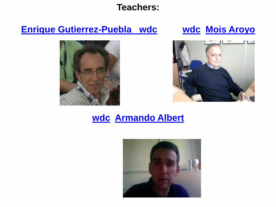
Teachers:
Enrique Gutierrez-Puebla wdc wdc Mois Aroyo
wdc Armando Albert

General Introduction to the module 02
Coffee Break
2-dimensions periodicity
Workshop on 2-D periodicity I
Lunch
Workshop on 2-D periodicity II
Open discussion
22.10.2012

3-dimensions periodicity I
Coffee Break
3-dimensions periodicity II
Workshop on 3-D periodicity I
Lunch
Workshop on 3-D periodicity II
Open discussion
23.10.2012
24.10.2011
Worshop on Space Groups I
Coffee Break
Worshop on Space Groups II
Crystal lattices I
Lunch
Crystal lattices II
Open discussion
25.10.2011
Crystal lattices III
Workshop on Crystal lattices I
Coffee Break
Workshop on Crystal lattices II
Lunch
Personal or Group Tutorials
Open discussion

26.10.2011
Math Review
Coffee Break
International Tables for Crystallography
Lunch
Workshop on Symmetry. International Tables I
29.10.2011
Workshop on Symmetry. International Tables II
Group-subgroup relations and lattice transformations I
Coffee Break
Group-subgroup relations and lattice transformations II
Lunch
Introduction to Reciprocal Space

30.10.2012
Single Crystal, Powder and Twins
Atoms, ions, molecules and macromolecules
Coffee Break
Bonds and interactions in the solid state
Conformations and chirality: Natural products and
Macromolecules
Lunch
Personal or Group Tutorials
Final Remarks


The first x-ray diffraction pattern (1912) von Laue, was the first person to perform a diffraction experiment with a crystal. He hypothesized that if atoms really existed, and if x-rays really were waves, then the wavelength of x-rays would approximate the distance between atoms in a crystal and diffraction would be observed. His brilliant insight thus proved both atomicity and the wave character of x-rays. This remarkable achievement was recognized by the Nobel prize. Laue did not actually do the experiment himself. Rather, he persuaded a couple of graduate students to do the experiment for him. Laue then set an example that has inspired PIs ever since – he was given all the credit!
W. L. (Lawrence) Bragg realized that von Laue’s diffraction pattern could be modeled as reflection from Miller planes -- i.e. the angle of incidence with Miller planes = the angle of reflection. This is the reason why diffracted x-rays are generally called reflections. Bragg published this work in 1913. His insight further substantiated the wave character of x-rays (thereby contradicting his father’s theory that x-rays are particles). This work also allowed the Bragg’s (father and son) to determine the first atomic resolution structures. The Bragg’s were jointly awarded the Nobel prize in 1915 – at which time W.L. Bragg was 25 years old and fighting in the trenches of world war I.
The first x-ray diffraction
picture, which was taken
from a crystal of copper
sulfate by von Laue’s
students, and dubbed the
“beerstein” pattern.

Crick & Watson 1953, published the double helical model for DNA based upon knowledge of chemical structure and geometry, possible chemical interactions, and fiber diffraction patterns. In outline, this is essentially the experimental approach of structure determination by x-ray crystallography. The structure immediately suggested the basis for replication and spurred, but did not illuminate, efforts to decipher the genetic code and mechanism of protein synthesis.
This reflects a common theme in structural biology. Function is most easily inferred when looking at the structure of a relevant complex.
DNA Crystallography is analogous to fiber diffraction
Photo 51 taken by Raymond Gosling under the supervision of Rosalind Franklin in 1952

Electron density is calculated from the diffraction pattern.
Structure determination by x-ray crystallography is analogous to light microscopy.
object
image
lens
Except that there is no lens that can focus x-rays. So we record the scattered x-rays and do the mathematical equivalent of focusing (electron density equation).
Electron Density equation.
Crystal = trillions of copies of the object.
Diffraction (scattering) pattern -- can’t be focused.
Data collection
Electron density map (image) and model.

Resolution of a Reflection. Distance between planes (Å) The term resolution is used to describe the details of features that can be seen in an image. It has a precise meaning in crystallography. It is determined by the angle through which x-rays are scattered with respect to the incident beam. The higher the scattering angle the higher the resolution and the more detail that can be visualized. The resolution of a data set is typically defined as the point at which the intensity of half of the reflections falls to less than 2 times their standard deviation.
Detector
High resolution reflection. (usually weaker)
Low resolution reflection. (usually stronger)

The electron density equation The electron density equation is used to calculate a focused image from the scattered x-rays, or diffraction pattern. This is a summation, in which the thousands of scattered x-rays are added together in a Fourier transform.
(xyz) = (1/V) |F(hkl)| exp[-2i(hx+ky+lz)-(hkl)]
• x, y, z = position in the crystal.
• (xyz) = electron density at the position x, y, z. Has units of e/Å3.
• V = volume of the unit cell
• h, k, l = position in diffraction space -- each of the diffracted rays has a coordinate.
• |F(hkl)| = Structure Factor Amplitude = Square root of the intensity of the diffracted x-ray at
position h, k, l. Has units of electrons (e).
• 2 = Just a constant. No big deal.
• i = square root of -1. Just a mathematical device to indicate direction. Because the electron
density equation is a summation of vectors, we need to indicate the direction of each vector.
No big deal.
• (hkl) = phase of the reflection at position h, k, l.
Amplitude
F
Origin
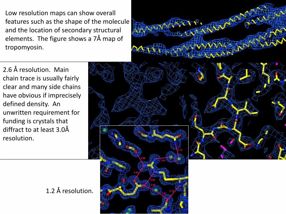
Low resolution maps can show overall features such as the shape of the molecule and the location of secondary structural elements. The figure shows a 7Å map of tropomyosin.
2.6 Å resolution. Main chain trace is usually fairly clear and many side chains have obvious if imprecisely defined density. An unwritten requirement for funding is crystals that diffract to at least 3.0Å resolution.
1.2 Å resolution.
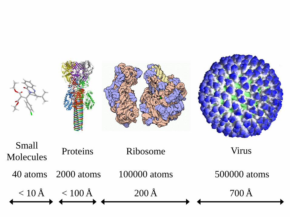
< 10 Å
40 atoms
Small
Molecules
< 100 Å
2000 atoms
Proteins
200 Å
100000 atoms
Ribosome
700 Å
500000 atoms
Virus

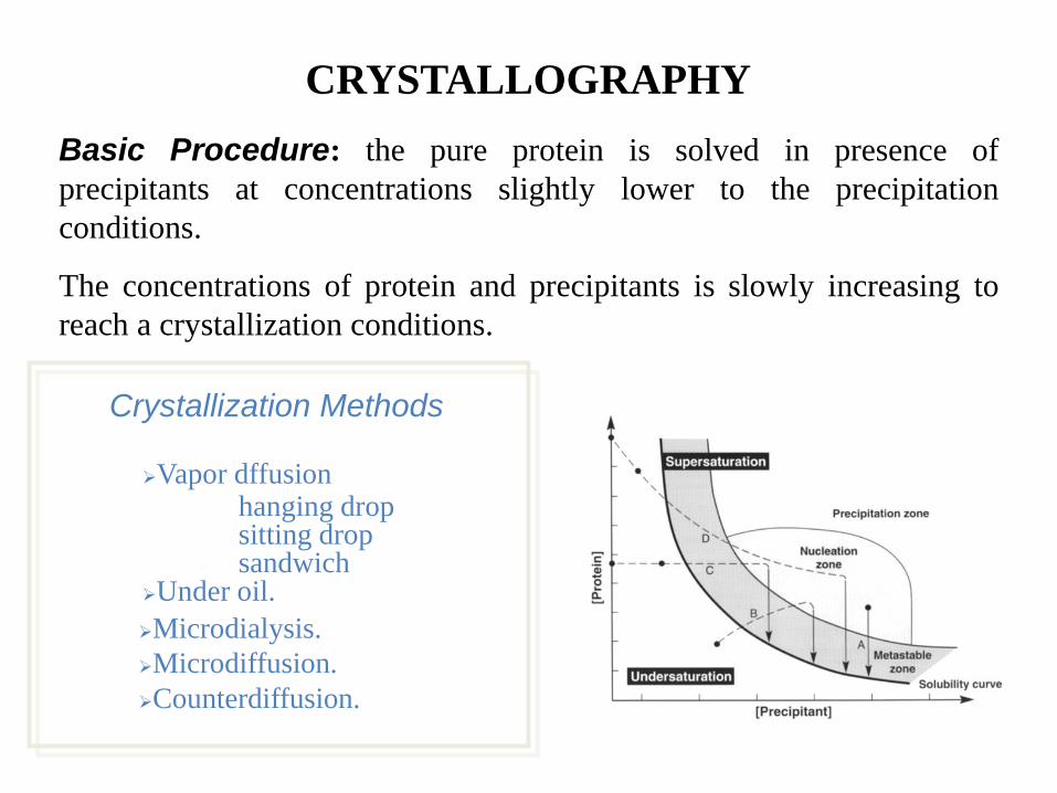
CRYSTALLOGRAPHY
Substance Data Crystals Structure Phases

CRYSTALLOGRAPHY
Substance Data
Crystals
Structure Phases
CRYSTALLOGRAPHY
Basic Procedure: the pure protein is solved in presence of
precipitants at concentrations slightly lower to the precipitation
conditions.
The concentrations of protein and precipitants is slowly increasing to
reach a crystallization conditions.
Crystallization Methods
Vapor dffusion hanging drop sitting drop sandwich Under oil.
Microdialysis.
Microdiffusion.
Counterdiffusion.

CRYSTALLOGRAPHY
Substance Crystals Structure Phases
Data

X RAY SOURCES
X Ray Tubes 106 fotons
Rotating Anode 109 fotons
Synchrotron 1019 fotons
Old X Ray Tube Synchrotron (European Synchrotron Radiation Facility, ESRF)

DIFFRACTION EXPERIMENT

CRYSTALLOGRAPHY
Substance Data Crystals Structure
Phases

FROM DIFFRACTION DATA TO
ELECTRON DENSITY
F T
F(x,y,z) = f(hkl)e d(hkl) i(xyz)(hkl)
FT
FT

THE PHASE PROBLEM

|Fp(h)|
Acentric Centric
|Fp(h)| |Fp(h)|
Argand Diagram – Complex Plane
THE PHASE PROBLEM

CRYSTALLOGRAPHY
Substance Data Crystals Phases
ESTRUCTURA

Jerome Karle Born New York City 18 June 1918
Herbert A. Hauptman Born New York City 14 February 1917 - October 23, 2011, Buffalo
Nobel Prize in Chemistry (1985) "for their outstanding achievements in the
development of direct methods for the determination of crystal structures”
Herbert A. Hauptman and Jerome Karle
Made structure solution generally accessible to non experts
ONE of the 27 Nobel Prizes Awarded by Crystallographers

Nobel Prize in Chemistry (1988)
"for the determination of the three-dimensional structure of a photosynthetic reaction centre"
Johann Deisenhofer, Robert Huber, Hartmut Michel
Understanding of the photosynthetic mechanism
The first high-resolution structure of a membrane protein and also the most
complex molecular structure which had been solved up to that moment (1984).
Membrane protein crystallisation (1980)

Order and disorder – The master of analogies
Nobel Prize in Physics (1991) "for discovering that methods developed for
studying order phenomena in simple systems can be generalized to more complex forms of matter, in
particular to liquid crystals and polymers"
Pierre-Gilles de Gennes
In very different systems such as liquid crystal, ferromagnet, superconductor or polymer, universal features can be identified and be
explained by simple scaling laws

Order and disorder – The master of analogies

Georges Charpak Born 1924 Dabrovica, Poland. Died 29 September 2010, Paris, France
Nobel Prize in Physics (1992) "for his invention and development of particle
detectors, in particular the multiwire proportional chamber"
Georges Charpak
In the multiwire proportional chamber each wire acts as a detector. The important breakthrough was mainly
due to the enormous increase in data-taking rate.

“for pioneering contributions to the development of neutron scattering
techniques for studies of condensed matter”
Bertram N. Brockhouse and Clifford G. Shull
Nobel Prize in Physics (1994)
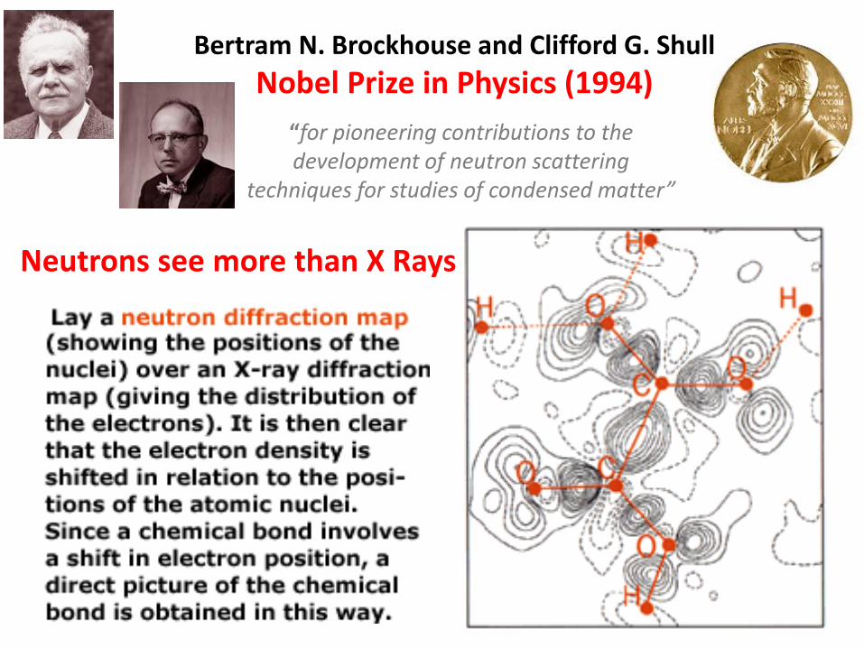
“for pioneering contributions to the development of neutron scattering
techniques for studies of condensed matter”
Bertram N. Brockhouse and Clifford G. Shull
Nobel Prize in Physics (1994)
Neutrons see more than X Rays

“for their elucidation of the enzymatic mechanism underlying the synthesis of adenosine
triphosphate (ATP)” and “ for the first discovery of an ion-transporting enzyme, Na + , K + -ATPase”
Paul D. Boyer, John E. Walker and Jens C. Skou
Nobel Prize in Chemistry (1997)
A molecular machine for ATP synthesis was discovered when
the enzyme ATP synthase was crystallized.

“for discoveries concerning channels in cell membranes ”
Peter Agre and Roderick MacKinnon
Nobel Prize in Chemistry (2003)
Unravelling the secrets of cell channels
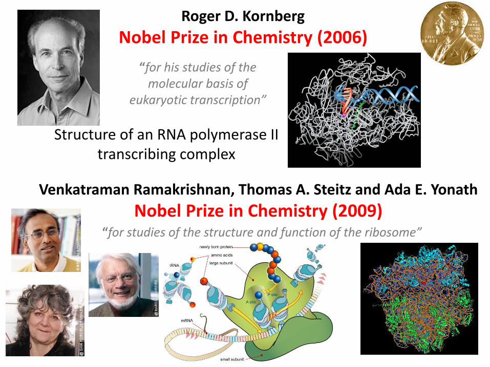
“for his studies of the molecular basis of
eukaryotic transcription”
Roger D. Kornberg
Nobel Prize in Chemistry (2006)
Structure of an RNA polymerase II transcribing complex
“for studies of the structure and function of the ribosome”
Venkatraman Ramakrishnan, Thomas A. Steitz and Ada E. Yonath
Nobel Prize in Chemistry (2009)

“for the discovery of quasicrystals”
Dan Shechtman
Nobel Prize in Chemistry (2011)
Where are the atoms? A 3 dimensional direct-space approach (tiling models) is usually used in combination with a 6 dimensional reciprocal-space approach.
The IUCr had to change the deffinition of crystal : A material is a crystal if it has essentially a sharp diffraction pattern
Direct methods for phasing diffraction data for periodic structures are not directly transferrable to higher dimensional problem; the development of novel method is needed.

“for studies of G-protein-coupled receptors”
Robert J. Lefkowitz, Brian K. Kobilka
Nobel Prize in Chemistry (2012)
A combination of several biochemical strategies produced a ternary complex suitable for crystallization and its high
resolution structure was finally determined.
G-protein-coupled receptors mediate a wide range of physiological signals from the outside of the cell.

“for studies of G-protein-coupled receptors”
Robert J. Lefkowitz, Brian K. Kobilka
Nobel Prize in Chemistry (2012)
When a hormone, olfactory molecule or a taste molecule couples with a receptor on the cell surface, a
chain of reactions inside the cell is triggered
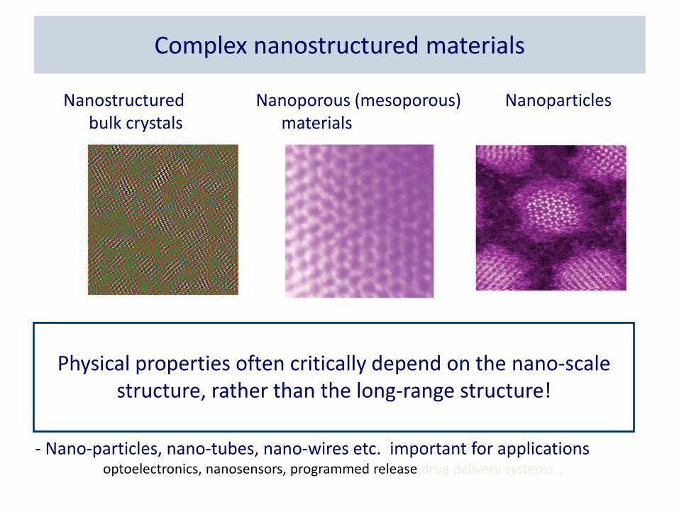
- Complex bulk systems with interesting physical properties are often inhomogeneous on a nanometer lengthscale
high-temperature superconductors, colossal magnetoresistive materials,
high performance thermoelectric materials …
- Nano-particles, nano-tubes, nano-wires etc. important for applications optoelectronics, nanosensors, programmed release drug delivery systems…
Physical properties often critically depend on the nano-scale structure, rather than the long-range structure!
Complex nanostructured materials
Nanoporous (mesoporous) materials
Nanoparticles Nanostructured bulk crystals

Example: Ho2(Ti2-xHox)O7-x/2 ”stuffed spin ice”
300K neutron diffraction patterns (GPPD, IPNS, Argonne)
x=0.00
x=0.30
x=0.50
x=0.67
pyrochlore fluorite
x=0.3
Crystallography challenged: materials w/ disorder C
HA
LLEN
GE
Rietveld approach assumption: crystals are perfectly periodic…
…but this is not always the case!

Crystallography challenged: nano-crystals C
HA
LLEN
GE
Figures: J.S.O. Evans et al.
and M. Tucker et al.
Bragg peak info ONLY
Rietveld method
in powders

Global Approach FU
TUR
E
• Add complementary information
– Extra experimental data
– Theoretical constraints
Science, 316, 561 (2007).