Systems/Circuits EnhancedGABAA ... · PDF fileDOI:10.1523/JNEUROSCI.5054-14.2015 ... (115 dB...
12
Systems/Circuits Enhanced GABA A -Mediated Tonic Inhibition in Auditory Thalamus of Rats with Behavioral Evidence of Tinnitus Evgeny A. Sametsky, Jeremy G. Turner, Deb Larsen, Lynne Ling, and Donald M. Caspary Department of Pharmacology, School of Medicine, Southern Illinois University, Springfield, Illinois 62794 Accumulating evidence suggests a role for inhibitory neurotransmitter dysfunction in the pathology of tinnitus. Opposing hypotheses proposed either a pathologic decrease or increase of GABAergic inhibition in medial geniculate body (MGB). In thalamus, GABA mediates fast synaptic inhibition via synaptic GABA A receptors (GABA A Rs) and persistent tonic inhibition via high-affinity extrasynaptic GABA A Rs. Given that extrasynaptic GABA A Rs control the firing mode of thalamocortical neurons, we examined tonic GABA A R currents in MGB neurons in vitro, using the following three groups of adult rats: unexposed control (Ctrl); sound exposed with behavioral evidence of tinnitus (Tin); and sound exposed with no behavioral evidence of tinnitus (Non-T). Tonic GABA A R currents were evoked using the selective agonist gaboxadol. Months after a tinnitus-inducing sound exposure, gaboxadol-evoked tonic GABA A R currents showed sig- nificant tinnitus-related increases contralateral to the sound exposure. In situ hybridization studies found increased mRNA levels for GABA A R -subunits contralateral to the sound exposure. Tin rats showed significant increases in the number of spikes per burst evoked using suprathreshold-injected current steps. In summary, we found little evidence of tinnitus-related decreases in GABAergic neu- rotransmission. Tinnitus and chronic pain may reflect thalamocortical dysrhythmia, which results from abnormal theta-range resonant interactions between thalamus and cortex, due to neuronal hyperpolarization and the initiation of low-threshold calcium spike bursts (Walton and Llina ´s, 2010). In agreement with this hypothesis, we found tinnitus-related increases in tonic extrasynaptic GABA A R currents, in action potentials/evoked bursts, and in GABA A R -subunit gene expression. These tinnitus-related changes in GABAergic function may be markers for tinnitus pathology in the MGB. Key words: auditory; GABA; GABA A R; thalamus; tinnitus Introduction Inhibitory neurotransmission is essential for accurate processing of acoustic information. It modulates input/output functions, alters frequency tuning, and sharpens temporal response accu- racy while gating neurotransmission to primary auditory cortex (AI; Wang et al., 2008; Edeline, 2011; Richardson et al., 2012). Downregulation of inhibitory function has been suggested to un- derpin tinnitus pathology in dorsal cochlear nucleus (DCN; Wang et al., 2009; Pilati et al., 2012a; Pilati et al., 2012b), inferior colliculus (IC; Bauer et al., 2008; Dong et al., 2009), and AI (Noren ˜a and Eggermont, 2003; Yang et al., 2007, 2011; Roberts et al., 2012) where tinnitus is accompanied by increased spontane- ous activity, bursting, enhanced sound-evoked responses, and reduction in markers of inhibitory neurotransmission (Egger- mont and Roberts, 2004; Middleton et al., 2011). This downregu- lation of inhibitory function is thought to reflect altered homeostatic plasticity compensating for the loss of peripheral excitatory drive (Noren ˜a, 2011; Richardson et al., 2012). Few studies have examined tinnitus pathophysiology in audi- tory thalamus/medial geniculate body (MGB). MGB gates the percept of sound as it projects to AI (Banks and Smith, 2011) and limbic structures, which are thought to be involved in individuals most disturbed by their tinnitus (Rauschecker et al., 2010; Leaver et al., 2011). While MGB was historically considered a simple relay of auditory signals between IC and AI, recent studies suggest additional signal processing in MGB (Bartlett and Smith, 1999; Winer et al., 2005; Edeline, 2011; Bartlett, 2013). MGB has intri- cate reciprocal connectivity with the GABAergic thalamic reticu- lar nucleus (TRN) that regulates attention and sleep (Jones, 1975; Rouiller et al., 1985). The unique connectivity and function of MGB strongly implicates it as a key link in the tinnitus network (Rouiller et al., 1985; Rauschecker et al., 2010; Leaver et al., 2011). Data from sodium salicylate models of tinnitus suggest altered excitation and inhibition in rat MGB (Su et al., 2012). Proper functioning of MGB is dependent on normal GABAergic inhibi- tion, which in sensory thalamus may account for 90% of total inhibitory conductance (Belelli et al., 2005; Cope et al., 2005, 2009). In rodents, MGB receives GABAergic projections from IC (Winer et al., 1996; Peruzzi et al., 1997; Ito and Oliver, 2012) and Received Dec. 12, 2014; revised March 31, 2015; accepted May 12, 2015. Author contributions: E.A.S., J.G.T., and D.M.C. designed research; E.A.S., D.L., and L.L. performed research; E.A.S., J.G.T., D.L., L.L., and D.M.C. analyzed data; E.A.S., L.L., and D.M.C. wrote the paper. This research was supported by Office of Naval Research Grant N00014-12-1-0214 and National Institute on Deafness and Other Communication Disorders Grant DC-000151. We thank Drs. Tom Brozoski, Brandon Cox, and Steve Sandstrom for their careful reading and comments on the manuscript. J.G.T. is the cofounder of OtoScience Labs, which conducts hearing and tinnitus research, and licenses the gap- based testing technology from Southern Illinois University School of Medicine. The authors declare no other com- peting financial interests. Correspondence should be addressed to Donald M. Caspary, Southern Illinois University, School of Medicine, Department of Pharmacology, 801 North Rutledge Street, P.O. Box 19629, Springfield, IL 62702. E-mail: [email protected]. DOI:10.1523/JNEUROSCI.5054-14.2015 Copyright © 2015 the authors 0270-6474/15/359369-12$15.00/0 The Journal of Neuroscience, June 24, 2015 • 35(25):9369 –9380 • 9369
Transcript of Systems/Circuits EnhancedGABAA ... · PDF fileDOI:10.1523/JNEUROSCI.5054-14.2015 ... (115 dB...
Systems/Circuits
Enhanced GABAA-Mediated Tonic Inhibition in AuditoryThalamus of Rats with Behavioral Evidence of Tinnitus
Evgeny A. Sametsky, Jeremy G. Turner, Deb Larsen, Lynne Ling, and Donald M. CasparyDepartment of Pharmacology, School of Medicine, Southern Illinois University, Springfield, Illinois 62794
Accumulating evidence suggests a role for inhibitory neurotransmitter dysfunction in the pathology of tinnitus. Opposing hypothesesproposed either a pathologic decrease or increase of GABAergic inhibition in medial geniculate body (MGB). In thalamus, GABA mediatesfast synaptic inhibition via synaptic GABAA receptors (GABAARs) and persistent tonic inhibition via high-affinity extrasynapticGABAARs. Given that extrasynaptic GABAARs control the firing mode of thalamocortical neurons, we examined tonic GABAAR currentsin MGB neurons in vitro, using the following three groups of adult rats: unexposed control (Ctrl); sound exposed with behavioral evidenceof tinnitus (Tin); and sound exposed with no behavioral evidence of tinnitus (Non-T). Tonic GABAAR currents were evoked using theselective agonist gaboxadol. Months after a tinnitus-inducing sound exposure, gaboxadol-evoked tonic GABAAR currents showed sig-nificant tinnitus-related increases contralateral to the sound exposure. In situ hybridization studies found increased mRNA levels forGABAAR �-subunits contralateral to the sound exposure. Tin rats showed significant increases in the number of spikes per burst evokedusing suprathreshold-injected current steps. In summary, we found little evidence of tinnitus-related decreases in GABAergic neu-rotransmission. Tinnitus and chronic pain may reflect thalamocortical dysrhythmia, which results from abnormal theta-range resonantinteractions between thalamus and cortex, due to neuronal hyperpolarization and the initiation of low-threshold calcium spike bursts(Walton and Llinas, 2010). In agreement with this hypothesis, we found tinnitus-related increases in tonic extrasynaptic GABAARcurrents, in action potentials/evoked bursts, and in GABAAR �-subunit gene expression. These tinnitus-related changes in GABAergicfunction may be markers for tinnitus pathology in the MGB.
Key words: auditory; GABA; GABAAR; thalamus; tinnitus
IntroductionInhibitory neurotransmission is essential for accurate processingof acoustic information. It modulates input/output functions,alters frequency tuning, and sharpens temporal response accu-racy while gating neurotransmission to primary auditory cortex(AI; Wang et al., 2008; Edeline, 2011; Richardson et al., 2012).Downregulation of inhibitory function has been suggested to un-derpin tinnitus pathology in dorsal cochlear nucleus (DCN;Wang et al., 2009; Pilati et al., 2012a; Pilati et al., 2012b), inferiorcolliculus (IC; Bauer et al., 2008; Dong et al., 2009), and AI(Norena and Eggermont, 2003; Yang et al., 2007, 2011; Roberts etal., 2012) where tinnitus is accompanied by increased spontane-ous activity, bursting, enhanced sound-evoked responses, and
reduction in markers of inhibitory neurotransmission (Egger-mont and Roberts, 2004; Middleton et al., 2011). This downregu-lation of inhibitory function is thought to reflect alteredhomeostatic plasticity compensating for the loss of peripheralexcitatory drive (Norena, 2011; Richardson et al., 2012).
Few studies have examined tinnitus pathophysiology in audi-tory thalamus/medial geniculate body (MGB). MGB gates thepercept of sound as it projects to AI (Banks and Smith, 2011) andlimbic structures, which are thought to be involved in individualsmost disturbed by their tinnitus (Rauschecker et al., 2010; Leaveret al., 2011). While MGB was historically considered a simplerelay of auditory signals between IC and AI, recent studies suggestadditional signal processing in MGB (Bartlett and Smith, 1999;Winer et al., 2005; Edeline, 2011; Bartlett, 2013). MGB has intri-cate reciprocal connectivity with the GABAergic thalamic reticu-lar nucleus (TRN) that regulates attention and sleep (Jones, 1975;Rouiller et al., 1985). The unique connectivity and function ofMGB strongly implicates it as a key link in the tinnitus network(Rouiller et al., 1985; Rauschecker et al., 2010; Leaver et al., 2011).
Data from sodium salicylate models of tinnitus suggest alteredexcitation and inhibition in rat MGB (Su et al., 2012). Properfunctioning of MGB is dependent on normal GABAergic inhibi-tion, which in sensory thalamus may account for �90% of totalinhibitory conductance (Belelli et al., 2005; Cope et al., 2005,2009). In rodents, MGB receives GABAergic projections from IC(Winer et al., 1996; Peruzzi et al., 1997; Ito and Oliver, 2012) and
Received Dec. 12, 2014; revised March 31, 2015; accepted May 12, 2015.Author contributions: E.A.S., J.G.T., and D.M.C. designed research; E.A.S., D.L., and L.L. performed research;
E.A.S., J.G.T., D.L., L.L., and D.M.C. analyzed data; E.A.S., L.L., and D.M.C. wrote the paper.This research was supported by Office of Naval Research Grant N00014-12-1-0214 and National Institute on
Deafness and Other Communication Disorders Grant DC-000151. We thank Drs. Tom Brozoski, Brandon Cox, andSteve Sandstrom for their careful reading and comments on the manuscript.
J.G.T. is the cofounder of OtoScience Labs, which conducts hearing and tinnitus research, and licenses the gap-based testing technology from Southern Illinois University School of Medicine. The authors declare no other com-peting financial interests.
Correspondence should be addressed to Donald M. Caspary, Southern Illinois University, School of Medicine,Department of Pharmacology, 801 North Rutledge Street, P.O. Box 19629, Springfield, IL 62702. E-mail:[email protected].
DOI:10.1523/JNEUROSCI.5054-14.2015Copyright © 2015 the authors 0270-6474/15/359369-12$15.00/0
The Journal of Neuroscience, June 24, 2015 • 35(25):9369 –9380 • 9369
TRN (Montero and Scott, 1983; Rouiller et al., 1985), whileGABAergic interneurons comprise �1% of MGB neurons(Winer and Larue, 1996). GABAergic inputs to MGB act via post-synaptic GABAA receptors (GABAARs) that are heterogeneous(Sur et al., 1999; Richardson et al., 2012). MGB synapses expresslow-affinity, fast-desensitizing GABAARs (�1, �2, �2 subunits)that mediate fast synaptic transmission and enable high temporalprecision (Mozrzymas, 2004, 2010; Farrant and Nusser, 2005;Barberis et al., 2011). In addition, MGB expresses high levels ofGABAARs containing �4- and �-subunits that mediate non-desensitizing tonic inhibition (Richardson et al., 2011). The pres-ence of the �-subunit in GABAARs results in extrasynapticlocation and high agonist affinity, which favors activation bylow ambient levels of GABA in extracellular space, which mayarise from synaptic spillover and/or release from glia (Farrantand Nusser, 2005; Brickley and Mody, 2012). These non-desensitizing GABAARs can generate long-lasting tonic currentsthat hyperpolarize and shift thalamocortical neurons from tonicto burst-firing mode. This shift could alter the salience of phan-tom acoustic signals to AI.
GABAergic inhibition is critically involved in determininghigher-frequency thalamocortical oscillations as well as slow-wave sleep (Llinas and Steriade, 2006). Llinas et al. (2005) postu-lated that excessive inhibition in thalamus could be the origin ofpathologically increased hyperpolarization of thalamic neurons,resulting in deinactivation of T-type Ca 2� channels. Upon depo-larization, these channels generate low-threshold burst spikesin the wakeful state. A proposed thalamocortical dysrhythmiamodel suggests that elevated inhibition and the resultant focalburst activity associated with abnormal low-frequency oscilla-tions in thalamus produce ectopic disinhibition of fast oscillatoryactivity in AI that could correlate with tinnitus (Llinas and Ste-riade, 2006).
Using in vitro patch-clamp recordings, we sought to clarify therole of GABAA inhibition in MGB as it relates to tinnitus patho-physiology in a sound-exposure behavioral model of tinnitus. Incontrast to findings from other auditory structures, MGB circuitsshowed an increase in GABAAR function in animals with behav-ioral evidence of tinnitus.
Materials and MethodsYoung-adult (3– 4 months) Long–Evans male rats (Harlan Laboratories)were housed on a 12 h reversed light/dark cycle with ad libitum access tofood and water. Forty-five Long–Evans rats were used for electrophysio-logical recordings in the MGB slice preparation and 13 Fischer BrownNorway (FBN) rats from earlier tinnitus experiments (Wang et al., 2009)were used for MGB in situ hybridization studies. Both strains have similarlife expectancy, with young adult animals used in both studies.
All experiments were conducted in strict accordance with a protocolapproved by the Animal Care and Use Committee of Southern IllinoisUniversity.
Noise exposure and induction of tinnitus. Tinnitus was induced between3 and 4 months of age, using methods similar to those described by Baueret al. (1999) and Bauer and Brozoski (2001). Briefly, acoustic exposurewas a 16 kHz octave band at 116 dB SPL into one ear of a ketamine/xylazine anesthetized rat for 1 h. This exposure typically results in anipsilateral 30 – 40 dB temporary threshold shift [measured as pre-to-postnoise exposure shift in auditory brainstem response (ABR) thresholds]across frequencies, with increased threshold elevations at frequenciesnear the 16 kHz exposure (Wang et al., 2009). The exposure also pro-duces a small, variable, permanent ipsilateral threshold shift as measuredby ABR (Wang et al., 2009). Acoustic nerve fiber losses were not mea-sured in this set of sound-exposed rats (Bauer et al., 2007; Kujawa andLiberman, 2009). Prepulse inhibition (PPI) was assessed in parallel withgap detection performance. ABR and prepulse findings suggest that gap
detection was not altered by unilateral hearing loss at the levels used inthese studies (Turner et al., 2006).
Behavioral testing. The gap detection reflex method was adapted fromTurner et al. (2006) and Turner (2007), and was based upon the ability ofthe acoustic startle reflex to be reduced when preceded by a silent gap ina constant acoustic background. As detailed in Wang et al. (2009), be-havioral testing used Kinder Scientific startle reflex hardware and soft-ware, which was customized for this application. Background soundswere presented through one speaker (XT25TG30-04, Vifa) with startlestimuli presented through a second speaker (CTS KSN-1005, Power-Line). A piezoelectric transducer provided the measure of startle forceapplied to the floor. An animal holder was suspended above the floor,allowing the animal to freely turn around while minimizing excessivemovement. The sound-gap envelope consisted of 60 dB peak (SPL)broadband noise (BBN) or filtered noise (one-third octave bandpass, 48dB/octave roll off, Model 3988, Krohn-Hite) centered at 10, 12.5, 16, 20,and 25 kHz, calibrated at 60 dB peak SPL. PPI was measured in parallel asa hearing level control and to test for hyperacusis (Turner and Parrish,2008). Animals with threshold elevation and/or showing hyperacusiswere eliminated from the study. Two months after noise exposure, ani-mals were behaviorally tested twice on different occasions, and the resultsof two sessions were averaged to determine tinnitus. Gap/PPI test ses-sions began with a 1 min acclimation period followed by two trials con-sisting of the startle-eliciting noise burst (115 dB SPL, 20 ms duration),which served to habituate the startle response to a more stable baseline.Data from these two initial trials were not used in the gap detectionanalysis. The remainder of the session consisted of startle-only trialsmixed with gap or PPI trials (Turner et al., 2006; Kalappa et al., 2014).
The intertrial interval varied throughout test sessions, with targetedfrequencies and BBN stimuli for both gap and PPI presented in ascendingorder from 10 kHz to BBN (380 trials per session). Gap and prepulsestimuli occurred 100 ms before the startle stimulus and were 50 ms longwith a 1.0 ms rise/fall time (Turner et al., 2006). Acoustic stimuli werecalibrated using a cloth model rat and a Bruel & Kjær Pulse System witha 0.5 inch free-field microphone (model 4191, Bruel & Kjær). Ambientsound levels in test chambers (with background test noise turned off)were �20 dB SPL in the 4 – 40 kHz range.
Tinnitus determination. Data from gap detection and PPI testing wereexpressed as the ratio of mean startle response for a gap or PPI trialrelative to a proximal startle-only trial (for details, see Kalappa et al.,2014). A response of 1.0 meant that the startle reflex was the samewhether or not it was preceded by a gap or prepulse stimulus. The lowerthe ratio is from 1.0, the greater the startle inhibition. An individualsubject’s average data from the two behavioral test sessions were in-spected for freedom from artifacts, such as a high level of ambulatorymovement. Tinnitus index scores were calculated by subtracting the PPIratio from the gap ratio. Higher scores are indicative of tinnitus-likedeficits in processing silent gap cues. This index score provides a metricthat describes the response of an animal to silent gap cues relative tocontrol sounds and helps control for several potential confounds in thegap ratio as a standalone metric for tinnitus. The index score helps tocontrol for hearing loss as an inability to hear or process the 60 dB teststimulus, which would result both in poor gap ratios and poor PPI ratios,hence a small score. The index also helps to control for startle reflexamplitude differences. If an animal has such a small startle reflex that itcannot be inhibited by a silent gap cue, the same impact would be foundin the PPI testing where small startle reflexes would also be expected,again resulting in a small index score. This metric controls for temporalprocessing deficits, which should be present in both the gap and PPIconditions, given their identical timing features. Finally, this index cor-rects for nonspecific sensory gating/filtering failures, which should im-pact both gap and PPI response ratios. Mean tinnitus index scores forcontrol animals were pooled to obtain the average control group startleratio, with 95% confidence intervals determined for each frequency andBBN.
The average tinnitus index score for each noise-exposed subject wascompared with the average control tinnitus index score for that fre-quency. Noise-exposed subject responses that fell within the 95% confi-dence interval for the control group were deemed no different from those
9370 • J. Neurosci., June 24, 2015 • 35(25):9369 –9380 Sametsky et al. • GABAA Inhibition and Tinnitus in Auditory Thalamus
of control subjects and were without tinnitus for that given frequency orBBN. Noise-exposed subjects with tinnitus index scores reduced beyondthe 95% confidence interval of the companion control group were con-sidered significantly different. These subjects were considered to havetinnitus at that given frequency or BBN.
In situ hybridization. Adult (4 months old) male FBN rats were usedfor in situ hybridization experiments. The animals were repurposed froma prior series of similarly conducted tinnitus experiments with the iden-tical sound exposure conditions. Previously, we published patch-clamprecordings of gaboxadol (GBX)-evoked tonic GABAAR currents fromMGB neurons in adult FBN rats (Richardson et al., 2011, 2013). Therecorded parameters of gaboxadol-evoked tonic GABAAR currents inFBN rats were comparable to those presented here for Long–Evans rats.Animals were divided into the following two groups: sound exposed andcontrol/sham exposed. These animals were not assigned a tinnitus score.In situ hybridization studies were conducted as described by Ling et al.(2005). Oligonucleotide probes for �4 (Wisden et al., 1991; 5�-CAA GTCGCC AGG CAC AGG ACG TGC AGG AGG GCG AGG CTG ACCCCG-3�) and � (provided by Merck Inc.; 5�-GAC CTT GGC TTT CCGTTT CTT CCT GTA GTC AGC ATT-3�) were synthesized and purifiedby Sigma-Genosys. The probes were then 3�-end labeled with 35S-dATP(NEN-PerkinElmer Life Science) by 3�-terminal deoxynucleotidyl trans-ferase using a commercially available kit (Fisher Scientific), and purifiedand desalted through a micro BioSpin-30 column (Bio-Rad). These la-beled probes were used to detect GABAAR subunits �4 and � in MGB.Additional methods, visualization, and quantification were performed asin the study by Richardson et al. (2013).
All slides were blinded to ensure unbiased measurement. The accumu-lation of silver grains over cell bodies (�10 times above slide back-ground) was interpreted as positive hybridization labeling for the �4 and� GABAAR subunits to their specific mRNAs. Only grains within theperimeter of the cell were counted. Data were collected from cells only ifthey were distinguishable as individual cells possessing a nucleolus. Thenumbers of grains over somata of MGB neurons (silver grains/100 �m 2)were counted, and values were corrected by subtracting background la-beling obtained from five random off-tissue areas on each slide.
Preparation of thalamic brain slices for electrophysiology. Two monthsfollowing sound exposure and within 2 weeks of behavioral testing, Lon-g–Evans rats (5–7 months old) were anesthetized with isoflurane anddecapitated. Brains were rapidly removed, and horizontal brain slices(300 �m) were cut using a VT 1000P vibratome (Leica Microsystems) inice-cold (�4°C) sucrose-rich artificial CSF (aCSF) containing the follow-ing (in mM): sucrose 250, KCl 3, NaH2PO4 1.23, MgCl2 5, CaCl2 0.5,NaHCO3 26, and glucose 10, pH 7.4, saturated with carbogen (95% O2/5%CO2). Slices were retained in a holding chamber filled with standardaCSF containing the following (in mM): NaCl 124, KCl 3, KH2PO4 1.25,MgCl2 1.3, CaCl2 2, NaHCO3 26, and glucose 10, pH 7.4, saturated withcarbogen and warmed to 32°C for 20 min before being allowed to equil-ibrate to room temperature (�22°C) for at least 1 h before recording.Slices were transferred to a submersion recording chamber mounted onthe stage of an upright Olympus BX-51WI microscope, maintained atroom temperature, and continuously perfused with standard aCSF satu-rated with carbogen, at a flow rate of 2 ml/min using a peristaltic P720pump (Instech Laboratories).
Electrophysiology. Using infrared differential interference contrast mi-croscopy, visually guided, somatic, whole-cell patch-clamp recordingswere obtained from MGB neurons using fire-polished borosilicate glasselectrodes (4 –5 M�). Two intracellular solutions were used in this study.In voltage-clamp experiments, patch electrodes were filled with the fol-lowing (in mM): 120 CsMeSO3, 2 CsCl, 5 NaCl, 1.5 MgCl2, 10 HEPES, 10EGTA, 5 TEA-Cl, 2.5 Mg-ATP, 0.3 Na-GTP, and 5 QX-314, pH 7.3,adjusted with CsOH, with osmolarity of 290 � 10 mOsm. In current-clamp experiments, a more “physiological” solution was used, as follows:K-gluconate140, NaCl 1, MgCl2 2, Mg-ATP 2, Na-GTP 0.3, and HEPES10, pH 7.3–7.4, adjusted with KOH, with osmolarity of 290 � 10 mOsm.Holding potentials were not corrected for the liquid junction potential.Voltage-clamp and current-clamp recordings were obtained using aMultiClamp-700B amplifier and Digidata-1440A analog-to-digital con-verter (Molecular Devices). The signal was low-pass filtered at 5 kHz and
digitized at 10 kHz. Data were acquired using pClamp 10 software (Mo-lecular Devices). For all recordings, series resistance was �20 M� andwas monitored continuously.
To isolate GABAergic currents, 20 �M 6,7-dinitroquinoxaline-2,3-dione (DNQX) and 50 �M D(�)-2-amino-5-phosphonopentanoic acid(D-AP5) were added to the standard aCSF. Extrasynaptic tonic GABAARcurrents were evoked by bath application of the selective agonist, 0.1–10�M gaboxadol [4,5,6,7-tetrahydroisoxazolo[5,4-c]pyridine-3(2H)-one],in the presence of glutamatergic receptor blockers. GABAA tonic currentsand spontaneous IPSCs (sIPSCs) were recorded at �10 and �20 mV,respectively. GABAA currents were blocked with 40 �M gabazine.
Drugs. Drugs were dissolved in distilled water to a concentration of1–10 mM, and the stock solutions were divided into small aliquots andfrozen. The following stock drug solutions were used to prepare freshfinal solutions every day: QX-314 from Tocris Bioscience; DNQX,D-AP-5, and gabazine from Abcam; with all other chemicals fromSigma-Aldrich.
Analysis. Postrecording analyses of extrasynaptic GABAAR currentsand action potential firing evoked using current steps were analyzedusing pClamp 10. Dose–response curve fitting was performed in Origin 8(OriginLab) using an iterative process to minimize the � 2 test. Sponta-neous IPSCs were analyzed using MiniAnalysis version 6.0.3 software(Synaptosoft). All spontaneous IPSPs were visually verified on the basisof event kinetics. Between 500 and 1500 individual events were analyzedfor each cell (4 min of gap-free acquisition).
All statistical values were evaluated with Origin 8 (OriginLab) or SAS/STAT (SAS Institute). Values are presented either as the mean � SEM ormean � SD (box plot). ANOVA with Tukey’s post hoc analysis was usedwhen multiple comparisons were made. Pairwise group differences wereevaluated using a Student’s t test. Statistical significance was establishedat p � 0.05.
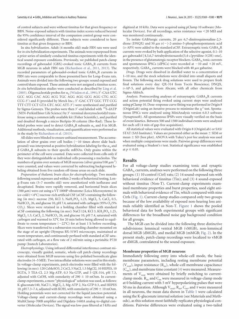
ResultsFor all voltage-clamp studies examining tonic and synapticGABAA currents, analyses were performed on the following threegroups: (1) 10 control (Ctrl) rats; (2) 14 sound-exposed rats withbehavioral evidence of tinnitus (Tin); and (3) 4 sound-exposedwithout tinnitus (Non-T). Current-clamp experiments exam-ined membrane properties and burst properties, used eight ani-mals with behavioral evidence of Tin, which compared with nineCtrls (Fig. 1). Current-clamp studies compared only two groupsbecause of the low availability of exposed non-hearing loss ani-mals reliably identified as Non-T. Figure 1 shows the pooledbehavioral data for both experimental groups with significantdifferences for the broadband noise gap background conditionfor all groups.
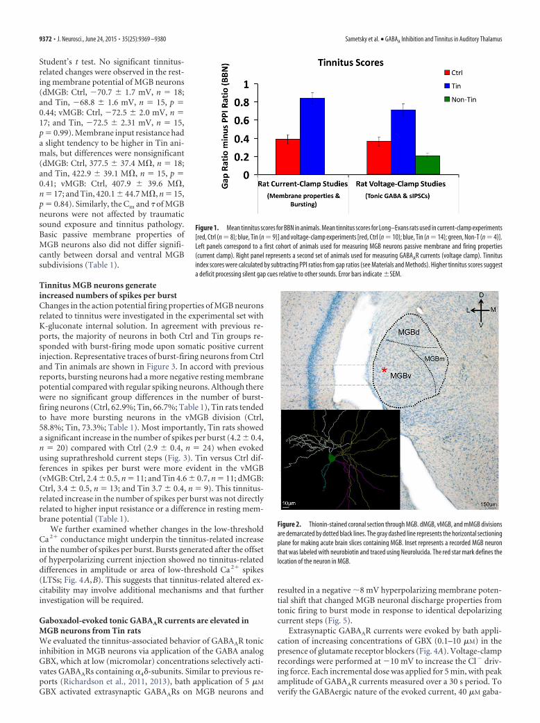
The MGB can be divided into the following three distinctivesubdivisions: lemniscal ventral MGB (vMGB), non-lemniscaldorsal MGB (dMGB), and medial MGB (mMGB; Fig. 2). In thepresent study, patch-clamp recordings were localized to vMGBor dMGB, contralateral to the sound exposure.
Membrane properties of MGB neuronsImmediately following entry into whole-cell mode, the basicmembrane parameters, including resting membrane potential(Vrest), input resistance (Rm), whole-cell membrane capacitance(Cm), and membrane time constant () were measured. Measure-ments of Vrest were obtained by briefly switching to current-clamp mode. Rm and Cm were measured in voltage-clamp modeat 0 holding current with 5 mV hyperpolarizing pulses that were50 ms in duration. Although Vrest, Rm, Cm, and were measuredin all neurons, the mean data shown in Table 1 were calculatedusing the K-gluconate internal solution (see Materials and Meth-ods), as this solution most faithfully replicates physiological con-ditions. Pairwise differences were evaluated using a two-tailed
Sametsky et al. • GABAA Inhibition and Tinnitus in Auditory Thalamus J. Neurosci., June 24, 2015 • 35(25):9369 –9380 • 9371
Student’s t test. No significant tinnitus-related changes were observed in the rest-ing membrane potential of MGB neurons(dMGB: Ctrl, �70.7 � 1.7 mV, n 18;and Tin, �68.8 � 1.6 mV, n 15, p 0.44; vMGB: Ctrl, �72.5 � 2.0 mV, n 17; and Tin, �72.5 � 2.31 mV, n 15,p 0.99). Membrane input resistance hada slight tendency to be higher in Tin ani-mals, but differences were nonsignificant(dMGB: Ctrl, 377.5 � 37.4 M�, n 18;and Tin, 422.9 � 39.1 M�, n 15, p 0.41; vMGB: Ctrl, 407.9 � 39.6 M�,n 17; and Tin, 420.1 � 44.7 M�, n 15,p 0.84). Similarly, the Cm and of MGBneurons were not affected by traumaticsound exposure and tinnitus pathology.Basic passive membrane properties ofMGB neurons also did not differ signifi-cantly between dorsal and ventral MGBsubdivisions (Table 1).
Tinnitus MGB neurons generateincreased numbers of spikes per burstChanges in the action potential firing properties of MGB neuronsrelated to tinnitus were investigated in the experimental set withK-gluconate internal solution. In agreement with previous re-ports, the majority of neurons in both Ctrl and Tin groups re-sponded with burst-firing mode upon somatic positive currentinjection. Representative traces of burst-firing neurons from Ctrland Tin animals are shown in Figure 3. In accord with previousreports, bursting neurons had a more negative resting membranepotential compared with regular spiking neurons. Although therewere no significant group differences in the number of burst-firing neurons (Ctrl, 62.9%; Tin, 66.7%; Table 1), Tin rats tendedto have more bursting neurons in the vMGB division (Ctrl,58.8%; Tin, 73.3%; Table 1). Most importantly, Tin rats showeda significant increase in the number of spikes per burst (4.2 � 0.4,n 20) compared with Ctrl (2.9 � 0.4, n 24) when evokedusing suprathreshold current steps (Fig. 3). Tin versus Ctrl dif-ferences in spikes per burst were more evident in the vMGB(vMGB: Ctrl, 2.4 � 0.5, n 11; and Tin 4.6 � 0.7, n 11; dMGB:Ctrl, 3.4 � 0.5, n 13; and Tin 3.7 � 0.4, n 9). This tinnitus-related increase in the number of spikes per burst was not directlyrelated to higher input resistance or a difference in resting mem-brane potential (Table 1).
We further examined whether changes in the low-thresholdCa 2� conductance might underpin the tinnitus-related increasein the number of spikes per burst. Bursts generated after the offsetof hyperpolarizing current injection showed no tinnitus-relateddifferences in amplitude or area of low-threshold Ca 2� spikes(LTSs; Fig. 4A,B). This suggests that tinnitus-related altered ex-citability may involve additional mechanisms and that furtherinvestigation will be required.
Gaboxadol-evoked tonic GABAAR currents are elevated inMGB neurons from Tin ratsWe evaluated the tinnitus-associated behavior of GABAAR tonicinhibition in MGB neurons via application of the GABA analogGBX, which at low (micromolar) concentrations selectively acti-vates GABAARs containing �4�-subunits. Similar to previous re-ports (Richardson et al., 2011, 2013), bath application of 5 �M
GBX activated extrasynaptic GABAARs on MGB neurons and
resulted in a negative �8 mV hyperpolarizing membrane poten-tial shift that changed MGB neuronal discharge properties fromtonic firing to burst mode in response to identical depolarizingcurrent steps (Fig. 5).
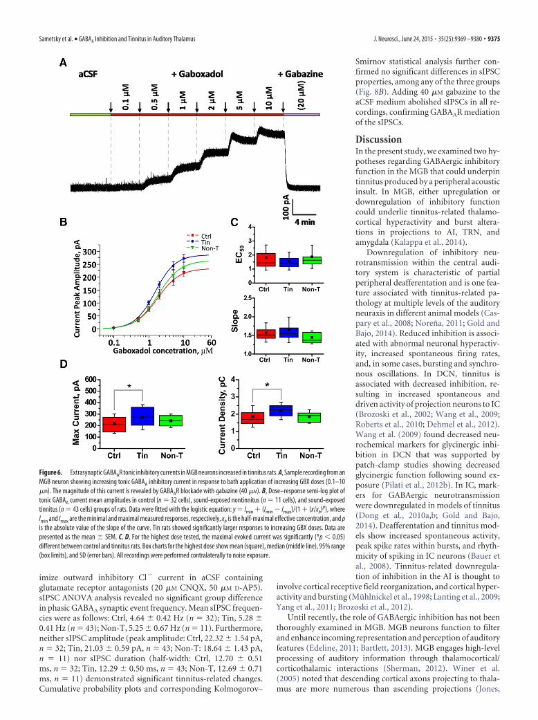
Extrasynaptic GABAAR currents were evoked by bath appli-cation of increasing concentrations of GBX (0.1–10 �M) in thepresence of glutamate receptor blockers (Fig. 4A). Voltage-clamprecordings were performed at �10 mV to increase the Cl� driv-ing force. Each incremental dose was applied for 5 min, with peakamplitude of GABAAR currents measured over a 30 s period. Toverify the GABAergic nature of the evoked current, 40 �M gaba-
Figure 1. Mean tinnitus scores for BBN in animals. Mean tinnitus scores for Long–Evans rats used in current-clamp experiments[red, Ctrl (n 8); blue, Tin (n 9)] and voltage-clamp experiments [red, Ctrl (n 10); blue, Tin (n 14); green, Non-T (n 4)].Left panels correspond to a first cohort of animals used for measuring MGB neurons passive membrane and firing properties(current clamp). Right panel represents a second set of animals used for measuring GABAAR currents (voltage clamp). Tinnitusindex scores were calculated by subtracting PPI ratios from gap ratios (see Materials and Methods). Higher tinnitus scores suggesta deficit processing silent gap cues relative to other sounds. Error bars indicate �SEM.
Figure 2. Thionin-stained coronal section through MGB. dMGB, vMGB, and mMGB divisionsare demarcated by dotted black lines. The gray dashed line represents the horizontal sectioningplane for making acute brain slices containing MGB. Inset represents a recorded MGB neuronthat was labeled with neurobiotin and traced using Neurolucida. The red star mark defines thelocation of the neuron in MGB.
9372 • J. Neurosci., June 24, 2015 • 35(25):9369 –9380 Sametsky et al. • GABAA Inhibition and Tinnitus in Auditory Thalamus

zine, a nonselective GABAAR antagonist, was applied at the endof each GBX dose series. The peak amplitude of the GABAARtonic current was measured with reference to current recordedfollowing gabazine application. Most MGB neurons showed cur-rent saturation at the maximum (10 �M) GBX concentrationused in these experiments. Higher GBX concentrations were notused to avoid concomitant activation of synaptic GABAARs.
We recorded from 32 MGB neurons from 10 Ctrl rats, 43neurons from 14 Tin rats, and 11 neurons from 4 Non-T rats. Allrecordings were contralateral to the sound exposure and wereparsed by subdivision into recordings from dMGB and vMGB(dMGB/vMGB, respectively: Ctrl, 16/16 neurons; Tin, 20/23neurons; Non-T, 5/6 neurons).
Repeated-measures ANOVA revealed significant differencesamong three experimental groups in the dose–response ampli-tude of GABAAR tonic currents, measured with bath applicationof 0.1–10 �M GBX (F 3.15; p 0.048; Fig. 6). There wassignificant interaction between GBX concentrations and groups(p 0.037). Additional t test analysis demonstrated significantincreases in the amplitude of GABAAR tonic currents for the Tingroup compared with the Ctrl group at 1–10 �M GBX concentra-tions (1 �M GBX, p 0.034; 2 �M GBX, p 0.022; 5 �M GBX,p 0.012; 10 �M GBX, p 0.019; Fig. 6). No differences werefound between the Non-T group and either the Ctrl or Tin
groups. Figure 6D shows the significant 25.5% increase in meancurrent amplitude in the Tin group compared with the Ctrl groupat 10 �M GBX (Ctrl, 217.11 � 14.86 pA; Tin, 272.54 � 16.66 pA;Non-T, 259.08 � 20.02 pA). Likewise, a 19.1% greater currentdensity occurred in the Tin group (p 0.02) relative to Ctrl (Ctrl,1.83 � 0.11 pA/pF; Tin, 2.18 � 0.08 pA/pF; Non-T, 2.09 � 0.1pA/pF; Fig. 6E). No significant differences were identified be-tween the Non-T group and either the Ctrl or Tin groups at a 10�M GBX concentration. GABAAR tonic currents were compara-ble in dMGB and vMGB across groups [e.g., the 25% tinnitus-related increase in maximal current was detected in dMGB andvMGB at 10 �M GBX, but only reached significance in dMGB(p 0.046) when comparing current density in the smallerparsed groups].
When the dose–response curves were normalized to the max-imal current recorded with 10 �M GBX, a simple logistic functionwas used to fit dose–response data from which the EC50 andactivation slope for GABAAR tonic current were calculated foreach individual neuron. Importantly, no decline in GABAARfunction was observed for any of the functional parameters ex-amined. No significant group differences were seen for meanEC50 (Ctrl, 2.60 � 0.36 �M; Tin, 1.95 � 0.21 �M; Non-T, 2.29 �0.49 �M) or activation slope (Ctrl, 1.6 � 0.06; Tin, 1.61 � 0.05;Non-T, 1.42 � 0.06; Fig. 6C). Since no group differences werefound in current kinetics, it was concluded that tinnitus pathol-ogy does not substantially alter the subunit makeup of extrasyn-aptic GABAARs.
This tinnitus-associated augmentation of GBX-evoked cur-rents suggests an increase in the number of extrasynapticGABAARs. The significant increase in tonic currents in Tin ani-mals differs dramatically from the near 50% loss of similarlyevoked tonic currents recorded previously from aged MGB neu-rons (Richardson et al., 2013).
�-Subunit expression is upregulated contralateral to soundexposure in Tin groupTo assess the possibility of GABAAR subunit changes underpin-ning the tinnitus-related increases in MGB neuronal GABAARtonic currents, in situ hybridization was used to examine theexpression of the GABAAR �4- and �-subunits, markers of tonic–current extrasynaptic GABAAR constructs. Previous radioligandbinding and immunohistochemical studies using [ 3H]gaboxadoland antibodies for �4- and �-subunits suggested exceptionallyhigh levels of the �4�-subunit containing extrasynapticGABAARs in MGB (Richardson et al., 2012; Richardson et al.,2013). Similarly, the present in situ hybridization study foundhigh expression levels of �4� GABAAR subunits throughout MGB(Fig. 7).
Again, contrary to what was predicted, t test hypothesis testingrevealed a significant increase in mRNA expression of theGABAAR �-subunit contralateral to sound exposure both indMGB and vMGB (dMGB: Ctrl, 2.9 � 0.34 silver grains/100
Table 1. Membrane properties and discharge mode of MGB neurons
Condition MGB division
Parameters
n Vrest (mV) Cm (pF) Rm (M�) (�s) Depth (�m)Bursting/regular spiking (n)
Ctrl Dorsal 18 �70.67 � 1.73 143.38 � 8.13 377.48 � 37.42 2.53 � 0.14 39.90 � 2.26 12/6Ventral 17 �72.50 � 1.97 129.50 � 6.64 407.89 � 39.58 2.23 � 0.12 41.39 � 2.58 10/7
Tin Dorsal 15 �68.80 � 1.64 133.27 � 7.98 422.87 � 39.10 2.51 � 0.20 41.00 � 2.77 9/6Ventral 15 �72.53 � 2.31 125.67 � 8.60 420.13 � 44.66 2.33 � 0.21 43.67 � 3.70 11/4
Values are given as the mean � SEM, unless otherwise indicated. Statistical analysis did not reveal significant difference between Ctrl and Tin groups in all parameters presented in the table. Similarly, no difference was found between dMGBand vMGB neurons. Depth, MGB neurons depth from a slice surface (�m).
Figure 3. MGB neurons from the Tin group demonstrated increased spiking during bursting.The injection of positive current, 500 ms, activates a low-threshold Ca 2� spike that generatesburst firing in a majority of MGB neurons. Representative recordings from MGB neurons show-ing burst firing from a control animal (red; Vrest, �76 mV; 80 pA current step) and from a ratwith behavioral evidence of tinnitus (blue; Vrest, �76 mV; 50 pA current step). The number ofspikes per burst was significantly increased in tinnitus animals. Burst per spike latency was notdifferent among groups. Representative traces with different burst/spike latency were selectedfor clarity of demonstration only. Insert showing the mean number of spikes per burst in the Ctrland Tin groups of rats. Error bars indicate �SEM. *p � 0.05, t test.
Sametsky et al. • GABAA Inhibition and Tinnitus in Auditory Thalamus J. Neurosci., June 24, 2015 • 35(25):9369 –9380 • 9373

�m 2; Tin, 5.6 � 0.27 grains/100 �m 2; p �0.001; vMGB: Ctrl, 3.0 � 0.29 grains/100�m 2; Tin, 5.5 � 0.22 grains/100 �m 2; p �0.001). Replacing the �-subunit with the�-subunit results in GABAARs with extra-synaptic receptor location and tonic in-hibitory currents. Figure 7 summarizeschanges in � and � GABAAR subunitmRNA levels associated with behavioralevidence of tinnitus. These gene expres-sion data support the patch-clamp find-ings described above, suggesting thattinnitus pathology may result in an in-crease in extrasynaptic GABAAR density.Interestingly,�-containingGABAARsshowedsignificant tinnitus-related mRNA in-creases in MGB ipsilateral to the soundexposure (dMGB: Ctrl, 6.0 � 0.2 grains/100 �m 2; Tin, 9.0 � 0.25 grains/100 �m 2;p � 0.001; vMGB: Ctrl, 5.5 � 0.21 grains/100 �m 2; Tin, 9.7 � 0.25 grains/100 �m 2;p � 0.001) with only modest, nonsignifi-cant increases contralateral to the soundexposure (Fig. 7).
Ambient GABA and resting membranepotential of MGB neuronsGiven the presence of high-affinity extra-synaptic GABAARs on MGB neurons, it isfeasible that these receptors would betonically active in the presence of low con-centrations of ambient GABA in the ex-tracellular space. We evaluated the possible role of endogenoustonic GABAAR currents on resting membrane potential and fir-ing properties of MGB neurons, with bath application of gaba-zine. MGB neurons from Ctrl and Tin groups were recorded incurrent-clamp mode with no correcting currents applied toclamped neurons, and blockers of glutamatergic synaptic transmis-sion omitted from the recording solution. Ten minutes after obtain-ing whole-cell configuration, baseline resting membrane potentialwas recorded for 5–10 min followed by recording in aCSF solutionwith gabazine (40 �M) for another 15 min. The membrane potentialchange (V) calculated by subtracting Vgabazine (10 min followinggabazine application) from Vrest (measured immediately beforegabazine application). Vrest and Vgabazine were mean values calcu-lated over a period of 1 min. Bath application of gabazine to MGBslices blocked synaptically mediated IPSPs, and in some recordingsresulted in a small depolarizing shift (3–4 mV) in Vrest. However, thedepolarizing shift of gabazine on baseline Vrest was not significant inboth the Ctrl and Tin groups.
Synaptic sIPSCs in auditory thalamus are not affected bysound exposureWe further examined whether GABAergic changes at extrasyn-aptic receptors were associated with changes in synaptic GABAtransmission. For this purpose, we compared sIPSCs in MGBneurons in Ctrl and Tin animal groups. sIPSC activity was re-corded for 4 –5 min from MGB neurons contralateral to soundexposure. Recordings were from the same set of neurons in whichextrasynaptic GABAAR currents were tested before GBX dose–response experiments.
Typical recordings of sIPSCs from MGB neurons are demon-strated in Figure 6A. Recordings were made at �20 mV to max-
Figure 5. Gaboxadol tonically activates extrasynaptic GABAARs, hyperpolarizes restingmembrane potential, and converts MGB neurons to burst mode. Top trace shows a representa-tive recording from a tonically firing MGB neuron. Bath application of 5 �M GBX hyperpolarizes(8 mV) the membrane potential, deactivating low-threshold Ca 2� channels, and convertedthis MGB neuron to burst-firing mode.
Figure 4. LTSs underlying MGB neuronal bursting showed no tinnitus-related differences in amplitude or area. A, Representa-tive recording from a Ctrl animal. MGB neurons were held at �50 mV to inactivate low-threshold Ca 2� channels. Subsequenthyperpolarizing voltage steps, 500 ms, remove channel inactivation and deactivate the channels. LTS current was generated afterthe offset of the hyperpolarizing voltage step. Inset, Magnified portion of the current traces after the hyperpolarizing voltage steps.B, LTS current amplitude and area [red, Ctrl (n 39 cells); blue, Tin (n 30)] plotted against varying hyperpolarizing steps. Dataare presented as the mean � SEM.
9374 • J. Neurosci., June 24, 2015 • 35(25):9369 –9380 Sametsky et al. • GABAA Inhibition and Tinnitus in Auditory Thalamus
imize outward inhibitory Cl� current in aCSF containingglutamate receptor antagonists (20 �M CNQX, 50 �M D-AP5).sIPSC ANOVA analysis revealed no significant group differencein phasic GABAA synaptic event frequency. Mean sIPSC frequen-cies were as follows: Ctrl, 4.64 � 0.42 Hz (n 32); Tin, 5.28 �0.41 Hz (n 43); Non-T, 5.25 � 0.67 Hz (n 11). Furthermore,neither sIPSC amplitude (peak amplitude: Ctrl, 22.32 � 1.54 pA,n 32; Tin, 21.03 � 0.59 pA, n 43; Non-T: 18.64 � 1.43 pA,n 11) nor sIPSC duration (half-width: Ctrl, 12.70 � 0.51ms, n 32; Tin, 12.29 � 0.50 ms, n 43; Non-T, 12.69 � 0.71ms, n 11) demonstrated significant tinnitus-related changes.Cumulative probability plots and corresponding Kolmogorov–
Smirnov statistical analysis further con-firmed no significant differences in sIPSCproperties, among any of the three groups(Fig. 8B). Adding 40 �M gabazine to theaCSF medium abolished sIPSCs in all re-cordings, confirming GABAAR mediationof the sIPSCs.
DiscussionIn the present study, we examined two hy-potheses regarding GABAergic inhibitoryfunction in the MGB that could underpintinnitus produced by a peripheral acousticinsult. In MGB, either upregulation ordownregulation of inhibitory functioncould underlie tinnitus-related thalamo-cortical hyperactivity and burst altera-tions in projections to AI, TRN, andamygdala (Kalappa et al., 2014).
Downregulation of inhibitory neu-rotransmission within the central audi-tory system is characteristic of partialperipheral deafferentation and is one fea-ture associated with tinnitus-related pa-thology at multiple levels of the auditoryneuraxis in different animal models (Cas-pary et al., 2008; Norena, 2011; Gold andBajo, 2014). Reduced inhibition is associ-ated with abnormal neuronal hyperactiv-ity, increased spontaneous firing rates,and, in some cases, bursting and synchro-nous oscillations. In DCN, tinnitus isassociated with decreased inhibition, re-sulting in increased spontaneous anddriven activity of projection neurons to IC(Brozoski et al., 2002; Wang et al., 2009;Roberts et al., 2010; Dehmel et al., 2012).Wang et al. (2009) found decreased neu-rochemical markers for glycinergic inhi-bition in DCN that was supported bypatch-clamp studies showing decreasedglycinergic function following sound ex-posure (Pilati et al., 2012b). In IC, mark-ers for GABAergic neurotransmissionwere downregulated in models of tinnitus(Dong et al., 2010a,b; Gold and Bajo,2014). Deafferentation and tinnitus mod-els show increased spontaneous activity,peak spike rates within bursts, and rhyth-micity of spiking in IC neurons (Bauer etal., 2008). Tinnitus-related downregula-tion of inhibition in the AI is thought to
involve cortical receptive field reorganization, and cortical hyper-activity and bursting (Muhlnickel et al., 1998; Lanting et al., 2009;Yang et al., 2011; Brozoski et al., 2012).
Until recently, the role of GABAergic inhibition has not beenthoroughly examined in MGB. MGB neurons function to filterand enhance incoming representation and perception of auditoryfeatures (Edeline, 2011; Bartlett, 2013). MGB engages high-levelprocessing of auditory information through thalamocortical/corticothalamic interactions (Sherman, 2012). Winer et al.(2005) noted that descending cortical axons projecting to thala-mus are more numerous than ascending projections (Jones,
Figure 6. Extrasynaptic GABAAR tonic inhibitory currents in MGB neurons increased in tinnitus rats. A, Sample recording from anMGB neuron showing increasing tonic GABAA inhibitory current in response to bath application of increasing GBX doses (0.1–10�M). The magnitude of this current is revealed by GABAAR blockade with gabazine (40 �M). B, Dose–response semi-log plot oftonic GABAA current mean amplitudes in control (n 32 cells), sound-exposed nontinnitus (n 11 cells), and sound-exposedtinnitus (n 43 cells) groups of rats. Data were fitted with the logistic equation: y Imin � (Imin � Imax)/(1 � (x/xh)p), whereImin and Imax are the minimal and maximal measured responses, respectively, xh is the half-maximal effective concentration, and pis the absolute value of the slope of the curve. Tin rats showed significantly larger responses to increasing GBX doses. Data arepresented as the mean � SEM. C, D, For the highest dose tested, the maximal evoked current was significantly (*p � 0.05)different between control and tinnitus rats. Box charts for the highest dose show mean (square), median (middle line), 95% range(box limits), and SD (error bars). All recordings were performed contralaterally to noise exposure.
Sametsky et al. • GABAA Inhibition and Tinnitus in Auditory Thalamus J. Neurosci., June 24, 2015 • 35(25):9369 –9380 • 9375

1975; Ojima and Rouiller, 2010; Sherman,2012). MGB has intricate connectionswith the limbic system that are likely in-volved in the emotional aspect of tinnitus(Sah et al., 2003; LeDoux, 2007; Kraus andCanlon, 2012). A potential role of MGB intinnitus pathology has been outlined byLeaver et al. (2011) and Rauschecker et al.(2010), and the role of GABA in the pa-thology of chronic pain and tinnitus at thelevel of thalamus is integral to thethalamocortical dysrhythmia hypothesis(Llinas et al., 1999, 2005; Llinas and Ste-riade, 2006).
GABAergic inhibition plays a criticalrole in shaping MGB neuronal responseproperties (Cotillon-Williams et al., 2008;Cai et al., 2014; Mellott et al., 2014). Apartfrom synaptic GABAARs that mediate fastsignal transmission, MGB neurons showrobust slow tonic inhibitory currents, me-diated by �4�2�-containing GABAARs(Richardson et al., 2011, 2013; Yague etal., 2013). These high-affinity extrasynap-tic GABAARs appear to detect fluctuationsin ambient GABA levels and may mediate�90% of total inhibitory current in sen-sory thalamus (Belelli et al., 2005; Cope etal., 2005). Both hypotheses regardingGABAergic dysfunction in tinnitus-relatedpathology in MGB suggest alteredGABAergic function related to deafferen-tation, but differ in the direction oftinnitus-related changes. DCN, IC, and AIcircuits in tinnitus models show down-regulation of inhibitory neurotransmis-sion. Neurons in these structures showincreased spontaneous activity, abnor-mal sound-evoked activity, spontaneousneuronal hyperactivity, and decreasedneurochemical markers of GABA and/or glycine.
Here we examined whether the loss or gain of GABAergicinhibition, underpinning thalamocortical dysrhythmia (Llinas etal., 1999, 2005; Llinas and Steriade, 2006), is the operative condi-tion in MGB tinnitus pathology. The thalamocortical dysrhyth-mia hypothesis suggests that increased GABAergic inhibitionleads to deinactivation of T-type Ca�2 channels for a subset ofthalamocortical neurons, resulting in increased/abnormal burst-ing and increased MGB output (Kalappa et al., 2014). The presentstudy found no evidence to support a downregulation of GABAe-rgic inhibitory function in MGB. We show for the first time that2 months following a tinnitus-inducing traumatic sound expo-sure, thalamocortical MGB neurons contralateral to sound expo-sure showed significantly increased duration of evoked burstingand increased gaboxadol-evoked tonic currents. The increase inmaximum gaboxadol-evoked tonic current was evident in boththe vMGB and dMGB, and suggests increased expression/densityof functional extrasynaptic GABAARs. This GBX-evoked increasein tonic current does not appear to be a simple consequence of thesound exposure since tonic GABAAR currents recorded in Non-Tanimals were not significantly different from Ctrl group. Consid-ering that tonic extrasynaptic GABAergic inhibition may accountfor �80 –90% of total inhibition in thalamus (Belelli et al., 2005;
Cope et al., 2005), this moderate increase in GABAA tonic currentin MGB neurons could significantly alter the response propertiesof thalamocortical neurons exposed to ambient GABA or GABAfrom synaptic spillover, altering auditory gating (Kalappa et al.,2014).
In support of our patch-clamp results, a significant tinnitus-related increase in GABAAR �-subunit mRNA level was foundcontralateral to the sound exposure. �-Subunits are associatedwith extrasynaptic GABAARs and tonic inhibition. The tinnitus-related upregulation of GABAAR tonic inhibition markers are instark contrast to a recent study (Richardson et al., 2013) thatshowed a near 50% age-related loss of functional and neuro-chemical markers of extrasynaptic GABAARs in MGB.
It is also plausible that �5-subunit-containing GABAARscould contribute to tinnitus-related pathology by increasingtonic inhibition observed in MGB. We feel that a tinnitus-relatedupregulation is unlikely to account for the observed changes inthe present study since �5-subunit expression in thalamus is lowcompared with that in �-containing GABAARs (Sur et al., 1999;Pirker et al., 2000). It is especially low when expression levels inhippocampus and cortical layer 5 are compared (Pirker et al.,2000; Atack et al., 2005; Walker and Semyanov, 2008). In con-trast, MGB has some of the highest expression levels of �4�-
Figure 7. Tinnitus-related increase in MGB neuronal gene expression levels for �4 and � GABAAR subunits. A, Silver grainsrepresenting �4 GABAAR subunit mRNA are seen over neurons in the vMGB. Note the higher numbers of grains over neurons fromsound-exposed animals. Scale bar, 10 �m. B, Histogram shows �4 and � MGB neuron GABAAR subunit mRNA changes (graincounts) from control (n 7) and sound-exposed (n 6) FBN rats. GABAAR �-subunit gene expression showed significanttinnitus-related increases both contralateral (Contra) and ipsilateral (Ipsi) to the sound exposure ( p � 0.001). MGB changes in �gene expression were observed both in dorsal and ventral MGB subdivisions. GABAARs formed by �-subunits are associated withextrasynaptic tonic inhibitory currents and extrasynaptic locations of GABAARs. Interestingly, the �4 mRNA level in MGB wassignificantly increased only ipsilateral to the sound exposure ( p � 0.001).
9376 • J. Neurosci., June 24, 2015 • 35(25):9369 –9380 Sametsky et al. • GABAA Inhibition and Tinnitus in Auditory Thalamus

receptors in the brain (Richardson et al., 2011; Wafford, 2014).Gaboxadol is a selective agonist for �4�-containing extrasynapticGABAARs at the concentrations used in the present study (Belelliet al., 2005). The extrasynaptic action of gaboxadol is supportedby its lack of significant effect on spontaneous or miniature IPSCsin MGB neurons. Future studies will examine the role of �5-containing constructs in the pathology of tinnitus.
The kinetics of gaboxadol-evoked extrasynaptic GABAARcurrents were not different among experimental groups, suggest-ing that GABAAR extrasynaptic subunit composition may not bealtered in animals with behavioral evidence of tinnitus.
Analysis of synaptic currents, sIPSCs, revealed no significantdifferences among groups. This suggests that synaptic MGBGABAA neurotransmission was not downregulated in tinnitus-related pathology. These data differ from findings with sodiumsalicylate-induced tinnitus that demonstrated reduced evokedsynaptic transmission in MGB (Su et al., 2012).
In the present study, passive membrane properties of MGBneurons, including resting membrane potential, were not af-fected by tinnitus-related pathology. While extrasynapticGABAARs can be activated at low ambient concentrations ofGABA, we were unable to consistently demonstrate an endoge-nous tonic current with GABAAR blockade. The lack of signifi-cant gabazine effect on MGB neuron Vrest could be a function ofaCSF flow rate in our slice preparation, resulting in low exoge-nous concentrations of ambient GABA. All recordings were madeat room temperature, which could negatively impact ambientGABA levels (Iversen et al., 1968). A similar lack of endogenousGABAAR tonic current was extensively examined for hyperglos-
sal motor neurons (Numata et al., 2012). The present study didnot test the possibility that changes in GABA uptake (GABAtransporter, GAT1 or GAT3/4) could underpin tinnitus-relatedpathology in light of studies in models of absence epilepsy, wherechanges in GAT1 function have been described (Pirttimaki etal., 2013; Walker and Pavlov, 2014). Future studies will exam-ine changes in GAT function.
The majority of MGB neurons responded to somatic depolar-ization with burst firing in Ctrl and Tin groups. However, neu-rons from Tin animals fired significantly more spikes per burstrelative to control MGB neurons, suggesting a tinnitus-relatedincrease in intrinsic membrane excitability, perhaps attributed toaltered K�, Na�, or Ca 2� conductance. The tinnitus-related in-crease in the number of spikes per burst was more common invMGB, which also showed a trend toward an increase in thenumber of bursting neurons. The present in vitro findings areconsistent with the recent in vivo single-unit findings from awakeanimals showing tinnitus-related increases in burst properties ofMGB neurons that directly correlated with an increase in tinnitusseverity score (Kalappa et al., 2014). Animals used in the study byKalappa et al. (2014) were similarly sound exposed and behavior-ally tested.
Thalamic neurons can switch from tonic firing to bursting(Llinas and Jahnsen, 1982; Carbone and Lux, 1984; Jahnsen andLlinas, 1984). Bursting is elicited when thalamic neurons are hy-perpolarized and is supported by the activation of low-voltageT-type calcium channels that, upon activation, generate LTSs.However, there was no evidence found to support the LTS differ-ence between the Ctrl and Tin groups. An increase in GABAergic
Figure 8. GABAergic synaptic inhibition is not altered in MGB neurons of tinnitus rats. A, Representative traces of sIPSC recordings from MGB neurons from the Ctrl, Non-T, and Tin groups.Recordings were performed at �20 mV in the presence of 20 �M DNQX and 50 �M AP-5. Bath application of gabazine (GBZ) blocked all inhibitory GABAAR-mediated synaptic events. B, Averagedcumulative probability plots of sIPSC interevent intervals, amplitude, and half-width of MGB neurons in the Ctrl and Tin groups. Statistical analysis revealed no significant difference in these threeGABAergic synaptic event parameters between the Ctrl and Tin groups (Ctrl, n 29 cells; Tin, n 29 cells). All recordings were contralateral to the sound exposure. Data are reported as themean � SEM.
Sametsky et al. • GABAA Inhibition and Tinnitus in Auditory Thalamus J. Neurosci., June 24, 2015 • 35(25):9369 –9380 • 9377

inhibition might indirectly affect Ca 2� balance and, in turn,modulate potassium channels. Brief activation of GABAergic in-hibitory synapses in hippocampus results in elevated neuronalexcitability by modulating intracellular calcium level and reduc-ing the sensitivity of calcium-activated potassium channels (Nel-son et al., 2003). A tinnitus-related increase in tonic GABAA
inhibition in MGB might similarly, but indirectly, increase MGBneuron intrinsic excitability. Future studies will examine the roleof tinnitus-related changes of ion channel conductances under-pinning increased burst parameters of MGB neurons.
While a large body of literature suggests that inhibition isreduced in tinnitus pathology, our results in MGB support thepossibility that excessive tonic inhibition in MGB may be in-volved in thalamocortical dysrhythmia (Llinas et al., 2005; DeRidder et al., 2015). Recent data supporting the thalamocorticaldysrhythmia hypothesis come from a human magnetoencepha-lography study (Adjamian et al., 2012) that found increased os-cillation in tinnitus patients in the delta band. Thalamocorticalbursting mode of operation in local circuitry during vigilanceleads to increase in a low-frequency oscillation. This creates thephenomenon of an edge effect when low-frequency firing maylead to cortical disinhibition (Llinas et al., 2005).
Enhanced tonic inhibition in the MGB neurons is one of manyfactors possibly initiating thalamocortical dysrhythmia. The shiftto abnormal periodic bursting activity is hypothesized to be re-lated to partial deafferentation of specific sensory thalamic nucleiand possibly reduced excitation, which could result in thalamicneurons becoming hyperpolarized (Llinas et al., 2005). In turn,reduced ascending thalamic input could lead to a decrease in theexcitatory drive/input to the TRN and reduced firing rates at thecortical level. Apart from the observed tinnitus-related increasein tonic inhibitory tone, other changes in thalamic neuronalmembrane currents could mediate abnormal hyperpolarizationassociated with bursting activity. For example, resting membranepotential of thalamic neurons depends on the activity of two-pore-domain potassium (KCNK) channels, which are major tar-gets for endogenous modulators (Talley et al., 2001; Musset et al.,2006). An increase in KCNK resting potassium current due tomodulation and concomitant membrane hyperpolarizationcould effectively shift thalamic neurons into burst mode.
Another candidate for abnormal increased thalamocorticalneurons bursting is hyperpolarization-activated cation (HCN)channels. HCN channels act as pacemaker channels where HCNcurrent produces a depolarization of the membrane potentialfollowed by LTS. Mice lacking HCN2 channels suffer from ab-sence epilepsy, probably due to the increased responsiveness ofthalamocortical neurons (Ludwig et al., 2003). Lack of HCN2channels leads to hyperpolarization of thalamocortical neurons,promoting the recovery of T-type calcium channels from inacti-vation. This results in increased burst firing and oscillatory activ-ity. Correspondingly, abnormal reduction in HCN current inthalamocortical neurons could be a mechanism generatingthalamocortical dysrhythmia. Finally, an alteration in T-type cal-cium channel kinetics similarly modulates the bursting activity ofthalamic neurons. It would be important to further exploreother functional and molecular mechanisms at the MGB levelinvolved in the thalamocortical dysrhythmia model and intinnitus pathology.
In conclusion, the present study demonstrated that contraryto findings in other central auditory structures that have atinnitus-related downregulation of functional and neurochemi-cal markers of inhibitory neurotransmission, MGB circuits showno reduction of function. Rather, there was evidence of increased
tonic GABAAR currents and an increased number of spikes perburst in animals with behavioral evidence of tinnitus. These re-sults agree with, and partially support, the thalamocortical dys-thymia model of tinnitus.
ReferencesAdjamian P, Sereda M, Zobay O, Hall DA, Palmer AR (2012) Neuromag-
netic indicators of tinnitus and tinnitus masking in patients with andwithout hearing loss. J Assoc Res Otolaryngol 13:715–731. CrossRefMedline
Atack JR, Alder L, Cook SM, Smith AJ, McKernan RM (2005) In vivo label-ling of alpha5 subunit-containing GABA(A) receptors using the selectiveradioligand [(3)H]L-655,708. Neuropharmacology 49:220–229. CrossRefMedline
Banks MI, Smith PH (2011) Thalamocortical relations. In: The auditorycortex (Winer JA, Schreiner CE, eds), pp 75–98. New York: Springer.
Barberis A, Petrini EM, Mozrzymas JW (2011) Impact of synaptic neu-rotransmitter concentration time course on the kinetics and pharmaco-logical modulation of inhibitory synaptic currents. Front Cell Neurosci5:6. CrossRef Medline
Bartlett EL (2013) The organization and physiology of the auditory thala-mus and its role in processing acoustic features important for speechperception. Brain Lang 126:29 – 48. CrossRef Medline
Bartlett EL, Smith PH (1999) Anatomic, intrinsic, and synaptic propertiesof dorsal and ventral division neurons in rat medial geniculate body.J Neurophysiol 81:1999 –2016. Medline
Bauer CA, Brozoski TJ (2001) Assessing tinnitus and prospective tinnitustherapeutics using a psychophysical animal model. J Assoc Res Otolaryn-gol 2:54 – 64. CrossRef Medline
Bauer CA, Brozoski TJ, Rojas R, Boley J, Wyder M (1999) Behavioral modelof chronic tinnitus in rats. Otolaryngol Head Neck Surg 121:457– 462.CrossRef Medline
Bauer CA, Brozoski TJ, Myers K (2007) Primary afferent dendrite degener-ation as a cause of tinnitus. J Neurosci Res 85:1489 –1498. CrossRefMedline
Bauer CA, Turner JG, Caspary DM, Myers KS, Brozoski TJ (2008) Tinnitusand inferior colliculus activity in chinchillas related to three distinct pat-terns of cochlear trauma. J Neurosci Res 86:2564 –2578. CrossRef Medline
Belelli D, Peden DR, Rosahl TW, Wafford KA, Lambert JJ (2005) Extrasyn-aptic GABAA receptors of thalamocortical neurons: a molecular target forhypnotics. J Neurosci 25:11513–11520. CrossRef Medline
Brickley SG, Mody I (2012) Extrasynaptic GABA(A) receptors: their func-tion in the CNS and implications for disease. Neuron 73:23–34. CrossRefMedline
Brozoski TJ, Bauer CA, Caspary DM (2002) Elevated fusiform cell activity inthe dorsal cochlear nucleus of chinchillas with psychophysical evidence oftinnitus. J Neurosci 22:2383–2390. Medline
Brozoski T, Odintsov B, Bauer C (2012) Gamma-aminobutyric acid andglutamic acid levels in the auditory pathway of rats with chronic tinnitus:a direct determination using high resolution point-resolved proton mag-netic resonance spectroscopy (H-MRS). Front Syst Neurosci 6:9.CrossRef Medline
Cai R, Kalappa BI, Brozoski TJ, Ling LL, Caspary DM (2014) Is GABA neu-rotransmission enhanced in auditory thalamus relative to inferior collicu-lus? J Neurophysiol 111:229 –238. CrossRef Medline
Carbone E, Lux HD (1984) A low voltage-activated, fully inactivating Cachannel in vertebrate sensory neurones. Nature 310:501–502. CrossRefMedline
Caspary DM, Ling L, Turner JG, Hughes LF (2008) Inhibitory neurotrans-mission, plasticity and aging in the mammalian central auditory system.J Exp Biol 211:1781–1791. CrossRef Medline
Cope DW, Hughes SW, Crunelli V (2005) GABAA receptor-mediated tonicinhibition in thalamic neurons. J Neurosci 25:11553–11563. CrossRefMedline
Cope DW, Di Giovanni G, Fyson SJ, Orban G, Errington AC, Lorincz ML,Gould TM, Carter DA, Crunelli V (2009) Enhanced tonic GABAA inhi-bition in typical absence epilepsy. Nat Med 15:1392–1398. CrossRefMedline
Cotillon-Williams N, Huetz C, Hennevin E, Edeline JM (2008) Tonotopiccontrol of auditory thalamus frequency tuning by reticular thalamic neu-rons. J Neurophysiol 99:1137–1151. CrossRef Medline
Dehmel S, Pradhan S, Koehler S, Bledsoe S, Shore S (2012) Noise overexpo-
9378 • J. Neurosci., June 24, 2015 • 35(25):9369 –9380 Sametsky et al. • GABAA Inhibition and Tinnitus in Auditory Thalamus

sure alters long-term somatosensory-auditory processing in the dorsalcochlear nucleus—possible basis for tinnitus-related hyperactivity?J Neurosci 32:1660 –1671. CrossRef Medline
De Ridder D, Vanneste S, Langguth B, Llinas R (2015) Thalamocortical dys-rhythmia: a theoretical update in tinnitus. Front Neurol 6:124.
Dong S, Mulders WH, Rodger J, Robertson D (2009) Changes in neuronalactivity and gene expression in guinea-pig auditory brainstem after uni-lateral partial hearing loss. Neuroscience 159:1164 –1174. CrossRefMedline
Dong S, Rodger J, Mulders WH, Robertson D (2010a) Tonotopic changes inGABA receptor expression in guinea pig inferior colliculus after partialunilateral hearing loss. Brain Res 1342:24 –32. CrossRef Medline
Dong S, Mulders WH, Rodger J, Woo S, Robertson D (2010b) Acoustictrauma evokes hyperactivity and changes in gene expression in guinea-pigauditory brainstem. Eur J Neurosci 31:1616 –1628. CrossRef Medline
Edeline J-M (2011) Physiological properties of neurons in the medial genic-ulate body. In: The auditory cortex (Winer JA, Schreiner CE, eds), pp251–274. New York: Springer.
Eggermont JJ, Roberts LE (2004) The neuroscience of tinnitus. Trends Neu-rosci 27:676 – 682. CrossRef Medline
Farrant M, Nusser Z (2005) Variations on an inhibitory theme: phasic andtonic activation of GABA(A) receptors. Nat Rev Neurosci 6:215–229.CrossRef Medline
Gold JR, Bajo VM (2014) Insult-induced adaptive plasticity of the auditorysystem. Front Neurosci 8:110. CrossRef Medline
Ito T, Oliver DL (2012) The basic circuit of the IC: tectothalamic neuronswith different patterns of synaptic organization send different messages tothe thalamus. Front Neural Circuits 6:48. CrossRef Medline
Iversen LL, Neal MJ (1968) The uptake of [3H]GABA by slices of rat cerebralcortex. J Neurochem 15:1141–1149. CrossRef Medline
Jahnsen H, Llinas R (1984) Voltage-dependent burst-to-tonic switching ofthalamic cell activity: an in vitro study. Arch Ital Biol 122:73– 82. Medline
Jones EG (1975) Some aspects of the organization of the thalamic reticularcomplex. J Comp Neurol 162:285–308. Medline
Kalappa BI, Brozoski TJ, Turner JG, Caspary DM (2014) Single-unit hyper-activity and bursting in the auditory thalamus of awake rats directly cor-relates with behavioral evidence of tinnitus. J Physiol 592:5065–5078.CrossRef Medline
Kraus KS, Canlon B (2012) Neuronal connectivity and interactions betweenthe auditory and limbic systems. Effects of noise and tinnitus. Hear Res288:34 – 46. CrossRef Medline
Kujawa SG, Liberman MC (2009) Adding insult to injury: cochlear nervedegeneration after “temporary” noise-induced hearing loss. J Neurosci29:14077–14085. CrossRef Medline
Lanting CP, de Kleine E, van Dijk P (2009) Neural activity underlying tin-nitus generation: results from PET and fMRI. Hear Res 255:1–13.CrossRef Medline
Leaver AM, Renier L, Chevillet MA, Morgan S, Kim HJ, Rauschecker JP(2011) Dysregulation of limbic and auditory networks in tinnitus. Neu-ron 69:33– 43. CrossRef Medline
LeDoux J (2007) The amygdala. Curr Biol 17:R868 –R874. CrossRefMedline
Ling LL, Hughes LF, Caspary DM (2005) Age-related loss of the GABA syn-thetic enzyme glutamic acid decarboxylase in rat primary auditory cortex.Neuroscience 132:1103–1113. CrossRef Medline
Llinas R, Jahnsen H (1982) Electrophysiology of mammalian thalamic neu-rones in vitro. Nature 297:406 – 408. CrossRef Medline
Llinas RR, Steriade M (2006) Bursting of thalamic neurons and states ofvigilance. J Neurophysiol 95:3297–3308. CrossRef Medline
Llinas RR, Ribary U, Jeanmonod D, Kronberg E, Mitra PP (1999) Thalamo-cortical dysrhythmia: a neurological and neuropsychiatric syndromecharacterized by magnetoencephalography. Proc Natl Acad Sci U S A 96:15222–15227. CrossRef Medline
Llinas R, Urbano FJ, Leznik E, Ramírez RR, van Marle HJ (2005) Rhythmicand dysrhythmic thalamocortical dynamics: GABA systems and the edgeeffect. Trends Neurosci 28:325–333. CrossRef Medline
Ludwig A, Budde T, Stieber J, Moosmang S, Wahl C, Holthoff K, LangebartelsA, Wotjak C, Munsch T, Zong X, Feil S, Feil R, Lancel M, Chien KR,Konnerth A, Pape HC, Biel M, Hofmann F (2003) Absence epilepsy andsinus dysrhythmia in mice lacking the pacemaker channel HCN2. EMBOJ 22:216 –224. CrossRef Medline
Mellott JG, Foster NL, Nakamoto KT, Motts SD, Schofield BR (2014) Dis-
tribution of GABAergic cells in the inferior colliculus that project to thethalamus. Front Neuroanat 8:17. CrossRef Medline
Middleton JW, Kiritani T, Pedersen C, Turner JG, Shepherd GM, Tzouno-poulos T (2011) Mice with behavioral evidence of tinnitus exhibit dorsalcochlear nucleus hyperactivity because of decreased GABAergic inhibi-tion. Proc Natl Acad Sci U S A 108:7601–7606. CrossRef Medline
Montero VM, Scott GL (1983) Ultrastructural identification of satellite in-terneurons in the rat dorsal lateral geniculate nucleus. Arch Biol Med Exp(Santiago) 16:343–360. Medline
Mozrzymas JW (2004) Dynamism of GABA(A) receptor activation shapesthe “personality” of inhibitory synapses. Neuropharmacology 47:945–960. CrossRef Medline
Mozrzymas JW (2010) Pharmacological studies reveal novel aspects of theversatility of GABAA receptors. J Physiol 588:1381–1382. CrossRefMedline
Muhlnickel W, Elbert T, Taub E, Flor H (1998) Reorganization of auditorycortex in tinnitus. Proc Natl Acad Sci U S A 95:10340 –10343. CrossRefMedline
Musset B, Meuth SG, Liu GX, Derst C, Wegner S, Pape HC, Budde T, Preisig-Muller R, Daut J (2006) Effects of divalent cations and spermine on theK� channel TASK-3 and on the outward current in thalamic neurons.J Physiol 572:639 – 657. CrossRef Medline
Nelson AB, Krispel CM, Sekirnjak C, du Lac S (2003) Long-lasting increasesin intrinsic excitability triggered by inhibition. Neuron 40:609 – 620.CrossRef Medline
Norena AJ (2011) An integrative model of tinnitus based on a central gaincontrolling neural sensitivity. Neurosci Biobehav Rev 35:1089 –1109.CrossRef Medline
Norena AJ, Eggermont JJ (2003) Changes in spontaneous neural activityimmediately after an acoustic trauma: implications for neural correlatesof tinnitus. Hear Res 183:137–153. CrossRef Medline
Numata JM, van Brederode JF, Berger AJ (2012) Lack of an endogenousGABAA receptor-mediated tonic current in hypoglossal motoneurons.J Physiol 590:2965–2976. CrossRef Medline
Ojima H, Rouiller EM (2010) Auditory cortical projections to the medialgeniculate body. In: The auditory cortex (Winer JA, Schreiner CE, eds),pp 171–188. New York: Springer.
Peruzzi D, Bartlett E, Smith PH, Oliver DL (1997) A monosynaptic GABAe-rgic input from the inferior colliculus to the medial geniculate body in rat.J Neurosci 17:3766 –3777. Medline
Pilati N, Large C, Forsythe ID, Hamann M (2012a) Acoustic over-exposuretriggers burst firing in dorsal cochlear nucleus fusiform cells. Hear Res283:98 –106. CrossRef Medline
Pilati N, Ison MJ, Barker M, Mulheran M, Large CH, Forsythe ID, Matthias J,Hamann M (2012b) Mechanisms contributing to central excitabilitychanges during hearing loss. Proc Natl Acad Sci U S A 109:8292– 8297.CrossRef Medline
Pirker S, Schwarzer C, Wieselthaler A, Sieghart W, Sperk G (2000)GABA(A) receptors: immunocytochemical distribution of 13 subunits inthe adult rat brain. Neuroscience 101:815– 850. CrossRef Medline
Pirttimaki T, Parri HR, Crunelli V (2013) Astrocytic GABA transporterGAT-1 dysfunction in experimental absence seizures. J Physiol 591:823–833. CrossRef Medline
Rauschecker JP, Leaver AM, Muhlau M (2010) Tuning out the noise:limbic-auditory interactions in tinnitus. Neuron 66:819 – 826. CrossRefMedline
Richardson BD, Ling LL, Uteshev VV, Caspary DM (2011) ExtrasynapticGABA(A) receptors and tonic inhibition in rat auditory thalamus. PLoSOne 6:e16508. CrossRef Medline
Richardson BD, Brozoski TJ, Ling LL, Caspary DM (2012) Targeting inhib-itory neurotransmission in tinnitus. Brain Res 1485:77– 87. CrossRefMedline
Richardson BD, Ling LL, Uteshev VV, Caspary DM (2013) Reduced GABAA
receptor-mediated tonic inhibition in aged rat auditory thalamus. J Neu-rosci 33:1218 –1227a. CrossRef Medline
Roberts LE, Eggermont JJ, Caspary DM, Shore SE, Melcher JR, Kaltenbach JA(2010) Ringing ears: the neuroscience of tinnitus. J Neurosci 30:14972–14979. CrossRef Medline
Roberts LE, Bosnyak DJ, Thompson DC (2012) Neural plasticity expressedin central auditory structures with and without tinnitus. Front Syst Neu-rosci 6:40. CrossRef Medline
Sametsky et al. • GABAA Inhibition and Tinnitus in Auditory Thalamus J. Neurosci., June 24, 2015 • 35(25):9369 –9380 • 9379

Rouiller EM, Colomb E, Capt M, De Ribaupierre F (1985) Projections of thereticular complex of the thalamus onto physiologically characterized re-gions of the medial geniculate body. Neurosci Lett 53:227–232. CrossRefMedline
Sah P, Faber ES, Lopez De Armentia M, Power J (2003) The amygdaloidcomplex: anatomy and physiology. Physiol Rev 83:803– 834. CrossRefMedline
Sherman SM (2012) Thalamocortical interactions. Curr Opin Neurobiol22:575–579. CrossRef Medline
Su YY, Luo B, Jin Y, Wu SH, Lobarinas E, Salvi RJ, Chen L (2012) Alteredneuronal intrinsic properties and reduced synaptic transmission of therat’s medial geniculate body in salicylate-induced tinnitus. PLoS One7:e46969. CrossRef Medline
Sur C, Farrar SJ, Kerby J, Whiting PJ, Atack JR, McKernan RM (1999) Pref-erential coassembly of alpha4 and delta subunits of the gamma-aminobutyric acidA receptor in rat thalamus. Mol Pharmacol 56:110 –115. CrossRef Medline
Talley EM, Solorzano G, Lei Q, Kim D, Bayliss DA (2001) CNS distributionof members of the two-pore-domain (KCNK) potassium channel family.J Neurosci 21:7491–7505. Medline
Turner JG (2007) Behavioral measures of tinnitus in laboratory animals.Prog Brain Res 166:147–156. CrossRef Medline
Turner JG, Parrish J (2008) Gap detection methods for assessing salicylate-induced tinnitus and hyperacusis in rats. Am J Audiol 17:S185–192.CrossRef Medline
Turner JG, Brozoski TJ, Bauer CA, Parrish JL, Myers K, Hughes LF, CasparyDM (2006) Gap detection deficits in rats with tinnitus: a potential novelscreening tool. Behav Neurosci 120:188 –195. CrossRef Medline
Wafford KA (2014) The pharmacology of extrasynaptic GABAA receptors.In: Extrasynaptic GABAA receptors (Errington AC, Di Giovanni G,Crunelli V, eds), pp 51–74. New York: Springer.
Walker MC, Pavlov I (2014) The role of extrasynaptic GABAA receptors infocal epilepsy. In: Extrasynaptic GABAA receptors (Errington AC, DiGiovanni G, Crunelli V, eds), pp 207–221. New York: Springer.
Walker MC, Semyanov A (2008) Regulation of excitability by extrasynaptic
GABA(A) receptors. Results Probl Cell Differ 44:29 – 48. CrossRefMedline
Walton KD, Llinas RR (2010) Central pain as a thalamocortical dysrhyth-mia: a thalamic efference disconnection? In: Translational pain research:from mouse to man, Chap 13 (Kruger L, Light AR, eds). Boca Raton, FL:CRC.
Wang H, Brozoski TJ, Turner JG, Ling L, Parrish JL, Hughes LF, Caspary DM(2009) Plasticity at glycinergic synapses in dorsal cochlear nucleus of ratswith behavioral evidence of tinnitus. Neuroscience 164:747–759.CrossRef Medline
Wang X, Lu T, Bendor D, Bartlett E (2008) Neural coding of temporal in-formation in auditory thalamus and cortex. Neuroscience 154:294 –303.CrossRef Medline
Winer JA, Larue DT (1996) Evolution of GABAergic circuitry in the mam-malian medial geniculate body. Proc Natl Acad Sci U S A 93:3083–3087.CrossRef Medline
Winer JA, Saint Marie RL, Larue DT, Oliver DL (1996) GABAergic feedfor-ward projections from the inferior colliculus to the medial geniculatebody. Proc Natl Acad Sci U S A 93:8005– 8010. CrossRef Medline
Winer JA, Miller LM, Lee CC, Schreiner CE (2005) Auditory thalamocorti-cal transformation: structure and function. Trends Neurosci 28:255–263.CrossRef Medline
Wisden W, Herb A, Wieland H, Keinanen K, Luddens H, Seeburg PH (1991)Cloning, pharmacological characteristics and expression pattern of the ratGABAA receptor alpha 4 subunit. FEBS Lett 289:227–230. CrossRefMedline
Yague JG, Cavaccini A, Errington AC, Crunelli V, Di Giovanni G (2013)Dopaminergic modulation of tonic but not phasic GABAA-receptor-mediated current in the ventrobasal thalamus of Wistar and GAERS rats.Exp Neurol 247:1–7. CrossRef Medline
Yang G, Lobarinas E, Zhang L, Turner J, Stolzberg D, Salvi R, Sun W (2007)Salicylate induced tinnitus: behavioral measures and neural activity inauditory cortex of awake rats. Hear Res 226:244 –253. CrossRef Medline
Yang S, Weiner BD, Zhang LS, Cho SJ, Bao S (2011) Homeostatic plasticitydrives tinnitus perception in an animal model. Proc Natl Acad Sci U S A108:14974 –14979. CrossRef Medline
9380 • J. Neurosci., June 24, 2015 • 35(25):9369 –9380 Sametsky et al. • GABAA Inhibition and Tinnitus in Auditory Thalamus