Systems analysis of protective immune responses …Systems analysis of protective immune responses...
6
Systems analysis of protective immune responses to RTS,S malaria vaccination in humans Dmitri Kazmin a,1 , Helder I. Nakaya b,1 , Eva K. Lee c , Matthew J. Johnson d , Robbert van der Most e , Robert A. van den Berg f , W. Ripley Ballou f , Erik Jongert e , Ulrike Wille-Reece g , Christian Ockenhouse g , Alan Aderem h , Daniel E. Zak h , Jerald Sadoff i , Jenny Hendriks i , Jens Wrammert a , Rafi Ahmed a,2 , and Bali Pulendran a,j,2 a Emory Vaccine Center, Emory University, Atlanta, GA 30329; b School of Pharmaceutical Sciences, University of Sao Paulo, São Paulo 05508, Brazil; c School of Industrial and Systems Engineering, Georgia Institute of Technology, Atlanta, GA 30332; d Center for Genome Engineering, University of Minnesota, Minneapolis, MN 55108; e GSK Vaccines, Rixensart 1330, Belgium; f GSK Vaccines, Rockville, MD 20850; g Program for Appropriate Technology in Health-Malaria Vaccine Initiative, Washington, DC 20001; h Center for Infectious Disease Research, Seattle, WA 98109; i Crucell, Leiden 2333, The Netherlands; and j Department of Pathology, Emory University School of Medicine, Atlanta, GA 30322 Contributed by Rafi Ahmed, January 4, 2017 (sent for review December 19, 2016; reviewed by Elias Haddad and Robert Seder) RTS,S is an advanced malaria vaccine candidate and confers significant protection against Plasmodium falciparum infection in humans. Little is known about the molecular mechanisms driving vaccine immunity. Here, we applied a systems biology approach to study immune responses in subjects receiving three consecutive immunizations with RTS,S (RRR), or in those receiving two immuni- zations of RTS,S/AS01 following a primary immunization with ade- novirus 35 (Ad35) (ARR) vector expressing circumsporozoite protein. Subsequent controlled human malaria challenge (CHMI) of the vac- cinees with Plasmodium-infected mosquitoes, 3 wk after the final immunization, resulted in ∼50% protection in both groups of vac- cinees. Circumsporozoite protein (CSP)-specific antibody titers, pre- challenge, were associated with protection in the RRR group. In contrast, ARR-induced lower antibody responses, and protection was associated with polyfunctional CD4 + T-cell responses 2 wk after priming with Ad35. Molecular signatures of B and plasma cells de- tected in PBMCs were highly correlated with antibody titers prechal- lenge and protection in the RRR cohort. In contrast, early signatures of innate immunity and dendritic cell activation were highly associ- ated with protection in the ARR cohort. For both vaccine regimens, natural killer (NK) cell signatures negatively correlated with and predicted protection. These results suggest that protective immu- nity against P. falciparum can be achieved via multiple mechanisms and highlight the utility of systems approaches in defining molecu- lar correlates of protection to vaccination. malaria | vaccine | systems vaccinology | systems biology | immune M alaria is a communicable disease transmitted by mosqui- toes from the genus Anopheles. There was an estimated 214 million cases of malaria in 2014, with an estimated 438,000 deaths, primarily in sub-Saharan Africa. Nearly three-quarters of malaria victims were children younger than 5, with an estimated 800 childhood deaths daily (1). A malarial vaccine candidate targeting circumsporozoite pro- tein (CSP), a major component of the Plasmodium falciparum sporozoite coat, has been developed and recommended for pilot implementation by the World Health Organization (2). The vac- cine candidate, named RTS,S/AS01, consists of 19 NANP repeats (R) and the C-terminal of CSP including T-cell epitopes (T) fused to hepatitis B surface antigen (HBsAg) (S) (3, 4). It is produced as a mixture of the fusion construct (RTS) with native HBsAg (S), which self-assembles into virus-like particles with the CSP portion of the fusion protein exposed on the surface. The RTS,S/AS01 vaccine candidate contains adjuvant system AS01, a liposome- based adjuvant comprising 3-O-desacyl-4′-monophosphoryl lipid A (MPL), a Toll-like receptor 4 ligand, and QS-21, a saponin extracted from the bark of the Quillaja saponaria Molina tree (5). To date, RTS,S/AS01 has been shown to have an acceptable safety and immunogenicity profile in controlled human malaria infection (CHMI) and field (6–8) studies. Phase IIa/IIb clinical trials conducted in malaria endemic areas in Africa proved the vaccine to be partially protective in adults (9), children (10, 11), and infants (12, 13). These results were further confirmed in a phase III trial in sub-Saharan Africa (14–17) in which 55.8% efficacy against clinical malaria was observed over the first 12 mo of follow-up in children of 5–17 mo (14). The magnitude of the CSP-specific antibody responses induced by RTS,S/AS01 vaccination has been correlated with protection in previous studies (18). However, RTS,S/AS01 vaccination does not induce CD8 + T cells, and because CD8 + T cells have a critical role in protection against malaria (19), this observation provided one rationale to include a viral vector in a prime-boost regimen with RTS,S/AS01 to determine whether this addition enhances antibody, CD4 + , and CD8 + T-cell responses, which synergize to confer enhanced protection against infection. In this context, replication-defective recombinant adenoviral vectors (rAds) are known to potently induce T-cell immunity and are lead vaccine candidates (20). Thus, to augment cellular responses to the RTS,S /AS01 vaccine, a com- bination of adenoviral vaccine candidates and RTS,S/AS01 has Significance The RTS,S malaria vaccine is the most advanced malaria vaccine candidate to be tested in humans. Despite its promise, there is little understanding of its mechanism of action. In this work, we describe the use of a systems biological approach to iden- tify “molecular signatures” that are induced rapidly after the standard RTS,S vaccination regimen, consisting of three RTS,S immunizations, or with a different regimen consisting of a primary immunization with recombinant adenovirus 35 (Ad35) expressing the circumsporozoite malaria antigen followed by two immunizations with RTS,S. These results reveal important insights about the innate and adaptive responses to vaccina- tion and identify signatures of protective immunity against malaria. Author contributions: D.K., R.v.d.M., R.A.v.d.B., W.R.B., E.J., U.W.-R., C.O., A.A., J.S., J.H., R.A., and B.P. designed research; D.K., H.I.N., E.K.L., M.J.J., and J.W. performed research; J.S. and J.H. contributed new reagents/analytic tools; D.K., H.I.N., A.A., D.E.Z., R.A., and B.P. analyzed data; R.v.d.M. and R.A.v.d.B. contributed to the development of the clinical study; R.A.v.d.B. contributed to the analysis plan; and D.K., H.I.N., and B.P. wrote the paper. Reviewers: E.H., Drexel University; and R.S., National Institutes of Health. Conflict of interest statement: R.v.d.M., R.A.v.d.B., W.R.B., and E.J. are employees of the GSK group of companies. They report ownership of GSK shares and/or restricted GSK shares. Data deposition: The data reported in this paper have been deposited in the Gene Ex- pression Omnibus (GEO) database, www.ncbi.nlm.nih.gov/geo (accession no. GSE89292). 1 D.K. and H.I.N. contributed equally to this work. 2 To whom correspondence may be addressed. Email: [email protected] or rahmed@ emory.edu. This article contains supporting information online at www.pnas.org/lookup/suppl/doi:10. 1073/pnas.1621489114/-/DCSupplemental. www.pnas.org/cgi/doi/10.1073/pnas.1621489114 PNAS | February 28, 2017 | vol. 114 | no. 9 | 2425–2430 SYSTEMS BIOLOGY Downloaded by guest on June 5, 2020
Transcript of Systems analysis of protective immune responses …Systems analysis of protective immune responses...

Systems analysis of protective immune responses toRTS,S malaria vaccination in humansDmitri Kazmina,1, Helder I. Nakayab,1, Eva K. Leec, Matthew J. Johnsond, Robbert van der Moste,Robert A. van den Bergf, W. Ripley Ballouf, Erik Jongerte, Ulrike Wille-Reeceg, Christian Ockenhouseg, Alan Aderemh,Daniel E. Zakh, Jerald Sadoffi, Jenny Hendriksi, Jens Wrammerta, Rafi Ahmeda,2, and Bali Pulendrana,j,2
aEmory Vaccine Center, Emory University, Atlanta, GA 30329; bSchool of Pharmaceutical Sciences, University of Sao Paulo, São Paulo 05508, Brazil; cSchool ofIndustrial and Systems Engineering, Georgia Institute of Technology, Atlanta, GA 30332; dCenter for Genome Engineering, University of Minnesota,Minneapolis, MN 55108; eGSK Vaccines, Rixensart 1330, Belgium; fGSK Vaccines, Rockville, MD 20850; gProgram for Appropriate Technology inHealth-Malaria Vaccine Initiative, Washington, DC 20001; hCenter for Infectious Disease Research, Seattle, WA 98109; iCrucell, Leiden 2333, The Netherlands;and jDepartment of Pathology, Emory University School of Medicine, Atlanta, GA 30322
Contributed by Rafi Ahmed, January 4, 2017 (sent for review December 19, 2016; reviewed by Elias Haddad and Robert Seder)
RTS,S is an advanced malaria vaccine candidate and conferssignificant protection against Plasmodium falciparum infection inhumans. Little is known about the molecular mechanisms drivingvaccine immunity. Here, we applied a systems biology approachto study immune responses in subjects receiving three consecutiveimmunizations with RTS,S (RRR), or in those receiving two immuni-zations of RTS,S/AS01 following a primary immunization with ade-novirus 35 (Ad35) (ARR) vector expressing circumsporozoite protein.Subsequent controlled human malaria challenge (CHMI) of the vac-cinees with Plasmodium-infected mosquitoes, 3 wk after the finalimmunization, resulted in ∼50% protection in both groups of vac-cinees. Circumsporozoite protein (CSP)-specific antibody titers, pre-challenge, were associated with protection in the RRR group. Incontrast, ARR-induced lower antibody responses, and protectionwas associated with polyfunctional CD4+ T-cell responses 2 wk afterpriming with Ad35. Molecular signatures of B and plasma cells de-tected in PBMCs were highly correlated with antibody titers prechal-lenge and protection in the RRR cohort. In contrast, early signaturesof innate immunity and dendritic cell activation were highly associ-ated with protection in the ARR cohort. For both vaccine regimens,natural killer (NK) cell signatures negatively correlated with andpredicted protection. These results suggest that protective immu-nity against P. falciparum can be achieved via multiple mechanismsand highlight the utility of systems approaches in defining molecu-lar correlates of protection to vaccination.
malaria | vaccine | systems vaccinology | systems biology | immune
Malaria is a communicable disease transmitted by mosqui-toes from the genus Anopheles. There was an estimated
214 million cases of malaria in 2014, with an estimated 438,000deaths, primarily in sub-Saharan Africa. Nearly three-quarters ofmalaria victims were children younger than 5, with an estimated800 childhood deaths daily (1).A malarial vaccine candidate targeting circumsporozoite pro-
tein (CSP), a major component of the Plasmodium falciparumsporozoite coat, has been developed and recommended for pilotimplementation by the World Health Organization (2). The vac-cine candidate, named RTS,S/AS01, consists of 19 NANP repeats(R) and the C-terminal of CSP including T-cell epitopes (T) fusedto hepatitis B surface antigen (HBsAg) (S) (3, 4). It is produced asa mixture of the fusion construct (RTS) with native HBsAg (S),which self-assembles into virus-like particles with the CSP portionof the fusion protein exposed on the surface. The RTS,S/AS01vaccine candidate contains adjuvant system AS01, a liposome-based adjuvant comprising 3-O-desacyl-4′-monophosphoryl lipidA (MPL), a Toll-like receptor 4 ligand, and QS-21, a saponinextracted from the bark of the Quillaja saponaria Molina tree (5).To date, RTS,S/AS01 has been shown to have an acceptable
safety and immunogenicity profile in controlled human malariainfection (CHMI) and field (6–8) studies. Phase IIa/IIb clinical
trials conducted in malaria endemic areas in Africa proved thevaccine to be partially protective in adults (9), children (10, 11),and infants (12, 13). These results were further confirmed in aphase III trial in sub-Saharan Africa (14–17) in which 55.8%efficacy against clinical malaria was observed over the first 12 moof follow-up in children of 5–17 mo (14).The magnitude of the CSP-specific antibody responses induced
by RTS,S/AS01 vaccination has been correlated with protection inprevious studies (18). However, RTS,S/AS01 vaccination does notinduce CD8+ T cells, and because CD8+ T cells have a critical rolein protection against malaria (19), this observation provided onerationale to include a viral vector in a prime-boost regimen withRTS,S/AS01 to determine whether this addition enhances antibody,CD4+, and CD8+ T-cell responses, which synergize to confer enhancedprotection against infection. In this context, replication-defectiverecombinant adenoviral vectors (rAds) are known to potently induceT-cell immunity and are lead vaccine candidates (20). Thus, toaugment cellular responses to the RTS,S /AS01 vaccine, a com-bination of adenoviral vaccine candidates and RTS,S/AS01 has
Significance
The RTS,S malaria vaccine is the most advanced malaria vaccinecandidate to be tested in humans. Despite its promise, there islittle understanding of its mechanism of action. In this work,we describe the use of a systems biological approach to iden-tify “molecular signatures” that are induced rapidly after thestandard RTS,S vaccination regimen, consisting of three RTS,Simmunizations, or with a different regimen consisting of aprimary immunization with recombinant adenovirus 35 (Ad35)expressing the circumsporozoite malaria antigen followed bytwo immunizations with RTS,S. These results reveal importantinsights about the innate and adaptive responses to vaccina-tion and identify signatures of protective immunity againstmalaria.
Author contributions: D.K., R.v.d.M., R.A.v.d.B., W.R.B., E.J., U.W.-R., C.O., A.A., J.S., J.H.,R.A., and B.P. designed research; D.K., H.I.N., E.K.L., M.J.J., and J.W. performed research;J.S. and J.H. contributed new reagents/analytic tools; D.K., H.I.N., A.A., D.E.Z., R.A., andB.P. analyzed data; R.v.d.M. and R.A.v.d.B. contributed to the development of the clinicalstudy; R.A.v.d.B. contributed to the analysis plan; and D.K., H.I.N., and B.P. wrotethe paper.
Reviewers: E.H., Drexel University; and R.S., National Institutes of Health.
Conflict of interest statement: R.v.d.M., R.A.v.d.B., W.R.B., and E.J. are employees of theGSK group of companies. They report ownership of GSK shares and/or restricted GSKshares.
Data deposition: The data reported in this paper have been deposited in the Gene Ex-pression Omnibus (GEO) database, www.ncbi.nlm.nih.gov/geo (accession no. GSE89292).1D.K. and H.I.N. contributed equally to this work.2To whom correspondence may be addressed. Email: [email protected] or [email protected].
This article contains supporting information online at www.pnas.org/lookup/suppl/doi:10.1073/pnas.1621489114/-/DCSupplemental.
www.pnas.org/cgi/doi/10.1073/pnas.1621489114 PNAS | February 28, 2017 | vol. 114 | no. 9 | 2425–2430
SYST
EMSBIOLO
GY
Dow
nloa
ded
by g
uest
on
June
5, 2
020

also been evaluated (21). Recently, an Ad35-CSP (AdVac)–RTS,S/AS01 prime-boost approach was tested in humans, and its ef-ficacy and immunogenicity was compared with the RTS,S/AS01vaccine candidate alone (18). Surprisingly, however, inclusion ofthe adenoviral prime immunization did not result in increasedvaccine efficacy (18).In this study, we sought to enhance our understanding of the
mechanisms of vaccine-induced protection against malaria. In re-cent years, the tools of systems biology (22, 23) have been applied toidentify signatures of immunogenicity to vaccination and haveprovided insights into the mechanisms of immune responses in-duced by vaccines such as the live attenuated yellow fever (YF-17D)and seasonal influenza vaccines (24–26). Here, we used systemsapproaches to trace the temporal variations of the transcriptionalresponse elicited by the two vaccines and to identify transcriptionalsignatures associated with protection and immunogenicity.
ResultsChallenge Model for the RTS,S/AS01 and AdVac Malaria Vaccines. Theclinical trial (NCT01366534) was conducted at Walter Reed ArmyInstitute of Research, as described (18). Forty-six healthy malaria-naïve volunteers, randomized to two study arms, participated in thisstudy testing the efficacy of RTS,S and AdVac malaria vaccinecandidates (Fig. 1), as described (18). Study arm 1 (hereafter re-ferred to as ARR), comprised of 25 volunteers who received theAdVac vaccine composed of Ad35 vector expressing full-lengthCSP, as a primary immunization, was followed by two doses of RTS,S/AS01 vaccine. The subjects in the second arm, consisting of 21volunteers, received three doses of RTS,S/AS01 (RRR regimen).Participants in both study arms were vaccinated at 28-d intervals,and subjected to CHMI 21 d following the final immunization.Parasitemia was monitored for 28 d, and immunomonitoring con-tinued for 159 d following challenge. The study also included 12nonvaccinated subjects as infectivity controls. Vaccine efficacy was44% and 52% in ARR and RR arms, respectively, and was notstatistically different between the two arms (18). All subjects in thecontrol group developed parasitemia (18).
Adaptive Immune Responses. The RRR regimen induced signifi-cantly greater antibody titers against CSP than ARR regimen at alltime points before or on the day of challenge (Fig. S1A and ref.18). Similar results were also seen for antibody titers againstHBsAg, the protein fused to CSP, although the differences at latertime points were modest (Fig. S1B). Two doses of RTS,S/AS01following the ARR were not able to induce as high a magnitude ofantibody titers as two doses of RTS,S/AS01 vaccine in the RRRarm (Fig. S1A). We also assessed the number of antibody secretingcells (ASCs) induced after immunization, using ELISPOT. Bothvaccines induced similar frequencies of CSP-specific (Fig. S1C),or HBsAg-specific ASCs, 6 d after the second and third
immunizations. This was surprising because RRR vaccinationinduced a greater magnitude of CSP-antibody titers comparedwith ARR vaccination (Fig. S1A). This discordance may reflectdifferences in kinetics of the ASC response induced by ARRversus RRR. Alternatively, a different population of ASC (whichwas not sampled in this study) may contribute to enhanced an-tibody response in the RRR group.CSP-specific CD4+ and CD8+ T-cell responses to vaccination
were also assessed (18). There was negligible induction of CD8+
T-cell responses by RRR and a modest induction by ARR. In con-trast, there was a significant induction of CSP-specific CD4+ T-cellresponse by ARR, and to a much weaker degree by RRR (18).The functionality of T cells was monitored by FACS analysis usinga panel including four markers: CD40L, IL-2, TNFα, and IFNγ. Inthe ARR vaccine group, there was a markedly enhanced frequencyof polyfunctional (expressing three or four functions) CSP-specificCD4+ T cells at D14, D42, and D77, and postchallenge (Fig. S1D).
Immunologic Correlates of Protection. As reported (18), in theRRR vaccine arm, individuals who did not develop parasitemiawithin 28 d after challenge (referred hereafter as “protected”)had higher concentration of anti-CSP antibodies at the time ofchallenge than nonprotected individuals (Fig. 2A). In the ARRarm, the concentration of anti-CSP antibodies was substantiallylower than that in the RRR arm (Fig. S1A), and there was nostatistically significant difference in the titers on the day ofchallenge, between the protected versus nonprotected subjects.Anti-HBsAg antibody concentrations were not significantly dif-ferent between protected and nonprotected individuals (Fig. 2B).In the ARR arm, it was in fact the frequency of CSP-specificpolyfunctional CD4+ T cells at day 14 that significantly corre-lated with protection (Fig. 2C). The frequencies of CSP-specificpolyfunctional T cells were similar between protected and non-protected groups at all later time points (Fig. 2C). The frequencyof polyfunctional CD4+ T cells did not correlate with protectionin the RRR arm (Fig. 2C).
Transcriptional Signatures Induced by Vaccination. Vaccination withARR or RRR induced potent transcriptional responses inPBMCs, with several thousands of genes being induced or re-pressed (Fig. 3A). The transcriptional responses at day 1 and day 6after vaccinations, which include signatures of inflammation/TLRsignaling and cell cycle genes in proliferating ASCs, correspond tothe early innate and the later ASC responses (Fig. S2 A and B).ARR and RRR induced a small number of differentiallyexpressed genes at D1 after primary vaccination (Fig. S2C). Thegenes more strongly induced by ARR included genes associatedwith the type I IFN antiviral response and innate immunity, suchas IFI27, IFI44L, IFI6, and HESX1 (27), consistent with previousstudies (28). We then investigated the regulation of known IFNtype I response-associated genes (27), as well as genes up-regu-lated in response to the live attenuated virus YF-17D vaccine (24).Both Ad35.CS and RTS,S/AS01 primary vaccinations induce po-tent expression of IFN type I and YF-17D signatures (Fig. S3),suggesting that the virus-like particles and the AS01 adjuvantcontained in RTS,S/AS01 induces a potent antiviral type I IFNresponse, similar to that observed with viruses such as Ad35 orYF-17D. The identity of such genes induced by Ad35.CS andRTS,S/AS01 was largely overlapping (Fig. S3).To identify functional pathways perturbed by the two regi-
mens, we used Gene Set Enrichment Analysis (GSEA) usingblood transcription modules (BTMs) (29) as gene sets. Tran-scripts were ranked according to a fold change differences rel-ative to the D0 baseline. The functional responses elicited by thetwo vaccines were broadly similar (Fig. S4 and Dataset S1). Bothvaccines induced strong innate responses (Fig. S4), including in-flammatory/TLR/chemokines BTMs, following each vaccination(Fig. S4). Enrichment of cell cycle and plasma and B-cell–related
2 60 1
n = 25
AFFYMETRIX
days14 34 422829 625657 827778n = 21
ARR
RRR
Ad35or RTS,S
RTS,S RTS,SChallenge
Transcriptomics
IFN-γ ELISPOTASC ELISPOT
anti-HepB/anti-CST cell subsets
ASC FACS
105 140 236Parasitemia
Fig. 1. Study design. Filled rectangles indicate the time points of data col-lection. ARR regimen was immunization with Ad35 followed by two immu-nizations with RTS,S. RRR regimen was three consecutive immunizationswith RTS,S.
2426 | www.pnas.org/cgi/doi/10.1073/pnas.1621489114 Kazmin et al.
Dow
nloa
ded
by g
uest
on
June
5, 2
020

BTMs was observed 6 d after each vaccination (Fig. S4). A no-ticeable difference was the contraction of B-cell and plasma cellBTMs at D2 following prime immunization, observed in the ARRregimen, but absent in the RRR arm (Fig. S4). Interestingly BTMsrelated to cell cycle were enhanced even at D14 after primaryvaccination, suggesting the persistence of cycling cells. Further-more, in both arms, we observed a repression of BTMs related toNK cells at D1 following each vaccination (Fig. S4).
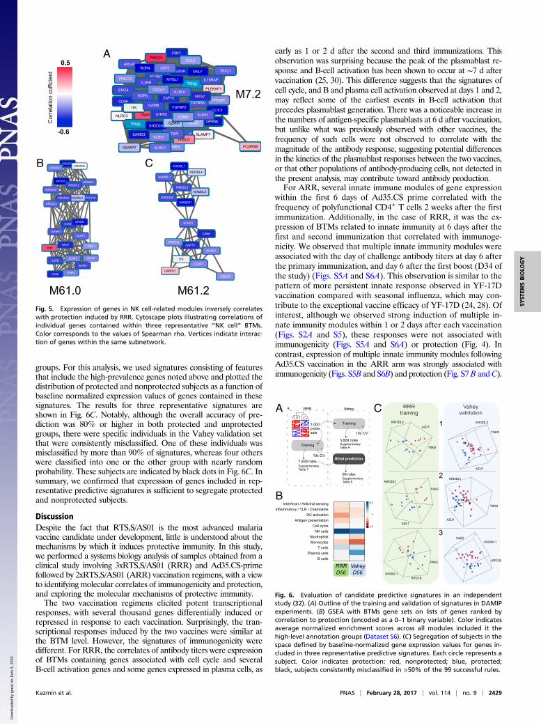
Molecular Signatures of Immunogenicity. We next analyzed thetranscriptional signatures that correlated with immunogenicity ofvaccination. In the case of RRR vaccination, we assessed tran-scriptional correlates of CSP-specific antibody titers on D77 (theday of challenge). Following RRR vaccination, the expression ofBTMs related to plasmablasts at D1 after each vaccination waspositively associated with the antibody titers on the day ofchallenge (Figs. S5A and S6A). This observation was surprising,given that the plasmablast response in humans to vaccinationwith other vaccines such as influenza (30) has been shown topeak at day 7 after vaccination, and with ARR and RRR vac-cination robust, plasmablast responses were observed 6 d aftereach boost (Fig. S1C). The observed correlation between BTMsrelated to B cells and plasmablasts, at day 1 after each boost, andimmunogenicity might reflect a transient burst of genes relatedto B-cell activation within a day of vaccination, but this hy-pothesis needs further exploration. Additionally, cell divisionBTMs showed positive correlation to the antibody titers even atlater time points (D14, 28, and 56), suggesting the persistence ofcycling cells (Figs. S5A and S6A). Furthermore, the expression ofseveral innate immunity modules (antigen presentation M95,dendritic cell activation M165), including many antiviral and typeI IFN-related modules at day 6 post primary and secondaryvaccinations, were positively correlated with the antibody titerson the day of challenge (Figs. S5A and S6A and Dataset S2).Most strikingly, on the day of first and second boosts, genemodules relevant to NK cells showed strong negative correlationto the antibody titers at the day of challenge (Fig. S5A andDataset S2). Indeed, we observed that the majority of genesincluded in these NK cell-related BTMs showed negative asso-ciation with antibody titers.In the case of ARR, the frequency of polyfunctional CD4+ T
cells at day 14 was associated with protection (Fig. 2C). We thusassessed whether early transcriptional signatures correlated withthe polyfunctional CD4+ T-cell response at day 14. At day 1,several modules representative of innate immune activation (an-tigen presentation M71, M95.1; activated dendritic cells andmonocytes M168, M11; TLR and inflammatory responses M16,M25, M146) were strongly associated with polyfunctional CD4+
T-cell response at day 14 (Figs. S5B and S6B). Interestingly, mod-ules representative of respiratory electron transport were stronglyassociated with the response. In contrast, modules representativeof NK cells and T cells were negatively associated with the re-sponse. Similar, but weaker, associations were observed at D2 afterprime Ad35.CS immunization. By day 6, the landscape of corre-lates changed, with many modules representing DC markers be-coming negatively enriched, whereas NK cell modules continued tobe negatively associated (Figs. S5B and S6B and Dataset S2).
Association of Molecular Signatures with Protection. We then ana-lyzed transcriptional signatures associated with protection. In theRRR group, BTMs related to plasma cells, B cells, and cell cycle atD1, D29, and D57 (i.e., 1 d after each vaccination) were positivelyassociated with protection (Fig. 4 and Fig. S7A), consistent with thecorrelations between the expression of such BTMs at 1 d after each
B
14 42 77
ARR
RRR
Non-Protected
Protected
Non-Protected
Protected
days post-primary immunization
CD40LIL-2TNF-αIFN-γ
A
p=0.01697 p=0.04427 p=0.72206
p=0.76508 p=0.64164 p=0.96988
0 14 4228 56 77
C
0 28 56 77 105 140 236days post-primary immunization
0
50
100
150
200
Ant
i-CS
repe
at re
gion
con
cent
ratio
n, E
U/m
L
0 28 56 77 105 140 236days post-primary immunization
0
50
100
150
200
ARRRRR* * *
NonProtectedProtectedStatus post-challenge
0 28 56 77 105 140 236days post-primary immunization
0 28 56 77 105 140 236days post-primary immunization
ARRRRR
5A
nti-H
BsA
g co
ncen
tratio
n, m
IU/m
L, 1
0
0
2
4
6
8
0
2
4
6
8
* * *
Fig. 2. Serological and cellular associations with protection. (A) Serum anti-CS IgG antibody titers. Lines indicate median values, shaded areas indicate25–75% interquartile range. *α = 0.05 level by Wilcoxon signed rank test.(B) Serum anti-HBsAg antibody titers in protected and nonprotected subjects.(C) SPICE plots indicate the functionality of CSP-specific CD4 T cells. Innersectors on SPICE plots indicate the number of markers expressed: Blue,green, orange, and red indicate 1, 2, 3, and 4 markers, respectively. Outsidearches indicate the identity of expressed markers, per legend in the figure.Sum of χ2 values was used as a test metric. P values were generated by
partial permutation test (37) and indicate the statistical significance of thedifferences in frequencies of T-cell subsets in P vs. NP.
Kazmin et al. PNAS | February 28, 2017 | vol. 114 | no. 9 | 2427
SYST
EMSBIOLO
GY
Dow
nloa
ded
by g
uest
on
June
5, 2
020

booster vaccination, and antibody titers (Figs. S5A and S6A). Wealso observed positive associations of multiple innate immunitymodules at 6 d after primary and secondary vaccination, similar tothe observed correlations with CSP-specific antibody titers (Fig. 4and Fig. S7A). Additionally, several NK cell modules at D56 (day ofthe second boost) negatively associate with protection (Fig. 4 andFig. S7A), consistent with their correlation with antibody titers (Figs.S5A and S6A). Strikingly, there were negative correlations of theexpression of almost all of the genes contained within the NK cell-related BTMs, at D56, and protection (Fig. 5).The transcriptional signatures of protection for the ARR arm
were different from those for RRR. Here, multiple innate im-munity modules positively associate with protection at D1 and 2after the prime, and D28 (day of the first boost) (Fig. S7 B and C),similar to the transcriptional correlates of polyfunctional CD4+ Tcells described above (Figs. S5B and S6B). Again, NK modulesdisplay strong negative association with protection at multiple timepoints (D2, D28, D29, D56) (Fig. S7 B and C and Dataset S3).We then determined the overlap between the molecular sig-
natures of protection and immunogenicity. For the ARR arm, atD1 and 2 BTMs related to antigen presentation, TLR signalingand dendritic cells were associated with both immunogenicity(i.e., polyfunctional CD4+ T cells at D14) and protection (Fig.S7C). For RRR, there was considerable overlap between signa-tures of protection and immunogenicity. Several BTMs thatcorrelate with protection were also correlated with immunoge-nicity (Fig. 4). BTMs related to plasma and B cells, and the cellcycle were correlated with both protection and immunogenicityat 1 d after the primary and secondary vaccinations (Fig. 4). Incontrast, several innate immunity modules, at day 6 after primeand day 6 after boost (D34), were correlated with both anti-CSP–specific antibody response and protection (Fig. 4). Strikingly, atD56 (the day of the final boost), we observed negative correla-tions of several NK cell-related BTMs with protection and im-munogenicity (i.e., CSP-specific antibody titers at D77) (Fig. 4).A full description of all common associations with immunoge-nicity and protection is provided in Dataset S4.
Predictive Modeling of Protection. We then developed predictivesignatures of protection based on the transcriptional response toRRR vaccination. To achieve this goal, a discriminant analysis viamixed integer programming (DAMIP) (31) was used to generatethe candidate predictive signatures. The two groups to classify aregroup 0 (protected) versus group 1 (not protected). For signaturevalidation, we used a transcriptional dataset from an independentmalaria challenge study with RTS,S/AS01 (32), NCT00075049,hereby referred to as “Vahey data set.” Responses in the two
studies were broadly similar (Fig. S8 and Datasets S5 and S6). Thebaseline-normalized expression values for the RRR cohort in thepresent study was used as a training set, and candidate signaturesthat passed a minimum accuracy threshold in the training set werethen applied to the independent validation set for blind pre-diction. The outline of the predictive modeling experiments isprovided in Fig. 6A. A full list of signatures and their performancemetrics is provided in Datasets S7 and S8. Analysis of the tran-scripts included in the successful predictive signatures revealed ahigh prevalence of transcripts that were commonly found in alarge number of signatures at D56 (Table S1). One such transcript,KIR2DS1 (an NK cell marker), was found in 57 of 99 successfulpredictive signatures. This observation is consistent with the factthat NK cell-related BTMs are negatively associated with pro-tection in both datasets at D56 (Fig. 6B). To validate these sig-natures, we used the set of transcripts identified in RRR togenerate and train signatures in the Vahey data set (Dataset S9).Many of the mRNAs that were highly represented in signaturestrained in RRR arm of this study are also highly represented insignatures trained in the Vahey data set (Datasets S10 and S11).Up-regulated mRNA common to both RRR- and Vahey-generatedsignatures include several NK markers KIR2DS1, KIR2DL2, andKIR3DL1. Notably, many of the mRNAs that were included in thepredictive signatures in 10-fold cross validation (10× CV) in RRRand the Vahey data set individually were also included in predictivesignatures that were trained by using RRR, and were shown toblind predict outcome in Vahey data set (Dataset S11). Therefore,we conclude that a small number of mRNAs with high prevalencein predictive signatures are likely to be determining factors thatdistinguish protected versus nonprotected individuals.Finally, we illustrated the ability of the generated signatures of
protection to segregate the samples in their respective protection
AFFYMETRIX
days
Ad35or RTS,S
RTS,S RTS,S Challenge
Transcriptomics
2 60 1 14 34 4228 29 56 57 8277 78 105 140 236
0
2000
4000
6000
8000
100001 14 28 29 56 57 8277 782 6 34 62
days post-primary immunization
DE
Gs
(# p
robe
set
s)
ARRRRR DOWNUP
DOWNUP
62
Fig. 3. Transcriptional responses to vaccination. The number of probe sets dis-playing significantly different signals compared with D0. Significance was de-termined as Benjamini–Hotchberg FDR q value <0.01 and jFold-changej >1.5-fold.
cell cycle (I) (M4.1)cell cycle (II) (M4.10)mitotic cell division (M6)E2F transcription factor network (M8)cell cycle (III) (M103)respiratory electron transport chain (mitochondrion) (M219)respiratory electron transport chain (mitochondrion) (M231)mitochondrial cluster (M235)respiratory electron transport chain (mitochondrion) (M238)complement and other receptors in DCs (M40)enriched in activated dendritic cells/monocytes (M64)enriched in activated dendritic cells (I) (M119)enriched in activated dendritic cells (II) (M165)enriched in dendritic cells (M168)TLR and inflammatory signaling (M16)MHC-TLR7-TLR8 cluster (M146)DC surface signature (S5)Resting dendritic cell surface signature (S10)inflammatory response (M33)type I interferon response (M127)viral sensing & immunity; IRF2 targets network (I) (M111.0)enriched in monocytes (II) (M11.0)RA, WNT, CSF receptors network (monocyte) (M23)enriched in monocytes (III) (M73)enriched in monocytes (IV) (M118.0)Monocyte surface signature (S4)enriched in monocytes (I) (M4.15)enriched in NK cells (II) (M61.0)enriched in NK cells (KIR cluster) (M61.1)enriched in NK cells (receptor activation) (M61.2)enriched in NK cells (I) (M7.2)NK cell surface signature (S1)PLK1 signaling events (M4.2)
40-4Indicates modules that constitute common correlates of immunogenicity and protection at at least one time point
Inflammatory / TLR / chemokinesInterferon / antiviralMonocytesNK cellsSignal transduction
B cellPlasma cellsCell cycleMitochondrialAntigen presentation and DC
cell cycle and transcription (M4.0)plasma cells, immunoglobulins (M156.1)plasma cells & B cells, immunoglobulins (M156.0)enriched in B cells (IV) (M47.3)enriched in B cells (III) (M47.2)
Day 14 29 34 57 62 28 561 2 6
RRR
Fig. 4. Transcriptional correlates of protection to RRR vaccination. Eachsquare represents a BTM. Color represents normalized enrichment scores forBTMs. Assignment of a BTM to a high-level annotation group is illustrated by acolored sidebar (Dataset S3). Modules that represent common associations ofboth immunogenicity and protection are highlighted (Dataset S4).
2428 | www.pnas.org/cgi/doi/10.1073/pnas.1621489114 Kazmin et al.
Dow
nloa
ded
by g
uest
on
June
5, 2
020
groups. For this analysis, we used signatures consisting of featuresthat include the high-prevalence genes noted above and plotted thedistribution of protected and nonprotected subjects as a function ofbaseline normalized expression values of genes contained in thesesignatures. The results for three representative signatures areshown in Fig. 6C. Notably, although the overall accuracy of pre-diction was 80% or higher in both protected and unprotectedgroups, there were specific individuals in the Vahey validation setthat were consistently misclassified. One of these individuals wasmisclassified by more than 90% of signatures, whereas four otherswere classified into one or the other group with nearly randomprobability. These subjects are indicated by black dots in Fig. 6C. Insummary, we confirmed that expression of genes included in rep-resentative predictive signatures is sufficient to segregate protectedand nonprotected subjects.
DiscussionDespite the fact that RTS,S/AS01 is the most advanced malariavaccine candidate under development, little is understood about themechanisms by which it induces protective immunity. In this study,we performed a systems biology analysis of samples obtained from aclinical study involving 3xRTS,S/AS01 (RRR) and Ad35.CS-primefollowed by 2xRTS,S/AS01 (ARR) vaccination regimens, with a viewto identifying molecular correlates of immunogenicity and protection,and exploring the molecular mechanisms of protective immunity.The two vaccination regimens elicited potent transcriptional
responses, with several thousand genes differentially induced orrepressed in response to each vaccination. Surprisingly, the tran-scriptional responses induced by the two vaccines were similar atthe BTM level. However, the signatures of immunogenicity weredifferent. For RRR, the correlates of antibody titers were expressionof BTMs containing genes associated with cell cycle and severalB-cell activation genes and some genes expressed in plasma cells, as
early as 1 or 2 d after the second and third immunizations. Thisobservation was surprising because the peak of the plasmablast re-sponse and B-cell activation has been shown to occur at ∼7 d aftervaccination (25, 30). This difference suggests that the signatures ofcell cycle, and B and plasma cell activation observed at days 1 and 2,may reflect some of the earliest events in B-cell activation thatprecedes plasmablast generation. There was a noticeable increase inthe numbers of antigen-specific plasmablasts at 6 d after vaccination,but unlike what was previously observed with other vaccines, thefrequency of such cells were not observed to correlate with themagnitude of the antibody response, suggesting potential differencesin the kinetics of the plasmablast responses between the two vaccines,or that other populations of antibody-producing cells, not detected inthe present analysis, may contribute toward antibody production.For ARR, several innate immune modules of gene expression
within the first 6 days of Ad35.CS prime correlated with thefrequency of polyfunctional CD4+ T cells 2 weeks after the firstimmunization. Additionally, in the case of RRR, it was the ex-pression of BTMs related to innate immunity at 6 days after thefirst and second immunization that correlated with immunoge-nicity. We observed that multiple innate immunity modules wereassociated with the day of challenge antibody titers at day 6 afterthe primary immunization, and day 6 after the first boost (D34 ofthe study) (Figs. S5A and S6A). This observation is similar to thepattern of more persistent innate response observed in YF-17Dvaccination compared with seasonal influenza, which may con-tribute to the exceptional vaccine efficacy of YF-17D (24, 28). Ofinterest, although we observed strong induction of multiple in-nate immunity modules within 1 or 2 days after each vaccination(Figs. S2A and S5), these responses were not associated withimmunogenicity (Figs. S5A and S6A) or protection (Fig. 4). Incontrast, expression of multiple innate immunity modules followingAd35.CS vaccination in the ARR arm was strongly associated withimmunogenicity (Figs. S5B and S6B) and protection (Fig. S7 B andC).
RRRtraining
VaheyvalidationA C
B
1
2
3
RRRtraining
VaheyVVvalidation
AZU1
AZU1
AZU1AZU1
TSKS
TSKS
TSKS
TSKS
KIR3DL2 KIR3DL2
KIR2DL1KIR2DL1
KIR2DL1KIF21B
PAK2
PAK2KIR2DL1
KIF21B
a
1,000probesets
Training
Training
7,829 rules
3,829 rules
10x CV
10x CV
99 rules
Blind prediction
RRR Vahey
Supplementary Table 7
Supplementary Table 9
Supplementary Table 8
RRRD56
VaheyD56
Interferon / Antiviral sensingInflammatory / TLR / Chemokine
DC activationAntigen presentation
Cell cycleNK cells
NeutrophilsMonocytes
T cellsPlasma cells
B cells
-2.5
0
2.5
Fig. 6. Evaluation of candidate predictive signatures in an independentstudy (32). (A) Outline of the training and validation of signatures in DAMIPexperiments. (B) GSEA with BTMs gene sets on lists of genes ranked bycorrelation to protection (encoded as a 0–1 binary variable). Color indicatesaverage normalized enrichment scores across all modules included it thehigh-level annotation groups (Dataset S6). (C) Segregation of subjects in thespace defined by baseline-normalized gene expression values for genes in-cluded in three representative predictive signatures. Each circle represents asubject. Color indicates protection: red, nonprotected; blue, protected;black, subjects consistently misclassified in >50% of the 99 successful rules.
TRD@PRKCQ KLRB1
IL2RBIL18RAPMYBL1
TARPNLRC3
TRA@
CD96
SH2D2A
S1PR5
ITK GZMB
ARL4CRORA
GZMH
PRF1
CST7TBX21
XCL2PRKCH
GNLY
TGFBR3
GZMM GPR56
CLIC3
CCL5
KLRF1
PTGDR
GZMA
FGFBP2
XCL1
EOMES
SLAMF7
FCGR3B
FASLG
GIMAP7 KLRC1
CTSWKLRK1
SAMD3
PLEKHF1
NKG7ZAP70
STAT4HOPX
CD247 KLRD1
CD247IL2RB
KLRB1
KLRK1
PVRIGCD96
KIR2DS1
KIR2DL1
KIR2DL5A
KIR3DL1
KIR2DL3
KIR2DL2
KIR3DL3
KIR2DS3
KIR2DS5
KIR2DS2
KIR2DS4
CD7
S1PR5GPR56
TARP
KLRF1TGFBR3
NKG7
KLRK1
KLRD1
CARD11
CD244
ZAP70
CD96
CD247
PRKCQ
ITK
KIR2DS2
KIR2DL2KIR2DL3
KIR2DS1
KIR2DL1KIR2DL4
KIR3DL1
A
B C
M7.2
M61.0 M61.2
0.5
-0.6
Cor
rela
tion
coffi
cien
t
Fig. 5. Expression of genes in NK cell-related modules inversely correlateswith protection induced by RRR. Cytoscape plots illustrating correlations ofindividual genes contained within three representative “NK cell” BTMs.Color corresponds to the values of Spearman rho. Vertices indicate interac-tion of genes within the same subnetwork.
Kazmin et al. PNAS | February 28, 2017 | vol. 114 | no. 9 | 2429
SYST
EMSBIOLO
GY
Dow
nloa
ded
by g
uest
on
June
5, 2
020

A surprising result is that at D56 (the day of the final boost), weobserved consistent signatures of immunogenicity and protection.In particular, the NK cell modules in peripheral blood at D56 (dayof the third immunization) correlate negatively with both antibodyresponse and protection. Furthermore, several NK cell-relatedgenes were observed in the predictive signatures delineated byDAMIP. Whether there is a causal link between the observed NKcell modules and the antibody response remains to be determined.In this context, a recent report shows that NK cells negativelyregulate germinal center and T follicular responses and memoryB-cell generation (33), so it is conceivable that such a mechanismmay be at play with the current vaccine. In addition, it is possiblethat this inverse correlation between the expression of NK cell-related genes and antibody titers and protection reflects the mi-gration of NK cells from the blood to the liver, where they mayhelp orchestrate antibody-mediated effector mechanisms such asantibody-dependent cell-mediated cytoxcity against infected cells inthe liver. In this context, several previous studies have implicatedNK cells in immunity against malaria (34–36).Together, our findings indicate that the RTS,S/AS01 vaccine
candidate elicits protective immunity against infection primarilythrough rapid accumulation of high levels of anti-CS antibodies. Incontrast, vaccination with ARR did not induce as high a magnitudeof antibody response, but rather enhanced frequencies of polyfunc-tional CD4+ T cells. Given the critical importance of CD4+ T cells inpromoting antibody responses, the failure of the ARR regimen toinduce as strong an antibody response as the RRR regimen was asurprise. However, it should be noted that RRR regimen involvedthree immunizations with CSP-expressing Hep B virus-like particles,whereas ARR only involved two immunizations. Furthermore, the
polyfunctional CD4+ T cells induced by the Ad35 prime may havealtered the quality of the antibody response, leading, for example, tohigher-affinity antibodies as a result of enhanced germinal centerresponse in the ARR vaccine compared with the RRR vaccine.Thus, the ARR and RRR vaccine might have conferred protectionagainst malaria via two distinct mechanisms, involving the magnitude(RRR) and the affinity (ARR) of the antibody response. Further-more our results demonstrate a potent and sustained transcriptionalresponse induced by this vaccine, and delineate several unappre-ciated molecular correlates of immunogenicity and protection (Fig.S9). Importantly, these results provide candidate molecular signa-tures that may have potential as biomarkers of protective efficacy ofvaccine-induced immunity against malaria.
MethodsThis study was conducted at the Walter Reed Army Institute of Research(WRAIR) between August 2011 and July 2012, and was approved by theWRAIR Institutional Review Board (IRB) and Program for AppropriateTechnology in Health-Malaria Vaccine Initiative’s Western IRB. The trial wasundertaken in accordance with the provisions of the International Confer-ence on Harmonization and Good Clinical Practice guidelines. Written in-formed consent was obtained from each subject before study procedureswere initiated. All laboratories received deidentified samples and performedtests according to protocol, and therefore their work was IRB-exempt.
ACKNOWLEDGMENTS. The clinical study was performed at the Walter ReedArmy Institute of Research Malaria Vaccine Branch, which provided thePBMCs for this study. We thank Dr. Matthew Woodruff for critically readingthe manuscript. This work was supported by a research grant from MVI-Path (to B.P.), National Institutes of Health Grants U19AI090023 (to B.P.)and U19AI057266 (to R.A.), and National Science Foundation Grants NSF-1516074 and NSF-1361532 (to E.K.L.).
1. World Health Organization (2015) World Malaria Report (World Health Organiza-tion, Geneva).
2. Strategic Advisory Group of Experts (2011) Meeting of the Strategic Advisory Groupof Experts on immunization, October 2015 - conclusions and recommendations. WklyEpidemiol Rec 90(50):681–699.
3. Cohen J, Nussenzweig V, Nussenzweig R, Vekemans J, Leach A (2010) From the cir-cumsporozoite protein to the RTS, S/AS candidate vaccine. Hum Vaccin 6(1):90–96.
4. Casares S, Brumeanu TD, Richie TL (2010) The RTS,S malaria vaccine. Vaccine 28(31):4880–4894.
5. Didierlaurent AM, et al. (2014) Enhancement of adaptive immunity by the human vac-cine adjuvant AS01 depends on activated dendritic cells. J Immunol 193(4):1920–1930.
6. Gordon DM, et al. (1995) Safety, immunogenicity, and efficacy of a recombinantlyproduced Plasmodium falciparum circumsporozoite protein-hepatitis B surface anti-gen subunit vaccine. J Infect Dis 171(6):1576–1585.
7. Kester KE, et al.; RTS,S Vaccine Evaluation Group (2009) Randomized, double-blind, phase2a trial of falciparummalaria vaccines RTS,S/AS01B and RTS,S/AS02A inmalaria-naive adults:Safety, efficacy, and immunologic associates of protection. J Infect Dis 200(3):337–346.
8. Agnandji ST, Fernandes JF, Bache EB, Ramharter M (2015) Clinical development ofRTS,S/AS malaria vaccine: A systematic review of clinical Phase I-III trials. FutureMicrobiol 10(10):1553–1578.
9. Bojang KA, et al.; RTS, S Malaria Vaccine Trial Team (2001) Efficacy of RTS,S/AS02malaria vaccine against Plasmodium falciparum infection in semi-immune adult menin The Gambia: A randomised trial. Lancet 358(9297):1927–1934.
10. Alonso PL, et al. (2004) Efficacy of the RTS,S/AS02A vaccine against Plasmodium fal-ciparum infection and disease in young African children: Randomised controlled trial.Lancet 364(9443):1411–1420.
11. Bejon P, et al. (2008) Efficacy of RTS,S/AS01E vaccine against malaria in children 5 to17 months of age. N Engl J Med 359(24):2521–2532.
12. Aponte JJ, et al. (2007) Safety of the RTS,S/AS02D candidate malaria vaccine in infantsliving in a highly endemic area of Mozambique: A double blind randomised con-trolled phase I/IIb trial. Lancet 370(9598):1543–1551.
13. Abdulla S, et al. (2008) Safety and immunogenicity of RTS,S/AS02D malaria vaccine ininfants. N Engl J Med 359(24):2533–2544.
14. Agnandji ST, et al.; RTS,S Clinical Trials Partnership (2011) First results of phase 3 trialof RTS,S/AS01 malaria vaccine in African children. N Engl J Med 365(20):1863–1875.
15. The RTS,S Clinical Trial Partnership (2015) Efficacy and safety of RTS,S/AS01 malariavaccine with or without a booster dose in infants and children in Africa: Final resultsof a phase 3, individually randomised, controlled trial. Lancet 386(9988):31–45.
16. RTS,S Clinical Trials Partnership (2014) Efficacy and safety of the RTS,S/AS01 malariavaccine during 18 months after vaccination: A phase 3 randomized, controlled trial inchildren and young infants at 11 African sites. PLoS Med 11(7):e1001685.
17. Agnandji ST, et al.; RTS,S Clinical Trials Partnership (2012) A phase 3 trial of RTS,S/AS01malaria vaccine in African infants. N Engl J Med 367(24):2284–2295.
18. Ockenhouse CF, et al. (2015) Ad35.CS.01-RTS,S/AS01 heterologous prime boost vaccine effi-cacy against sporozoite challenge in healthy malaria-naïve adults. PLoS One 10(7):e0131571.
19. Epstein JE, et al. (2011) Live attenuated malaria vaccine designed to protect throughhepatic CD8+ T cell immunity. Science 334(6055):475–480.
20. O’Brien KL, et al. (2009) Adenovirus-specific immunity after immunization with anAd5 HIV-1 vaccine candidate in humans. Nat Med 15(8):873–875.
21. Stewart VA, et al. (2007) Priming with an adenovirus 35-circumsporozoite protein (CS)vaccine followed by RTS,S/AS01B boosting significantly improves immunogenicity toPlasmodium falciparum CS compared to that with either malaria vaccine alone. InfectImmun 75(5):2283–2290.
22. Pulendran B, Li S, Nakaya HI (2010) Systems vaccinology. Immunity 33(4):516–529.23. Pulendran B (2014) Systems vaccinology: Probing humanity’s diverse immune systems
with vaccines. Proc Natl Acad Sci USA 111(34):12300–12306.24. Querec TD, et al. (2009) Systems biology approach predicts immunogenicity of the
yellow fever vaccine in humans. Nat Immunol 10(1):116–125.25. Nakaya HI, et al. (2011) Systems biology of vaccination for seasonal influenza in hu-
mans. Nat Immunol 12(8):786–795.26. Gaucher D, et al. (2008) Yellow fever vaccine induces integrated multilineage and
polyfunctional immune responses. J Exp Med 205(13):3119–3131.27. Schoggins JW, et al. (2011) A diverse range of gene products are effectors of the type
I interferon antiviral response. Nature 472(7344):481–485.28. Zak DE, et al. (2012) Merck Ad5/HIV induces broad innate immune activation that
predicts CD8+ T-cell responses but is attenuated by preexisting Ad5 immunity. ProcNatl Acad Sci USA 109(50):E3503–E3512.
29. Li S, et al. (2014) Molecular signatures of antibody responses derived from a systemsbiology study of five human vaccines. Nat Immunol 15(2):195–204.
30. Wrammert J, et al. (2008) Rapid cloning of high-affinity human monoclonal anti-bodies against influenza virus. Nature 453(7195):667–671.
31. Lee EK (2007) Large-scale optimization-based classification models in medicine andbiology. Ann Biomed Eng 35(6):1095–1109.
32. Vahey MT, et al. (2010) Expression of genes associated with immunoproteasomeprocessing of major histocompatibility complex peptides is indicative of protectionwith adjuvanted RTS,S malaria vaccine. J Infect Dis 201(4):580–589.
33. Rydyznski C, et al. (2015) Generation of cellular immune memory and B-cell immunityis impaired by natural killer cells. Nat Commun 6:6375.
34. Filtjens J, et al. (2014) Contribution of the Ly49E natural killer receptor in the immuneresponse to Plasmodium berghei infection and control of hepatic parasite develop-ment. PLoS One 9(1):e87463.
35. Roland J, et al. (2006) NK cell responses to Plasmodium infection and control of in-trahepatic parasite development. J Immunol 177(2):1229–1239.
36. Gonzalez-Aseguinolaza G, et al. (2002) Natural killer T cell ligand alpha-galactosylceramideenhances protective immunity induced by malaria vaccines. J Exp Med 195(5):617–624.
37. Subramanian A, et al. (2005) Gene set enrichment analysis: A knowledge-based ap-proach for interpreting genome-wide expression profiles. Proc Natl Acad Sci USA102(43):15545–15550.
38. Roederer M, Nozzi JL, Nason MC (2011) SPICE: Exploration and analysis of post-cytometric complex multivariate datasets. Cytometry A 79(2):167–174.
2430 | www.pnas.org/cgi/doi/10.1073/pnas.1621489114 Kazmin et al.
Dow
nloa
ded
by g
uest
on
June
5, 2
020