Systemic Lupus Erythematosus: Prevalence, Severity, and ... · Objectives 1. Estimate the...
54
Elizabeth D. Ferucci, MD, MPH June 25, 2015 IHS Clinical Rounds Systemic Lupus Erythematosus: Prevalence, Severity, and Identification in American Indian/Alaska Native Populations
Transcript of Systemic Lupus Erythematosus: Prevalence, Severity, and ... · Objectives 1. Estimate the...

Elizabeth D. Ferucci, MD, MPH
June 25, 2015
IHS Clinical Rounds
Systemic Lupus Erythematosus: Prevalence, Severity, and Identification in American
Indian/Alaska Native Populations

Disclosures
• I have no relationships with any entity producing, marketing, re-selling, or distributing health care goods or services.
• The standard of care for treatment of lupus is almost all off-label. I will discuss off-label medication use.

Objectives
1. Estimate the prevalence of systemic lupus erythematosus (SLE) in the US, and compare the prevalence in AI/AN populations to other racial/ethnic minorities.
2. Recognize possible clinical presentations of SLE and use these to guide the initial evaluation and assist with appropriate referrals.
3. Describe the range of severity that can be seen in SLE and understand that the disease is often worse in racial/ethnic minority populations.
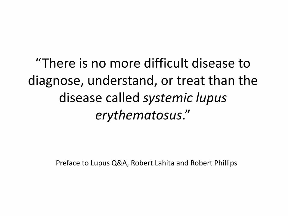
“There is no more difficult disease to diagnose, understand, or treat than the
disease called systemic lupus erythematosus.”
Preface to Lupus Q&A, Robert Lahita and Robert Phillips

Systemic Lupus Erythematosus (SLE)
An inflammatory, multisystem, autoimmune disease of unknown etiology
with protean clinical and laboratory manifestations and a variable course and prognosis. Lupus can be a mild disease, a severe and life-threatening illness, or
anything in between.
PREVALENCE AND INCIDENCE OF SLE
Part 1
Arthritis and Rheumatology, September 2014, Vol. 66, No. 9, pp. 2494-2502.

Background: SLE in Populations
• US population rates
– Prevalence: 15-144 per 100,000
– Incidence: 1.8-23.2 per 100,000 per year
• Highest rates in women and US blacks
– 10:1 female:male ratio
– Rates up to 5 times higher in blacks than whites
– Prevalence in black women: 58-286 per 100,000
1. Lim SS, Drenkard C. Curr Rheumatol Rep 2008; 10:265 2. Feldman CH, et al. Arthritis Rheum 2013;65:753.

Disparities in Lupus Prevalence
0
50
100
150
200
250
300
350
Siegel 1970 Fessel 1974 Feldman 2013
Pre
vale
nce
pe
r 1
00
,00
0
WM
WF
AAM
AAF
WM: white men; WF: white females; AAM: African-American men; AAF: African-American females

Objectives of IHS Lupus Registry
• This population-based registry was created with the objective to determine the prevalence (2007) and incidence (2007-2009) of SLE in the Indian Health Service (IHS) active clinical population in 3 regions of the US.
• Using comparable methods to 4 other CDC-funded registries in order to compare rates by race/ethnicity. – Georgia, Michigan—1st round
– New York City, California, IHS—2nd round

Indian Health Service Area Map
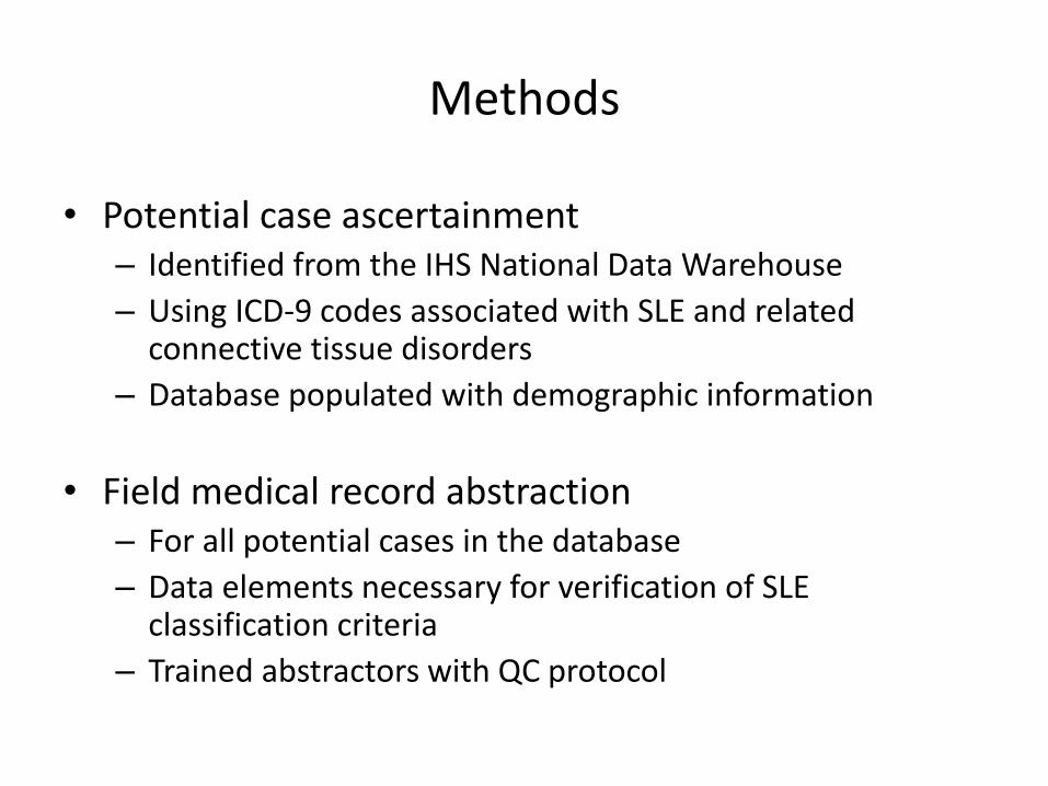
Methods
• Potential case ascertainment – Identified from the IHS National Data Warehouse
– Using ICD-9 codes associated with SLE and related connective tissue disorders
– Database populated with demographic information
• Field medical record abstraction – For all potential cases in the database
– Data elements necessary for verification of SLE classification criteria
– Trained abstractors with QC protocol

Primary Case Definition
• 4 or more of the 11 American College of Rheumatology (ACR) classification criteria for SLE documented in the medical record

Prevalence of SLE in CDC registries
0
50
100
150
200
250
300
Female Male
AIAN
GA-B
MI-B
GA-W
MI-W
pe
r 1
00
, 00
0 p
op
ula
tio
n
AIAN: American Indian/Alaska Native from IHS registry; GA-B: Georgia Registry—Black; MI-B: Michigan Registry—Black; GA- W: Georgia Registry—White; MI-W: Michigan Registry—White

Incidence of SLE in CDC registries
0
2
4
6
8
10
12
14
16
Female Male
AIAN
GA-B
MI-B
GA-W
MI-W
pe
r 1
00
, 00
0 p
op
ula
tio
n p
er
year
AIAN: American Indian/Alaska Native from IHS registry; GA-B: Georgia Registry—Black; MI-B: Michigan Registry—Black; GA- W: Georgia Registry—White; MI-W: Michigan Registry—White

Possible explanations for high rates in AI/AN populations
• Genetic factors
– HLA, multiple other loci associated with SLE in large genomewide association studies
– Frequency/role of individual genes/SNPs unknown in AI/AN populations
• Environmental facotrs
– Tobacco, UV light exposure, infections all found to play a role in SLE development
– Role of these unknown in AI/AN populations

SLE in US Hispanics and Asians
• California Lupus Surveillance program found highest prevalence in Black women, with Hispanic and Asian women’s prevalence intermediate between Black and White
• Hawaiian study from 1970s found rates in Asian and Native Hawaiian population were higher than Whites in Hawaii
1. Arthritis Research and Therapy 2014, Volume 16 Suppl 1; 2. Arthritis Rheum. 1979;22(4):328.

CLINICAL PRESENTATION AND INITIAL EVALUATION
Part 2

Case 1
• 39 year old woman has had swollen and stiff hand joints for one year (MCP and PIPs), with morning stiffness, referred to rheumatology for suspected rheumatoid arthritis
• ROS: photosensitivity
• Exam: patchy alopecia, inflammatory arthritis
• Labs: WBC 3.0, ALC 0.8, SCr 0.6, UA normal, ANA+ 1:320 titer, RF-, CCP-, Sm+, dsDNA-, SSA/SSB-, RNP-
• What is the diagnosis, and why?

Case 2
• 33 year old woman presents with:
• Inflammatory arthritis, +ANA for 4 years
• 1 year ago: pericardial and pleural effusions, resolved with prednisone
• Now: fever, cough. Labs: SCr 3.8, WBC 3.4, C3 61↓, C4 9↓, 3.1 g proteinuria, UA lrg bld
• Autoantibodies: ANA/dsDNA/SSA/SSB/RNP+
• Other sx’s: photosensitivity, Raynaud’s, pruritic rash, oral ulcers
• What is the diagnosis, and why?

Systemic lupus erythematosus
• Systemic autoimmune disease
• Not organ-specific – Diverse presentations, evolve over time
• Hallmark is autoantibodies
• Characterized by remissions and exacerbations (“flares”)
• Prognosis (and therapy) varies by organ involvement and severity

ACR Classification Criteria
(4 of 11 required for classification as SLE)
1. Malar rash
2. Discoid rash
3. Photosensitivity
4. Oral ulcers
5. Arthritis
6. Serositis
7. Renal disorder: proteinuria, cellular casts
8. Neurologic disorder: Seizures and/or psychosis
9. Hematologic disorder: Immune-mediated hemolytic anemia,
leukopenia, lymphopenia, thrombocytopenia
10. Antinuclear antibodies (ANA)
11. Immunologic disorder: anti-DNA antibody, anti-Sm antibody, or
antiphospholipid antibodies
Tan EM, et al. The 1982 revised criteria for the classification of systemic lupus erythematosus. Arthritis Rheum 1982;25:1271-7.
Hochberg MC. Updating the American College of Rheumatology revised criteria for the classification of systemic lupus erythematosus [letter].
Arthritis Rheum 1997;40:1725.

Diagnostic Criteria for SLE
• Clinical (history and physical) – Rashes (malar, discoid, photosensitivity)
– Oral ulcers
– Arthritis
– Pleuritis or pericarditis
– Seizures, psychosis
• Lab – Renal (proteinuria, cellular casts)
– Hematologic (leukopenia, lymphopenia, hemolytic anemia, thrombocytopenia)
– Autoantibodies (ANA, dsDNA, Sm, antiphospholipid antibodies)

SLE Rashes

Systemic lupus erythematosus: photosensitivity, face and neck

Other manifestations of SLE
• Acute or chronic cutaneous LE, not malar or discoid
• Neurologic disorders, other than seizures or psychosis
• Alopecia
• Low complements (C3, C4)
• Pneumonitis
• Myocarditis, Libman-Sacks endocarditis
• Autoimmune hepatitis


serositis pericardial effusion cerebral infarct
glomerulonephritis spherocytes
brain atrophy
Lupus
on the Inside
Lupus
on the Inside


Tests to order if SLE is suspected
• Need CBC with diff, UA, renal function
– Urine protein/creatinine ratio if any proteinuria on UA
• Antinuclear antibody
– Positive in vast majority of patients
– Titer and pattern may be helpful at diagnosis
– If ANA is positive, order additional autoantibodies • dsDNA, Sm most specific
• SSA (Ro), SSB (La), and RNP can be found in SLE)

Shmerling R. N Engl J Med 2003;349:1499-1500

Case 1
• 39 year old woman has had swollen and stiff hand joints for one year (MCP and PIPs), with morning stiffness, referred for suspected RA
• ROS: photosensitivity
• Exam: patchy alopecia, inflammatory arthritis
• Labs: WBC 3.0, ALC 0.8, SCr 0.6, UA normal, ANA+ 1:320 titer, RF-, CCP-, Sm+, dsDNA-, SSA/SSB-, RNP-
• What is the diagnosis, and why?
SLE (criteria met: +ANA, hematologic, arthritis, immunologic, and photosensitivity)
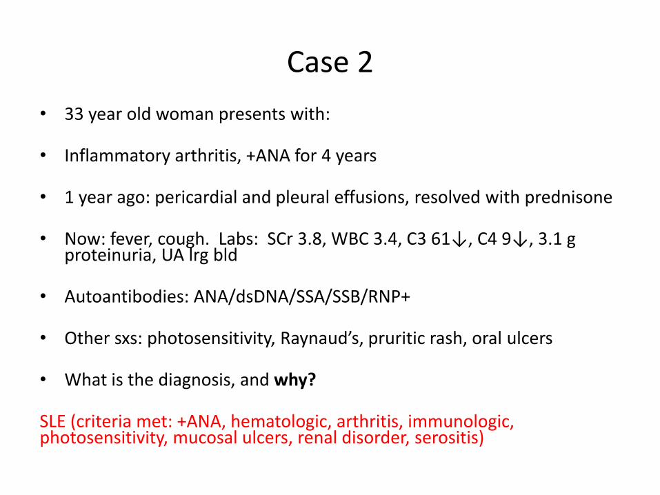
Case 2
• 33 year old woman presents with:
• Inflammatory arthritis, +ANA for 4 years
• 1 year ago: pericardial and pleural effusions, resolved with prednisone
• Now: fever, cough. Labs: SCr 3.8, WBC 3.4, C3 61↓, C4 9↓, 3.1 g proteinuria, UA lrg bld
• Autoantibodies: ANA/dsDNA/SSA/SSB/RNP+
• Other sxs: photosensitivity, Raynaud’s, pruritic rash, oral ulcers • What is the diagnosis, and why?
SLE (criteria met: +ANA, hematologic, arthritis, immunologic, photosensitivity, mucosal ulcers, renal disorder, serositis)

SEVERITY OF SLE AND HEALTH DISPARITIES
Part 3

Major organ vs. non-major organ “Bad” vs. “Not so bad” SLE
Major organ:
• Glomerulonephritis
• CNS
• Pneumonitis
• Myocarditis
• Severe hematologic involvement
Non-major organ:
• Arthritis
• Rashes
• Oral ulcers

Disease Activity
• Important determinant of need for ongoing immunosuppressive therapy
• Defined by lupus manifestations present NOW
• In contrast to “Damage” or organ dysfunction that has accumulated over time due to previous SLE disease activity

Available Therapy for SLE
• Corticosteroids
• Anti-malarials
• Immunosuppressive agents
– Azathioprine, mycophenolate mofetil (CellCept)
• Cytotoxics
– Cyclophosphamide
• DMARDs
– Methotrexate, leflunomide (Arava)
• Biologics
– Belimumab, rituximab

Corticosteroids
• IV pulse dosing often used early in severe disease – 1 gram IV methylprednisolone daily for 3 days
• 1 mg/kg/day oral prednisone in organ-threatening disease
• Rarely use more than 10-15 mg/day in non-organ-threatening disease
• Dose should be minimized in long term

Case 3 • 24 year old woman newly diagnosed with SLE
• Manifestations include: – Inflammatory arthritis
– Lab abnormalities: +ANA, +dsDNA, lymphopenia
• She was given a prednisone taper by her PCP and her joint symptoms improved
• She is very concerned about internal organ involvement and how to avoid complications of SLE in the long-term.
• What medication is best for her? What other advice can we give her?

Hydroxychloroquine is the most commonly prescribed anti-malarial medication. Chloroquine is available but more toxic.
Quinacrine may be used in some cases, but availability is limited.
First-line therapy in SLE

Benefits of hydroxychloroquine
• Controls skin and joint disease
• Long-term use prevents major renal or CNS damage
• Protective effect on survival in SLE
• Lower fasting glucose in women with SLE or RA taking hydroxychloroquine
• Pre-clinical use may delay onset of SLE
1. Fessler BJ et al Arthritis Rheum 2005;52:1473. 2. Penn SK et al. J Rheumatol. 2010 Jun;37(6):1136-42
3. Alarcon GS et al. Ann Rheum Dis 2007;66:1168. 4. James JA et al. Lupus. 2007;16(6):401-9.

Other advice for this young woman
• Avoid sun exposure
• Pregnancy is possible but should be planned when lupus is under control – SSA (Ro) antibody increases risk of neonatal lupus
• Stay up to date on immunizations
• Do not use tobacco
• Prognosis appears to be good at this time
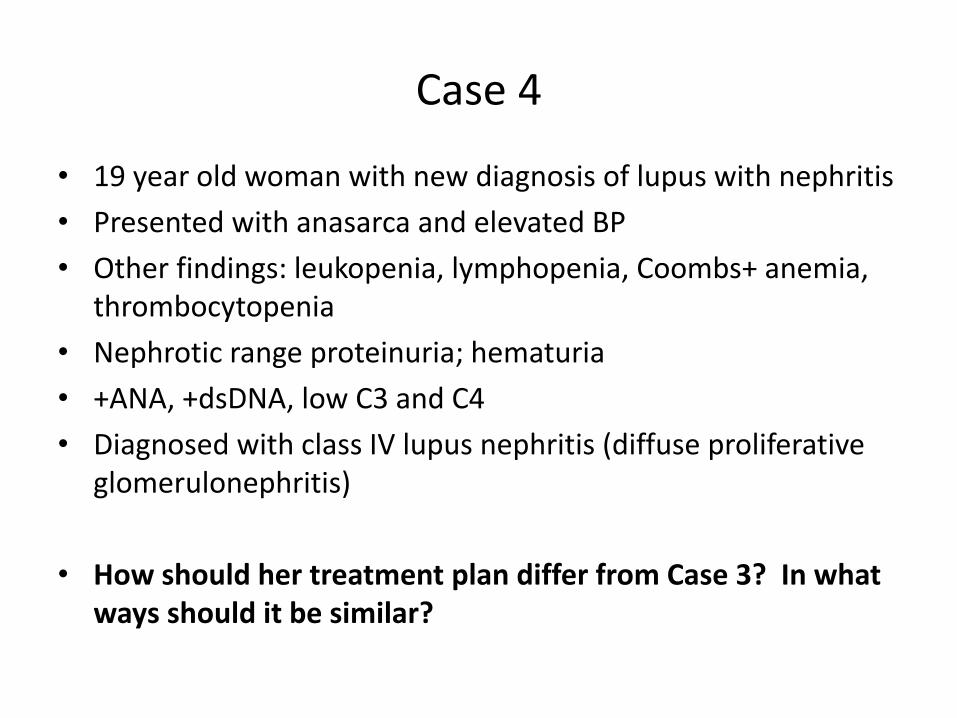
Case 4
• 19 year old woman with new diagnosis of lupus with nephritis
• Presented with anasarca and elevated BP
• Other findings: leukopenia, lymphopenia, Coombs+ anemia, thrombocytopenia
• Nephrotic range proteinuria; hematuria
• +ANA, +dsDNA, low C3 and C4
• Diagnosed with class IV lupus nephritis (diffuse proliferative glomerulonephritis)
• How should her treatment plan differ from Case 3? In what ways should it be similar?

Lupus Nephritis
• Aggressive Rx in short term
– Induce remission
• Long term goals:
– Prevent damage
– Reduce corticosteroid exposure

Recommendations in this case
• Same as Case 3 PLUS
• Appropriate nephritis therapy
– Corticosteroids and immunosuppressive agent
– Aim to taper off steroids
• Blood pressure control
• ACE or ARB if proteinuria

Disparities in Lupus Severity
• Racial/ethnic minorities are more likely to develop lupus at a younger age and to have more severe symptoms at onset.
• Manitoba First Nations data:
– Mean age of onset, 31 vs. 37 years
– More severe disease at diagnosis (SLEDAI score)
McCarty DJ, et al. Arthritis Rheum 1995; 38:1260-1270. Cooper GS, et al. Lupus 2002; 11:161-167. Peschken, et al. J Rheumatol 2000;27:1884-91.

Renal disease
• Most important predictor of mortality in SLE
• Present more often in black and AI/AN people with SLE in CDC registries
0%
5%
10%
15%
20%
25%
30%
35%
40%
45%
Renal Disorder
AIAN
GA-B
MI-B
GA-W
MI-W
AIAN: American Indian/Alaska Native from IHS registry; GA-B: Georgia Registry—Black; MI-B: Michigan Registry—Black; GA- W: Georgia Registry—White; MI-W: Michigan Registry—White

Disparities in End-Stage Renal Disease
Costenbader KH et al, Arthritis Rheum, 2011;63(6):1681-8.
Standardized Incidence Rates, End-stage Renal Disease due to Lupus Nephritis, U.S., 2001-2006
SIR*
* Standardized Incidence Rate: end-stage renal disease cases/million person-years

Survival in SLE in Manitoba

Infection Rates
• In nationwide Medicaid dataset, adjusted hazardratio for serious hospitalized infections in SLEpatients was high in these groups:
– Men 1.33 (95% CI 1.20-1.47)
– Blacks 1.14 (95% CI 1.06-1.21)
– AI/AN 1.37 (95% CI 1.12-1.67)
Feldman CH et al. Arthritis Rheumatol 2015; doi: 10.1002/art.39070
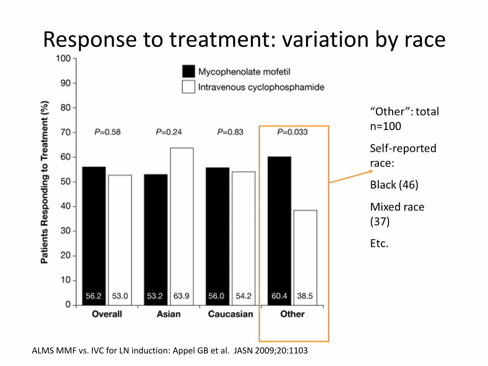
Response to treatment: variation by race
ALMS MMF vs. IVC for LN induction: Appel GB et al. JASN 2009;20:1103

Risk Factors for Health Disparities in SLE
• Non-modifiable:
– Genetics, sex
– Intrinsic severity of disease
• Potentially modifiable:
– Environmental factors
– Health system factors
– Knowledge of SLE

• The Lupus Initiative® is a national education programdesigned to reduce health disparities in lupus.
• ANTHC has an educational series calledLupusConnect that was developed in partnershipwith The Lupus Initiative® as an interactiveeducational series for providers working in IndianHealth Service or tribal facilities.
The Lupus Initiative