Syringocystadenomapapilliferum Arising from the External...
3
J Int Adv Otol 2016; 12(1): 116-8 • DOI: 10.5152/iao.2016.2036 INTRODUCTION Any mass or polypoid lesion in the external auditory canal is usually challenging as a proper diagnosis cannot be made. This is due to its nonspecific clinical and radiological findings [1] . To accurately diagnose the nature of any aural polyp, imaging techniques and polypectomy should be considered to be mindful that it may uncover a serious disease process or even a surprising one [2] . Syringocystadenomapapilliferum (SCAP) is an extremely rare benign tumor originating from modified apocrine sweat (ceruminous) glands showing extensive papillary growth of epithelial elements deep into the dermis [3] . According to the World Health Organiza- tion, ceruminous gland tumors are classified into benign ceruminous adenomas, chondroidsyringomas, SCAPs and malignant ade- nocarcinomas, adenoid cystic carcinomas, and mucoepidermoid carcinomas [4] . Eleven cases of SCAP of the cartilaginous par0000t of the external auditory canal have been mentioned in the literature till 2006 [5] . To my knowledge, another two cases have been mentioned since that time: one associated with lipomatous apocrine adenomas [6] and the other originating from the bony part of the ear canal [7] . CASE PRESENTATION A 36-year-old female presented to our tertiary hospital with right ear pain for a duration of 3 days duration following self-manipu- lation with a cotton bud. The patient did not have a history of a previous similar condition, ear discharge, or hearing loss before this episode. She had no history of general organ system problems. She also had a history of diminished hearing in the right ear and a sense of right ear fullness for a duration of 2 months. No tragal or mastoid tenderness was found in both ears. A left ear examination showed normal findings. A right ear examination by otoscopy and ear endoscopy showed a well-circumscribed soft mass lesion filling the deep bony part of the ear canal (Figure 1). It was not tender to touch, and the probe easily passed around the swelling from all directions except the posterior part. The right ear drum could not be visualized as it was totally obscured by the swelling. The Rinne test was positive in both ears, and Weber test was central. Pure tone audiometry showed mild conductive hearing loss of the right ear. Aspiration was attempted, with no fluid coming out from the swelling. Computed tomography (CT) with contrast for the right petrous bone was performed to identify the mass extension and condition of the middle ear. It revealed a nonhomogenous soft tissue lesion of a density of 30–40 HU and measuring approximately 1.3×0.7 cm, which filled the deep bony part of the right ear canal starting from the bony cartilaginous junction, and the right ear drum could not be separately visualized from the lesion. The middle ear cleft was intact (Figure 2). Because there was no destruction of the bony meatal wall as seen in the CT scan, a suggestion of a benign pathology was raised and a decision of excision biopsy of the lesion was taken. Corresponding Address: Hazem M. Abdel Tawab E-mail: [email protected] Submitted: 23.12.2015 Revision received: 06.01.2016 Accepted: 13.01.2016 ©Copyright 2016 by The European Academy of Otology and Neurotology and The Politzer Society - Available online at www.advancedotology.org 116 Syringocystadenomapapilliferum Arising from the External Auditory Canal: A Rare Tumor in a Rare Site Syringocystadenomapapilliferum is an extremely rare benign adnexal tumor of the scalp and face region. This is a report of the case of a female patient with syringocystadenomapapilliferum originating from the bony cartilaginous junction of the external auditory canal. A provisional diag- nosis of an inflammatory polyp was made based on the clinical and radiological findings. Diagnosis was established only after the histopatholog- ical examination. This article represents a report of a rare skin disease and a rare site of affection. It emphasizes the role of histopathology in the diagnosis of such a condition with debatable clinical and radiological findings. KEYWORDS: Syringocystadenomapapilliferum, adnexal, external auditory canal Hazem M. Abdel Tawab, Khalid F. Amish, Ibrahim A. Sulaiman, Rubyath C. Rajib Department of Otorhinolaryngology, Cairo University School of Medicine, Cairo, Egypt (HMAT) Department of Otorhinolaryngology, Sultan Qaboos Hospital, Salalah, Oman (HMAT, KFA) Department of Pathology, Sultan Qaboos Hospital, Salalah, Oman (IAS, RCR) Case Report
Transcript of Syringocystadenomapapilliferum Arising from the External...

J Int Adv Otol 2016; 12(1): 116-8 • DOI: 10.5152/iao.2016.2036
INTRODUCTIONAny mass or polypoid lesion in the external auditory canal is usually challenging as a proper diagnosis cannot be made. This is due to its nonspecific clinical and radiological findings [1]. To accurately diagnose the nature of any aural polyp, imaging techniques and polypectomy should be considered to be mindful that it may uncover a serious disease process or even a surprising one [2].
Syringocystadenomapapilliferum (SCAP) is an extremely rare benign tumor originating from modified apocrine sweat (ceruminous) glands showing extensive papillary growth of epithelial elements deep into the dermis [3]. According to the World Health Organiza-tion, ceruminous gland tumors are classified into benign ceruminous adenomas, chondroidsyringomas, SCAPs and malignant ade-nocarcinomas, adenoid cystic carcinomas, and mucoepidermoid carcinomas [4]. Eleven cases of SCAP of the cartilaginous par0000t of the external auditory canal have been mentioned in the literature till 2006 [5]. To my knowledge, another two cases have been mentioned since that time: one associated with lipomatous apocrine adenomas [6] and the other originating from the bony part of the ear canal [7].
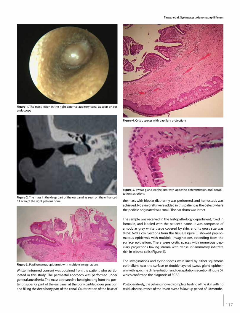
CASE PRESENTATIONA 36-year-old female presented to our tertiary hospital with right ear pain for a duration of 3 days duration following self-manipu-lation with a cotton bud. The patient did not have a history of a previous similar condition, ear discharge, or hearing loss before this episode. She had no history of general organ system problems. She also had a history of diminished hearing in the right ear and a sense of right ear fullness for a duration of 2 months. No tragal or mastoid tenderness was found in both ears. A left ear examination showed normal findings. A right ear examination by otoscopy and ear endoscopy showed a well-circumscribed soft mass lesion filling the deep bony part of the ear canal (Figure 1). It was not tender to touch, and the probe easily passed around the swelling from all directions except the posterior part. The right ear drum could not be visualized as it was totally obscured by the swelling.
The Rinne test was positive in both ears, and Weber test was central. Pure tone audiometry showed mild conductive hearing loss of the right ear. Aspiration was attempted, with no fluid coming out from the swelling.
Computed tomography (CT) with contrast for the right petrous bone was performed to identify the mass extension and condition of the middle ear. It revealed a nonhomogenous soft tissue lesion of a density of 30–40 HU and measuring approximately 1.3×0.7 cm, which filled the deep bony part of the right ear canal starting from the bony cartilaginous junction, and the right ear drum could not be separately visualized from the lesion. The middle ear cleft was intact (Figure 2). Because there was no destruction of the bony meatal wall as seen in the CT scan, a suggestion of a benign pathology was raised and a decision of excision biopsy of the lesion was taken.
Corresponding Address: Hazem M. Abdel Tawab E-mail: [email protected]
Submitted: 23.12.2015 Revision received: 06.01.2016 Accepted: 13.01.2016 ©Copyright 2016 by The European Academy of Otology and Neurotology and The Politzer Society - Available online at www.advancedotology.org116
Syringocystadenomapapilliferum Arising from the External Auditory Canal: A Rare Tumor in a Rare Site
Syringocystadenomapapilliferum is an extremely rare benign adnexal tumor of the scalp and face region. This is a report of the case of a female patient with syringocystadenomapapilliferum originating from the bony cartilaginous junction of the external auditory canal. A provisional diag-nosis of an inflammatory polyp was made based on the clinical and radiological findings. Diagnosis was established only after the histopatholog-ical examination. This article represents a report of a rare skin disease and a rare site of affection. It emphasizes the role of histopathology in the diagnosis of such a condition with debatable clinical and radiological findings.
KEYWORDS: Syringocystadenomapapilliferum, adnexal, external auditory canal
Hazem M. Abdel Tawab, Khalid F. Amish, Ibrahim A. Sulaiman, Rubyath C. RajibDepartment of Otorhinolaryngology, Cairo University School of Medicine, Cairo, Egypt (HMAT)Department of Otorhinolaryngology, Sultan Qaboos Hospital, Salalah, Oman (HMAT, KFA)Department of Pathology, Sultan Qaboos Hospital, Salalah, Oman (IAS, RCR)
Case Report
Written informed consent was obtained from the patient who partic-ipated in this study. The permeatal approach was performed under general anesthesia. The mass appeared to be originating from the pos-terior superior part of the ear canal at the bony cartilaginous junction and filling the deep bony part of the canal. Cauterization of the base of
the mass with bipolar diathermy was performed, and hemostasis was achieved. No skin grafts were added in this patient as the defect where the pedicle originated was small. The ear drum was intact.
The sample was received in the histopathology department, fixed in formalin, and labeled with the patient’s name. It was composed of a nodular grey white tissue covered by skin, and its gross size was 0.8×0.6×0.2 cm. Sections from the tissue (Figure 3) showed papillo-matous epidermis with multiple invaginations extending from the surface epithelium. There were cystic spaces with numerous pap-illary projections having stroma with dense inflammatory infiltrate rich in plasma cells (Figure 4).
The invaginations and cystic spaces were lined by either squamous epithelium near the surface or double-layered sweat gland epitheli-um with apocrine differentiation and decapitation secretion (Figure 5), which confirmed the diagnosis of SCAP.
Postoperatively, the patient showed complete healing of the skin with no residualor recurrence of the lesion over a follow-up period of 10 months.
Figure 1. The mass lesion in the right external auditory canal as seen on ear endoscopy
Figure 2. The mass in the deep part of the ear canal as seen on the enhanced CT scan pf the right petrous bone
Figure 3. Papillomatous epidermis with multiple invaginations
Figure 4. Cystic spaces with papillary projections
Figure 5. Sweat gland epithelium with apocrine differentiation and decapi-tation secretions
117
Tawab et al. Syringocystadenomapapilliferum
DISCUSSIONCeruminous gland tumors are rare and represent less than 5% of all tumors of the external auditory canal and auricular tumors [8].
Ceruminous glands are found in the outer cartilaginous part of the external auditory canal, and there is a general acceptance that the bony part of the canal is devoid of them.
Syringocystadenomapapilliferum is one of these rare tumors that has been mentioned in the literature in the early 20th century in dermato-logical case reports as nevus syringocystadenomatosuspapilliferus [9].
It has been also reported that SCAP is usually associated with a nevus in the head and neck region [10]. However, there was no nevus in the head and neck region in our patient.
In this presented case, SCAP was found to originate from the bony cartilaginous junction and fill the deep bony part of the canal. To my knowledge, only one study has demonstrated the case of a patient with SCAP arising from the bony part of the canal [7].
Radiological examinations such as computed tomography are man-datory in a case of polypoid lesions obstructing the ear canal as pos-sibilities of a surprising pathology like foreign body [2] or any other unexpected pathology.
Magnetic resonance (MR) imaging is also a useful technique for pol-ypoid lesions of the external auditory canal. Recently, Kamakura et al. demonstrated the MR imaging criteria of SCAP of the external au-ditory canal as intermediate signal intensities on T1- and T2-weight-ed images and slight enhancements on gadolinium-enhanced T1-weighted images [5].
Histological diagnosis is the best tool for such polypoid lesions in the ear canal, and in these cases, excision biopsy is better than incision biopsy to avoid the risk of being potentially malignant mass [11], not having sufficient biopsy tissue, or uncontrollable hemorrhage in glo-mus or facial palsy in schwannomas [5].
Regular follow-up should be done for such a tumor for the fear of recurrence.The patient with SCAP presented in this study had been followed for 9 months after complete excision, and no recurrence oc-curred during this follow-up period.
Mass lesions in the external auditory canal are difficult to diagnose based on clinical and radiological data. Histopathology usually rep-resents the main diagnostic tool in these cases. SCAP, despite its rari-ty, should be kept in mind in the differential diagnosis of mass lesions
in the external auditory canal. A rare site of origin of such a lesion had been described in the present study.
Informed Consent: Written informed consent was obtained from the patient who participated in this study.
Peer-review: Externally peer-reviewed.
Author Contributions: Concept - H.M.A.T.; Design - H.M.A.T., K.F.A.; Super-vision - H.M.A.T.; Resources - H.M.A.T., I.A.S.; Materials - H.M.A.T., R.C.R.; Data Collection and/or Processing - H.M.A.T, R.C.R.; Analysis and/or Interpreta-tion - H.M.A.T., K.F.A., R.C.R.; Literature Search - H.M.A.T.; Writing Manuscript - H.M.A.T.; Critical Review - H.M.A.T., I.A.S.
Conflict of Interest: No conflict of interest was declared by the authors.
Financial Disclosure: The authors declared that this study has received no financial support.
REFERENCES1. Markou K, Karasmanis I, Vlachtsis K, Petridis D, Nikolaou A, Vital V. Primary
pleomorphic adenoma of the external ear canal. Report of a case and literature review. Am J Otolaryngol 2008; 29: 142-6. [CrossRef]
2. Abdel Tawab HM, Kumar VR, Tabook SM. A surprising finding after exter-nal ear polypectomy in a deaf mute patient. Case Rep Otolaryngol 2015 (2015); Article ID: 401708, 3 pages. [CrossRef]
3. Marioni G, Brescia G, Staffieri C, Poletti A, Staffieri A. Syringocystoade-nomapapilliferum of the external ear canal: an immunohistochemical study. Acta Oto-Laryngologica 2004; 124: 761-2.
4. Michaels L, Thompson LDR. Ceruminous gland neoplasms of external au-ditory canal and cylindrom inWorld Health Organization Classification of Tumors. Pathology and Genetics. Head and Neck Tumors, L. Barnes, J. W. Eveson, P. Reichart et al., 2005; Eds., pp. 331–333, IARC Press, Lyon, France.
5. Kamakura T, Horii A, Mishiro Y, Takashima S, Kubo T. Magnetic resonance imaging of syringocystadenomapapilliferum of the external auditory ca-nal. AurisNasus Larynx 2006; 33: 53-5. [CrossRef]
6. Su TC, Shen KH, Wang HK, Chu PY, Chen ML. Lipomatous apocrine ad-enoma with syringocystadenomapapilliferum arising from the external auditory canal. Head Neck Oncol 2011; 3: 36. [CrossRef]
7. Arechvo A, Balseris S, Neverauskiene L, Arechvo I. Syringocystade-nomapapilliferumofthe bony external auditory canal: a rare tumor in a rare location. Case Rep Otolaryngol 2013; Article ID 541679.
8. Thompson LD, Nelson BL, Barnes EL. Ceruminous adenomas: a clinic pathologic study of 41 cases with a review of the literature. Am J Surg Pathol 2004; 28: 308-18. [CrossRef]
9. Werther L. Syringadenomapapilliferum (Naevussyringadenomatosuspa-pilliferus. Archivf¨ urDermatologie und Syphilis 1913; 116: 865-70.
10. Monticciolo NL, Schmidt JD, Morgan MB. Verrucous carcinoma arising with-in syringocystadenomapapilliferum. Ann Clin Lab Sci 2002; 32: 434-7.
11. Diaz RC, Babu SC. Ductal carcinoma arising from syringocystadenomapapil-liferumin the external auditory canal. Otol Neurotol 2007; 28: 873-4. [CrossRef]
118
J Int Adv Otol 2016; 12(1): 116-8
![Journal of Neurological Sciences [Turkish] 33:(4 http ...nsnjournal.org/sayilar/96/buyuk/pdf_JNS_1021.pdf · verb epilambanein, meaning to be seized, to be overwhelmed by surprise.](https://static.fdocuments.in/doc/165x107/5c67f65409d3f28e058ca306/journal-of-neurological-sciences-turkish-334-http-verb-epilambanein.jpg)