Synthetic biology approaches for the production of chiral ...
200
1 Synthetic biology approaches for the production of chiral aminoalcohols in engineered E. coli strains by Panwajee Payongsri A thesis submitted for the degree of Doctor of Philosophy of University College London Department of Biochemical Engineering University College London 2014
Transcript of Synthetic biology approaches for the production of chiral ...
1
Synthetic biology approaches for the
production of chiral aminoalcohols in
engineered E. coli strains
by
Panwajee Payongsri
A thesis submitted for the degree of Doctor of Philosophy of University
College London
Department of Biochemical Engineering
University College London
2014
2
Declaration
‘I, Panwajee Payongsri, confirm that the work presented in this thesis is my own. Where
information has been derived from other sources, I confirm that this has been indicated in
the thesis.’
Candidate’s signature
..................................................................
Panwajee Payongsri
3
Acknowledgement
Completing my PhD at the Biochemical Engineering Department of UCL has
provided me not only with the research skills but also numerous invaluable experiences.
The research would never have been successfully accomplished without a number of
people around me.
First of all I would like to thank my supervisor Professor Paul Dalby for his
consistent support, dedication and patience. He has given me so much freedom to explore
my curiosities and allowed me to freely share my research ideas. This approach led to very
useful discussions, rapid progress with my research and a truly enjoyable project. I would
like to further extend my gratitude for his firm support for my decisions while I was the
demonstrator for the biotransformation practical. Throughout the three years of research, I
also collaborated with Professor Helen Hailes and David Steadman from the Chemistry
Department. Professor Hailes has been very dedicated and assertive which assured the
direction and the progress of the project. David Steadman provided me not only the
synthetic materials but also his time for discussions. This significantly improved my
understanding of the chemistry side of the project which greatly facilitated my research. In
sum, it was a great honour to work with such a fantastic research team.
My PhD would have never started smoothly without Leonardo Rios and
Phattaraporn Morris who spent their time training me to safely use and maintain all the
laboratory facilities. I would also like to thank Dhushy Stanislaus, Facilities Manager, who
has put tremendous care and attention into maintaining laboratory safety, ensuring a good
working environment and quickly resolving laboratory issues. I really appreciated her
support for my role as lab manager during my final year. This was a once in a lifetime
experience that I would never be able to find in day-to-day research activities.
The opportunity to study abroad was financially sponsored by the Royal Thai
Government and I feel truly thankful to all Thai tax payers. I would also like to thank the
Biochemical Engineering Department for additional funding for my international
conference attendances. I would like to mention that I would never be able to apply for this
degree without the support and recommendations of both Professor Gary Lye and Dr. Paul
Dalby.
Throughout my life, my family has consistently supported me and I would never
have achieved what I have without them. I feel especially grateful to my parents, Pichai and
Sumalee Payongsri. I would also like to thank my brothers and sister, Nut, Srisakul and
Titipol Payongsri, for their advice and moral support which has always lifted my spirits.
Lastly, I would like to thank the Osters who made me feel that the UK is my second home.

4
Abstract
Transketolase catalyses asymmetric carbon-carbon bond formation and produces
1,3-dihydroxyketones, a functionality that is found in a vast number of natural and
synthetic compounds. The wild-type transketolase enzymes from several species can
accept a wide range of aldehydes, but industrially exploitable levels of activity tend to be
limited to natural substrates and small aliphatic aldehydes. Several single mutants of the
transketolase from E. coli were found to have enhanced activity towards non-
phosphorylated substrates, non-hydroxylated aldehydes, and cyclic aldehydes. However,
aromatic aldehydes still suffer from poor activities and yields while the key bottlenecks
have not been identified. In order to improve transketolase activity towards aromatic
aldehydes, small transketolase libraries were created with the aid of available crystal
structures, and computational modelling of substrate binding. The new mutants were then
assessed alongside transaminase for the ability to synthesise novel aromatic amino
alcohols, which would provide building blocks for chloramphenicol and its derivatives.
The strategy for creating new libraries from combining single mutations can have
significant impact not only on the activity but also the stability of the enzyme due to the
synergy between residues. Random recombination of previously identified single mutants,
which had higher activities on either glycolaldehyde or propionaldehyde, produced new
double and triple mutants which all had lower activities for both substrates, and poor
stabilities as illustrated by formation of inclusion bodies. On the other hand, the
combination of D469 and R520 variants, two sites identified within a co-evolved network,
created mutants that maintained the activity towards propionaldehyde, while significantly
reducing the inclusion body formation. This implied that they were more stable and could
be suitable templates for further rounds of mutation.
In the second stage, the factors influencing the bioconversion of aromatic
aldehydes by transketolase were identified through crystal structure analysis. 3-
formylbenzoic acid (3-FBA) and 4-formylbenzoic acid (4-FBA) were chosen as substrates
that might re-establish hydrogen bonds between the carboxylate group and the so-called
phosphate-binding residues within the active site. Both substrates were tested with wild
type and a set of mutants that were previously reported to accept aromatic aldehydes. This
showed that the steric hindrance from D469 impeded the aromatic substrate to access to
the active site, and that D469T was the most active mutant for both new substrates. Site-
directed mutagenesis at the phosphate-binding residues confirmed that R358, H461, and
R520 interacted with carboxylate groups of both substrates and also suggested that the

5
affinity between the enzyme and the aromatic aldehyde, as well as their proximal
orientations, was the key factor governing the reaction rate. This was also supported by the
computational modelling of 3-FBA and 4-FBA binding in energy-minimised D469Tand
D469T/R520Qstructures.
Site-saturation mutagenesis at S385 and R358 was performed to further improve
the activity of transketolase for 3-formylbenzoic acid, 4-formylbenzoic acid, and also 3-
hydroxybenzaldehyde (3-HBA). Due to the stability, D469T/R520Q was chosen as a
template. S385E/D469T/R520Q was found with improved activity on 3-FBA and relieved
substrate inhibition. Both S385Y/D469T/R520Q and S385T/D469T/R520Q were found to
have higher activity and yields with both 4-FBA and 3-HBA. The enhancements in all the
mutants were due to significant improvements in kcat. Very large KM values were observed
for 3-HBA which confirmed that enzyme-substrate affinity was the major factor limiting the
bioconversion rate. The synergic and kinetic study of S385Y/D469T and S385T/D469T
illustrated that the triple mutants followed a different adaptive walk to reach the same
optima.
In order to directly use 3-(1,3-Dihydroxy-2-oxopropyl) benzoic acid (3-DOPBA) and
4-(1,3-Dihydroxy-2-oxopropyl) benzoic acid (4-DOPBA) products from the transketolase
reaction, as substrates in the transaminase reaction, with minimal competition from the
leftover aldehydes, their conversion yields would have to be optimised. This was achieved
by supplementing the reactions with an additional 50% HPA, which increased the yield for
3-DOPBA and 4-DOPBAto 90% and 70%, respectively. Both optimised reactions were fed
into the transaminase reactions to synthesise novels aromatic aminodiols. However, none
of the available transaminases appeared to accept either of the compounds. The
competitive reaction between 4-FBA and 4-DOPBA in an amination reaction suggested that
the major cause of the inability of CV2025 transaminase to catalyse 4-DOPBA was the
inability of 4-DOPBA to access into the active site.

6
Abbreviations
3-DOPBA 3-(1,3-Dihydroxy-2-oxopropyl )benzoic acid
3-FBA 3-Formylbenzoic acid
3-HBA 3-hydroxybenzaldehyde
3-NBA 3-Nitrobenzaldehyde
4-DOPBA 4-(1,3-Dihydroxy-2-oxopropyl )benzoic acid
4-FBA 4-Formylbenzoic acid
AMP Ampicillin
APD (2S,3S)-2-aminopentane-1,3-diol
BA Benzaldehyde
BSA Bovine Serum Albumin
DHEThDP Dihydroxyethyl-Thiamine diphosphate
DHP 1,3 dihydroxypentane-2one
DPP 1,3-dihydroxy-1-phenylpropane-2-one
E-4P Erythrulose 4-phosphate
ee enantiomeric excess
F6P fructose 6-phosphate
GA Glycolaldehyde
HPA Hydroxypyruvic acid
HPLC High-performance liquid chromatography
LB Lysogenic broth
MBA Methylbenzylamine
MW Molecular Weight
PA propionaldehyde
PAGE polyacrylamide gel electrophoresis
PCR polymerase chain reaction
PLP pyridoxal 5-phosphate
PP domain Phosphate binding domain
Pyr domain pyrimidine binding domain
SDS sodium dodecylsulphate
TAm Transaminase
ThDP Thiamine Diphosphate
TK Transketolase
X5P xylulose 5-phosphate

7
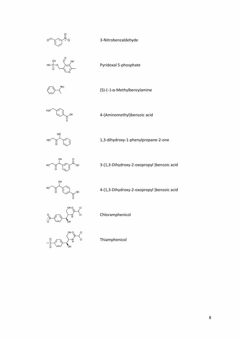
List of chemical structures
Hydroxypyruvic acid
Glycolaldehyde
Propionaldehyde
Thiamine diphosphate
Erythrulose
1,3 dihydroxypentane-2one
3-Formylbenzoic acid
4-Formylbenzoic acid
3-Hydroxybenzaldehyde
Benzaldehyde
8
3-Nitrobenzaldehyde
Pyridoxal 5-phosphate
(S)-(−)-α-Methylbenzylamine
4-(Aminomethyl)benzoic acid
1,3-dihydroxy-1-phenylpropane-2-one
3-(1,3-Dihydroxy-2-oxopropyl )benzoic acid
4-(1,3-Dihydroxy-2-oxopropyl )benzoic acid
Chloramphenicol
Thiamphenicol
9
Table of Contents
Declaration 2
Acknowledgement 3
Abstract 4
Abbreviations 6
List of chemical structures 7
List of Figure 14
List of Tables 17
List of equation 18
List of reaction scheme 19
1. Chapter 1: Literature review 20
1.1. Introduction 20
1.1.1. Biocatalysis 20
1.1.2. Protein engineering: applications and strategies 21
1.1.3. Evaluation of the kinetic parameters for organic synthesis 27
1.1.4. Transketolase 29
1.2. Research aims 44
2. Chapter 2: Materials and methods 46
2.1. Chemicals 46
2.2. Media and buffer reagent preparation 46
2.2.1. Ampicillin (Amp) 46
2.2.2. Lysogenic broth (LB) 47
2.2.3. LB agar with 150 μg/ml ampicillin (LB-Amp) 47
2.2.4. Tris buffer 47
2.3. Transketolase production 47
2.4. Preparation of clarified lysate 48
2.5. Protein concentration quantification 49
2.5.1. Bradford assay 49

10
2.5.2. SDS-PAGE and densitometry 49
2.6. Enzymatic reaction 52
2.6.1. Cofactor solution preparation 52
2.6.2. Substrate preparations 52
2.6.3. Activity assay 53
2.7. HPLC system 53
2.7.1. HPLC system for aliphatic compounds 53
2.7.2. HPLC system for aromatic compounds 54
2.8. Standard product and substrate graphs 54
2.9. Initial rate and specific activity calculation 55
2.10. Synergy calculation 55
2.11. E. coli strain and glycerol stock preparation 56
2.12. Plasmid extraction and storage 57
2.13. DNA gel electrophoresis 57
2.14. Transketolase library construction 58
2.14.1. Primer design and preparation 58
2.14.2. Polymerase Chain Reaction Conditions 60
2.14.3. Transformation 60
2.15. Purified enzyme preparation 61
2.15.1. Buffers used in purification 61
2.15.2. Purification procedure 61
3. Chapter 3: Recombination of existing single mutants 63
3.1. Introduction 63
3.2. Materials and method 65
3.2.1. Materials 65
3.2.2. Mutant construction 65
3.2.3. Protein quantification 65
3.2.4. Expression level study 65

11
3.2.5. Identification of the protein in the insoluble fraction 66
3.2.6. Activity assay 66
3.2.7. HPLC analysis 67
3.3. Results and discussion 68
3.3.1. The analysis of the reaction samples by HPLC 68
3.3.2. The specific activities of transketolase mutants towards glycolaldehyde and
propionaldehyde 69
3.3.3. The locations of the targets residues, their functions, and interactions 72
3.3.4. H26Y, the inactive mutant 77
3.3.5. The expression level of all mutants 78
3.3.6. Correlation of expression and activity 83
3.4. Conclusion 84
4. Chapter 4: Rational design of substrate and TK for aromatic aldehyde 86
4.1. Introduction 86
4.2. Materials and methods 87
4.2.1. Chemicals and reagents 87
4.2.2. Transketolase library 88
4.2.3. Enzyme preparation and quantification 88
4.2.4. Screening assay when using 3-FBA and 4-FBA as a substrates 88
4.2.5. Enzyme kinetics for 3-FBA and 4-FBA 89
4.2.6. Propionaldehyde activity 90
4.2.7. Synthesis of 3- and 4-(1,3-Dihydroxy-2-oxopropyl)benzoic acid 90
4.2.8. Computational docking of 3-FBA and 4-FBAinto TK mutant structures 91
4.2.9. Reaction of D469T with 3-nitrobenzaldehyde (unpublished data) 91
4.3. Results and discussions 93
4.3.1. Screening for 3-FBA and 4-FBA 93
4.3.2. Kinetics of the mutants towards 3-FBA and 4-FBA 100
4.3.3. Computational docking of 3-FBA and 4-FBA 107

12
4.4. Conclusion 109
5. Chapter 5: Second Generation Engineering of Transketolase for Aromatic Ketodiols 110
5.1. Introduction 110
5.2. Materials and methods 112
5.2.1. Chemicals and reagents 112
5.2.2. Transketolase library 112
5.2.3. Library Screening 112
5.2.4. Cell culture and protein quantification for detailed enzyme kinetics 114
5.2.5. Detailed enzyme kinetics 114
5.2.6. Computational modelling of the binding of aldehyde substrate 115
5.3. Results and discussions 116
5.3.1. The 3-HBA reaction samples when analysed by HPLC 116
5.3.2. High-throughput screening 116
5.3.3. Kinetic parameters of the mutants with 3-FBA, 4-FBA, and 3-HBA 121
5.3.4. The relationship between the enzyme activity and the final yields of
different aromatic aldehydes 127
5.3.5. Computational docking of substrates into the energy minimised
S385E/D469T/R520Q and S385Y/D469T/R520Q structures 128
5.3.6. Interactions between S385 and R520 mutations 132
5.4. Conclusion 135
6. Chapter 6: Synthesis of novel aromatic aminodiols through the coupling of
transketolase with transaminase 136
6.1. Introduction 136
6.2. Materials and methods 139
6.2.1. Materials 139
6.2.2. Optimising the synthesis of the aromatic dihydroxy ketones by TK 140
6.2.3. Transaminase enzyme preparation 140
6.2.4. Protein quantification 141

13
6.2.5. HPLC 141
6.2.6. MBA inhibition study 142
6.2.7. Screening transaminases for aromatic dihydroxy ketones 142
6.2.7.1. Temperature and pH optimisation 143
6.2.7.2. Control reactions 143
6.2.8. Competitive reaction 144
6.2.9. Kinetic study of 3-FBA and 4-FBA with CV2025 145
6.3. Results and discussions 146
6.3.1. The yield of aromatic dihydroxy ketones after HPA fed-batch 146
6.3.2. MBA inhibition study 148
6.3.3. Screening transaminases for aromatic dihydroxy ketones 149
6.3.4. Competitive reaction 155
6.3.5. Kinetics study for 3-FBA and 4-FBA 157
6.3.6. Active site structure and possible reasons for slow reaction with aromatic
dihydroxy ketones and 3-FBA 159
6.4. Conclusion 161
7. Conclusion and future research recommendation 162
7.1. Future research recommendation for transketolase 164
7.2. Future research recommendation for transketolase-transaminase pathway for the
synthesis of aromatic aminoalcohols 165
7.3. Overall evaluation and recommendation 165
8. References 167
9. Appendices 180
9.1. Directed evolution to re-adapt a co-evolved network within an enzyme 180
9.2. Rational substrate and enzyme engineering of transketolase for aromatics 201
9.3. Second Generation Engineering of Transketolase for Polar Aromatic Aldehyde
Substrates 210
14
List of Figure
Figure 1.1 The Fitness landscape of a protein. ...................................................................... 23
Figure 1.2 The process of directed evolution. ....................................................................... 24
Figure 1.3 Transketolase structure from E. coli (PDB ID: 1QGD). .......................................... 30
Figure 1.4 Two conformations of ThDP. ................................................................................ 31
Figure 1.5 The Catalytic mechanism of transketolase. .......................................................... 33
Figure 1.6 Residues within E. coli transketolase active site................................................... 36
Figure 2.1 The map of pQR791 plasmid. ............................................................................... 48
Figure 2.2 An example of SDS-PAGE gel with standard TK gradient. .................................... 50
Figure 2.3 The plot between the migration distances of the protein markers and their
log(MW). ................................................................................................................................ 51
Figure 2.4 The relationship between the intensity of the band and the amount of �
purified TK and � BSA ............................................................................................................ 52
Figure 2.5 The calculation of initial rate using SigmaPlot. ..................................................... 55
Figure 3.1 Standard graphs of a) HPA, b) erythrulose, and c) DHP. ...................................... 67
Figure 3.2 The chromatograms of reaction samples analysed by Aminex HPx-87H,
300x7.8mm column (Bio-Rad, UK) according to the protocol in Chapter 2, section 2.7.1. ... 68
Figure 3.3 The location of the residues subject to mutation in this study. Black dotted lines
are hydrogen bonds. .............................................................................................................. 73
Figure 3.4 The relative activity of each mutant in group I towards a) glycolaldehyde and b)
propionaldehyde. ................................................................................................................... 75
Figure 3.5 The relative activity of group 2 mutants towards propionaldehyde. ................... 77
Figure 3.6 The fraction of transketolase. ............................................................................... 79
Figure 3.7 The scatter plot between the P2 concentration and the TK soluble: inclusion
body ratio. .............................................................................................................................. 83
Figure 3.8 The relationship between the specific activity towards glycolaldehyde and
propionaldehyde and the expression level of the mutant. ................................................... 84
Figure 4.1 The standard graph of 3-FBA and 4-FBA. .............................................................. 90
Figure 4.2 The ribose 5-phosphate-bound E. coli transketolase crystal structure and the
computational docking of 3-FBA and 4-FBA into the energy-minimised transketolase
mutant structures. ................................................................................................................. 94
Figure 4.3 The Chromatograms of 3-FBA and 4-FBA reactions when analysed by ACE5 C18
reverse phase column (150 × 4.6 mm) and Aminex HPx-87H column respectively. ............. 96

15
Figure 4.4 The chromatogram of 3-NBA reaction when incubated the substrate with
different mutants. .................................................................................................................. 99
Figure 4.5 The Michaelis-Menten plot of all the six mutants for 3-FBA. ............................. 101
Figure 4.6 The Michaelis-Menten plot of D469T and D469T/R520Q for 4-FBA. ................. 102
Figure 4.7 Comparison between the experimentally determined binding constants (KM) and
the calculated Kd from binding energy using AudoDOCK . .................................................. 108
Figure 5.1 The standard graph of 3-HBA. ............................................................................ 115
Figure 5.2 The chromatograms of the 3-HBA reaction samples. ......................................... 116
Figure 5.3 Comparison of the specific activities by clarified lysate and the 1-hour
conversions in whole cell which were standardised by OD600. .......................................... 117
Figure 5.4 The comparison of the 18-hour yields by clarified lysate and whole cells in high
throughput screen towards 4-FBA � and 3-HBA �. ........................................................... 118
Figure 5.5 The cross comparison of the yields between three substrates by
S385X/D469T/R520Q library. ............................................................................................... 119
Figure 5.6 The cross comparison of the yields between three substrates by
R358X/D469T/R520Q library. .............................................................................................. 120
Figure 5.7 The kinetic plots of all three mutants for 3-FBA, 4-FBA, and 3-HBA. ................. 122
Figure 5.8 The relationship between the activities of all the mutants and the % yields of all
the aromatic substrates. ...................................................................................................... 128
Figure 5.9 The computational docking of 3-FBA into the energy minimised a)
S385E/D469T/R520Q, b) D469T, c) D469T/R520Q. ............................................................. 130
Figure 5.10 The computational docking of a) 4-FBA, and b) 3-HBA into the energy
minimised S385Y/D469T/R520Q. ........................................................................................ 131
Figure 6.1 The reaction mechanism of transaminase enzymes when catalysing the transfer
of an amine group from one amino acid to an alpha keto acid. .......................................... 137
Figure 6.2 Chromatogram of the 1-minute reaction sample from the amination reaction of
3-DOPBA obtained from transketolase reaction. ................................................................ 142
Figure 6.3 The reaction profile of equimolar3-FBA reaction catalysed by
S385E/D469T/R520Q. .......................................................................................................... 147
Figure 6.4 The acetophenone (�) and MBA (�) concentrations at 18 hours after the
reaction between DPP and MBA was started. ..................................................................... 149
Figure 6.5 The overlapped chromatograms of the sample at 3 minutes (�) and 16 hours (�)
of the 3-DOPBA amination reaction by CV2025. ................................................................. 151
16
Figure 6.6 The overlapped chromatograms of the 4-DOPBA reaction samples at �2
minutes, �6 hours, and � 26 hours. ................................................................................... 152
Figure 6.7 Comparison of the peak areas of different compounds at different temperatures,
pH 7.5. .................................................................................................................................. 154
Figure 6.8 The overlapped chromatograms of CV2025 lysate when incubated with 50 mM
Tris buffer, pH 7.5 with 0.4 mM PLP, 0.08 mM ThDP, and 0.3 MgCl2 and at 30°C for (�) 1
minute, (�) 3 hours, and (�) 18 hours. ............................................................................... 155
Figure 6.9 The overlapped chromatograms of purified CV2025 when incubated with 50 mM
Tris buffer, pH 7.5 with 0.4 mM PLP, 0.08 mM ThDP, and 0.3 MgCl2 and at 30°C for (�) 1
minute, (�) 1 hour, and (�) 2 hours. ................................................................................... 155
Figure 6.10 The relationship between the 3-FBA concentration and the initial rate of 3-FBA
amination by CV2025. .......................................................................................................... 158
Figure 6.11 The relationship between the 4-FBA concentration and the initial rate of 4-FBA
amination. ............................................................................................................................ 159
Figure 6.12 The active site channel of CV2025 (PDB ID: 4A6T). The channel was illustrated
as the surface of amino acids constituting the active site. .................................................. 161

17
List of Tables
Table 1.1 The summary of the functions of each residue. .................................................... 41
Table 2.1 The migration distances of the protein markers, their molecular weights, and
their log(molecular weight). .................................................................................................. 50
Table 2.2 The sequences of all the primers used. ................................................................. 59
Table 2.3 The conditions of the PCR reaction. ....................................................................... 60
Table 3.1 The specific activities of all the mutants towards glycolaldehyde and
propionaldehyde determined in this study and previously determined. .............................. 70
Table 3.2 The comparison of the experimental outcomes and conditions between this study
and the previous study when using glycolaldehyde as a substrate. ...................................... 71
Table 3.3 The possible P2 proteins. ....................................................................................... 82
Table 4.1 The KM and kcat values of yeast and E. coli transketolases towards different
acceptor substrates................................................................................................................ 93
Table 4.2 The bioconversion of different aromatic aldehydes by transketolase mutants. ... 95
Table 4.3 The kinetic parameters of all the mutants towards 3-FBA and 4-FBA. ................ 101
Table 4.4 The % HPA left after incubating in different conditions for 18 hours. ................ 104
Table 4.5 The free energy binding (ΔG), the predicted Kd and experimental KM towards 3-
FBA and 4-FBA for D469T and D469T/R520Q. ..................................................................... 108
Table 5.1 The kinetic parameters of all the mutants towards 3-FBA, 4-FBA, and 3-HBA. .. 126
Table 5.2 The kinetic parameters and the changes in the ΔG from D469T (ΔΔG) upon the
introduction of other mutations. ......................................................................................... 135
Table 6.1 The initial rate of 4-FBA amination under different conditions. .......................... 156

18
List of equation
Equation 1.1 Michaelis-Menten equation. Etis the total enzyme concentration, V is velocity,
[S] is substrate concentration ................................................................................................ 28
Equation 4.1 The modified Michaelis-Menten equation used for the calculation of the
kinetic parameters ................................................................................................................. 89
19
List of reaction scheme
Scheme 1.1 General reaction catalysed by transketolase. .................................................... 29
Scheme 1.2 The reaction of transketolase when HPA is used as a substrate. ...................... 29
Scheme 1.3 The novel transketolase-transamiase pathway for the production of aminodiol
via dihydroxyketone intermediate. ........................................................................................ 43
Scheme 6.1 The reaction scheme for the production of chloramphenicol amine analogues
using TK and TAm ................................................................................................................. 138
Scheme 6.2 The degradation of the aromatic dihydroxy ketone through the rearrangement
at the 2-hydroxyl group ....................................................................................................... 148

20
1. Chapter 1: Literature review
1.1. Introduction
1.1.1. Biocatalysis
Enzymes have gained a huge interest in chemosynthesis due to their ability to
catalyse reactions at low temperature with up to 109 folds improvement in the reaction
rates (Koeller and Wong, 2001) together with high stereo- and regio-selectivity (Dalby,
2011; Koeller and Wong, 2001; Turner, 2009). These specificities are vital for the synthesis
of pharmaceutical intermediates. Since they are able to catalyse their reaction with such
high selectivity, the blocking and deblocking of functional groups can be avoided, which
leads to fewer reaction steps (Ishige et al., 2005). The conditions required for the enzymes
to function are often mild and involve less hazardous chemicals making the process more
environmental friendly (Ishige et al., 2005) and minimise side reactions arisen from
extreme pH and temperatures (Patel, 2008). If the harsh process conditions are
unavoidable, the existence of extremophilic organisms have opened the possibilities of
using biocatalysts in extreme conditions (Robertson and Steer, 2004).
Although biocatalysis is an attractive option, naturally exist variants may not have
all the characteristics required for a particular industrial processes such as their stabilities
in the required conditions, substrate spectrum and reactivity with non-natural substrates,
and selectivity. Genomic approaches have been employed to find an enzyme with some
desirable properties (Robertson and Steer, 2004) and protein engineering is often used to
further improve other properties (Dalby, 2011).
The main focus of this research is to engineer a transketolase enzyme to accept
aromatic aldehydes. This could provide an effective tool for the synthesis of
chloramphenicol analogues. Therefore, this chapter focuses on the variety of protein
engineering approaches available, and the background of previous research on
transketolase. In the first half of this chapter, different methodologies of protein
engineering were described together with the advantages and drawbacks of each
approach. The second half of this chapter focuses on the mechanism of transketolase
enzyme, the functions of certain residues within the active site, and the previous
engineered transketolase variants and their substrate spectrum. The information from both

21
sections is vital for designing an approach fur further engineer transketolase. In the last
section, the applications of transketolase in organic synthesis were reviewed.
1.1.2. Protein engineering: applications and strategies
Methods in protein engineering can be classified into two strategies; rational
design, and directed evolution (Behrens et al., 2011; Lehmann and Wyss, 2001).
1.1.2.1. Rational design
Rational design requires sequence information as well as high quality structural
information of the target enzyme and its closely related enzymes in order to select which
residues should be replaced (Bornscheuer and Pohl, 2001; Cedrone et al., 2000). In recent
years, this approach has also begun to employ computational modelling in order to justify
the target, and predict the effect of the mutation prior introducing the mutation (Behrens
et al., 2011). The ultimate holy grail of rational design is de novo enzyme design (Bolon et
al., 2002; Otten et al., 2010). Since several enzymes can adopt the same fold while
catalysing different overall reactions, different sets of catalytic residues within an active
site could also generate several enzymes with diverse mechanisms (Glasner et al., 2006).
Such a principle could be exploited in de novo enzyme design. Such a de novo design was
achieved by the aid of computational modelling where force-field was applied to protein
sequences in order to identify which sequence and conformation is the most stable for a
particular backbone structure. This sequence is then used as a scaffold protein (Poole and
Ranganathan, 2006; Street and Mayo, 1999). The positions and type of catalytic residues
are then calculated and placed to generate active site (Bolon et al., 2002; Dalby, 2007). The
use of computational modelling allows a vast sequence space search and narrow the library
size by eliminating the sequences that are unlikely to fold (Chica et al., 2005). An
alternative strategy to construct a novel enzyme is to design the positions and orientations
of the catalytic residues while the scaffold proteins that could support these requirements
are searched. Once the scaffold is found, the catalytic residues are then grafted into the
scaffold (Röthlisberger et al., 2008). An enzyme catalysing a Kemp elimination, a reaction
mechanism that does not occur in natural enzymes, has been successfully constructed by
this method with a kcat of 0.29 s-1. Directed evolution has further improved this kcat to 1.37
s-1 (Röthlisberger et al., 2008).
Amino acid substitutions are normally achieved by site directed mutagenesis where
the mutations are incorporated into the primers which are then used in polymerase chain
22
reaction to synthesise a new DNA with the replaced codon. The amino acid can be
replaced, deleted, or inserted depending on the primer design. Rational design has been
shown to be successful in several cases. The thermostability of a protein can be improved
by introducing more proline residues, salt bridge, and side chain-side chain hydrogen bonds
but these are not universal rules (Lehmann and Wyss, 2001). Oxidation tolerance could be
improved by the removal of cysteine and methionine (Bornscheuer and Pohl, 2001). The
rational design approach can also improve the enantiomeric excess of an enzyme (Otten et
al., 2010). In addition to improving the properties of an enzyme, rational design is usually
employed when the relationship between amino acid residues and their function need to
be determined.
The library size required for structural-based rational design tends to be smaller
than that of directed evolution discussed in the next section. Consequently, less screening
effort is required in theory (Kazlauskas and Bornscheuer, 2009). However, the limitation of
rational design methods is the requirement of extensive information on the structure and
sequences of enzymes. The unavailability of the structure could be overcome by
computational modelling (Behrens et al., 2011). However, the dynamic and flexible nature
of the protein structure still brings difficulties to rational design. Conformational changes
upon mutation are rather difficult to predict and a small degree of differences can
dramatically influence enzyme function. Therefore, it is extremely difficult to predict the
outcome (Romero and Arnold, 2009).
1.1.2.2. Directed evolution
In contrast to rational design, directed evolution approaches do not require
structural information. The process of directed evolution has been compared with natural
evolution where the fitter species under a certain conditions is the most successful at
passing on their genes to their offspring. The evolution of proteins could be viewed in a
similar way where their function is expressed as their fitness. The mutation is artificially
introduced into the gene and the natural selection occurs in the laboratory where the
mutants are screened and selected. It could be viewed that different mutants have
different degrees of fitness and mapping these sequences with their fitness has been
described as fitness landscape Figure 1.1. In this map, each single sequence differing by one
amino acid is aligned next to each other forming sequence space. Each of them has
different fitness or its ability to function (Smith, 1970). This sequence space also illustrates
evolutionary paths that a functional protein can take by acquiring one mutation at a time

23
with improving fitness. Under the screening or selection pressure, only fitter mutants are
picked for further mutation which can be viewed as walking up the functional hill. This has
been described as adaptive walk (Romero and Arnold, 2009). This sequence space is vast
due to the fact that each position along the sequence can be assigned by 20 amino acids.
For a small peptide of 100-amino acid long, there are already 10020 possible sequences.
However, the majority of these sequences are not functional protein whereas the
functional proteins tend to lie next to each other in the sequence space making hills in the
protein sequence space (Smith, 1970).
Figure 1.1 The Fitness landscape of a protein.
This figure illustrates the relationship between the sequence space and the activity of the enzyme which is
represented by the intensity of the colour as well as the height of the structure. Higher and paler point
illustrates higher activity. a) It is usually believed that among the vast sequence, only a small set of sequences is
functional proteins. In reality, non-functional sequences are often found adjacent to the functional ones. b)
Functional and non-functional sequences are often clustered together due to the epistatic interactions between
amino acids which produce “Badlands” landscape. These interactions make it impossible to jump from one local
optimum to another when the stepwise mutations are introduced. c) A Fujiyama landscape is less rough and the
optimum can be easily reached than b). d) Multiple routes (yellow and green) can be found to reach the optimal
point while certain path (yellow) may lead to local optimum.
The process of directed evolution starts with selecting a template protein with the
some desired functions. This parental protein is then mutated to create a library of mutants

24
whose functions are assessed by screening or selection in order to identify any variants that
have the desired properties. These mutants become the new templates and are subject to
further rounds of mutations and screenings until the desired function is achieved.
Figure 1.2 The process of directed evolution.
Starting with a good choice of parental protein could increase the chances of
success with directed evolution. A good parental protein should have the desired function
to a certain degree. However, mutations could destabilise the protein, so a stable parental
protein is also preferable and can make protein more evolvable (Bloom et al., 2006;
Tokuriki and Tawfik, 2009).
Once the parent is chosen, a library can be created. The strategies that are
normally used to introduce mutations are error prone PCR (Cadwell and Joyce, 1992),
recombinant techniques such as DNA shuffling (Stemmer, 1994a, 1994b) and staggered
extension process (StEP) (Zhao et al., 1998). These recombinant techniques, however,
require high sequence homology (70%) and the crossovers only occur in the regions with
the highest sequence identity (Sieber et al., 2001). In addition, residues around the active
site and substrate binding region tend to be highly conserved, so the chance that they are
replaced by recombination is low (Paramesvaran et al., 2009). As a result, the mutation rate
is still low and new functional proteins are less likely to be created while the library is also
rather biased (Otten et al., 2010). In order to overcome these problems in the early
recombinant techniques, several recombinant strategies that do not require sequence
homology were developed. These include sequence homology–independent protein
recombination (SHIPREC) (Sieber et al., 2001), incremental truncation for the creation of
hybrid enzymes (ITCHY) (Ostermeier et al., 1999), a combination of ITCHY and DNA
shuffling technique called SCRATCHY (Lutz et al., 2001) and several others reviewed by
(Otten and Quax, 2005).

25
Screening or selection processes are methods for assessing the function of
individual mutants within the library in order to identify better variants. The rule “you get
what you screen for” (You and Arnold, 1996) must always be considered when designing
the screening assay in order that the screening result can represent the function of interest
as much as possible and avoid screening for something else. A large library requires the
ability to individually culture a large number of variants as well as a high throughput
screening method to analyse these variants. Screening technologies available include agar
plates, microtitre plates, cell in droplet, cell as microreactor, cell surface display, and in
vitro compartmentalisation (Leemhuis et al., 2009; Yang and Withers, 2009). The first two
technologies are capable to work with the library size not more than 105 variants per day
whereas the last four techniques are normally coupled to fluorescence-activated cell
sorting system (FACS) and can handle between 109 - 1010 variants per day (Leemhuis et al.,
2009). Most of the high throughput assays are coupled with absorption detection methods,
either plate reader or FACS. The use of chromophoric or fluorophoric substrate or product
could ease the process. However, the use of surrogate substrates or the indirect
measurement of product such as derivatives could give inaccurate screening results. This is
because the screened enzymes may evolve to work with the surrogate substrate instead of
the actual substrate (Romero and Arnold, 2009). Despite the risk of inaccuracy, surrogate
substrates still have other applications. If the protein is evolved to accept a new substrate
that is structurally very different from the natural substrate, it could be impossible to
screen the desired substrate directly. Instead, surrogate substrates or intermediates that
their structures are gradually changed to the desired substrates could be used for stepwise
screening (Arnold, 1998; Chen and Zhao, 2005; Savile et al., 2010).
Directed evolution has repeatedly shown to be a powerful tool for protein
engineering without neither structure information nor sequence-function relationship.
Apart from this purpose, it also shows several alternative adaptive pathways together with
their probability of each pathway to occur in natural evolution (Romero and Arnold, 2009).
For a functional multiple-mutation protein where one mutation can be introduced at a
time, there are several routes to introduce each mutation. Any pathway that encounters
deleterious mutations which are recovered by other mutations due to epistatic interaction
is unlikely to be found in natural evolution. Therefore, directed evolution can suggest
possible evolution pathway and mechanism. Epistatic interaction between residues or
properties can also be studied (Romero and Arnold, 2009; Smith, 1970)

26
Substitution of one base in a codon does not always replace an amino acid due to
the degeneracy of the genetic code. On average, one base pair replacement can result in
5.7 alternative amino acids (Kuchner and Arnold, 1997). Therefore, synonymous sequences
(DNA variants giving identical protein sequence) can often be found within the library
population, and the number of unique sequences is effectively lower. As a result, the
number of samples to be screened has to be much larger in order to find all possible unique
sequences (Drummond et al., 2005). In addition to degenerative code, one substitution
tends not to improve the protein function. High mutation rate could, therefore, introduce
multiple mutations. This comes with an expensive cost of even larger sequence space,
hence, the library size (Drummond et al., 2005). If sufficient number of variants is screened,
the chance of finding an improved protein is higher. All these complications make directed
evolution labour intensive and require effective high throughput screening method. In
addition, developing the screening method is not always easy and the properties or
functions of interest may not be easily screened (Chica et al., 2005). The capacity of the
high throughput screening still only cover a small fraction of the sequence space (Lutz,
2010).
1.1.2.3. Semi-rational design
As mentioned above, directed evolution tends to create a large library which
requires intensive labour automated machinery while the accuracy of the screening can be
doubtful (Romero and Arnold, 2009). Therefore, smaller and higher quality library
construction can be preferable (Lutz, 2010).This strategy is often called semi-rational
design or a smart library (Dalby, 2003; Lutz, 2010). Such strategies exploit the structure-
function relationship, protein sequence, computational searches and predictive algorithms,
to assist the prediction of the target sites in order to reduce the library size (Lutz, 2010).
Site directed saturation mutagenesis is then introduced to those target sites. This could be
used in combination with error-prone polymerase chain reaction to increase the mutation
rate at other sites in addition to the target residue (Chica et al., 2005). Four main redesign
strategies are often employed to find the targets and these are sequence-based, structure-
based, computational-based redesign, and computational de novo enzyme design as
reviewed by (Lutz, 2010). A combination of several strategies is also possible.
Even though the library can be narrowed down due to the reduction in the target
sites, multi-site saturation mutagenesis can still give very large number of possible
combinations. Degenerative codon is still an issue but this can be minimised by using NDT

27
codon which codes for 12 amino acids (Reetz and Carballeira, 2007) instead of NNH that
codes for 20 amino acids. The size of the library could be minimised by employing in silico
screening of all mutation combinations (Hayes et al., 2002). This screening ranks the
mutants according to their global minimum energy conformation which is associated with
folding and protein stability. The mutants predicted to be stable are then created in the
laboratory and their functions are experimentally assessed. Although mutations at the
residues close to the catalytic centre have more potential to improve enzyme activity,
choosing these residues is not always easy. One approach to identify potential targets is to
calculate sequence entropy which represents the degree of sequence conservation.
Mutation at the site with high entropy is more likely to improve the function of an enzyme
(You and Arnold, 1996). Besides sequence entropy analysis, structure, functional and
evolutionary databases could all be used to predict the target sites as shown in HotSpot
Wizard (Pavelka et al., 2009). The hot spots are highly variable functional residues which
their replacements have a tendency to change the enzyme activity (Pavelka et al., 2009).
This wizard also annotates highly conserved catalytic residues, ranks the mutability of each
residue on both sequence and structure. However, the improvement of enzymatic activity
and selectivity after mutation may not be significant compared with mutations at the
moderate to highly conserved residues which have been shown to be successful in several
cases (Morley and Kazlauskas, 2005; Paramesvaran et al., 2009; Toscano et al., 2007).
Semi-rational design can provide several advantages over the traditional directed
evolution. These include but not limited to the non-requirement of high throughput
screening and more accurate method can be used for screening (Lutz, 2010).
1.1.3. Evaluation of the kinetic parameters for organic synthesis
Protein engineering can improve several properties of an enzyme to meet the
performance required for a particular process condition. These properties are the stability,
kcat, Michaelis-Menten constant (KM), and substrate and product inhibition constants (Fox
and Clay, 2009; Koeller and Wong, 2001).
Traditionally, the catalytic efficiency of an enzyme is expressed as kcat/KM and the
higher the value is, the higher the efficiency. In addition, this value is also used to compare
the specificity of an enzyme towards different substrates. However, during protein
engineering, two enzymes may have been identified to have very similar kcat/KM values but
their individual kcat and KM values may be significantly different. This becomes a question

28
which enzyme is more suitable for the process. Several works have shown that the term
kcat/KM is a rather meaningless and shall not be used to compare enzyme efficiency
(Ceccarelli et al., 2008; Eisenthal et al., 2007; Fox and Clay, 2009). This term, however,
excludes several factors that are important for space-time yield consideration (Fox and
Clay, 2009). In industrial process where enzymes are used, substrate concentrations usually
exceed what are found in physiological conditions which can cause enzyme inhibition.
Considering Michaelis-Menten equation below, it could be seen that at extremely high
substrate concentration where KM<< [S], the reaction is operating solely depending on the
kcat and KM is much less important.
V=kcat Et [S]
KM+�S�
Equation 1.1 Michaelis-Menten equation. Etis the total enzyme concentration, V is velocity, [S] is substrate
concentration
The enzyme with the highest kcat will perform faster, hence, reaching the highest
conversion and productivity. This also means the highest substrate concentration possible
should be used during the screening and the time required to reach the highest conversion
of each individual enzyme should be determined (Fox and Clay, 2009). However, the kcat/KM
values towards different substrates can still be used to compare substrate preference or
specificity only when the concentrations of both substrates do not exceed their
KM(Ceccarelli et al., 2008). This further limits the application of this term in industrial
processes, especially in one-pot synthesis where all the highly concentrated substrates are
added simultaneously and an enzyme can react with several substrates.
Since the kcat/KM value is not an appropriate parameter to justify which enzyme
should be used in an process, Fox and Clay (2009) suggested to compare different enzymes
by their average velocity which integrated inhibition constants and other kinetic
parameters into rate equation (Fox and Clay, 2009). This equation is applicable for batch
and fed-batch reaction systems.
Once an enzyme with enhanced activity is created, the next consideration is how
good is good enough. This depends on the turnover required by the process. A kcat of 2 s-1
can generate 1 mmol of product per day (Koeller and Wong, 2001). Although the above
enzyme efficiency may discard the KM value from its consideration, the low KM can cause
substrate inhibition.

29
1.1.4. Transketolase
Transketolase (E.C. 2.2.1.1) belongs to thiamine diphosphate (ThDP) dependent
enzyme family. It catalyses an asymmetric reversible transfer of a dihydroxyethyl group
from a ketose phosphate to an aldose phosphate in non-oxidative phase of pentose
phosphate pathway and Calvin cycle (Kochetov and Sevostyanova, 2005; Schenk et al.,
1998). Transketolases have been shown to have high stereospecificity and selectivity. They
prefer hydroxylated aldehydes with a 2R configuration and the transfer of the
dihydroxyethyl group to a 2R hydroxylated aldehyde produces a dihydroxy ketone with a
3S,4R or D-threo configuration (Hecquet et al., 1994; Hobbs et al., 1993; Turner, 2000)(see
Scheme 1.1). The use of hydroxypyruvate (HPA) as a ketol donor leads to the production of
carbon dioxide which pulls the reaction to completion (Scheme 1.2).
Scheme 1.1 General reaction catalysed by transketolase.
Scheme 1.2 The reaction of transketolase when HPA is used as a substrate.
Molecular evolutionary analysis revealed that several other enzymes belong to this
family and they have been classified into six groups according to the domain arrangements
and additional domain recruitments within an enzyme (Costelloe et al., 2008). These six
groups are transketolase-like, pyruvate ferredoxin reductase, pyruvate decarboxylase-like,
2-oxoisovalerate dehydrogenase-like, sulfopyruvate decarboxylase, and phosphopyruvate
decarboxylase (Costelloe et al., 2008). All the members in these groups have two highly
conserved domains namely pyrophosphate binding domain (PP) and pyrimidine binding
domain (Pyr). In most of these members, both domains are found on the same subunits
except for 2-oxoisovalerate dehydrogenase like enzymes (Costelloe et al., 2008). The
multiple sequence alignment of the PP and Pyr domains of the enzymes within this family
revealed several highly conserved residues. Some residues are found to be conserved only
within a few groups but the residues that are responsible for metal binding, ThDP binding,
and ThDP activation are conserved in all six groups (Costelloe et al., 2008). In addition to
these highly conserved residues, multiple sequence alignment of ThDP dependent enzymes
30
also revealed that there is a common ThDP binding motif which consists of GDGX26NN or
GDGX26NCN (Hawkins et al., 1989). This sequence adopts αβα fold and involves in the
binding of divalent cations (Lindqvist and Schneider, 1993; Nemeria et al., 2009) which in
turn bind to the diphosphate group of the ThDP molecule.
1.1.4.1. Crystal structures of transketolases
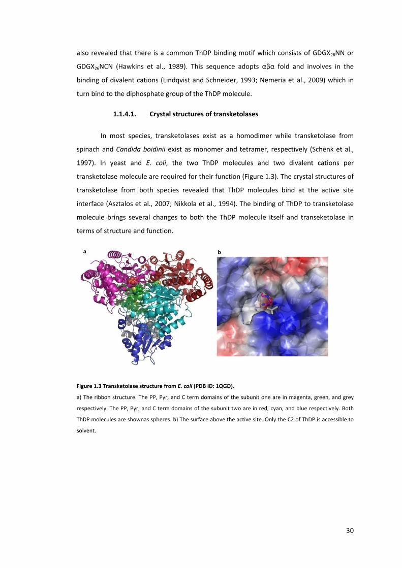
In most species, transketolases exist as a homodimer while transketolase from
spinach and Candida boidinii exist as monomer and tetramer, respectively (Schenk et al.,
1997). In yeast and E. coli, the two ThDP molecules and two divalent cations per
transketolase molecule are required for their function (Figure 1.3). The crystal structures of
transketolase from both species revealed that ThDP molecules bind at the active site
interface (Asztalos et al., 2007; Nikkola et al., 1994). The binding of ThDP to transketolase
molecule brings several changes to both the ThDP molecule itself and transeketolase in
terms of structure and function.
Figure 1.3 Transketolase structure from E. coli (PDB ID: 1QGD).
a) The ribbon structure. The PP, Pyr, and C term domains of the subunit one are in magenta, green, and grey
respectively. The PP, Pyr, and C term domains of the subunit two are in red, cyan, and blue respectively. Both
ThDP molecules are shownas spheres. b) The surface above the active site. Only the C2 of ThDP is accessible to
solvent.

31
1.1.4.2. Mechanism of transketolase
1.1.4.2.1. The role of ThDP in transketolase reaction
ThDP is a derivative of vitamin B1. The structure consists of one pyrimidine ring
linked to a thiazolium ring, and a diphosphate group. The C2 atom is the centre of all ThDP
dependent enzymes. In transketolase, this is the only atom that can be accessed by solvent
(see also Figure 1.3B) (Nikkola et al., 1994; Sundström et al., 1992). The conformation of
ThDP molecule in free solution differs from the enzyme-bound where the 4’-NH2 is placed
next to the C2 atom. The latter conformation is called the “V conformation”. Both
conformations are shown in Figure 1.4 below.
Figure 1.4 Two conformations of ThDP.
a) inactive conformation. b) the V conformation.
ThDP is directly involved in the catalytic reaction of all ThDP dependent enzymes
without being released from the active site (Kluger and Tittmann, 2008). Free thiamine and
thiazolium derivatives can catalyse similar reactions observed in the ThDP dependent
enzyme but with several orders of magnitude slower (Kluger and Tittmann, 2008; Mizuhara
and Handler, 1954; Schenk et al., 1998). The mechanism of ThDP has long been studied but
the early researches proposed that the amine group on the pyrimidine formed a Schiff base
with the carbonyl group(Langenbeck, 1932; Wiesner and Valenta, 1956). However, it was
illustrated that this amine cannot be deprotonated (Breslow, 1958; Fry et al.,
1957).Therefore, the formation of a Schiff base by the amine on the pyrimidine and
mechanism involved are rather impossible. Breslow (1957) used NMR spectroscopy to
monitor the deuterium exchange rate of ThDP and revealed for the first time that the C2 of
the thiazolium ring can be deprotonated. He then proposed the catalytic mechanism of
ThDP where the deprototanted C2 forms a carbanion and acts as the nucleophile that
attacks the carbonyl group(Breslow, 1958). The enzymes within this family employ this
mechanism for all their substrates (Kern et al., 1997). The formation of this carbanion has
been investigated in terms of deprotonation rate by 1H NMR spectroscopy and the
equilibrium position of the carbanion by the pKa value of this C2 atom. The H/D exchange

32
at the C2 position in solvent deuterium has a half-life of 20 minutes (Breslow, 1957)which is
too small to catalyse any reaction (Kern et al., 1997). The pKa of this CH atom is between
17-20 (Kemp and O’Brien, 1970)which makes a very small fraction of ThDP exists in the
carbanion form (Hübner et al., 1998). These evidences suggest these enzymes play a crucial
role in accelerating the reaction but they do not catalyse the reaction themselves (Kluger
and Tittmann, 2008).
The role of the enzyme in activating ThDP was revealed when Kern and colleagues
investigated the exchange rate at the C2 atom with pyruvate decarboxylase and
transketolase (Kern et al., 1997). These enzymes can increase the exchange rate up to three
orders of magnitude higher than the free ThDP but most of the C2 atoms are still
protonated. Pyruvate decarboxylase requires allosteric or substrate activations to further
increase the exchange rate to meet its observed kcat(Kern et al., 1997).
The crystal structures several ThDP dependent enzymes show the three essential
roles of these enzymes in ThDP activation; the V conformation, the position of 4’-NH2, and
the glutamate side chain next to the N’1 atom (Schellenberger, 1998). Their crystal
structures show that the ThDP molecule is in the V conformation which brings the 4’-NH2
group in close proximity to the C2 atom (Figure 1.4). One glutamate that forms a hydrogen
bond with the N’1 atom is highly conserved in all the ThDP dependent enzymes. Mutation
at this residue in both pyruvate decarboxylase and transketolase from yeast results in
severely impaired kcat. This implies that this glutamate residue is involved in the activation
of ThDP through the proton abstraction at the N1’ atom which transfers the negative
charge to the 4’-N (Hübner et al., 1998; Kern et al., 1997; Schellenberger, 1998). 4’-NH2 was
identified to act as the proton acceptor of the C2 atom through the use of 4’-desamino-
ThDP and site directed mutagenesis at the histidine residue close to the C2. Replacing the
histidine residue does not change the deprotonation rate of the C2 atom but removing the
4’-NH2 significantly impairs the kcat of both enzymes (Kern et al., 1997). In addition, the
deprotonation rate of the C2 atom is at least 5 orders of magnitude lower (Hübner et al.,
1998). This concludes that the 4’-NH2 promotes the C2 deprotonation.
1.1.4.2.2. Reaction mechanism: active site residues and ThDP
In addition to activating ThDP, certain residues in the active site of transketolase
were identified to act as acid/base catalysts which transfer protons between substrates as
illustrated in the reaction mechanism in Figure 1.5 (Wikner et al., 1997).

33
Figure 1.5 The Catalytic mechanism of transketolase.
R is the pyrimidine ring and R’ is the rest of the ThDP molecule. B1, B2, and B3 are acid/base involved in the
catalysis.
The reaction mechanism of transketolase is divided into two stages (Nilsson et al.,
1997; Wikner et al., 1997) and follows the ping-pong mechanism. Both stages require
proton transfers between ThDP, the active site residues, and the substrates. In the first
stage, proton abstraction at the C2 atom by B1 must occur to form a carbanion which then
acts as a nucleophile that attacks the carbonyl group of the donor substrate. Delocalisation
of electrons in the thiazolium ring of ThDP stabilises this carbanion and this mode of
stabilisation is often described as electron sink. The carbonyl group is then protonated by
B2 to form a hydroxyl group. The proton abstraction at the C3 hydroxyl group of the
intermediate by B3 then leads to the carbon-carbon bond cleavage. At the end of this first
stage, the carbanion of dihydroxyethyl-ThDP intermediate is formed and an aldehyde is
released from the active site (Wikner et al., 1997). In the absence of an acceptor aldehyde,
the dihydroxyethyl group can be released from ThDP and forms glycolaldehyde (Fiedler et
al., 2001). The second stage is the ligation of the dihydroxyethyl group to an acceptor. The
carbanion acts as a nucleophile attacking the carbonyl group of the aldehyde acceptor
forming another covalent-linked intermediate. The later steps are the reverse process of
the first stage and the release of the dihydroxy ketone from the active site. In both stages,

34
the oxygen on the carbonyl group of the donor and acceptor must be stabilised through
either charge compensation or proton transfer (Nilsson et al., 1997; Wikner et al., 1997).
It can be seen from this mechanism that several acid/base catalysts are involved in
the reaction. Enzyme crystallography, site-directed mutagenesis in yeast (Wikner et al.,
1997) and E. coli transketolase (Asztalos et al., 2007), and the use of ThDP analogues were
employed to identify these acid/base catalysts. In the active site of transketolase, there are
four highly conserved histidine residues which can involve in the proton transfer; H26, H69,
H261, on the PP domain, and H473 on the Pyr domain of E. coli transketolase as shown in
Figure 1.6a.
As previously mentioned, 4’-NH2 directly associates with the proton abstraction at
the C2 atom when it forms 4’-imino-ThDP (Hübner et al., 1998; Kern et al., 1997).
Therefore, this 4’-NH2 was identified as the proton acceptor in this step. The carbonyl
group of the donor molecule was suggested to be stabilised either by the H481 (H473 in E.
coli) or 4’-imino group (Wikner et al., 1997). The crystal structure of yeast transketolase
with erythrose 4-phosphate (PDB ID: 1NGS) suggested that the residues that abstract the
proton of the donor C3 hydroxyl could be H30 and H263 due to the fact that these residues
form hydrogen bonds with the carbonyl group of erythrulose 4-phosphate (Nilsson et al.,
1997). Since erythrose is the product from the cleavage of fructose, the location of the
carbonyl group of erythrose and the C3 hydroxyl group of fructose would be nearly
identical (Nilsson et al., 1997). This proposal was further supported when the crystal
structures of E. coli transketolase with xylulose-5-phosphate (PDB ID: 2R8O) and fructose-6-
phosphate (2R8P) showed that the C3 hydroxyl groups of both substrates interact with H26
and H261 (Asztalos et al., 2007). This carbonyl group is formed upon the deprotonation of
the C3 hydroxyl group of during the first stage of the reaction. In the double mutant
H26A/H261A in E. coli transketolase, much fewer donor-ThDP adduct species also formed
(Asztalos et al., 2007). These evidences lead to a suggestion that both histidine residues
could also be involved in the proton transfer and be nominated as B3 in Figure 1.5(Nilsson
et al., 1997).
1.1.4.2.3. Other residues involved in mechanism: substrate
recognition and stereoselectivity
In addition to acid/base catalysts and ThDP activation, several highly conserved
residues in transketolase active site and in vicinity serve other various functions including

35
substrate recognition, stereoselectivity control, transition state stabilisation, ThDP binding,
divalent cation metal binding, and phosphate binding. Mutagenesis studies and the crystal
structures of transketolases from both yeast and E. coli with natural substrates were used
to interpret the functions of these residues. In addition, the crystal structure of yeast
transketolase with enamine-ThDP intermediate was also available (1GPU). These studies
revealed that some residues perform multiple functions at the same time through their
complex hydrogen bond network with ThDP, substrates, and intermediate. Here, the
position of each amino acid is displayed in Figure 1.6 a) and b) and the functions of each
residue are described. All the residue positions refer to E. coli transketolase unless stated.
The positions in yeast transketolase are in parenthesises when both species were
mentioned together.

36
Figure 1.6 Residues within E. coli transketolase active site.
a) The hydrogen bond network between donor-ThDP adduct (PDB ID: 2R8P). b) The conserved residues
important for ThDP binding, substrate recognition, and catalysis. c) A schematic draw representing the
conserved residues involved in catalysis and substrate recognition. Hydrogen bond network was adapted from
the E. coli TK crystal structure (PDI ID: 2R8P) and are shown as dotted lines. For a summary of the functions of
each residue, see Table 1.1.

37
As previously mentioned, H30 and H263 in yeast transketolase were proposed to
be part of the proton transfer in the catalytic step because the mutation at this residue in
yeast transketolase significantly reduces the kcat value (Wikner et al., 1997) and the
formation of donor-ThDP adduct (Asztalos et al., 2007). In addition, mutation at H30 also
increases the KM value towards the donor xylulose-5-phosphate without noticeably altering
the KM towards the acceptor ribose-5-phosphate (Wikner et al., 1997). The different effects
of the mutation upon the KM values towards the donor and acceptor could arise from the
fact that hydrogen bonds only form between H30 and the C3 hydroxyl group of the donor
substrate but not with the acceptor. Therefore, removing this hydrogen bond can lead to a
lower enzyme-donor substrate affinity but not the affinity towards the acceptor (Nilsson et
al., 1997). However, a rather contradictory observation was found in E. coli transketolase
where the wild type and the double mutation H26A/H261A have similar KM values towards
donor and acceptor substrates although the activity of the double mutant is less than 1% of
the wild type (Asztalos et al., 2007). The crystal structure of E. coli TK with ribose-5-
phosphate (PDB ID: 2R5N) also shows that the H26 forms hydrogen bond with the C3
hydroxyl group (Asztalos et al., 2007).
It was observed that the hydroxylated donor substrate must be in D-threo
configuration at the C3 position (Hecquet et al., 1994; Morris et al., 1996) and the C3
hydroxyl group forms hydrogen bonds with H26. Therefore, the additional function of H26
was suggested to control the stereoselectivity of the enzyme. Later on, H26Y was identified
to reverse the stereoselectivity from 3S to 3R with an enantiomeric excess greater than
88% when using propylaldehyde as an acceptor (Smith et al., 2008). This further supports
the role of H26 in stereo-control and substrate recognition.
H66 is one of the conserved histidine residues within the active site but it locates
close to the phosphate group of the ThDP molecule. The crystal structures of E. coli
transketolase (PDB ID: 2R8P, 2R8O) show that one of the nitrogen atoms on the imidazole
ring forms hydrogen bonds with the phosphate group of ThDP and the other nitrogen atom
forms hydrogen bonds with a water molecule which in turn forms hydrogen bonds with the
C1-hydroxyl group of the donor-ThDP adducts. The DHEThDP intermediate in yeast
transketolase (PDB ID: 1GPU) also shows that the imidazole ring of H69 also forms
hydrogen bond with the hydroxyl group of the DHEThDP. However, both crystal structures
from E. coli (PDB ID: 2R5N) and yeast (PDB ID: 1NGS) do not show that this residue does not
form hydrogen bonds with acceptor substrates. Mutation at H69 in yeast transketolase
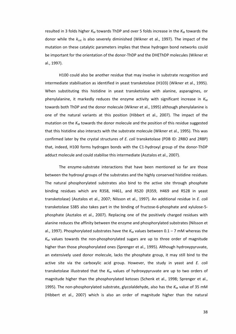
38
resulted in 3 folds higher KM towards ThDP and over 5 folds increase in the KM towards the
donor while the kcat is also severely diminished (Wikner et al., 1997). The impact of the
mutation on these catalytic parameters implies that these hydrogen bond networks could
be important for the orientation of the donor-ThDP and the DHEThDP molecules (Wikner et
al., 1997).
H100 could also be another residue that may involve in substrate recognition and
intermediate stabilisation as identified in yeast transketolase (H103) (Wikner et al., 1995).
When substituting this histidine in yeast transketolase with alanine, asparagines, or
phenylalanine, it markedly reduces the enzyme activity with significant increase in KM
towards both ThDP and the donor molecule (Wikner et al., 1995) although phenylalanine is
one of the natural variants at this position (Hibbert et al., 2007). The impact of the
mutation on the KM towards the donor molecule and the position of this residue suggested
that this histidine also interacts with the substrate molecule (Wikner et al., 1995). This was
confirmed later by the crystal structures of E. coli transketolase (PDB ID: 2R8O and 2R8P)
that, indeed, H100 forms hydrogen bonds with the C1-hydroxyl group of the donor-ThDP
adduct molecule and could stabilise this intermediate (Asztalos et al., 2007).
The enzyme-substrate interactions that have been mentioned so far are those
between the hydroxyl groups of the substrates and the highly conserved histidine residues.
The natural phosphorylated substrates also bind to the active site through phosphate
binding residues which are R358, H461, and R520 (R359, H469 and R528 in yeast
transketolase) (Asztalos et al., 2007; Nilsson et al., 1997). An additional residue in E. coli
transketolase S385 also takes part in the binding of fructose-6-phosphate and xylulose-5-
phosphate (Asztalos et al., 2007). Replacing one of the positively charged residues with
alanine reduces the affinity between the enzyme and phosphorylated substrates (Nilsson et
al., 1997). Phosphorylated substrates have the KM values between 0.1 – 7 mM whereas the
KM values towards the non-phosphorylated sugars are up to three order of magnitude
higher than those phosphorylated ones (Sprenger et al., 1995). Although hydroxypyruvate,
an extensively used donor molecule, lacks the phosphate group, it may still bind to the
active site via the carboxylic acid group. However, the study in yeast and E. coli
transketolase illustrated that the KM values of hydroxypyruvate are up to two orders of
magnitude higher than the phosphorylated ketoses (Schenk et al., 1998; Sprenger et al.,
1995). The non-phosphorylated substrate, glycolaldehyde, also has the KM value of 35 mM
(Hibbert et al., 2007) which is also an order of magnitude higher than the natural

39
phosphorylated substrates. This emphasised the strength of the substrate binding through
the phosphate group and how it can have an impact on the substrate conversion rate.
However, mutation at these residues have been shown to improve the activity towards
glycolaldehyde (Hibbert et al., 2007).
As previously mention, transketolase is highly stereoselective and this is controlled
by the hydrogen bond network formed between H26, H261, and the substrate. In addition
to these two residues, D469 (D477 in yeast transketolase) was also found to interact with
the C3 and C4 hydroxyl group of the donor and C2 hydroxyl group of an acceptor (Asztalos
et al., 2007; Nilsson et al., 1998, 1997). The importance of the C2 hydroxyl group in
acceptor molecules was demonstrated by removing the C2 hydroxyl group from the
acceptor substrates or replacing this aspartate with alanine. In both cases, very low
substrate conversion was observed together with a significant increase in kcat/KM(Nilsson et
al., 1998). Transketolase from yeast and E. coli also poorly accept alternative diastereomers
which have the opposite configuration at the C2 position (Morris et al., 1996; Nilsson et al.,
1998). Although the interaction between the C2-hydroxyl group and D469 seems to be
crucial, several D469 mutants have been shown to improve specific activity towards
propionaldehyde (Hibbert et al., 2008) with improved and reversed stereoselectivity (Smith
et al., 2008), longer chain aliphatic aldehydes with higher enantiomeric excess than wild
type (Cázares et al., 2010), and even aromatic aldehydes (Galman et al., 2010; Payongsri et
al., 2012).
During the catalysis of transketolase, the DHEThDP intermediate has to be
stabilised and the stabilising functions were proposed to arise from H473. The crystal
structure of yeast transketolase with DHEThDP (1GPU) revealed that H481 forms hydrogen
bond with the α hydroxyl group of the DHEThDP in addition to other histidine residues
(Schneider et al., 2002). This residue is also in close proximity to the 4’-NH2 group, and it
was proposed that H473 may accept a proton from 4’-NH2 to form 4’-imino group to
abstract the C2-proton (Singleton et al., 1996). However, amino acid substitution at this
residue does not influence the deprotonation rate of the C2 atom on the thiazolium ring
suggesting that it does not promote the ThDP activation (Hübner et al., 1998). In addition,
H473 is only conserved in non-mammalian species (Wikner et al., 1997) and only some
ThDP dependent enzymes (Costelloe et al., 2008). In human transketolase, this residue is
replaced with glutamine and it has been shown a higher degree of tolerating amino acid
substitution (Singleton et al., 1996) compared with H481 mutation in yeast transketolase

40
(Wikner et al., 1997). It should be noted that the human transketolase shows 24%
sequence identity to yeast transketolase and the variation in the sequence may lead to the
alteration in the function of the residue. This leads to a suggestion that H110 in human
transketolase (H100 in E. coli) may serve the function of H473 instead (Singleton et al.,
1996).
As illustrated in Figure 1.3, each ThDP molecule binds at the interface of the two
subunits where the phosphate group binds to the PP domain of subunit one through the
interaction with divalent metal ion and the pyrimidine ring binds to a hydrophobic pocket
in the Pyr domain of the other subunit. The thiazolium ring forms a hydrophobic interaction
with both subunits (Schneider and Lindqvist, 1998). Upon the ThDP binding, two disordered
regions undergo structural rearrangement and become more ordered. These regions are
then identified as cofactor loops (Sundström et al., 1992). In yeast transketolase, the two
regions consist of N187-W198 and L383-G398 (Sundström et al., 1992) whereas in E. coli
transketolase, these two regions are N185-W196 and L382-G392 (Martinez-Torres et al.,
2007). The divalent metal ion forms electrostatic interaction with the side chain of D155
(D157), N185 (N187), and the main chain oxygen of I187 (I189) (Sundström et al., 1992).
The loop closure is stabilised through several inter-subunit interactions and the second
loop forms hydrophobic interaction with the first loop (Sundström et al., 1992). Besides the
interaction between two loops, an additional aspartate residue also involves in the loop
closing. The amino acid substitution at D382 in yeast transketolase leads to the second loop
disorder which suggested that D382 stabilises the loop formation although this residue is
not part of the cofactor loop (Meshalkina et al., 1997). In fact, this residue involves in
subunit-subunit interaction by forming hydrogen bond with the first loop of the other
subunit (Sundström et al., 1992). The loss of this interaction leads to a further loss of loop-
loop interaction and second loop disorder. Besides that, substituting this aspartate for
alanine or asparagine reduces the enzymatic activity and increases the KM towards both
phosphorylated donor and acceptor molecules (Meshalkina et al., 1997). In addition to
providing the inter-subunit interaction, it was observed that ThDP binding and the loop
closure influence the thermal stability of transketolase (Esakova et al., 2005).
The dimeric enzyme is not only stabilised through the interaction between the
cofactor loops. The study in yeast transketolase illustrate that the two subunits are also
held together by hydrogen bond network formed by highly conserved acid residues; E162
of the first subunit with G167 and E418 of the other subunit (Meshalkina et al., 1997).
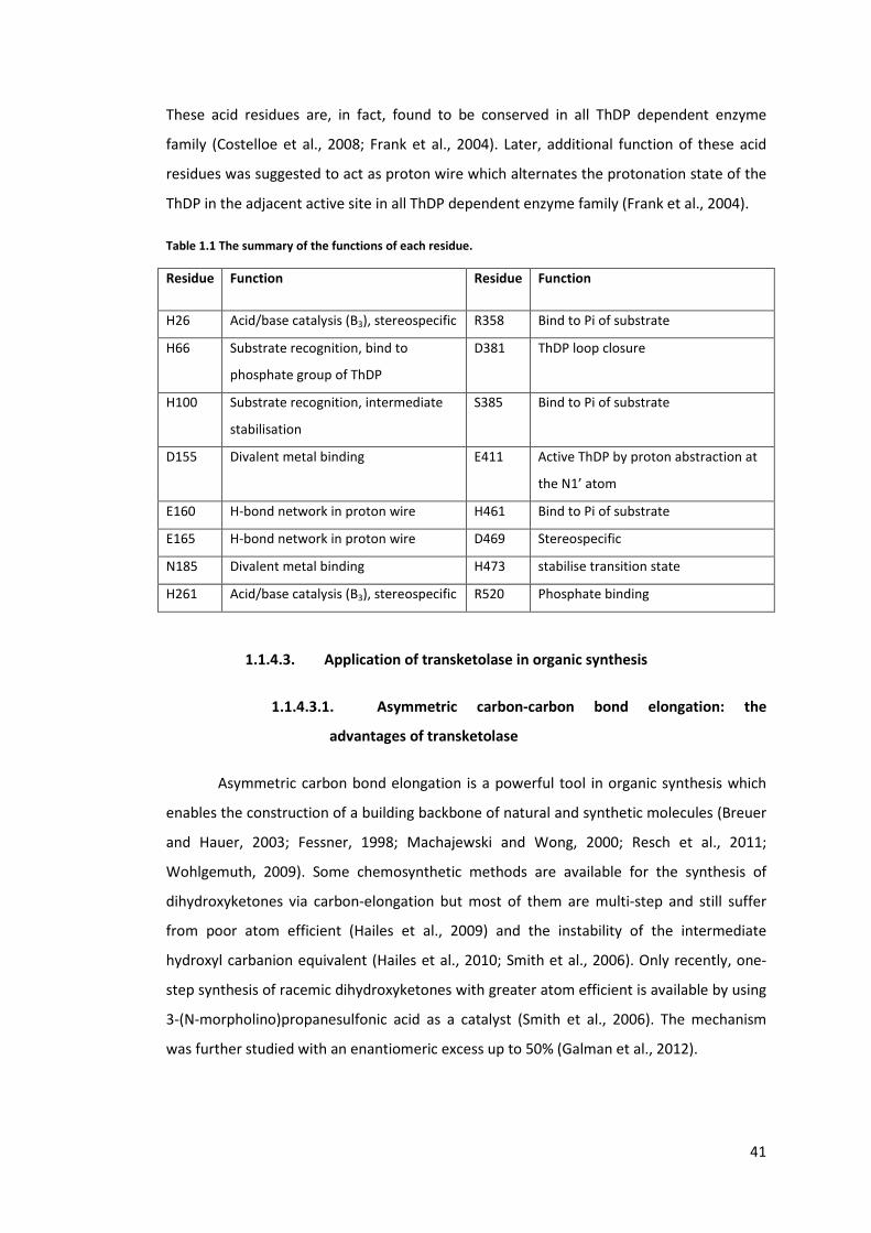
41
These acid residues are, in fact, found to be conserved in all ThDP dependent enzyme
family (Costelloe et al., 2008; Frank et al., 2004). Later, additional function of these acid
residues was suggested to act as proton wire which alternates the protonation state of the
ThDP in the adjacent active site in all ThDP dependent enzyme family (Frank et al., 2004).
Table 1.1 The summary of the functions of each residue.
Residue Function Residue Function
H26 Acid/base catalysis (B3), stereospecific R358 Bind to Pi of substrate
H66 Substrate recognition, bind to
phosphate group of ThDP
D381 ThDP loop closure
H100 Substrate recognition, intermediate
stabilisation
S385 Bind to Pi of substrate
D155 Divalent metal binding E411 Active ThDP by proton abstraction at
the N1’ atom
E160 H-bond network in proton wire H461 Bind to Pi of substrate
E165 H-bond network in proton wire D469 Stereospecific
N185 Divalent metal binding H473 stabilise transition state
H261 Acid/base catalysis (B3), stereospecific R520 Phosphate binding
1.1.4.3. Application of transketolase in organic synthesis
1.1.4.3.1. Asymmetric carbon-carbon bond elongation: the
advantages of transketolase
Asymmetric carbon bond elongation is a powerful tool in organic synthesis which
enables the construction of a building backbone of natural and synthetic molecules (Breuer
and Hauer, 2003; Fessner, 1998; Machajewski and Wong, 2000; Resch et al., 2011;
Wohlgemuth, 2009). Some chemosynthetic methods are available for the synthesis of
dihydroxyketones via carbon-elongation but most of them are multi-step and still suffer
from poor atom efficient (Hailes et al., 2009) and the instability of the intermediate
hydroxyl carbanion equivalent (Hailes et al., 2010; Smith et al., 2006). Only recently, one-
step synthesis of racemic dihydroxyketones with greater atom efficient is available by using
3-(N-morpholino)propanesulfonic acid as a catalyst (Smith et al., 2006). The mechanism
was further studied with an enantiomeric excess up to 50% (Galman et al., 2012).
42
The complications of the stereoselectivity in carbon-carbon bond elongation have
drawn a huge attention from a number of researches to use enzymes in asymmetric
synthesis due to their naturally high stereoselectivity. In addition to transketolase and
other ThDP dependent enzymes (Breuer and Hauer, 2003; Resch et al., 2011; Sprenger and
Pohl, 1999), several enzymes including aldolases (Enders et al., 2005; Fessner, 1998;
Machajewski and Wong, 2000; Samland and Sprenger, 2006), hydroxynitrile lyase (Breuer
and Hauer, 2003) have gained interests for similar purposes. In the carbohydrate synthesis,
transketolases can provide some advantages over some aldolases and these are non-
requirement of dihydroxyacetone phosphate, simple purification process and monitoring
due to the fact that a non-phosphorylated compound is produced (Kobori et al., 1992;
Toone and Whitesides, 1991). In addition, since transketolase only accepts hydroxylated
substrates with 2R configuration, racemic hydroxylated aldehydes can be used as a starting
material (Kobori et al., 1992).
1.1.4.3.2. Substrate spectrum of TK and its potential
The product of transketolase contains a 1,3-dihydroxy ketone motif, a functional
group that can be found in carbohydrates(Kobori et al., 1992), and corticosteriods (Hailes et
al., 2009; Hailes et al., 2010). The substrate spectrum studies of transketolases from
different species revealed transketolases can accept not only natural substrates but also
non-phosphorylated sugars (Demuynck et al., 1991; Kobori et al., 1992), non-hydroxylated
aldehydes, cyclic aldehydes (Hobbs et al., 1993; Morris et al., 1996), andheteroaromatics
(Humphrey et al., 1995). In addition to aldehyde substrates, transketolase can also catalyse
the transfer of the ketol group to nitroso aromatics (Corbett and Corbett, 1986).
Mutagenesis studies of transketolase from E. coli have further expanded the synthetic
potential of transketolase. Initially, E. coli transketolase libraries were found to have
improved activity towards propionaldehyde (Hibbert et al., 2008) with enhanced and
reverse stereoselectivity (Smith et al., 2008). Several mutants also were identified to accept
cyclic and longer chain non-hydroxylated aldehydes (Cázares et al., 2010) heteroaromatics
and benzaldehyde derivatives (Galman et al., 2010) with high enatiomeric excess in all
cases. So far, transketolase has been used in mostly multi-step synthesis of several
compounds including furaneol which is a caramel-like flavour (Hecquet et al., 1996;
Hecquet et al., 1994), glycosidase inhibitors; 1,4-Dideoxy-1,4-imino-D-arabinitol (Ziegler et
al., 1988) and N-hydroxypyrrolidine (Humphrey et al., 2000), (+)-exo-brevicomin (Myles et
al., 1991). For single step synthesis, transketolase tends to be used for simple carbohydrate
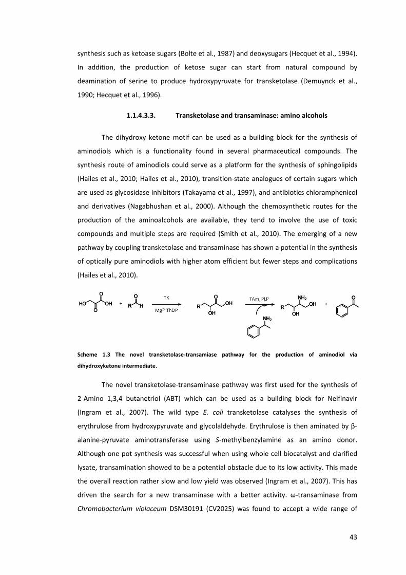
43
synthesis such as ketoase sugars (Bolte et al., 1987) and deoxysugars (Hecquet et al., 1994).
In addition, the production of ketose sugar can start from natural compound by
deamination of serine to produce hydroxypyruvate for transketolase (Demuynck et al.,
1990; Hecquet et al., 1996).
1.1.4.3.3. Transketolase and transaminase: amino alcohols
The dihydroxy ketone motif can be used as a building block for the synthesis of
aminodiols which is a functionality found in several pharmaceutical compounds. The
synthesis route of aminodiols could serve as a platform for the synthesis of sphingolipids
(Hailes et al., 2010; Hailes et al., 2010), transition-state analogues of certain sugars which
are used as glycosidase inhibitors (Takayama et al., 1997), and antibiotics chloramphenicol
and derivatives (Nagabhushan et al., 2000). Although the chemosynthetic routes for the
production of the aminoalcohols are available, they tend to involve the use of toxic
compounds and multiple steps are required (Smith et al., 2010). The emerging of a new
pathway by coupling transketolase and transaminase has shown a potential in the synthesis
of optically pure aminodiols with higher atom efficient but fewer steps and complications
(Hailes et al., 2010).
Scheme 1.3 The novel transketolase-transamiase pathway for the production of aminodiol via
dihydroxyketone intermediate.
The novel transketolase-transaminase pathway was first used for the synthesis of
2-Amino 1,3,4 butanetriol (ABT) which can be used as a building block for Nelfinavir
(Ingram et al., 2007). The wild type E. coli transketolase catalyses the synthesis of
erythrulose from hydroxypyruvate and glycolaldehyde. Erythrulose is then aminated by β-
alanine-pyruvate aminotransferase using S-methylbenzylamine as an amino donor.
Although one pot synthesis was successful when using whole cell biocatalyst and clarified
lysate, transamination showed to be a potential obstacle due to its low activity. This made
the overall reaction rather slow and low yield was observed (Ingram et al., 2007). This has
driven the search for a new transaminase with a better activity. ω-transaminase from
Chromobacterium violaceum DSM30191 (CV2025) was found to accept a wide range of

44
substrates including aliphatic aldehydes, cyclic aldehydes, aromatic aldehydes, keto acids,
and erythrulose (Kaulmann et al., 2007). In addition, a few engineered E. coli transketolase
variants have been shown to have higher activity towards non-hydroxylated aldehyde
(Hibbert et al., 2008), cyclic aldehydes (Cázares et al., 2010), and aromatic aldehydes
(Galman et al., 2010). This expands the range of the products that could be synthesised by
the two enzymes. Propionaldehyde, together with the best transketolase mutant D469T,
was further coupled with CV2025 transaminase to explore this feasibility and to model the
production process of (2S,3S)-2-aminopentane-1,3-diol (APD) (Smith et al., 2010). All the
information was then integrated to design the processes and tools required for the
synthesis of other2-amino-1,3-diols (Hailes et al., 2010). With the aid of process design, the
coupling of transketolase wild type and mutant with CV2025 transaminase was shown to
give higher yields and optical purity for both ABT and APD (Rios-Solis et al., 2011).
1.2. Research aims
The overall aim of this research is to construct transketolase mutants with higher
activity towards aromatic aldehydes and integrate these variants with transaminase to
produce novel chiral amino alcohols. This research consists of four major sequential steps
as designated into four chapters. Each is briefly described below.
In the first chapter, two sets of libraries were constructed and the target residues
were chosen from two different rationales. Their stabilities and activities towards
glycolaldehyde and propionaldehyde were then compared. Eight variants including single,
double, and a triple mutants from the first library were constructed by randomly combining
three existing single-mutant transketolase variants that were reported to have higher
activity towards glycolaldehyde than wild type. In the second library, the double mutants
that were constructed and the two target residues were co-evolved residues. The
information from this study will aid the design of the future library in order to compromise
both activity and stability.
The second aim is to identify the factors governing the bioconversion of the
aromatic aldehydes by transketolase and to find which mutants have the highest activity
towards some aromatic aldehydes. Although a few transketolase mutants were identified
to accept benzaldehyde and derivatives, very slow conversion rates were observed
(Galman et al., 2010). Acid aromatic aldehydes were chosen to study if enzyme-substrate
affinity could be the key factor. Site directed mutagenesis was employed to identify the key

45
mutation residues that allow transketolase to accept aromatic aldehyde which can be used
as a pseudo wild type for the next library design. This strategy was also used to study how
these residues control the bioconversion of aromatic aldehydes.
The third aim is to create a small transketolase library by introducing site saturation
mutagenesis to certain residues within the active site. The knowledge from the
combination of the single mutants in the first part will be used in designing the stable
library while the literature review on the functions of certain residues would justify which
residues should be targets or remained untouched. The improved mutants and the
substrates of interests will be part of the novel transketolase-transaminase pathway.
Lastly, transketolase and transaminase will be coupled in vitro to produce a novel
aromatic amino alocohol, a new derivative of chloramphenicol building block. The factors
and bottlenecks associated with the process will be identified for the process improvement
and strategy design.

46
2. Chapter 2: Materials and methods
2.1. Chemicals
Unless otherwise stated, all the chemicals were purchased from Sigma-Aldrich,
Poole, UK. Lithium hydroxypyruvate was prepared from bromopyruvic acid and lithium
hydroxide according to the previous method and stored at -20 °C (Morris et al., 1996).The
purity of each batch was tested by HPLC according to the protocol below. Propionaldehyde
was purchased from AcroSeal (Acros Organics, Fisher Scientific, UK). Standard dihydroxy
ketones were synthesised by biomimetric reaction (Smith et al., 2006) except for 3-(1,3-
dihydroxy-2-oxopropyl)benzoic acid and 4-(1,3-dihydroxy-2-oxopropyl)benzoic acid which
were synthesised by transketolase mutants and characterised according to the publication
(Payongsri et al., 2012). The standard dihydroxy ketones were kept at 4 °C.
All the buffers, liquid phase of the HPLC, and liquid media were prepared using RO
water which was purified by Elix Water Purification system (Millipore, UK). This produces
water with greater than 5 MΩ.cm resistivity at 25 °C, less than 30 ppb of total organic
carbon (TOC), and less than 1cfu/ml of bacteria.
The water used in PCR reaction including dissolving primers was ultrapure water
which was purified by Milli-Q® Advantage A10® system (Millipore, UK).
Unless otherwise stated, the pH of all the solutions wasadjusted by the addition of
sodium hydroxide solution or hydrochloric acid.
2.2. Media and buffer reagent preparation
2.2.1. Ampicillin (Amp)
Ampicillin stock solution was prepared by dissolving ampicillin powder in RO water
to the final concentration of 50 mg/ml. This was then filtered through sterile 0.2 μm
filter.The ampicillin stock can be aliquoted into 1.5-ml sterile eppendorf tubes and stored
at -20 °C. The working concentration of ampicillin in the culture media was 150 μg/ml
which was achieved by adding 30 μl of this stock solution per 10 ml of the media.

47
2.2.2. Lysogenic broth (LB)
Lysogenic broth, known asLuria-Bertani broth, was prepared by dissolving 5 g yeast
extract, 10 g tryptone, and 10 g sodium chloride in RO water with a final volume of 1 l. The
pH of the mixture was adjusted to 7.0. The liquid was then autoclaved for sterilisation.
2.2.3. LB agar with 150 μg/ml ampicillin (LB-Amp)
The composition and the preparation of LB agar was the same as LB broth but
select agar was also added to the final concentration of 15 g/l. After autoclave, the liquid
was left to cool down to approximately 50 °C before adding ampicillin. 90 μl of this
ampicillin stock solution was added to 20 ml of LB agar which was then poured into a petri
dish and allowed to set. The LB-Amp plates can be stored at 4 °C for a month.
2.2.4. Tris buffer
50 mM Tris buffer was prepared by dissolving 7.88 g of Tris-HCl in 1 l ofRO water in
a borosilicate bottle. The pH was then adjusted to 7.0 by the addition of 46 % (W/V) sodium
hydroxide. To prevent microbial contamination and precipitation, Tris buffer was only
taken out from the bottle by directly pouring into a smaller container which was then
pipetted to the desired volume.
2.3. Transketolase production
All transketolase mutants with 6 his-tag at the N-terminuswere expressed under
the control of its own nativepromoter in the tktA gene which was cloned into pQR791
plasmid(Martinez-Torres et al., 2007) which is a modification of pQR711 plasmid (French
and Ward, 1995). The amino acid sequence of wild type TK is illustrated below. The
positions of all amino acids mentioned throughout this thesis excluded the 6 histidine
residues (underlined) used for affinity purification. The plasmid map is shown in Figure 2.1.
1 MHHHHHHSSR KELANAIRAL SMDAVQKAKS GHPGAPMGMA DIAEVLWRDF LKHNPQNPSW 61 ADRDRFVLSN GHGSMLIYSL LHLTGYDLPM EELKNFRQLH SKTPGHPEVG YTAGVETTTG 121 PLGQGIANAV GMAIAEKTLA AQFNRPGHDI VDHYTYAFMG DGCMMEGISH EVCSLAGTLK 181 LGKLIAFYDD NGISIDGHVE GWFTDDTAMR FEAYGWHVIR DIDGHDAASI KRAVEEARAV 241 TDKPSLLMCK TIIGFGSPNK AGTHDSHGAP LGDAEIALTR EQLGWKYAPF EIPSEIYAQW 301 DAKEAGQAKE SAWNEKFAAY AKAYPQEAAE FTRRMKGEMP SDFDAKAKEF IAKLQANPAK 361 IASRKASQNA IEAFGPLLPE FLGGSADLAP SNLTLWSGSK AINEDAAGNY IHYGVREFGM 421 TAIANGISLH GGFLPYTSTF LMFVEYARNA VRMAALMKQR QVMVYTHDSI GLGEDGPTHQ 481 PVEQVASLRV TPNMSTWRPC DQVESAVAWK YGVERQDGPT ALILSRQNLA QQERTEEQLA 541 NIARGGYVLK DCAGQPELIF IATGSEVELA VAAYEKLTAE GVKARVVSMS STDAFDKQDA 601 AYRESVLPKA VTARVAVEAG IADYWYKYVG LNGAIVGMTT FGESAPAELL FEEFGFTVDN 661 VVAKAKELL

48
Figure 2.1 The map of pQR791 plasmid.
In the chapter 3, the E. coli strain used is BL21Gold(DE3) while the rest of the study,
the host cell is XL10 gold (Stratagene, La Jolla, California, USA).
The glycerol stock of each mutant was streaked on an LB-Amp plate and incubated
at 37 °C for 18 hours. A single colony was picked by a sterile tip and inoculated into 10 mlLB
media with 150 μg/ml ampicillin in a falcon tube. The culture was then shaken at 250 rpm,
37 °C for 16-18 hours before harvested or transferred to a larger volume of fermentation.
For a larger scale, 10% inoculums volume was used and the fermentation was performed at
the same conditions for 6 hours before harvesting.
To harvest, the culture was aliquoted into 50-ml falcon tubes and centrifuged at
400 rpm for 10 minutes. The supernatant was discarded and the cell pellet was either used
fresh or store at -20 °C.
2.4. Preparation of clarified lysate
The frozen cell paste was thawed on ice and resuspended in 2-5 ml 50 mM Tris
buffer, pH 7.0. The volume of Tris buffer added depends on the culture volume and the
required transketolase concentration for further use. This was then transferred to 7-ml
bijou tube and lysed by sonication (MSE Soniprep 150 probe, Sanyo) on ice with 10 s on, 15
s off for 10 cycles. The mixture was then centrifuged at 17,700 g for 10 min at 4 °C. The
supernatant containing transketolase was used fresh or stored at -80 °C in 150 μl aliquots
for up to 1 month.

49
2.5. Protein concentration quantification
2.5.1. Bradford assay
The total protein concentration was measured by Bradford assay using bovine
serum albumin (BSA) as a standard protein. 20 – 70 μlof 0.6 mg/ml BSA stock solution was
added into the 1 ml cuvettes. 1 ml of Bradford reagent was then added to each cuvette at
15 s interval. The mixture was incubated for 5 minutes before measuring the absorbance at
595 nm. The final BSA concentration in the cuvette was then plotted against the OD595 to
make a standard graph. Only the linear data points were taken. To measure the total
protein concentration in the clarified lysate, 1 ml of Bradford reagent was added into a 1 ml
cuvette containing 10 μl of the clarified lysate. After 5 minutes of incubation, the OD595
was read and the final concentration of the total protein in the cuvette was calculated. The
actual protein concentration before the addition of Bradford reagent was then re-
calculated back through the dilution factor. If the OD595 of the clarified lysate was beyond
standard graph, it was diluted to fit within the range of the standard graph. All
quantifications were done in duplicated.
Example
The final concentration of BSA in the cuvette when adding 1 ml of Bradford assay to
20 μl of 0.6 mg/ml BSA is
= 0.6 mg/ml * 0.020 ml / (1ml+ 0.020ml)
= 0.0118 mg/ml
2.5.2. SDS-PAGE and densitometry
After measuring the total protein concentration within the clarified lysate, the
lysate was mixed with 2xLaemmli Sample Buffer (Bio-Rad Laboratories, UK) to reach the
final protein concentration of 1.0 – 1.5 μg/μl. When the total protein concentration is very
high, the lysate may be diluted with 50 mM Tris buffer prior mixed with 2xLaemmli Sample
Buffer. The mixture was then heated at 95 °C for 10 minutes to fully denature the protein.
To minimise the experimental error due to evaporation, all samples were prepared in 500
μl tube with at least 50 μl of sample. After heating, the sample was briefly centrifuged,
gently mixed and briefly centrifuged again to get rid of any bubbles. 10 – 15 μl of the
denatured sample was loaded into each well so that the final amount of protein per well is
between 10 – 15 μg. Denatured BSA was also loaded into the same gel at the final quantity

50
of 1.2 – 3.6 μg per gel to make a standard correlation between the intensity of the band
and the amount of protein within the band as showed in Figure 2.2. 5 μl of prestained
protein ladder PageRuler™ (Fermentas, UK) was loaded into one of the well to confirm the
size of transketolase if necessary.
Figure 2.2 An example of SDS-PAGE gel with standard TK gradient.
a) Lanes 1-3 are whole cell sample. Lanes 3-4 are clarified lysate. Lanes 6-8 are resuspended pellets. Lanes 9 -12
are TK gradient. b) The SDS-PAGE of His-tag purified TK and clarified lysate. The molecular weights of the
markers are shown accordingly.
The migration distance of each marker protein in Figure 2.2 b was plotted against
the log(molecular weight kDa), a linear best fit line was obtained. The migration distances
were measured when the image was 16.58 cm long and 13.5 cm wide. The top most part of
the image was assigned to be the starting point of protein migration.
Table 2.1 The migration distances of the protein markers, their molecular weights, and their log(molecular
weight).
MW(kDa) Migration distance
(cm) Log (MW)
17 15.68 1.230
26 13.89 1.415
34 12.41 1.531
43 10.51 1.633
55 8.93 1.740
72 7.14 1.857
95 5.81 1.978
130 4.16 2.114
170 3.17 2.230
TK 7.54
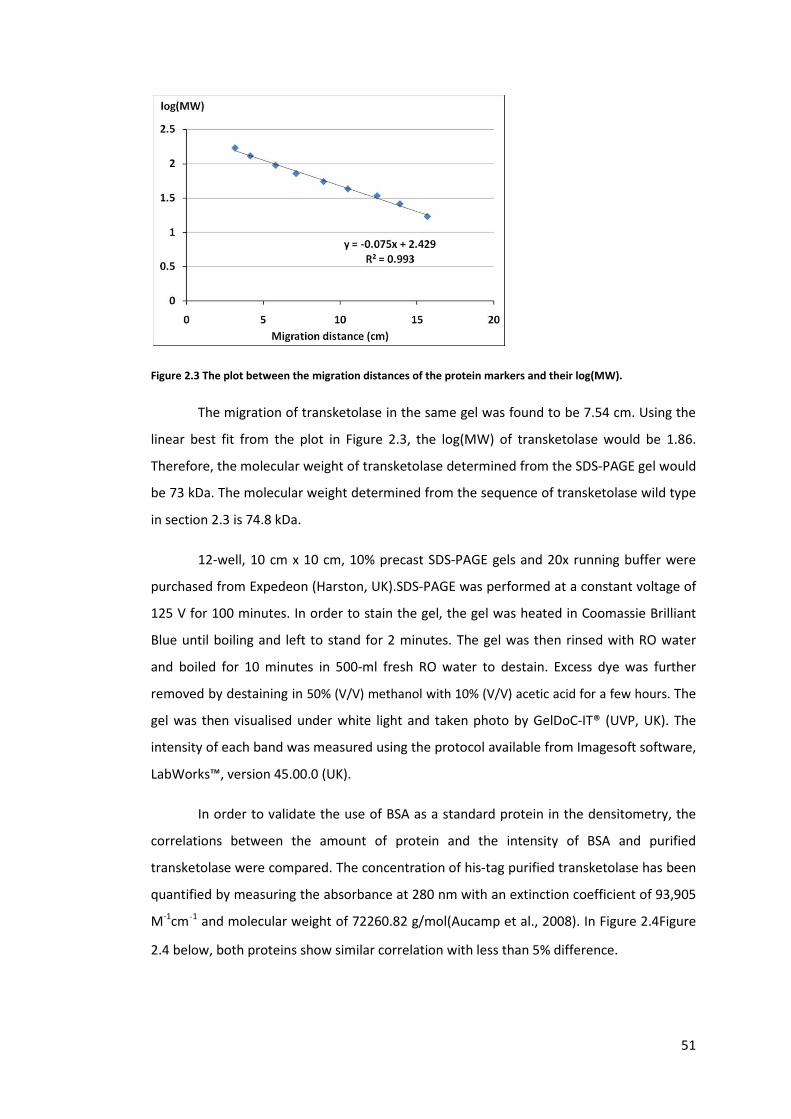
51
Figure 2.3 The plot between the migration distances of the protein markers and their log(MW).
The migration of transketolase in the same gel was found to be 7.54 cm. Using the
linear best fit from the plot in Figure 2.3, the log(MW) of transketolase would be 1.86.
Therefore, the molecular weight of transketolase determined from the SDS-PAGE gel would
be 73 kDa. The molecular weight determined from the sequence of transketolase wild type
in section 2.3 is 74.8 kDa.
12-well, 10 cm x 10 cm, 10% precast SDS-PAGE gels and 20x running buffer were
purchased from Expedeon (Harston, UK).SDS-PAGE was performed at a constant voltage of
125 V for 100 minutes. In order to stain the gel, the gel was heated in Coomassie Brilliant
Blue until boiling and left to stand for 2 minutes. The gel was then rinsed with RO water
and boiled for 10 minutes in 500-ml fresh RO water to destain. Excess dye was further
removed by destaining in 50% (V/V) methanol with 10% (V/V) acetic acid for a few hours. The
gel was then visualised under white light and taken photo by GelDoC-IT® (UVP, UK). The
intensity of each band was measured using the protocol available from Imagesoft software,
LabWorks™, version 45.00.0 (UK).
In order to validate the use of BSA as a standard protein in the densitometry, the
correlations between the amount of protein and the intensity of BSA and purified
transketolase were compared. The concentration of his-tag purified transketolase has been
quantified by measuring the absorbance at 280 nm with an extinction coefficient of 93,905
M-1cm-1 and molecular weight of 72260.82 g/mol(Aucamp et al., 2008). In Figure 2.4Figure
2.4 below, both proteins show similar correlation with less than 5% difference.
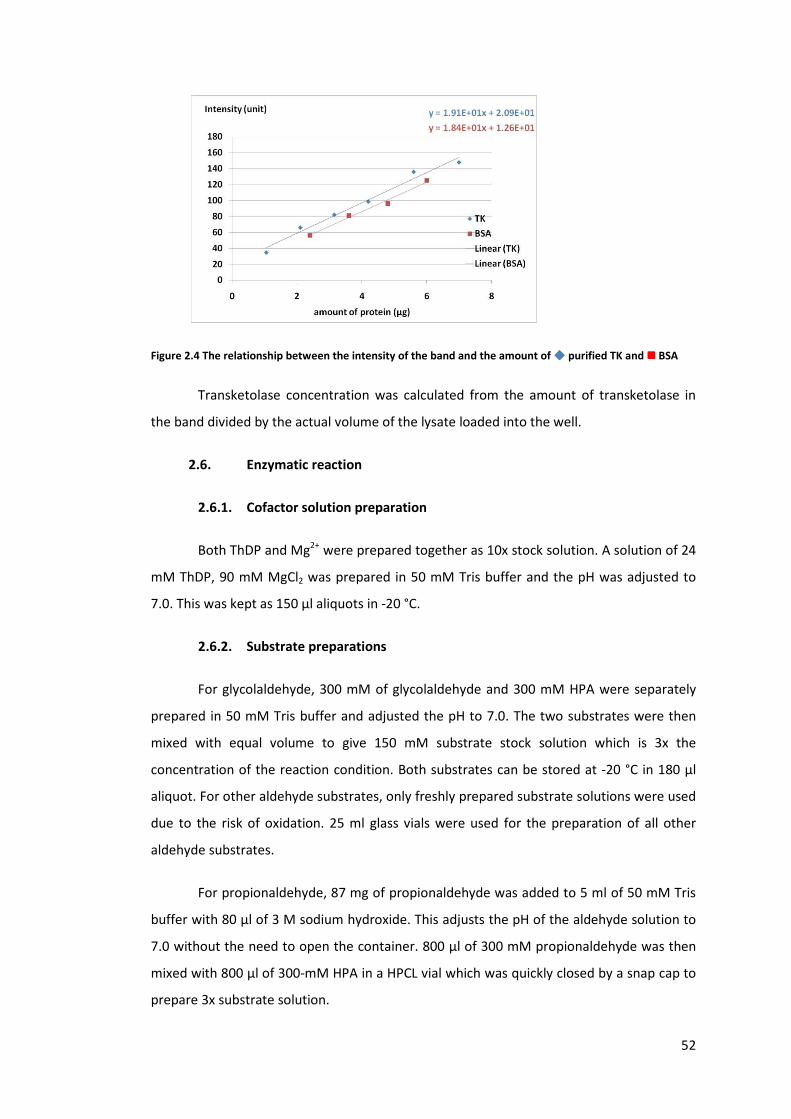
52
Figure 2.4 The relationship between the intensity of the band and the amount of ���� purified TK and ���� BSA
Transketolase concentration was calculated from the amount of transketolase in
the band divided by the actual volume of the lysate loaded into the well.
2.6. Enzymatic reaction
2.6.1. Cofactor solution preparation
Both ThDP and Mg2+ were prepared together as 10x stock solution. A solution of 24
mM ThDP, 90 mM MgCl2 was prepared in 50 mM Tris buffer and the pH was adjusted to
7.0. This was kept as 150 μl aliquots in -20 °C.
2.6.2. Substrate preparations
For glycolaldehyde, 300 mM of glycolaldehyde and 300 mM HPA were separately
prepared in 50 mM Tris buffer and adjusted the pH to 7.0. The two substrates were then
mixed with equal volume to give 150 mM substrate stock solution which is 3x the
concentration of the reaction condition. Both substrates can be stored at -20 °C in 180 μl
aliquot. For other aldehyde substrates, only freshly prepared substrate solutions were used
due to the risk of oxidation. 25 ml glass vials were used for the preparation of all other
aldehyde substrates.
For propionaldehyde, 87 mg of propionaldehyde was added to 5 ml of 50 mM Tris
buffer with 80 μl of 3 M sodium hydroxide. This adjusts the pH of the aldehyde solution to
7.0 without the need to open the container. 800 μl of 300 mM propionaldehyde was then
mixed with 800 μl of 300-mM HPA in a HPCL vial which was quickly closed by a snap cap to
prepare 3x substrate solution.

53
For 3-formylbenzoic acid and 4-formylbenzoic acid, their solubilities are lower than
glycolaldehyde and propionaldehyde, so they were prepared at 150 mM and adjusted the
pH to 7.0. This was then used to dissolve HPA to get the final concentration of 150 mM
each.
2.6.3. Activity assay
All reactions were run in triplicate at the final volume of 300 μl at 22 °C. Prior
starting the reaction, 30 μl of clarified lysated was incubated with 30 μl of 10X cofactor and
140 μl of 50 mM Tris buffer, pH 7.0 which makes the total volume of 200 μl. After 20
minutes of incubation, 100 μl of 3X substrate stock solution was added to the enzyme-
cofactor solution to start the reaction at 15 s interval. 20 μl of reaction sample was taken at
certain time points and quenched in 180 μl 0.1% TFA for glycolaldehyde and
propionaldehyde. For aromatic substrate, 20 μl samples were quenched in 380 μl 0.1% TFA.
All samples were quenched in 500-μl reaction tubes which were centrifuged at 13,000 rpm
for 3 minutes to remove any precipitate. The supernatants were analysed by HPLC to
quantify the substrate and product concentration. The reaction of glycolaldehyde was
performed in polystyrene 96-well plate which was covered by its lid. For other aldehydes,
the reactions were performed in 1.5-ml HPLC vials which were closed by snap caps.
2.7. HPLC system
Two separate HPLC systems were employed in this study for different types of
compound separation. Both HPLCs were controlled by Chromeleon software (Dionex, UK).
The chromatograms and peak analysis were also processed using the same software.
2.7.1. HPLC system for aliphatic compounds
At least 170 μl of the supernatants was loaded onto a polystyrene 96 well plate and
covered with aluminium foil to prevent evaporation. The sample was taken by FAMOS
autosampler (Dionex, UK) which was then injected into Aminex HPx-87H, 300x7.8mm
column (Bio-Rad, UK) which was maintained at 60 °C. Micro-Guard Cation H Refill
Cartridges, 30 x 4.6 mm (Bio-Rads, UK) was also used to prevent column contamination.
The separation was performed by isocratic flow of 0.1% TFA in water at the flow rate of 0.6
ml/min which was maintained by Ultimate 3000 isocratic analytical pump (Dionex, UK). The
UV absorption at 210 nm was monitored at the data collection rate of 5Hz by AD20
absorbance detector (Dionex, UK) for the detection of HPA, erythrulose, 1,3 dihydroxy
54
pentan-2one which have the retention time of 8.4, 11.5, and 15.2 minutes, respectively.
For glycolaldehyde reaction, the HPLC assay time was 15 minutes whereas the
propionaldehyde is 20 minutes. The 0.1 % TFA mobile phase was degassed by sparging with
helium for 10 minutes before use.
2.7.2. HPLC system for aromatic compounds
360 μl of the supernatant was transferred into 1.5-ml HPLC vials and closed with
crimp caps. The sample was taken by Ultimate 3000 autosampler (Dionex, UK) and injected
into ACE5 C18 reverse phase column (150 × 4.6 mm) which was maintained at 30 °C. The
mobile phase was pumped in at the flow rate of 1.0 ml/min starting with 85% of 0.1% TFA
and 15% of 100% acetonitrile (CHROMASOLV® HPLC gradient grade) which was linearly
increased to 72% in 9 minutes. This followed by 2-minute equilibration period by pumping
85% of 0.1% TFA and 15% acetonitrile. UV detection at 210, 250, and 275 nm were
simultaneously used throughout the process.
For 3-hydroxybenzaldehyde, the same column was used but the mobile phases
were changed to 0.2 M acetic acid and 80% (V/V) methanol (in water). The mobile phase
starts with constant flow of 90% 0.2 M acetic acid and 10% of 80% methanol for 5 minutes.
This is followed by a linear increase of 80% methanol from 10% to 60% in the 14th minute
which was maintained for another 6 minutes. Then the column was equilibrated for 3
minutes by 90% 0.2M acetic acid and 10% 80% (V/V) methanol.
2.8. Standard product and substrate graphs
The commercially available standard substrates and products were purchased from
SigmaAldrich. The racemic aromatic dihydroxyketones were synthesised by biomimetic
reaction (Smith et al., 2006) which have been purified and characterised by NMR (David
Steadman, Chemistry Department, UCL). All reagents were dissolved in 0.1% TFA.
For HPA and erythrulose which were analysed by the first HPLC system, their final
concentrations in TFA were between 0.05 – 5.0 mM. This range represents 0.5 – 50 mM
concentration before the reaction samples were diluted 10 times upon quenching in 0.1%
TFA. For aromatic aldehydes, the final concentrations of the aldehydes were between 0.05
– 2.5 mM which represents 1 – 50 mM of aldehydes before 20 times dilution. At least five
concentration points were prepared and the R2 value of the linear standard graph must be
greater than 0.96.

55
2.9. Initial rate and specific activity calculation
The graph of product versus time was plotted in Microsoft Excel 2007. If the data
are linear, linear best-fit line was used to find the initial rate.
Specific accvity (μmol mg-1
min-1
)=inical rate (
mM
min)
enzyme concentracon (mg
ml)
If the data are not in the linear range, the whole data set was analysed by
SigmaPlot 12. The exponential rise to maximum; y =a �1-e-b x� was fitted into the data and
SigmaPlot then predicted the equation that fits the data. Y is product concentration and x is
time. SigmaPlot also returns the predicted values of the product concentration at time zero
until the last data point. The initial values of product concentration and time were then
used to calculate the initial rate.
Figure 2.5 The calculation of initial rate using SigmaPlot.
The raw data are shown as � and the calculated exponential rise to the maximum fitted the data is showed in
black line. The linear range at the beginning of the reaction time was calculated which was then used to
calculate the linear best fit as showed in blue line.
2.10. Synergy calculation
The synergetic effect upon multiple mutations was calculated based on the changes
of free energy calculation relative to wild type or pseudo wild type (Mildvan et al., 1992;
Schreiber and Fersht, 1995; Wells, 1990).
ΔΔG(x,y) = ΔΔG(x) + ΔΔG(y) + ΔG(I)

56
Or ΔG(I) = ΔΔG(x,y) – (ΔΔG(x) + ΔΔG(y))
ΔΔG(x) = RT ln (Ax/AWT); the change in the free energy from wild type due to the mutation at
site x
ΔΔG(y) = RT ln (Ay/AWT); the change in the free energy from wild type due to the mutation at
site y
ΔΔG(x,y) = RT ln (Ax,y/AWT); the change in the free energy from wild type due to the double
mutation at site x and y
ΔG(I) is the coupling energy
This could be simplified as ΔG(I) = RT{ ln (Ax,y/AWT) – ln (Ax/AWT) - ln(Ay/AWT)}
ΔG(I) = RT ln {(Ax,y AWT)/(AxAy)}
A is the kinetic parameter such as specific activity, KM, and kcat.
R is gas constant; 8.3144621J K−1 mol−1
T is temperature in K.
The value of ΔG(I) determines the effects of the double mutation whether both sites
are additive, partially additive, neutral, negative, or epistatic (antagonistic) (Mildvan et al.,
1992).
If ΔG(I)≈ 0, both mutants have additive effect
If ΔG(I)< - ΔG(y) , the second mutant exerts epistatic effect on the first on
If ΔG(I) = - ΔG(y), no synergy between x and y
If -ΔG(y)<ΔG(I)< 0 , the second mutant has partially positive effect on the first one
Since all the ΔG terms have RT terms, the (Ax,y AWT)/(AxAy) and Ax or Ay can be
compared directly as well.
2.11. E. coli strain and glycerol stock preparation
The initial BL21Gold DE3 carrying wild type tktA gene in pQR791 plasmid was
provided by Leonardo Rios. The D469T mutant, also in pQR791 plasmid but expressed in
XL10 Gold strain, was provided by Phattaraporn Morris. Both were streaked on LB-Amp
57
plates to get single colonies which were used to inoculate in 10 ml LB broth with 150 μg/ml
ampicillin. They were cultured overnight according to the above protocol. 4 ml of the
cultured was mixed with 4 ml of 40 % filtered-sterile glycerol which was then stored at -80
°C in 1 ml aliquots in sterile eppendorf tubes.
2.12. Plasmid extraction and storage
A single colony was picked and cultured in 10 ml LB media with 150 μg/ml
ampicillin as above protocol. 1.5 ml of the culture was centrifuged at 17,000 g for 5 minutes
in 1.5 ml eppendorf tube. The supernatant was discarded and another 1.5 ml of the culture
was added to the same tube which was centrifuged for another round. This cell paste from
3 ml of the culture was used for plasmid extraction by QIAprep Spin Miniprep Kit (QIAGEN,
UK). The plasmid was eluted from the column by Milli-Q® water. The unused pellets and
the extracted plasmid were stored in -20 °C. The integrity of the plasmid was checked by
DNA gel electrophoresis.
2.13. DNA gel electrophoresis
0.7 % (W/V) agarose gel was used throughout the study. 0.21 g of agarose and 30
ml of TAE buffer were added into a 100 ml flask which was then heated in the microwave
until boiling and all agarose was dissolved. 5 μl of 10,000X Sybr® safe (Invitrogen, UK) was
added while the mixture was still hot. Once the gel was cooled down to approximately 50
°C, it was poured into the casting tray with the comb inserted and sealed by silicone
gaskets. Once the gel was set, the gaskets and the comb were removed. The tray was
transferred to the horizontal gel tank where the TAE buffer was poured into until the gel
was submerged.
5 μl of DNA samples was mixed with 5 μl 6x DNA loading buffer (Merck,
Nottingham, UK). 8 μl of the mixture was loaded into each well. 5 μl Hyperladder I (Bioline,
UK) was loaded onto the gel to be used as DNA marker. The gel was run at a constant
voltage of 70 volt for 45 minutes. The gel was visualised under the UV light by GelDoC-IT®
(UVP, UK).

58
2.14. Transketolase library construction
2.14.1. Primer design and preparation
All the primers were designed to have two GC at the 3’ end with the length
between 28-33 bases. The base pairs to be replaced are at the middle of the primer. The
codon with high usage and least substitution was selected for a single amino acid
replacement. For random mutation, NHS was used. All the primers are listed in Table 2.2
below.
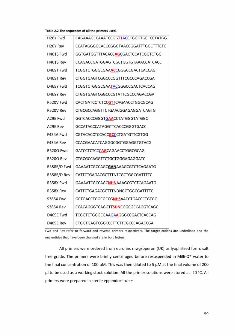
59
Table 2.2 The sequences of all the primers used.
H26Y Fwd CAGAAAGCCAAATCCGGTTACCCGGGTGCCCCTATGG
H26Y Rev CCATAGGGGCACCCGGGTAACCGGATTTGGCTTTCTG
H461S Fwd GGTGATGGTTTACACCAGCGACTCCATCGGTCTGG
H461S Rev CCAGACCGATGGAGTCGCTGGTGTAAACCATCACC
D469T Fwd TCGGTCTGGGCGAAACCGGGCCGACTCACCAG
D469T Rev CTGGTGAGTCGGCCCGGTTTCGCCCAGACCGA
D469Y Fwd TCGGTCTGGGCGAATACGGGCCGACTCACCAG
D469Y Rev CTGGTGAGTCGGCCCGTATTCGCCCAGACCGA
R520V Fwd CACTGATCCTCTCCGTTCAGAACCTGGCGCAG
R520V Rev CTGCGCCAGGTTCTGAACGGAGAGGATCAGTG
A29E Fwd GGTCACCCGGGTGAACCTATGGGTATGGC
A29E Rev GCCATACCCATAGGTTCACCCGGGTGACC
F434A Fwd CGTACACCTCCACCGCCCTGATGTTCGTGG
F434A Rev CCACGAACATCAGGGCGGTGGAGGTGTACG
R520Q Fwd GATCCTCTCCCAGCAGAACCTGGCGCAG
R520Q Rev CTGCGCCAGGTTCTGCTGGGAGAGGATC
R358E/D Fwd GAAAATCGCCAGCGANAAAGCGTCTCAGAATG
R358E/D Rev CATTCTGAGACGCTTTNTCGCTGGCGATTTTC
R358X Fwd GAAAATCGCCAGCNHNAAAGCGTCTCAGAATG
R358X Rev CATTCTGAGACGCTTTNDNGCTGGCGATTTTC
S385X Fwd GCTGACCTGGCGCCGNHSAACCTGACCCTGTGG
S385X Rev CCACAGGGTCAGGTTSDNCGGCGCCAGGTCAGC
D469E Fwd TCGGTCTGGGCGAAGAAGGGCCGACTCACCAG
D469E Rev CTGGTGAGTCGGCCCTTCTTCGCCCAGACCGA
Fwd and Rev refer to forward and reverse primers respectively. The target codons are underlined and the
nucleotides that have been changed are in bold letters.
All primers were ordered from eurofins mwg/operon (UK) as lyophilised form, salt
free grade. The primers were briefly centrifuged before resuspended in Milli-Q® water to
the final concentration of 100 μM. This was then diluted to 5 μM at the final volume of 200
μl to be used as a working stock solution. All the primer solutions were stored at -20 °C. All
primers were prepared in sterile eppendorf tubes.

60
2.14.2. Polymerase Chain Reaction Conditions
All the mutations were introduced to the DNA template by site directed
mutagenesis kit (Stratagene, Cheshire, UK). A 50-μl PCR reaction consisted of 2 μl of
forward primer, 2 μl of reverse primer, 5 μl of 10X reaction buffer, 3 μl Quick solution, 1 μl
dNTP mix, 1 μl DNA template, and 36 μl of Milli-Q® water. After a brief mixing, 1 μl of DNA
polymerase was added. The PCR tube was transferred to the thermal cycler(Techgene,
version 13, block 20 x 0.5 ml). The details of the PCR cycle are illustrated in Table 2.3.
Table 2.3 The conditions of the PCR reaction.
Segment number of cycle Step Temperature (°C) Time
1 1 Initial denaturation 95 30 s
2 25
Denaturation 95 30 s
Annealing 55 1 min
Elongation 68 15 min
3 Hold
4
For double mutant construction, two sets of primers were added to the reaction
volume at the same time but the 1 μl of forward and reverse primer of each primer set
were added in order to make the total concentration of all the forward and reverse primer
constant. For the triple mutant, 0.7 μl was used.
All the PCR products were checked by DNA gel electrophoresis before any further
use. After confirming that PCR was successful and gave the right product size, 1 μl of DpnI
enzyme was added to 45 μl of PCR reaction mixture which was then left at 37 °C for 1 hour
to remove the DNA template. After that, the sample can be stored at -20 °C or used straight
away.
2.14.3. Transformation
1 μl of the DpnI treated PCR reaction was added to a sterile eppendorf tube which
was kept on ice. The competent cell was taken from the -80 °C and left on ice for 5 minutes.
30 μl of the competent cell was added to the pre-chilled eppendorf containing the DNA and
left for 20 minutes. The competent cells were subject to heat shock at 42 °C for 45 s and
brought back to ice for 3 minutes. 1 ml of S.O.C media (Invitrogen, UK) was added to the
competent cells which were then incubated at 37 °C, shaking at 250 rpm for 1 hour. 150 μl

61
of the mixture was spread on LB-Amp plate and the rest was spread onto another plate.
These plates were pre dried in the incubator at 37 °C for 1 hour prior spreading the cells.
These plates were left to dry then incubated at 37 °C for 18 hours. At least 6 colonies were
picked to prepare glycerol stock and extract plasmid to confirm the mutation by DNA
sequencing of which the service was provided by Wolfson Institute for Biomedical Research
and the UCL Cancer Institute, UK.
2.15. Purified enzyme preparation
2.15.1. Buffers used in purification
2.15.1.1. Equilibration and wash buffers
The equilibration and wash buffers consist of 50 mM sodium phosphate monobasic
(NaH2PO4), 300 mM sodium chloride, and 10 mM imidazole. The solution was prepared
dissolving 6 g of NaH2PO4, 17.5 g of sodium chloride, and 0.68 g of imidazole in 1 l of RO
water and the pH was adjusted to 8.0 by addition of 46 % (W/V) sodium hydroxide.
2.15.1.2. Elution buffer
The elution buffer consists of 50 mM sodium phosphate monobasic (NaH2PO4), 300
mM sodium chloride, and 250 mM imidazole. This was prepared by dissolving 6 g of
NaH2PO4, 17.5 g of sodium chloride, and 17 g of imidazole in 1 l of RO and adjusting the pH
to 8.0.
2.15.2. Purification procedure
Both transketolase and transaminase enzymes contain 6 histidine residues at their
N-termini for affinity purification using His-select™ gel (Sigma-Aldrich, UK). E. coli strains
producing either transketolase or transaminase were cultured according to the standard
procedure at the fermentation volume of 200 ml (for TK see section 2.3 and for
transaminase see section 6.2.3). Cell pasted obtained from 50 ml culture was resuspended
in the appropriate buffer prior the preparation of clarified lysate. For the cell paste for
transketolase, 5 ml of 50 mM Tris buffer, pH 7.0 was used while the cell paste for
transaminase was resuspended in 5 ml of 50 mM Tris buffer, pH 7.5 with 0.4 mM PLP.
Lysate was used fresh and must be kept on ice at all time.
62
The nickel beads were stored in 30% ethanol solution and must be resuspended in
its own container prior to transferring 5 ml of these fully mixed beads to a 10-ml centrifugal
column (Thermo Scientific Pierce, UK) which is fitted with a falcon tube to collect the flow-
through. The column was spun at 1000 rpm for 5 minutes to remove ethanol which was
collected in the falcon tube. 10 ml of equilibration buffer was added to the centrifugal
column and the column was inverted a few times to mix the beads. This was then
centrifuged at 1000 rpm for 5 minutes and the flow-through buffer was discarded. After
this step was repeated three times, 5-ml clarified lysate was then transferred to the beads.
The column was closed and mixed by attaching the column to a Rotospin Test Tube Rotator
and spun at 40 rpm for 30 minutes. This binding procedure was performed in cold cabinet
to maintain the temperature at 4 °C. The column was spun at 1000 rpm for 5 minutes and
the flow through was discarded. 10 ml of wash buffer was added to the beads and the
column was mixed by Test Tube Rotator as above for 5 minutes. The column was then spun
at 1000 rpm for 5 minutes and the flow-through was discarded. This was repeated twice
more times to remove nonspecific binding. 5 ml of elution buffer was then added to the
beads and mixed by Test Tube Rotator for 15 minutes. The column was then spun at 1000
rpm for 5 minutes. The flow-through containing purified enzyme was collected for dialysis.
The beads were resuspended in 10 ml of elution buffer for two more times but the flow
through was discarded. The beads were washed with RO water and stored in 30% ethanol
at 4 °C. The used beads were only used with the same enzyme variant.
The collected flow-through containing purified enzyme was transferred to dialysis
tube with a molecular weight cut-off of 10 kDa (Perbio Science UK Ltd., Northumberland,
UK). The tube was sealed and dialysed in 4 l of 50 mM Tris bufferat 4°C for 18 hours. For
transketolase, the pH of the buffer was 7.0 while the pH for transaminase was 7.5 and the
buffer also contains 0.4 mM PLP. The buffer was gently mixed by a magnetic stirrer
throughout the dialysis. After 18 hours, the sample in the dialysis tube was transferred to a
clean tube and stored at 4°C to be used within 7 days. The purity was assessed by SDS-
PAGE gel as shown in Figure 2.2b.

63
3. Chapter 3: Recombination of existing single mutants
3.1. Introduction
Protein engineering is a powerful tool for improving properties of an enzyme. In
order to expand an enzyme substrate spectrum using a more efficient small library size,
mutations could be focussed around the active site of an enzyme (Bougioukou et al., 2009;
Morley and Kazlauskas, 2005; Paramesvaran et al., 2009; Reetz et al., 2005; Toscano et al.,
2007). Mutations further away from the active site could also cause conformational
changes within an enzyme which result in reshaping of the active site and improving
activity (Lee and Goodey, 2011; Wu et al., 2010). When considering the use of enzymes in
industrial processes where organic solvents, elevated temperature, or non-physiological pH
may be required, catalytic activity is not the only important parameter, as enzyme stability
must also be considered.
The effects of environmental factors on the stability of the wild type E. coli
transketolase have been investigated (Dalby et al., 2007; Jahromi et al., 2011; Martinez-
Torres et al., 2007). At the same time, transketolase has been engineered to accept a wider
range of substrates. In recent years, directed evolution of transketolase has led to several
mutants with improved activity towards glycolaldehyde (Hibbert et al., 2007), and
propionaldehyde (Hibbert et al., 2008). The D469X library has gained a particular interest
due to the fact that some variants have also yielded higher and reversed stereoselectivity
(Smith et al., 2008), and accepted a wider range of aldehydes including cyclic, longer chain
(Cázares et al., 2010) and aromatic aldehydes (Galman et al., 2010). Besides that, the
enzyme kinetic study of the D469T mutant revealed that substituting the negatively
charged residue with a hydrophobic residue not only enhanced the affinity towards
propionaldehyde but also significantly improved the kcat (Hibbert et al., 2008).
Having showed several synthetic potential, the substrate spectrum and activity of
transketolase could be further improved, possibly by introducing multiple mutations.
However, the stability of the mutant has to be considered and so far, only the wild type
stability has been documented. The stability of a protein can be determined from in vitro
studies of the kinetics and the thermodynamic stability (Tokuriki and Tawfik, 2009). Since
the functional protein is in a folded form, thermostability assays usually involve monitoring
folding/unfolding and aggregation. Previous in vitro studies suggested that the more
thermostable enzymes unfold and aggregate at higher temperatures (Wang et al., 2010).

64
Aggregation could also be observed within a cell as inclusion bodies. Previous in vivo
studies of destabilising mutations suggested that they can influence the formation of
inclusion bodies and reduce the amount of the soluble fraction within the cell (Calloni et
al., 2005; Tokuriki and Tawfik, 2009; Wetzel, 1994). These stabilities, however, have been
observed to trade off with the enzyme activity such that when the activity is improved,
stability tends to decline (Arnold et al., 2001; Dalby, 2011; Tokuriki et al., 2008). This is
because there is compensation between structural rigidity and flexibility, where a rigid
structure makes an enzyme more stable, whereas flexibility is required for an enzyme to
function (Arnold et al., 2001). This suggests it is best to use a more stable variant as a
template for directed evolution as it can accumulate more mutations (Bloom et al., 2006;
Tokuriki and Tawfik, 2009). Additional stability can be brought to the target protein through
introducing consensus sequence (Lehmann and Wyss, 2001) or global suppressors (Brown
et al., 2010).
Another approach to target mutations with potentially less deleterious impact is to
use the evolutionary information of the proteins. Multiple sequence alignments of enzymes
within the same familycan reveal that that certain residues mutate in a manner that is
dependent on mutations at other residues, to maintain a structure compatible for a new
function (Göbel et al., 1994; Neher, 1994). This pattern of mutation forms a network which
has been expressed as statistically coupled residues (Lockless and Ranganathan, 1999) and
their coupling energy (Süel et al., 2003). It was suggested that this network may be
important for maintaining the stability of the folded state of a protein (Göbel et al., 1994;
Socolich et al., 2005) and also the long-range residue-residue interaction (Lockless and
Ranganathan, 1999).
In this study, I carried out kinetic analyses and mutagenesis work as part of a
collaborative project that initially aimed to create a highly active mutant towards
glycolaldehyde and propionaldehyde by recombining all the single mutants previously
identified to have enhanced activities towards both substrates (Strafford et al., 2012).
Preliminary work by Ed Hibbert had created several double mutants and found that all of
them lost activity as well as soluble expression of the enzyme through destabilisation. I
created a complementary series of rationally designed recombinant mutants and
characterised them in this Chapter. Another group of double mutants were also created by
John Strafford using saturation mutagenesis at two or three residues within a network of
residues that were found to co-evolve in TK. The combinations between D469 and R520

65
were the most beneficial for stability. I also carried out repeats and additional
characterisation of some of these mutants as presented here. These studies were all
combined into a single paper (Strafford et al., 2012), the overall results of which will be
summarised here.
3.2. Materials and method
3.2.1. Materials
The sources of all chemicals are listed in chapter 2. The bottle of propionaldehyde
was closed with septum cap. The liquid was drawn out using syringe and needle. The bottle
was then filled with 100% nitrogen to prevent oxidation.
3.2.2. Mutant construction
The primers used in this study were listed in Table 2.2, chapter 2 and confirmed by
DNA sequencing. These residues were selected because they are either highly conserved
residues in close proximity to the active centre or and medium range residues that are
phylogenetic variants (Hibbert et al., 2008, 2007). All the plasmids bearing mutations were
transformed into BL21Gold(DE3)because it was suggested to minimise proteolysis of the
recombinant protein (Miller et al., 2007). In addition, this strain was also chosen for the
expression of both transketolase and transaminase (Ingram et al., 2007; Rios-Solis et al.,
2011). Since the highly active mutants would be co-expressed with transaminase for the
coupling reaction, this strain was chosen.
3.2.3. Protein quantification
Total protein concentration was quantified by Bradford assay according to the
protocol. The concentration of transketolase within the clarified lysate was determined
from SDS-PAGE and densitometry.
3.2.4. Expression level study
Single colonies of each mutant were inoculated into 10 ml LB broth containing 150
μg/ml ampicillin. They were cultured for 18 hours at 37 °C, shaking at 250 rpm. Cells were
harvested by centrifugation at 4000 rpm, 4 °C, for 10 minutes. The supernatant was
discarded and the pellets were resuspened in 2 ml 50 mM Tris buffer, pH 7.0. This was split
into 2 portions; 1 ml was kept as whole cell sample, another 1 ml was sonicated to analyse

66
the soluble and insoluble fraction. The 1 ml of the resuspended cells was transferred into 2-
ml tubes and sonicated on ice for 10 cycles with 10 s on, 15 s off. They were then
centrifuged at 17,000 g, 4°C, for 10 minutes. The supernatants were transferred into clean
2-ml eppendorf tubes. All the pellets were fully resuspended in 1 ml 50 mM Tris buffer, pH
7.0.
The 50 μl of whole cell, lysate, and resuspended pellet were mixed with 50 μl 2X
Laemmli buffer and heat to 95 °C for 10 minutes. Up to this point, the lysate, and the
pellets always had the same final volume as the whole cell sample. The total protein
concentrations in the clarified lysate were determined by Bradford assay to adjust the
volume of lysate to be loaded into the SDS-PAGE gel. The same volumes were applied for
the whole cell and resuspended pellets. Different amount of purified transketolase were
added into all the gels to make a calibration curve for each individual gel. The gels were
analysed by the protocol in materials and methods. Expression level refers to the
percentage concentration of transketolase in the lysate as defined below.
Percentage concentracon= [transketolase concentracon in lysate]
[total protein concentracon in lysate] x 100
Soluble :inclusion body raco = [transketolase in lysate]
[transketolase in debris pellet]
3.2.5. Identification of the protein in the insoluble fraction
The insoluble fraction was prepared for SDS-PAGE analysis according to the
protocol above. Protein ladder was also added to the gel. The plot of log(molecular weight)
versus the migration distance of the marker was used to construct a calibration curve to
identify the molecular weight of P2 protein. The band was then cut from the gel and sent
for protein sequencing (Alta Bioscience, University of Birmingham, UK).
3.2.6. Activity assay
Clarified lysate was prepared according to the protocol. The 30 μl of lysate was
incubated with 30 μl of 10xcofactor solution (24 mM ThDP, 90 mM MgCl2) and 140 μl 50
mM Tris buffer, pH 7.0 at room temperature for 20 minutes. 3X substrate solution (150 mM
HPA, 150 mM aldehyde) were prepared in 50 mM Tris buffer and adjusted pH to 7.0. The
reaction was started by adding 100 μl of 3x substrate solution to the enzyme-cofactor
solution. All reactions were done in triplicate. For the glycolaldehyde activity study, the

67
reaction was run in polystyrene, round-bottom microtitre plate and covered with a lid
during the assay. The samples were taken at every 1 minute. All the propionaldehyde
reactions were run in glass vials and covered with snap caps. The samples were taken at
every 10 minutes for the variants containing D469T/Y for 1 hour. For other mutants, the
samples were taken at every 60 minutes for 5 hours.
In order to directly compare the effect of mutation upon the substrate preference
between glycolaldehyde and propionaldehyde, the same batch of clarified lysate were used
to identify the activities of all the mutants towards both substrates. This eliminated any
possible error from protein concentration quantification. Only freshly prepared
propionaldehyde was used in all experiments to avoid substrate oxidation.
3.2.7. HPLC analysis
HPA, erythrulose, and 1,3-dihydroxypentane-2one (DHP) were quantified by HPLC
using Aminex HPx-87H, 300x7.8mm column (Bio-Rad, UK) according to the protocol in
chapter 2, section 2.7.1.
3.2.7.1. Standard graphs
Figure 3.1 Standard graphs of a) HPA, b) erythrulose, and c) DHP.
The standard graphs were plotted based on the final concentrations of the solutions in the wells.
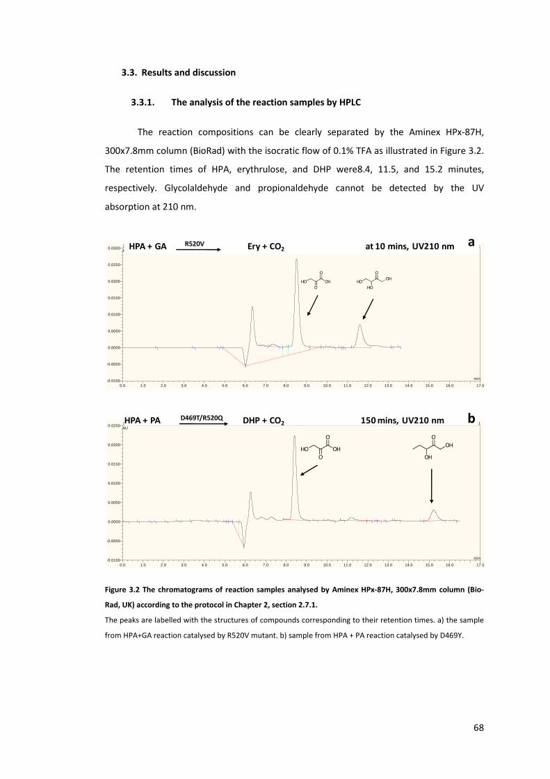
68
3.3. Results and discussion
3.3.1. The analysis of the reaction samples by HPLC
The reaction compositions can be clearly separated by the Aminex HPx-87H,
300x7.8mm column (BioRad) with the isocratic flow of 0.1% TFA as illustrated in Figure 3.2.
The retention times of HPA, erythrulose, and DHP were8.4, 11.5, and 15.2 minutes,
respectively. Glycolaldehyde and propionaldehyde cannot be detected by the UV
absorption at 210 nm.
Figure 3.2 The chromatograms of reaction samples analysed by Aminex HPx-87H, 300x7.8mm column (Bio-
Rad, UK) according to the protocol in Chapter 2, section 2.7.1.
The peaks are labelled with the structures of compounds corresponding to their retention times. a) the sample
from HPA+GA reaction catalysed by R520V mutant. b) sample from HPA + PA reaction catalysed by D469Y.
0.0 1.0 2.0 3.0 4.0 5.0 6.0 7.0 8.0 9.0 10.0 11.0 12.0 13.0 14.0 15.0 16.0 17.5-0.0100
-0.0050
0.0000
0.0050
0.0100
0.0150
0.0200
0.0250
0.0300 2010-11-29 KINETICS R520V HPA + 10-50 MMGA #114 [modified by All Users] 50 mM 10 UV_VIS_1AU
min
HPA + GA R520V Ery + CO2 at 10 mins, UV210 nm a
0.0 1.0 2.0 3.0 4.0 5.0 6.0 7.0 8.0 9.0 10.0 11.0 12.0 13.0 14.0 15.0 16.0 17.5-0.0100
-0.0050
0.0000
0.0050
0.0100
0.0150
0.0200
0.0250 2010-09-07 A29ED469Y D469YR520Q D469TR520Q F434AR520Q HPA PA #15 [modified by All Users] UV_VIS_1AU
min
HPA + PA D469T/R520Q DHP + CO2 150 mins, UV210 nm b

69
3.3.2. The specific activities of transketolase mutants towards glycolaldehyde
and propionaldehyde
The previous work illustrated that the wild type transketolase from the host strain
has very low activity compared with the highly expressed wild type and this background
can be neglected (Miller et al., 2007). In addition, two mutants in this study, H26Y and
H26Y/D469Y, were found to be inactive. When assessing the activities of these mutants
towards both glycolaldehyde and propionaldehyde, there were no background activities of
the wild type enzyme from the host strain.
The mutants in this study were separated into two groups. The first group are those
that have been studied with glycolaldehyde in the previous section and the mutants in the
second group are those that were reported to enhance activities towards propionaldehyde
(Hibbert et al., 2008) or influence the stereoselectivity of the enzyme (Smith et al., 2008).
The specific activities of all the single and combined mutants towards glycolaldehyde and
propionaldehyde are listed in Table 3.1. below. The activities of the wild type and mutants
previously identified to have higher activities towards glycolaldehyde (Hibbert et al., 2007)
and propionaldehyde (Hibbert et al., 2008) were repeated in this study and added for
comparison. Both studies showed that D469T and D469Y had improved activities towards
propionaldehyde although the percentage increase from wild type still differed. The double
mutants containing D469T/Y also had higher activities than wild type. Since the previous
report suggested that there was a strong shift in substrate preference upon the mutation at
H26 and D469, the two groups will be discussed separately (Hibbert et al., 2008).
From Table 3.1, it can be seen that the specific activity of the wild type in this study
was significantly higher than what was determined by Hibbert et al., (2007). Yet, the
mutants identified with enhaced activity towards glycolaldehyde were found to have either
similar (A29E, H461S) or lower activity (R520V) than wild type. Therefore, the experimental
conditions were compared in Table 3.2 to identify the sources of deviation.

70
Table 3.1 The specific activities of all the mutants towards glycolaldehyde and propionaldehyde determined in this study and previously determined.
Mutant
Glycolaldehyde activity Propionaldehyde activity Rate GA:PA (from this study only)
This study Previous study b This study Previous study c
μmol mg-1 min-1 μmol mg-1 min-1 μmol mg-1 min-1 μmol mg-1 min-1
Group I
WT 36 (1.1) 0.65 (0.04) 0.20 0.029 (0.001) 183
A29E 38 (6.2) 1.95 (0.17) 0.16 0.10 (0.02) 232
H461S 32 (11) 3.14 (0.54) 0.14 0.040 (0.007) 223
R520V 22 (3.6) 2.3 (0.5) 0.11 0.14(0.05) 206
A29E H461S 28 (4.2) 0.2 142
A29E R520V 34 (11) 0.14 249
H461S R520V 15 (4.2) 0a n.d.
A29E H461S R520V 37 0.17 224
Group II
H26Y 0 a 0 a n.d.
D469Y n.d 2.4 0.127 (0.01) n.d.
H26Y D469Y 0 a 0 a
D469Y R520V n.d. 1.5
D469T 1.4 0.14 (0.001) n.d.
D469T R520Q 1.9
D469Y R520Q 1.2
A29E D469Y 0 a
R520Q 0.69d a No activity was observed, b(Hibbert et al., 2007), c(Hibbert et al., 2008), d(Strafford et al., 2012)

71
Table 3.2 The comparison of the experimental outcomes and conditions between this study and the previous
study when using glycolaldehyde as a substrate.
This study Previously determined b
Mutant Activity
μmol mg-1 min-1
Initial rate a
mM min-1
[TK]
mg/ml
Activity
μmol mg-1 min-1
Initial rate
mM min-1
[TK]
mg/ml
WT 36 (1.1) 6.4 (0.68) 0.18 0.65 (0.04) 0.29 (0.01) 0.44
A29E 38 (6.2) 4.56 (0.18) 0.11 1.95 (0.17) 0.66 (0.01) 0.34
H461S 32 (11) 0.85 (0.03) 0.03 3.14 (0.54) 0.60 (0.04) 0.19
R520V 22 (3.6) 1.73 (0.09) 0.09 2.3 (0.5) 2.16 (0.03) 0.92
Standard deviation is in parenthesis.a an example of one batch, b(Hibbert et al., 2007)
From Table 3.2, it can be seen that the previously determined specific activities of
wild type and the single mutants were much lower than what have been identified in this
study. The initial rates of all the reactions in the previous study were much lower although
the enzyme concentrations used were much higher. The causes of the discrepancies may
be the differences in the enzyme and substrate preparations. I also discovered that certain
mutants were unstable while their expression levels fluctuated from batch to batch and
depended on the culture condition (see later on). Propionaldehyde is also easily oxidised
and degraded. Since more details in the activity assays for both substrates are not available
in their publications (Hibbert et al., 2008, 2007), other causes of deviations cannot be
determined. Therefore, the relative influences of each mutation here will be discussed
based on comparison of the results from this study only. It should be noted that the specific
activities of the single mutants in the publication Straffordet al., 2012)differed from those
listed in Table 3.1 because the specific activities in the publication were the averaged
results from this work and all previous repeats (Hibbert et al., 2008, 2007)(Strafford,
personal communication).
Prior to any discussion, two important observations must be noted as they have the
potential to influence the apparent characteristic of the mutants. Firstly, the mutants in the
first group strongly preferred glycolaldehyde over propionaldehyde as an acceptor
substrate with an approximately 200-fold difference in the bioconversion rates. These
differences made the reaction times of propionaldehyde much longer and it is possible that
the mutants in the first group were subject to a higher degree of inactivation by
propionaldehyde. This inactivation could also occur to the mutants in the second group to a
lesser extent. This is because the specific activities of the mutants evolved for
propionaldehyde were still 15-35 times slower than the activity of wild type towards

72
glycolaldehyde. This factor may be more pronounced for inherently more unstable mutants
and lead to lower observed activities. However, this factor will be neglected throughout the
study. Secondly, in this experiment, I found that the H461S mutation had a tendency to
cause enzyme inactivation. When the H461S mutant was grown and prepared for the
activity assay, half of the batches did not show any enzymatic activity. The specific activity
also greatly fluctuated which resulted in a high standard deviation as showed in Table 3.1.
In addition, occasionally inactivated enzymes were also observed in A29E/H461S,
H461S/R520V, and A29E/H461S/R520V mutants suggesting that H461S may cause a global
instability or time-dependent enzyme inactivation.
3.3.3. The locations of the targets residues, their functions, and interactions
Among all three target residues in group I, A29 was the only residue that is not part
of the active site. It is actually located at the phosphate binding pocket of ThDP as showed
in Figure 3.3. Therefore, amino acid substitution at this residue is unlikely to alter the
substrate binding. However, replacing alanine with a negatively charged amino acid can
cause electrostatic repulsion between the side chain and the phosphate group of the ThDP
molecule which could affect the affinity between the enzyme and cofactor or the
reposition of the ThDP. These could potentially result in lower enzyme activity as observed
in this experiment.

73
Figure 3.3 The location of the residues subject to mutation in this study. Black dotted lines are hydrogen
bonds.
a) the covalent-linked intermediate between fructose 6-phosphate and ThDP in wild type TK from E. coli(PDB
ID: 2R8P). b) the binding of ribose 5-phosphate into E. coli transketolase active site (PDB ID: 2R5N). c) the
binding of erytrhose 4-phosphate into yeast transketolase active site (PDB IB: 1NGS). d) aschematic illustration
of the hydrogen bonds between certain residues and covalent-linked intermediate between fructose 6-
phosphate and ThDP.
The previous directed evolution study showed that replacing the positively charged
phosphate-binding residues with neutral amino acids appear to improve the activity
towards glycolaldehyde (Hibbert et al., 2007). In this study, however, the mutations at the

74
phosphate binding residues; H461S and R520V did not appear to improve the activity
towards glycolaldehyde. In addition, all other single mutants in the first group were found
to have lower activities towards propionaldehyde. Besides that, the single, double, and
triple mutants constructed from combining H461S and R520V did not show a significant
shift in substrate preference from hydroxylated to non-hydroxylated substrate. By analysis
of the crystal structure of transketolase, it can be seen that these residues are located at
the entrance of the transketolase active site and the distance from their amino groups to
the C2 atom of the thiazolium ring are further than 10 Å. This distance is too far for the
hydroxyl group of the glycolaldehyde to interact with while the carbonyl group is in close
proximity to the C2 atom. This distance is, however, compatible with phosphorylated
substrates as showed in the crystal structures of yeast and E. coli transketolases (Figure
3.3a and b) (Asztalos et al., 2007; Nilsson et al., 1997; Wikner et al., 1997). In the the crystal
structure of E. coli transketolase (PDB ID: 2R5N, Figure 3.3b), it shows that the 3-hydroxyl
group of ribose 5-phosphate forms a hydrogen bond with the side chain carboxylic group of
D469 residue (Asztalos et al., 2007). On the other hand, D477 in yeast transketolase
interacts with C2 hydroxyl group of a shorter acceptor molecule erythrose 4-phosphate
(Figure 3.3c) (Nilsson et al., 1997). In addition, D469Y hada very low activity towards
glycolaldehyde but significantly improved activity towards propionaldehyde, a dramatic
change in the substrate preference from hydroxylated substrate to non-hydroxylated
substrate as showed in Table 3.1. This combinded evidence suggest that the hydroxyl group
of glycolaldehyde does not interact with the phosphate binding site. Therefore, it is
possible that mutations at these residues may have small impacts on the activity towards
glycolaldehyde or propionaldehyde.
Although none of the residues in groupI appeared to directly interact with
glycolaldehyde, the mutation at H461 and R520 can cause some changes in the
microenvironments in the active site channel or small structural arrangement which
influences the overall activity and stability. In addition, there could also be synergy
between these residues due to their spatially close positions. Therefore, the experimentally
determined activities and expected activities were compared to find these interactions as
showed in Figure 3.4a.
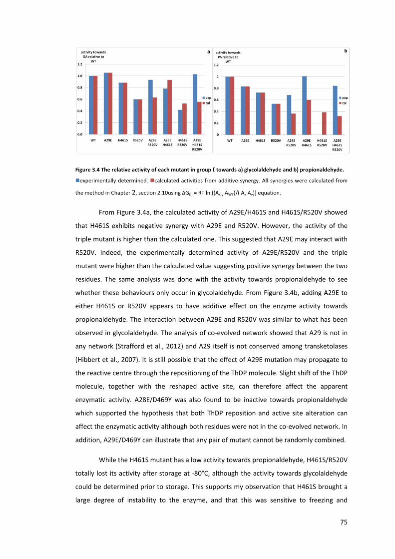
75
Figure 3.4 The relative activity of each mutant in group I towards a) glycolaldehyde and b) propionaldehyde.
�experimentally determined. �calculated activities from additive synergy. All synergies were calculated from
the method in Chapter 2, section 2.10using ΔG(I) = RT ln {(Ax,y AWT)/( Ax Ay)} equation.
From Figure 3.4a, the calculated activity of A29E/H461S and H461S/R520V showed
that H461S exhibits negative synergy with A29E and R520V. However, the activity of the
triple mutant is higher than the calculated one. This suggested that A29E may interact with
R520V. Indeed, the experimentally determined activity of A29E/R520V and the triple
mutant were higher than the calculated value suggesting positive synergy between the two
residues. The same analysis was done with the activity towards propionaldehyde to see
whether these behaviours only occur in glycolaldehyde. From Figure 3.4b, adding A29E to
either H461S or R520V appears to have additive effect on the enzyme activity towards
propionaldehyde. The interaction between A29E and R520V was similar to what has been
observed in glycolaldehyde. The analysis of co-evolved network showed that A29 is not in
any network (Strafford et al., 2012) and A29 itself is not conserved among transketolases
(Hibbert et al., 2007). It is still possible that the effect of A29E mutation may propagate to
the reactive centre through the repositioning of the ThDP molecule. Slight shift of the ThDP
molecule, together with the reshaped active site, can therefore affect the apparent
enzymatic activity. A28E/D469Y was also found to be inactive towards propionaldehyde
which supported the hypothesis that both ThDP reposition and active site alteration can
affect the enzymatic activity although both residues were not in the co-evolved network. In
addition, A29E/D469Y can illustrate that any pair of mutant cannot be randomly combined.
While the H461S mutant has a low activity towards propionaldehyde, H461S/R520V
totally lost its activity after storage at -80°C, although the activity towards glycolaldehyde
could be determined prior to storage. This supports my observation that H461S brought a
large degree of instability to the enzyme, and that this was sensitive to freezing and

76
thawing. The instability or loss in activity was not observed previously after mutagenesis of
yeast transketolase at the equivalent residue (Nilsson et al., 1997). In the study of yeast
transketolase, H469A still had 77% activity remaining, but the affinities towards
phosphorylated substrates were much lower, which implied that this residue formed
hydrogen bonds with the phosphate group of the substrate (Nilsson et al., 1997). However,
different substitutions could also cause different effects on the enzyme function and
stability. The function-instability relationship upon H461S introduction will be discussed
with the expression study later on.
D469T/Y were previously identified with improved activity for propionaldehyde
(Hibbert et al., 2008). The mutagenesis study in yeast transketolase together with the use
of deoxy aldose sugars and an opposite stereoisomer at the C2, led to a proposal that
Asp477 interacts with the hydroxyl group at the C2 of acceptor aldehyde which directly
controls the stereoselectivity of the enzyme (Nilsson et al., 1998). In the E. coli TK structure
(PDB ID: 1QGD) D469 and R520 do not form a direct hydrogen bond between them but
rather via a water molecule and the backbone carbonyl group of L466. This also supports
my speculation that H461 and R520 do not form any direct interaction with the 2-hydroxyl
group of glycolaldehyde or the methyl group of propionaldehyde but the interactions
would partly arise from the D469 residue (Hibbert et al., 2008). However, the effect of
combining the mutants is still observable.
Some non-natural D469 variants, especially D469T, were found to have the highest
activities towards propionaldehyde among several other mutants (Hibbert et al., 2008).
D469T was previously combined with D259 mutants in order to further improve the
activity. However, the double mutants D469T/D259S and D469T/D259Y had lower activities
than the respective single mutants, while recombinants containing D469S did not suffer the
same issue (Strafford et al., 2012). This led to a hypothesis of whether D469T was the cause
of the loss of enzyme activities within the double mutants. In addition, a statistical coupling
analysis of ThDP dependent enzyme sequences revealed that the D469 residue co-evolved
with R520 (Strafford et al., 2012). Therefore, substituting R520 with other amino acids
could potentially improve the enzyme activity or stability. Indeed this was observed, and in
this study R520Q was chosen for further work because it was found to enhance the
solubility but reduce the enzyme activity slightly (Strafford et al., 2012). In order to assess
whether this was the case more generally, I recombined D469T/Y mutants with R520V/Q to
study if each combination will have the same effects. None of these variants naturally exist

77
in transketolase which therefore has significant implications for enzyme engineering
(Hibbert et al., 2007). In Figure 3.5, it can be seen that positive synergy was observed for
D469T/R520Q whereas negative synergy was observed for D469Y/R520Q. However, when
R520V was added into D469Y, only a slightly positive synergy was observed. This suggested
that different substitutions at the first position (R520) can have a different effect on the
second position.
Figure 3.5 The relative activity of group 2 mutants towards propionaldehyde.
�experimentally determined � calculated activities from additive synergy.
3.3.4. H26Y, the inactive mutant
Another residue in the second group is H26Y. In the previous studies, H26Y was
identified to reverse the stereoselectivity (Smith et al., 2008) and accept other aliphatic and
cyclic aldehydes (Cázares et al., 2010).Other variants; A, T, K, and V were also found to have
higher activity than wild type (Hibbert et al., 2008). In this study, however, H26Y was found
to have no activity towards any substrate. Although D469Y can improve the activity
towards propionaldehyde, the double mutant H26Y/D469Y did not restore the activity
either. In addition, H26Y was reconstructed by another research group and was also found
to have very little activity (Yi et al., 2012). In fact, H26 is highly conserved in transketolase-
like enzyme family (Costelloe et al., 2008). The transketolase crystal structures from yeast
(PDB ID: 1NGS) (Nilsson et al., 1997) and E. coli (PDB ID: 2R8O, 2R8P) (Asztalos et al., 2007)
illustrated that H26 forms a hydrogen bond to the C3 hydroxyl groups of the donor
molecules (Figure 3.3), which needs to be deprotonated during the catalytic cycle. Due to
the close proximity and hydrogen bond interaction with the C3 hydroxyl group, H26 was
proposed to abstract proton from this hydroxyl group (Wikner et al., 1997). The study of
the equivalent histidine residue in yeast transketolase also showed that H30A/N mutants
showed severely impaired kcat (Wikner et al., 1997), although in the E. coli transketolase,
78
the H26A mutant was observed to have 2.3 times higher activity towards propionaldehyde
than the wild type (Hibbert et al., 2008). Therefore, it is highly possible that H26Y will
produce an inactive enzyme, particularly given the large steric bulk of the tyrosine residue.
The study in both groups of transketolase so far illustrated that combining high
activity-mutants would not always improve the activity of the enzyme due to the
synergistic interaction between these residues. Although the two distal residues are not in
the same co-evolved network, they may appear to interact upon mutations but these
interactions could possibly arise from a different reason to those driving the natural co-
evolution (charge stabilisation, steric minimisation).
3.3.5. The expression level of all mutants
Enzyme activity is not the only parameter to be considered for synthetic purposes.
Higher temperature or non-physiological pH can be encountered, and enzyme stability
must also be considered. The effect of mutation does not only influence the enzymatic
activity but also the stability of the enzyme. The in vitro stability of an enzyme can be
determined from the thermodynamic stability study or kinetic stability study. In contrast, it
was suggested that mutations within a protein can have an impact on protein folding
mechanism which leads to the formation of inclusion bodies (Calloni et al., 2005). In the
study of Strafford et al., (2012), the expression levels of some single and multiple mutants
were already assessed to investigate the instability upon the accumulated
mutation(Strafford et al., 2012). Here, I further looked into the effect of the combination of
the mutations in group I and II that I created to find out whether there was a relationship
between their activities and stabilities. The expression level data were also employed in
with the previous study to get a larger picture effect of the mutation on enzyme stability
(Strafford et al., 2012). The mutation itself may promote the expression but not stability or
folding. As a result, higher expression may not reflect higher stability. Therefore, I
additionally looked into the ratio between the soluble and inclusion body content formed.
The expression patterns of all the mutants were analysed and illustrated for comparison in
Figure 3.6 below.

79
Figure 3.6 The fraction of transketolase.
� represents the percentage concentration of transketolase in the clarified lysate. � is the ratio of soluble TK :
inclusion body.
In order to avoid a lower expression due to plasmid instability, the percentage
expression was determined with the master glycerol stocks as these were not obtained
from sub-cultures but rather represent the first generation after transformation in which
the plasmid copy number is expected to be at its highest. Although the expression of all the
mutants appeared to be at least 8 %, some quickly lost their expression levels even though
the same glycerol stock was repeatedly used. These include H26Y, H26Y/D469Y, H461S, and
H461S containing mutants. For H26Y and H26Y/D469Y, no enzyme was produced after the
second time that the same glycerol stock was used. Since these phenomena only occurred
in certain mutants, it suggested the rapid reductions in the expression level of these
mutants were not due to the overall instability of the glycerol stock but rather the
instability or the toxicity of the mutants themselves.
The blue line in Figure 3.6 illustrates that most mutants had lower expression levels
than the wild type, while the soluble: inclusion body ratio (red line) decreased below that
of the wild type. This suggested that larger fractions of the produced enzymes were in the
inclusion body and implied that these mutants are beginning to become unstable.
However, some double mutants appear to restore the solubility of the enzyme.
As mentioned above, H461S containing mutants quickly lost their expression levels
when the same glycerol stock was repeatedly used. It is not surprising that several single
mutations cause significant reduction in both expression level and activity. It has been
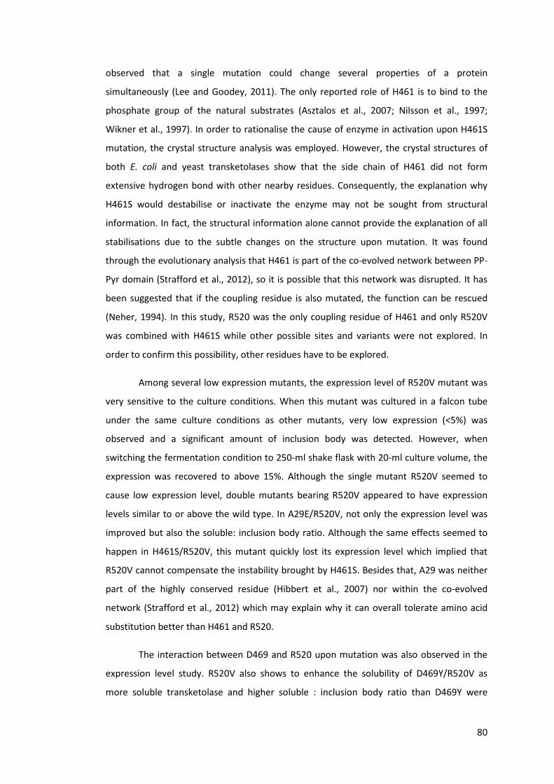
80
observed that a single mutation could change several properties of a protein
simultaneously (Lee and Goodey, 2011). The only reported role of H461 is to bind to the
phosphate group of the natural substrates (Asztalos et al., 2007; Nilsson et al., 1997;
Wikner et al., 1997). In order to rationalise the cause of enzyme in activation upon H461S
mutation, the crystal structure analysis was employed. However, the crystal structures of
both E. coli and yeast transketolases show that the side chain of H461 did not form
extensive hydrogen bond with other nearby residues. Consequently, the explanation why
H461S would destabilise or inactivate the enzyme may not be sought from structural
information. In fact, the structural information alone cannot provide the explanation of all
stabilisations due to the subtle changes on the structure upon mutation. It was found
through the evolutionary analysis that H461 is part of the co-evolved network between PP-
Pyr domain (Strafford et al., 2012), so it is possible that this network was disrupted. It has
been suggested that if the coupling residue is also mutated, the function can be rescued
(Neher, 1994). In this study, R520 was the only coupling residue of H461 and only R520V
was combined with H461S while other possible sites and variants were not explored. In
order to confirm this possibility, other residues have to be explored.
Among several low expression mutants, the expression level of R520V mutant was
very sensitive to the culture conditions. When this mutant was cultured in a falcon tube
under the same culture conditions as other mutants, very low expression (<5%) was
observed and a significant amount of inclusion body was detected. However, when
switching the fermentation condition to 250-ml shake flask with 20-ml culture volume, the
expression was recovered to above 15%. Although the single mutant R520V seemed to
cause low expression level, double mutants bearing R520V appeared to have expression
levels similar to or above the wild type. In A29E/R520V, not only the expression level was
improved but also the soluble: inclusion body ratio. Although the same effects seemed to
happen in H461S/R520V, this mutant quickly lost its expression level which implied that
R520V cannot compensate the instability brought by H461S. Besides that, A29 was neither
part of the highly conserved residue (Hibbert et al., 2007) nor within the co-evolved
network (Strafford et al., 2012) which may explain why it can overall tolerate amino acid
substitution better than H461 and R520.
The interaction between D469 and R520 upon mutation was also observed in the
expression level study. R520V also shows to enhance the solubility of D469Y/R520V as
more soluble transketolase and higher soluble : inclusion body ratio than D469Y were

81
observed. However, at least 20 % of the total D469Y/R520V produced by the cell was in the
form of inclusion body. R502Q, in contrast, significantly reduced the amount of inclusion
body in D469T/R520Q and D469Y/R520Q to less than 15% which increased the soluble :
inclusion body ratio above the wild type. However, the % of total protein for D469Y/R520Q
in the lysate is much lower than D469Y/R520V. It could be said that R520V enhances the
production of the mutant within the cell whereas R520Q slightly reduces the total
production but facility the folding and the formation of soluble enzyme. This is another
difference between R520V and R520Q mutation on D469Y.
H26Y and H26Y/D469Y although showed 9.2 and 11 % expression level of the total soluble
protein in Figure 3.6, their expression can only be detected straight after transformation.
The expression level in the same glycerol stock that has been used for streaking twice
declined to less than 5%. I initially suspected that H26Y and H26Y/D469Y were not stable
and may precipitate upon sonication or form a large amount of inclusion body during cell
culture. Therefore, both mutants were cultured for 24 hours at 25°C with and without 10%
ethanol supplement in the media. This is to slow down cell growth and allow more time for
the protein to fold. Ethanol was added to induce heat-shock protein to aid protein folding
(Thomas and Baneyx, 1997, 1996). The half of the harvested cells was then lysed by freeze-
thaw lysis while the other half was sonicated. None of these have improved the expression
level of both mutants. The SDS-PAGE of the cell debris did not show that these mutants
form any inclusion body either which suggested that both H26Y and H26Y/D469Y did not
express. These evidences imply that H26 is not only important for enzymatic activity but
may also the control of transketolase expression.
The loss of expression level and activities in certain mutants may suggest that these
mutants may have lost higher order structure or become soluble multimeric enzymes. In
order to see whether this is the actual cause, these mutants could be purified and the
distribution of the molecular weights analysed by gel filtration. It should still be noted that
the purification process itself may inactive transketolase or cause it to aggregate as shown
by Stratford et al. ( 2012).
During the study of the insoluble fraction of all the mutants, one band always
appeared in the cell debris samples but showed differences in the expression level. The size
of this protein was determined from the log(molecular weight) versus the migration
distance and was estimated to be approximately 40 kDa. In order to identify whether this is
a fraction transketolase, the sample was sent for protein sequencing. The result showed

82
that the first 5 amino acids from the N terminus are AEIYN which are not found in
transketolase sequence. This sequence was then BLAST search in BL21(DE3) (taxid:469008).
Table 3.3 shows the proteins with AEIYN sequence and the position of this sequence.
Table 3.3 The possible P2 proteins.
Protein AC/ID Name
Length
(aa)
Match
range
C6EA44/C6EA44_
ECOBD
Outer membrane p
hosphoporin protein E precursor 345 22-26
C6EER5/C6EER5_
ECOBD
GCN5-related
N-acetyltransferase precursor 172 15-19
C6EI53/C6EI53_E
COBD
OmpF, subunit of outer membrane porin F and The
Colicin A Import System precursor 362 23-27
C6EKF5/C6EKF5_
ECOBD Flavohemoprotein 396 136-140
E5QQ21/E5QQ21
_ECOBD Outer membrane porin protein, N-ter 201 24-28
From the position of the sequence and the molecular weight of the protein, the
most likely protein is OmpF. OmpF is associated with stress response and control the
material across the cell membrane. In a low nutrient environment, high expression level of
OmpF is to be expected to facilitate nutrient uptake (Delihas and Forst, 2001). Therefore, it
could be argued that OmpF may be highly expressed towards the end of the culture when
the nutrient was low. Therefore, P2 concentration was plotted against the harvest OD600.
However, this plot has no strong correlation to the expression level of OmpF. The high
expression is unlikely to be due to low nutrient level at the end of culture. I further
investigated to see whether the expression of this protein is related to the formation of the
transketolase inclusion body by plotting the P2 concentration against soluble: inclusion
body ratio as showed in Figure 3.7. The P2 concentration has an inverse relationship with
the soluble: inclusion body ratio. The higher ratio arose from either low soluble fraction or
high inclusion body and both situations can trigger stress response within the cell.

83
Figure 3.7 The scatter plot between the P2 concentration and the TK soluble: inclusion body ratio.
3.3.6. Correlation of expression and activity
As part of the aim of the study in the publication (Strafford et al., 2012), the activity
and stability relationship of the mutants was investigated and a trade off between the
enzyme activity and stability was observed. All the expression and activity data here were
compiled to compare this relationship when the mutants were constructed based on two
different approaches. All the mutants in this study were created from combining the single
mutants which were previously reported to be highly active towards glycolaldehyde
(Hibbert et al., 2007) and propionaldehyde (Hibbert et al., 2008). In the previous study,
some double mutants were the created with the same approach while some double
mutants were constructed by choosing target residues within the co-evolved network
(Strafford et al., 2012). Assuming that the % TK concentration in the clarified lysate
represents the stability of the protein, I plotted the % TK concentration versus activity
towards propionaldehyde to see if this trend could be observed as showed in Figure 3.8a.
Since group I mutants strongly preferred glycolaldehyde over propionaldehyde, their
expression levels were also plotted against their activities towards glycolaldehyde (Figure
3.8b).
84
Figure 3.8 The relationship between the specific activity towards glycolaldehyde and propionaldehyde and
the expression level of the mutant.
�mutants in group 1. �mutants in group 2
Unfortunately, the number of mutants from this study alone is insufficient to draw
this conclusion. In addition, several mutants in this study contain a global inactivating
mutant; H461S, and catalytically important residue; H26, which can disrupt the relationship
between stability-activity. Furthermore, drastic decreases in expression levels of several
mutants were observed within a few times that the same glycerol stock was used. Including
such uncertain-characteristic mutants in the analysis would lead to inaccurate
interpretation of the activity-stability relationship.
3.4. Conclusion
Here we have illustrated that most mutants that were created by pairwise
recombining two previously active single mutants within the enzyme active site resulted in
a rapid loss of expression level, suggesting that this recombination strategy could have a
detrimental effect on the enzyme stability. There could also be strong synergy between
them although this was not always showed by analysis of the natural co-evolved network.
Since the consistent expression level was only observed in the double mutants in which
both residues were within the same co-evolved cluster, this method of designing a library
could be more attractive. In addition, a single mutation may have negative effect on the
enzyme but this could have a positive effect when recombining with other residues in the
coupled network.

85
In this study, we also found that the single mutant H461S appears to globally
destabilise or inactivate the enzyme. This destabilisation cannot be deduced from the
crystal structure information but the intrinsic stabilising function may only be determined
from mutagenesis and statistical analysis of kinetic data, which has to be further studied.
86
4. Chapter 4: Rational design of substrate and TK for aromatic
aldehyde
4.1. Introduction
Dihydroxy ketone functionalilty is found in a number of high value, natural
compounds (Enders and Narine, 2008; Enders et al., 2005; Wohlgemuth, 2009) such as
carbohydrate (Kobori et al., 1992), and cortocosteriods (Hailes et al., 2009; Hailes et al.,
2010). The stereoselective synthesis of these chemicals, however, is rather complicated,
low atom efficient, and involves protecting groups and requires multi-steps (Hailes et al.,
2009; Hailes et al., 2010). Transketolase catalyse the asymmetric carbon elongation by
transferring dihydroxy ketone group to an aldehyde to form dihydroxy ketone product with
high stereospecicity and selectivity and this makes transketolase attractive in organic
synthesis.
The natural substrate spectrum of wild type transketolase from several species
includes non-phosphorylated sugar (Demuynck et al., 1991; Kobori et al., 1992), non-
hydroxylated aldehydes, cyclic aldehydes (Hobbs et al., 1993; Morris et al., 1996), and
nitroso aromatics (Corbett and Corbett, 1986). This makes the earlier work with
transketolase focus on the synthesis of ketose sugars (Bolte et al., 1987), deoxysugars
(Hecquet et al., 1994) which could be a building block for other compounds when
integrated with other enzymatic systems or chemical routes. These include the synthesis of
a caramel-like flavour; furaneol, (Hecquet et al., 1996; Hecquet et al., 1994), glycosidase
inhibitors; 1,4-Dideoxy-1,4-imino-D-arabinitol (Ziegler et al., 1988) and N-
hydroxypyrrolidine (Humphrey et al., 2000), beetle pheromone (+)-exo-brevicomin(Myles
et al., 1991), and 2-amino 1,3,4-butanetriol which is a building block for an antiviral drug;
Nelfinavir (Ingram et al., 2007).
Transketolase could be coupled with transaminase where the dihydroxy ketone is
aminated by transaminase to produce chiral amino alcohols such as glycosidase inhibitor,
sphingolipids, and chlorampenicol and analogues(Hailes et al., 2010; Hailes et al., 2010;
Smith et al., 2010). In the case of chloramphenicol synthesis, the reaction has to start with
benzaldehyde derivatives (Hailes et al., 2010) which is not accepted by wild type E. coli
transketolase (Galman et al., 2010) and very low activities were reported with wild type
spinach transketolase (Demuynck et al., 1991).

87
Transketolases from many species prefer hydroxylated over non-hydroxylated
substrates (Demuynck et al., 1991; Hobbs et al., 1993; Morris et al., 1996) due to the
extensive hydrogen bonds with conserved histidine and aspartate residues within the
active site channel (Asztalos et al., 2007; Wikner et al., 1997) and these network makes
transketolase more readily for engineering towards these substrates. Among these
residues, D469 was randomly substituted and several variants show significant enhanced
activity towards propionaldehyde (Hibbert et al., 2008) with higher enantiomeric excess
(Smith et al., 2008). Several variants were also reported to accept longer chain aldehydes
with high stereoselectivity (Cázares et al., 2010). Recently, D469T, D469K, and D469E were
found to be able to catalyse aromatic and heteroaromatic aldehydes with the yield below
10% and side reactions (Galman et al., 2010). The low yields were suggested to be due to
steric hindrance upon substrate binding which lead to the rational design of F434A mutant
which shows to improve the overnight yield of benzaldehyde to 10% with no side reaction
(Galman et al., 2010).
This chapter aims to identify the causes of low bioconversion of aromatic aldehydes
by transketolase. Benzaldehyde does not have any functional group that could form a
specific interaction with the active site of transketolase. Therefore, 3-formylbenzoic acid
and 4-formylbenzoic acid were chosen in order to reconstitute the hydrogen bonds
between the highly conserved phosphate binding sites and the substrates as observed in
natural substrates. Both substrates were screened against all the mutants that were
reported to accept benzaldehyde and 3-hydroxybenzaldehyde to investigate the residues
that contribute the steric hindrance. The mutant that accepts these substrates was then
subject to mutagenesis at the phosphate binding sites and kinetic study in order to identify
their influences in the bioconversion of aromatic substrates. The experimental results were
compared with computational docking of the both substrates in the modelled mutant
structures.
4.2. Materials and methods
4.2.1. Chemicals and reagents
All the reagents were purchased from Sigma Aldrich, UK, unless otherwise stated.

88
4.2.2. Transketolase library
Transketolase mutants were expressed under the same expression system as the
wild type of which the details were provided in Chapter 2. The sequences of the forward
primers are listed below. The target codons are in bold letter and the substituted bases are
underlined.
R358P: GAAAATCGCCAGCCCAAAAGCGTCTCAGAATG
R358L: GAAAATCGCCAGCCTTAAAGCGTCTCAGAATG
H461S: GGTGATGGTTTACACCAGCGACTCCATCGGTCTGG
D469Y: TCGGTCTGGGCGAATACGGGCCGACTCACCAG
R520Q: GATCCTCTCCCAGCAGAACCTGGCGCAG
F434A: CGTACACCTCCACCGCCCTGATGTTCGTGG
D469T: TCGGTCTGGGCGAAACCGGGCCGACTCACCAG
The plasmids of all the mutants were extracted and sequenced to confirm the
amino acid substitution according to the standard protocol.
4.2.3. Enzyme preparation and quantification
Transketolase mutants were cultured according to the standard protocol in chapter
2. The fermentation volume of all the cultures was 50 ml which was performed in 250-ml
flask. The total protein concentration was quantified by Bradford assay and the
transketolase concentration was identified by densitometry.
4.2.4. Screening assay when using 3-FBA and 4-FBA as a substrates
Wild type, the mutants with enhanced activities towards non-hydroxylated
substrates; D469Y, D469T (Cázares et al., 2010; Galman et al., 2010; Hibbert et al., 2008;
Smith et al., 2008; Strafford et al., 2012) and the mutant that accepts aromatic substrates;
F434A (Galman et al., 2010) were screened for 3-FBA and 4-FBA. 30 μl of clarified lysate
was incubated with 30 μl of 10x-cofactor solution (24 mM ThDP, 90 mM MgCl2 in 50 mM
Tris buffer, pH 7.0) and 140 μl of 50 mM Tris buffer, pH 7.0 for 20 minutes in 1.5 ml glass
vials. The reaction was started by the addition of 100 μl of 3x substrate solution (150 mM
HPA and 150 mM 3-FBA or 90 mM HPA and 90 mM 4-FBA in 50 mM Tris buffer, pH 7.0)
which was prepared in glass vial. 24 hours after the reaction starts at 25 °C, 20 μl of the
reaction sample was quenched in 180 μl of 0.1% TFA. The samples were centrifuged at

89
13,000 rpm for 3 minutes. The supernatants were analysed by HPLC with Aminex HPx-87H
column and UV detection at 210 nm. This is the same system as for the detection of
erythrulose and HPA but the retention time was extended to 120 minutes. The retention
times of 3-FBA and 4-FBA were 64 minutes.
4.2.5. Enzyme kinetics for 3-FBA and 4-FBA
All the reactions were performed in glass vials at 25 °C in triplicate. Due to the
instability of these mutants, clarified lysate was used throughout the study instead of the
affinity purified enzyme. 3x HPA and aldehyde substrate stock solutions were freshly
prepared prior the reaction. All substrates were prepared in 50 mM Tris buffer and
adjusted the pH to 7.0. The concentration of HPA was maintained constant at 50 mM in all
the reactions. Transketolase concentrations within the reactions were between 0.04 – 0.07
mg/ml. Due to the inhibition above 50 mM, the concentrations of 3-FBA in the reaction
were varied between 10 – 40 mM. The concentrations of 4-FBA in the reaction were
between 6 – 30 mM. All the enzymes were prepared in the same way as for the screening.
The triplicate reactions were started by adding 100 μl of the 3x substrate stock solution at
15-second interval. After adding the substrates, the vials were quickly closed by snap caps.
20 μl samples of 3-FBA were taken every 3 minutes for 15 minutes and quenched in 380 μl
0.1% TFA. The samples were centrifuged and the supernatants were analysed by HPLC with
ACE5 C18 reverse phase column (150 × 4.6 mm). The details of the mobile phase are in
Chapter 2, section 2.7.2. The retention times of 3-FBA and its product are 5.47 and 2.97
minutes, respectively. Michaelis-Menten plot was used to determine the KM and kcat of 3-
FBA. Instead of plotting the initial rate against the substrate concentration, specific
activities at each concentration were plotted in order to combine the repeated data. All the
data were plotted into SigmaPlot 12.0 as illustrated in Chapter 2, section 2.9. The unit of
μmol mg-1 min-1 was converted to s-1 as below in order to calculate the kcat as showed in
Equation 4.1
μmol mg-1 min-1 = 10-6 mol x (72197 g mol-1
10-3 g ) x
1 s-1
60
specific accvity �s-1�= kcat [S]
KM+[S]
Equation 4.1 The modified Michaelis-Menten equation used for the calculation of the kinetic parameters

90
The 4-FBA reactions were performed in a similar manner but the samples were
taken every 30 minutes. Due to a slower reaction, the enzyme concentrations within the
reaction were 0.104 mg/ml for D469T and 0.171 mg/ml for D469T/R520Q. The retention
times of 4-FBA and its product are 5.41 and 2.45 minutes respectively. The KM and kcat of 4-
FBA were determined in the same manner. However, due to the linearity of the data when
using D469T, a double reciprocal Lineweaver Burk was also used to compare the kinetic
parameters.
Standard substrates and the dihydroxy-ketone products of 3-FBA and 4-FBA were
used to quantify the conversion yields at different time points.
4.2.5.1. Standard graphs of 3-FBA and 4-FBA
Figure 4.1 The standard graph of 3-FBA and 4-FBA.
The standard solutions were analysed by ACE5 C18 column and monitored by UV absorption at 275 nm. The
graphs were plotted basedon the peak areas and the final concentration of the aldehydes in the vials.
4.2.6. Propionaldehyde activity
Specific activity towards propionaldehyde was determined using clarified lysate
which was prepared in the same way as the screening method. The specific activities were
assessed at 50 mM HPA and 50 mM propionaldehyde. The activity assay was performed
according to the standard protocol in chapter 2.
4.2.7. Synthesis of 3- and 4-(1,3-Dihydroxy-2-oxopropyl)benzoic acid
Both are the dihydroxy ketone products of 3-FBA and 4-FBA. They were prepared
by David Steadman (Chemistry Department). The details of the preparation and
characterisation are available from the publication (Payongsri et al., 2012).

91
4.2.8. Computational docking of 3-FBA and 4-FBAinto TK mutant structures
The modelled D469T and D469T/R520Q structures were prepared by introducing
mutations into the E. coli transketolase crystal structure (PDB ID: 1QGD). Residues at 10 Å
around the ThDP molecule were selected and then energy minimised using Charm
forcefield Adopted Basic NR, Implicit Generalised Born solvent model, True SHAK E
constant, and 1000 steps in Discovery Studio 2.0 (Accelrys, Inc. San Diego, California, USA).
The energy minimised ThDP-enamine structure and 3-FBA or 4-FBA were then docked into
these energy-minimised structures using AutoDock 4.2 and Auto-Dock tools 1.5.4. This
computational docking was performed by David Steadman (Chemistry Department).
The binding energies of 3-FBA and 4-FBA were used to calculate the Kd values of the
substrates and compared with the KM values of all the acceptor substrates that have been
previously reported. All the calculated KM values of other natural substrates were
performed by John Strafford (former PhD at Biochemical Engineering Department).
4.2.9. Reaction of D469T with 3-nitrobenzaldehyde (unpublished data)
The nitro group on 3-nitrobenzaldehyde is a less electronegative group than the
carboxylic acid group on 3-FBA and 4-FBA. This substrate was used to study how the
strength of the negative charge on the substrate influences the enzyme activity. Only 3-
nitrobenzaldehyde consumption was monitored in this study.
Due to the low solubility of 3-nitrobenzaldehyde, 20 mM substrate solution was
prepared and used as 2x stock solution. 30.2 mg of 3-nitrobenzaldehyde was added into 10
ml of 50 mM Tris buffer. The mixture was stirred in a 60 °C water bath until all the powder
dissolved. The solution was then left to cool down to room temperature. This solution was
then used to dissolve HPA to the final concentration of 100 mM and the pH of this 2x stock
solution was adjusted to 7.0.
30 μl of clarified lysate was incubated with 30 μl 10x cofactor solution (as above)
and 90 μl 50 mM Tris buffer, pH 7.0 for 20 minutes. 150 μl of the 2X substrate solution was
added to the enzyme-cofactor solution to start the reaction.
To monitor the reaction, 20 μl samples were taken at time zero, 5 hours, 24 hours,
and 48 hours and added into 380 μl of 0.1% TFA. The samples were centrifuged and the
supernatants were analysed by HPLC with ACE5 C18 reverse phase column (150 × 4.6 mm).
The flow rate was maintained at 1.0 ml/min. The mobile phase uses 2 solvents which are

92
0.2 M acetic acid and 80% (V/V) methanol. The flow profile comprises of 3 phases. In the 1st
phase, the mobile phase was maintained at 90% acetic acid and 10 % methanol (80% V/V)
for 5 minutes. In the 2nd phase, the composition was linearly changed to 40% 0.2 M acetic
acid and 60% methanol over 14 minutes. This final point was maintained for 5 minutes in
the 3rd phase. This was then followed by a 3-minute equilibration step which consists of
90% 0.2 M acetic acid and 10% methanol at 1.0 ml/min. 3-nitrobenzaldehyde substrate was
eluted at 17.5 minutes.

93
4.3. Results and discussions
4.3.1. Screening for 3-FBA and 4-FBA
Transketolase crystal structures and previous mutagenesis studies revealed that
there are several highly conserved residues that are essential for substrate specificity
including phosphate binding residues (Nilsson et al. 1997; Schneider and Lindqvist 1998;
Asztalos et al. 2007). Among several natural substrates, ribose 5-phosphate and fructose 6-
phosphate naturally exit in cyclic structures. In addition, the cyclic structure of ribose 5-
phosphate was also found in the active site of the crystal structure of E. coli transketolase
(PDB ID: 2R5N) as showed in Figure 4.2a. This suggests that previously generated
transketolase mutants may be able accept the aromatic six-membered ring of
benzaldehyde. However, the lack of specific interaction in benzaldehyde can lead to low
enzyme-substrate affinity, hence, low activity. This was highlighted when comparing the KM
values between the phosphorylated and non-phosphorylated substrates where the KM
values of non-phosphorylated substrates are at least 3 orders of magnitude higher than
the phosphorylated ones whereas their kcat values are similar as showed in (Table 4.1)
(Nilsson et al., 1998; Sprenger et al., 1995). I hypothesised that the negatively charged
carboxylic acid at the 3- or 4- position on the benzaldehyde ring could enhance the
enzyme-substrate affinity. This was later supported when comparing the docking of 3-FBA
and 4-FBA in the D469T and D469T/R520Q modelled structures with the E. coli
transketolase structure with bound cyclic ribose 5-phosphate (PDB ID: 2R5N).
Table 4.1 The KM and kcat values of yeast and E. coli transketolases towards different acceptor substrates.
TK Acceptor
substrate
KM (mM) kcat
(s-1)
kcat/KM
s-1 M-1
Reference
E. coli TK Ribose-5P 1.4 (Sprenger et al., 1995)
Yeast TK Ribose-5P 0.146 46.3 3.2x10⁵ (Nilsson et al., 1997)
E. coli TK Erythrose-4P 0.090 (Sprenger et al., 1995)
Yeast TK Erythrose-4P 0.0406 69 1.7x10⁶ (Nilsson et al., 1998)
E. coli TK Glycolaldehyde 14 (Sprenger et al., 1995)
E. coli TK Glycolaldehyde 35 9.9 283 (Hibbert et al., 2007)
E. coli TK Ribose 1400 (Sprenger et al., 1995)
E. coli TK Erythrose 150 (Sprenger et al., 1995)
E. coli TK D469T Propionaldehyde 55 5 91 (Hibbert et al., 2008)

94
Figure 4.2 The ribose 5-phosphate-bound E. coli transketolase crystal structure and the computational
docking of 3-FBA and 4-FBA into the energy-minimised transketolase mutant structures.
a) The crystal structure of wild type E. coli transketolase with cyclic ribose 5-phosphate (PDB ID: 2R5N). b) The
computational docking of 3-FBA into D469T. c) The computational docking of 3-FBA into D469T/R520Q. d) The
computational docking of 4-FBA into D469T.
The crystal structure of E. coli transketolase with cyclic ribose 5-phosphate showed
that the phosphate group of ribose 5-phosphate interacts with R358, S385, H461, and R520
(Asztalos et al., 2007) and these interactions were also observed in yeast transketolase in
complex with erythrose 4-phosphate (Nilsson et al., 1997). Computational docking of 3-FBA
into D469T also suggested that the carboxylic acid group also interact with those
phosphate groups in a similar manner. In addition, the orientation of the aromatic ring is
also similar to the ring of ribose 5-phosphate. With the pKa values of 3.8 and 3.7 for 3-FBA
and 4-FBA,respectively (Jover et al., 2008),both substrates are fully ionised and bear
negatively charged carboxylate group at pH 7.0 which is the reaction condition. Therefore,
both substrates can potentially restore the electrostatic interaction with the phosphate
binding residues at the active site.
In order to investigate whether 3-FBA and 4-FBA bind to the active site in the
predicted manner and to see if other mutants would accept 3-FBA and 4-FBA, both
substrates were tested with wild type and previously constructed mutants; D469Y, D469T,
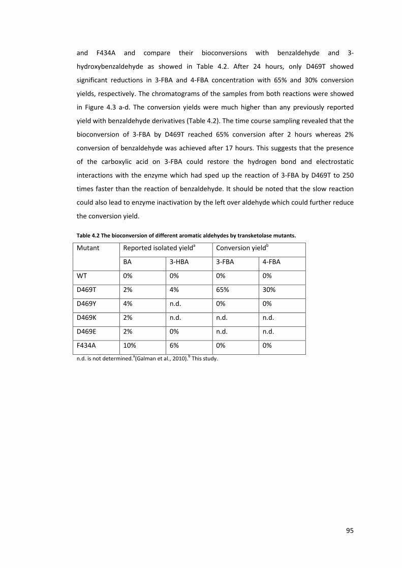
95
and F434A and compare their bioconversions with benzaldehyde and 3-
hydroxybenzaldehyde as showed in Table 4.2. After 24 hours, only D469T showed
significant reductions in 3-FBA and 4-FBA concentration with 65% and 30% conversion
yields, respectively. The chromatograms of the samples from both reactions were showed
in Figure 4.3 a-d. The conversion yields were much higher than any previously reported
yield with benzaldehyde derivatives (Table 4.2). The time course sampling revealed that the
bioconversion of 3-FBA by D469T reached 65% conversion after 2 hours whereas 2%
conversion of benzaldehyde was achieved after 17 hours. This suggests that the presence
of the carboxylic acid on 3-FBA could restore the hydrogen bond and electrostatic
interactions with the enzyme which had sped up the reaction of 3-FBA by D469T to 250
times faster than the reaction of benzaldehyde. It should be noted that the slow reaction
could also lead to enzyme inactivation by the left over aldehyde which could further reduce
the conversion yield.
Table 4.2 The bioconversion of different aromatic aldehydes by transketolase mutants.
Mutant Reported isolated yielda Conversion yieldb
BA 3-HBA 3-FBA 4-FBA
WT 0% 0% 0% 0%
D469T 2% 4% 65% 30%
D469Y 4% n.d. 0% 0%
D469K 2% n.d. n.d. n.d.
D469E 2% 0% n.d. n.d.
F434A 10% 6% 0% 0%
n.d. is not determined.a(Galman et al., 2010).b This study.

96
Figure 4.3 The Chromatograms of 3-FBA and 4-FBA reactions when analysed by ACE5 C18 reverse phase
column (150 × 4.6 mm) and Aminex HPx-87H column respectively.
a) The 8-minute sample from 3-FBA reaction catalysed by D469T, analysed by ACE5 C18 column. b) The 18-hour
sample from 3-FBA reaction catalysed by D469T/R520Q, analysed by Aminex column. c) The 2-hour sample
from 4-FBA reaction catalysed by D469T, analysed by ACE5 C18 column. d) The 4-hour sample from 4-FBA
reaction catalysed by D469T, analysed by Aminex column. All reactions were started with 50 mM of HPA and 50
mM aldehydes.
0.00 0.50 1.00 1.50 2.00 2.50 3.00 3.50 4.00 4.50 5.00 5.50 6.00 6.30-20
25
50
75
100
125
150
175
2002011-07-14 3FBA SXDTRQ RXDTRQ #87 D469T 8 UV_VIS_3mAU
min
WVL:275 nm3-FBA + HPA D469T 3-DOPBA + CO2 8 mins, UV 275nm, C18 a
0.0 5.0 10.0 15.0 20.0 25.0 30.0 35.0 40.0 45.0 50.0 55.0 60.0 65.0 70.0 75.0 80.0 85.0 90.0-0.100
-0.000
0.100
0.200
0.300
0.400
0.500
0.600 2010-10-25 retest 3 and 4 formylbenzoic acid #6 [modified by General] 3' D469T R520Q 19h UV_VIS_1AU
min
3-FBA + HPA D469T/R520Q 3-DOPBA + CO2 19 hours, UV 210nm, Aminex b
0.00 0.50 1.00 1.50 2.00 2.50 3.00 3.50 4.00 4.50 5.00 5.50 6.00 6.30-50
100
200
300
4502011-09-21 TEST 4FBA SDR7 23 DT DTRQ #44 4FBA DT 2h UV_VIS_3mAU
min
WVL:275 nm4-FBA + HPA D469T 4-DOPBA + CO2 2 hours, UV 275nm, C18 c
0.0 5.0 10.0 15.0 20.0 25.0 30.0 35.0 40.0 45.0 50.0 55.0 60.0 65.0 68.0-0.0100
0.0000
0.0100
0.0200
0.0300
0.0400
0.0500
0.0600
0.0700 2011-04-04 D469T HPA consumption and 4FBA+HPA #34 FBA 240 UV_VIS_1AU
min
4-FBA + HPA D469T 4-DOPBA + CO2 4 hours, UV 210nm, Aminex d

97
Several D469 mutants are capable to catalyse benzaldehyde and reach similar
yields but only D469T was reported to accept 3-hydroxybenzaldehyde(Galman et al., 2010).
Crystal structures of yeast and E. coli transketolase reveal that D469 residue interacts with
hydroxyl group of the substrates (Nilsson et al., 1998; Smith et al., 2008). Replacing this
negatively charged amino acid with a more hydrophobic amino acids such as threonine and
tyrosine was reported to enhance the activity toward non-hydroxylated substrates due to
the more favourable interactions with non-polar groups on substrates ( Cázares et al.,
2010; Hibbert et al., 2008; Smith et al., 2008). This favourable hydrophobic interaction may
be one of the key factors that allow D469T to catalyse aromatic aldehydes that were not
accepted by wild type. In addition, the side chain of threonine is also shorter than the side
chain of aspartate and replacing aspartate with threonine can widen the active site and
relieve the steric hindrance. This is particular true if the aromatic ring aligned in the active
site as predicted in Figure 4.2 where D469 is at the bottom of the ring.
Although D469Y was one of the mutants with the highest activity for
propionaldehyde by providing a similar hydrophobic interaction (Hibbert et al., 2008; Smith
et al., 2008; Strafford et al., 2012), it was unable to catalyse the bioconversion of neither 3-
FBA nor 4-FBA. By considering the size of the side chain, it can be seen that tyrosine is
much larger than threonine and this can cause steric hindrance upon the access of 3-FBA
and 4-FBA into the active site. This evidence from D469Y supports my earlier hypothesis
that steric is one of the factor that controls the reaction of transketolase but the main
source would arise from the side chain of D469. It should be noted that D469Y was
reported to accept benzaldehyde with up to 4% isolated yield as showed in Table 4.2, this is
not a significant improvement over D469T which achieved 2% isolated yield especially
when taken 17-hour reaction time into account. It is possible that substituting aspartate
with tyrosine could significantly reshape the active site structure which leads to a new
substrate binding site. This assumption was supported by the fact that D469Y reversed the
stereoselectivity of 1,3-dihydroxypentane-2-one from 3S to 3R (Smith et al., 2008; Strafford
et al., 2012). However, this new binding site cannot accommodate 3-FBA and 4-FBA.
F434A was reported to give the highest yields for both benzaldehyde and 3-
hydroxybenzaldehyde in the previous report with up to 10% and 6%, respectively, but it
cannot catalyse 3-FBA and 4-FBA. Replacing the bulky phenylalanine side chain with alanine
may enlarge the active site channel enough to accommodate benzaldehyde but this space
may not be sufficient when a functional group is presence on the ring. This may explain the
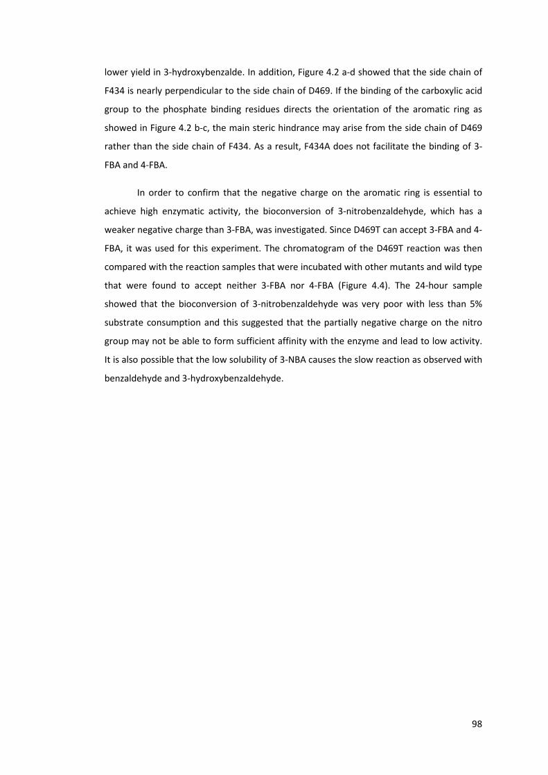
98
lower yield in 3-hydroxybenzalde. In addition, Figure 4.2 a-d showed that the side chain of
F434 is nearly perpendicular to the side chain of D469. If the binding of the carboxylic acid
group to the phosphate binding residues directs the orientation of the aromatic ring as
showed in Figure 4.2 b-c, the main steric hindrance may arise from the side chain of D469
rather than the side chain of F434. As a result, F434A does not facilitate the binding of 3-
FBA and 4-FBA.
In order to confirm that the negative charge on the aromatic ring is essential to
achieve high enzymatic activity, the bioconversion of 3-nitrobenzaldehyde, which has a
weaker negative charge than 3-FBA, was investigated. Since D469T can accept 3-FBA and 4-
FBA, it was used for this experiment. The chromatogram of the D469T reaction was then
compared with the reaction samples that were incubated with other mutants and wild type
that were found to accept neither 3-FBA nor 4-FBA (Figure 4.4). The 24-hour sample
showed that the bioconversion of 3-nitrobenzaldehyde was very poor with less than 5%
substrate consumption and this suggested that the partially negative charge on the nitro
group may not be able to form sufficient affinity with the enzyme and lead to low activity.
It is also possible that the low solubility of 3-NBA causes the slow reaction as observed with
benzaldehyde and 3-hydroxybenzaldehyde.
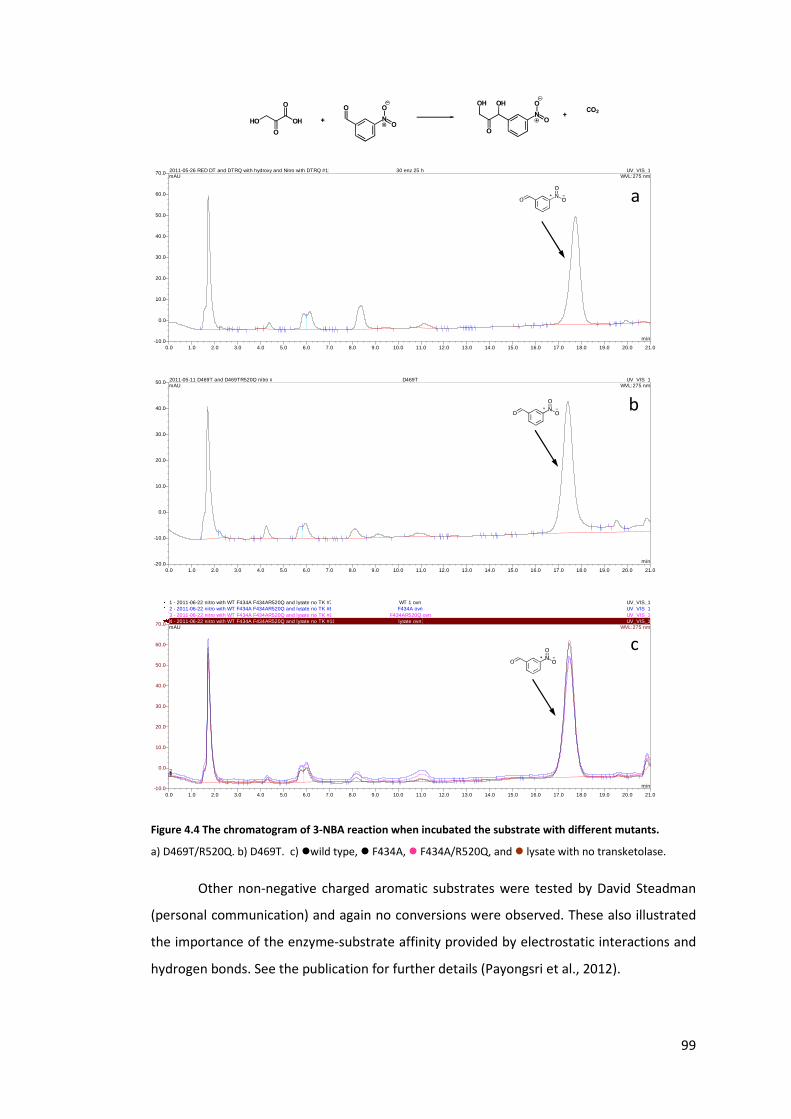
99
Figure 4.4 The chromatogram of 3-NBA reaction when incubated the substrate with different mutants.
a) D469T/R520Q. b) D469T. c) �wild type, � F434A, � F434A/R520Q, and � lysate with no transketolase.
Other non-negative charged aromatic substrates were tested by David Steadman
(personal communication) and again no conversions were observed. These also illustrated
the importance of the enzyme-substrate affinity provided by electrostatic interactions and
hydrogen bonds. See the publication for further details (Payongsri et al., 2012).
0.0 1.0 2.0 3.0 4.0 5.0 6.0 7.0 8.0 9.0 10.0 11.0 12.0 13.0 14.0 15.0 16.0 17.0 18.0 19.0 20.0 21.0-10.0
0.0
10.0
20.0
30.0
40.0
50.0
60.0
70.0 2011-05-26 RED DT and DTRQ with hydroxy and Nitro with DTRQ #11 30 enz 25 h UV_VIS_1mAU
min
WVL:275 nm
a
0.0 1.0 2.0 3.0 4.0 5.0 6.0 7.0 8.0 9.0 10.0 11.0 12.0 13.0 14.0 15.0 16.0 17.0 18.0 19.0 20.0 21.0-20.0
-10.0
0.0
10.0
20.0
30.0
40.0
50.0 2011-05-11 D469T and D469TR520Q nitro #3 D469T UV_VIS_1mAU
min
WVL:275 nm
b
0.0 1.0 2.0 3.0 4.0 5.0 6.0 7.0 8.0 9.0 10.0 11.0 12.0 13.0 14.0 15.0 16.0 17.0 18.0 19.0 20.0 21.0-10.0
0.0
10.0
20.0
30.0
40.0
50.0
60.0
70.0
1 - 2011-06-22 nitro with WT F434A F434AR520Q and lysate no TK #7 WT 1 ovn UV_VIS_12 - 2011-06-22 nitro with WT F434A F434AR520Q and lysate no TK #8 F434A ovn UV_VIS_13 - 2011-06-22 nitro with WT F434A F434AR520Q and lysate no TK #9 F434AR520Q ovn UV_VIS_14 - 2011-06-22 nitro with WT F434A F434AR520Q and lysate no TK #10 lysate ovn UV_VIS_1mAU
min
4321
WVL:275 nm
c
100
4.3.2. Kinetics of the mutants towards 3-FBA and 4-FBA
The influences of the interaction between the phosphate binding residues and the
carboxylic acid group of 3-FBA and 4-FBA on the improved bioconversion were investigated
through site directed mutagenesis at the positively-charged phosphate binding residues
which are R358, H461, and R520. Similar experiment in yeast transketolase showed that
substituting these equivalent residues with alanine significantly increased the KM values for
xylulose 5-phosphate and ribose 5-phosphate (Nilsson et al., 1997). Due to these influences
on the activity with phosphorylated substrates, they were the target for the enzyme-
substrate affinity study. In this case, the carboxylic acid of 3-FBA and 4-FBA mimicked the
phosphate group of the natural substrates.
The yields and kinetic parameters of all the mutants are showed in Table 4.3 and
the Michaelis Menten plots used for deriving the kinetic constants are illustrated in Figure
4.5 and Figure 4.6. For 3-FBA, most of the mutants gave similar yields after 24 hours except
a less active mutant R358L/D469T which achieved less than 10% and R358P/D469T/R520Q
which gave 14% yield. The latter mutant has the highest 3-FBA affinity and could be
inhibited by substrate and product to a larger extent than other mutants. Although these
mutants were found to have different kinetic parameters, their yields are somewhat
similar. In addition, there was up to 20% of HPA left once the product concentration
reached stationary. This suggested that all these mutants suffer from a similar degree of
product inhibition.

101
Table 4.3 The kinetic parameters of all the mutants towards 3-FBA and 4-FBA.
Mutant Substrate Specific activity
(relative to
D469T)
Convers
ion yield
(%)
KM (mM) kcat (s-1) kcat/ KM
(s-1 M-1)
WT 3-FBA 0 0 n.db n.d. n.d.
D469T 3-FBA 1.0a 67 (1.0) 56 (10) 13 (1.5) 236 (50)
H461S/D469T 3-FBA 0.08 65 (0.6) 29 (15) 0.8 (0.2) 28 (16)
D469T/R520Q 3-FBA 0.75 67 (0.1) 13 (4) 6 (0.75) 468 (170)
H461S/D469T/R520Q 3-FBA 0.21 63 (0.7) 11 (3) 1.5 (0.2) 138 (45)
R358P/D469T 3-FBA 1.2 65 (2.5) 20 (2.5) 11.0 (0.7) 553 (80)
R358L/D469T 3-FBA <0.1 <10 n.d. n.d. n.d.
R358P/D46T/R520Q 3-FBA 0.42 14 (1.3) 5.5 (1) 3.4 (0.2) 625 (115)
WT 4-FBA 0.0 0.0 n.d. n.d. n.d.
D469T 4-FBA 1.0a 30(1.7) 251 (240) 5.0 (4.4) 20 (26)
D469T/R520Q 4-FBA 0.45 13(0.4) 25 (7.6) 0.20 (0.03) 8 (2.8)
R358P/D469T 4-FBA <0.005 1(0.1) n.d. n.d. n.d.
R358L/D469T 4-FBA 0.30 5(0.1) n.d. n.d. n.d.
R358P/D469T/R520Q 4-FBA <0.001 <1 n.d. n.d. n.d.
Standard errors are in parentheses. The yield and specific activities of both aldehydes were determined at
equimolar concentrations of HPA and aldehyde which are 50 mM for 3-FBA and 30 mM for 4-FBA. The yield and
activity were determined using 10% V/V clarified lysate, 2.4 mM ThDP, 9.0 mM MgCl2 in 50 mM Tris buffer, pH
7.0.
a The specific activity of D469T towards 3-FBA and 4-FBA are 4.6 μmol/mg/min and 0.45 μmol/mg/min
respectively.
b n.d is not determined due to very low activity.
Figure 4.5 The Michaelis-Menten plot of all the six mutants for 3-FBA.
[3-FBA] (mM)
0 10 20 30 40 50
Sp
eci
fic
act
ivit
y (s
-1)
0
2
4
6
8
D469T H461S/D469T D469T/R520Q H461S/D469T/R520Q R358P/D469T R358P/D469T/R520Q D469TH461S/D469TD469T/R520QH461S/D469T/R520QR358P/D469T R358P/D469T/R520Q

102
Figure 4.6 The Michaelis-Menten plot of D469T and D469T/R520Q for 4-FBA.
Lineweaver Burk plots are also shown to compare the data fitting.
The reactions of 4-FBA by all the mutants were much slower than that of 3-FBA and
the conversion yields were also much lower. H461S/D469T and H461S/D469T/R520Q were
far too inactive to detect any activity towards 4-FBA and were not further studied with this
substrate. It is possible that in such slow reaction, enzyme inactivation, in particular by
aldehyde, may be one of the key factors for low yields. In addition, we observed that HPA
was degraded over the course of reaction time when the D469T containing mutants were
used. When using D469T in the equimolar reaction of 3-FBA, the conversion reached 65%
after 2 hours and the reaction did not progress further. The product concentration became
stationary and 3-FBA remained constant but there was only 20% of HPA left and it
continued to decrease. Similar trends were observed in the reaction with propionaldehyde
and 4-FBA where the HPA consumption is higher than the product formation. I further
investigated to see whether this degradation was catalysed by D469T or other components
in the clarified lysate, buffer, background transketolase wild type, or ThDP. 50 mM HPA was
incubated for 18 hours in the presence and absence of ThDP, lysate with overexpressed
D469T or D469T/R520Q, wild type, XL10 Gold cells with no overexpressed TK, and 50 mM
Tris as a control as showed in Table 4.4. From this table, it can be seen that the background
HPA degradation occurred at 2-14 % under the presence of cell-free lysate. This
degradation also occurred in Tris buffer alone. The highly expressed wild type appeared to
contribute to this degradation but to a much lower degree. These results showed that
other components in the reaction contribute very little effect on HPA degradation.
However, only the D469T and D469T/R520Q with ThDP reactions caused greater than 90 %
103
HPA consumption which illustrated that the D469T mutant directly involved in HPA
degradation which can influence the conversion yield especially for a long reaction time.
The kinetic study for 3-FBA showed that substituting the amino acids at the
phosphate binding sites resulted in mutants with lower activity than D469T except
R358P/D469T which had 20 % improvement. In this study, substrate inhibition was
observed at the 3-FBA concentration above 40 mM which was excluded from further
analysis. The mutation at both R358 and R520 significantly reduced the KM values to a
higher degree than the kcat. Consequently, the overall the kcat/KM value of R358P/D469T and
D469T/R520Q increased to 553 ± 80 s-1 M-1 and 468 ± 170 s-1 M-1compared with 236 ± 50 s-1
M-1in D469T. The KM was further reduced when R358P was combined with R520Q in
R358P/D469T/R520Q which resulted in a mutant with the lowest KM value of 5.5 mM with
the kcat of 3.4 ±0.2 s-1. With such a low KM, this triple mutant has the highest kcat/KM with
value of 625 ±115 s-1 M-1. The kcat/KM values of the double and triple mutants are higher
than that the value for glycolaldehyde by the wild type (283 s-1 M-1) (Hibbert et al., 2007)
and the D469T towards propionaldehyde (91 s-1 M-1) (Hibbert et al., 2008). However, they
are still much lower than the kcat/KM values for phosphorylated aldoses erythrose 4-
phosphate (1.7x10⁶s-1 M-1) and ribose 5-phosphate (3.1x10⁵s-1 M-1) catalysed by yeast
transketolase (Nilsson et al., 1998). D469T/R520Q may appear to have a better
performance than D469T due to the higher kcat/KM value. This double mutant, however, has
higher activities than D469T only at the concentration of 3-FBA below 30 mM. Replacing
the positively charge arginine could impair the enzyme activity as observed in R358L/D469T
which has less than 10% of the activity and this was far too low to study the kinetic
parameters. In addition, the presence of H461S mutation in any mutant results in severely
impaired enzymatic activities. The H461S/R520Q mutant has the lowest activity in all the
mutants created and this was slightly recovered by the R520Q mutation in the triple
mutant H461S/D469T/R520Q.
Interestingly, replacing any positively charged residue at the phosphate binding site
decreased the KM values. The alterations in the KM values were observed in H461S/D469T
and H461S/D469T/R520Q mutants where their KM values are 29 mM and 11 mM. More
significant changes in the KM values were observed in R538P/D469T and D469T/R520Q
where KM values significantly decrease from 56 ± 10 mM in D469T to 20 ± 2.5 mM and 13 ±
4 mM and the lowest KM value in R358P/D469T/R520Q. The fact that H461S causes a
smaller change in the KM value may imply that H461 may contribute a weaker interaction
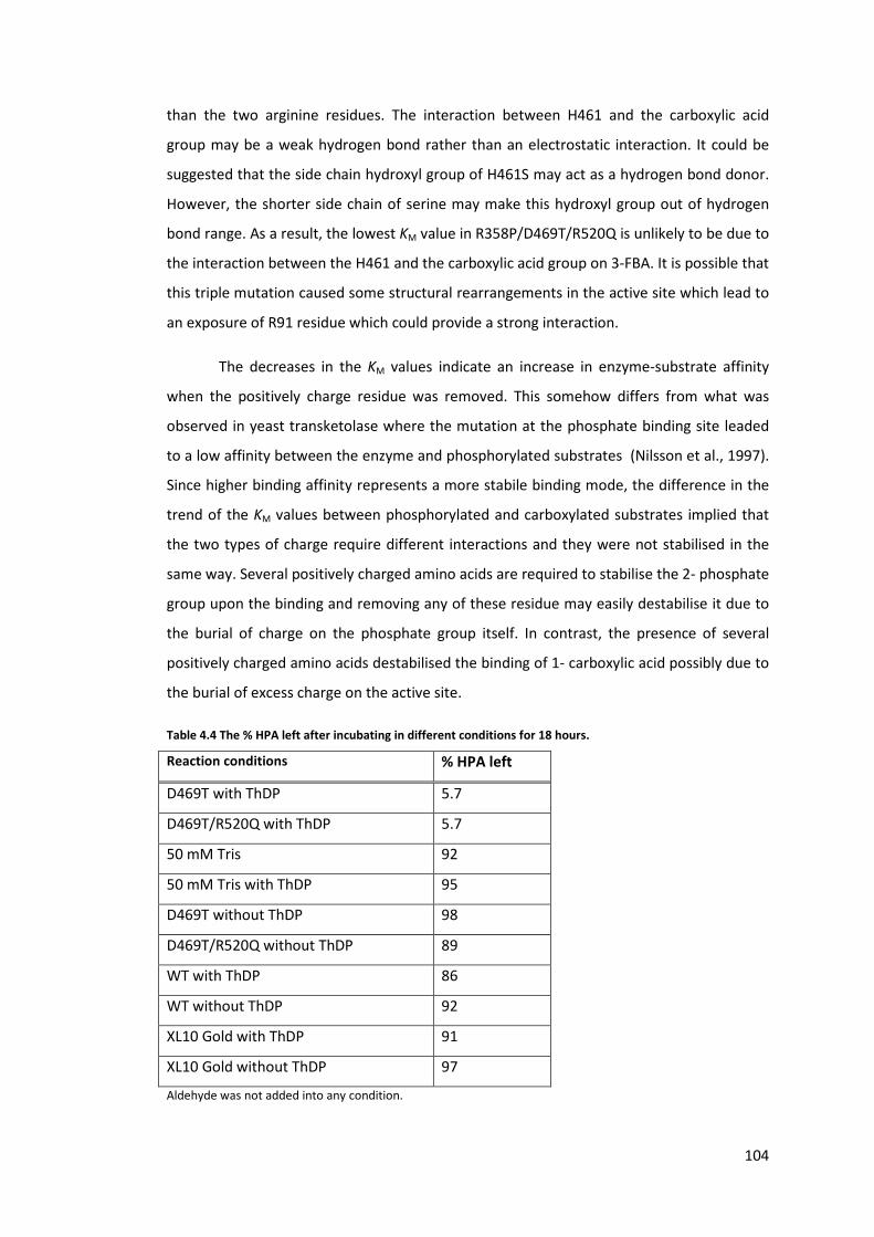
104
than the two arginine residues. The interaction between H461 and the carboxylic acid
group may be a weak hydrogen bond rather than an electrostatic interaction. It could be
suggested that the side chain hydroxyl group of H461S may act as a hydrogen bond donor.
However, the shorter side chain of serine may make this hydroxyl group out of hydrogen
bond range. As a result, the lowest KM value in R358P/D469T/R520Q is unlikely to be due to
the interaction between the H461 and the carboxylic acid group on 3-FBA. It is possible that
this triple mutation caused some structural rearrangements in the active site which lead to
an exposure of R91 residue which could provide a strong interaction.
The decreases in the KM values indicate an increase in enzyme-substrate affinity
when the positively charge residue was removed. This somehow differs from what was
observed in yeast transketolase where the mutation at the phosphate binding site leaded
to a low affinity between the enzyme and phosphorylated substrates (Nilsson et al., 1997).
Since higher binding affinity represents a more stabile binding mode, the difference in the
trend of the KM values between phosphorylated and carboxylated substrates implied that
the two types of charge require different interactions and they were not stabilised in the
same way. Several positively charged amino acids are required to stabilise the 2- phosphate
group upon the binding and removing any of these residue may easily destabilise it due to
the burial of charge on the phosphate group itself. In contrast, the presence of several
positively charged amino acids destabilised the binding of 1- carboxylic acid possibly due to
the burial of excess charge on the active site.
Table 4.4 The % HPA left after incubating in different conditions for 18 hours.
Reaction conditions % HPA left
D469T with ThDP 5.7
D469T/R520Q with ThDP 5.7
50 mM Tris 92
50 mM Tris with ThDP 95
D469T without ThDP 98
D469T/R520Q without ThDP 89
WT with ThDP 86
WT without ThDP 92
XL10 Gold with ThDP 91
XL10 Gold without ThDP 97
Aldehyde was not added into any condition.

105
In addition to the effect on the KM, all these mutations reduced the kcat values
which may arise from repositioning the whole 3-FBA molecule in such a way that the
aldehyde group of 3-FBA was located further away from the reactive carbanion-ThDP
intermediate. In R358P/D469T, D469T/R520Q, and R358P/D469T/R520Q, their kcat were
reduced from 13 s-1 to 11 s-1, 6 s-1, and 3.4 s-1, respectively. The most significantly impaired
kcat mutant was found to be H461S/D469T (0.8 s-1) which also has the lowest activity. This
was slightly recovered in H461S/D469T/R520Q to 1.5 s-1. In fact, H461S mutant and any
mutants bearing H461S were found to have lower enzymatic activity towards
glycolaldehyde and propionaldehyde as reported in Chapter 3. I previously suggested that
this mutation causes a global instability and overall reduction in the enzymatic activity.
Here, the kcatvalues of H461S/D469T and H461S/D469T/R520Q further support my
hypothesis. The instability of H461S mutant was further illustrated by the lower soluble
fraction which was found to be 10 % of the total soluble cellular protein while the wild type
has 16% expression level. In order to further confirm that H461S causes a general reduction
in the enzymatic activity, I tested H461S/D469T and H461S/d469T/R520Q with
propionaldehyde. Propionaldehyde is a very small substrate and unlikely to form a strong
specific interaction with H461. The specific activities of H461S/D469T and
H461S/D469T/R520Q towards propionaldehyde are 0.08 μmol/mg/min and 0.22
μmol/mg/min, respectively. These are 5.8% and 16% of the activity of D469T towards
propionaldehyde. The slight improvements on the activities for both substrates upon
R520Q mutation was thought to be due to the stabilising tendency of R520Q on certain
D469 variants through the co-evolved network (Strafford et al., 2012).
D469T catalysed the bioconversion of 4-FBA at a specific activity of 0.45
μmol/mg/min which is approximately 10 times slower than the reaction of 3-FBA. The kcat
for 4-FBA is 5 s-1, approximately 2.5 times lower than 3-FBA, while the KM value towards 4-
FBA is nearly five times higher than 3-FBA although both substrates carry the same charge.
These leaded to the kcat/KM value of 20 s-1M-1 which is nearly 20 folds lower than the kcat/KM
value for 3-FBA and this implied that 4-FBA is a much less favourable substrate than 3-FBA.
Assuming that the carboxylic acid groups of both substrates bind to the active site at a
similar position as suggested in Figure 4.2 b and d, the aldehydes of the two substrates are
not at the same position which resulted in the significant difference in their kcat. This also
implied that the relative orientations of the aldehyde and the carboxylic acid group are
important for a productive binding and catalysis. Although the para position of the
carboxylic acid in 4-FBA would electronically favour nucleophilic attack at the carbonyl
106
group than the meta position in 3-FBA, the kcat values between the two substrates
illustrated that electronic effects may not be a major factor influencing the enzymatic
activity. In addition, 3-nitro group is much more electron withdrawing than the carboxylic
acid group, the enzymatic activity towards 3-nitrobenzaldehyde is much poorer than 3-FBA
and 4-FBA.
The removal of any positively charged residue significantly impaired the enzymatic
activity and yield towards 4-FBA. Most of the double mutants have very low activities to
analyse the cause of the loss in activity except D469T/R520Q. In the case of D469T/R520Q,
this was due to the 25-fold reduction in the kcat rather than losing the enzyme-substrate
affinity. In fact, the introduction of R520Q significantly decreased the KM value to 25 mM
which is still approximately 2 times higher than the KM value for 3-FBA by the same mutant.
R358P/D469T was found to have much lower activity towards 4-FBA than D469T and
D469T/R520Q while it has higher activity towards 3-FBA than both mutants. Therefore, the
improvement is only specific for 3-FBA through the improved affinity. In R358L/D469T, the
activity towards 4-FBA was partially impaired and the activity towards 3-FBA was reduced
to a larger extent.
The mutagenesis and kinetic studies so far illustrated that removing a positively
charged amino acid resulted in a lowering ofKM and kcat for both 3-FBA and 4-FBA. However,
3-FBA was positioned in a more active orientation than 4-FBA in all double and triple
mutants.
Throughout my study, the bioconversions of 3-FBA and 4-FBA at 10 mM were
found to be much faster and higher than the reaction of benzaldehyde which was
performed at 50 mM. This suggested that solubility of the substrate may not be an issue in
controlling the rate of the reaction. It was illustrated that the replacements of negatively
charged D469 to a neutral charge threonine or tyrosine have improved the enzyme-
substrate affinity towards non-hydroxylated substrate propionaldehyde (Hibbert et al.,
2008). The interaction between propionaldehyde and the transketolase active site would
rely on the hydrophobic interaction alone. To test whether neutralising the positive charge
on the active site would favour the hydrophobic interaction, D469T/R520Q was tested with
benzaldehyde at 50 mM. The conversion yield was less than 3% which is not significantly
improved over D469T. This suggested that hydrophobic interaction alone is insufficient to
achieve high yield and activity in aromatic substrates.

107
4.3.3. Computational docking of 3-FBA and 4-FBA
The binding energies, distances between the carbonyl group to the enamine in
D469T and D469T/R520Q were summarised in Table 4.5. The computational docking of 3-
FBA into D469T showed that the carboxylic acid of 3-FBA form hydrogen bond and
electrostatic interactions with R358, S385, H461, R520 as showed in Figure 4.2 b and c. 2
binding clusters of 3-FBA in D469T were predicted. In the first cluster, the carbonyl group
form hydrogen bond with the side chain of H26 which placed the carbonyl group 4.9 Å
away from the enamine-ThDP. In the second cluster, the carbonyl group of 3-FBA forms
hydrogen bond with the main chain O-atom of H473 which places the carbonyl group 7.2Å
away from the enamine-ThDP making this conformation rather unproductive due to a
longer distance to the enamine.
The docking of 3-FBA into D469T/R520Q is slightly different from D469T. The whole
3-FBA molecule was moved further away from the enamine-ThDP and S385 is not forming
hydrogen bond with the carboxylic group of 3-FBA but it forms the hydrogen bond with the
carbonyl group instead (Figure 4.2c). This placed the aldehyde 6.6Å away from the enamine
which may reflect the 2-fold lower in kcat in D469T/R520Q.
4-FBA was predicted to form one binding cluster in D469T (Figure 4.2d). Unlike 3-
FBA, only one of the oxygen atom on the carboxylic acid was predicted to interact with
R358, H461, and R520 but not S385. This interaction network was less extensive than that
of 3-FBA in D469T which may reflect the 5-fold higher KM in 4-FBA. The aldehyde of 4-FBA
was placed 5.1Å away from the enamine-ThDP which is 0.16 Å further than the aldehyde of
3-FBA. Although this further distance may suggest the 2.5-fold lower kcat in 4-FBA, this
distance was 1.5 Å closer than the aldehyde of 3-FBA to the enamine-ThDP in
D469T/R520Q which has similar kcat value to the kcat of 4-FBA in D469T. Therefore, in
addition to the distance between the carbonyl group to the enamine-ThDP, the relative
orientation of the carbonyl group relative to the enamine intermediate may also be a
crucial factor governing the reaction. Finally, 4-FBA was found to have multiple binding
clusters in D469T/R520Q and these clusters were found to have similar binding energies.
The distances between the carbonyl group to the enamine were 4.6 Å, 6.5 Å, 8.3 Å, and 8.9
Å with the population of 35%, 10%, 16%, and 26% respectively. The non-productive clusters
could block the active site and prevent other 4-FBA molecule to bind in a productive
conformation which resulted in low observed kcat value.
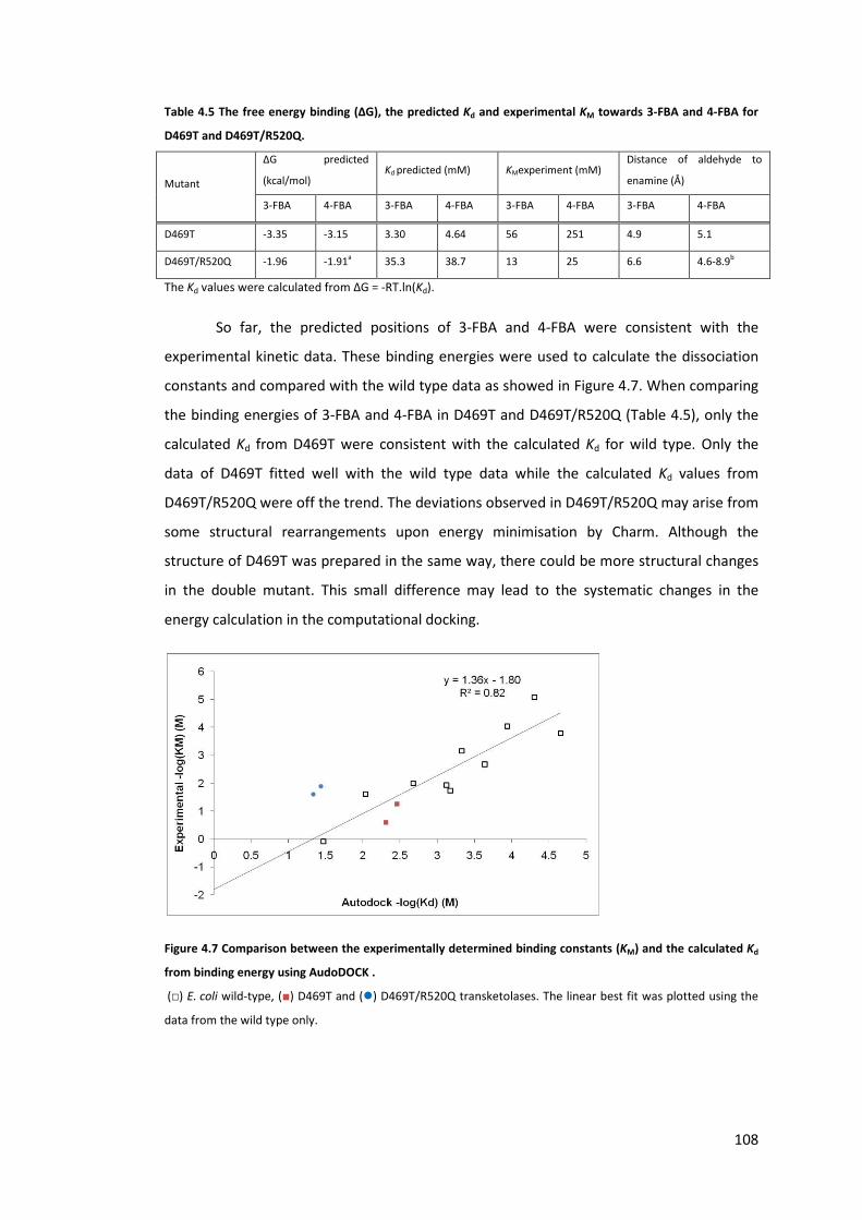
108
Table 4.5 The free energy binding (ΔG), the predicted Kd and experimental KM towards 3-FBA and 4-FBA for
D469T and D469T/R520Q.
Mutant
ΔG predicted
(kcal/mol) Kd predicted (mM) KMexperiment (mM)
Distance of aldehyde to
enamine (Å)
3-FBA 4-FBA 3-FBA 4-FBA 3-FBA 4-FBA 3-FBA 4-FBA
D469T -3.35 -3.15 3.30 4.64 56 251 4.9 5.1
D469T/R520Q -1.96 -1.91a 35.3 38.7 13 25 6.6 4.6-8.9b
The Kd values were calculated from ΔG = -RT.ln(Kd).
So far, the predicted positions of 3-FBA and 4-FBA were consistent with the
experimental kinetic data. These binding energies were used to calculate the dissociation
constants and compared with the wild type data as showed in Figure 4.7. When comparing
the binding energies of 3-FBA and 4-FBA in D469T and D469T/R520Q (Table 4.5), only the
calculated Kd from D469T were consistent with the calculated Kd for wild type. Only the
data of D469T fitted well with the wild type data while the calculated Kd values from
D469T/R520Q were off the trend. The deviations observed in D469T/R520Q may arise from
some structural rearrangements upon energy minimisation by Charm. Although the
structure of D469T was prepared in the same way, there could be more structural changes
in the double mutant. This small difference may lead to the systematic changes in the
energy calculation in the computational docking.
Figure 4.7 Comparison between the experimentally determined binding constants (KM) and the calculated Kd
from binding energy using AudoDOCK .
(□) E. coli wild-type, (■) D469T and (�) D469T/R520Q transketolases. The linear best fit was plotted using the
data from the wild type only.

109
4.4. Conclusion
I have illustrated that the phosphate binding site at the entrance of the
transketolase active site can be used to enhance the bioconversion of benzaldehyde
derivatives that contain negatively charged carboxylic acid. The position of the carboxylic
acid itself on the aromatic ring influences the position of the aldehyde group. This affects
the catalytic activity as illustrated that the meta position appears to have higher activity as
shown in 3-FBA than 4-FBA. The difference may be due to the relative positions of their
aldehyde where the meta position is placed in a more productive location. In addition,
replacing the positively charged residues leads to higher affinity but lower kcat and
hydrophobic interaction alone is insufficient for a fast conversion and high yield for
aromatic aldehydes.
110
5. Chapter 5: Second Generation Engineering of Transketolase for
Aromatic Ketodiols
5.1. Introduction
Transketolase accepts aromatic aldehydes and heteroaromatic aldehydes with poor
activity and yield (Galman et al., 2010). In Chapter 4, it was found that the presence of the
carboxylic group on 3-formylbenzoic acid can improve the activity up to 250 times faster
than benzaldehyde with the yield up to 67% within 2 hours (Payongsri et al., 2012). These
enhancements were found to be due to restoring the hydrogen bonds and electrostatic
interaction between the carboxylic acid group and the phosphate binding residues of the
enzyme. D469T was found to be the most active mutant towards 3-FBA and 4-FBA with the
specific activities of 4.6 μmol/mg/min and 0.45 μmol/mg/min respectively. The activity of
D469T for 3-FBA is actually higher than any previously reported aromatic aldehydes and
non-hydroxylated aldehydes. However, the catalytic turnovers of these aromatic substrates
are still much lower than natural substrates (13.2 s-1 and 5.0 s-1 for 3-FBA and 4-FBA
respectively). The study in yeast transketolase revealed that, ribose 5-phosphate has a kcat
value of 46.3 s-1 (Wikner et al., 1997) and for erythrose 4-phosphate the kcat value is 69 s-
1(Nilsson et al., 1998). These relatively low kcat values suggested some possibilities for
improving transketolase by protein engineering. In addition to improving the performances
towards 3-FBA and 4-FBA, protein engineering is a powerful tool to further enhance the
activity of transketolase towards neutral and polar aromatic aldehydes which are still
poorly accepted. This is especially important for the synthesis of chloramphenicol and its
derivatives where the substituents are non-charged and the synthetic potential of
transketolase could be further expanded.
A number of directed evolution strategies are available with their own advantages
and drawbacks. However, they tend to involve the handling of large library size which can
be resource intensive. An alternative option is to construct smart libraries by targeting
residues to those within active site close to the reaction centre and highly conserved
residues. This strategy has been shown to improve substrate activity and also influence the
stereoselectivity of the enzymes (Morley and Kazlauskas, 2005; Paramesvaran et al., 2009;
Toscano et al., 2007). As a result, the size of the library is not too large to manually handle
and I adopted this strategy to design my library.

111
The analysis of E. coli transketolase active site in the previous study has classified
active site residues into two shells (Hibbert et al., 2007). Residues within the first shells are
located within 4 Å from the substrate molecule whereas residues in the second shells are
within 10 Å from the ThDP molecule (Hibbert et al., 2007). Among these residues, the
active site of transketolase consists of a number of highly conserved histidine residues on
the phosphate binding domain and these residues are H26, H69, H100, and H261 (Costelloe
et al., 2008; Schenk et al., 1997). Mutagenesis studies in yeast transketolase on the highly
conserved histidine residues showed that H30 and H263 (H26 and H261 in E. coli) may act
as an acid/base catalyst (Wikner et al., 1997). My previous study in Chapter 3 showed that
H26Y has no activity and recently, this mutant was also recreated by another research
group and it was found to be inactive (Yi et al., 2012). H103 was suggested to be involved in
substrate recognition and stabilisation of the intermediate (Wikner et al., 1995). H69 was
identified to bind to the phosphate group of ThDP and may be involved in the proton
transfer between a water molecule and C1-hydroxyl group of the donor-ThDP adduct
(Wikner et al., 1997). Therefore, these histidine residues were excluded from the library
constructed in this Chapter. My previous study in Chapter 4 illustrated that substituting
H461 with serine may lead to enzyme instability as a lower fraction of soluble protein was
observed. This mutation also globally reduced the enzyme activity, although this residue
does not appear to interact with nearby amino acids (Payongsri et al., 2012). Therefore,
H461 was also excluded from the library. From all these eliminations, the target residues
were limited to only R358 and S385. The crystal structure of E. coli transketolase revealed
that the side-chain hydroxyl group of S385 and the guanidinium group of R358 form
hydrogen bonds with the phosphate group of xylulose 5-phosphate (PDB ID: 2R5O) and
fructose 6-phosphate (PDB ID: 2R8P) (Asztalos et al., 2007). These hydrogen bonds were
also observed in the crystal structure of yeast transketolase with erythrulose 4-phosphate
(PDB ID: 1NGS) and replacing R358 equivalent residue in yeast transketolase significantly
decreases the affinities towards xylulose 5-phosphate and ribose 5-phosphate (Nilsson et
al., 1997). I have previously confirmed in Chapter 4, that R358 in E. coli transketolase
interacts with the carboxylate group of both 3-FBA and 4-FBA from the R358P and R358L
mutants (Payongsri et al., 2012). The previous saturation mutagenesis studies at both
residues showed that only R358I has approximately 2 times higher activity than wild type
towards both glycolaldehyde (Hibbert et al., 2007) and propionaldehyde (Hibbert et al.,
2008). However, the library of S385 did not produce any mutant with improved activity
towards glycolaldehyde or propionaldehyde. In this Chapter, the saturation mutagenesis of

112
these sites has been introduced within the D469T/R520Q mutant in order to improve the
activity of transketolase towards aromatic aldehydes.
5.2. Materials and methods
5.2.1. Chemicals and reagents
All aldehyde substrates were purchased from Sigma-Aldrich (Aldrich® Chemistry,
UK). HPA was synthesised according to the protocol referred in Chapter 2.
5.2.2. Transketolase library
Although D469T has the highest activities and yields for 3-FBA and 4-FBA in the last
chapter, all the libraries were constructed on D469T/R520Q due to its stability which allows
it to withstand further mutations and also minimise inclusion body formation (Strafford et
al., 2012). The target residues were limited to those that are within the 1st shell (Hibbert et
al., 2007) but excluding the catalytic histidine residues (H26, H69, H100, H261) and H461
where its mutation was shown to have an impact on enzyme stability and activity
(Payongsri et al., 2012). These eliminations left only R358, S385, D469, and R520. Since
D469T/R520Q was used as a template, R358 and S385 are the only two targets available.
The triple mutants of both libraries were constructed by Quickchange site directed
mutagenesis according to the standard protocol. All the dpnI digested PCR products were
transformed into XL10 Gold competent cells (Stratagene, La Jolla, California, USA). The
sequences of the primers were used as listed below. The target codons are underlined and
the bases to be changed are in bold letters. N represents an equal concentration of A, T, C,
and G. H is 50% T and 50% A. S is 50% C and 50% G.
R358X GAAAATCGCCAGCNHNAAAGCGTCTCAGAATG
S385X GCTGACCTGGCGCCGNHSAACCTGACCCTGTGG
Q520R CACTGATCCTCTCCCGTCAGAACCTGGCGCAG
The last primer is to reverse the triple mutants to S385Y/D469T and S385T/D469T.
5.2.3. Library Screening
According to the sequences of the primers above, both libraries were expected to
produce 16 possible amino acids. The library coverage was calculated by the online tool:
GLUE1AA (http://guinevere.otago.ac.nz/cgi-bin/aef/glue1AA.pl) (Patrick et al., 2003) and

113
picking 24 colonies was expected to give 11.6 distinct amino acids under the assumption
that the libraries are diverse. 6 random colonies from each library were picked and their
plasmids were sequenced in order to verify the diversity of each library.
Uneven cell lysis can influence the amount of released enzyme, hence the reaction
rate. Therefore, whole cells were used in the screening of all three substrates. 24 colonies
from each library were picked and inoculated into 12 ml of LB media with 150 μg/ml
ampicillin. They were then cultured at 37 °C, shaking at 250 rpm for 18 hours. Their final
OD600 values were measured before harvest. These cells were aliquoted into 2 ml
eppendorf tubes which were then centrifuged at 17,000 g for 10 minutes to harvest and
the supernatant was discarded. To each eppendorf tube containing supernatant-free cells,
200 μl of 2x cofactor solution (4.8 mM ThDP and 18 mM MgCl2 in 50 mM Tris buffer, pH
7.0) was added. After the cells were fully resuspended, they were left to incubate with the
cofactors for 20 minutes. 150 μL of these cell-cofactor mixtures were transferred to a
borosilicate microplate (Radleys, Essex, UK). The reaction was then started by the addition
of 150 μl of 2x substrate (100 mM 3-FBA and 100 mM HPA, 80 mM 4-FBA and 80 mM HPA,
or 30 mM 3-HBA and 60 mM HPA). All substrates were prepared in 50 mM Tris-HCl buffer
and adjusted the pH to 7.0. The plate was then seal to prevent evaporation (Thermo
Scientific Nunc) and shaken at 200 rpm, 3 mm amplitude to avoid sedimentation of the
cells. 3-FBA samples were taken at 1 hour and 24 hours after the reaction was started in
order to screen for mutants with higher activities and yields. The higher activity mutants
would assume to give higher conversion at 1 hour. My previous experiments illustrated
that the reactions of 4-FBA and 3-HBA are very slow compared with 3-FBA and their
conversion yields by D469T, which is the best mutant, were also low (Payongsri et al.,
2012). Taking samples within 1 hour would give low signal to noise ratio. In addition, HPA
degradation also competes with the ketol transfer reaction of transketolase. As a result,
the mutants with higher activity tend to give higher yields. Therefore, only the samples at
18 hours were taken to quantify the final yields. This method assumes that all the mutants
have similar expression levels of transketolase.
380 μl of 0.1% TFA was added to 20 μl of sample to quench the reaction. These
were then centrifuged for 3 minutes at 13,300 rpm before the supernatants were analysed
by HPLC according to the methods stated in Chapter 2. The conversion of 3-FBA and 4-FBA
reactions were quantified by the same method as stated in Chapter 4 (Payongsri et al.,
2012). Racemic dihydroxy ketone of 3-HBA, 1,3-dihydroxy-1-(3-hydroxyphenyl)propan-2-

114
one, was synthesised and purified according to the published protocol (Galman et al., 2010)
and this was used as a standard for quantification.
5.2.4. Cell culture and protein quantification for detailed enzyme kinetics
Cell cultures of all the mutants started from streaking glycerol stock onto LB-plates
containing 150 μg/ml of ampicillin and cultured according to the standard protocol. All of
them were cultured at the final volume of 50 ml in LB media with 150 μg/ml of ampicillin in
250-ml shake flasks. Clarified lysate was prepared according to the protocol in Chapter 2,
section 2.4.
For the 3-HBA kinetic study, cells harvested from 50 ml culture were resuspended
in 2 ml of 50 mM Tris buffer, pH 7.0 before sonication. This is to produce clarified lysate
with transketolase concentration at least 0.8 mg/ml. The total protein concentration was
quantified by Bradford assay and the expression level of TK was quantified by SDS-PAGE
and densitometry. The expression levels of transketolase mutants from both libraries are
approximately 20 % of the total soluble protein in the cell-free lysate.
5.2.5. Detailed enzyme kinetics
All kinetic studies were performed in triplicate at the reaction volume of 300 μl in
1.5 ml glass vials. In all the reactions, the concentration of HPA maintained at 50 mM and
the concentration of the aldehydes were varied. For 3-FBA, the concentrations were from
3-90 mM while 4-FBA the concentrations were between 6-30 mM and for 3-HBA, the
concentrations were 5-25 mM. For the reaction of 3-FBA and 4-FBA, 30 μl of clarified lysate
was incubated with 30 μl 10x cofactor solution and 140 μl 50 mM Tris buffer, pH 7.0 for 20
minutes. 100 μl of 3x substrate solutions were then added to the lysate-cofactor solutions
to start the reaction. For 3-FBA, the reaction samples were taken every taken every 3
minutes for 15 minutes while the samples of 4-FBA reaction were taken at every 15
minutes for 90 minutes.
Due to the lower solubility of 3-HBA, the substrate stock solutions were prepared
as 2x concentration. The reactions of 3-HBA were prepared by incubating 50 μl of clarified
lysate with 30 μl of 10X cofactor solution and 70 μl of 50 mM Tris buffer, pH 7.0 for 20
minutes. 150 μl of 2x substrate solutions were then added to start the reaction. The
samples were taken every 30 minutes for 180 minutes.
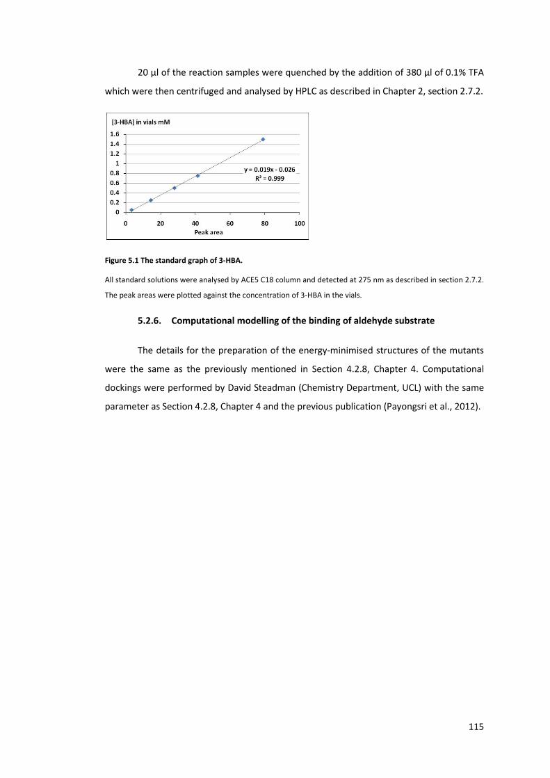
115
20 μl of the reaction samples were quenched by the addition of 380 μl of 0.1% TFA
which were then centrifuged and analysed by HPLC as described in Chapter 2, section 2.7.2.
Figure 5.1 The standard graph of 3-HBA.
All standard solutions were analysed by ACE5 C18 column and detected at 275 nm as described in section 2.7.2.
The peak areas were plotted against the concentration of 3-HBA in the vials.
5.2.6. Computational modelling of the binding of aldehyde substrate
The details for the preparation of the energy-minimised structures of the mutants
were the same as the previously mentioned in Section 4.2.8, Chapter 4. Computational
dockings were performed by David Steadman (Chemistry Department, UCL) with the same
parameter as Section 4.2.8, Chapter 4 and the previous publication (Payongsri et al., 2012).
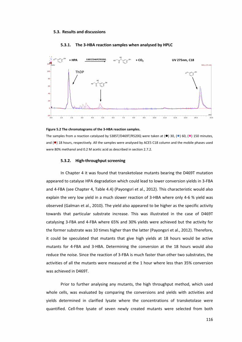
116
5.3. Results and discussions
5.3.1. The 3-HBA reaction samples when analysed by HPLC
Figure 5.2 The chromatograms of the 3-HBA reaction samples.
The samples from a reaction catalysed by S385T/D469T/R520Q were taken at (�) 30, (�) 60, (�) 150 minutes,
and (�) 18 hours, respectively. All the samples were analysed by ACE5 C18 column and the mobile phases used
were 80% methanol and 0.2 M acetic acid as described in section 2.7.2.
5.3.2. High-throughput screening
In Chapter 4 it was found that transketolase mutants bearing the D469T mutation
appeared to catalyse HPA degradation which could lead to lower conversion yields in 3-FBA
and 4-FBA (see Chapter 4, Table 4.4) (Payongsri et al., 2012). This characteristic would also
explain the very low yield in a much slower reaction of 3-HBA where only 4-6 % yield was
observed (Galman et al., 2010). The yield also appeared to be higher as the specific activity
towards that particular substrate increase. This was illustrated in the case of D469T
catalysing 3-FBA and 4-FBA where 65% and 30% yields were achieved but the activity for
the former substrate was 10 times higher than the latter (Payongsri et al., 2012). Therefore,
it could be speculated that mutants that give high yields at 18 hours would be active
mutants for 4-FBA and 3-HBA. Determining the conversion at the 18 hours would also
reduce the noise. Since the reaction of 3-FBA is much faster than other two substrates, the
activities of all the mutants were measured at the 1 hour where less than 35% conversion
was achieved in D469T.
Prior to further analysing any mutants, the high throughput method, which used
whole cells, was evaluated by comparing the conversions and yields with activities and
yields determined in clarified lysate where the concentrations of transketolase were
quantified. Cell-free lysate of seven newly created mutants were selected from both
0.0 1.0 2.0 3.0 4.0 5.0 6.0 7.0 8.0 9.0 10.0 11.0 12.0 13.0 14.0 15.5-20
0
20
40
60
80
100
120
1 - 2011-07-21 15mM HBA 30 HPA SDR 7, 21, 23 D469T #15 SDR23 30min UV_VIS_12 - 2011-07-21 15mM HBA 30 HPA SDR 7, 21, 23 D469T #24 SDR23 1h UV_VIS_13 - 2011-07-21 15mM HBA 30 HPA SDR 7, 21, 23 D469T #31 SDR23 4h30m UV_VIS_14 - 2011-07-21 15mM HBA 30 HPA SDR 7, 21, 23 D469T #39 SDR23 18h UV_VIS_1mAU
min
4321
WVL:275 nm
ThDP
+ HPA S385T/D469T/R520Q + CO2 UV 275nm, C18

117
libraries to quantify their specific activities towards 50 mM 3-FBA which were compared
with the 1-hour conversion yield in whole cell system. Since different mutants were
harvested at slightly different OD600, there would be a small variation in the transketolase
concentration in the screening reaction and their 1-hour conversions would need to be
standardised by their OD600. These were then plotted against the clarified lysate activities
as shown in Figure 5.3. The whole cell data correlated well with those of clarified lysate
which illustrated that the whole cell data can represent the activities obtained in the lysate.
Only the clarified lysate activity of S385E/D469T/R520Q was found to be much higher than
the whole cell system. This mutant was later found to be highly active towards 3-FBA and
the deviation from the high throughput may arise from the mass transfer across the cell
membrane that slowed down the whole cell catalysis.
Figure 5.3 Comparison of the specific activities by clarified lysate and the 1-hour conversions in whole cell
which were standardised by OD600.
For 4-FBA and 3-HBA, five mutants were selected. A good agreement between the
clarified lysate and high throughput screen was illustrated by the linear relationship
between the two data sets as shown in Figure 5.4. The time course sampling of both 4-FBA
and 3-HBA also support my initial hypothesis that higher activity mutants gave higher 18-
hour yields (see later for integrated results). However, systematically higher yields were
obtained from clarified lysate which implied that mass transfer into the cells could limit the
conversion yields in the whole cell system. For example, D469T gave 30% conversion yield
in clarified lysate (Payongsri et al., 2012) whereas the whole cell D469T gave only 15.6 %
yield after 18 hours. The conversion of 3-HBA by D469T also follows the same trend where
the whole cell achieved 4.9% yield while lysate gave 6.7% (Table 5.1). The good correlation
between the lysate and whole cell data allow me to further analyse all the mutants in both
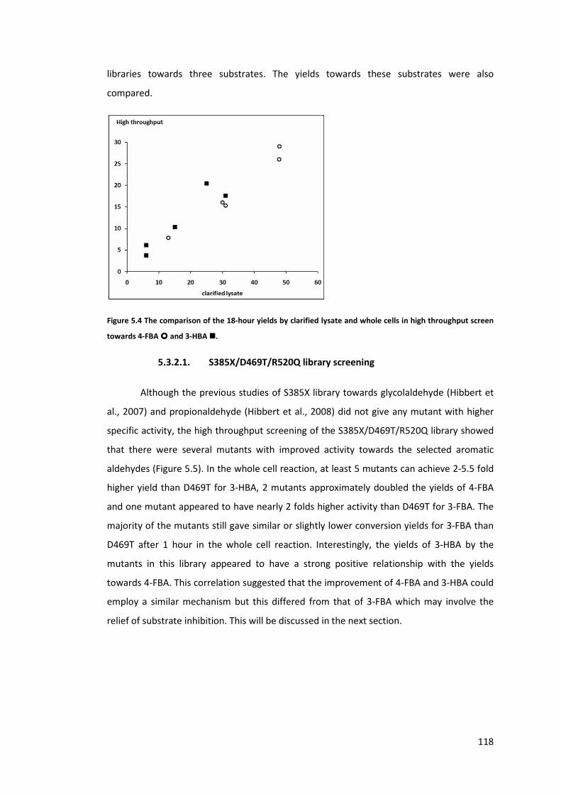
118
libraries towards three substrates. The yields towards these substrates were also
compared.
Figure 5.4 The comparison of the 18-hour yields by clarified lysate and whole cells in high throughput screen
towards 4-FBA ���� and 3-HBA ����.
5.3.2.1. S385X/D469T/R520Q library screening
Although the previous studies of S385X library towards glycolaldehyde (Hibbert et
al., 2007) and propionaldehyde (Hibbert et al., 2008) did not give any mutant with higher
specific activity, the high throughput screening of the S385X/D469T/R520Q library showed
that there were several mutants with improved activity towards the selected aromatic
aldehydes (Figure 5.5). In the whole cell reaction, at least 5 mutants can achieve 2-5.5 fold
higher yield than D469T for 3-HBA, 2 mutants approximately doubled the yields of 4-FBA
and one mutant appeared to have nearly 2 folds higher activity than D469T for 3-FBA. The
majority of the mutants still gave similar or slightly lower conversion yields for 3-FBA than
D469T after 1 hour in the whole cell reaction. Interestingly, the yields of 3-HBA by the
mutants in this library appeared to have a strong positive relationship with the yields
towards 4-FBA. This correlation suggested that the improvement of 4-FBA and 3-HBA could
employ a similar mechanism but this differed from that of 3-FBA which may involve the
relief of substrate inhibition. This will be discussed in the next section.

119
Figure 5.5 The cross comparison of the yields between three substrates by S385X/D469T/R520Q library.
The activities towards 3-FBA were measured after 1 hour where the reaction achieve less than 35% conversion.
The final yields of 4-FBA and 3-HBA were measured at 18 hours. All reactions were performed in freshly harvest
whole cells.
In addition, I initially speculated that HPA degradation is competing with the
productive ketol transfer reaction and this dictated the final yield of aromatic aldehydes.
This leads to a hypothesis that higher activity mutants will give higher final yields. The
specific activities and yields for 3-HBA determined by clarified lysate under the same
substrate concentrations as the high throughput screen showed that D469T had an activity
of 0.0071 μmol/mg/min with the 6.7% conversion after 18 hours (Table 5.1) while
S385V/D469T/R520Q,S385Y/D469T/R520Q and S385T/D469T/R520Q achieved 15%, 25%,
and 31% respectively. The specific activities of these mutants by clarified lysate were 0.03,
0.071, and 0.073 μmol/mg/min, respectively which were 5-12 times higher than D469T.
This positive correlation between yield and activity was also observed in the bioconversion
of 4-FBA. This increase in the final yield in higher activity mutants confirmed that the ketol
transfer reaction is competing with the HPA degradation. This characteristic was previously
studied and identified that D469T bearing mutants, but not the background host enzyme or
buffer, causes the observed HPA degradation(Payongsri et al., 2012). This relationship
between yield and specific activity was, however, not obvious in the 3-FBA reaction which
was partly due to substrate inhibition and relatively fast catalysis which will be discussed
later.
The DNA sequencing revealed that substituting S385 with hydrophobic amino acids
(V, A, Y), polar amino acid (T), and negatively charged amino acid (E) can improve the
bioconversion of 3-HBA. Since rather diverse amino acids can substitute S385, it implied
that these mutants had improved kcat instead of providing better interactions with the
hydroxyl or carboxylic acid groups of the substrates with an exception for S385E. This may
be unexpected because S385 binds to the phosphate group of natural substrates.

120
5.3.2.2. R358X/D469T/R520Q library screening
The previous attempt at introducing saturation mutagenesis at R358 gave R358P
and R358I mutants with 1.5 and 2.1 fold improvements of activity for glycolaldehyde
(Hibbert et al., 2007). Both mutants also had 1.5 and 1.9 times higher activity than wild
type towards propionaldehyde (Hibbert et al., 2008). In this study, however, saturation
mutagenesis at this position on D469T/R520Q did not appear to yield a mutant with higher
activity towards the selected aromatic aldehydes (Figure 5.6). In contrast to the
S385X/D469T/R520Q library, the activity towards 3-FBA has a positive correlation with the
conversion yield of 4-FBA.
Figure 5.6 The cross comparison of the yields between three substrates by R358X/D469T/R520Q library.
The activities towards 3-FBA were measured after 1 hour where the reaction achieve less than 35% conversion.
The final yields of 4-FBA and 3-HBA were measured at 18 hours. All reactions were performed in freshly harvest
whole cells.
In addition to screening this library, some triple mutants obtained from random
sampling were sequenced and assessed their performances using clarified lysate.
R358E/D469T/R520Q and R358Q/D469T/R520Q were found to have diminished activity for
3-FBA. The activities of R358H/D469T/R520Q, and R358I/D469T/R520Q were marginally
lower than D469T. The high throughput screen showed that approximately a third of the
mutants from the R358X/D469T/R520Q library had 1-hour conversions of 3-FBA similar to
D469T while nearly half of the library had significantly lower activities towards 3-FBA. The
rest of the library had partially impaired activity towards 3-FBA. In addition, the yields of 4-
FBA by the majority of the mutants were lower than 50% of D469T while more than half of
the mutants from this library achieved similar yields to D469T for 3-HBA. Since I previously
showed in Chapter 4 that R358 interacted with the carboxylate group of 3-FBA and 4-FBA
and stabilised their bindings, it is possible that the removal of the positively charged
arginine would have a detrimental effect on the bioconversion of the carboxylated

121
aromatic aldehydes to a greater extent than 3-HBA. This could also explain the correlation
between the 3-FBA activity and 4-FBA yield obtained from this library (Figure 5.6). Besides
that, it could imply that the mutation at R358 did not have a strong influence on 3-HBA
compared with 3-FBA and 4-FBA. Alternatively, 3-HBA may have a rather different
orientation than 3-FBA and 4-FBA. The evidence for this hypothesis arose from two
mutants as follows. The R358T/D469T/R520Q mutant maintained 60% activity towards 3-
FBA and 4-FBA but it had significantly lower activity towards 3-HBA. R358H/D469T/R520Q
only maintained the activity towards 3-HBA but not 3-FBA or 4-FBA despite the fact that it
still had a positively charged histidine. It is still surprising that none of the mutants from
this library has improved activity towards any of the substrates.
The high-throughput screen of both libraries illustrated that the S385X library can
tolerate a wider range of amino acid substitution than the R358X for 3-FBA and 4-FBA.
Replacing R358 with other amino acids had a tendency to maintain the enzymatic activity
only towards 3-HBA while the substituting S385 was more likely to improve the activity
towards 3-HBA. Although both E. coli and yeast transketolase crystal structures illustrated
that S385 forms hydrogen bonds with the phosphate group of natural substrates (Asztalos
et al., 2007; Nilsson et al., 1997), it may not contribute significant interaction with the
aromatic substrates. On the other hand, the R358 residue, together with H461 and R520,
strongly interacted with the carboxylic acid groups of both 3-FBA and 4-FBA as shown in
the previous study (Payongsri et al., 2012).
5.3.3. Kinetic parameters of the mutants with 3-FBA, 4-FBA, and 3-HBA
The high-throughput screen illustrated that none of these mutants simultaneously
had higher activity towards all three substrates and all the mutants appeared to have lower
activity towards at least one substrate. This apparent shift in substrate specificity can be
investigated in detail by kinetic studies which can reveal whether this is due to a loss in
enzyme-substrate affinity or a reduction in the catalytic activity kcat. Therefore, the mutants
with higher activities towards at least one substrate were further analysed for their kinetic
parameters in order to gain an insight into their mechanisms on enhancing the activity
towards each substrate as well as the causes of the shift in substrate specificity. The
specific activities of all three mutants towards different aldehyde concentrations were
plotted in order to find the kcat and KM of each mutants for different aldehydes as shown in
Figure 5.7. The details for the calculation are the same as those in Chapter 4, section4.2.5,
Equation 4.1. These kinetic parameters are summarised in Table 5.1.
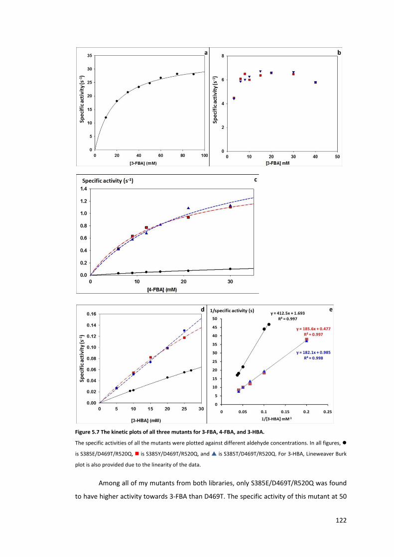
122
Figure 5.7 The kinetic plots of all three mutants for 3-FBA, 4-FBA, and 3-HBA.
The specific activities of all the mutants were plotted against different aldehyde concentrations. In all figures, �
is S385E/D469T/R520Q, � is S385Y/D469T/R520Q, and � is S385T/D469T/R520Q. For 3-HBA, Lineweaver Burk
plot is also provided due to the linearity of the data.
Among all of my mutants from both libraries, only S385E/D469T/R520Q was found
to have higher activity towards 3-FBA than D469T. The specific activity of this mutant at 50
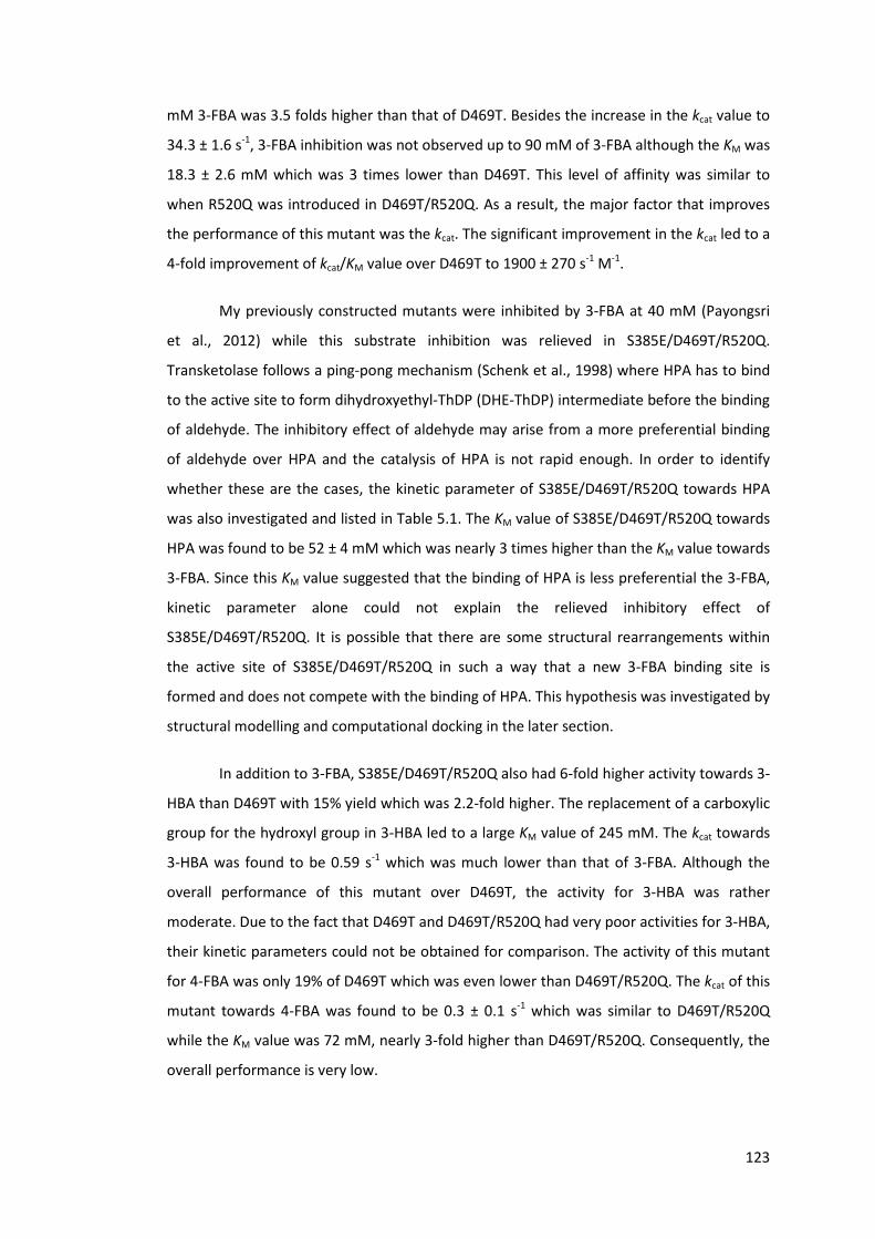
123
mM 3-FBA was 3.5 folds higher than that of D469T. Besides the increase in the kcat value to
34.3 ± 1.6 s-1, 3-FBA inhibition was not observed up to 90 mM of 3-FBA although the KM was
18.3 ± 2.6 mM which was 3 times lower than D469T. This level of affinity was similar to
when R520Q was introduced in D469T/R520Q. As a result, the major factor that improves
the performance of this mutant was the kcat. The significant improvement in the kcat led to a
4-fold improvement of kcat/KM value over D469T to 1900 ± 270 s-1 M-1.
My previously constructed mutants were inhibited by 3-FBA at 40 mM (Payongsri
et al., 2012) while this substrate inhibition was relieved in S385E/D469T/R520Q.
Transketolase follows a ping-pong mechanism (Schenk et al., 1998) where HPA has to bind
to the active site to form dihydroxyethyl-ThDP (DHE-ThDP) intermediate before the binding
of aldehyde. The inhibitory effect of aldehyde may arise from a more preferential binding
of aldehyde over HPA and the catalysis of HPA is not rapid enough. In order to identify
whether these are the cases, the kinetic parameter of S385E/D469T/R520Q towards HPA
was also investigated and listed in Table 5.1. The KM value of S385E/D469T/R520Q towards
HPA was found to be 52 ± 4 mM which was nearly 3 times higher than the KM value towards
3-FBA. Since this KM value suggested that the binding of HPA is less preferential the 3-FBA,
kinetic parameter alone could not explain the relieved inhibitory effect of
S385E/D469T/R520Q. It is possible that there are some structural rearrangements within
the active site of S385E/D469T/R520Q in such a way that a new 3-FBA binding site is
formed and does not compete with the binding of HPA. This hypothesis was investigated by
structural modelling and computational docking in the later section.
In addition to 3-FBA, S385E/D469T/R520Q also had 6-fold higher activity towards 3-
HBA than D469T with 15% yield which was 2.2-fold higher. The replacement of a carboxylic
group for the hydroxyl group in 3-HBA led to a large KM value of 245 mM. The kcat towards
3-HBA was found to be 0.59 s-1 which was much lower than that of 3-FBA. Although the
overall performance of this mutant over D469T, the activity for 3-HBA was rather
moderate. Due to the fact that D469T and D469T/R520Q had very poor activities for 3-HBA,
their kinetic parameters could not be obtained for comparison. The activity of this mutant
for 4-FBA was only 19% of D469T which was even lower than D469T/R520Q. The kcat of this
mutant towards 4-FBA was found to be 0.3 ± 0.1 s-1 which was similar to D469T/R520Q
while the KM value was 72 mM, nearly 3-fold higher than D469T/R520Q. Consequently, the
overall performance is very low.
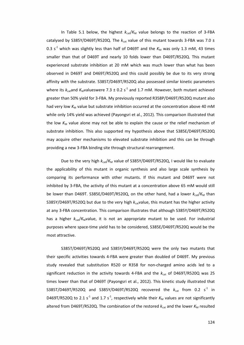
124
In Table 5.1 below, the highest kcat/KM value belongs to the reaction of 3-FBA
catalysed by S385Y/D469T/R520Q. The kcat value of this mutant towards 3-FBA was 7.0 ±
0.3 s-1 which was slightly less than half of D469T and the KM was only 1.3 mM, 43 times
smaller than that of D469T and nearly 10 folds lower than D469T/R520Q. This mutant
experienced substrate inhibition at 20 mM which was much lower than what has been
observed in D469T and D469T/R520Q and this could possibly be due to its very strong
affinity with the substrate. S385T/D469T/R520Q also possessed similar kinetic parameters
where its kcatand KMvalueswere 7.3 ± 0.2 s-1 and 1.7 mM. However, both mutant achieved
greater than 50% yield for 3-FBA. My previously reported R358P/D469T/R520Q mutant also
had very low KM value but substrate inhibition occurred at the concentration above 40 mM
while only 14% yield was achieved (Payongsri et al., 2012). This comparison illustrated that
the low KM value alone may not be able to explain the cause or the relief mechanism of
substrate inhibition. This also supported my hypothesis above that S385E/D469T/R520Q
may acquire other mechanisms to elevated substrate inhibition and this can be through
providing a new 3-FBA binding site through structural rearrangement.
Due to the very high kcat/KM value of S385Y/D469T/R520Q, I would like to evaluate
the applicability of this mutant in organic synthesis and also large scale synthesis by
comparing its performance with other mutants. If this mutant and D469T were not
inhibited by 3-FBA, the activity of this mutant at a concentration above 65 mM would still
be lower than D469T. S385E/D469T/R520Q, on the other hand, had a lower kcat/KM than
S385Y/D469T/R520Q but due to the very high kcatvalue, this mutant has the higher activity
at any 3-FBA concentration. This comparison illustrates that although S385Y/D469T/R520Q
has a higher kcat/KMvalue, it is not an appropriate mutant to be used. For industrial
purposes where space-time yield has to be considered, S385E/D469T/R520Q would be the
most attractive.
S385T/D469T/R520Q and S385Y/D469T/R520Q were the only two mutants that
their specific activities towards 4-FBA were greater than doubled of D469T. My previous
study revealed that substitution R520 or R358 for non-charged amino acids led to a
significant reduction in the activity towards 4-FBA and the kcat of D469T/R520Q was 25
times lower than that of D469T (Payongsri et al., 2012). This kinetic study illustrated that
S385T/D469T/R520Q and S385Y/D469T/R520Q recovered the kcat from 0.2 s-1 in
D469T/R520Q to 2.1 s-1 and 1.7 s-1, respectively while their KM values are not significantly
altered from D469T/R520Q. The combination of the restored kcat and the lower KM resulted

125
in an overall higher enzymatic activity towards 4-FBA. Besides that, the kcat/KM values of
both mutants were 4.5-5 times higher than that of D469T, and over 10 folds when
compared with D469T/R520Q.
All the kinetic studies so far illustrated that the mutation at S385 can alter the kcat
for both 3-FBA and 4-FBA and one mutant may have very different kcat values towards both
substrate. Given than that the carboxylate group of both 3-FBA and 4-FBA interacts with
R358, the relative position of the aldehyde of both 3-FBA and 4-FBA in all three mutants
would be different. These suggested that the enzymatic activity towards aromatic aldehyde
does depend on the type and the position of the substituent group on the aromatic ring
relative to the aldehyde which in turn affects the position of the aldehyde relative to the
catalytic centre. The alteration enzymatic activity upon mutation towards certain aromatic
substrates is not due to the change in the general catalytic activity but rather a shift in
substrate orientation and proximity to the reactive centre.
My previous study on 3-FBA and 4-FBA indirectly illustrated the importance of the
enzyme-substrate affinity but none of the mutants could be studied with non-charged
aromatic aldehyde due to a very low activity of D469T and D469T/R520Q (Payongsri et al.,
2012). Here is the first time that the kinetic parameters could be determined. In addition to
4-FBA, S385T/D469T/R520Q and S385Y/D469T/R520Q also had considerably higher
activities and yields towards 3-HBA than D469T. The kcat values of S385Y/D469T/R520Q and
S385T/d469T/R520Q were 2.09 s-1 and 1.01 s-1 which were similar to their kcat values
towards 4-FBA but up to 4 folds lower than the kcat for 3-FBA. Their KM towards 3-HBA,
however, were 100-300 times higher than the KM for 3-FBA, and 8-25 times higher than KM
for 4-FBA. These very large KM values led to the kcat/KM values of approximately 5.5 s-1 M-1
which was 1000 times lower than the kcat/KM for 3-FBA. These kinetic parameters from both
mutants supported my previous hypothesis that the affinity between transketolase and
aromatic aldehyde is one of the major factors governing the biocatalytic conversion of
aromatic aldehydes (Payongsri et al., 2012). These KM values were also much higher than
the concentration of 3-HBA within the reaction and such high KM values gave rise to a very
low specific activity towards 3-HBA compared with 3-FBA and 4-FBA.

126
Table 5.1 The kinetic parameters of all the mutants towards 3-FBA, 4-FBA, and 3-HBA.
Substrate
Specific activity
(relative to
D469T)
Yield
(%)
KM
(mM)
kcat
(s-1)
kcat/KM
(s-1 M-1)
D469T 3-FBA 1.00a 67 (1.0)b 56 (10) 13.2 (1.5) 236 (50)
D469T/R520Q 3-FBA 0.75 67 (0.1)b 13 (4) 6.0 (0.75) 468 (170)
S385E/D469T/R520Q 3-FBA 3.5 63 (1.0) 18 (3) 34.3 (1.6) 1900 (270)
S385Y/D469T/R520Q 3-FBA 0.47 53 (0.8) 1.3 (0.4) 7.0 (0.3) 5400 (1490)
S385T/D469T/R520Q 3-FBA 0.52 59 (0.5) 1.7 (0.25) 7.3 (0.2) 4255 (620)
S385Y/D469T 3-FBA 41 (1.1) n.d. n.d. n.d.
S385T/D469T 3-FBA 63 (1.4) 23 (10) 9 (2) 394 (185)
S385E/D469T/R520Q HPA 52 (4) 46 (2) 880 (70)
D469T 4-FBA 1.00a 30(1.7) b 251 (240) 5.0 (4.4) 20 (26)
D469T/R520Q 4-FBA 0.45 13(0.4) b 25 (8)
0.20
(0.03) 8.1 (2.8)
S385E/D469T/R520Q 4-FBA 0.19 <5 72 (30) 0.3 (0.1) 4.6 (2.4)
S385Y/D469T/R520Q 4-FBA 3.38 48 (0.4) 15 (3) 1.7(0.1) 109 (21)
S385T/D469T/R520Q 4-FBA 2.09 48 (1.7) 23 (6) 2.1(0.3) 90 (26)
S385Y/D469T 4-FBA 1.49 35 (0.3) 12.1 (0.5)
1.13
(0.02) 94 (4)
S385T/D469T 4-FBA <0.2 <5 >>30
D469T 3-HBA 1.00a 6.7c n.d.d n.d. n.d.
D469T/R520Q 3-HBA n.d. n.d. n.d. n.d. n.d.
S385E/D469T/R520Q 3-HBA 6.0 15 245 0.59 2.42
S385Y/D469T/R520Q 3-HBA 12.4 25 387 2.09 5.40
S385T/D469T/R520Q 3-HBA 12.7 31 184 1.01 5.50
S385Y/D469T 3-HBA 5.5 122 0.56 4.58
S385T/D469T 3-HBA <0.8 n.d. n.d. n.d.
a The specific activities of D469T determined at 50 mM 3-FBA/50 mM HPA, 30 mM 4-FBA/50 mM HPA, and 15
mM 3-HBA/30 mM HPA are 4.63 μmol/mg/min, 0.45 μmol/mg/ min, and 0.0071 μmol/mg/ min, respectively.
b the kinetic data of D469T and D469T/R520Q were from Chapter 4,Table 4.3(Payongsri et al., 2012).c Yields
were determined at 18 hours.d values were not determined (n.d.) due to very low activity.

127
So far, I illustrated that mutations can improve the enzymatic activity through 2
routes which can be either through improving enzyme-substrate affinity or the kcat. If the
concentration of substrate is much lower than the KM value, lowering KM will lead to higher
catalytic activity. S385Y/D469T/R520Q and S385T/D469T/R520Q are the example of this
case where their KM values towards 4-FBA are 10 to 16 times lower than that of D469T.
However, once the substrate concentration is much higher than the KM, improving the kcat
will enhance the enzymatic activity. This is illustrated by the catalysis of 3-FBA where
S385E/D469T/R520Q has similar KM to D469T/R520Q but much higher kcat.
5.3.4. The relationship between the enzyme activity and the final yields of
different aromatic aldehydes
I previously mentioned that the low bioconversion rate was associated with low
conversion yield in the mutant bearing D469T, possibly due to the competition with HPA
degradation. In order to justify at which point HPA degradation has an influence on the
conversion yield, the specific activities of the mutants towards each substrate were plotted
against the percentage yields as shown in Figure 5.8. This plot clearly illustrated that as the
specific activity increases, the percentage yield is improved which is similar to what I
speculated earlier. However, the two parameters become independent when the specific
activity is greater than 2.5 μmol/mg/min which is normally observed in the catalysis of 3-
FBA by most mutants. Due to the fact that several mutants were strongly inhibited by 3-
FBA, some mutants did not fit the trend. H461S containing mutants also globally lost the
activity and did not follow the pattern. It should be noted that S385E/D469T/R520Q has
much higher activity than D469T for 3-FBA but the yield did not improve. The 2-hour
sample showed that there was HPA left in the reaction but the ketodiol product
concentration becomes stationary which suggested that it is probably inhibited by the
product to a similar degree as D469T. The catalysis of HPA degradation could also be faster
in S385E/D469T/R520Q. The time course sampling revealed that HPA consumption is still
faster than that of 3-FBA which supports the latter characteristics.

128
Figure 5.8 The relationship between the activities of all the mutants and the % yields of all the aromatic
substrates.
5.3.5. Computational docking of substrates into the energy minimised
S385E/D469T/R520Q and S385Y/D469T/R520Q structures
The screening of S385X/D469T/R520Q library revealed that several mutants
maintained their activities towards a least one substrate. The kinetic study also illustrated
that the mutation at S385 had a rather diverse effect on the kcat of the enzyme. These
evidences suggested that each amino acid substitution has different catalytic effects for all
three substrates rather than a general change in the catalytic activity. These may arise from
the alteration in the substrate binding orientations or the position of the aldehyde relative
to the reactive enamine when the mutation was introduced. In order to rationalise this,
computational docking of substrates into the energy minimised structures of the mutants
was employed.
The kinetic study of S385E/D469T/R520Q alone could not explain the relief of
substrate inhibition and the increase in kcat towards 3-FBA. I speculated that phenomenon
may arise from structural rearrangement together with a new 3-FBA binding site. The
energy minimised S385E/D469T/R520Q structure was prepared according to the standard
protocol (Payongsri et al., 2012). The structures of DHE-ThDP and 3-FBA were then docked
into the active site. The computational docking predicted two binding clusters with
different binding energy and population. The first cluster showed in green structure in
Figure 5.9a had a lower binding energy together with a lower population (38%). It anchored
to phosphate binding residues similar to the previous prediction of 3-FBA in D469T (Figure

129
5.9b) and D469T/R520Q (Figure 5.9c). In the previous study, 3-FBA was predicted to form
two binding clusters in both D469T and D469T/R520Q where one cluster is a 180° rotation
of the other (Figure 5.9b and c). In D469T, one cluster placed the aldehyde 4.9Å away from
the enamine while the other was 7.2 Å which made it rather less active. Unlike D469T and
D469T/R520Q, the binding of 3-FBA to H461 and R358 in S385E/D469T/R520Q appeared to
have only unproductive conformations as the aldehyde groups were 7.2to 9.0 Å away from
enamine. The second cluster (yellow stick in Figure 5.9a) was more populated (62%) and
catalytically productive. It employed a new binding site as carboxylic acid cluster formed
hydrogen bonds with the side chain of R91, S24, and H26, and the backbone NH group from
G25. Only one conformation was predicted because the aldehyde group was anchored to
the side chain of H26 and H261 via hydrogen bonds. This placed the aldehyde 4.6Å away
from the enamine which is slightly shorter than that of D469T (4.9Å) (Payongsri et al.,
2012). Since the aldehyde was held in placed by the hydrogen bonds, the aldehyde would
be more likely to react with the enamine compared with the freely rotating aldehyde in
D469T. This, together with shorter distance, could contribute to the improved kcat.
Furthermore, since the productive and non-productive conformations did not employ the
same binding site and the productive conformation is also more populated, this could be
the mechanism that substrate inhibition was relieved in S385E/D469T/R520Q.
130
Figure 5.9 The computational docking of 3-FBA into the energy minimised a) S385E/D469T/R520Q, b) D469T,
c) D469T/R520Q.
In all figure, the productive conformations are in yellow while the unproductive ones are in green. a), the 3-FBA
in the first binding cluster is in green while the second cluster is in yellow. DHE-ThDP is in magenta. Hydrogen
bonds are in black dotted lines.
S385Y/D469T/R520Q and S385T/D469T/R520Q had higher activity for both 4-FBA
and 3-HBA. The kcat values of both mutants for 4-FBA and 3-HBA are actually very similar
but the reactions of 3-HBA are much slower due to the very large KM. Computational
docking of both substrates into the structures of the mutants could further give an insight
into the similarities and differences between the two substrates. Here, only
S385Y/D469T/R520Q was chosen to study the computational docking of 4-FBA and 3-HBA
because the side chain of tyrosine is much more different from serine than threonine. The
location and projection of the tyrosine side chain may be different from the side chain of
serine in the wild type so that this large aromatic side chain does not block the access of
the aromatic molecule. The accessibility of the aromatic aldehyde in the energy minimised
131
S385Y/D469T/R520Q upon computational docking would also assess the validity and
reliability of the computational docking.
Unlike the side chain of S385 in wild type, the side chain of tyrosine in the energy
minimised S385Y/D469T/R520Q structure did not project into the active site cavity. The
computational docking of 4-FBA into this triple mutant predicted only one cluster whereas
multiple clusters were found in D469T/R520Q which has very poor kcat towards 4-FBA. This
binding cluster in the triple mutant still employed the interaction between the carboxylic
acid group with R358 and H461 (Figure 5.10a). In addition, the benzene ring of 4-FBA can
form pi stacking with the aromatic ring of S385Y. These interactions hold 4-FBA in a
location that placed the aldehyde group 5.1Å away from the enamine and this distance is
also similar to the prediction in D469T 5.1Å (Payongsri et al., 2012). This similar distance
together with only one predicted cluster could be the explanation for the similar kcat to
D469T.
Figure 5.10 The computational docking of a) 4-FBA, and b) 3-HBA into the energy minimised
S385Y/D469T/R520Q.
The catalytically active conformations in yellow. Hydrogen bonds are in black dotted lines.
The computational docking of 3-HBA in S385Y/D469T/R520Q predicted four major
clusters but only two clusters showed in yellow and green in Figure 5.10b were predicted to
be in the active site. However, they did not form pi stacking as 4-FBA did. The first cluster,
showed as yellow stick in Figure 5.10b, formed a hydrogen bond with the side chain of
S385Y through its hydroxyl group. This placed the aldehyde 4.6Å away from the enamine
which is similar distance to that of 4-FBA in this triple mutant and may be the explanation
for the similar kcat between 4-FBA and 3-HBA by this mutant. The second cluster, as shown

132
in green stick, was catalytically inactive because the aldehyde group is pointing away from
the active site. None of the clusters was found to interact with R358 which supported my
early finding that the mutation at R358 tend to have very low influence on the activity for
3-HBA.
The reason for multiple clusters of 3-HBA could be the very low affinity between
the enzyme and 3-HBA itself and hydrophobic interaction through pi stacking is probably
insufficient for computational docking either. It should be noted that the computational
docking of 4-FBA into the active site of D469T gave only one cluster (Payongsri et al.,
2012)despite the fact that the KM was 251 mM which is the same order of magnitude as the
KM between 3-HBA and S385Y/D469T/R520Q.
5.3.6. Interactions between S385 and R520 mutations
The kinetic studies of the two triple mutants S385Y/D469T/R520Q and
S385T/D469T/R520Q revealed that they carry very similar characteristics towards all three
substrates. In my previous study with 4-FBA, the replacing one of the phosphate binding
arginine (R358 or R520) with a neutral amino acid led to a severely impaired kcat. This was
recovered by introducing either S385Y or S385T into D469T/R520Q. However, the side
chains of threonine and tyrosine are very different in size and polarity. This raised a
question whether both triple mutants follow the same path along of adaptive walk to reach
the same optimum. Therefore, the double mutant S385Y/D469T and S385T/D469T were
constructed and their kinetic parameters towards all three substrates were assessed. In
order to identify the type of synergic interaction between S385T, S385Y and R520Q, the
changes in the ΔG of binding and transition state from that of D469T were calculated and
these were represented as ΔΔG (Mildvan et al., 1992). The ΔΔG calculated from the kcat is
associated with the change in the energy barrier or the stability of the transition state
whereas the ΔΔG calculated from the KM is associated with the stability of substrate
binding. Therefore, a mutant with a negative ΔΔG value has a more stable substrate
binding or transition state than D469T. The coupling energy (ΔG(I)) was calculated as below.
ΔΔG (X,Y) = ΔΔG(X) + ΔΔG(Y) + ΔG(I)
ΔG(I) = ΔΔG(X,Y) - ΔΔG(X) - ΔΔG(Y)
ΔΔG(X) represents the changes in the free energy from the wild type upon the mutation at x
position.

133
ΔΔG(X,Y) is the change in the free energy from the wild type in the double mutant XY.
ΔΔGX = ΔGD469T – ΔGX
= RT ln(KX/KD469T)
ΔG(I) = RT ln {(KX,Y KD469T)/( KX KY)}
K is either the KM or kcat. In all cases, D469T is the pseudo wild type.
S385T/D469T was found to be active only towards 3-FBA. The kcat value was 9.0 s-1
which is slightly lower than that of D469T but higher than that of D469T/R520Q. Therefore,
both S385T and R520Q destabilised the transition state or increased the energy barrier
from D469T which resulted in the decreases in the kcat value. Consequently, both double
mutants had positive ΔΔGtransition state values. The summation of the destabilising effect from
both double mutants is less than the destabilising effect in the triple mutant which made
the ΔG(I) negative. This means S385T and R520Q displayed partially additive effect on
increasing the energy barrier for the transition state. The KM of S385T/D469T towards 3-
FBA was 23 mM which was between that of D469T and D469T/R520Q. This reduction in the
KM value was observed upon mutation of all phosphate binding residues (Payongsri et al.,
2012). However, this KM value was similar to that of H461S/D469T which suggested that
S385 may not strongly participate in the interaction with the carboxylic acid group of 3-
FBA. The ΔΔGbinding of the triple mutant is greater than the summation of the ΔΔGbinding of
the double mutants which illustrates that S385T and R520Q further stabilise substrate
binding than the simple additive combination of the effects of S385T and R520Q alone.
Therefore, the stabilising effect on the binding from S385T and R520Q exhibited positive
synergy.
S385T/D469T had a very low activity towards both 4-FBA and 3-HBA. As a result,
the kinetic parameters towards both substrates cannot be determined. This also illustrated
that S385T and R520Q must be introduced into D469T simultaneously in order to improve
the activity towards 4-FBA and 3-HBA.
S385Y/D469T, on the other hand, maintained good activities towards all three
substrates. However, 3-FBA inhibition was observed at even lower concentration than its
triple mutant and the KM was estimated to be lower than 10 mM. The kinetic study at lower
3-FBA concentration cannot be performed due to the detection limit of the HPLC. These
evidences suggested that the KM of the triple mutant was between the KM of S385Y/D469T

134
and D469T/R520Q. The triple mutant did not stabilise 3-FBA binding further than the
combination of the two mutants which represents a partially additive synergy between
S385Y and R520Q. Due to the fact that 3-FBA inhibition in S385Y/D469T started at 10 mM,
the kcat cannot be determined. However, the specific activity of S385Y/D469T at 15 mM 3-
FBA was somewhat similar to that of D469T which suggests that the kcat was not impaired.
The specific activity of S385Y/D469T towards 4-FBA was 1.49 fold higher than that
of D469T. This was largely due to the significant reduction in the KM to 12.1 mM from 251
mM in D469T although the kcat value of this mutant for 4-FBA was nearly 4.4 times lower
than that of D469T. Both S385Y and R520Q dramatically increased the affinity for 4-FBA
together with reducing the kcat in the double mutants. The kcat of the triple mutant was
improved from the double mutants and this characteristic may suggest an epistatic or
antagonistic interaction between S385Y and R520Q. However, the recovery of the kcat in
the triple mutant was still less than that of D469T which makes the coupling energy for the
kcat negative. Consequently, the destabilisation of the transition state from both S385Y and
R520Q was a partially additive effect. Both S385Y and R520Q improved the binding stability
from D469T. However, the absolute summation of the stabilising effect of S385Y and
R520Q was greater than the stabilising effect in the triple mutant which made the coupling
energy positive. Therefore, the stabilising effects from both mutations were partially
additive. S385Y/D469T did not only have higher activity towards 4-FBA but also 3-HBA. The
kcat of the double mutant was 4-fold lower than that of the triple mutant while the KM of
the double mutant towards 3-HBA was 122 mM. Among the mutants that their kinetics can
be studied, this was the lowest KM value towards 3-HBA. Since the kinetic parameter of
D469T cannot be determined, it is impossible to identify the interaction between S385Y
and R520Q in the reaction of 3-HBA.
Here, I have illustrated that S385Y can improve the activity towards 4-FBA and 3-
HBA independently to R520Q. Therefore, S385Y/D469T/R520Q and S385T/D469T/R520Q
follow different paths to achieve a similar characteristic.

135
Table 5.2 The kinetic parameters and the changes in the ΔG from D469T (ΔΔG) upon the introduction of other
mutations.
Mutant Substrate KM (mM) ΔΔG binding (kJ mol-1)
kcat (s-1)
ΔΔG transition state (kJ mol-1)
D469T 3-FBA 56 0.00 13.2 0 D469T/R520Q 13 -3.60 6.0 1.93 S385T/D469T 23 -2.18 9 0.89 S385T/D469T/R520Q 1.7 -8.57 7.3 1.46 ΔG(I)(kJ mol-1) -2.79 -1.36 D469T 4-FBA 251 0.00 5.0 0 D469T/R520Q 25 -5.68 0.20 7.87 S385Y/D469T 12.1 -7.44 1.13 3.62 S385Y/D469T/R520Q 15 -6.88 1.7 2.68 ΔG(I)(kJ mol-1) 6.24 -8.81
5.4. Conclusion
Site directed mutagenesis at the residues close to the reactive centre has improved
the specific activity of transketolase towards negatively-charged and polar aromatic
aldehydes. The kinetic study revealed that mutation at S385 can improve the activity via
restoring the kcat or improving the affinity. Computational docking was able to provide
some insights into how the mutation at S385 influenced the kinetic behaviours of the
enzyme for these substrates.A triple mutant, S385E/D469T/R520Q was found to have the
highest kcat for a non-natural substrate that has ever been reported in transketolase. The
three triple mutants were found to have 5 to 12-fold improvements for the activity of 3-
HBA which were high enough to determine the kinetic parameters for 3-HBA for the first
time. The kinetic study of hydrophobic aromatic aldehyde 3-HBA could be done for the
first time and it also supported my previous hypothesis that the affinity between the
enzyme and the aromatic substrates was one of the most important factors that controls
the reaction rate. The study of the synergy between S385Y/T and R520Q illustrated that
different mechanisms were employed by S385Y/D469T/R520Q and S385Y/D469T/R520Q to
reach similar enzymatic performance. This synergy also implied that random mutation at
R520 on S385T/D469T and S385Y/D469T may provide additional beneficial mutants.

136
6. Chapter 6: Synthesis of novel aromatic aminodiols through the
coupling of transketolase with transaminase
6.1. Introduction
Chiral amines and amino acids are the building blocks of several natural and
synthetic compounds, some of which are important in pharmaceutical, agrochemical,
(Höhne and Bornscheuer, 2009; Mutti et al., 2011; Rudat et al., 2012; Shin and Kim, 2001;
Tufvesson et al., 2011). Several production routes of amino acids and chiral amines are
available (Breuer et al., 2004). However, chemosynthesis of chiral amines are still
challenging and involve toxic chemicals (Savile et al., 2010; Tufvesson et al., 2011). An
alternative method is to use enzymes which becomes increasingly attractive with a range of
enzymes available including hydrolases, oxidoreductases, and transferases (Höhne and
Bornscheuer, 2009).
Transaminases (TAms) are transferase enzymes that belong to pyridoxal phosphate
dependent enzyme family. These enzymes catalyse oxidative deamination of a donor and
reductive amination of an acceptor. The reaction mechanism of transaminase is illustrated
in Figure 6.1 below.

137
Figure 6.1 The reaction mechanism of transaminase enzymes when catalysing the transfer of an amine group
from one amino acid to an alpha keto acid.
They have gained interests for the synthesis of chiral amines either by kinetic
resolution and asymmetric synthesis (Koszelewski et al., 2010). The multiple sequence
alignment by Protein family database (Pfam) classified transaminases into 6 classes (class I
to IV) (Rudat et al., 2012; Ward and Wohlgemuth, 2010). In addition, transaminases were
broadly classified according to the substrate and position of carbonyl group that is
aminated (Ward and Wohlgemuth, 2010; Yonaha et al., 1983, 1976). Alpha, beta, and
gamma transaminases transfer an amine group to a carbonyl group that is located at the
alpha, beta, and gamma position relative to the carboxylic acid group in the substrates,
respectively. ω transaminases belong to class III of the PLP-dependent enzyme sub-family
and can transfer the amine group from a primary amine to aldehydes and ketones without
the requirement of the carboxylic acid group (Rudat et al., 2012; Ward and Wohlgemuth,
2010). As a result, ω-transaminases have a much wider substrate spectrum than other
classes of transaminases which makes them attractive in organic synthesis. Besides that, ω-
transaminases also have rapid turnover, high stability (Park et al., 2012; Shin et al., 2003),
their catalytic routes are more environmentally friendly (Ward and Wohlgemuth, 2010),
and they do not have a requirement for cofactor recycling (Rudat et al., 2012). However,
one major obstacle of using ω-transaminases is that their equilibrium constant for the

138
amino transferring reaction is close to 1 in most cases which means that their conversion
yields are only 50% (Shin and Kim, 2001).
Chiral amino alcohols are a functionality that is found in a number of natural
compounds including antibiotics such as chloramphenicols and derivatives, sphingolipids,
and glycosidase inhibitors (Hailes et al., 2010; Smith et al., 2010). The chemical synthesis of
chloramphenicol (Controulis et al., 1949) and thiamphenicol (Nagabhushan et al., 2000) are
multi-step reactions whereas the synthesis of florphenicol requires thaimphenicol as
starting material (Nagabhushan et al., 2000). Developing a shorter and simpler alternative
method could provide numerous advantages over the traditional one.
Here, my research aim is to enzymatically produce novel aromatic aminodiols
which can be used as building blocks in the synthesis of chloramphenicol analogues. A
sequential reaction starts with the reaction of transketolase (TK) to produce aromatic 1,3-
diol compound. In the next step, a transaminase catalyses the transfer of an amine group
from an amine donor to the carbonyl group of the 1,3-diol as illustrated in Scheme 6.1
below.
Scheme 6.1 The reaction scheme for the production of chloramphenicol amine analogues using TK and TAm
This novel enzymatic route for the synthesis of aminoalcohols was reported in the
synthesis of 2-amino-1,3,4-butanetriol (ABT) which is a functionality found in Nelfinavir
(Ingram et al., 2007). This concept was further studied and developed in a number of
aspects. Their kinetic constants towards the production of ABT were available for reaction
modelling which aids the process design (Chen et al., 2006). A number of transketolase
mutants were constructed and screened with improvements for wider range of substrates
(Galman et al., 2010; Hibbert et al., 2008, 2007; Payongsri et al., 2012). Consequently,
numerous amino alcohols can be produced. Our research group at UCL has also optimised
aliphatic amino alcohol synthesis through transketolase-transaminase coupling system
(Rios-Solis et al., 2011; Smith et al., 2010). In addition to chloramphenicol and
thiamphenicol, yeast TK can accept halogenated pyruvate derivative as a donor (Esakova et
al., 2009). If E. coli TK does the same as well as accepting an aromatic aldehyde, Florfenicol
TK
ThDP, MgCl2
TAm, PLP
+ +

139
and other flouroanalogues can be synthesised. Alongside the transketolase research, the
recently discovered ω-transaminase from Chromobacterium violaceum DSM30191
(CV2025) was also found to accept large aromatic substrates (Kaulmann et al., 2007).
Therefore, the transketolase-transaminase coupling system could have a large synthetic
potential.
For the synthesis of chloramphenicol analogues, I have previously illustrated that
engineered transketolase mutants accept 3-FBA and 4-FBA and produce novel aromatic
dihydroxy ketones. In recent years, numerous ω-transaminases were discovered and found
to have different substrate preferences (Koszelewski et al., 2010). This work assessed a
small range of ω-transaminases for their ability to aminate aromatic dihydroxy ketones,
although the main focus was on CV2025. This was because the substrate spectrum of
CV2025 has been thoroughly investigated and aromatic substrates are more preferable. In
addition, it was the only enzyme that was reported to accept 1,3-dihydroxy-1-
phenylpropane-2-one (DPP) with high stereoselectivity (Smithies et al., 2009). Although this
is S selective transaminase, protein engineering to reverse the stereoselectivity would be
feasible especially due to the availability of the crystal structure of CV2025 (PDB ID:
4A6T)(Humble et al., 2012). In addition, the key residues that can reverse and improve the
selectivity on certain aromatic substrates have been identified through rational design and
3D structure analysis (Humble et al., 2012). The enantioselectivity can be increased by a
single mutation W60C and can be reversed by double mutation F88A/A231F (Humble et al.,
2012).
6.2. Materials and methods
6.2.1. Materials
All the substrates for transaminase were freshly prepared in 50 mM Tris buffer and
adjusted the pH to 7.5. A 40 mM PLP stock solution was prepared in 50 mM Tris buffer, pH
7.5 and stored in 80-μl aliquots at -20 °C. The concentration of the TAm in the lysate was
adjusted by diluting with 50 mM Tris buffer with 0.4 mM PLP, 7.5. This solution was kept at
4 °C. Kanamycin was filtered sterile and stored in the same way as ampicillin but the final
concentration was 30 mg/ml. The working concentration of kanamycin was 30 μg/ml.

140
6.2.2. Optimising the synthesis of the aromatic dihydroxy ketones by TK
The reactions of 3-FBA and 4-FBA were driven to completion by addition of HPA to
the reaction. Both reactions started with 45 mM of HPA and 45 mM aldehyde which was
prepared by addition of 2X substrate solution (90 mM HPA, 90 mM FBA in 50 mM Tris
buffer, pH 7.0) into the TK lysate that has been incubated with cofactor for 20 minutes. 750
mM HPA in 50 mM Tris buffer, pH 7.0 was added to the reaction to reach the final
concentration of 22.5 mM. HPA was added to the reaction when the formation of the
dihydroxy ketone and the consumption of the aldehyde slowed down. All substrates were
prepared in glass vials. All the reactions were performed in 1.5-ml vials and closed with
snap caps.
S385E/D469T/R520Q displays the highest activity towards 3-FBA. Our previous
experiment on the stereoselectivity of different mutants for 3-FBA showed that
S385E/D469T/R520Q gave a racemic mixture of the 3-(1,3-Dihydroxy-2-oxopropyl )benzoic
acid (3-DOPBA) (Steadman, et al, unpublished data). It was demonstrated that CV2025 can
accept racemic DPP (Kaulmann et al., 2007; Smithies et al., 2009). However, the presence
of the substituent group on the ring may influence the stereospecificity. As a result,
S385E/D469T/R520Q was used to produce a racemic 3-DOPBA. HPA was added at 1 hour
after the reaction was started and the reaction was then left overnight at 22 °C.
For 4-FBA reaction, S385Y/D469T/R520Q and S385T/D469T/R520Q were used due
to their high yield and activity. 3 hours after the reaction was started, HPA was added. This
was maintained at 22 °C for another 6 hours and then reduced to 4 °C for 12 hours.
6.2.3. Transaminase enzyme preparation
All the strains were provided by Prof John Ward (through Maria F Villegas-Torres).
The CV2025 mutants were provided by Dawid Deszcz. The strain of TAm used in the
screening are CV2025, pQR1006, pQR958, pQR1019, pQR1021, and CV2025 mutant
Y153M/S/F.
A glycerol stock of each strain was streaked on LB plates with kanamycin at a
concentration of 30 μg/ml. Single colonies were picked and inoculated into 250-ml flasks
containing 50 ml of LB broth with kanamycin. This was cultured overnight at 37 °C, shaking
at 250 rpm. These cultures were then transferred into 2-L flasks containing 450 ml of LB
media with kanamycin. They were then cultured at 37 °C, shaking at 250 rpm for 3 hours.
The temperature was then dropped to 30 °C prior induction by addition of IPTG to the final

141
concentration of 1 mM. They were cultured at 30 °C, shaking at 250 rpm for another 5
hours. 50 mg of pyridoxal-5'-phosphate was added to each culture 30 minutes before
harvesting. The culture was aliquoted into 4 of 50-ml falcon tubes and centrifuged at 4000
rpm for 10 minutes. The supernatant was discarded and another 50 ml-of culture was
added into these 4 tubes which were then centrifuged for another 10 minutes. This was
repeated until all the culture was harvested. Each falcon tube contains cells from
approximately 125 ml of culture.
Cell pellets were resuspended in 4 ml of 50 mM Tris buffer, pH 7.5 with 0.4 mM
PLP. The resuspended cells were sonicated on iced according to the standard protocol.
These were then centrifuged at 13,000 rpm for 15 minutes at 4 °C. The supernatant
containing TAms can be used fresh or stored at -80 °C in 120-μl aliquots to be used within 1
month.
For the purification of TAm, see section 2.15.2.
6.2.4. Protein quantification
The total protein concentration within the clarified lysate was quantified by
Bradford assay. The percentage of TAm within the lysate was analysed by SDS-PAGE gel and
densitometry according to the protocol using BSA as a protein standard due to
unavailability of purified with known concentration of transaminase.
6.2.5. HPLC
All the compounds in this study were quantified by reverse phase column ACE5 C18
according to the protocol. All samples were centrifuged at 13,000 rpm for 3 minutes before
loading into the HPLC. The retention times of S-methylbenzylamine (MBA), 3-
aminomethylbenzoic acid, 4-aminomethylbenzoic acid, and acetophenone (AP) are 3.59,
2.55, 2.26, and 7.8 minutes, respectively (see Figure 6.5 and Figure 6.6 for example). It
should be noted that MBA has a much lower absorption at nearly all wavelengths than
other aromatic compounds used in this study. Its absorption at 275 nm is also very low and
it is recommended to use either 210 or 250 nm for the detection and quantification of
MBA. An example chromatogram is shown in Figure 6.2.

142
Figure 6.2 Chromatogram of the 1-minute reaction sample from the amination reaction of 3-DOPBA obtained
from transketolase reaction.
The reaction consisted of 10 mM 3-DOPBA, 40 mM MBA, and less than 1 mM 3-FBA. The sample was analysed
by ACE5 C18 column and detected by UV absorption at 250 nm.
6.2.6. MBA inhibition study
Throughout this study, S-MBA was used as the amino donor to thermodynamically
drive the reaction forward due to the formation of acetophenone (Tufvesson et al., 2011).
Higher concentration of MBA could also increase the reaction rate as well as shifts the
equilibrium. However, it can also cause substrate inhibition. Therefore, the maximum
concentration has to be determined.
The initial rates of the 3-FBA-MBA reactions where compared when using 20 mM
and 40 mM MBA. Both reactions were performed in triplicate. 68 μl of clarified lysate
CV2025, together with 6 μl of 40 mM PLP and 76 μl of 50 mM Tris buffer, pH 7.5, was
added to 1.5-ml glass vials. The reaction was started by addition of 150 μl of the 2X
substrate stock solution (40 mM 3FBA with 40 mM MBA or 80 mM MBA in 50 mM Tris
buffer, pH 7.5). The vials were quickly closed by snap caps. 20 μl of samples were taken at
3, 10, 80, 120, 150, and 180 minutes and quenched by added into 380 μl of 0.1% TFA. These
samples were centrifuged and the supernatants were analysed by HPLC. The peak areas of
3-aminomethylbenzoic acid were used directly to compare the initial rates.
6.2.7. Screening transaminases for aromatic dihydroxy ketones
The concentrations of the aromatic dihydroxy ketones were kept constant at 10
mM while the concentration of MBA was maintained at 40 mM.
Different ω-TAms were screened against DPP and the 3-DOPBA to see which TAm
consume the highest amount of dihydroxy ketone and MBA as well as the generation of AP.
0.00 0.50 1.00 1.50 2.00 2.50 3.00 3.50 4.00 4.50 5.00 5.50 6.00 6.50 7.00 7.50 8.00 8.50 9.00-50
0
50
100
150
200
250
300 2012-03-07 RETEST TAM 3FBA KETODIOL #3 [modified by General] control 3min rt UV_VIS_2mAU
min
0.153 - 0.0010.320 - 0.003 1.033 - 0.000 1.500 - 0.000
1.607 - 29.456
1.833 - 0.756
2.220 - 0.9902.393 - 1.251
3.053 - 69.228
4.460 - 1.3944.553 - 0.546
4.753 - 5.100
5.400 - 0.004
5.567 - 27.255
6.087 - 0.0026.307 - 0.0086.473 - 0.0036.633 - 0.0006.753 - 0.0197.053 - 0.0137.427 - 0.0017.633 - 0.233
7.853 - 2.957
8.567 - 0.0008.753 - 0.0818.860 - 0.006
WVL:250 nmC18, UV 250 nm

143
The TAms used are CV2025, pQR1006, pQR958, pQR1019, pQR1021, and CV2025 mutant
Y153M/S/F.
The 450-μl reaction was prepared by adding 200 μl of TAms lysate, 9 μl of 40 mM
PLP, and 90 μl of 200 mM MBA to a 1.5-ml vial. The reaction was then started by the
addition of 151 μl of optimised TK reaction sample into the TAm mixture. The vial was
closed by snap cap and incubated at 22 °C by Thermomixer Comfort (eppendorf, UK). 75 μl
of samples were added into 225 μl of 0.1% TFA to quench the reaction. This was then
centrifuged at 13,000 rpm for 3 minutes and the supernatant was analysed by the HPLC.
The samples were taken at 1 minute, 2 hours, 6 hours, and 26 hours. The screening for the
4-DOPBA was conducted in the same way but only CV2025 was used.
6.2.7.1. Temperature and pH optimisation
All substrate stock solutions were prepared in 50 mM Tris buffer and adjusted the
final pH to 7.5 and 8.5. Clarified lysate were prepared by resuspending cell pellets in 50 mM
Tris buffer, pH 7.5 and 8.5 with 0.4 mM PLP. All different pH reactions were performed at
22 °C, 30 °C, 37 °C, and 45 °C which were maintained by Thermomixer Comfort (Eppendorf,
UK). A control reaction was set up by mixing crude TK reaction with TAm lysate, PLP to the
same final concentration as the TAm reaction but MBA was not added. This was done in
order to compare the degree of dihydroxy ketone rearrangement over the period of time.
6.2.7.2. Control reactions
Several control reactions were set up in order to confirm that the growing peak is
associated with the utilisation of the aromatic dihydroxy ketones and to assess the
background acetophenone production. The reactions using cell-free and his-tag
purifiedCV2025 were compared in order to compare whether the presence of cellular
metabolites causes the side reactions or the increase in the area of certain peaks on the
chromatogram. Clarified lysate was prepared in 50 mM Tris and HEPES, pH 7.5 in order to
find out whether the background reactions were due to the buffer used. Both purified and
clarified lysate CV2025 were incubated with and without MBA in both buffers. To confirm
that Tris or HEPES does not catalyse the reaction, two sets of reactions were set up where
the enzyme was substituted with Tris and HEPES and they were incubated under the same
conditions. 20 μl of samples were taken at 1 minute, 3 hours, and 20 hours to be analysed
by HPLC. All experiments were assessed without the addition of known amino acceptors
and all the reactions were performed at 30 °C.

144
Clarified CV2025 was prepared in 50 mM Tris buffer pH 7.5 and 50 mM HEPES pH
7.5 as above procedure. Less than 1-week old, his-tag purified CV2025 was provided by
Amanatuzzakiah Abdul Halim. 200 mM MBA was prepared in 50 mM Tris and HEPES and
adjusted the pH to 7.5. 40 mM PLP was prepared in 50 mM Tris buffer, pH 7.5.
6.2.8. Competitive reaction
In order to find out whether the 4-DOPBA could access into the active site of
CV2025, a competitive reaction between the 4-DOPBA and 4-FBA itself was performed. Due
to the fact that 4-DOPBA was synthesised using clarified lysate, other components within
the lysate may influence the activity of CV2025. Therefore, the 4-FBA amination rate was
also compared with the presence and absence of TK lysate and ThDP.
The 4-DOPBA was enzymatically synthesised from 45 mM reaction with 10%
clarified lysate at 500-μl scale. 50 μl of S385T/D469T/R520Q was incubated with 50 μl of
10X cofactor solution and 150 μl of 50 mM Tris buffer, pH 7.0 for 20 minutes before the
addition of 250 μl 2X substrate solution (90 mM HPA, 90 mM 4-FBA in 50 mM Tris buffer,
pH 7.0). The reaction temperature was maintained at 22 °C for 18 hours which gives 50 %
conversion making the leftover 4-FBA concentration of 22.5 mM. All the concentrations at
the end of the reaction were confirmed by HPLC. The left over 4-FBA concentration was
then used as a starting substrate concentration in the TAm reaction. In parallel, 50 μl TK
lysate was mixed with 50 μl of 10X cofactor solution and 400 μl of 50 mM Tris buffer, pH
7.0. This TK mixture was incubated at 18 hours at 22 °C as the reaction and was used in the
second reaction below.
The final volumes of all three TAm reactions were 450 μl containing 10 μl of
CV2025 clarified lysate, and 9 μl of 40 mM PLP. All the reactions had 10 mM of 4-FBA and
40 mM MBA at the beginning. In the first reaction, 200 μl of the 4-FBA/4-DOPBA reaction
was added to the TAm-PLP mixture. In the second reaction, 56.6 μl of 90 mM 4-FBA and
200 μl of TK-ThDP mixture were added to the reaction. In the last reaction, only 56.6 μl of
90 mM 4-FBA was added to the TAm. 50 mM Tris buffer with 0.4 mM PLP at pH 7.5 was
added to all these solutions to reach the final volume of 360 μl. All three reactions were
started by adding 90 μl of 200 mM MBA to the reaction. All the reactions were done in
duplicate. The 20-μl samples were taken at every 1 minute and quenched in 380 μl 0.1%
TFA before analysed by HPLC. Standard graph of 4-aminomethylbenzoic acid was used for
the quantification in the reaction.

145
6.2.9. Kinetic study of 3-FBA and 4-FBA with CV2025
The concentration of MBA in the reaction was kept constant at 50 mM. The
concentrations of 3-FBA and 4-FBA in the reaction were varied from 3-15 mM. The enzyme
concentration in the reaction was 0.18 mg/ml. The reactions were done in triplicate at the
final volume of 300 μl.
50 μl of clarified CV2025 lysate, 6 μl of 40 mM PLP, and 94 μl of 50 mM Tris buffer
pH 7.5 were added to the 1.5-ml vials. The reaction was started by adding 150 μl of 2X
substrate solution to the reactions at every 15-second interval and the vials were quickly
closed with snap cap. 20 μl of the reaction were taken and quenched in 380 μl 0.1% TFA
before analysed by the HPLC according to the standard protocol. The samples of 3-FBA
reaction were taken every 10 minutes while the samples of 4-FBA reactions were taken
every 1 minute. Commercially available of 4-aminomethylbenzoic acid was used to prepare
standard concentration for the quantification of this compound in the reaction.
For 3-FBA reaction, the rate of 3-FBA consumption was used to calculate the initial
rate. Standard 3-FBA was prepared for quantification.

146
6.3. Results and discussions
6.3.1. The yield of aromatic dihydroxy ketones after HPA fed-batch
All the aromatic substrates that I used so far with transketolase mutants never
reached 100 % conversion when the reaction was started with equimolar concentrations of
HPA and aldehyde. If the dihydroxy ketone from the reaction was used directly without
purification steps, the leftover aldehyde could compete with the dihydroxy ketone in the
TAm reaction step. Since CV2025 was reported to catalyse aromatic aldehydes at a much
higher rate than DPP (Kaulmann et al., 2007), S-MBA would be quickly consumed to
aminate the aromatic aldehyde leaving a lower concentration of S-MBA to aminate the
dihydroxy ketones. In addition, the consumption of MBA leads to the reduction of pH and
the acetophenone formation could inhibit the enzyme. The overall effect results in lower
enzyme performance and aminodiol yield. Therefore, driving the TK reaction to completion
is crucial for the subsequent reaction.
In an equimolar concentration reaction, the yield of 3-FBA reaction by D469T
bearing mutants reached approximately 65 % conversion after 2 hours and this was
suggested to be due to the product inhibition and HPA degradation by TK mutant
(Payongsri et al., 2012) (and Chapter4, Table 4.4). Using a higher amount of HPA could,
therefore, improve the yield by compensating the degraded HPA. The time course sampling
of S385E/D469T/R520Q (Figure 6.3) showed that the increase in 3-DOPBA concentration, as
well as the 3-FBA consumption rate, started to level out after 1 hour while HPA
concentration still decreased. Addition of HPA at 1 hour after the reaction started may
increase the chance of using the leftover HPA and consuming up 3-FBA.

147
Figure 6.3 The reaction profile of equimolar3-FBA reaction catalysed by S385E/D469T/R520Q.
The 18-hour sample showed the concentration of 3-FBA was less than 3 mM left
which illustrated that the fed-batch of HPA has improved the conversion yield to greater
than 90 %. This percentage yield can still be achieved when the starting substrate
concentrations were changed as long as 1.5 equivalent of HPA was added to the reaction at
approximately 1 hour. However, using excess HPA at the beginning of the reaction does not
improve the yield to the same degree.
Before the optimisation of the 4-FBA reaction at 3 hours after the 45-mM-
equimolar reaction was started using S385Y/D469T/R520Q or S385T/D469T/R520Q, there
was remaining 28 mM of 4-FBA, while the concentration of HPA was only 12 mM. This
showed a greater degree of HPA degradation than the 3-FBA reaction and possibly due to
the fact that the reactions of 4-FBA by both mutants were slower than the reaction of 3-
FBA by S385E/D469T/R520Q (see Chapter5, Figure 5.8). Therefore, a greater degree of HPA
degradation was observed. Adding 1.5 equivalent of HPA to the reaction at 3 hours has
increased the 4-FBA consumption in both S385T/Y/D469T/R520Q to a similar level. The 18-
hour samples showed that both mutants achieved greater than 70 % substrate
consumption with 31 mM of 4-DOPBA.
3-DOPBA and 4-DOPBA from the TK reactions were used as 3X substrate solution
and the 3-DOPBA and 4-DOPBA concentrations in the TAm reactions were kept constant at
10 mM. Therefore, the 3-FBA and 4-FBA reactions needed to be started with 35 mM and 45
mM, respectively. The concentrations of the dihydroxy ketone from both reactions were
approximately 31 mM while the leftover 3-FBA and 4-FBA concentrations were less than 3
0
20
40
60
80
100
120
0 20 40 60 80 100 120 140
% Conversion
Time (mins)
%HPA left %3FBA left % conversion

148
mM and 10 mM. Since some dihydroxy ketones underwent degradation, more aldehydes
were consumed than the formation of the dihydroxy ketones. The major route of
degradation was expected to be through the rearrangement at the 2- hydroxyl group as
illustrated in Scheme 6.2(Galman et al., 2010). This was supported by a continuous increase
in the formation of a compound that was highly absorbed at 250 nm.
Scheme 6.2 The degradation of the aromatic dihydroxy ketone through the rearrangement at the 2-hydroxyl
group
(Galman et al., 2010).
6.3.2. MBA inhibition study
It was previously reported that MBA can have an inhibitory effect on CV2025 and
other ω-TAms (Rios-Solis et al., 2011). Since the reaction with DPP was very slow compared
with aldehydes (Kaulmann et al., 2007), the reaction rate could be increased by using
higher substrate concentrations. In addition, using high substrate concentrations can also
push the equilibrium forwards. Before screening with the aromatic dihydroxy ketones using
MBA as a donor, it was therefore important to identify the maximum MBA concentration to
be used without inhibiting the enzyme. Although one may speculate that MBA inhibition
may occur at a different concentration when using different acceptor substrates, here, I
identified if 40 mM MBA can inhibit CV2025 in 3-FBA/MBA reactions. The initial rates of the
reaction at the concentration of 20 and 40 mM MBA were compared.
The equimolar reaction showed that the rate of 3-FBA consumption was 0.068
mM/min while the reaction with doubled MBA concentration had the initial rate of 0.069
mM/min. These initial rates were not significantly different and illustrated that 40 mM
MBA can be used. If there is an inhibitory effect, it will have the same effect as at 20 mM.
Although using higher substrate concentration does not seem to increase the initial rate, it
still has the tendency to thermodynamically drive the reaction forwards. Therefore, 40 mM
MBA was used through this study.
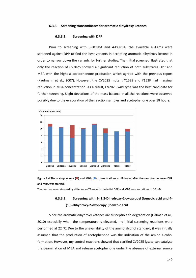
149
6.3.3. Screening transaminases for aromatic dihydroxy ketones
6.3.3.1. Screening with DPP
Prior to screening with 3-DOPBA and 4-DOPBA, the available ω-TAms were
screened against DPP to find the best variants in accepting aromatic dihydroxy ketone in
order to narrow down the variants for further studies. The initial screened illustrated that
only the reaction of CV2025 showed a significant reduction of both substrates DPP and
MBA with the highest acetophenone production which agreed with the previous report
(Kaulmann et al., 2007). However, the CV2025 mutant Y153S and Y153F had marginal
reduction in MBA concentration. As a result, CV2025 wild type was the best candidate for
further screening. Slight deviations of the mass balance in all the reactions were observed
possibly due to the evaporation of the reaction samples and acetophenone over 18 hours.
Figure 6.4 The acetophenone (����) and MBA (����) concentrations at 18 hours after the reaction between DPP
and MBA was started.
The reaction was catalysed by different ω-TAms with the initial DPP and MBA concentrations of 10 mM.
6.3.3.2. Screening with 3-(1,3-Dihydroxy-2-oxopropyl )benzoic acid and 4-
(1,3-Dihydroxy-2-oxopropyl )benzoic acid
Since the aromatic dihydroxy ketones are susceptible to degradation (Galman et al.,
2010) especially when the temperature is elevated, my initial screening reactions were
performed at 22 °C. Due to the unavailability of the amino alcohol standard, it was initially
assumed that the production of acetophenone was the indication of the amino alcohol
formation. However, my control reactions showed that clarified CV2025 lysate can catalyse
the deamination of MBA and release acetophenone under the absence of external source

150
of amino acceptor. This deamination of MBA occurred in both Tris and HEPES buffers and
up to 2 mM of acetophenone can be released within 3 hours when using 40 mM of MBA.
This production of acetophenone and the consumption of MBA may be due to the
amination of certain cellular metabolites such as pyruvate. When the dihydroxy ketones
were added, at least 5 mM of acetophenone was produced while the less than 1 mM of the
3-DOPBA or 4-DOPBA was utilised and the presence of the leftover aldehydes from the TK
reaction were less than 1 mM. Since acetophenone is produced even in the absence of any
acceptor and the concentration is higher than the concentration of PLP alone, MBA was
assumed to be used by CV2025 to aminate cellular metabolites.
In order to confirm that CV2025 can aminate the 3-DOPBA and 4-DOPBA, the
chromatograms of all the reaction samples were overlayed and compared instead. In Figure
6.5, it can be seen that the peak area of the 3-DOPBA hardly changed. The aromatic amino
alcohol is more polar than the dihydroxy ketones, so it was expected to be eluted much
earlier. The superimposition the 2-minute sample chromatogram with the 16-hour sample
showed that there was a small increase in the peak area with a retention time of 1.75
minutes which suggested that CV2025 may be able to accept the 3-DOPBA but that the
reaction was very slow.

151
Figure 6.5 The overlapped chromatograms of the sample at 3 minutes (����) and 16 hours (����) of the 3-DOPBA
amination reaction by CV2025.
The reaction was performed at 40 mM MBA, 10 mM 3-DOPBA, in 50 mM Tris buffer, pH 7.5, at 22 °C. The peaks
of the compounds of interests were labelled with the structures corresponding to their retention times. The
samples were analysed byACE5 C18 column and detected by 275 nm absorption.
0.00 0.50 1.00 1.50 2.00 2.50 3.00 3.50 4.00 4.50 5.00 5.50 6.00 6.50 7.00 7.50 8.00 8.50 9.00-20
0
20
40
60
80
100
120
140
160
180
200
1 - 2012-03-07 retest TAm 3FBA ketodiol #5 [modified by General] CV2025 3min rt UV_VIS_32 - 2012-03-07 retest TAm 3FBA ketodiol #19 [modified by General] CV2025 16h rt UV_VIS_3mAU
min
21
WVL:275 nm
1.79 min peak
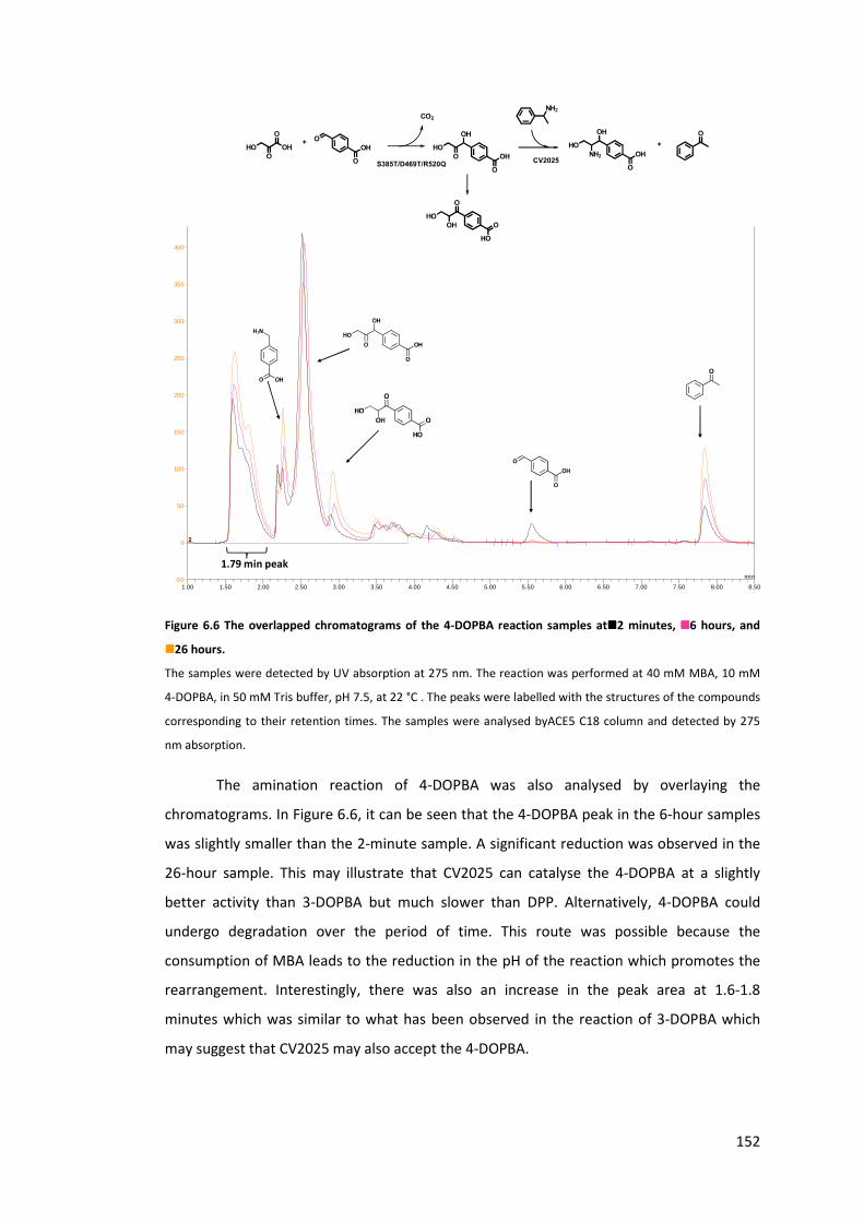
152
Figure 6.6 The overlapped chromatograms of the 4-DOPBA reaction samples at����2 minutes, ����6 hours, and
����26 hours.
The samples were detected by UV absorption at 275 nm. The reaction was performed at 40 mM MBA, 10 mM
4-DOPBA, in 50 mM Tris buffer, pH 7.5, at 22 °C . The peaks were labelled with the structures of the compounds
corresponding to their retention times. The samples were analysed byACE5 C18 column and detected by 275
nm absorption.
The amination reaction of 4-DOPBA was also analysed by overlaying the
chromatograms. In Figure 6.6, it can be seen that the 4-DOPBA peak in the 6-hour samples
was slightly smaller than the 2-minute sample. A significant reduction was observed in the
26-hour sample. This may illustrate that CV2025 can catalyse the 4-DOPBA at a slightly
better activity than 3-DOPBA but much slower than DPP. Alternatively, 4-DOPBA could
undergo degradation over the period of time. This route was possible because the
consumption of MBA leads to the reduction in the pH of the reaction which promotes the
rearrangement. Interestingly, there was also an increase in the peak area at 1.6-1.8
minutes which was similar to what has been observed in the reaction of 3-DOPBA which
may suggest that CV2025 may also accept the 4-DOPBA.
1.00 1.50 2.00 2.50 3.00 3.50 4.00 4.50 5.00 5.50 6.00 6.50 7.00 7.50 8.00 8.50-50
0
50
100
150
200
250
300
350
400
450
1 - 2012-03-23 TAm with prod of SDR7-23 4FBA #7 [modified by General] SY CV 2min UV_VIS_32 - 2012-03-23 TAm with prod of SDR7-23 4FBA #13 [modified by General] SY CV 6h UV_VIS_33 - 2012-03-23 TAm with prod of SDR7-23 4FBA #25 [modified by General] SY CV 26h UV_VIS_3mAU
min
321
WVL:275 nm
1.79 min peak
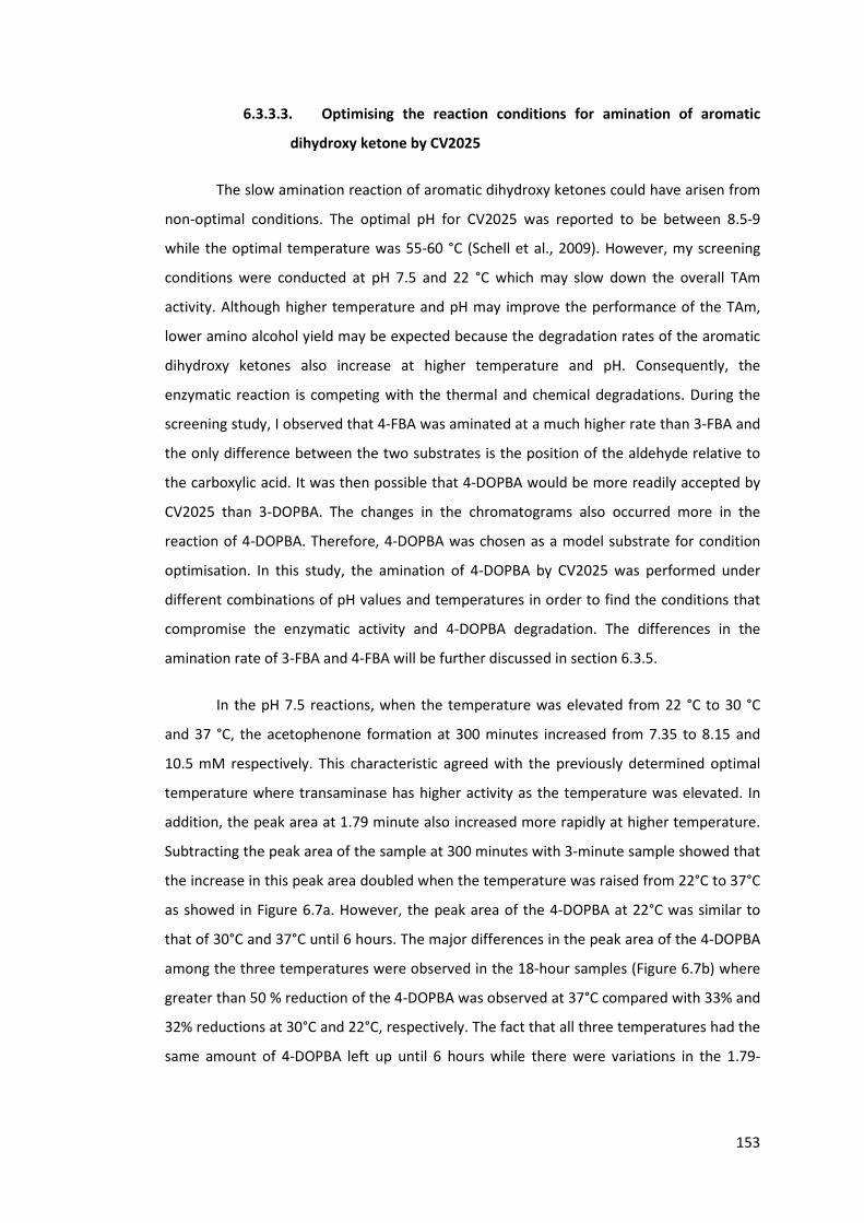
153
6.3.3.3. Optimising the reaction conditions for amination of aromatic
dihydroxy ketone by CV2025
The slow amination reaction of aromatic dihydroxy ketones could have arisen from
non-optimal conditions. The optimal pH for CV2025 was reported to be between 8.5-9
while the optimal temperature was 55-60 °C (Schell et al., 2009). However, my screening
conditions were conducted at pH 7.5 and 22 °C which may slow down the overall TAm
activity. Although higher temperature and pH may improve the performance of the TAm,
lower amino alcohol yield may be expected because the degradation rates of the aromatic
dihydroxy ketones also increase at higher temperature and pH. Consequently, the
enzymatic reaction is competing with the thermal and chemical degradations. During the
screening study, I observed that 4-FBA was aminated at a much higher rate than 3-FBA and
the only difference between the two substrates is the position of the aldehyde relative to
the carboxylic acid. It was then possible that 4-DOPBA would be more readily accepted by
CV2025 than 3-DOPBA. The changes in the chromatograms also occurred more in the
reaction of 4-DOPBA. Therefore, 4-DOPBA was chosen as a model substrate for condition
optimisation. In this study, the amination of 4-DOPBA by CV2025 was performed under
different combinations of pH values and temperatures in order to find the conditions that
compromise the enzymatic activity and 4-DOPBA degradation. The differences in the
amination rate of 3-FBA and 4-FBA will be further discussed in section 6.3.5.
In the pH 7.5 reactions, when the temperature was elevated from 22 °C to 30 °C
and 37 °C, the acetophenone formation at 300 minutes increased from 7.35 to 8.15 and
10.5 mM respectively. This characteristic agreed with the previously determined optimal
temperature where transaminase has higher activity as the temperature was elevated. In
addition, the peak area at 1.79 minute also increased more rapidly at higher temperature.
Subtracting the peak area of the sample at 300 minutes with 3-minute sample showed that
the increase in this peak area doubled when the temperature was raised from 22°C to 37°C
as showed in Figure 6.7a. However, the peak area of the 4-DOPBA at 22°C was similar to
that of 30°C and 37°C until 6 hours. The major differences in the peak area of the 4-DOPBA
among the three temperatures were observed in the 18-hour samples (Figure 6.7b) where
greater than 50 % reduction of the 4-DOPBA was observed at 37°C compared with 33% and
32% reductions at 30°C and 22°C, respectively. The fact that all three temperatures had the
same amount of 4-DOPBA left up until 6 hours while there were variations in the 1.79-
154
minute peaks suggested that the disappearance of the 4-DOPBA at 18 hours may not be
due to the enzymatic reaction.
Figure 6.7 Comparison of the peak areas of different compounds at different temperatures, pH 7.5.
a) at 300 minutes and b) at 18 hours. � 1.79 min peak increased from 3 min, �rearranged compound (see
Scheme 6.2) (250 nm detection), � remaining 4-DOPBA.
At pH 8.5 and 45 °C, both rearranged product and the 1.79-minute peak increased
at higher proportion than the reaction at 37 °C with pH 7.5. However, the changes in 4-
DOPBA concentrations up until 6 hours were similar at all temperatures. All the results so
far suggested that it is unlikely that a further increase in the pH and temperature would
promote the amination of 4-DOPBA by CV2025. However, the degradation of 4-DOPBA will
become the major concern. Since none of the conditions appeared to increase the
consumption of 4-DOPBA, it is likely that CV2025 cannot accept 4-DOPBA, and possibly 3-
DOPBA.
In order to confirm whether the 1.79-minute peak is the amino alcohol, several
control reactions were conducted. Changing the reaction buffer from Tris to HEPES still
produced this peak in a similar proportion which suggested that the formation of this peak
is independent to the buffer. When CV2025 clarified lysate was incubated with MBA
without any external source of amino acceptor, this peak still became larger and this
suggested that this peak cannot be the amino alcohol. In addition, when MBA was not
added to the clarified lysate CV2025, the peak area still increased (Figure 6.8). However,
this peak was not changed when usingonly his-tag purified CV2025 (Figure 6.9). All the
evidence above suggested that cellular metabolites were the cause of the side reactions.

155
Figure 6.8 The overlapped chromatograms of CV2025 lysate when incubated with 50 mM Tris buffer, pH 7.5
with 0.4 mM PLP, 0.08 mM ThDP, and 0.3 MgCl2 and at 30°C for (����) 1 minute, (����) 3 hours, and (����) 18 hours.
Figure 6.9 The overlapped chromatograms of purified CV2025 when incubated with 50 mM Tris buffer, pH 7.5
with 0.4 mM PLP, 0.08 mM ThDP, and 0.3 MgCl2 and at 30°C for (����) 1 minute, (����) 1 hour, and (����) 2 hours.
6.3.4. Competitive reaction
All my evidence so far suggested that CV2025 accepted neither 3-DOPBA nor 4-
DOPBA which led to a speculation whether both aromatic dihydroxy ketones could bind
into the CV2025 active site in the first place. Since both 3-DOPBA and 4-DOPBA contain an
aromatic ring and carboxylic group, one of these moieties may facilitate their binding as
observed in the substrate spectrum of CV2025 (Kaulmann et al., 2007). In order to confirm
whether 4-DOPBA could access into the active site of CV2025, a competitive reaction was
performed. Here, the rates of 4-FBA amination were compared under the presence and
absence of 4-DOPBA. If 4-DOPBA can access into the active site, it will slow down the rate
of 4-FBA amination. In order to investigate the possibility that other components in the TK
0.00 0.50 1.00 1.50 2.00 2.50 3.00 3.50 4.00 4.50 5.00 5.50 6.00 6.50 7.00 7.50 8.00 8.50 9.00 9.50-20
0
20
40
60
80
100
120
140
160
1 - 2012-08-21 TK TAm reaction in HEPES #18 [modified by General] C 1min UV_VIS_32 - 2012-08-21 TK TAm reaction in HEPES #33 C 3h UV_VIS_33 - 2012-08-21 TK TAm reaction in HEPES #45 C 20h UV_VIS_3mAU
min
321 0.520 - 0.0000.700 - 0.0030.827 - 0.0070.880 - 0.0031.067 - 0.001
1.647 - 16.7851.800 - 24.450
2.160 - 9.791
2.620 - 0.4383.053 - 0.1623.353 - 0.0153.480 - 0.0893.667 - 0.0063.887 - 0.0054.000 - 0.0884.140 - 0.152 4.867 - 0.0685.313 - 0.0025.580 - 0.0005.787 - 0.0276.027 - 0.0246.147 - 0.0226.247 - 0.0016.620 - 0.0006.947 - 1.6647.200 - 0.045 7.700 - 0.083
8.053 - 0.779
WVL:275 nm
0.00 0.50 1.00 1.50 2.00 2.50 3.00 3.50 4.00 4.50 5.00 5.50 6.00 6.50 7.00 7.50 8.00 8.50 9.00 9.50-20
0
20
40
60
80
100
120
140
160
1 - 2012-08-21 TK TAm reaction in HEPES #55 H 1min UV_VIS_22 - 2012-08-21 TK TAm reaction in HEPES #58 H 1h UV_VIS_23 - 2012-08-21 TK TAm reaction in HEPES #64 H 2h UV_VIS_2mAU
min
321 0.907 - 0.0001.020 - 0.002
1.620 - 33.406
4.147 - 0.0014.267 - 0.0004.587 - 0.0014.687 - 0.0004.793 - 0.0005.013 - 0.0005.220 - 0.0255.620 - 0.0005.693 - 0.0005.780 - 0.0006.140 - 0.0006.413 - 0.0016.693 - 0.0007.000 - 0.377 7.587 - 0.0007.780 - 0.0007.993 - 0.0298.187 - 0.0018.240 - 0.0018.560 - 0.0008.593 - 0.0008.667 - 0.0008.887 - 0.0129.113 - 0.0009.147 - 0.000
WVL:250 nm

156
reaction may inhibit CV2025 activity, one of the control reactions was set up by adding TK
lysate and ThDP to the TAm reaction as showed in Table 6.1.
Table 6.1 The initial rate of 4-FBA amination under different conditions.
Reaction composition Initial rate mM min-1
4-FBA, 4-DOPBA, ThDP, TK 0.299
4-FBA, TK, ThDP 0.316
4-FBA 0.414
All the reactions were performed at 22°C , 40 mM MBA, 10 mM 4-FBA. The concentrations of ThDP in the first
two reactions are 1.07 mM.
This competitive reaction showed that the rate of CV2025 was slowed down by
24% when TK lysate and ThDP were added alone. The presence of 4-DOPBA, however, did
not significantly further reduce the rate of 4-FBA amination. These results suggested that
the 4-DOPBA could not bind well in the active site of CV2025. Although ω-TAms may have
dual substrate recognitions which allow them to bind to both aromatic substrates and
alpha-keto acids (Cho et al., 2008; Shin and Kim, 2002), the presence of the carboxylic acid
and aromatic ring of 4-DOPBA did not facilitate the amination at the carbonyl group of 4-
DOPBA due to inaccessibility of the substrate itself. Since the competitive reaction between
3-FBA and 3-DOPBA was not performed, it cannot be ruled out that 3-DOPBA cannot access
into the active site. The causes of the inability to catalyse 3-DOPBA and 4-DOPBA may be
different due to their differences in the position of the carboxylic acid group. This was
suggested by the kinetic study of 3-FBA and 4-FBA in the next section.
The analysis of the amino donors and acceptors of ω transaminase from Vibrio
fluvialis JS17 suggested that the active site of ω transaminase consists of one large pocket
and one small pocket (Shin and Kim, 2002) and CV2025 also show similar patterns and most
dihydroxy ketones were poorly catalysed by CV2025 (Kaulmann et al., 2007). The initial rate
of erythrulose and DPP aminations which were followed by the rate of acetophenone
production suggested that erythrulose was aminated at approximately 4 times faster than
DPP. This implied that the presence of the aromatic side chain appeared to significantly
slow down the amination of the dihydroxy ketone in DPP (Kaulmann et al., 2007).
Therefore, the inability of CV2025 to accept 3-DOPBA and 4-DOPBA was not surprising.
Engineering to enlarge the active site is possible as illustrated in R-selective transaminase
ATA-117 where the both binding pockets were enlarged to be able to accommodate
prositagliptin (Savile et al., 2010).
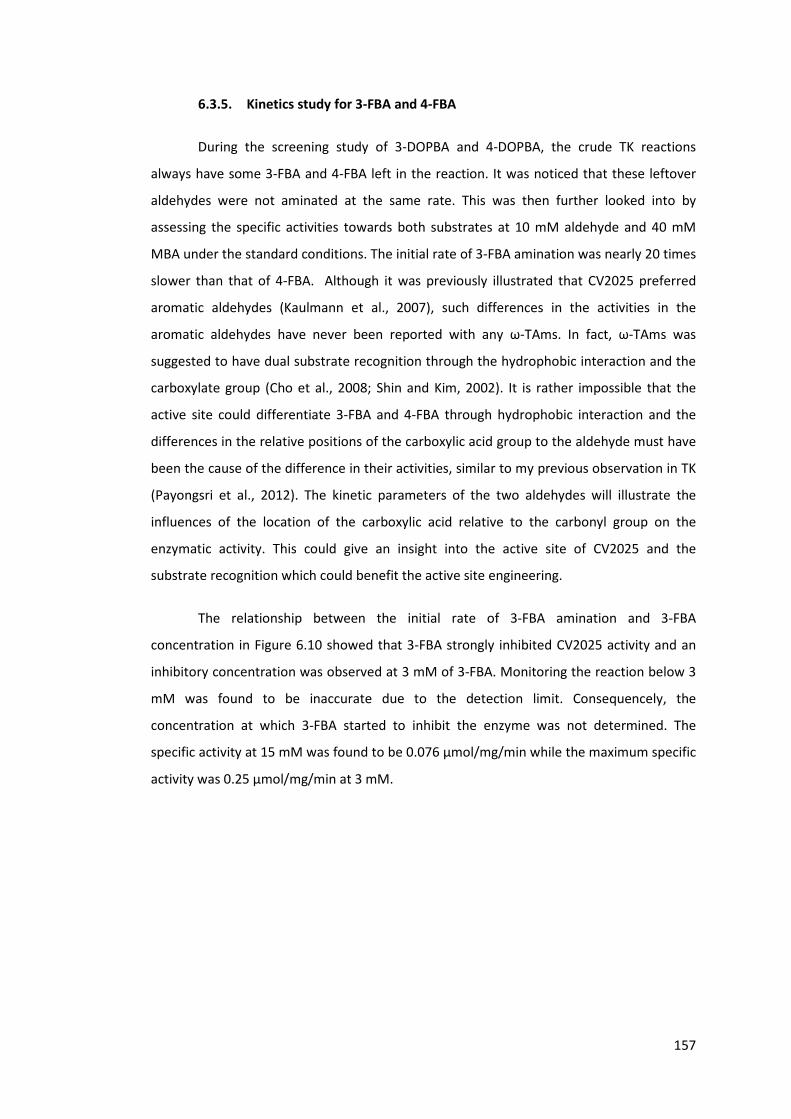
157
6.3.5. Kinetics study for 3-FBA and 4-FBA
During the screening study of 3-DOPBA and 4-DOPBA, the crude TK reactions
always have some 3-FBA and 4-FBA left in the reaction. It was noticed that these leftover
aldehydes were not aminated at the same rate. This was then further looked into by
assessing the specific activities towards both substrates at 10 mM aldehyde and 40 mM
MBA under the standard conditions. The initial rate of 3-FBA amination was nearly 20 times
slower than that of 4-FBA. Although it was previously illustrated that CV2025 preferred
aromatic aldehydes (Kaulmann et al., 2007), such differences in the activities in the
aromatic aldehydes have never been reported with any ω-TAms. In fact, ω-TAms was
suggested to have dual substrate recognition through the hydrophobic interaction and the
carboxylate group (Cho et al., 2008; Shin and Kim, 2002). It is rather impossible that the
active site could differentiate 3-FBA and 4-FBA through hydrophobic interaction and the
differences in the relative positions of the carboxylic acid group to the aldehyde must have
been the cause of the difference in their activities, similar to my previous observation in TK
(Payongsri et al., 2012). The kinetic parameters of the two aldehydes will illustrate the
influences of the location of the carboxylic acid relative to the carbonyl group on the
enzymatic activity. This could give an insight into the active site of CV2025 and the
substrate recognition which could benefit the active site engineering.
The relationship between the initial rate of 3-FBA amination and 3-FBA
concentration in Figure 6.10 showed that 3-FBA strongly inhibited CV2025 activity and an
inhibitory concentration was observed at 3 mM of 3-FBA. Monitoring the reaction below 3
mM was found to be inaccurate due to the detection limit. Consequencely, the
concentration at which 3-FBA started to inhibit the enzyme was not determined. The
specific activity at 15 mM was found to be 0.076 μmol/mg/min while the maximum specific
activity was 0.25 μmol/mg/min at 3 mM.

158
Figure 6.10 The relationship between the 3-FBA concentration and the initial rate of 3-FBA amination by
CV2025.
All substrates were prepared in 50 mM Tris buffer, pH 7.5. The MBA concentration was kept constant at 50 mM
and the enzyme concentration in the reaction was 0.18 mg/min.
In contrast, 4-FBA was not found to inhibit the reaction at 15 mM. However, the
results did not fit to a simple Michaelis-Menten curve either but rather a sigmoidal curve
seemed to fit the data instead (Figure 6.11). This sigmoidal curve suggested that there
could be allosteric control although both MBA and 4-FBA are non-natural substrates for
CV2025. The reaction at very low 4-FBA concentration was found to be very fast and the
full conversion was achieved instantly after the addition of MBA. Therefore, the initial rate
at the concentration lower than 3 mM was not studied. The maximum specific activity was
5.15 μmol/mg/min which makes the kcat become 4.4 s-1. This specific activity is much higher
than the activity of the best variant TK for 4-FBA which makes the true one-pot synthesis
rather improbable.
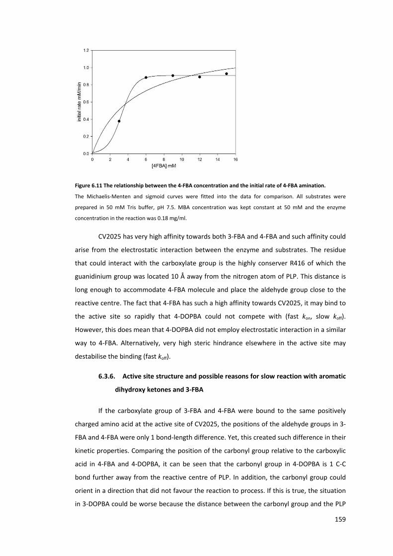
159
Figure 6.11 The relationship between the 4-FBA concentration and the initial rate of 4-FBA amination.
The Michaelis-Menten and sigmoid curves were fitted into the data for comparison. All substrates were
prepared in 50 mM Tris buffer, pH 7.5. MBA concentration was kept constant at 50 mM and the enzyme
concentration in the reaction was 0.18 mg/ml.
CV2025 has very high affinity towards both 3-FBA and 4-FBA and such affinity could
arise from the electrostatic interaction between the enzyme and substrates. The residue
that could interact with the carboxylate group is the highly conserver R416 of which the
guanidinium group was located 10 Å away from the nitrogen atom of PLP. This distance is
long enough to accommodate 4-FBA molecule and place the aldehyde group close to the
reactive centre. The fact that 4-FBA has such a high affinity towards CV2025, it may bind to
the active site so rapidly that 4-DOPBA could not compete with (fast kon, slow koff).
However, this does mean that 4-DOPBA did not employ electrostatic interaction in a similar
way to 4-FBA. Alternatively, very high steric hindrance elsewhere in the active site may
destabilise the binding (fast koff).
6.3.6. Active site structure and possible reasons for slow reaction with aromatic
dihydroxy ketones and 3-FBA
If the carboxylate group of 3-FBA and 4-FBA were bound to the same positively
charged amino acid at the active site of CV2025, the positions of the aldehyde groups in 3-
FBA and 4-FBA were only 1 bond-length difference. Yet, this created such difference in their
kinetic properties. Comparing the position of the carbonyl group relative to the carboxylic
acid in 4-FBA and 4-DOPBA, it can be seen that the carbonyl group in 4-DOPBA is 1 C-C
bond further away from the reactive centre of PLP. In addition, the carbonyl group could
orient in a direction that did not favour the reaction to process. If this is true, the situation
in 3-DOPBA could be worse because the distance between the carbonyl group and the PLP

160
reactive centre would be even further. The fact that DPP could be accepted by CV2025 also
supports this hypothesis. The lack of the carboxylic acid group on DPPwould allow this
molecule to move more freely within the active site and increase the chance of amination
at the carbonyl group of DPP.
However, our competitive reaction illustrated that 4-DOPBA, and possibly 3-
DOPBA, may not be able to access into the active site of CV2025. Therefore, there could be
some other steric hindrance upon the binding of both compounds. In fact, the crystal
structure of CV2025 (PDB ID: 4A6T) (Humble et al., 2012) showed that the active site was a
narrow channel mainly consisting of aromatic amino acids as showed in Figure 6.12a and b.
The aromatic rings of two amino acids Y153 and W60 were sandwiched and create a small
channel which was suggested to be important in the stereoselective control of ω-TAms
(Shin and Kim, 2002). This was confirmed by the ability to enhance the stereoselectivity by
a single mutation at W60 (Humble et al., 2012). The width of this channel due to the
sandwich packing between Y153 and W60 was 7.56 Å. A 90°-rotation view showed that
A231 and F88 constricted the active site which was due to the fact that their side chains
were protruding perpendicularly to the planes of T153 and W60. However, the distance
between A231 and F88 was approximately 8.64 Å which was large enough to allow
erythrulose and other molecules with dihydroxy ketone moiety to reach PLP, including DPP.
These 4 amino acids could be the target for site directed mutagenesis study to improve the
enzymatic activity towards aromatic dihydroxy ketones. However, a few residues may need
to be mutated simultaneously. In addition, the kinetic study of 3-FBA and 4-FBA illustrated
that the position of the carbonyl group is also crucial. Since TK extends 1 additional bond
from the carbonyl group, repositioning the whole aromatic dihydroxy ketone may need to
be considered to allow the carbonyl group to be places at close proximity to the reactive
centre.

161
Figure 6.12 The active site channel of CV2025 (PDB ID: 4A6T). The channel was illustrated as the surface of
amino acids constituting the active site.
a) the side view image and b) the 90°-rotated image of a). The PLP was showed in green stick and catalytic
Lys288 (as showed in Figure 6.1) was showed in magenta stick.
6.4. Conclusion
The enzymatic synthesis of 3-DOPBA and 4-DOPBA has been optimised by addition
of HPA at a certain time after the reaction started. The conversion yield can be up to 90 %
and 70% yields for 3-FBA and 4-FBA respectively. However, CV2025 did not appear to be
able to aminate either 3-DOPBA or 4-DOPBA to form aromatic amino alcohols. The
competitive reaction on 4-DOPBA suggested that this was due to inaccessibility to the
active site of 4-DOPBA itself. Some residues were suggested to be the target for
mutagenesis.

162
7. Conclusion and future research recommendation
The novel synthetic pathway of aminoalcohol by transketolase and transaminase
was proposed to be a more effective and efficient synthetic pathway than the traditional
organic synthetic routes. In the past, glycolaldehyde and propionaldehyde were extensively
studied in the above enzymatic system. When using different aromatic aldehydes,
derivatives of chloramphenicol can also be synthesised. However, only benzaldehyde has
ever been used as a substrate for certain transketolase mutants and the yield of 1,3-
dihydroxy-1-phenylpropane-2-one was far too low to be used as a substrate for
transaminase reaction and only the synthetic compound was ever used. Since the yield and
activity of transketolase with aromatic aldehyde was the bottleneck, it can limit the
application of this novel pathway to just aliphatic aldehydes.
In order to expand the application of this novel pathway, the overall goal of this
research was to construct a small library of transketolase mutants with higher activities
towards different aromatic aldehydes in order to synthesise novel aromatic dihydroxy
ketones. The aromatic dihydroxy ketones were then fed into the transaminase reaction to
produce aromatic aminoalcohols which can be used as a building block for chloramphenicol
derivatives. The stability of the accumulation of mutation was also considered because
accumulation of mutations usually destabilise the protein and lead to aggregate formation
and lower enzyme titre. Therefore, the desired parental mutant must have high activity as
well as stability.
During this research, the quantification of the enzyme concentration in the lysate
(densitometry) was revisited and redeveloped together with the reaction monitoring
procedure in order to achieve reliable and repeatable specific activity of the enzyme. The
degradation of hydroxypyruvate by transketolase mutants and a number of side reactions
were observed during this research. They were speculated to compete with the ketol
transfer reaction which also resulted in lower final yields if the specific activity of the
reaction is low. For a slow to moderate reaction rate, hydroxypyruvate degradation has a
more pronounced effect on the yield. As a result, the improved yields can be used as an
indicator for enhanced activity. The final yield could still be further improved by the fed-
batch regime of hydroxypyruvate. These side reactions required a new screening method
that is more accurate and specific than the colorimetric assay and HPLC was inevitably
developed as a tool although it has lower throughput. This methodology was shown to be
compatible for screening whole cell biocatalyst.

163
In the previous studies (Hibbert et al., 2008, 2007), several singletransketolase
mutants were found to have higher activities towards glycolaldehyde and propionaldehyde.
In the attempt to further improve their activities, two transketolase libraries were created.
Random combination of active mutants was showed to be ineffective to produce highly
active mutants but it led to unstable enzymes. In contrast, considering co-evolved residues
and finding the right pair to be simultaneously replaced has showed to stabilise the enzyme
as well as maintaining the activity.H26 and H461 were showed to be inappropriate target
because their mutations drastically reduced the enzyme stability and the overall enzymatic
activities for both substrates.
Prior to constructing the new transketolase library for aromatic aldehydes, the
available transketolase crystal structures were analysed and a rational design approach was
employed to find the mutants that accepted aromatic aldehyde. The use of natural
substrate analogues, namely 3-formylbenzoic acid (3-FBA) and 4-formylbenzoic acid (4-
FBA), has showed that D469T was the best mutant and it should be the template for
further protein engineering. The enzyme-substrate affinity and the substrate orientation
within the active sites were the two key factors controlling the bioconversion rate of the
aromatic aldehydes. Computational docking of both substrates into D469T and
D469T/R520Q suggested that the carboxylate group on both substrates interacts with the
positively charged residues that bind to the phosphate group of natural substrates.
The study of a pair of co-evolved residues in Chapter 3 suggested that
D469T/R520Q is more stable than D469T. Site-saturation mutagenesis at the residues
within the first shell produced several triple mutants with higher activities and yields
towards 3-FBA, 4-FBA, and 3-hydroxybenzaldehyde. The kinetic study of these mutants
illustrated that the kcat of each mutant differ from one aldehyde to another which implied
that these aromatic aldehyde with different substituents bind to the active site in different
orientations.At this stage, the overall goal of the research was reached.
The optimised 3-FBA and 4-FBA reactions were fed into the transaminase reaction
in order to synthesise novel aromatic dihydroxy ketones in Chapter 6. Unfortunately, none
of the available omega transaminases including CV2025 appeared to accept any newly
synthesised aromatic dihydroxy ketones. The bottleneck for the transketolase-
transaminase pathway was identified as the inability of transaminase to accept the
aromatic dihydroxy ketone with ring substituents.

164
The current outcomes have showed the potential and also new obstacles of the
transketolase-transaminase reaction and they have to be carefully evaluated for the
research direction.
7.1. Future research recommendation for transketolase
David Steadman (Chemistry Department) and I have explored several additional
aromatic aldehydes with 3-substituents. However, the 4-substituents have not been fully
researched and very few mutants have rather moderate activities for 4-FBA. For an
immediate plan, aromatic aldehydes with 4-substituents could be further explored and the
S385X/D469T/R520Q library can still be a good starting point. Since most of the mutants
have relatively low kcat of the 4-FBA, it is still possible that additional mutations can further
improve the activity of the enzyme. In addition, other D469 variants could be investigated
even the D469Y mutant. Mutation at this residue has been showed to alter the
stereoselectivity as well as improving the activity for propionaldehyde before (Smith et al.,
2008) but several other residues may need to be simultaneously mutated in order to allow
D469Y to accept aromatic aldehydes. Besides the research in engineering transketolase,
the enzymatic synthesis 3-(1,3-Dihydroxy-2-oxopropyl )benzoic acid (3-DOPBA) and 4-(1,3-
Dihydroxy-2-oxopropyl )benzoic acid (4-DOPBA) may be used as a substrate in traditional
chemical synthesis to produce two different chloramphenicol analogues and tested their
antibacterial properties.
It should be noted that most of the screening methods only quantify the product
concentration while the stereoselectivity of the mutants cannot be immediately
determined. It should be noted that a parallel study by David Steadman illustrated that the
absolute stereoselectivity depends on the substituent and the highly active mutants appear
to have S selective for most aldehydes with the 3- substituent. A different strategy for
designing transketolase library will need to be considered to alter the stereoselectivity.
Amino acid addition or deletion may be considered to alter the position of the acid/base
catalysts relative to the aldehyde. The availability of the crystal structure of the mutants
especially the substrate-bound structures would significantly facilitate the next library
design and computational docking of other substrates.
Most of the neutral-charged aromatic aldehydes are poorly soluble in aqueous
solution while the KM values for these aromatic aldehydes are likely to be large. If the
apparent aldehyde concentration is higher, possibly with the aid of co-solvent, the

165
bioconversion rate could be further enhanced. However, this strategy requires
transketolase and transaminase mutants that well tolerate organic solvents. Certain
mutants have been studied and shown to meet the requirement and they can be
cooperated into the highly active mutants for this purpose. The bioconversion rate is a
critical parameter because it has a direct impact on the productivity and the overall
production cost which determines the economic viability of the process.
7.2. Future research recommendation for transketolase-transaminase pathway for
the synthesis of aromatic aminoalcohols
The experiment in Chapter 6 illustrated that the major bottleneck of the
transketolase-transaminase pathway is the transaminase reaction. This is because none of
the available transaminases appeared to accept aromatic dihydroxy ketones with ring
substituent and most of the omega transaminases are also S selective. However, some R-
selective transaminases are commercially available and they should be tested with 3-
DOPBA and 4-DOPBA to assess the process feasibility and other challenges.
The equilibrium is still one of the major issues for most transaminase reactions
especially when cheaper amino donors such as isopropylamine or amino acids are to be
used and this issue makes the overall yield very low. To overcome this issue, an engineering
strategy such as product removal is required to overcome this issue. However, the final
yield, production rate, the overall production cost and its commercial value has to be
evaluated since the final product is still cheaply available.
7.3. Overall evaluation and recommendation
The research at this stage suggested that the synthesis of chloramphenicol and its
analogues by transketolase-transaminase cascade is unlikely to be feasible. For economic
evaluation, the cost of starting materials from both reaction especially hydroxypyruvic acid
and (S)-methylbenzylamine is still higher than the selling price of chloramphenicol. To
minimise the reagent cost, an alternative substrate, isopropylamine, could be used. In
addition, the yield of the process would be very low due to the equilibrium position of the
transaminase reaction and this could make the final product uneconomically viable. Using
excess isopropylamine and the removal of acetone which is the product of isopropylamine
deaminationby applying negative pressure, the reaction could be driven to completion.
Nonetheless, this research has its own merit. This thesis unravelled the key mechanism of
transketolase for non-natural substrates which can give further insight into the

166
understanding of the substrate specificity of the enzymes within the same family. Other
enzyme within the same family may be considered such as pyruvate decarboxylase which
already gives R selective for the ligation between pyruvate and benzaldehyde (Sprenger
and Pohl, 1999).

167
8. References
Arnold, F.H., 1998. Design by Directed Evolution. Accounts of Chemical Research 31, 125–131.
Arnold, F.H., Wintrode, P.L., Miyazaki, K., Gershenson, A., 2001. How enzymes adapt: lessons from directed evolution. Trends in biochemical sciences 26, 100–6.
Asztalos, P., Parthier, C., Golbik, R., Kleinschmidt, M., Hübner, G., Weiss, M.S., Friedemann, R., Wille, G., Tittmann, K., 2007. Strain and near attack conformers in enzymic thiamin catalysis: X-ray crystallographic snapshots of bacterial transketolase in covalent complex with donor ketoses xylulose 5-phosphate and fructose 6-phosphate, and in noncovalent complex with acceptor aldo. Biochemistry 46, 12037–52.
Aucamp, J.P., Martinez-Torres, R.J., Hibbert, E.G., Dalby, P.A., 2008. A microplate-based evaluation of complex denaturation pathways: structural stability of Escherichia coli transketolase. Biotechnology and bioengineering 99, 1303–10.
Behrens, G.A., Hummel, A., Padhi, S.K., Schätzle, S., Bornscheuer, U.T., 2011. Discovery and Protein Engineering of Biocatalysts for Organic Synthesis. Advanced Synthesis & Catalysis 353, 2191–2215.
Bloom, J.D., Labthavikul, S.T., Otey, C.R., Arnold, F.H., 2006. Protein stability promotes evolvability. Proceedings of the National Academy of Sciences of the United States of America 103, 5869–74.
Bolon, D.N., Voigt, C. a, Mayo, S.L., 2002. De novo design of biocatalysts. Current opinion in chemical biology 6, 125–9.
Bolte, J., Demuynck, C., Samaki, H., 1987. Utilization of enzymes in organic chemistry: Transketolase catalyzed synthesis of ketoses. Tetrahedron Letters 28, 5525–5528.
Bornscheuer, U.T., Pohl, M., 2001. Improved biocatalysts by directed evolution and rational protein design. Current opinion in chemical biology 5, 137–43.
Bougioukou, D., Kille, S., Taglieber, A., Reetz, M., 2009. Directed Evolution of an Enantioselective Enoate-Reductase: Testing the Utility of Iterative Saturation Mutagenesis. Advanced Synthesis & Catalysis 351, 3287–3305.
Breslow, R., 1957. RAPID DEUTERIUM EXCHANGE IN THIAZOLIUM SALTS 1. Journal of the American Chemical Society 79, 1762–1763.
Breslow, R., 1958. On the Mechanism of Thiamine Action. IV. 1 Evidence from Studies on Model Systems. Journal of the American Chemical Society 80, 3719–3726.
Breuer, M., Ditrich, K., Habicher, T., Hauer, B., Kesseler, M., Stürmer, R., Zelinski, T., 2004. Industrial methods for the production of optically active intermediates. Angewandte Chemie (International ed. in English) 43, 788–824.
168
Breuer, M., Hauer, B., 2003. Carbon–carbon coupling in biotransformation. Current Opinion in Biotechnology 14, 570–576.
Brown, N.G., Pennington, J.M., Huang, W., Ayvaz, T., Palzkill, T., 2010. Multiple global suppressors of protein stability defects facilitate the evolution of extended-spectrum TEM β-lactamases. Journal of molecular biology 404, 832–46.
Cadwell, R.C., Joyce, G.F., 1992. Randomization of genes by PCR mutagenesis. Genome Research 2, 28–33.
Calloni, G., Zoffoli, S., Stefani, M., Dobson, C.M., Chiti, F., 2005. Investigating the effects of mutations on protein aggregation in the cell. The Journal of biological chemistry 280, 10607–13.
Cázares, A., Galman, J.L., Crago, L.G., Smith, M.E.B., Strafford, J., Ríos-Solís, L., Lye, G.J., Dalby, P.A., Hailes, H.C., 2010. Non-alpha-hydroxylated aldehydes with evolved transketolase enzymes. Organic & biomolecular chemistry 8, 1301–9.
Ceccarelli, E.A., Carrillo, N., Roveri, O.A., 2008. Efficiency function for comparing catalytic competence. Trends in biotechnology 26, 117–8.
Cedrone, F., Ménez, A, Quéméneur, E., 2000. Tailoring new enzyme functions by rational redesign. Current opinion in structural biology 10, 405–10.
Chen, B.H., Sayar, A., Kaulmann, U., Dalby, P.A., Ward, J.M., Woodley, J.M., 2006. Reaction modelling and simulation to assess the integrated use of transketolase and ω-transaminase for the synthesis of an aminotriol. Biocatalysis and Biotransformation 24, 449–457.
Chen, Z., Zhao, H., 2005. Rapid creation of a novel protein function by in vitro coevolution. Journal of molecular biology 348, 1273–82.
Chica, R.A., Doucet, N., Pelletier, J.N., 2005. Semi-rational approaches to engineering enzyme activity: combining the benefits of directed evolution and rational design. Current opinion in biotechnology 16, 378–84.
Cho, B.K., Park, H.Y., Seo, J.H., Kim, J., Kang, T.J., Lee, B.S., Kim, B.G., 2008. Redesigning the substrate specificity of omega-aminotransferase for the kinetic resolution of aliphatic chiral amines. Biotechnology and bioengineering 99, 275–84.
Controulis, J., Rebstock, M.C., Crook, H.M., 1949. Chloramphenicol (chloromycetin). Journal of the American Chemical Society 71, 2463–2468.
Corbett, M.D., Corbett, B.R., 1986. Effect of ring substituents on the transketolase-catalyzed conversion of nitroso aromatics to hydroxamic acids. Biochemical pharmacology 35, 3613–21.
Costelloe, S.J., Ward, J.M., Dalby, P.A., 2008. Evolutionary analysis of the TPP-dependent enzyme family. Journal of molecular evolution 66, 36–49.

169
Dalby, P.A., 2003. Optimising enzyme function by directed evolution. Current Opinion in Structural Biology 13, 500–505.
Dalby, P.A., 2007. Engineering enzymes for biocatalysis. Recent patents on biotechnology 1, 1–9.
Dalby, P.A., 2011. Strategy and success for the directed evolution of enzymes. Current opinion in structural biology 21, 473–80.
Dalby, P.A., Aucamp, J.P., George, R., Martinez-Torres, R.J., 2007. Structural stability of an enzyme biocatalyst. Biochemical Society transactions 35, 1606–9.
Delihas, N., Forst, S., 2001. MicF: an antisense RNA gene involved in response of Escherichia coli to global stress factors. Journal of molecular biology 313, 1–12.
Demuynck, C., Bolte, J., Hecquet, L., Dalmas, V., 1991. Enzyme-catalyzed synthesis of carbohydrates: synthetic potential of transketolase. Tetrahedron Letters 32, 5085–5088.
Demuynck, C., Bolte, J., Hecquet, L., Samaki, H., 1990. Enzymes as reagents in organic chemistry: transketolase-catalysed synthesis of d-[1,2-13C2]xylulose. Carbohydrate Research 206, 79–85.
Drummond, D.A., Iverson, B.L., Georgiou, G., Arnold, F.H., 2005. Why high-error-rate random mutagenesis libraries are enriched in functional and improved proteins. Journal of molecular biology 350, 806–16.
Eisenthal, R., Danson, M.J., Hough, D.W., 2007. Catalytic efficiency and kcat/KM: a useful comparator? Trends in biotechnology 25, 247–9.
Enders, D., Narine, A.A., 2008. Lessons from nature: biomimetic organocatalytic carbon-carbon bond formations. The Journal of organic chemistry 73, 7857–70.
Enders, D., Voith, M., Lenzen, A., 2005. The dihydroxyacetone unit--a versatile C(3) building block in organic synthesis. Angewandte Chemie (International ed. in English) 44, 1304–25.
Esakova, O.A., Meshalkina, L.E., Kochetov, G.A., 2005. Effects of transketolase cofactors on its conformation and stability. Life sciences 78, 8–13.
Esakova, O.A., Meshalkina, L.E., Kochetov, G.A., Golbik, R., 2009. Halogenated pyruvate derivatives as substrates of transketolase from Saccharomyces cerevisiae. Biochemistry (Moscow) 74, 1234–1238.
Fessner, W.D., 1998. Enzyme mediated C-C bond formation. Current opinion in chemical biology 2, 85–97.
Fiedler, E., Golbik, R., Schneider, G., Tittmann, K., Neef, H., König, S., Hübner, G., 2001. Examination of donor substrate conversion in yeast transketolase. The Journal of biological chemistry 276, 16051–8.

170
Fox, R.J., Clay, M.D., 2009. Catalytic effectiveness, a measure of enzyme proficiency for industrial applications. Trends in biotechnology 27, 137–40.
Frank, R.A.W., Titman, C.M., Pratap, J.V., Luisi, B.F., Perham, R.N., 2004. A molecular switch and proton wire synchronize the active sites in thiamine enzymes. Science 306, 872–6.
French, C., Ward, J.M., 1995. Improved production and stability ofE. coli recombinants expressing transketolase for large scale biotransformation. Biotechnology Letters17, 247–252.
Fry, K., Ingraham, L.L., Westheimer, F.H., 1957. The Thiamin-Pyruvate Reaction. Journal of the American Chemical Society 79, 5225–5227.
Galman, J.L., Steadman, D., Bacon, S., Morris, P., Smith, M.E.B., Ward, J.M., Dalby, P.A., Hailes, H.C., 2010. α,α’-Dihydroxyketone formation using aromatic and heteroaromatic aldehydes with evolved transketolase enzymes.Chemical communications (Cambridge, England) 46, 7608–10.
Galman, J.L., Steadman, D., Haigh, L.D., Hailes, H.C., 2012. Investigating the reaction mechanism and organocatalytic synthesis of α,α’-dihydroxy ketones. Organic & biomolecular chemistry 10, 2621–8.
Glasner, M.E., Gerlt, J. a, Babbitt, P.C., 2006. Evolution of enzyme superfamilies.Current opinion in chemical biology 10, 492–7.
Göbel, U., Sander, C., Schneider, R., Valencia, A., 1994. Correlated mutations and residue contacts in proteins. Proteins 18, 309–17.
Hailes, H., Dalby, P.A., Lye, G., Ward, J., 2009. Biocatalytic approaches to ketodiols and aminodiols. Chimica Oggi Chemistry Today 27, 28–31.
Hailes, H.C., Dalby, P.A., Lye, G., Baganz, F., Micheletti, M., Szita, N., Ward, J., 2010. alpha,alpha ’-Dihydroxy Ketones and 2-Amino-1,3-diols: Synthetic and Process Strategies Using Biocatalysts. Current Organic Chemistry 14, 1883–1893.
Hailes, Helen C., Dalby, P.A., Lye, G.J., Ward, J.M., 2010. ChemInform Abstract: Biocatalytic Approaches to Ketodiols and Aminodiols. ChemInform 41, no–no.
Hawkins, C.F., Borges, A., Perham, R.N., 1989. A common structural motif in thiamin pyrophosphate-binding enzymes. FEBS letters 255, 77–82.
Hayes, R.J., Bentzien, J., Ary, M.L., Hwang, M.Y., Jacinto, J.M., Vielmetter, J., Kundu, A., Dahiyat, B.I., 2002. Combining computational and experimental screening for rapid optimization of protein properties. Proceedings of the National Academy of Sciences of the United States of America. 99, 15926–31.
Hecquet, L., Bolte, J., Demuynck, C., 1994. Chemoenzymatic synthesis of 6-deoxy-D-fructose and 6-deoxy-L-sorbose using transketolase. Tetrahedron 50, 8677–8684.
Hecquet, L., Bolte, J., Demuynck, C., 1996. Enzymatic synthesis of “natural-labeled” 6-deoxy-L-sorbose precursor of an important food flavor. Tetrahedron 52, 8223–8232.

171
Hibbert, E.G., Senussi, T., Costelloe, S.J., Lei, W., Smith, M.E.B., Ward, J.M., Hailes, H.C., Dalby, P.A., 2007. Directed evolution of transketolase activity on non-phosphorylated substrates. Journal of biotechnology 131, 425–32.
Hibbert, E.G., Senussi, T., Smith, M.E.B., Costelloe, S.J., Ward, J.M., Hailes, H.C., Dalby, P.A., 2008. Directed evolution of transketolase substrate specificity towards an aliphatic aldehyde.Journal of biotechnology 134, 240–5.
Hobbs, G.R., Lilly, M.D., Turner, N.J., Ward, J.M., Willets, A.J., Woodley, J.M., 1993. Enzyme-catalysed carbon?carbon bond formation: use of transketolase from Escherichia coli. Journal of the Chemical Society, Perkin Transactions 1 165.
Höhne, M., Bornscheuer, U.T., 2009. Biocatalytic Routes to Optically Active Amines. ChemCatChem 1, 42–51.
Hübner, G., Tittmann, K., Killenberg-Jabs, M., Schäffner, J., Spinka, M., Neef, H., Kern, D., Kern, G., Schneider, G., Wikner, C., Ghisla, S., 1998. Activation of thiamin diphosphate in enzymes.Biochimica et biophysica acta 1385, 221–8.
Humble, Maria Svedendahl, Cassimjee, K.E., Abedi, V., Federsel, H.-J., Berglund, P., 2012. Key Amino Acid Residues for Reversed or Improved Enantiospecificity of an ω-Transaminase. ChemCatChem 4, 1167–1172.
Humble, Maria S, Cassimjee, K.E., Håkansson, M., Kimbung, Y.R., Walse, B., Abedi, V., Federsel, H.-J., Berglund, P., Logan, D.T., 2012. Crystal structures of the Chromobacterium violaceumω-transaminase reveal major structural rearrangements upon binding of coenzyme PLP. The FEBS journal 279, 779–92.
Humphrey, A.J., Parsons, S.F., Smith, M.E.., Turner, N.J., 2000. Synthesis of a novel N-hydroxypyrrolidine using enzyme catalysed asymmetric carbon–carbon bond synthesis. Tetrahedron Letters 41, 4481–4485.
Humphrey, A.J., Turner, N.J., McCague, R., Taylor, S.J.C., 1995. Synthesis of enantiomerically pure ?-hydroxyaldehydes from the corresponding ?-hydroxycarboxylic acids: novel substrates for Escherichia coli transketolase. Journal of the Chemical Society, Chemical Communications 2475.
Ingram, C.U., Bommer, M., Smith, M.E.B., Dalby, P.A., Ward, J.M., Hailes, H.C., Lye, G.J., 2007. One-pot synthesis of amino-alcohols using a de-novo transketolase and beta-alanine: pyruvate transaminase pathway in Escherichia coli. Biotechnology and bioengineering 96, 559–69.
Ishige, T., Honda, K., Shimizu, S., 2005. Whole organism biocatalysis. Current opinion in chemical biology 9, 174–80.
Jahromi, R.R.F., Morris, P., Martinez-Torres, R.J., Dalby, P.A., 2011. Structural stability of E. coli transketolase to temperature and pH denaturation. Journal of biotechnology 155, 209–16.
Jover, J., Bosque, R., Sales, J., 2008. QSPR Prediction of pKa for Benzoic Acids in Different Solvents. QSAR & Combinatorial Science 27, 563–581.

172
Kaulmann, U., Smithies, K., Smith, M.E.B., Hailes, H.C., Ward, J.M., 2007. Substrate spectrum of ω-transaminase from Chromobacterium violaceum DSM30191 and its potential for biocatalysis. Enzyme and Microbial Technology 41, 628–637.
Kazlauskas, R.J., Bornscheuer, U.T., 2009. Finding better protein engineering strategies. Nature chemical biology 5, 526–9.
Kemp, D.S., O’Brien, J.T., 1970. Base catalysis of thiazolium salt hydrogen exchange and its implications for enzymic thiamine cofactor catalysis. Journal of the American Chemical Society 92, 2554–2555.
Kern, D., Kern, G., Neef, H., Tittmann, K., Killenberg-Jabs, M., Wikner, C., Schneider, G., Hübner, G., 1997. How thiamine diphosphate is activated in enzymes. Science 275, 67–70.
Kluger, R., Tittmann, K., 2008. Thiamin diphosphate catalysis: enzymic and nonenzymic covalent intermediates. Chemical reviews 108, 1797–833.
Kobori, Y., Myles, D.C., Whitesides, G.M., 1992. Substrate specificity and carbohydrate synthesis using transketolase. The Journal of Organic Chemistry 57, 5899–5907.
Kochetov, G.A., Sevostyanova, I.A., 2005. Binding of the coenzyme and formation of the transketolase active center. IUBMB Life 57, 491–7.
Koeller, K.M., Wong, C.H., 2001. Enzymes for chemical synthesis. Nature 409, 232–40.
Koszelewski, D., Tauber, K., Faber, K., Kroutil, W., 2010. omega-Transaminases for the synthesis of non-racemic alpha-chiral primary amines. Trends in biotechnology28, 324–32.
Kuchner, O., Arnold, F.H., 1997. Directed evolution of enzyme catalysts. Trends in biotechnology 15, 523–30.
Langenbeck, W., 1932. Fermentproblem und organische Katalyse. Angewandte Chemie 45, 97–99.
Lee, J., Goodey, N.M., 2011. Catalytic contributions from remote regions of enzyme structure.Chemical reviews 111, 7595–624.
Leemhuis, H., Kelly, R.M., Dijkhuizen, L., 2009. Directed evolution of enzymes: Library screening strategies. IUBMB Life 61, 222–8.
Lehmann, M., Wyss, M., 2001. Engineering proteins for thermostability: the use of sequence alignments versus rational design and directed evolution. Current opinion in biotechnology 12, 371–5.
Lindqvist, Y., Schneider, G., 1993. Thiamin diphosphate dependent enzymes: transketolase, pyruvate oxidase and pyruvate decarboxylase. Current Opinion in Structural Biology 3, 896–901.

173
Lockless, S.W., Ranganathan, R., 1999. Evolutionarily conserved pathways of energetic connectivity in protein families. Science 286, 295–9.
Lutz, S., 2010. Beyond directed evolution--semi-rational protein engineering and design. Current opinion in biotechnology 21, 734–43.
Lutz, S., Ostermeier, M., Moore, G.L., Maranas, C.D., Benkovic, S.J., 2001. Creating multiple-crossover DNA libraries independent of sequence identity. Proceedings of the National Academy of Sciences of the United States of America 98, 11248–53.
Machajewski, T., Wong, C., 2000. The Catalytic Asymmetric Aldol Reaction. Angewandte Chemie (International ed. in English) 39, 1352–1375.
Martinez-Torres, R.J., Aucamp, J.P., George, R., Dalby, P.A., 2007. Structural stability of E. coli transketolase to urea denaturation. Enzyme and Microbial Technology 41, 653–662.
Meshalkina, L., Nilsson, U., Wikner, C., Kostikowa, T., Schneider, G., 1997. Examination of the thiamin diphosphate binding site in yeast transketolase by site-directed mutagenesis. European journal of biochemistry / FEBS 244, 646–52.
Mildvan, A.S., Weber, D.J., Kuliopulos, A., 1992. Quantitative interpretations of double mutations of enzymes. Archives of biochemistry and biophysics 294, 327–40.
Miller, O.J., Hibbert, E.G., Ingram, C.U., Lye, G.J., Dalby, P.A., 2007. Optimisation and evaluation of a generic microplate-based HPLC screen for transketolase activity. Biotechnology letters 29, 1759–70.
Mizuhara, S., Handler, P., 1954. Mechanism of Thiamine-catalyzed Reactions. Journal of the American Chemical Society 76, 571–573.
Morley, K.L., Kazlauskas, R.J., 2005. Improving enzyme properties: when are closer mutations better? Trends in biotechnology 23, 231–7.
Morris, K.G., Smith, M.E.B., Turner, N.J., Lilly, M.D., Mitra, R.K., Woodley, J.M., 1996. Transketolase from Escherichia coli: A practical procedure for using the biocatalyst for asymmetric carbon-carbon bond synthesis. Tetrahedron: Asymmetry 7, 2185–2188.
Mutti, F.G., Fuchs, C.S., Pressnitz, D., Sattler, J.H., Kroutil, W., 2011. Stereoselectivity of Four (R)-Selective Transaminases for the Asymmetric Amination of Ketones.Advanced Synthesis & Catalysis 353, 3227–3233.
Myles, D.C., Andrulis, P.J., Whitesides, G.M., 1991. A transketolase-based synthesis of (+)-exo-brevicomin. Tetrahedron Letters 32, 4835–4838.
Nagabhushan, T., Miller, G.H., Varma, K.J., 2000. Chloramphenicol and Analogues, in: Kirk-Othmer Encyclopedia of Chemical Technology. John Wiley & Sons, Inc.
Neher, E., 1994. How frequent are correlated changes in families of protein sequences? Proceedings of the National Academy of Sciences of the United States of America 91, 98–102.

174
Nemeria, N.S., Chakraborty, S., Balakrishnan, A., Jordan, F., 2009. Reaction mechanisms of thiamin diphosphate enzymes: defining states of ionization and tautomerization of the cofactor at individual steps. The FEBS journal 276, 2432–2446.
Nikkola, M., Lindqvist, Y., Schneider, G., 1994. Refined structure of transketolase from Saccharomyces cerevisiae at 2.0 A resolution. Journal of molecular biology 3, 387-404.
Nilsson, U., Hecquet, L., Gefflaut, T., Guerard, C., Schneider, G., 1998. Asp477 is a determinant of the enantioselectivity in yeast transketolase. FEBS letters 424, 49–52.
Nilsson, U., Meshalkina, L., Lindqvist, Y., Schneider, G., 1997. Examination of substrate binding in thiamin diphosphate-dependent transketolase by protein crystallography and site-directed mutagenesis. The Journal of biological chemistry 272, 1864–1869.
Ostermeier, M., Shim, J.H., Benkovic, S.J., 1999. A combinatorial approach to hybrid enzymes independent of DNA homology. Nature biotechnology 17, 1205–9.
Otten, L.G., Hollmann, F., Arends, I.W.C.E., 2010. Enzyme engineering for enantioselectivity: from trial-and-error to rational design? Trends in biotechnology 28, 46–54.
Otten, L.G., Quax, W.J., 2005. Directed evolution: selecting today’s biocatalysts. Biomolecular engineering 22, 1–9.
Paramesvaran, J., Hibbert, E.G., Russell, A.J., Dalby, P.A., 2009. Distributions of enzyme residues yielding mutants with improved substrate specificities from two different directed evolution strategies. Protein engineering, design & selection : PEDS 22, 401–11.
Park, E.-S., Kim, M., Shin, J.-S., 2012. Molecular determinants for substrate selectivity of ω-transaminases. Applied microbiology and biotechnology 6, 2425-35.
Patel, R.N., 2008. Synthesis of chiral pharmaceutical intermediates by biocatalysis. Coordination Chemistry Reviews 252, 659–701.
Patrick, W.M., Firth, A.E., Blackburn, J.M., 2003. User-friendly algorithms for estimating completeness and diversity in randomized protein-encoding libraries. Protein engineering 6, 451-457
Pavelka, A., Chovancova, E., Damborsky, J., 2009. HotSpot Wizard: a web server for identification of hot spots in protein engineering. Nucleic acids research 37, W376–83.
Payongsri, P., Steadman, D., Strafford, J., MacMurray, A., Hailes, H.C., Dalby, P.A., 2012. Rational substrate and enzyme engineering of transketolase for aromatics. Organic & biomolecular chemistry 10, 9021–9.
Poole, A.M., Ranganathan, R., 2006. Knowledge-based potentials in protein design. Current opinion in structural biology 16, 508–13.
Reetz, M.T., Bocola, M., Carballeira, J.D., Zha, D., Vogel, A., 2005. Expanding the range of substrate acceptance of enzymes: combinatorial active-site saturation test. Angewandte Chemie (International ed. in English) 44, 4192–6.

175
Reetz, M.T., Carballeira, J.D., 2007. Iterative saturation mutagenesis (ISM) for rapid directed evolution of functional enzymes. Nature protocols 2, 891–903.
Resch, V., Schrittwieser, J.H., Siirola, E., Kroutil, W., 2011. Novel carbon-carbon bond formations for biocatalysis. Current opinion in biotechnology 22, 793–799.
Rios-Solis, L., Halim, M., Cázares, A., Morris, P., Ward, J.M., Hailes, H.C., Dalby, P.A., Baganz, F., Lye, G.J., 2011. A toolbox approach for the rapid evaluation of multi-step enzymatic syntheses comprising a “mix and match” E. coli expression system with microscale experimentation. Biocatalysis and Biotransformation 29, 192–203.
Robertson, D.E., Steer, B.A., 2004. Recent progress in biocatalyst discovery and optimization. Current opinion in chemical biology 8, 141–9.
Romero, P.A., Arnold, F.H., 2009. Exploring protein fitness landscapes by directed evolution. Nature reviews. Molecular cell biology 10, 866–76.
Röthlisberger, D., Khersonsky, O., Wollacott, A.M., Jiang, L., DeChancie, J., Betker, J., Gallaher, J.L., Althoff, E. a, Zanghellini, A., Dym, O., Albeck, S., Houk, K.N., Tawfik, D.S., Baker, D., 2008. Kemp elimination catalysts by computational enzyme design. Nature 453, 190–5.
Rudat, J., Brucher, B.R., Syldatk, C., 2012. Transaminases for the synthesis of enantiopure beta-amino acids. AMB Express 2, 11.
Samland, A.K., Sprenger, G. a, 2006. Microbial aldolases as C-C bonding enzymes--unknown treasures and new developments. Applied microbiology and biotechnology 71, 253–64.
Savile, C.K., Janey, J.M., Mundorff, E.C., Moore, J.C., Tam, S., Jarvis, W.R., Colbeck, J.C., Krebber, A., Fleitz, F.J., Brands, J., Devine, P.N., Huisman, G.W., Hughes, G.J., 2010. Biocatalytic Asymmetric Synthesis of Chiral Amines from Ketones Applied to Sitagliptin Manufacture. Science329, 305-309.
Schell, U., Wohlgemuth, R., Ward, J.M., 2009. Synthesis of pyridoxamine 5′-phosphate using an MBA:pyruvate transaminase as biocatalyst. Journal of Molecular Catalysis B: Enzymatic 59, 279–285.
Schellenberger, a, 1998. Sixty years of thiamin diphosphate biochemistry. Biochimica et biophysica acta1385, 177–86.
Schenk, G., Duggleby, R.G., Nixon, P.F., 1998. Properties and functions of the thiamin diphosphate dependent enzyme transketolase. The international journal of biochemistry & cell biology 30, 1297–318.
Schenk, G., Layfield, R., Candy, J.M., Duggleby, R.G., Nixon, P.F., 1997. Molecular evolutionary analysis of the thiamine-diphosphate-dependent enzyme, transketolase. Journal of molecular evolution 44, 552–72.
Schneider, G., Fiedler, E., Thorell, S., Sandalova, T., Golbik, R., Ko, S., 2002. Snapshot of a key intermediate in enzymatic thiamin catalysis : Crystal structure of the

176
alpha,betacarbanion of the active site of transketolase from Saccharomyces cerevisiae. Proceedings of the National Academy of Sciences of the United States of America 99, 591–595.
Schneider, G., Lindqvist, Y., 1998. Crystallography and mutagenesis of transketolase: mechanistic implications for enzymatic thiamin catalysis. Biochimica et biophysica acta 1385, 387–98.
Schreiber, G., Fersht, A.R., 1995. Energetics of protein-protein interactions: analysis of the barnase-barstar interface by single mutations and double mutant cycles. Journal of molecular biology 248, 478–86.
Shin, J.S., Kim, B.G., 2001. Comparison of the omega-transaminases from different microorganisms and application to production of chiral amines. Bioscience, biotechnology, and biochemistry 65, 1782-1788.
Shin, J.-S., Kim, B.-G., 2002. Exploring the active site of amine:pyruvate aminotransferase on the basis of the substrate structure-reactivity relationship: how the enzyme controls substrate specificity and stereoselectivity. The Journal of organic chemistry 9, 2848-2853.
Shin, J.-S., Yun, H., Jang, J.-W., Park, I., Kim, B.-G., 2003. Purification, characterization, and molecular cloning of a novel amine:pyruvate transaminase from Vibrio fluvialis JS17. Applied microbiology and biotechnology61, 463–71.
Sieber, V., Martinez, C.A., Arnold, F.H., 2001. Libraries of hybrid proteins from distantly related sequences. Nature biotechnology 19, 456–60.
Singleton, C.K., Wang, J.J., Shan, L., Martin, P.R., 1996. Conserved residues are functionally distinct within transketolases of different species. Biochemistry 35, 15865–9.
Smith, J.M., 1970. Natural selection and the concept of a protein space. Nature 225, 563–4.
Smith, M.E.B., Chen, B.H., Hibbert, E.G., Kaulmann, U., Smithies, K., Galman, J.L., Baganz, F., Dalby, P.A., Hailes, H.C., Lye, G.J., Ward, J.M., Woodley, J.M., Micheletti, M., 2010. A Multidisciplinary Approach Toward the Rapid and Preparative-Scale Biocatalytic Synthesis of Chiral Amino Alcohols: A Concise Transketolase-/ω-Transaminase-Mediated Synthesis of (2 S ,3 S )-2-Aminopentane-1,3-diol. Organic Process Research & Development 14, 99–107.
Smith, M.E.B., Hibbert, E.G., Jones, A.B., Dalby, P.A., Hailes, H.C., 2008. Enhancing and Reversing the Stereoselectivity of Escherichia coli Transketolase via Single-Point Mutations. Organic Process Research & Development 350, 2631–2638.
Smith, M.E.B., Smithies, K., Senussi, T., Dalby, P.A., Hailes, H.C., 2006. The First Mimetic of the Transketolase Reaction. European Journal of Organic Chemistry 2006, 1121–1123.
Smithies, K., Smith, M.E.B., Kaulmann, U., Galman, J.L., Ward, J.M., Hailes, H.C., 2009. Stereoselectivity of an ω-transaminase-mediated amination of 1,3-dihydroxy-1-phenylpropane-2-one. Tetrahedron: Asymmetry 20, 570–574.

177
Socolich, M., Lockless, S.W., Russ, W.P., Lee, H., Gardner, K.H., Ranganathan, R., 2005. Evolutionary information for specifying a protein fold. Nature 437, 512–8.
Sprenger, G.A., Pohl, M., 1999. Synthetic potential of thiamin diphosphate-dependent enzymes. Journal of Molecular Catalysis B: Enzymatic 6, 145–159.
Sprenger, G.A., Schörken, U., Sprenger, G., Sahm, H., 1995. Transketolase A of Escherichia coli K12. Purification and properties of the enzyme from recombinant strains. European journal of biochemistry / FEBS 230, 525–32.
Stemmer, W.P., 1994a. DNA shuffling by random fragmentation and reassembly: in vitro recombination for molecular evolution. Proceedings of the National Academy of Sciences of the United States of America 91, 10747–51.
Stemmer, W.P., 1994b. Rapid evolution of a protein in vitro by DNA shuffling. Nature370, 389–91.
Strafford, J., Payongsri, P., Hibbert, E.G., Morris, P., Batth, S.S., Steadman, D., Smith, M.E.B., Ward, J.M., Hailes, H.C., Dalby, P.A., 2012. Directed evolution to re-adapt a co-evolved network within an enzyme.Journal of biotechnology 157, 237–245.
Street, A.G., Mayo, S.L., 1999. Computational protein design. Structure (London, England : 1993) 7, R105–9.
Süel, G.M., Lockless, S.W., Wall, M. a, Ranganathan, R., 2003. Evolutionarily conserved networks of residues mediate allosteric communication in proteins. Nature structural biology 10, 59–69.
Sundström, M., Lindqvist, Y., Schneider, G., 1992. Three-dimensional structure of apotransketolase. Flexible loops at the active site enable cofactor binding. FEBS letters 313, 229–31.
Takayama, S., McGarvey, G.J., Wong, C.-H., 1997. Enzymes in organic synthesis: recent developments in aldol reactions and glycosylations. Chemical Society Reviews 26, 407.
Thomas, J.G., Baneyx, F., 1996. Protein misfolding and inclusion body formation in recombinant Escherichia coli cells overexpressing Heat-shock proteins. The Journal of biological chemistry271, 11141–7.
Thomas, J.G., Baneyx, F., 1997. Divergent effects of chaperone overexpression and ethanol supplementation on inclusion body formation in recombinant Escherichia coli. Protein expression and purification 11, 289–96.
Tokuriki, N., Stricher, F., Serrano, L., Tawfik, D.S., 2008. How protein stability and new functions trade off. PLoS computational biology 4, e1000002.
Tokuriki, N., Tawfik, D.S., 2009. Stability effects of mutations and protein evolvability. Current opinion in structural biology 19, 596–604.
Toone, E.J., Whitesides, G.M., 1991. Enzymes as Catalysts in Carbohydrate Synthesis., in: Symposium A Quarterly Journal In Modern Foreign Literatures. pp. 1–22.
178
Toscano, M.D., Woycechowsky, K.J., Hilvert, D., 2007. Minimalist active-site redesign: teaching old enzymes new tricks. Angewandte Chemie (International ed. in English) 46, 3212–36.
Tufvesson, P., Lima-Ramos, J., Jensen, J.S., Al-Haque, N., Neto, W., Woodley, J.M., 2011. Process considerations for the asymmetric synthesis of chiral amines using transaminases. Biotechnology and bioengineering 108, 1479–93.
Turner, N.J., 2000. Applications of transketolases in organic synthesis.Current opinion in biotechnology 11, 527–31.
Turner, N.J., 2009. Directed evolution drives the next generation of biocatalysts. Nature chemical biology 5, 567–73.
Wang, W., Nema, S., Teagarden, D., 2010. Protein aggregation--pathways and influencing factors. International journal of pharmaceutics390, 89–99.
Ward, J., Wohlgemuth, R., 2010. High-Yield Biocatalytic Amination Reactions in Organic Synthesis. Current Organic Chemistry 14, 1914–1927.
Wells, J.A., 1990. Additivity of mutational effects in proteins. Biochemistry 29, 8509–8517.
Wetzel, R., 1994. Mutations and off-pathway aggregation of proteins. Trends in biotechnology 12, 193–8.
Wiesner, K., Valenta, Z., 1956. The mechanism of reactions catalyzed by thiamine pyrophosphate. Experientia 12, 190–193.
Wikner, C., Meshalkina, L., Nilsson, U., Bäckström, S., Lindqvist, Y., Schneider, G., 1995. His103 in yeast transketolase is required for substrate recognition and catalysis. European journal of biochemistry / FEBS 233, 750–5.
Wikner, C., Nilsson, U., Meshalkina, L., Udekwu, C., Lindqvist, Y., Schneider, G., 1997. Identification of catalytically important residues in yeast transketolase. Biochemistry 36, 15643–9.
Wohlgemuth, R., 2009. C2-Ketol elongation by transketolase-catalyzed asymmetric synthesis. Journal of Molecular Catalysis B: Enzymatic 61, 23–29.
Wu, S., Acevedo, J.P., Reetz, M.T., 2010. Induced allostery in the directed evolution of an enantioselective Baeyer-Villiger monooxygenase. Proceedings of the National Academy of Sciences of the United States of America 107, 2775–80.
Yang, G., Withers, S.G., 2009. Ultrahigh-throughput FACS-based screening for directed enzyme evolution. Chembiochem : a European journal of chemical biology 10, 2704–15.
Yi, D., Devamani, T., Abdoul-Zabar, J., Charmantray, F., Helaine, V., Hecquet, L., Fessner, W.-D., 2012. A pH-based high-throughput assay for transketolase: fingerprinting of

179
substrate tolerance and quantitative kinetics. Chembiochem : a European journal of chemical biology 13, 2290–300.
Yonaha, K., Toyama, S., Kagamiyama, H., 1983. Properties of the bound coenzyme and subunit structure of omega-amino acid:pyruvate aminotransferase. The Journal of biological chemistry 258, 2260–5.
Yonaha, K., Toyama, S., Yasuda, M., Soda, K., 1976. Purification and crystallization of bacterial ω-amino acid-pyruvate aminotransferase. FEBS Letters 71, 21–24.
You, L., Arnold, F.H., 1996. Directed evolution of subtilisin E in Bacillus subtilis to enhance total activity in aqueous dimethylformamide. Protein engineering 9, 77–83.
Zhao, H., Giver, L., Shao, Z., Affholter, J.A., Arnold, F.H., 1998. Molecular evolution by staggered extension process (StEP) in vitro recombination. Nature biotechnology 16, 258–61.
Ziegler, T., Straub, A., Effenberger, F., 1988. Enzyme-Catalyzed Synthesis of 1-Deoxymannojirimycin, 1-Deoxynojirimycin, and 1,4-Dideoxy-1,4-imino-D-arabinitol. Angewandte Chemie International Edition in English 27, 716–717.

210
9.3. Second Generation Engineering of Transketolase for Polar Aromatic Aldehyde
Substrates

211
212
213

214

215

216
217

218
219
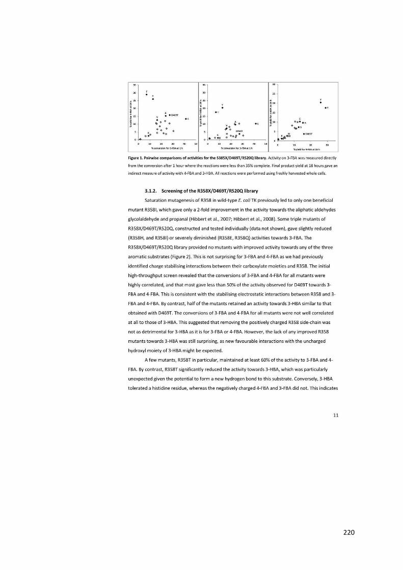
220

221
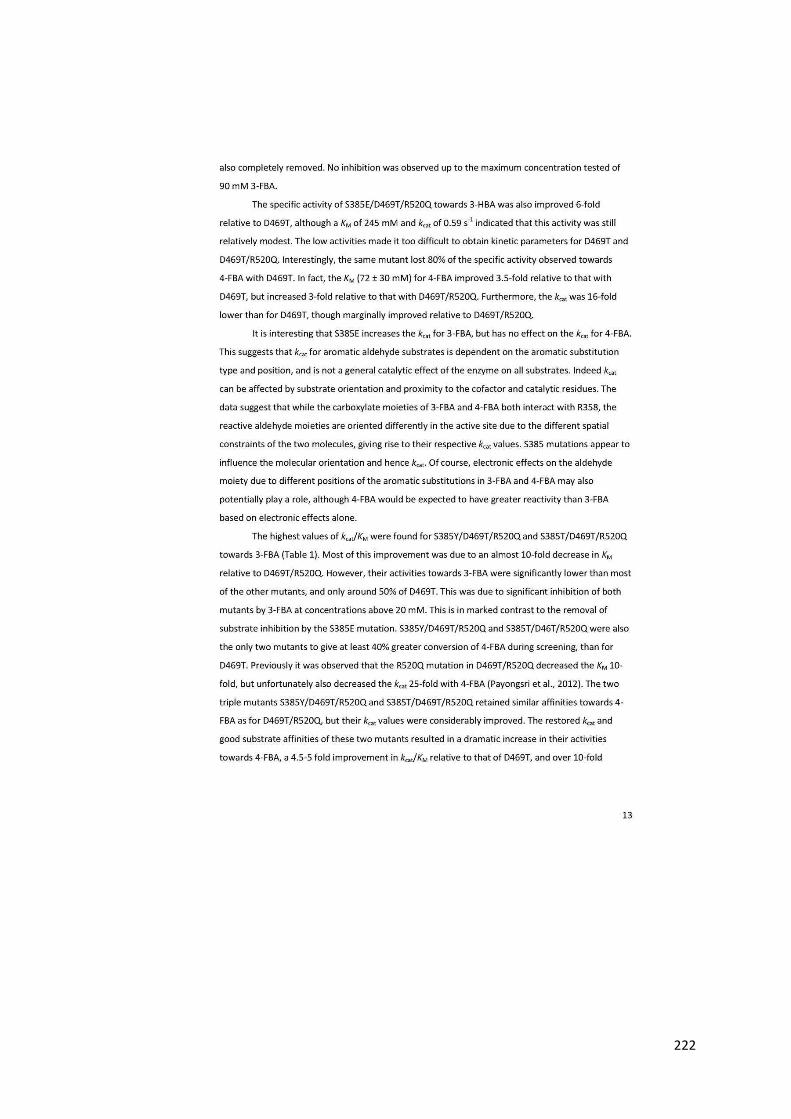
222

223
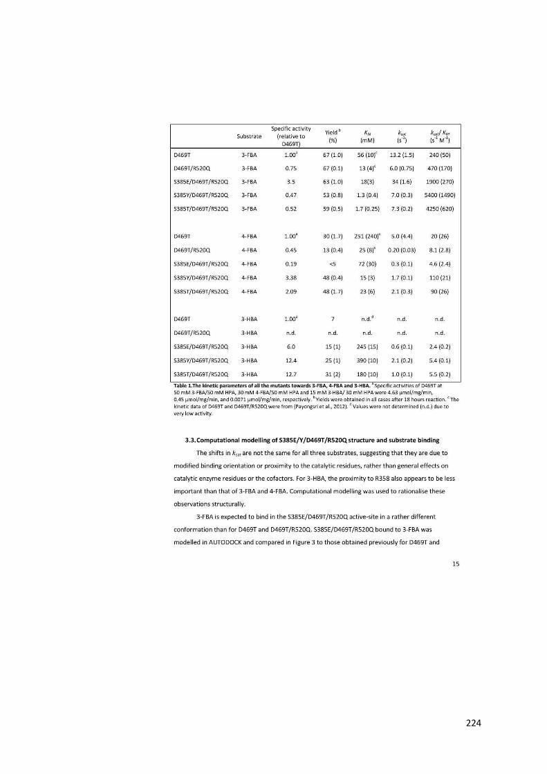
224
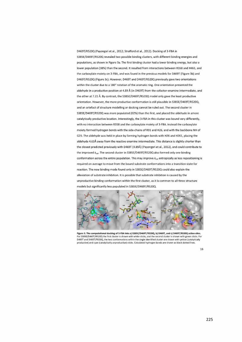
225

226

227
228

229

230