SynthesisandCharacterizationofPseudocantharidins,Novel ... splicing factor tra2-beta1 promotes...
12
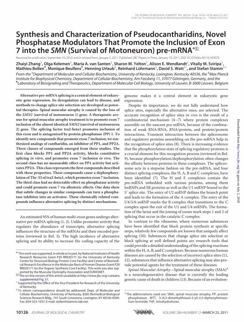
Synthesis and Characterization of Pseudocantharidins, Novel Phosphatase Modulators That Promote the Inclusion of Exon 7 into the SMN (Survival of Motoneuron) pre-mRNA * □ S Received for publication, September 10, 2010, and in revised form, January 3, 2011 Published, JBC Papers in Press, January 10, 2011, DOI 10.1074/jbc.M110.183970 Zhaiyi Zhang ‡ , Olga Kelemen ‡ , Maria A. van Santen § , Sharon M. Yelton ‡ , Alison E. Wendlandt ‡ , Vitaliy M. Sviripa ‡ , Mathieu Bollen ¶ , Monique Beullens ¶ , Henning Urlaub § , Reinhard Lu ¨ hrmann § , David S. Watt ‡1 , and Stefan Stamm ‡2 From the ‡ Department of Molecular and Cellular Biochemistry, University of Kentucky, Lexington, Kentucky 40536, the § Max Planck Institute for Biophysical Chemistry, Department of Cellular Biochemistry, Am Fassberg 11, 37077 Go ¨ttingen, Germany, and the ¶ Laboratory of Biosignaling and Therapeutics, Department of Molecular Cell Biology, University of Leuven, B-3000 Leuven, Belgium Alternative pre-mRNA splicing is a central element of eukary- otic gene expression. Its deregulation can lead to disease, and methods to change splice site selection are developed as poten- tial therapies. Spinal muscular atrophy is caused by the loss of the SMN1 (survival of motoneuron 1) gene. A therapeutic ave- nue for spinal muscular atrophy treatment is to promote exon 7 inclusion of the almost identical SMN2 (survival of motoneuron 2) gene. The splicing factor tra2-beta1 promotes inclusion of this exon and is antagonized by protein phosphatase (PP) 1. To identify new compounds that promote exon 7 inclusion, we syn- thesized analogs of cantharidin, an inhibitor of PP1, and PP2A. Three classes of compounds emerged from these studies. The first class blocks PP1 and PP2A activity, blocks constitutive splicing in vitro, and promotes exon 7 inclusion in vivo. The second class has no measurable effect on PP1 activity but acti- vates PP2A. This class represents the first compounds described with these properties. These compounds cause a dephosphory- lation of Thr-33 of tra2-beta1, which promotes exon 7 inclusion. The third class had no detectable effect on phosphatase activity and could promote exon 7 via allosteric effects. Our data show that subtle changes in similar compounds can turn a phospha- tase inhibitor into an activator. These chemically related com- pounds influence alternative splicing by distinct mechanisms. An estimated 95% of human multi-exon genes undergo alter- native pre-mRNA splicing (1, 2). Unlike promoter activity that regulates the abundance of transcripts, alternative splicing influences the structure of the mRNAs and their encoded pro- teins (reviewed in Ref. 3). The high incidence of alternative splicing and its ability to increase the coding capacity of the genome makes it a central element in eukaryotic gene expression. Despite its importance, we do not fully understand how splice sites, especially the alternative ones, are selected. The accurate recognition of splice sites in vivo is the result of a combinatorial mechanism (4 –7) where protein complexes assemble on the nascent pre-mRNA, because of the combina- tion of weak RNA/RNA, RNA/protein, and protein/protein interactions. Transient interaction between the spliceosome and regulatory proteins assembling on the pre-mRNA help in the recognition of splice sites (8). There is increasing evidence that the phosphorylation state of splicing regulatory proteins is a crucial element for this recognition process (reviewed in Ref. 9), because phosphorylation/dephosphorylation often changes the affinity between proteins in these complexes. The spliceo- some acts in a stepwise fashion on the pre-mRNA, and four distinct splicing complexes, the H, A, B and C complexes, have been identified (7). The H and E complexes contain the pre-mRNA imbedded with various proteins, among them hnRNPs and SR proteins as well as the U1 snRNP bound to the 5 splice site. The entry of U2 snRNP defines the branch point and leads to the formation of the A complex. The entry of the U4/5/6 snRNP marks the B complex that transitions to the C complex upon the exit of the U1 and U4 snRNPs. The forma- tion of the lariat and the joining of exons mark steps 1 and 2 of splicing that occur in the catalytic C complex. In contrast to the ribosome, where numerous substances have been identified that block protein synthesis at specific steps, relatively few compounds are known that uniquely affect splicing (10). Substances that change splice site selection or block splicing at well defined points are research tools that could provide a detailed understanding of the splicing reactions within the H, A, B, and C complexes. Because numerous human diseases are caused by the selection of incorrect splice sites (11, 12), substances that influence alternative splicing may also pro- vide potential agents for the treatment of these diseases. Spinal Muscular Atrophy—Spinal muscular atrophy (SMA) 3 is a neurodegenerative disease that is currently the leading genetic cause of death in children (13). Because of an evolution- * This work was supported, in whole or in part, by National Institutes of Health Research Resources Grant P20 RR020171 (to the University of Kentucky Center for Structural Biology Protein Core Facility) and Center of Biomedi- cal Research Excellence in the Molecular Basis of Human Disease Grant P20 RR020171 (to the Organic Synthesis Core Facility). This work was also sup- ported by the Muscular Dystrophy Association and EURASNET. □ S The on-line version of this article (available at http://www.jbc.org) contains supplemental Fig. S1. 1 Supported by the Office of the Vice President for Research of the University of Kentucky. 2 To whom correspondence should be addressed: Dept. of Molecular and Cellular Biochemistry, University of Kentucky, B283 Biomedical Biological Sciences Research Bldg., 741 South Limestone, Lexington, KY 40536-0509. Fax: 859-323-1037; E-mail: [email protected]. 3 The abbreviations used are: SMA, spinal muscular atrophy; PP, protein phosphatase; MTT, 3-(4,5-dimethylthiazol-2-yl)-2,5-diphenyltetrazo- lium bromide; THF, tetrahydrofuran. THE JOURNAL OF BIOLOGICAL CHEMISTRY VOL. 286, NO. 12, pp. 10126 –10136, March 25, 2011 © 2011 by The American Society for Biochemistry and Molecular Biology, Inc. Printed in the U.S.A. 10126 JOURNAL OF BIOLOGICAL CHEMISTRY VOLUME 286 • NUMBER 12 • MARCH 25, 2011 by guest on July 2, 2018 http://www.jbc.org/ Downloaded from
Transcript of SynthesisandCharacterizationofPseudocantharidins,Novel ... splicing factor tra2-beta1 promotes...
Synthesis and Characterization of Pseudocantharidins, NovelPhosphatase Modulators That Promote the Inclusion of Exon7 into the SMN (Survival of Motoneuron) pre-mRNA*□S
Received for publication, September 10, 2010, and in revised form, January 3, 2011 Published, JBC Papers in Press, January 10, 2011, DOI 10.1074/jbc.M110.183970
Zhaiyi Zhang‡, Olga Kelemen‡, Maria A. van Santen§, Sharon M. Yelton‡, Alison E. Wendlandt‡, Vitaliy M. Sviripa‡,Mathieu Bollen¶, Monique Beullens¶, Henning Urlaub§, Reinhard Luhrmann§, David S. Watt‡1, and Stefan Stamm‡2
From the ‡Department of Molecular and Cellular Biochemistry, University of Kentucky, Lexington, Kentucky 40536, the §Max PlanckInstitute for Biophysical Chemistry, Department of Cellular Biochemistry, Am Fassberg 11, 37077 Gottingen, Germany, and the¶Laboratory of Biosignaling and Therapeutics, Department of Molecular Cell Biology, University of Leuven, B-3000 Leuven, Belgium
Alternative pre-mRNAsplicing is a central element of eukary-otic gene expression. Its deregulation can lead to disease, andmethods to change splice site selection are developed as poten-tial therapies. Spinal muscular atrophy is caused by the loss ofthe SMN1 (survival of motoneuron 1) gene. A therapeutic ave-nue for spinal muscular atrophy treatment is to promote exon 7inclusion of the almost identical SMN2 (survival ofmotoneuron2) gene. The splicing factor tra2-beta1 promotes inclusion ofthis exon and is antagonized by protein phosphatase (PP) 1. Toidentify new compounds that promote exon 7 inclusion, we syn-thesized analogs of cantharidin, an inhibitor of PP1, and PP2A.Three classes of compounds emerged from these studies. Thefirst class blocks PP1 and PP2A activity, blocks constitutivesplicing in vitro, and promotes exon 7 inclusion in vivo. Thesecond class has no measurable effect on PP1 activity but acti-vates PP2A.This class represents the first compounds describedwith these properties. These compounds cause a dephosphory-lation ofThr-33 of tra2-beta1,which promotes exon 7 inclusion.The third class had no detectable effect on phosphatase activityand could promote exon 7 via allosteric effects. Our data showthat subtle changes in similar compounds can turn a phospha-tase inhibitor into an activator. These chemically related com-pounds influence alternative splicing by distinct mechanisms.
An estimated 95%of humanmulti-exon genes undergo alter-native pre-mRNA splicing (1, 2). Unlike promoter activity thatregulates the abundance of transcripts, alternative splicinginfluences the structure of the mRNAs and their encoded pro-teins (reviewed in Ref. 3). The high incidence of alternativesplicing and its ability to increase the coding capacity of the
genome makes it a central element in eukaryotic geneexpression.Despite its importance, we do not fully understand how
splice sites, especially the alternative ones, are selected. Theaccurate recognition of splice sites in vivo is the result of acombinatorial mechanism (4–7) where protein complexesassemble on the nascent pre-mRNA, because of the combina-tion of weak RNA/RNA, RNA/protein, and protein/proteininteractions. Transient interaction between the spliceosomeand regulatory proteins assembling on the pre-mRNA help inthe recognition of splice sites (8). There is increasing evidencethat the phosphorylation state of splicing regulatory proteins isa crucial element for this recognition process (reviewed in Ref.9), because phosphorylation/dephosphorylation often changesthe affinity between proteins in these complexes. The spliceo-some acts in a stepwise fashion on the pre-mRNA, and fourdistinct splicing complexes, the H, A, B and C complexes, havebeen identified (7). The H and E complexes contain thepre-mRNA imbedded with various proteins, among themhnRNPs and SR proteins as well as the U1 snRNP bound to the5� splice site. The entry of U2 snRNP defines the branch pointand leads to the formation of the A complex. The entry of theU4/5/6 snRNP marks the B complex that transitions to the Ccomplex upon the exit of the U1 and U4 snRNPs. The forma-tion of the lariat and the joining of exons mark steps 1 and 2 ofsplicing that occur in the catalytic C complex.In contrast to the ribosome, where numerous substances
have been identified that block protein synthesis at specificsteps, relatively few compounds are known that uniquely affectsplicing (10). Substances that change splice site selection orblock splicing at well defined points are research tools thatcould provide a detailed understanding of the splicing reactionswithin theH,A, B, andC complexes. Because numerous humandiseases are caused by the selection of incorrect splice sites (11,12), substances that influence alternative splicingmay also pro-vide potential agents for the treatment of these diseases.Spinal Muscular Atrophy—Spinal muscular atrophy (SMA)3
is a neurodegenerative disease that is currently the leadinggenetic cause of death in children (13). Because of an evolution-
* This work was supported, in whole or in part, by National Institutes of HealthResearch Resources Grant P20 RR020171 (to the University of KentuckyCenter for Structural Biology Protein Core Facility) and Center of Biomedi-cal Research Excellence in the Molecular Basis of Human Disease Grant P20RR020171 (to the Organic Synthesis Core Facility). This work was also sup-ported by the Muscular Dystrophy Association and EURASNET.
□S The on-line version of this article (available at http://www.jbc.org) containssupplemental Fig. S1.
1 Supported by the Office of the Vice President for Research of the Universityof Kentucky.
2 To whom correspondence should be addressed: Dept. of Molecular andCellular Biochemistry, University of Kentucky, B283 Biomedical BiologicalSciences Research Bldg., 741 South Limestone, Lexington, KY 40536-0509.Fax: 859-323-1037; E-mail: [email protected].
3 The abbreviations used are: SMA, spinal muscular atrophy; PP, proteinphosphatase; MTT, 3-(4,5-dimethylthiazol-2-yl)-2,5-diphenyltetrazo-lium bromide; THF, tetrahydrofuran.
THE JOURNAL OF BIOLOGICAL CHEMISTRY VOL. 286, NO. 12, pp. 10126 –10136, March 25, 2011© 2011 by The American Society for Biochemistry and Molecular Biology, Inc. Printed in the U.S.A.
10126 JOURNAL OF BIOLOGICAL CHEMISTRY VOLUME 286 • NUMBER 12 • MARCH 25, 2011
by guest on July 2, 2018http://w
ww
.jbc.org/D
ownloaded from
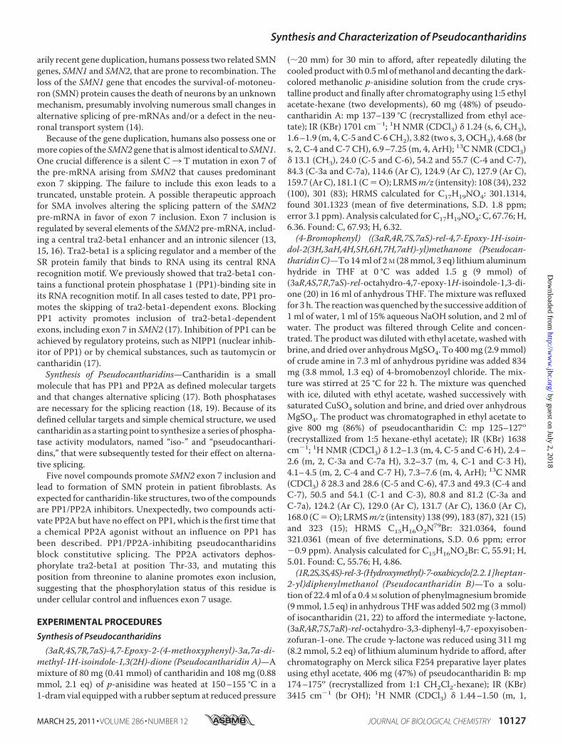
arily recent gene duplication, humans possess two related SMNgenes, SMN1 and SMN2, that are prone to recombination. Theloss of the SMN1 gene that encodes the survival-of-motoneu-ron (SMN) protein causes the death of neurons by an unknownmechanism, presumably involving numerous small changes inalternative splicing of pre-mRNAs and/or a defect in the neu-ronal transport system (14).Because of the gene duplication, humans also possess one or
more copies of the SMN2 gene that is almost identical to SMN1.One crucial difference is a silent C3 T mutation in exon 7 ofthe pre-mRNA arising from SMN2 that causes predominantexon 7 skipping. The failure to include this exon leads to atruncated, unstable protein. A possible therapeutic approachfor SMA involves altering the splicing pattern of the SMN2pre-mRNA in favor of exon 7 inclusion. Exon 7 inclusion isregulated by several elements of the SMN2 pre-mRNA, includ-ing a central tra2-beta1 enhancer and an intronic silencer (13,15, 16). Tra2-beta1 is a splicing regulator and a member of theSR protein family that binds to RNA using its central RNArecognition motif. We previously showed that tra2-beta1 con-tains a functional protein phosphatase 1 (PP1)-binding site inits RNA recognition motif. In all cases tested to date, PP1 pro-motes the skipping of tra2-beta1-dependent exons. BlockingPP1 activity promotes inclusion of tra2-beta1-dependentexons, including exon 7 in SMN2 (17). Inhibition of PP1 can beachieved by regulatory proteins, such as NIPP1 (nuclear inhib-itor of PP1) or by chemical substances, such as tautomycin orcantharidin (17).Synthesis of Pseudocantharidins—Cantharidin is a small
molecule that has PP1 and PP2A as defined molecular targetsand that changes alternative splicing (17). Both phosphatasesare necessary for the splicing reaction (18, 19). Because of itsdefined cellular targets and simple chemical structure, we usedcantharidin as a starting point to synthesize a series of phospha-tase activity modulators, named “iso-” and “pseudocanthari-dins,” that were subsequently tested for their effect on alterna-tive splicing.Five novel compounds promote SMN2 exon 7 inclusion and
lead to formation of SMN protein in patient fibroblasts. Asexpected for cantharidin-like structures, two of the compoundsare PP1/PP2A inhibitors. Unexpectedly, two compounds acti-vate PP2A but have no effect on PP1, which is the first time thata chemical PP2A agonist without an influence on PP1 hasbeen described. PP1/PP2A-inhibiting pseudocantharidinsblock constitutive splicing. The PP2A activators dephos-phorylate tra2-beta1 at position Thr-33, and mutating thisposition from threonine to alanine promotes exon inclusion,suggesting that the phosphorylation status of this residue isunder cellular control and influences exon 7 usage.
EXPERIMENTAL PROCEDURES
Synthesis of Pseudocantharidins
(3aR,4S,7R,7aS)-4,7-Epoxy-2-(4-methoxyphenyl)-3a,7a-di-methyl-1H-isoindole-1,3(2H)-dione (Pseudocantharidin A)—Amixture of 80 mg (0.41 mmol) of cantharidin and 108 mg (0.88mmol, 2.1 eq) of p-anisidine was heated at 150–155 °C in a1-dram vial equippedwith a rubber septum at reduced pressure
(�20 mm) for 30 min to afford, after repeatedly diluting thecooled productwith 0.5ml ofmethanol anddecanting the dark-colored methanolic p-anisidine solution from the crude crys-talline product and finally after chromatography using 1:5 ethylacetate-hexane (two developments), 60 mg (48%) of pseudo-cantharidin A: mp 137–139 °C (recrystallized from ethyl ace-tate); IR (KBr) 1701 cm�1; 1H NMR (CDCl3) � 1.24 (s, 6, CH3),1.6–1.9 (m, 4, C-5 andC-6 CH2), 3.82 (two s, 3, OCH3), 4.68 (brs, 2, C-4 and C-7 CH), 6.9–7.25 (m, 4, ArH); 13C NMR (CDCl3)� 13.1 (CH3), 24.0 (C-5 and C-6), 54.2 and 55.7 (C-4 and C-7),84.3 (C-3a and C-7a), 114.6 (Ar C), 124.9 (Ar C), 127.9 (Ar C),159.7 (ArC), 181.1 (C�O); LRMSm/z (intensity): 108 (34), 232(100), 301 (83); HRMS calculated for C17H19NO4: 301.1314,found 301.1323 (mean of five determinations, S.D. 1.8 ppm;error 3.1 ppm). Analysis calculated for C17H19NO4: C, 67.76; H,6.36. Found: C, 67.93; H, 6.32.(4-Bromophenyl) ((3aR,4R,7S,7aS)-rel-4,7-Epoxy-1H-isoin-
dol-2(3H,3aH,4H,5H,6H,7H,7aH)-yl)methanone (Pseudocan-tharidinC)—To14ml of 2M (28mmol, 3 eq) lithium aluminumhydride in THF at 0 °C was added 1.5 g (9 mmol) of(3aR,4S,7R,7aS)-rel-octahydro-4,7-epoxy-1H-isoindole-1,3-di-one (20) in 16 ml of anhydrous THF. The mixture was refluxedfor 3 h. The reactionwas quenched by the successive addition of1 ml of water, 1 ml of 15% aqueous NaOH solution, and 2 ml ofwater. The product was filtered through Celite and concen-trated. The product was diluted with ethyl acetate, washedwithbrine, and dried over anhydrousMgSO4. To 400mg (2.9mmol)of crude amine in 7.3 ml of anhydrous pyridine was added 834mg (3.8 mmol, 1.3 eq) of 4-bromobenzoyl chloride. The mix-ture was stirred at 25 °C for 22 h. The mixture was quenchedwith ice, diluted with ethyl acetate, washed successively withsaturated CuSO4 solution and brine, and dried over anhydrousMgSO4. The product was chromatographed in ethyl acetate togive 800 mg (86%) of pseudocantharidin C: mp 125–127o(recrystallized from 1:5 hexane-ethyl acetate); IR (KBr) 1638cm�1; 1H NMR (CDCl3) � 1.2–1.3 (m, 4, C-5 and C-6 H), 2.4–2.6 (m, 2, C-3a and C-7a H), 3.2–3.7 (m, 4, C-1 and C-3 H),4.1–4.5 (m, 2, C-4 and C-7 H), 7.3–7.6 (m, 4, ArH); 13C NMR(CDCl3) � 28.3 and 28.6 (C-5 and C-6), 47.3 and 49.3 (C-4 andC-7), 50.5 and 54.1 (C-1 and C-3), 80.8 and 81.2 (C-3a andC-7a), 124.2 (Ar C), 129.0 (Ar C), 131.7 (Ar C), 136.0 (Ar C),168.0 (C�O); LRMSm/z (intensity) 138 (99), 183 (87), 321 (15)and 323 (15); HRMS C15H16O2N79Br: 321.0364, found321.0361 (mean of five determinations, S.D. 0.6 ppm; error�0.9 ppm). Analysis calculated for C15H16NO2Br: C, 55.91; H,5.01. Found: C, 55.76; H, 4.86.(1R,2S,3S,4S)-rel-3-(Hydroxymethyl)-7-oxabicyclo[2.2.1]heptan-
2-yl)diphenylmethanol (Pseudocantharidin B)—To a solu-tion of 22.4ml of a 0.4 M solution of phenylmagnesiumbromide(9mmol, 1.5 eq) in anhydrous THFwas added 502mg (3mmol)of isocantharidin (21, 22) to afford the intermediate �-lactone,(3aR,4R,7S,7aR)-rel-octahydro-3,3-diphenyl-4,7-epoxyisoben-zofuran-1-one. The crude �-lactone was reduced using 311 mg(8.2 mmol, 5.2 eq) of lithium aluminum hydride to afford, afterchromatography on Merck silica F254 preparative layer platesusing ethyl acetate, 406 mg (47%) of pseudocantharidin B: mp174–175o (recrystallized from 1:1 CH2Cl2-hexane); IR (KBr)3415 cm�1 (br OH); 1H NMR (CDCl3) � 1.44–1.50 (m, 1,
Synthesis and Characterization of Pseudocantharidins
MARCH 25, 2011 • VOLUME 286 • NUMBER 12 JOURNAL OF BIOLOGICAL CHEMISTRY 10127
by guest on July 2, 2018http://w
ww
.jbc.org/D
ownloaded from
H-5�), 1.57–1.66 (m, 2, H-5� and H-6�), 1.77–1.86 (m, 1,H-6�), 2.25 (ddd, 1, H-3), 2.74 (dd, J � 3.6 and 7.2 Hz,1, CH2OH), 3.20 (ddd, 1, CH2OH), 3.33 (d, J2,3� 8.8Hz, 1,H-2),3.43 (ddd, 1, CH2OH), 4.43 (d, J� 4.8Hz, 1,H-1), 4.59 (d, J� 5.2Hz, 1, H-4), 5.14 (s, 1, C(OH)(C6H5)2), 7.07–7.17 (m, 2, para-ArH), 7.21–7.29 (m, 4, meta-ArH), 7.52–7.61 (m, 4, ortho-ArH); 13CNMR (CDCl3) � 29.4 and 30.7 (C-5 andC-6), 50.1 and55.3 (C-2 andC-3), 62.4 and 78.1 (CH2OHandC(OH)Ph2), 79.1and 81.3 (C-1 andC-4), 125.1 (Ar C), 125.5 (Ar C), 126.4 (Ar C),126.8 (Ar C), 128.5 (Ar C), 128.7 (Ar C), 148.2 (Ar C), 148.6 (ArC); LRMSm/z (intensity) 105 (100), 183 (79), 215 (25), 292 (2);HRMS calculated for C20H20O2 (M�-H2O) 292.1463, found292.1457 (mean of five determinations, S.D. 1.8 ppm; error�1.9 ppm). Analysis calculated for C20H22O3: C, 77.39; H, 7.14.Found: C, 77.32; H, 7.28.(3aR,4S,7R,7aS)-4,7-Epoxy-3a,7a-dimethyl-2-phenyl-1H-iso-
indole-1,3(2H)-dione (Pseudocantharidin D)—Amixture of 72mg (0.37mmol) of cantharidin and 342mg (3.7mmol, 10 eq) ofaniline was heated at 160–170 °C (760 mm) for 4 h to afford,after chromatography using 1:2 ethyl acetate-hexane, 94 mg(94%) of pseudocantharidin D: mp 114–116 °C (recrystallizedfrom 1:5 ethyl acetate-hexane); IR (KBr) 1709 cm�1; 1H NMR(CDCl3) � 1.26 (s, 6, CH3), 1.58–1.94 (m, 4, C-5 and C-6 CH2),4.70 (dd, 2, J � 2.4 and 3.2 Hz, C-4 and C-7 CH), 7.25–7.55 (m,5, ArH); 13C NMR (CDCl3) � 13.1 (CH3), 24.0 (C-5 and C-6),54.3 (C-4 and C-7), 84.3 (C-3a and C-7a), 126.7 (Ar C), 128.8(Ar C), 129.3 (Ar C), 132.3 (Ar C), 180.9 (C � O); LRMS m/z(intensity): 203 (100), 271 (9); HRMS calculated forC16H17NO3: 271.1208, found 271.1206 (mean of five determi-nations, S.D. 1.8 ppm; error �0.7 ppm). Analysis calculated forC16H17NO3: C, 70.83; H, 6.32. Found: C, 70.93; H, 6.27.
Phosphatase Assays
PP1, PP2A, and glycogen phosphorylase were purified fromrabbit skeletal muscle, and the phosphorylase phosphataseassays were performed as described previously (23).
Drug Treatment of Cells
Four hours after transfection with the reporter minigene,HEK293 cells were treated with the indicated concentration ofcompounds for an additional 12 h. At 16 h post-transfection,total RNA was extracted using Qiagen RNAeasy kit. To deter-mine toxicity, 106 cells/cm2 SMAI cells were seeded into24-well plates the day before treatment. The MTT assay wasperformed after adding different concentration of compoundsto SMAI cells for 24 h. For protein level detection, 106 cells/cm2
SMAI cells were seeded into six-well plates. The cells weretreated with compounds every other day for 15 days. Thegrowthmediumwas changed prior to compound addition. Thecells were lysed with radioimmune precipitation assay bufferafter treatment with compounds.
RT-PCR
Reverse transcription was performed using SV40pA RT(TGGTTTGTCCAAACTCATCAA). PCR was performedusing pCI For (GGTGTCCACTCCCAGTTCAA) andSMNex8rev (GCCTCACCACCGTGCTGG). For reverse tran-scription, 400 ng of total RNA (200 ng/�l), 5 pmol of reverse
primer, and 40 units of SuperScript III reverse transcriptaseweremixed in 5�l of RT buffer. To reverse transcribe the RNA,the reactionwas incubated at 55 °C for 50min. One-third of theRT reaction was used for cDNA amplification. The reactionwas performed in 25 �l and contained 10 pmol of specific for-ward and reverse primers, 200 �M dNTPs, 1� Taq polymerasebuffer, and 1 unit of Taq DNA polymerase. The amplificationwas carried out in an Eppendorf PCR system thermocyclerunder the following conditions: initial denaturation for 4min at94 °C; 28 cycles of 30 s at 94 °C, 30 s at 60 °C; and extension of45 s at 72 °C. After the last cycle, the reactionwas held for 5minat the extension temperature to complete the amplification ofall products.
SMA Cells
Fibroblasts from a 7-month-old,male, Caucasian SMA type Ipatient was obtained from the Coriell Institute for MedicalResearch (clone number GM00232).
In Vitro Splicing and Spliceosomal Assembly
A transcription template for the MINX pre-mRNAwas gen-erated from the pMINX plasmid (24) by PCR. HeLa nuclearextract was prepared according to Dignam et al. (25). Splicingreactions contained 40% (v/v) HeLa nuclear extract in buffer D(20 mM HEPES-KOH, pH 7.9, 100 mM KCl, 1.5 mM MgCl2, 0.2mM EDTA, 10% (v/v) glycerol, 0.5 mM DTT, 0.5 mM PMSF), 25mMKCl, 3 mMMgCl2, 20 mM creatine phosphate, 2 mMATP, 3nM 32P-labeled pre-mRNA, and the indicated concentration ofisocantharidin or pseudocantharidins. The nuclear extract waspreincubated with the compounds (or just water as a control)for 10 min at 30 °C, and the reactions were then started by theaddition of the other components. For the analysis of the splic-ing products, RNAwas isolated by proteinaseK treatment, phe-nol extraction, and ethanol precipitation, separated by denatur-ing polyacrylamide gel electrophoresis on an 8.3 M urea, 14%(w/v) polyacrylamide gel, and visualized by autoradiography.For the analysis of the spliceosomal complexes, 10 �l of thesplicing reaction were added to 2.5 �l of load buffer (1� Trisbase/boric acid/EDTA, 30% (v/v) glycerol, 1.25 �g/�l heparin)at the time points indicated and then placed on ice. The com-plexes were separated on 1.8% (w/v) agarose gels (26).
Cell Viability
Viability was tested by metabolizing MTT by mitochondrialdehydrogenases (Sigma): 1.000,000 cells were treated with thecompounds overnight, and the cells were subsequently ana-lyzed withMTT. TheMTT staining obtained from cells receiv-ing just Me2SO control was set as 100% signal.
Antisera
Anti-SAP130 and anti-actin antisera were from Abcam. Theanti-Thr-33 phospho-specific antiserum was made by immu-nizing rabbits with CKSARH-pT-PARSR peptide, followed bysubsequent affinity purification (Biogenes, Germany).
RESULTS
Synthesis of Pseudocantaridins, a New Series of Cantharidin-like Compounds—We previously showed that PP1-inhibitingsubstances, like cantharidin or tautomycin, influence alterna-
Synthesis and Characterization of Pseudocantharidins
10128 JOURNAL OF BIOLOGICAL CHEMISTRY VOLUME 286 • NUMBER 12 • MARCH 25, 2011
by guest on July 2, 2018http://w
ww
.jbc.org/D
ownloaded from
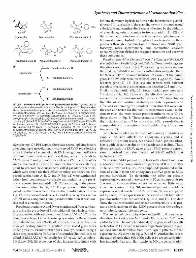
tive splicing (17). PP1 dephosphorylates several splicing factorsafter binding to an evolutionarily conservedRVxF-type dockingmotif in the beta 4 strand of their RNA recognition motif. Oneof these proteins is tra2-beta1, a splicing factor that binds toSMN2 exon 7 and promotes its inclusion (27). Because of itssimple chemical structure, we used cantharidin as a startingpoint to generate new substances, called pseudocantharidins,which were tested for their effect on splice site selection. Thepseudocantharidins A, B, C, and D (Fig. 1A) were synthesizedeither from commercially available cantharidin or the previ-ously reported isocantharidin (21, 22) according to the proce-dures summarized in Fig. 1B. For purposes of this paper,pseudocantharidin refers to the cantharidin-like structures inFig. 1A. Pseudocantharidins A, C, and D were synthesized asachiral meso-compounds, and pseudocantharidin B was syn-thesized as a racemic mixture.Pseudocantharidins A andDwere synthesized from canthar-
idin using the procedure of Bockstahler (28) in which canthar-idinwasmeltedwith aniline or p-anisidine at 150–170 °C in theabsence of solvent. Other reported procedures for the synthesisof similar derivatives (29–34) of cantharidin or isocantharidininvolved heating amines in solution or in sealed glass tubesunder pressure. Pseudocantharidin C was synthesized using athree-step procedure: (i) fusion of isocantharidin with urea toafford (3aR,4S,7R,7aS)-rel-octahydro-4,7-epoxy-1H-isoindole-1,3-dione (20); (ii) reduction of this intermediate imide with
lithium aluminum hydride to furnish the intermediate pyrroli-dine; and (iii) acylation of the pyrrolidine with 4-bromobenzoylchloride. Pseudocantharidin B was synthesized by the additionof phenylmagnesium bromide to isocantharidin (21, 22) andthe subsequent reduction of the intermediate �-lactone withlithium aluminum hydride. Complete characterization of theseproducts through a combination of infrared and NMR spec-troscopy, mass spectrometry, and combustion analysesunequivocally established the assigned structures and purity ofthese compounds.Pseudocantharidins ChangeAlternative Splicing of the SMN2
pre-mRNA and Exhibit Different Cellular Toxicity—Using can-tharidin or isocantharidin (21, 22) as startingmaterials, we syn-thesized over 50 different pseudocantharidins and tested themfor their ability to promote inclusion of exon 7 of the SMN2gene. HEK293 cells were transfected with 1 �g of pCl-SMN2reporter gene (27, 35) (Fig. 2A) and treated with differentpseudocantharidins at a concentration between 0.25 and 1mM.Similar to cantharidin (Fig. 2B), isocantharidin promotes exon7 inclusion (Fig. 2C). However, the effective concentrationrange (0.25–1 mM) for isocantharidin was �250 times higherthan that of cantharidin that already exhibited a pronouncedeffect at 4 �M. Among the pseudocantharidins that were syn-thesized and tested for their ability to promote exon 7 inclu-sion in vivo, the most active pseudocantharidins A–D arethose shown in Fig. 1. These pseudocantharidins increasedthe inclusion of exon 7 by more than 40%, a result that iscomparable with the action of cantharidin (Fig. 2B) or tau-tomycin (17).To determine whether the effect of pseudocantharidins on
exon 7 inclusion affects the endogenous genes and isreflected in protein levels, we treated SMA patient fibro-blasts with isocantharidin or the pseudocantharidins. Thesefibroblasts lack the SMN1 gene, and all SMN protein expres-sion is derived from the fraction of SMN2 mRNA thatincludes exon 7.We treated SMA patient fibroblasts with a final 1 mM con-
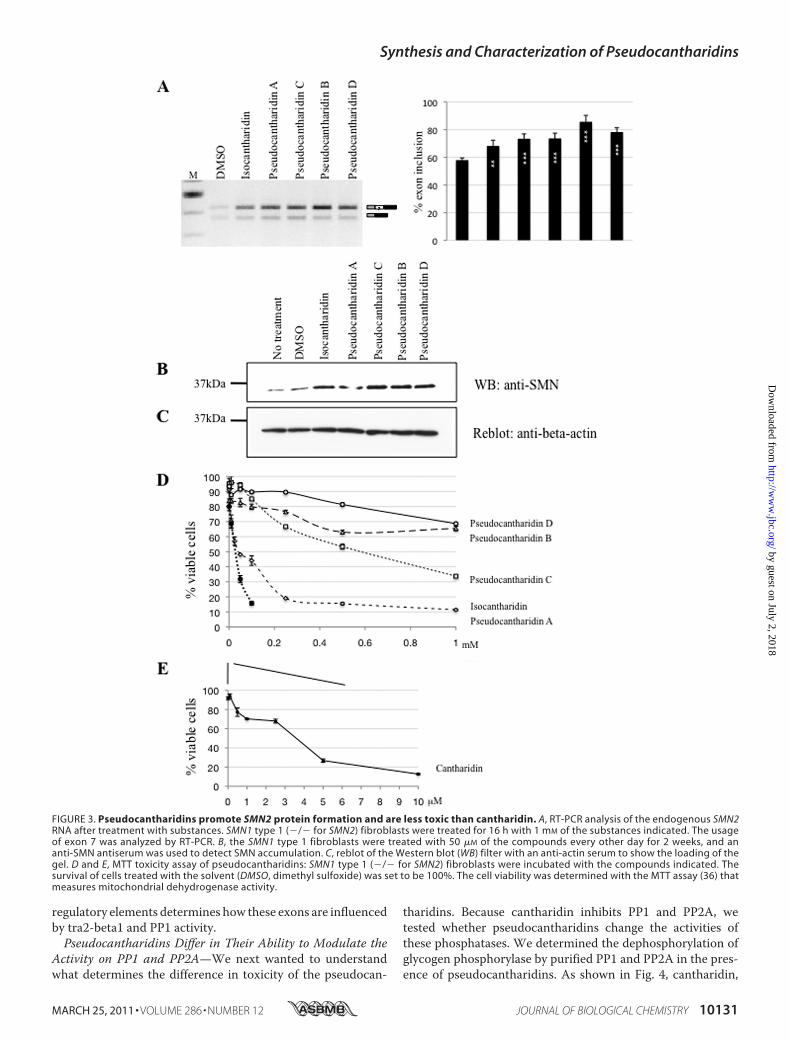
centration of the compounds and performed RT-PCR after16 h. As shown in Fig. 3A, the compounds promoted inclu-sion of exon 7 from the endogenous SMN2 gene in SMApatient fibroblasts. To determine the effect on proteinexpression, we treated these cells with 50 �M compounds for2 weeks, a concentration where we observed little toxiceffect. As shown in Fig. 3B, untreated patient fibroblastsexpress residual levels of SMN protein. When comparedwith �-actin, this expression is increased 2–3.8-fold whenpseudocantharidins are added (Fig. 3, B and C). The datashow that isocantharidin and pseudocantharidins A–D pro-mote the formation of the SMN protein from the endoge-nous, physiological relevant gene.We next tested the toxicity of isocantharidin and pseudocan-
tharidins A–D using the MTT test (36), in which MTT wasadded to cells. The mitochondrial dehydrogenases of live cellsmetabolize MTT, which is measured spectroscopically. Again,we used human fibroblast from SMA type I patients for theexperiments. As shown in Fig. 3 (D and E), cantharidin causesthe death of more than 80% of the cells at 10 �M concentration.Isocantharidin had a similar toxicity at 500 �M concentration.
FIGURE 1. Structure and synthesis of pseudocantharidins. A, structures ofpseudocantharidins used in this study. The 7-oxabicyclo[2.2.1]heptane skel-eton common to all compounds is shown in bold. The formal names for thepseudocantharidins are: A, (3aR,4S,7R,7aS)-4,7-epoxy-2-(4-methoxyphe-nyl)-3a,7a-dimethyl-1H-isoindole-1,3(2H)-dione; B, (1R,2S,3S,4S)-rel-3-(hy-droxymethyl)-7-oxabicyclo[2.2.1]heptan-2-yl)diphenylmethanol; C, (4-bro-mophenyl) (3aR,4R,7S,7aS)-rel-4,7-epoxy-1H-isoindol-2(3H,3aH,4H,5H,6H,7H,7aH)-yl)methanone; D, (3aR,4S,7R,7aS)-4,7-epoxy-3a,7a-dimethyl-2-phenyl-1H-isoindole-1,3(2H)-dione; B, schematic overview of the synthesis ofpseudocantharidins. a, aniline, 160 –170 °C; b, p-anisidine, 150 –155 °C (20mm); c, urea, 150 °C (20 mm); d, LiA1H4, THF; e, 5-bromobenzoyl chloride, Py;f, C6H5MgBr.
Synthesis and Characterization of Pseudocantharidins
MARCH 25, 2011 • VOLUME 286 • NUMBER 12 JOURNAL OF BIOLOGICAL CHEMISTRY 10129
by guest on July 2, 2018http://w
ww
.jbc.org/D
ownloaded from
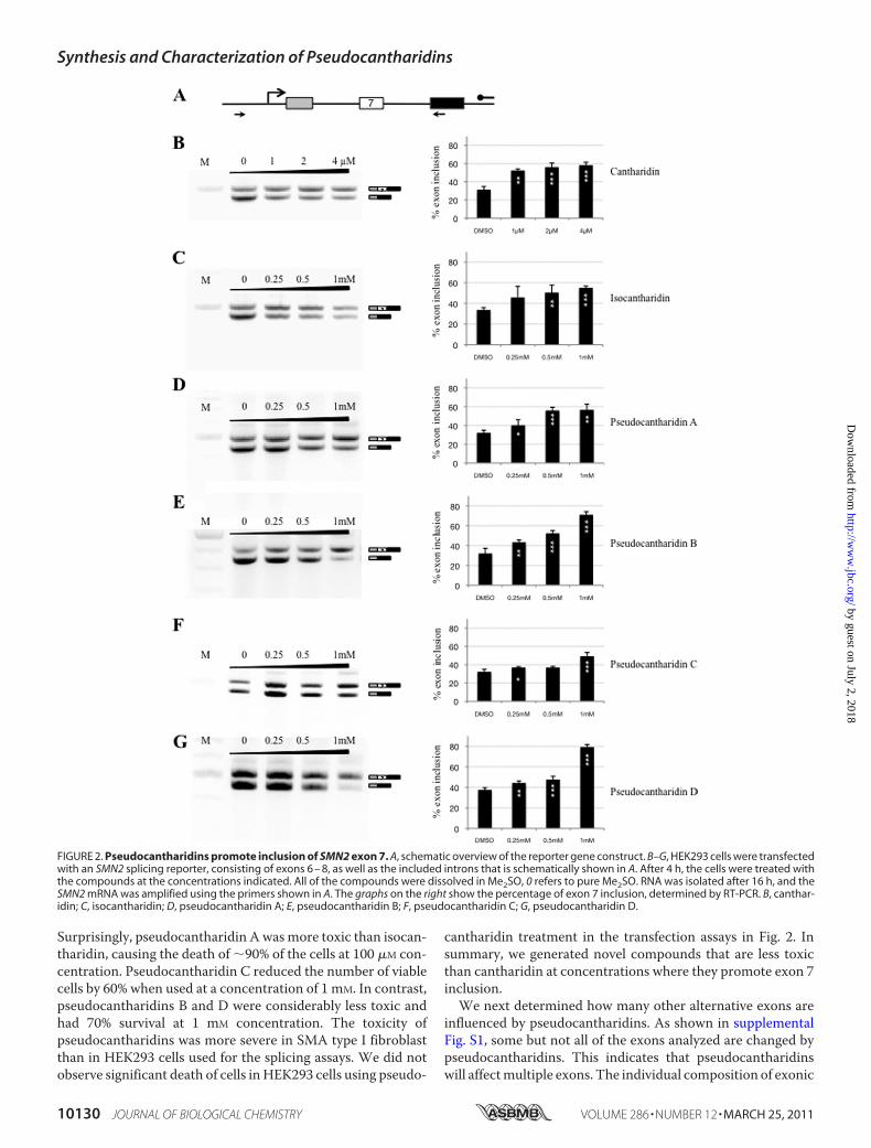
Surprisingly, pseudocantharidin Awasmore toxic than isocan-tharidin, causing the death of �90% of the cells at 100 �M con-centration. Pseudocantharidin C reduced the number of viablecells by 60% when used at a concentration of 1 mM. In contrast,pseudocantharidins B and D were considerably less toxic andhad 70% survival at 1 mM concentration. The toxicity ofpseudocantharidins was more severe in SMA type I fibroblastthan in HEK293 cells used for the splicing assays. We did notobserve significant death of cells inHEK293 cells using pseudo-
cantharidin treatment in the transfection assays in Fig. 2. Insummary, we generated novel compounds that are less toxicthan cantharidin at concentrations where they promote exon 7inclusion.We next determined how many other alternative exons are
influenced by pseudocantharidins. As shown in supplementalFig. S1, some but not all of the exons analyzed are changed bypseudocantharidins. This indicates that pseudocantharidinswill affectmultiple exons. The individual composition of exonic
FIGURE 2. Pseudocantharidins promote inclusion of SMN2 exon 7. A, schematic overview of the reporter gene construct. B–G, HEK293 cells were transfectedwith an SMN2 splicing reporter, consisting of exons 6 – 8, as well as the included introns that is schematically shown in A. After 4 h, the cells were treated withthe compounds at the concentrations indicated. All of the compounds were dissolved in Me2SO, 0 refers to pure Me2SO. RNA was isolated after 16 h, and theSMN2 mRNA was amplified using the primers shown in A. The graphs on the right show the percentage of exon 7 inclusion, determined by RT-PCR. B, canthar-idin; C, isocantharidin; D, pseudocantharidin A; E, pseudocantharidin B; F, pseudocantharidin C; G, pseudocantharidin D.
Synthesis and Characterization of Pseudocantharidins
10130 JOURNAL OF BIOLOGICAL CHEMISTRY VOLUME 286 • NUMBER 12 • MARCH 25, 2011
by guest on July 2, 2018http://w
ww
.jbc.org/D
ownloaded from
regulatory elements determines how these exons are influencedby tra2-beta1 and PP1 activity.Pseudocantharidins Differ in Their Ability to Modulate the
Activity on PP1 and PP2A—We next wanted to understandwhat determines the difference in toxicity of the pseudocan-
tharidins. Because cantharidin inhibits PP1 and PP2A, wetested whether pseudocantharidins change the activities ofthese phosphatases. We determined the dephosphorylation ofglycogen phosphorylase by purified PP1 and PP2A in the pres-ence of pseudocantharidins. As shown in Fig. 4, cantharidin,
FIGURE 3. Pseudocantharidins promote SMN2 protein formation and are less toxic than cantharidin. A, RT-PCR analysis of the endogenous SMN2RNA after treatment with substances. SMN1 type 1 (�/� for SMN2) fibroblasts were treated for 16 h with 1 mM of the substances indicated. The usageof exon 7 was analyzed by RT-PCR. B, the SMN1 type 1 fibroblasts were treated with 50 �M of the compounds every other day for 2 weeks, and ananti-SMN antiserum was used to detect SMN accumulation. C, reblot of the Western blot (WB) filter with an anti-actin serum to show the loading of thegel. D and E, MTT toxicity assay of pseudocantharidins: SMN1 type 1 (�/� for SMN2) fibroblasts were incubated with the compounds indicated. Thesurvival of cells treated with the solvent (DMSO, dimethyl sulfoxide) was set to be 100%. The cell viability was determined with the MTT assay (36) thatmeasures mitochondrial dehydrogenase activity.
Synthesis and Characterization of Pseudocantharidins
MARCH 25, 2011 • VOLUME 286 • NUMBER 12 JOURNAL OF BIOLOGICAL CHEMISTRY 10131
by guest on July 2, 2018http://w
ww
.jbc.org/D
ownloaded from
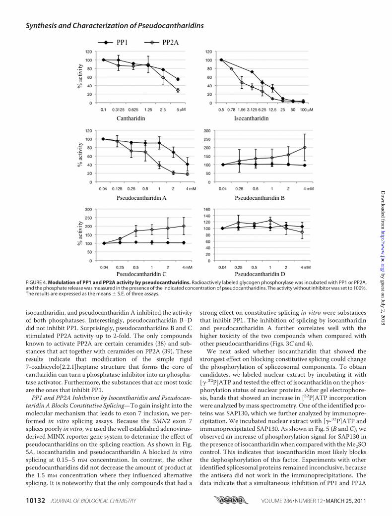
isocantharidin, and pseudocantharidin A inhibited the activityof both phosphatases. Interestingly, pseudocantharidin B–Ddid not inhibit PP1. Surprisingly, pseudocantharidins B and Cstimulated PP2A activity up to 2-fold. The only compoundsknown to activate PP2A are certain ceramides (38) and sub-stances that act together with ceramides on PP2A (39). Theseresults indicate that modification of the simple rigid7-oxabicyclo[2.2.1]heptane structure that forms the core ofcantharidin can turn a phosphatase inhibitor into an phospha-tase activator. Furthermore, the substances that are most toxicare the ones that inhibit PP1.PP1 and PP2A Inhibition by Isocantharidin and Pseudocan-
taridin A Blocks Constitutive Splicing—To gain insight into themolecular mechanism that leads to exon 7 inclusion, we per-formed in vitro splicing assays. Because the SMN2 exon 7splices poorly in vitro, we used the well established adenovirus-derived MINX reporter gene system to determine the effect ofpseudocantharidins on the splicing reaction. As shown in Fig.5A, isocantharidin and pseudocantharidin A blocked in vitrosplicing at 0.15–5 mM concentration. In contrast, the otherpseudocantharidins did not decrease the amount of product atthe 1.5 mM concentration where they influenced alternativesplicing. It is noteworthy that the only compounds that had a
strong effect on constitutive splicing in vitro were substancesthat inhibit PP1. The inhibition of splicing by isocantharidinand pseudocantharidin A further correlates well with thehigher toxicity of the two compounds when compared withother pseudocantharidins (Figs. 3C and 4).We next asked whether isocantharidin that showed the
strongest effect on blocking constitutive splicing could changethe phosphorylation of spliceosomal components. To obtaincandidates, we labeled nuclear extract by incubating it with[�-32P]ATP and tested the effect of isocantharidin on the phos-phorylation status of nuclear proteins. After gel electrophore-sis, bands that showed an increase in [32P]ATP incorporationwere analyzed bymass spectrometry. One of the identified pro-teins was SAP130, which we further analyzed by immunopre-cipitation. We incubated nuclear extract with [�-32P]ATP andimmunoprecipitated SAP130. As shown in Fig. 5 (B and C), weobserved an increase of phosphorylation signal for SAP130 inthe presence of isocantharidinwhen comparedwith theMe2SOcontrol. This indicates that isocantharidin most likely blocksthe dephosphorylation of this factor. Experiments with otheridentified splicesomal proteins remained inconclusive, becausethe antisera did not work in the immunoprecipitations. Thedata indicate that a simultaneous inhibition of PP1 and PP2A
FIGURE 4. Modulation of PP1 and PP2A activity by pseudocantharidins. Radioactively labeled glycogen phosphorylase was incubated with PP1 or PP2A,and the phosphate release was measured in the presence of the indicated concentration of pseudocantharidins. The activity without inhibitor was set to 100%.The results are expressed as the means � S.E. of three assays.
Synthesis and Characterization of Pseudocantharidins
10132 JOURNAL OF BIOLOGICAL CHEMISTRY VOLUME 286 • NUMBER 12 • MARCH 25, 2011
by guest on July 2, 2018http://w
ww
.jbc.org/D
ownloaded from

activity evoked by isocantharidin has a strong influence on thephosphorylation of the U2 snRNP component SAP130.It is well established that PP1 and PP2A activity is necessary
for the constitutive splicing reaction (19). The phosphatases acton proteins in the second step of the splicing reaction (18). Wetherefore examined which stage in the spliceosomal reactionwas blocked by isocantharidin and pseudocantharidin A andperformed assembly assays using the MINX substrate. Asshown in Fig. 5D, isocantharidin slowed down the transitionfrom the H to A complex and blocks C complex formation.Pseudocantharidin A has no discernable effect on H complexformation but again blocks C complex formation. These dataare in agreement with an inhibition of phosphatase activityneeded for the B to C transition. It is likely that a partial inhibi-
tion of constitutive splicing is a reason for the toxicity of iso-cantharidin and pseudocantharidin A.PP2A Activation by Pseudocantharidin B and C Changes
Phosphorylation of tra2-beta1 at Position Thr-33 and PromotesExon 7 Inclusion—We next asked why pseudocantharidins Band C, which activate PP2A but have no measurable effect onPP1, promote exon 7 inclusion. Previously, we used mass spec-trometry to characterize the phosphorylation status of splicingregulatory proteins and found that the phosphorylation of tra2-beta1 at positionThr-33 is changed during the splicing reactionin vitro (40). Phosphorylated Thr-33 could be detected in the Acomplex assembling on the MINX substrate but not in othercomplexes. Tra2-beta1 is an SR-like protein that binds to thecentral enhancer of exon 7 (27). Its binding is most likely stabi-
FIGURE 5. PP1-inhibiting substances block constitutive splicing and increase phosphorylation of the spliceosomal component SAP130. A, in vitrosplicing assay of a MINX substrate in HeLa nuclear extract. The compounds were added at 0.05, 0.15, 0.5, 1.5, and 5 mM concentrations. B, in vitro spliceosomalassembly assay using the MINX substrate. C and D, isocantharidin promotes phosphorylation of SAP130 in nuclear extract. HeLa nuclear extract was incubatedwith 4 �Ci of [�-32P]ATP for 30 min at 37 °C in the presence of isocantharidin or its solvent, dimethyl sulfoxide (DMSO). SAP130 was immunoprecipitated (IP), andthe immunoprecipitates were analyzed by autoradiography (C). The same filter was then reblotted with anti-SAP130 antiserum (D) to show loading of theprotein. WB, Western blot.
Synthesis and Characterization of Pseudocantharidins
MARCH 25, 2011 • VOLUME 286 • NUMBER 12 JOURNAL OF BIOLOGICAL CHEMISTRY 10133
by guest on July 2, 2018http://w
ww
.jbc.org/D
ownloaded from

lized by interactions with hnRNPG and SRp30c (41). Together,this enhancer complex causes recognition of the exon by thespliceosome and its subsequent inclusion. To investigatewhether pseudocantharidins interfere with the phosphoryla-tion at Thr-33, we created a phosphorylation-selective poly-clonal antisera. As shown in Fig. 6A, this antiserum recognizesEGFP-tra2-beta1 that is transfected into HEK293 cells. Theantiserumdid not detect tra2-beta1mutants wherewe changedthe threonine 33 to either alanine (T33A) or glutamic acid(T33E), which demonstrates its selectivity. Because tra2-beta1binds directly to PP1, we tested the influence of PP1 on thephosphorylation on this site and found that transfection of PP1expression constructs completely abolishes the phosphoryla-tion signal. Similarly, because PP2A activity is necessary for thesplicing reaction, we tested the influence of PP2A overexpressionon this site and found a significant reduction (Fig. 6A). The SRprotein kinase CLK2 was included as a positive control. We nexttested the effect of pseudocantharidins on the phosphorylation oftra2-beta1 on this site. We treated HEK293 cells with 1 mM
pseudocantharidins and tested the phosphorylation of endoge-nous tra2-beta1.As shown inFig. 6 (C andD), pseudocantharidinsB and C cause a dephosphorylation at this site, which is in agree-ment with their ability to promote PP2A activity when assayed onthe glycogen phosphorylate substrate (Fig. 4). The data suggestthat PP2A activation, caused by pseudocantharidin C and B,causes a dephosphorylation of tra2-beta1 at a specific site.
Phosphorylation of tra2-beta1 Thr-33 Influences Exon 7Inclusion—To test the role of Thr-33 in SMN2 exon 7 inclu-sion, we analyzed Thr-33 mutants in cotransfection experi-ments, using the SMN2 reporter minigene. We used cDNAconstructs expressing Tra2-beta1 wild type, as well asmutants where Thr-33 was changed to alanine (T33A) orglutamic acid (T33E). As shown in Fig. 7 (A and B), the T33Amutant that mimics the dephosphorylated form of tra2-beta1 induced exon 7 inclusion stronger than the wild type.The T33E mutant that mimics the phosphorylated form hadan effect similar to the wild type. To test the effect of thesemutants quantitatively, we performed real time PCR exper-iments and found a 2-fold stronger effect of the T33A com-pared with wild type (Fig. 7E).These experiments support the idea that dephosphorylation
of tra2-beta1 at residue 33 promotes exon 7 inclusion. In vivo,this dephosphorylation can be caused by pseudocantharidins Band C acting on the endogenous gene.
FIGURE 6. PP2A activation causes dephosphorylation of tra2-beta1 resi-due Thr-33. A, characterization of the antiserum. The tra2-beta1expressingcDNAs were transfected with cDNAs expressing the proteins indicated andanalyzed with an affinity-purified antiserum that detects the phosphorylatedform of Thr-33. B, reblot of the proteins in A with �-actin, which shows load-ing. C, HEK293 cells were treated with the pseudocantharidins indicated, andthe effect of the phosphorylation on Thr-33 was determined by Western blot(WB). D, reblot of the proteins in C with �-actin.
FIGURE 7. Mutation of tra2-beta1 residue Thr-33 to alanine promotesexon 7 inclusion. A, the SMN2 reporter minigene, schematically shown inFig. 2A, was transfected with cDNAs expressing tra2-beta1 wild type andthe mutants indicated. The RNAs were analyzed by end point RT-PCR.B, quantification of three independent experiments from A. C, quantifica-tion of the tra2-beta1 expression constructs and in the experiment in A byWestern blot (WB) of cellular lysates. �-Actin was used as a loading control.D and E, real time PCR analysis of RNAs from three independent experi-ments from A. The location of the primers is shown in D, and the quantifi-cation is in E.
Synthesis and Characterization of Pseudocantharidins
10134 JOURNAL OF BIOLOGICAL CHEMISTRY VOLUME 286 • NUMBER 12 • MARCH 25, 2011
by guest on July 2, 2018http://w
ww
.jbc.org/D
ownloaded from
DISCUSSION
Modification of Natural Products Allows Generation of NewCompounds That Influence Alternative Splicing—We used theestablished PP1 and PP2A inhibitor cantharidin as a startingpoint to generate new compounds that change alternative splic-ing and promote inclusion of SMN2 exon 7. Previous efforts todevelop broad spectrum, PP1/PP2A inhibitors as antineoplas-tic agents led to published chemical modifications of iso-cantharidin (21, 34, 42–46) principally in two ways: (i) acylsubstitution reactions of the anhydride and (ii) substitution-elimination reactions of the anhydride with amines to giveimide products. We used both routes to generate approxi-mately 50 pseudocantharidins and tested their effect on SMN2exon 7 inclusion. We selected the compounds that had thestrongest effect on exon 7 inclusion and that are shown in Fig.1A. These compounds activate exon 7 when used in reportergene assays and promote production of full-length SMN pro-tein in patient fibroblasts, demonstrating their effect in a phys-iological system. Most importantly, some pseudocantharidinsexhibit a lower toxicity than cantharidin at the concentrationwhere they promote exon 7 inclusion.Because PP1 and PP2A are structurally similar in their active
center, it was not surprising that inhibitors related to canthar-idin, such as isocantharidin and pseudocantharidin A, inhibitboth phosphatases. However, further chemical modificationsresulted in pseudocantharidin B andC that activate PP2Awhilehaving no effect on PP1. Currently, several ceramides are theonly substances known to activate PP2A, but they also activatePP1 (47).Themechanism of PP2A activation is not clear, and there are
no chemical similarities between pseudocantharidin B or C andceramides. Cantharidinmost likely binds to the active center ofPP2A (21), and given their chemical resemblance, pseudocan-tharidins B and C could stabilize a more open and accessibleform of the active center of the phosphatase, leading to itsactivation.Mechanism of Isocantharidin and Pseudocantharidin A on
Splice Site Selection—Because all pseudocantharidins arederived from the PP1/PP2A inhibitor cantharidin, we firstdetermined their effect on these phosphatases, using glycogenphosphorylase as the traditional substrate. Based on these stud-ies, the compounds fall into three groups: (i) isocantharidin andpseudocantharidin A that inhibit both PP1 and PP2A, (ii)pseudocantharidin B and C that activate PP2A and have noeffect on PP1, and (iii) pseudocantharidin D that does not showany phosphatase inhibition.The phosphatase inhibitors isocantharidin and pseudocan-
tharidin A block constitutive splicing, which is in agreementwith previous studies indicating that PP1 and PP2A activity isnecessary for the splicing reaction (18, 19). We next used thesecompounds in a cell-free system to determine what phosphor-ylation events they regulate and identified SAP130 (SF3b3), aU2 component as a target. SAP130 is part of the SF3a/3b com-plex that binds to introns upstream of the branch point andhelps to position U2. It has been reported that PP1 and PP2Aact on the U2 and U5 components SAP155 and U5–116kd dur-ing the second step of splicing (18). Using assembly assays, we
found that both phosphatase inhibitors act earlier, in the tran-sition from B to C complex. Furthermore, Sap130 is destabi-lized and partially dissociates from the C complex prior to step2 (40). Therefore, our data support an earlier finding that thephosphatases are required for both splicing steps (19) and fur-thermore identify SAP130 as another protein that is subject toreversible phosphorylation during the splicing reaction.Our in vitro assays indicate that the splicing of the MINX
substrate is not completely blocked but severely slowed down.The reaction starts slowing down in the 0.5–1.5 mM range,comparable with the concentrations where we see a change inalternative splicing in vivo. It is possible that the lack of a nec-essary dephosphorylation of SAP130 slows down the spliceo-somal assembly or necessary rearrangements within the splic-ing complexes. It is not clear how this favors exon 7 inclusion.One possibility is that the commitment complex for exon 7inclusion is less dependent on an efficient dephosphorylation ofsome of its components.Pseudocantharidins B and C Activate PP2A and Change the
Phosphorylation of tra2-beta1, a Protein Promoting Exon 7Inclusion—The second group of compounds, pseudocanthari-din B and C, activates PP2A and has no effect on PP1. This is ahighly surprising result, because currently only ceramides areknown to be PP2A activators (47, 48). Ceramides activate bothPP1 and PP2A. They influence alternative splicing of caspase-9and bcl-x by activating PP1 (49). Therefore pseudocantharidinB and C appear to be the first substances that are activatingPP2A without influencing PP1. Because they show low toxicityand, compared with ceramides, improved water solubility, thecompounds are the most interesting ones that promote exon 7inclusion.We used a phospho-selective antisera against the residue
Thr-33 of tra2-beta1 to show that pseudocantharidin B and Cpromote dephosphorylation of this amino acid. Tra2-beta1binds to the central enhancer of exon 7 and strongly promotesexon inclusion (27). The role of its Thr-33 residue in exon 7inclusion was supported by mutational analysis, because thealanine mutant had a stronger effect than the wild type and theglutamic acid mutant. The exact molecular role of this phos-phorylation remains to be determined. It is likely that Thr-33indicates the dephosphorylation of other, as yet unidentifiedresidues. Collectively, these changes could influence splice siteselection. Although the composition of the complexes formingon exon 7 has not been determined, it is likely that similar toother SR proteins (7), tra2-beta1 is present in spliceosomalA, B,and C complexes. A dephosphorylation on this and possiblyother sites could increase the affinity of the spliceosome to exon7, resulting in improved exon recognition.Action of Pseudocantharidin D—Pseudocantharidin D has
no influence on PP1 or PP2 activity and does not block splicingin vitro. However, it strongly promotes inclusion of exon 7,indicating that it has an effect on alternative splice site selec-tion. It is possible that this effect is due to an allosteric effect onPP1. Crystallographic studies of PP1 bound to a peptide repre-senting its targeting unit GM (50) indicated that the conforma-tion of GM changes upon PP1 binding. This suggests that PP1acting on the spliceosome and pre-mRNP not only causesdephosphorylation but also causes conformational changes. It
Synthesis and Characterization of Pseudocantharidins
MARCH 25, 2011 • VOLUME 286 • NUMBER 12 JOURNAL OF BIOLOGICAL CHEMISTRY 10135
by guest on July 2, 2018http://w
ww
.jbc.org/D
ownloaded from
is notable that PP1-binding sites of several splicing proteins,including tra2-beta1, are located in the beta 4 strand of theRRM. This part of the RRM contributes to RNA binding, and itis possible that binding of PP1 and RNA to an RRM are mutu-ally exclusive. One possible mechanism of action for pseudo-cantharidin D is that the compound interferes with thisregulation.Do Pseudocantharidins Recapitulate a Physiological Process?—
It is likely that reversible phosphorylation achieved by a tightlyregulated interplay of kinases andphosphatases plays an impor-tant role in alternative splice site selection (9, 51). Phosphatasesare typically present in complexes with inhibiting proteins andare activated by cellular signals. The example of ceramidesshows that this activation can also be achieved by lowmolecularweight substances (52). We showed that pseudocantharidinsthat are derived from a natural compound have a similar abilityto alter alternative splicing, although they are not chemicallyrelated to ceramides. This suggests the existence of more natu-ral metabolites that influence alternative splicing. Such metab-olites could contribute to tissue-specific alternative splicingpatterns and could also be useful lead compounds to combatdiseases caused by mis-splicing.
Acknowledgments—We thankDr. H. Peter Spielmann for discussions.The mass spectrometry analysis was performed at the University ofKentucky Center for Structural Biology Protein Core Facility.
REFERENCES1. Pan,Q., Shai, O., Lee, L. J., Frey, B. J., and Blencowe, B. J. (2008)Nat. Genet.
40, 1413–14152. Wang, E. T., Sandberg, R., Luo, S., Khrebtukova, I., Zhang, L., Mayr, C.,
Kingsmore, S. F., Schroth, G. P., and Burge, C. B. (2008) Nature 456,470–476
3. Stamm, S., Ben-Ari, S., Rafalska, I., Tang, Y., Zhang, Z., Toiber, D.,Thanaraj, T. A., and Soreq, H. (2005) Gene 344, 1–20
4. Black, D. L. (2003) Annu. Rev. Biochem. 72, 291–3365. Maniatis, T., and Tasic, B. (2002) Nature 418, 236–2436. Maniatis, T., and Reed, R. (2002) Nature 416, 499–5067. Wahl, M. C., Will, C. L., and Luhrmann, R. (2009) Cell 136, 701–7188. Berget, S. M. (1995) J. Biol. Chem. 270, 2411–24149. Stamm, S. (2008) J. Biol. Chem. 283, 1223–122710. Sumanasekera, C., Watt, D. S., and Stamm, S. (2008) Biochem. Soc. Trans.
36, 483–49011. Tazi, J., Bakkour, N., and Stamm, S. (2009) Biochim. Biophys. Acta 1792,
14–2612. Cooper, T. A., Wan, L., and Dreyfuss, G. (2009) Cell 136, 777–79313. Lunn, M. R., and Wang, C. H. (2008) Lancet 371, 2120–213314. Burghes, A. H., and Beattie, C. E. (2009) Nat. Rev. Neurosci. 10, 597–60915. Singh, N. N., Shishimorova, M., Cao, L. C., Gangwani, L., and Singh, R. N.
(2009) RNA Biol. 6, 341–35016. Singh, R. N. (2007) RNA Biol. 4, 7–1017. Novoyatleva, T., Heinrich, B., Tang, Y., Benderska, N., Butchbach, M. E.,
Lorson, C. L., Lorson, M. A., Ben-Dov, C., Fehlbaum, P., Bracco, L., Bur-ghes, A. H., Bollen, M., and Stamm, S. (2008)HumMol. Genet. 17, 52–70
18. Shi, Y., Reddy, B., and Manley, J. L. (2006)Mol. Cell 23, 819–82919. Mermoud, J. E., Cohen, P., and Lamond, A. I. (1992)Nucleic Acids Res. 20,
5263–526920. Jolivet, J. (1960) Ann. Chim. 5, 1165–1213
21. Baba, Y., Hirukawa,N., Tanohira, N., and Sodeoka,M. (2003) J. Am.Chem.Soc. 125, 9740–9749
22. Politis, J. K., Nemes, J. C., and Curtis, M. D. (2001) J. Am. Chem. Soc. 123,2537–2547
23. Beullens, M., Van Eynde, A., Stalmans, W., and Bollen, M. (1992) J. Biol.Chem. 267, 16538–16544
24. Zillmann, M., Zapp, M. L., and Berget, S. M. (1988) Mol. Cell. Biol. 8,814–821
25. Dignam, J. D., Lebovitz, R. M., and Roeder, R. G. (1983)Nucleic Acids Res.11, 1475–1489
26. Das, R., and Reed, R. (1999) RNA 5, 1504–150827. Hofmann, Y., Lorson, C. L., Stamm, S., Androphy, E. J., and Wirth, B.
(2000) Proc. Natl. Acad. Sci. U.S.A. 97, 9618–962328. Bockstahler, E. R. (July 19, 1966) U. S. Patent 3,261,84529. Nakatani, T., Konishi, T., Miyahara, K., and Noda, N. (2004) Chem.
Pharm. Bull 52, 807–80930. Kok, S. H., Chui, C. H., Lam,W. S., Chen, J., Tang, J. C., Lau, F. Y., Cheng,
G. Y., Wong, R. S., and Chan, A. S. (2006) Int. J. Mol. Med. 17, 151–15731. Kok, S. H., Chui, C.H., Lam,W. S., Chen, J., Lau, F. Y., Cheng, G. Y.,Wong,
R. S., Lai, P. P., Leung, T.W., Tang, J. C., and Chan, A. S. (2006) Int. J. Mol.Med. 17, 945–949
32. Kok, S. H., Chui, C. H., Lam,W. S., Chen, J., Lau, F. Y.,Wong, R. S., Cheng,G. Y., Tang, W. K., Teo, I. T., Cheung, F., Cheng, C. H., Chan, A. S., andTang, J. C. (2006) Int. J. Mol. Med. 18, 1217–1221
33. Kok, S. H., Chui, C. H., Lam,W. S., Chen, J., Lau, F. Y.,Wong, R. S., Cheng,G. Y., Lai, P. B., Leung, T.W., Yu,M.W., Tang, J. C., andChan, A. S. (2007)Bioorg. Med. Chem. Lett. 17, 1155–1159
34. Hill, T. A., Stewart, S. G., Ackland, S. P., Gilbert, J., Sauer, B., Sakoff, J. A.,and McCluskey, A. (2007) Bioorg. Med. Chem. 15, 6126–6134
35. Lorson, C. L., Hahnen, E., Androphy, E. J., andWirth, B. (1999) Proc. Natl.Acad. Sci. U.S.A. 96, 6307–6311
36. Mosmann, T. (1983) J. Immunol. Methods 65, 55–6337. Deleted in proof38. Galadari, S., Kishikawa, K., Kamibayashi, C., Mumby, M. C., and Hannun,
Y. A. (1998) Biochemistry 37, 11232–1123839. Leoni, L. M., Shih, H. C., Deng, L., Tuey, C.,Walter, G., Carson, D. A., and
Cottam, H. B. (1998) Biochem. Pharmacol. 55, 1105–111140. Bessonov, S., Anokhina, M., Will, C. L., Urlaub, H., and Luhrmann, R.
(2008) Nature 452, 846–85041. Singh, N. N., Androphy, E. J., and Singh, R. N. (2004) Crit. Rev. Eukaryot.
Gene Expr. 14, 271–28542. Hill, T. A., Stewart, S. G., Sauer, B., Gilbert, J., Ackland, S. P., Sakoff, J. A.,
and McCluskey, A. (2007) Bioorg. Med. Chem. Lett. 17, 3392–339743. Stewart, S. G., Hill, T. A., Gilbert, J., Ackland, S. P., Sakoff, J. A., and
McCluskey, A. (2007) Bioorg. Med. Chem. 15, 7301–731044. McCluskey, A., Ackland, S. P., Bowyer, M. C., Baldwin, M. L., Garner, J.,
Walkom, C. C., and Sakoff, J. A. (2003) Bioorg. Chem. 31, 68–7945. Hart, M. E., Chamberlin, A. R., Walkom, C., Sakoff, J. A., and McCluskey,
A. (2004) Bioorg. Med. Chem. Lett. 14, 1969–197346. Pang, S. K., Yu, C. W., Au-Yeung, S. C., and Ho, Y. P. (2007) Biochem.
Biophys. Res. Commun. 363, 235–24047. Chalfant, C. E., Szulc, Z., Roddy, P., Bielawska, A., and Hannun, Y. A.
(2004) J. Lipid Res. 45, 496–50648. Chalfant, C. E., Kishikawa, K., Mumby,M. C., Kamibayashi, C., Bielawska,
A., and Hannun, Y. A. (1999) J. Biol. Chem. 274, 20313–2031749. Chalfant, C. E., Rathman, K., Pinkerman, R. L., Wood, R. E., Obeid, L. M.,
Ogretmen, B., and Hannun, Y. A. (2002) J. Biol. Chem. 277, 12587–1259550. Egloff, M. P., Johnson, D. F., Moorhead, G., Cohen, P. T., Cohen, P., and
Barford, D. (1997) EMBO J. 16, 1876–188751. Shin, C., and Manley, J. L. (2004) Nat. Rev. Mol. Cell Biol. 5, 727–73852. Pettus, B. J., Chalfant, C. E., and Hannun, Y. A. (2002) Biochim. Biophys.
Acta 1585, 114–125
Synthesis and Characterization of Pseudocantharidins
10136 JOURNAL OF BIOLOGICAL CHEMISTRY VOLUME 286 • NUMBER 12 • MARCH 25, 2011
by guest on July 2, 2018http://w
ww
.jbc.org/D
ownloaded from
Reinhard Lührmann, David S. Watt and Stefan StammWendlandt, Vitaliy M. Sviripa, Mathieu Bollen, Monique Beullens, Henning Urlaub,
Zhaiyi Zhang, Olga Kelemen, Maria A. van Santen, Sharon M. Yelton, Alison E.Motoneuron) pre-mRNA
(Survival ofSMNModulators That Promote the Inclusion of Exon 7 into the Synthesis and Characterization of Pseudocantharidins, Novel Phosphatase
doi: 10.1074/jbc.M110.183970 originally published online January 10, 20112011, 286:10126-10136.J. Biol. Chem.
10.1074/jbc.M110.183970Access the most updated version of this article at doi:
Alerts:
When a correction for this article is posted•
When this article is cited•
to choose from all of JBC's e-mail alertsClick here
Supplemental material:
http://www.jbc.org/content/suppl/2011/01/10/M110.183970.DC1
http://www.jbc.org/content/286/12/10126.full.html#ref-list-1
This article cites 50 references, 11 of which can be accessed free at
by guest on July 2, 2018http://w
ww
.jbc.org/D
ownloaded from