Synthesis of Cartilage Matrix by Mammalian Chondrocytes In ...
10
Synthesis of Cartilage Matrix by In Vitro. III. Effects of Ascorbate Mammalian Chondrocytes JON C. DANIEL,* BENDICHT U. PAULI, *s and KLAUS E. KUETTNER I1~ * Department of Histology, University of Illinois Dental College, and Departments of Pathology,* Biochemistry,~ and Orthopedic Surgery,ll Rush Medical College, Rush-Presbyterian-St. Luke's Medical Center, Chicago, Illinois 60612 ABSTRACT Chondrocytes isolated from bovine articular cartilage were plated at high density and grown in the presence or absence of ascorbate. Collagen and proteoglycans, the major matrix macromolecules synthesized by these cells, were isolated at times during the course of the culture period and characterized. In both control and ascorbate-treated cultures, type II collagen and cartilage proteoglycans accumulated in the cell-associated matrix. Control cells secreted proteoglycans and type II collagen into the medium, whereas with time in culture, ascorbate-treated cells secreted an increasing proportion of types I and III collagens into the medium. The ascorbate-treated cells did not incorporate type I collagen into the cell-associated matrix, but continued to accumulate type II collagen in this compartment. Upon removal of ascorbate, the cells ceased to synthesize type I collagen. Morphological examination of ascorbate-treated and control chondrocyte culture revealed that both collagen and proteoglycans were deposited into the extracellular matrix. The ascorbate-treated cells accumulated a more extensive matrix that was rich in collagen fibrils and ruthenium red-positive proteoglycans. This study demonstrated that although ascorbate facilitates the formation of an extracellular matrix in chondrocyte cultures, it can also cause a reversible alteration in the phenotypic expression of those cells in vitro. Embryonic chick chondrocytes easily modulate or "dediffer- entiate" in culture (19). They tend to lose their characteristic extracellular matrix, become motile, and assume a spindle shape similar to that of fibroblasts (18). These fibroblastlike chick chondrocytes synthesize predominantly hyaluronic acid and small amounts of chondroitin sulfate-containing proteo- glycans. The shift from a polygonal to a spindle-shaped chon- drocyte is also accompanied by a change in synthesis from the cartilage-specific type II collagen to the typical interstitial type I collagen ( 19, 32). Thus, the switching of collagen gene expression is accompanied by alterations in both cell mor- phology and matrix biosynthesis. The present study was undertaken to examine the effects of ascorbate on long-term chondrocyte cultures. We used bovine articular chondrocytes, cultured under conditions that have been shown to preserve the phenotype of these cells for long periods of time in vitro (13, 14). In the absence of ascorbate, these chondrocytes synthesize and accumulate a metachromatic matrix (14) that contains type II collagen, as well as monomeric and aggregated proteoglycan species char- acteristic of the parent cartilage (13). In the presence of ascorbate, the extracellular matrix showed significantly in- creased amounts of collagen and ruthenium red-positive pro- teoglycans. Biochemical data indicate that ascorbate-treated chondrocytes synthesize collagens of types I, II, and III. Type II collagen was incorporated into the matrix, and collagens of types | and III were released into the culture medium. Type I collagen synthesis ceased when ascorbate is removed from the medium. Proteoglycan synthesis was stimulated by ascorbate, yet the proteoglycan hydrodynamic size was not altered and remained identical to that of the parent cartilage. MATERIALS AND METHODS Materials used in this study and their sources were as follows: PD-10 Sepharose CL-2B (Pharmacia Fine Chemicals, Piscataway, NJ); ultrapure guanidine HC1 (GdnHCl) (Research Plus Lab, Denville, NJ); cesium chloride (CsC1)(Beckman Instruments, Inc., Palo Alto, CA); pepsin (Millipore Corp., Bedford, MA); [3H]- proline 2,3,4,5aH; 90 Ci/mmol; ICN Biochemicals, lrvine, CA); Na235SO~(840 mCi/mmol), Aquasol II, and NCS (New England Nuclear, Boston, MA); 2,5- diphenyloxazole and 1,4-bis[2-(5-phenoxazoyl)]benzene (Research Products International, Elk Grove, IL); 6-aminohexanoic acid and phenylmethyisuifonyl fluoride (Sigma Chemical Co., St. Louis, MO); benzamidine hydrochloride, glucaronolactone, DMSO, and XRP-l x-ray film (Eastman Kodak Co., Roch- THE JOURNAL OF CELL BIOLOGY • VOLUME 99 DECEMBER 1984 1960-1969 1960 © The Rockefeller University Press • 0021-9525/84/12/1960/10 $1.00 Downloaded from http://rupress.org/jcb/article-pdf/99/6/1960/1390740/1960.pdf by guest on 09 February 2022
Transcript of Synthesis of Cartilage Matrix by Mammalian Chondrocytes In ...

Synthesis of Cartilage Matrix by In Vitro. III. Effects of Ascorbate
Mammalian Chondrocytes
JON C. DANIEL,* BENDICHT U. PAULI, *s and KLAUS E. KUETTNER I1~ * Department of Histology, University of Illinois Dental College, and Departments of Pathology,* Biochemistry, ~ and Orthopedic Surgery, ll Rush Medical College, Rush-Presbyterian-St. Luke's Medical Center, Chicago, Illinois 60612
ABSTRACT Chondrocytes isolated from bovine articular cartilage were plated at high density and grown in the presence or absence of ascorbate. Collagen and proteoglycans, the major matrix macromolecules synthesized by these cells, were isolated at times during the course of the culture period and characterized. In both control and ascorbate-treated cultures, type II collagen and cartilage proteoglycans accumulated in the cell-associated matrix. Control cells secreted proteoglycans and type II collagen into the medium, whereas with time in culture, ascorbate-treated cells secreted an increasing proportion of types I and III collagens into the medium. The ascorbate-treated cells did not incorporate type I collagen into the cell-associated matrix, but continued to accumulate type II collagen in this compartment. Upon removal of ascorbate, the cells ceased to synthesize type I collagen.
Morphological examination of ascorbate-treated and control chondrocyte culture revealed that both collagen and proteoglycans were deposited into the extracellular matrix. The ascorbate-treated cells accumulated a more extensive matrix that was rich in collagen fibrils and ruthenium red-positive proteoglycans. This study demonstrated that although ascorbate facilitates the formation of an extracellular matrix in chondrocyte cultures, it can also cause a reversible alteration in the phenotypic expression of those cells in vitro.
Embryonic chick chondrocytes easily modulate or "dediffer- entiate" in culture (19). They tend to lose their characteristic extracellular matrix, become motile, and assume a spindle shape similar to that of fibroblasts (18). These fibroblastlike chick chondrocytes synthesize predominantly hyaluronic acid and small amounts of chondroitin sulfate-containing proteo- glycans. The shift from a polygonal to a spindle-shaped chon- drocyte is also accompanied by a change in synthesis from the cartilage-specific type II collagen to the typical interstitial type I collagen ( 19, 32). Thus, the switching of collagen gene expression is accompanied by alterations in both cell mor- phology and matrix biosynthesis.
The present study was undertaken to examine the effects of ascorbate on long-term chondrocyte cultures. We used bovine articular chondrocytes, cultured under conditions that have been shown to preserve the phenotype of these cells for long periods of time in vitro (13, 14). In the absence of ascorbate, these chondrocytes synthesize and accumulate a metachromatic matrix (14) that contains type II collagen, as well as monomeric and aggregated proteoglycan species char- acteristic of the parent cartilage (13). In the presence of
ascorbate, the extracellular matrix showed significantly in- creased amounts of collagen and ruthenium red-positive pro- teoglycans. Biochemical data indicate that ascorbate-treated chondrocytes synthesize collagens of types I, II, and III. Type II collagen was incorporated into the matrix, and collagens of types | and III were released into the culture medium. Type I collagen synthesis ceased when ascorbate is removed from the medium. Proteoglycan synthesis was stimulated by ascorbate, yet the proteoglycan hydrodynamic size was not altered and remained identical to that of the parent cartilage.
MATERIALS AND METHODS
Materials used in this study and their sources were as follows: PD-10 Sepharose CL-2B (Pharmacia Fine Chemicals, Piscataway, N J); ultrapure guanidine HC1 (GdnHCl) (Research Plus Lab, Denville, N J); cesium chloride (CsC1) (Beckman Instruments, Inc., Palo Alto, CA); pepsin (Millipore Corp., Bedford, MA); [3H]- proline 2,3,4,5aH; 90 Ci/mmol; ICN Biochemicals, lrvine, CA); Na235SO~ (840 mCi/mmol), Aquasol II, and NCS (New England Nuclear, Boston, MA); 2,5- diphenyloxazole and 1,4-bis[2-(5-phenoxazoyl)]benzene (Research Products International, Elk Grove, IL); 6-aminohexanoic acid and phenylmethyisuifonyl fluoride (Sigma Chemical Co., St. Louis, MO); benzamidine hydrochloride, glucaronolactone, DMSO, and XRP-l x-ray film (Eastman Kodak Co., Roch-
THE JOURNAL OF CELL BIOLOGY • VOLUME 99 DECEMBER 1984 1960-1969 1960 © The Rockefeller University Press • 0021-9525/84/12/1960/10 $1.00
Dow
nloaded from http://rupress.org/jcb/article-pdf/99/6/1960/1390740/1960.pdf by guest on 09 February 2022

ester, NY); cyanogen bromide (Aldrich Chemical Co.,, Milwaukee, WI); puri- fied collagenase (Form lid (Advance Biofaetors Co., Lynbrook, NY); hyaluron- idase (Worthington Biochemical Corp., Freehold, N J); all other chemicals were reagent grade (Fisher Scientific Co., Lynbrook, NY). Assonatively extracted Swarm rat chondrosarcoma proteoglycan aggregate and monomer were gener- ous gifts from Dr. J. Kimura, Rush Medical College, Chicago, IL; monomeric pig dermal fibroblast prote~aglycans from Dr. J. Gregory, Rockefeller University, New York; and purified high-molecular-weight rooster comb hyaluronic acid from Dr. A. Balazs, Columbia University, New York.
The GdnHO solutions used for the isolation and characterization of proteo- glycans were buffered with 0.05 M sodium acetate to pH 6.2. They routinely contained the following proteina~ inhibitors at the concentrations indicated: 0.01 M 6-aminohexanoic acid, 0.01 M Na2EDTA, 0.005 M benzamidine hydrochloride, and 0.001 M phenylmethylsnlfonyl fluoride. The 0.5 and 4 M GdnHCI solutions are referred to as the associative and dissociative extraction solutions, respectively. When proteoglycans were isolated from medium, equal volumes of a double concentration of the associative or dissociative solutions were added. Proteoglycan aggregates (Al) and monomers (AID1) from bovine nasal septum were prepared by CsCl density gradient centrifogation, according to the method of Sajdera and Hascall (26) and used as cartier proteoglycans. Bovine types I and II carrier collagens were prepared from calf skin and nasal septum cartilage, respectively, by mild pepsin digestion, acid extraction, and salt precipitation (22). Type V collagen was extracted from 14-d-old chick embryos, and purified by differential salt precipitation (31).
Isolation and Culture Characteristics of Chondrocgtes: Procedures for the isolation and characterization of bovine articular chondro- cytes have been described in detail elsewhere (13). Cells were plated at high density (2 x l05 cells/cm 2) in 35- or 60-mm culture dishes, 75-cm 2 flasks, or in roller bottles, and were grown for various periods before labeling or fixation. The growth medium was Ham's F-12, supplemented with 10% fetal bovine serum, 25 mM HEPES (pH 7.2), 50 ug/ml gentamycin, and 50 ug/ml ampho- teflon B. In the experimental series, ascorbate was added at a concentration of 50 vg/ml. Cultures were refed with fresh medium every 48 h.
Morphology: Chondrocytes were grown for 14 d in roller bottles or on Thermanox coverslips that had been placed into the bottoms of 8-well Multi- plate dishes (Lux Scientific, Inc., Thousand Oaks, CA). Cells were fixed with 2% glutaraldehyde in 0.1 M cacodylate buffer, pH 7.4, dehydrated, and em- bedded in EPOn, as described in detail elsewhere (14). Some cultures were exposed to 0. 1% ruthenium red during Pre7 and postfixation. Sections were cut on a LKB 8810A ultramicrotome (LKB Instruments, Inc., Rockville, MD) with glass or diamond knives. Thick sections were stained with toluidine blue, thin sections with uranyl acetate followed by lead citrate. Thin sections were examined in a Philips EM-301 electron microscope (Philips Electronic Instru- ments, Inc., Mahwah, NJ).
Determination of 35S-labeled Proteoglycans Synthesized in Culture: The formation of3SS-labeled proteoglycan aggregated in vitro was investigated, using 35-mm dish cultures that were pulse labeled with Na2~5SO4 (20 #Ci/ml for 15 h) on days 5 and 14 of incubation. The medium and the associatively extracted (4°C, 4 h) matrix proteoglycans were frozen at -70"C for further analysis. The remaining matrix proteoglycans were dissociatively extracted (4°C, 2 h). The unextractable residue was digested with papain (100 ug/ml, in 0.05 M sodium acetate buffer, pH 5.8, containing 20 mM L-Cysteine and 5 mM disodium EDTA (60°C, 24 li). The papain digest contained freed 35S-labeled glycosaminoglycans, which were counted for 35S. Proteoglycans were analyzed from medium fraction after addition of an equal volume of 8 M GdnHCl (4°C, 4 h). In order to separate 3~S incorporated into macromolecules from incorporated 35S in the medium and matrix extracts, 250-#1 aiiquots of each sample were applied to PD-10 columns (5.5 x I 1.8 cm), and equilibrated and eluted with 4 M GdnHCl (12). Unlabeled proteoglycan monomer (AID1; 1.5 mg/ml) from bovine nasal septum was added to all sample solutions to serve as carrier in subsequent analyses and chromatographic separations. 1-ml fractions were collected in scintillation vials, mixed with 0.9 ml of 70% ethanol and 14 ml of Aquasol II, then counted in a Packard model 3333 scintillation counter (Packard Instrument Co., Downers Grove, IL). The excluded volume (Vo), which contained 35S-labeled macromolecules, was used to determine net proteoglycan synthesis.
Determination of Proteoglycan Monomer Size Synthesized by Chondrocytes In Vitro: Dish cultures were pulse labeled with Na235SO4 for 15 h on days 5 and 14. After dissociative extraction, the hydro- dynamic size of 35S-labeled proteoglycan monomers was determined by molec- ular sieve chromatography. Portions of the pooled Vo fractions from PD-10 columns were chromatographed on a Sepharose CL-2B column (120 x 0.6 cm), equilibrated in 4 M GdnHC1. The sample was eluted in 4 M GdnHC1 and collected in 0.5-ml fractions. Each fraction was counted as described in the previous section. The column was precalibrated with a variety of macromole- cules: (a) purified high-molecular-weight hyaluronic acid; (b) proteoglycan
monomers (AID1) from bovine nasal septum and bovine articular cartilage; (c) Swarm rat chondrosarcoma proteoglycan monomer (A1D1); (d) ftbroblast proteoglycan monomer from pig dermis; and (e) glucaronolaetone. Free 35SO4 was added to all samples in order to determine total column volume (V0. Macromolecnles used in the calibration of the column were monitored for uronic acid after fractionation, according to the technique of Bitter and Muir (4). Proteoglycan aggregates of the associatively extracted cell-assonated matrix were examined, utilizing a separate CL-2B column and 0.5 M GdnHCI as a
eluent. Fractions were collected and the radioactivity was determined as above.
Labeling of Collagen Synthesized by Chondrocyte Cul- tures: Chondrocytes grown in flasks or roller bottles were exposed for 14 days to medium containing 5 ~mCi/ml [3H]proline. Labeled media were collected every 48 h and frozen for subsequent analyses. The long-term labeled (14 d) cell layers were scraped from the dishes or bottles and suspended in cold 0.5 M acetic acid solution (pH adjusted to 2.5 with 0.I N HCI). In some experiments, [3H]proline (5 ~Ci/ml) was administered to previously unlabeled cultures for a 12-h period. The media were removed and the cell layers treated as outlined above. In pulse- or long-term labeled, ascorbate-treated cultures, fresh ascorbate was added at each feeding.
Isolation of Collagen from Cell Cultures: Media from labeling experiments were adjusted to 30% saturation with ammonium sulfate and kept at 4°C for 18 h. The precipitated collagen was collected by centflfugation at 10,000 g for 30 rain, then dissolved in 0.5 M acetic acid. After removal of undissolved material by ceutrifugation, the acetic acid solution was brought to 100 #g/ml pepsin and incubated at 4°C for 18 h. The digest was adjusted to 2 M NaCI and kept at 4"(2 for 18 h in order to reprecipitate all collagen types (22). The salt-precipitated media collagens were collected by centrifugation, dissolved in 5 ml of 0.5 M acetic acid, dialyzed against the same solution for 48 h at 4°C, and then lyophilized.
Cell layers from short- and long-term labeling experiments were extracted with 0.5 M acetic acid containing 100 pg/ml pepsin at 4°C for 48 h. The cell layer residue was further extracted with 50 mM Tris-HCl buffer, pH 7.5, 1 M NaCI for 24 h at 4°C. The extractions were combined and neutralized, and collagen was selectively precipitated from solution with a final 4.4 M NaCI concentration. After centrifugation, the precipitated collagens were resuspended in 5 ml of 0.5 M acetic acid, dialyzed against the same solution, and lyophilized.
Determination of Total Protein and Collagen: Chondrocytes, grown in the presence or absence of ascorbate for 5 d, were pulse-labeled with 5 pCi/ml [3H]proline for 18 h. Total protein and collagen in the medium and cell layer were determined separately for each of three dishes, according to a modification of the "bacterial collagenas¢ digestion procedure" of Peterkofsky (25). Trichloroacetic acid pellets from the media and cell-assonated matrix were pretreated with bovine testicular hyaluronidase (50 pg/ml in 0.5 M phosphate buffer, pH 5.5, 0.1 M NaCI) for 1 h at 37°C. The hyaluronidase digests were trichloroacetic acid precipitated, then subjected to the collagenase digestion procedure of Peterkofsky (25). Collagenase digestion was started with an excess of enzyme, and additional enzyme was added half way through the incubation to insure complete digestion.
SDS Gel Electrophoresis: The procedures of Laemmli (15) were followed, using either 11 x 0.5-cm tube gels or 1.5-mm-thick slab gels of 6% polyacrylamide. Collagen samples were dissolved in 6 M urea containing 1% SDS + 1% mercaptoethanol, and were heat denatured at 60°C for I0 rain. After electrophoresis, gels were stained with Coomassie Blue, destained, and scanned in an ISCO gel scanner (ISCO Inc., Lincoln, NE) at 580 nm. Tube gels were sliced into l-mm sections on a Miclde gel slicer, dissolved in NCS tissue solubilizer at 60°C for 1 h, and then counted in a Packard model 3333 scintillation counter.
After destaining the slab gels and after indicating the positions of the e- chains, gels were prepared for fluorography, according to the techniques of Bonner and Laskey (6). Dried gels were exposed to XRP-1 film for a period of 1-2 wk at -70°C. For quantitation of collagen types synthesized over various time periods in culture, x-ray fluorograms were placed over gels, and the regions on the gels corresponding to the dark bands on the x-ray fluorograms film were excised for scintillation counting. The gel slices were solubilized in NCS at 60°C for 1 h, and then counted in a liquid scintillation counter.
RESULTS
Chondrocytes, grown in the presence of ascorbate, formed multilayers in dishes, and monolayers alternating with ran- dom streaks (100 x 3 x l mm) in roller bottles. Multilayers and streaks contained several layers of cells interspersed in an abundant extracellular matrix. Superficial cells were sur- rounded by territorial matrix, which was reduced at the lateral surfaces, yet prevented direct contact between cells. Deeper
DANIEL ET AL. Chondrocytic Phenotype and Ascorbate 1961
Dow
nloaded from http://rupress.org/jcb/article-pdf/99/6/1960/1390740/1960.pdf by guest on 09 February 2022

within the cultures, cells were sparse, and were surrounded by dense, ruthenium red-positive rims of territorial matrix (Fig. 1). The extraterritorial matrix was abundant and consisted of polydispersed, ruthenium red-positive proteoglycans that had precipitated along the dense fibrillar network of collagen (Figs. 1 and 2). Cells displayed well-developed strands of round endoplasmic reticulum and Golgi complex, numerous poly- somes, clusters of mitochondria, and a few bundles of peri-
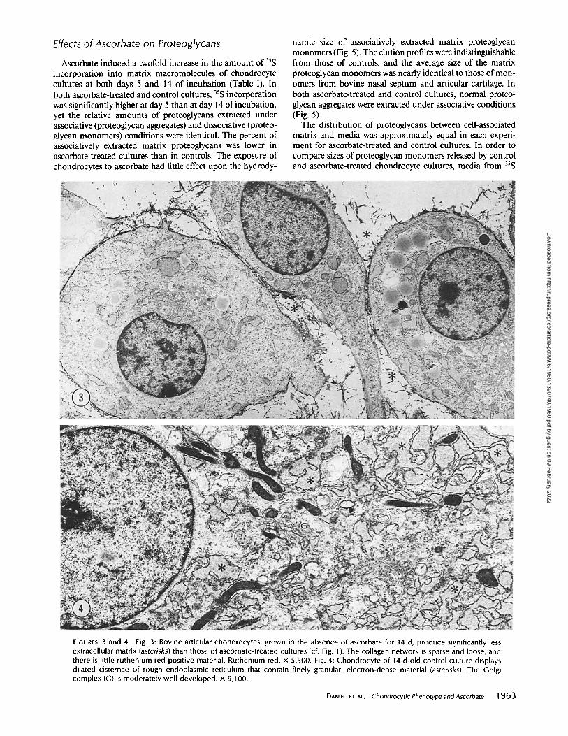
nuclear filaments. Chondrocyte cultures that were not exposed to ascorbate
produced significantly less extracellular matrix (Fig. 3). The density of the matrix framework was decreased, relative to ascorbate-treated cultures, and there was little ruthenium red- positive material. The chondrocytes displayed dilated cister- nae of rough endoplasmic reticulum, which contained a finely granular electron-dense material (Fig. 4).
FIGURES I and 2 Fig. 1: Bovine articular chondrocytes, grown in the presence of ascorbate for 14 d, display well-developed strands of rough endoplasmic reticulum (RER) and Golgi complex (G). Chondrocytes are surrounded by dense rims of ruthenium red-positive territorial matrix (arrow). The extraterritorial matrix is abundant and consists of dense fibrillar collagen. Ruthenium red, x 4,500. Fig. 2: The extraterritorial matrix of a 14-d-old, ascorbate-treated chondrocyte culture consists of a rather dense network of collagen fibrils. Globules of ruthenium red-positive proteoglycans are present at regular intervals along the collagen fibrils (arrow). Ruthenium red, x 27,000.
1962 THE JOURNAL OF CELL BIOLOGY • VOLUME 99, 1984
Dow
nloaded from http://rupress.org/jcb/article-pdf/99/6/1960/1390740/1960.pdf by guest on 09 February 2022
Effects of Ascorbate on Proteoglycans
Ascorbate induced a twofold increase in the amount of 35S incorporation into matrix macromolecules of chondrocyte cultures at both days 5 and 14 of incubation (Table I). In both ascorbate-treated and control cultures, 35S incorporation was significantly higher at day 5 than at day 14 of incubation, yet the relative amounts of proteoglycans extracted under associative (proteoglycan aggregates) and dissociative (proteo- glycan monomers) conditions were identical. The percent of associatively extracted matrix proteoglycans was lower in ascorbate-treated cultures than in controls. The exposure of chondrocytes to ascorbate had little effect upon the hydrody-
namic size of associatively extracted matrix proteoglycan monomers (Fig. 5). The elution profiles were indistinguishable from those of controls, and the average size of the matrix proteoglycan monomers was nearly identical to those of mon- omers from bovine nasal septum and articular cartilage. In both ascorbate-treated and control cultures, normal proteo- glycan aggregates were extracted under associative conditions (Fig. 5).
The distribution of proteoglycans between cell-associated matrix and media was approximately equal in each experi- ment for ascorbate-treated and control cultures. In order to compare sizes of proteoglycan monomers released by control and ascorbate-treated chondrocyte cultures, media from 35S
FIGURES 3 and 4 Fig. 3: Bovine articular chondrocytes, grown in the absence of ascorbate for 14 d, produce significantly less extracellular matrix (asterisks) than those of ascorbate-treated cultures (cf. Fig. 1). The collagen network is sparse and loose, and there is little ruthenium red-positive material. Ruthenium red, x 5,500. Fig. 4: Chondrocyte of 14-d-old control culture displays dilated cisternae of rough endoplasmic reticulum that contain finely granular, electron-dense material (asterisks). The Golgi complex (G) is moderately well-developed, x 9,100.
DAN,EL ET AL. Chondrocytic Phenotype andAscorbate 1963
Dow
nloaded from http://rupress.org/jcb/article-pdf/99/6/1960/1390740/1960.pdf by guest on 09 February 2022

TABLE I
Matrix Proteoglycans in 5- and 14-d Cultures
Control Ascorbate-treated
cpm extracted % extracted cpm extracted % extracted
Day 5 Day 14 Day 5 Day 14 Day 5 Day 14 Day 5 Day 14 cpm %
Associative extract (0.5 M GdnHCl) 39,046 24,235 59.5 57.9 Dissociative extract (4.0 M GdnHCl) 19,711 11,720 30.0 28.0 Papain extract 6,920 5,928 10.5 14.1 Total 65,677 41,883
cpm % 65,339 37,953 45.7 46.2 60,083 38,178 42.0 46.5 17,694 5,977 12.3 7.3
143,116 82,108
Ascorbate:control total ratio = 2.0. cpm, counts per minute .
0
x
D= 0
16 I 20 24 28 32
VO Fraction Number
36
pulse-labeled cultures were dissociatively extracted and chro- matographed on a Sepharose CL-2B column (Fig. 6). The major species of medium proteoglycan monomer had an average partition coefficient that was practically identical to those of monomers in cartilage explants and in parent tissues.
C o l l a g e n B iosynthes is
h collagenase digestion experiment was performed to de- termine the influence of ascorbate on total protein and col- lagen synthesis and the distribution of labeled proteins be- tween the cell layer and the medium (Table II). Ascorbate treatment stimulated [3H]proline incorporation into protein. Labeling ratios of ascorbate-treated to control cultures were 2.6 for the cell layer and 1.6 for the medium. Corresponding ratios for collagenase-sensitive proteins were 2.7 for the cell layer and 13.2 for the medium, indicating a marked stimula- tion of collagen released into the media of ascorbate-treated cells.
Collagen type analysis was used to monitor the phenotypic expression of ascorbate-exposed chondrocytes in long-term
1964 THE JOURNAL OF CELL BIOLOGY • VOLUME 99, 1984
1
FIGURE 5 Sepharose CL-2B elution profiles of 3SS-labeled proteoglycans, chromatographed under associative conditions. Chondrocyte cultures are pulse labeled with Na235SO4 on d 14, and the cell layers extracted in 0.5 M GdnHCl. Aliquots of the extracts from ascorbate-treated (filled circles) and con- trol (empty circles) cultures are placed on the column (the arrow indicates the location of the peak obtained from bo- vine nasal septum monomer [A1 D1 ]).
4o 44 [
V t
culture. Collagens were analyzed after limited pepsinization and neutral salt extraction to ensure maximum recovery of collagen from the cell layers and to remove noncollagenous peptides, as well as to cleave procollagen peptides. Successive extractions with acid and neutral salt removed 97% of the total collagenase susceptible counts from the cell-associated matrices. The majority of the cell-associated matrix collagens in long-term labeled, ascorbate-treated chondrocyte cultures was present in a band that co-migrated with the a~-(I) cartier. A minor collagen fraction migrated with a slightly slower mobility, and co-electrophoresed with a~-(V) chains (Fig. 7). There was a trace amount of collagen that co-migrated with a2-(I) chains. Pulse-labeled cell-associated matrix collagens from control cultures were also electrophoresed for a shorter time to facilitate visualization of low-molecular-weight bands (Fig. 8). Under this condition the a~ and B bands showed little separation. Both the a, and B bands were sensitive to bacterial collagenase, whereas all of the low-molecular-weight species were insensitive. The control cell-associated matrix did not exhibit an a2 component, indicating a complete lack of type I collagen biosynthesis, although we can not rule out
Dow
nloaded from http://rupress.org/jcb/article-pdf/99/6/1960/1390740/1960.pdf by guest on 09 February 2022

i . Vo
;~4 2'a
? o
x
0.. o
a2 aa 4¢ ' 4~ 4`8 s: I /
Fract ion Number Vt
TABLE II
Total Protein and Collagen Synthesis in 5-d Chondrocyte Cultures
FIGURE 6 Sepharose CL-2B elution profiles of 3SS-labeled proteoglycans, chromatographed under dissociative conditions. Dish cultures are pulse la- beled with Na23SSO4 on day 14, and the medium adjusted to 4M GdnHCI using 8 M GdnHCl. Aliquots of the ascorbate- treated (filled circles} and control (empty circles} culture extracts are placed on the column (the arrow indicates the lo- cation of the peak obtained from bovine nasal septum monomer [A1 D1 ]).
Cell layer Media
Percent colla- . Percent
Total protein* Collagen* gen s Total protein Collagen collagen cpm % cpm %
Control 27,918 _+ 2,906 7,150 + 1,238 4.5 33,285 _ 3,551 3,297 + 205 1.8 Ascorbate 71,378 _+ 4,670 19,185 _+ 4,276 4.7 52,283 _ 691 43,444 + 2,122 13 Ascorbate:control 2.6 2.7 1.6 13.2
* Total counts per minute (cpm) of proline incorporated into trichloroacetic acid-precipitable protein after hyaluroidase treatment. Mean and SD of three determinations.
* Collagenase-released cpm. Mean and SD of three determinations. i Calculated according to the formula: (collagen cpm x 100)/(noncollagen cpm x F) + collagen cpm, where F = 5.4 to reflect the amount of enrichment of
proline in collagen with respect to the average protein {25).
synthesis of type I trimer. Over a 2-wk period control chondrocytes synthesized and
released types II and III collagens, and an uncharacterized chain, which co-migrated with a~-(V), aE-Chains were not observed (see reference 13). However, ascorbate-treated cells released a2-chains into the medium (Fig. 9). Appropriate bands were excised from the gels and subjected to scintillation counting. Control medium contained no radioactivity in the a 2 region, even after 2 wk of culture (Table III). In the medium of ascorbate-treated cultures, however, the Otl;a2 ratios fell from 3.6 at day 5 to 2.7 at day 14. These data indicated that increasing amounts of type I collagen were released into the culture medium with time. Upon reduction, the collagenase- sensitive material, which originally ran near the top of the gel (Fig. 9), migrated as an a-sized chain (Fig. 10). Such behavior is characteristic of type III collagen, although further analysis will be required to verify the identity of this material.
Cells were pulse labeled with [3H]proline on day 14, to characterize the collagens that were synthesized late in the culture period and retained in the extracellular matrix (Fig. 11). Both ascorbate-treated and control matrices contained at-chains, and chains that co-migrated with a, (V), but neither contained a2-chains, nor putative type III chains. Ascorbate- treated, pulse-labeled matrix collagens were further quanti- tated to confirm the fluorographic data. Labeled collagens, along with unlabeled carrier collagens, were electrophoresed in tube gels, which were stained, sliced into 1-mm sections, and the slices counted in a scintillation counter (Fig. 12). The complete lack of a2-chains confirmed that there was no type I collagen retained in the matrix, even at a time when the a~:a2 ratio in the medium was 2.5:1.
The reversibility of the ascorbate effect was tested by grow- ing chondrocytes in ascorbate-supplemented medium for 12 d, followed by growth in ascorbate-free medium for 5 d. After
DANIEL ET AL. Chondrocytic Phenotype andAscorbate 1965
Dow
nloaded from http://rupress.org/jcb/article-pdf/99/6/1960/1390740/1960.pdf by guest on 09 February 2022

FIGURE 7 Fluorographof3H-labeled collagens from cell layer. Ascorbate-treated chondrocyte cultures are labeled with 5 #Ci/ml SH[proline] for 14 d and the collagens extracted as de- scribed in Materials and Methods. The colla- gens are electrophoresed on a 6% gel and fluorographed (only the upper portion of the gel is shown; the lower portion does not reveal any collagenase-sensitive bands). The positions of al (I) chains, a2 (I) chains, and fl-chains are indicated. B designates the position of an un- characterized collagen, which migrates in the position of ~ (V).
rices were abundant and consisted of dense networks of collagen fibrils, interdispersed with ruthenium red-positive proteoglycans. In the absence of ascorbate, bovine articular chondrocytes showed dilated cisternae of rough endoplasmic reticulum and sparse extracellular matrix. These findings were similar to those reported for chick embryonic chondrocytes in vitro (21). Chick chondrocytes, grown in the absence of ascorbate, displayed extremely dilated cisternae of rough en- doplasmic reticulum, a condition that was rapidly reversed by addition of ascorbate to the culture medium. In the same system, the typical 65-nm cross-banding pattern of collagen fibrils was only observed in ascorbate-treated cultures.
The proteoglycans synthesized by bovine articular chondro- cytes were characteristic of hyaline cartilage. Their hydrody- namic size was indistinguishable from that of proteoglycans extracted from slices of bovine articular cartilage, from which
FIGURE 8 Fluorograph of 3H[pro- line], pulse-labeled collagens from control cell-associated matrix. Con- trol chondrocytes, grown in 75-cm 2 flasks for 5 d, are pulse labeled for 18 h with 5/~Ci/ml 3H[proline]. The collagens are extracted, electropho- resed on a 6% polyacrylamide gel, and fluorographed. In this and sub- sequent gels, approximately equal amounts of radioactive protein was added to each channel. Channel 1 is exposed to purified bacterial colla- genase before electrophoresis. Channel 2 is untreated. The position of ~1 (I) chains is indicated (the entire gel is shown).
2 d in ascorbate-free medium the a2-chains, previously syn- thesized and released by these cells, were no longer evident (Fig. 10).
DISCUSSION
In the present study, we examined the influence of ascorbate on the phenotypic expression of bovine articular chondrocytes in vitro. In the presence of ascorbate, these chondrocytes displayed prominent rough endoplasmic reticulum, Golgi complex, and polysomes. Territorial and extraterritorial mat-
1966 THE JOURNAL OF CELL BIOLOGY • VOLUME 99, 1984
FIGURE 9 Fluorograph of 3H-labeled collagens isolated from as- corbate-treated medium. The isolated collagens are electropho- resed on 6% polyacrylamide gels and fluorographed. The media are collected from the roller bottles after cultures have been labeled from days 0 to 2 ( 1 ), from days 2 to 4 (2), from days 4 to 7 (3), from days 7 to 9 (4), from days 9 to 11 (5), and from days 11 to 14 (6). The positions of a, (I) and a2 (I) chains are indicated.
TABLE III
Quantitation of Collagen Types in Media of Ascorbate-treated and Control Cultures at Days 5 and 14 of Incubation
Ascorbate- Control treated
Collagen type Day 5 Day 14 Day 5 Day 14 % cpm % cpm
Type III 52 68 87 46 Unknown chain (B) 12 11.5 1.5 6 al 36 20 9 35 a2 0.1 0.5 2.5 13 ~1:~2 360 40 3.6 2.7
Values are given as the percentage of counts per minute per collagen region.
Dow
nloaded from http://rupress.org/jcb/article-pdf/99/6/1960/1390740/1960.pdf by guest on 09 February 2022

FIGURE 11 Fluorograph of 3H[proline], pulse-la- beled cell-layer collagens. Roller bottles of ascorbate- treated and control cul- tures are pulse labeled for 12 h with 5 #Ci/ml 3H- [proline] on day 14 of cul- ture. Channel A shows the collagen extracted from the control cell layer, and B shows the collagen ex- tracted from the ascorbate- treated cell layer. The up- per portion of the gel, con- taining the a-chains, has been magnified. The lower portion does not reveal any collagenase-sensitive bands.
FIGURE 10 Fluorographs of 3H-labeled collagens isolated from as- corbate-treated and post-ascorbate-treated cell culture media. The media are collected from the roller bottles after labeling the cultures with 3H[proline] from days 2 to 4 (1), from days 4 to 7 (2), and from days 7 to 9 (3). Ascorbate is removed on day 12 and the cells are exposed to ascorbate-free medium. Ascorbate-free cultures are labeled with 3H[proline] from days 13 to 14 (4) and from days 14 to 17 (5). After reduction with 1%/~-mercaptoethanol, the media collagens are electrophoresed on a 6% polyacrylamide gel.
the chondrocytes had been isolated. There was a twofold increase in the amount of 35S-labeled proteoglycan retained by the ascorbate-treated, cell-associated matrix, relative to controls, at both days 5 and 14. The ascorbate-treated cell- associated proteoglycans were less extractable under associa- tive conditions than were those of control cultures. This phenomenon was observed in both short-term (5-d) and long- term (14-d) cultures. The difference in extractability might reflect a higher molecular organization of the matrix in ascor- bate-treated cultures.
Despite these differences in extractability of proteoglycan aggregates, control and ascorbate-treated cultures were similar in the following ways: (a) both ascorbate-treated and control cells produced abundant extracellular matrix, which was rich in cartilage proteoglycans; (b) proteoglycans synthesized by bovine articular chondrocytes and released into the medium, represented about one-half of the total proteoglycan elabo- rated; (c) the molecular sizes of proteoglycan monomers from matrix and medium were identical in ascorbate-treated and control cultures. Proteoglycans of small hydrodynamic size, characteristic of fibroblasts, were not observed.
The matrix collagens of control and ascorbate-treated cul- tures were similar, with type II collagen predominating. Al- though we did not directly examine for the presence of type 1 trimer, a CnBr peptide analysis of the collagen control cell layers confirmed the presence of type II collagen with no detectable type I peptides (13). The media of both ascorbate- treated and control cultures contained a high-molecular- weight collagen. After reduction, this collagen migrated as an
a-sized chain, and was probably type III collagen. The pre- dominance of this type III-like collagen in the media might indicate that this collagen is incompletely processed or present in a pro-a, (III) form. This has been suggested for rabbit chondrocyte (2) as well as for fibroblast cultures (l 7). It should also be noted from studies by Benya and Nimni (l) that long- term cultures of cartilage slices contained up to 8% type III collagen. The significance of high levels of type III collagen synthesis by our chondrocyte cultures is unclear.
In addition the media of ascorbate-treated cultures con- tained a2-chains, which were indicative of type I collagen. The a2 chain of type I collagen was evident 2 d after initiation of ascorbate treatment. The ratio a,:a2 ratio approached that of type I collagen by 9 d in culture, and remained stable thereafter. The ascorbate effect was reversible, as a2-chains disappeared from the medium after removal of the vitamin. Ascorbate-treated cells, which were pulse labeled with [3H]- proline at day 14, synthesized type II collagen, some of which was incorporated into the matrix. There was no indication of a2-chains in the matrix. Thus, the cells seemed to concurrently synthesize a variety of collagen types, some of which were selectively incorporated into the matrix. The synthesis and release of type I collagen into the medium ofascorbate-treated cultures may be due to the presence of two distinct cell populations. One cell population may require ascorbate to produce type I collagen, while the other may remain insensi- tive to the vitamin.
Our findings are at variance with those of Capasso et al. (7), who found no type I collagen in ascorbate-treated cultures of chick embryonic chondrocytes. The reason for this discrep- ancy may be a different ascorbate-treatment schedule. Al- though the ascorbate concentrations were similar in both studies, Capasso et al. (7) added fresh ascorbate every 3-4 d, as contrasted to every 2 d in our studies. Given the short half- life of this vitamin in media and the rapid reversal of the ascorbate effect, maintenance of a minimal ascorbate level may be critical for type I collagen biosynthesis. In no case did
DANIEL ET AL. Chondrocytic Phenotype andAscorbate 1967
Dow
nloaded from http://rupress.org/jcb/article-pdf/99/6/1960/1390740/1960.pdf by guest on 09 February 2022

311 B 01 O 2
I ; I
t~
b X
:E
o
20
FIGURE 12 T u b e gel o f a s c o r b a t e - treated cell layer collagens (Fig. 11 ) is cut into 1-mm sections, dissolved in NCS, and counted for radioactivity. The ar- rows indicate migration distances of ~1, a2, and the uncharacterized B chains.
5 10 15 20 25 30
Slice Number
~¢e observe the short chain collagen which has been observed in other systems (28). However, we have observed this short chain collagen in control and ascorbate-treated chick chon- drocyte cultures (Daniel, J. C., manuscript in preparation).
There is extensive literature on the effects of ascorbate on connective tisssue cells in vitro. It is generally acknowledged that ascorbate acts as a co-factor for the lysyl- and prolyl- hydroxylases (5). Hydroxylation of proline to hydroxyproline stabilizes the collagen triple helix and is necessary for normal collagen secretion (11, 24). Alternatively, it has been suggested that ascorbate can influence the phenotypic expression of fibroblasts with respect to collagen synthesis (23). Ascorbate may regulate collagen production by independently control- ling collagen polypeptide synthesis, posttranslational hydrox- ylations, and the activities of the two hydroxylases. A 4-d exposure of fibroblasts to ascorbate caused a twofold increase in the amount of procollagen mRNA, yet no change in the amount of noncollagenous mRNA was observed (30). Bovine smooth muscle cells required ascorbate in their growth me- dium in order to maintain their morphology (29). In this system, ascorbate increased protein synthesis, but decreased cell-doubling times. Other investigators, using rabbit smooth muscle cells, have noted that ascorbate decreased the amount of insoluble elastin that was synthesized and incorporated into the extracellular matrix (10). These differences may be related to ascorbate-induced alterations in both the hydroxylation of proline and the cross-linking of lysine.
It has long been known that agents, such as chick embryo extract (27), BudR (20), and high concentrations of potassium (8), induce phenotypic instability in chondrocyte cultures. Repeated cell passage also causes a modulation of phenotype sometimes referred to as "dedifferentiation," which has been shown to be reversible under specific culture conditions (3).
1968 THe JOURNAL OF CELL BIOLOGY • VOLUME 99, 1984
In each case, cell morphology, as well as collagen and proteo- glycan biosynthesis, are altered. In the present experiments, only collagen expression is altered, whereas proteoglycan hy- drodynamic size is unaffected. Recent immunohistochemical studies have shown that polygonal, as well as fibroblastlike chick chondrocytes in monolayer culture can synthesize type I collagen (9, 32). These data suggest that the various pheno- typic traits of chondrocytes are not necessarily coordinately expressed. Several different phenotypic properties should be examined whenever one is attempting to characterize the effects of any agent upon chondrocytes in vitro. In light of our results, caution must be exercised when interpreting data from ascorbate-exposed cultures. The failure to see normal type II collagen from long-term monolayer cultures of ar- ticular chondrocytes may be explained by the routine use of ascorbate in the culture medium (16) or the analysis of combined cell layers and media.
Received for publication 19May1984, and in revised form 27July 1984.
REFERENCES
1. Benya, P. D., and M. E. Nimni. 1979. The stability of the collagen phenotype during stimulated collagen, glycosaminoglycan, and DNA synthese by articular cartilage organ slices. Arch. Biochem. Biophys. 192:327-335.
2. Benya, P. D., S. R. Padina, and M. E. Nimni. 1977. The progeny of rabbit articular chondrocytes synthesize collagen types 1 and Ill and type I trimer but not type ll: verification by cyanogen bromide peptide analysis. Biochemistry. Z 16:865-872.
3. Benya, P. D., and J. D. Shaffer. 1982. Dedifferentiated chondrocytes rcexpress the differentiated phenotype when cultured in agarose. Cell. 20:215-224.
4. Bitter, T., and H. Muir. 1962. A modified uronic acid carbazole reaction. Anal. Biochem. 4:320-334.
5. Blanck, T. J. J., and B. Peterkofsky. 1975. The stimulation of collagen secretion by ascorbate as a result of increased proline hydroxylation in chick embry fibroblasts. Arch. Biochem. Biophys. 171:250-26 I.
6. Bonner, W. M., and R. A. Laskey. 1974. A film detection method for tritium-labelled proteins and nucleic acids in polyacrylamide gels. Eur. £ Biochem. 46:83-88.
Dow
nloaded from http://rupress.org/jcb/article-pdf/99/6/1960/1390740/1960.pdf by guest on 09 February 2022

7. Capasso, O., E. Gionti, G. Pontarelli, F. S. Ambesi-lmpiombato, L. Nitch. G. Tajana, and R. Cancedda. 1982. The culture of chick emhry chondrocytes and the control of their differentiated function in vitro. Exp. Cell Res. 142:197-206.
8. Daniel, J. C., R. A. Kosher, J. E. Hamos, and J. W. Lash. 1974. Influence of external potassium on the synthesis and deposition of matrix components by chondrocytes in vitro. J. Cell. Biol. 63:843-854.
9. Dorfman, A., B. M. Vertel, and N. B. Schwartz. 1980. Immunological methods in the study of chondroitin sulfate proteoglycans. Curt. Top. Dev. Biol. 14:169-198.
10. Dunn, D. M., and C. Franzhlau. 1982. Effects of ascorbate on insoluble elastin accu- mulation and cross-link formation in rabbit pulmonary artery smooth muscle cultures. Biochemistry. J. 21:4195-4202.
11. Fessler, J. H., and L. I. Fessler. 1978. Biosynthesis of procollagen. Annu. Rev. Biochem. 47:129-162.
12. Kimura, J. H., C. B. Caputo, and V. C. Hascall. 1981. The effect of cycloheximide on synthesis of proteoglycans by cultured chondrocytes from the Swarm rat chondrosar- coma. Z Biol. Chem. 256:4368-4376.
13. Kuetmer, K. E., V. A. Memoli, B. U. Pauli, N. C. Wrob¢l, E. J.-M. A. Thonar, and J. C. Daniel. 1982. Synthesis of cartilage matrix by mammalian cbondrocytes in vitro. II. Maintenance of collagen and proteoglycan phenotype. J. Cell Biol. 93:751-757.
14. Kuetmer, K. E., B. U. Pauli, G. Gall, V. A. Memoli, and R. K. Schenk. 1982. Synthesis of cartilage matrix by mammalian chondrocytes in vitro. I. Isolation, culture character- isties, and morphology. Z Cell Biol. 93:743-750.
15. Laemmli, U. K. 1970. Cleavage 9f structural protein during the assembly of head of bacteriophage 1"4. Nature (Lond.). 227:680-685.
16. Layman, D. L., L. Sokoloff, and E. J. Miller. 1972. Collagen synthesis by articular chondrocytes in monolayer culture. Exp. Cell Res. 73:107-112.
17. Lichtenstein, J. R., P. H. Byers, B. D. Smith, andG. R. Martin. 1975. Identification of the eollagenoos proteins synthesized by cultured ceils from human skin. Biochem. J. 14:589-1594.
18. Mayne, R., J. R. Schlitz, and H. Holtzer. 1973. Some overt and covert properties of chondrogenic cells. In Biology of the Fibroblast. E. Kulonen and J. Pikkarainen, editors. Academic Press, Inc., New York. 61-68.
19. Mayne, R., M. S. Vail, P. M. Mayne, and E. J. Miller. 1976. Changes in type of collagen synthesized as clones of chick chondrocytes grow and eventually lose division capacity.
Proc. Natl. Acad. Sci. USA 72:1674-1678, 20. Mayne, R.,M.S.Vail, andE.J. Miller. 1975. Analysis ofchanges in collagen biosynthesis
that occur when chick chondrocytes are grown in 5-bromo-2'-deoxyuridine. Proc. Natl. Acad. Sci. USA. 72:4511-4515.
21. Meier, S., and M. Solursh. 1978. Ultrastructural analysis of the effect of ascorbic acid on secretion and assembly of extracellular matrix by cultured chick embryo choodro- cytes. J. Ultrastruct. Res. 65:48-59.
22. Miller, E. J., and R. K. Rhodes. 1982. Preparation and characterization of the different types of collagen. Methods Enzymol. 82:33-64.
23. Murad, S., D. Grove, K. A. Lindberg, G. Reynolds, A. Sivarajah, and S. R. Pinnell. 1981. Regulation of collagen synthesis by ascorbic acid. Proc. Natl. Acod. Sci. USA 78:2879-2882.
24. Peterkofsky, B. 1972. Regulation of collagen secretion by ascorhic acid in 3T3 and chick embryo fibroblasts. Biochem. Biophys. Res. Commun. 49:1343-1350.
25. Peterkofsky, B. 1982. Bacterial collagenase. Methods Enzymol. 82:453-471. 26. Sajdera, S. W., and V. C. Hascall. 1969. Proteinpolysaceharide complex from bovine
nasal cartilage. J. Biol. Chem. 244:77-87. 27. Schlitz, J. R., and S. Ward. 1980. Effects of chick embryo extract fractions on collagen
and glycosaminoglycan metabolism by chick chondroblasts. Biochim. Biophys. Acta. 628:343-354.
28. Schmid, T. M., and H. E. Conrad. 1982. A unique how molecular collagen secreted by cultured chick embryo chondrocytes. J. Biol. Chem. 257:12444-12450.
29. Schwartz, E., R. S. Bienkowski, B. Coltoff-Schifier, S. Goldfischer, and O. O. Blumenfeld. 1982. Changes in the components of extracellular matrix and in growth properties of cultured aortic smooth muscle cells upon ascorbate feeding. J. Cell Biol. 92:462-470.
30. Tajima, S., and S. R. Pinnell. 1982. Regulation of conagen synthesis by ascorbic acid. Ascorbic acid increases type 1 procollagen mRNA. Biochem. Biophys. Res. Commun. 106:632-637.
31. Von der Mark, H., and K. Von der Mark. 1979. Isolation and characterization of collagen A and B chains from chick embryos. FEBS (Fed. Eur. Biochem. Soc.) Lett. 99:101-105.
32. Von der Mark, K. V. Gauss, J. Von der Mark, and P. Muller. 1977. Relationship between cell shape and type of collagen synthesized as chondrocytes lose their cartilage phenotype in culture. Nature (Lond.). 267:531-532.
DANIEL ET At . Chondrocytic Phenotype and Ascorbate 1969
Dow
nloaded from http://rupress.org/jcb/article-pdf/99/6/1960/1390740/1960.pdf by guest on 09 February 2022
![Exercise – evidence of a benefit and biological actions3. Anti-apoptopic effects on chondrocytes reducing cartilage degeneration [Shen]. 4. Modulation metalloproteinases - remodeling](https://static.fdocuments.in/doc/165x107/5f9e513a79dfeb15207b3972/exercise-a-evidence-of-a-benefit-and-biological-3-anti-apoptopic-effects-on-chondrocytes.jpg)