Synthesis, Biological Activity, Molecular Docking and ... · PDF fileSynthesis, Biological...
11
International Journal of Scientific & Engineering Research, Volume 6, Issue 3, March-2015 1546 ISSN 2229-5518 IJSER © 2015 http://www.ijser.org Synthesis, Biological Activity, Molecular Docking and Structural Studies of N-(O- Fluor phenyl)-4,6-Dimethyl-2-Pyridone-3- Nitrile Subhadramma, S1., Saravanan, J2., Vinaya Kumar, R3 and Naveen Chandra4* Abstract: The title compound crystallizes in space group, P21/c with unit cell parameters, a=7.926(5), b=11.156(3), c=14.261(3) Å, β = 102.01(1)°,V = 1233.4(9) Å3 containing four asymmetric molecules. The molecules are packed in the crystal lattice with bifurcated hydrogen bonding of the type C-H---O. Oxygen atom O(1) is involved in the bifurcated hydrogen bonding with bridge distances 3.511(2) Å and 3.466(2) Å, respectively. Fluorine F(1) of the phenyl ring does not involve in either intra- or inter-hydrogen bonding. The dihedral angle between the phenyl group and pyridone ring is 87.7(1)° indicating that pyridonyl group is perpendicular to the phenyl group making the way for its maximum interaction through weak hydrogen bondings. Molecular docking study done on this compound with Phospholipase (PLA2) indicates that there are hydrogen bond and hydrophobic interactions at the active sites on the PLA2. The molecule is stabilized through these interactions with distances varying from 2.5 to 4 Å and hence it is a good candidate for the study of inhibition activity of the enzyme based on its experimental evidences. Also a study of in-vitro biological activity carried out indicates that the compound has antioxidant and hydrogen-peroxide-radical scavenging properties and more potent than ascorbic acid taken as standard. Index Terms- Anti-inflammatory, Active site, Crystal, Molecular Docking, Structure. —————————— —————————— 1. Introduction The title compound is a derivative of the compound, 3- cyano-4,6-dimethyl-2-pyridone (4,6-dimethyl-2- pyridone-3-nitrile), known as 'Guareschi pyridone', has been known for long time perhaps more than a century [1]. It is known that organic compounds containing fluorine atoms exhibit biological and pharmacological significance [2]. The fluorine atoms in such compounds play a very significant role in biomolecular interactions [2, 3]. The study of such interactions involving fluorine atoms in crystal structures has been considered to be of great importance in crystal engineering [4]. It is well known that a number of Schiff’s bases and thiophene derivatives exhibit different kinds of biological activities [5,6,7,8]. In order to study the biological and pharmacological properties different types of thiophene derivatives which have been prepared by us, the synthesized compound is used as a starting material for the synthesis of such derivatives. The synthesized compound has been screened for its anti- bacterial and anti-oxidant properties. The conducted in-vitro study indicates that the compound exhibits significant bacterial inhibition at low-to-moderate concentrations. Also it is nice to know its three dimensional structure as it exhibits a pronounced biological activity. The molecular docking study of the compound with the Phospholipase A 2 (PLA 2 ) enzyme (E.C. 3.1.1.4). shows that the it has anti-inflammatory activity due to atoms involvement in interactions less than 4.0 Å with the atoms of amino acid residues of Phospholipase (PLA 2 ) enzyme. The molecule is stabilized through hydrogen bonding and hydrophobic interactions with distances varying from 2.5 to 4Å, respectively. Also a study of in-vitro biological activity carried out indicates that the compound has anti-oxidant and hydrogen-peroxide-radical-scavenging properties and it can be considered more potent than ascorbic acid taken as standard. The crystal structure of the title compound determined by X-ray method shows that its staking pattern in the crystalline state exhibits along its crystallographic b- axis. The dihedral angle between the phenyl group and pyridone ring is found to be 87.7(1)° indicating that pyridonyl group is perpendicular to the phenyl group and probably making the way for its interaction through hydrogen bonds. 1. is a Research Scholar at Dr MGR Educational and Research Institute, MGR University, Chennai, Presently she is working as an associate professor of Physics, Vijaya College, Bangalore, India. E-mail: [email protected] 2. is a Professor and Head of the Department of Pharmaceutical Chemistry, PES College of Pharmacy Bangalore, India. E-mail: [email protected] 3. is a Professor, Department of Chemistry, KLES’, S. Nijalingappa College, Rajajinagar, Bangalore, India. E-mail: IJSER
Transcript of Synthesis, Biological Activity, Molecular Docking and ... · PDF fileSynthesis, Biological...

International Journal of Scientific & Engineering Research, Volume 6, Issue 3, March-2015 1546 ISSN 2229-5518
IJSER © 2015 http://www.ijser.org
Synthesis, Biological Activity, Molecular Docking and Structural Studies of N-(O-
Fluor phenyl)-4,6-Dimethyl-2-Pyridone-3-Nitrile
Subhadramma, S1., Saravanan, J2., Vinaya Kumar, R3 and Naveen Chandra4*
Abstract: The title compound crystallizes in space group, P21/c with unit cell parameters, a=7.926(5), b=11.156(3), c=14.261(3) Å, β = 102.01(1)°,V = 1233.4(9) Å3 containing four asymmetric molecules. The molecules are packed in the crystal lattice with bifurcated hydrogen bonding of the type C-H---O. Oxygen atom O(1) is involved in the bifurcated hydrogen bonding with bridge distances 3.511(2) Å and 3.466(2) Å, respectively. Fluorine F(1) of the phenyl ring does not involve in either intra- or inter-hydrogen bonding. The dihedral angle between the phenyl group and pyridone ring is 87.7(1)° indicating that pyridonyl group is perpendicular to the phenyl group making the way for its maximum interaction through weak hydrogen bondings. Molecular docking study done on this compound with Phospholipase (PLA2) indicates that there are hydrogen bond and hydrophobic interactions at the active sites on the PLA2. The molecule is stabilized through these interactions with distances varying from 2.5 to 4 Å and hence it is a good candidate for the study of inhibition activity of the enzyme based on its experimental evidences. Also a study of in-vitro biological activity carried out indicates that the compound has antioxidant and hydrogen-peroxide-radical scavenging properties and more potent than ascorbic acid taken as standard.
Index Terms- Anti-inflammatory, Active site, Crystal, Molecular Docking, Structure.
—————————— ——————————
1. Introduction The title compound is a derivative of the compound, 3-cyano-4,6-dimethyl-2-pyridone (4,6-dimethyl-2-pyridone-3-nitrile), known as 'Guareschi pyridone', has been known for long time perhaps more than a century [1]. It is known that organic compounds containing fluorine atoms exhibit biological and pharmacological significance [2]. The fluorine atoms in such compounds play a very significant role in biomolecular interactions [2, 3]. The study of such interactions involving fluorine atoms in crystal structures has been considered to be of great importance in crystal engineering [4]. It is well known that a number of Schiff’s bases and thiophene derivatives exhibit different kinds of biological activities [5,6,7,8]. In order to study the biological and pharmacological properties different types of thiophene derivatives which have been prepared by us, the synthesized compound is used as a starting material for the synthesis of such derivatives. The synthesized compound has been screened for its anti-bacterial and anti-oxidant properties. The conducted in-vitro study indicates that the compound exhibits significant bacterial inhibition at low-to-moderate concentrations. Also it is nice to know its three dimensional structure as it exhibits a pronounced biological activity. The molecular docking study of the compound with the Phospholipase A2 (PLA2) enzyme (E.C. 3.1.1.4). shows that the it has anti-inflammatory activity due to atoms involvement in interactions less than 4.0 Å with
the atoms of amino acid residues of Phospholipase (PLA2) enzyme. The molecule is stabilized through hydrogen bonding and hydrophobic interactions with distances varying from 2.5 to 4Å, respectively. Also a study of in-vitro biological activity carried out indicates that the compound has anti-oxidant and hydrogen-peroxide-radical-scavenging properties and it can be considered more potent than ascorbic acid taken as standard. The crystal structure of the title compound determined by X-ray method shows that its staking pattern in the crystalline state exhibits along its crystallographic b-axis. The dihedral angle between the phenyl group and pyridone ring is found to be 87.7(1)° indicating that pyridonyl group is perpendicular to the phenyl group and probably making the way for its interaction through hydrogen bonds. 1. is a Research Scholar at Dr MGR Educational and Research Institute, MGR University, Chennai, Presently she is working as an associate professor of Physics, Vijaya College, Bangalore, India. E-mail: [email protected] 2. is a Professor and Head of the Department of Pharmaceutical Chemistry, PES College of Pharmacy Bangalore, India. E-mail: [email protected] 3. is a Professor, Department of Chemistry, KLES’, S. Nijalingappa College, Rajajinagar, Bangalore, India. E-mail:
IJSER

International Journal of Scientific & Engineering Research, Volume 6, Issue 3, March-2015 1547 ISSN 2229-5518
IJSER © 2015 http://www.ijser.org
4*.Corresponding author, is a professor (Former Head of the Department of Chemistry), Post-graduate & Research Centre, St.Joseph's College (Autonomous), Langford Road, Bangalore, India. Present Address: Professor, and Coordinator for PG Course in Chemistry, KLES’, S. Nijalingappa College, Rajajinagar, Bangalore, India. E-mail: [email protected] 2. Materials and methods Title compound was synthesized using reactants O-fluoroaniline and ethylcyanoacetate in acetic
anhydride medium by heating the mixture taken in a conical flask for about seven hours at 160-165ºC on an oil bath. The reaction was the initial step for the expected final synthesis of pharmaceutically important thiophenyl derivatives based on the Gewald’s reaction mechanism [9]. The resultant compound obtained from the reaction was found to be the title compound. Its molecular constituents were found to be C14H11FN2O. Its experimentally determined melting point was found to be 156°C. The yield of the product was found to be more than 90%. The probable reaction scheme could be represented as:
F
NH2 N O
F
CH3
CN
CH3+O
CH2
CN
O C2H5
Fig. 1. Schematic representation of the reaction.
3. X-Ray Crystal Structure Determination 3.1. Crystallization of the Compound Single crystals of the compound were obtained from a solution in isopropanol by slow evaporation technique [10] at room temperature. Several pale yellow parallelepiped crystals were obtained. 3.2. X-ray Data Collection, Reduction and Structure Solution A suitable crystal was selected after examining it under an analytical polarizing microscope for its uniform extinction for data collection. Unit cell parameters were obtained from a set of different weighted intensity reciprocal lattice points. These parameters were refined to obtain unit cell parameters and based on these parameters a set of weighted intensity reflections data were collected after correcting for Lorentz and polarization effects [10]. These intensities were then put on an absolute scale and used in the structure determination of the molecule by direct method of phase determination [10]. The structure of the molecule thus obtained was refined by the several cycles of the least-squares method by incorporating positional parameters and isothermal parameters followed by anisothermal parameters for non-hydrogen atoms. All the positions of hydrogen atoms were geometrically fixed and their parameters were not refined. The final values of R and Rw as 0.0407 and 0.1040 were obtained,
respectively. The refinement of the structure was considered to be satisfactory as parameters obtained were within the allowed range [11,12]. SHELXL97 [13] was used in the structure determination and refinement of the structure. The data collection parameters and other associated parameters are given in Table 1.
4. Results and Discussion
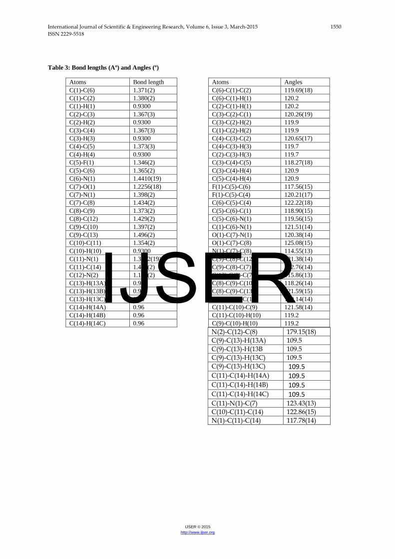
The final fractional coordinates and the equivalent isothermal parameters of the non-hydrogen-atoms along with the hydrogen atoms are given in Table 2. The bond distances and angles are given in Table 3. A molecular ORTEP [14] diagram representing the thermal ellipsoids plot is depicted in Fig. 2, and the crystal packing of the asymmetric molecules is depicted in Fig 3. The six-membered heterocyclic pyridonyl group has a well-defined diene-like structure with the bond distances C(8) - C(9) and C(10) - C(11) are shorter than the bonds C(7) – C(8) and C(9) – C(10) by more than 3 e.s.d’s and these parameters agree with the reported values [1] . It may be noted that all the bond distances are found to be normal within 3 times of their e.s.d’s. However, the distance between C(5) – F(1) is found to be 1.346 (3) Å slightly less than the expected value of 1.362(3) Å. But, this value agrees with the reported values [11.12]. This may be probably due to the partial negative charge present on the fluorine atom on account of resonance nature of the phenyl group. The molecules are packed nicely in
IJSER

International Journal of Scientific & Engineering Research, Volume 6, Issue 3, March-2015 1548 ISSN 2229-5518
IJSER © 2015 http://www.ijser.org
the crystal lattice with weak bifurcated hydrogen bonding involving C(4)-H(4) and C(8)-H(8) atoms of the pyridone ring with O(1) of carbonyl group of the phenyl with bridge distances 3.511(2) Å and 3.466(2)
Å, respectively. The display of the hydrogen bonding connecting molecules occurs in layers along the longest crystallographic c-axis (Fig.3). [15,16].
Table 1. Shows the crystal unit cell parameters and other associated data collection Parameters.
Crystal Data Identification code Shelexl Empirical formula C14 H11 F N2 O Formula weight 242.25 Temperature 293(2) K Wavelength 0.71073 Å Crystal system, space group Monoclinic, P21/c
Unit cell dimensions
a = 7.926(5) A alpha = 90.000(2) deg. b = 11.156(3) A beta = 102.007(2) deg c = 14.261(3) A gamma = 90.000(4) deg.
Volume 1233.4(9) Å3 Z, Calculated density 4, 1.305 Mg/m3
Absorption coefficient 0.094 mm-1 F(000) 504 Crystal size 0.30 x 0.25 x 0.20 mm Theta range for data collection 2.34 to 26.83 deg. Limiting indices -10<=h<=9, -14<=k<=14, -18<=l<=12 Reflections collected / unique 11917 / 2630 [R(int) = 0.0288] Completeness to theta = 26.83 99.8 % Absorption correction Semi-empirical from equivalents Max. and min. transmission 0.9814 and 0.9222 Refinement method Full-matrix least-squares on F2
Data / restraints / parameters 2630 / 0 / 165 Goodness-of-fit on F^2 1.048 Final R indices [I ≥2σ (I)] R1 = 0.0407, wR2 = 0.1040 R indices (all data) R1 = 0.0628, wR2 = 0.1186 Largest diff. peak and hole 0.200 and -0.188 e. Å-3
IJSER

International Journal of Scientific & Engineering Research, Volume 6, Issue 3, March-2015 1549 ISSN 2229-5518
IJSER © 2015 http://www.ijser.org
Table 2. The final fractional coordinate (X104 Å) and the equivalent isothermal parameters (X102 Å2) of the non-hydrogen atoms and hydrogen atoms.
Atom X Y Z U (eq)
C (1) 8739(2) 2486(2) -882(1) 57(1)
C (2) 9695(3) 2002(2) -1495(1) 67(1)
C (3) 9161(3) 974(2) -1991(1) 65(1)
C (4) 7682(3) 410(2) -1885(1) 65(1)
C (5) 6760(2) 895(2) -1263(1) 53(1)
C (6) 7260(2) 1924(1) -764(1) 43(1)
C (7) 4865(2) 3169(1) -599(1) 41(1)
C (8) 3832(2) 3648(1) 27(1) 39(1)
C (9) 4087(2) 3351(1) 981(1) 44(1)
C (10) 5441(2) 2575(2) 1358(1) 47(1)
C (11) 6492(2) 2124(1) 808(1) 44(1)
C (12) 2504(2) 4463(2) -402(1) 44(1)
C (13) 2980(3) 3858(2) 1615(1) 67(1)
C (14) 7960(2) 1316(2) 1191(1) 64(1)
N (1) 6201(2) 2415(1) -150(1) 40(1)
N (2) 1450(2) 5124(2) -735(1) 61(1)
O (1) 4645(2) 3372(1) -1460(1) 60(1)
F (1) 5291(2) 372(1) -1141(1) 83(1)
H (1) 9097 3191 -552 69
H (2) 10707 2376 -1571 80
H (3) 9810 657 -2405 78
H (4) 7310 -286 -2225 78
H (10) 5625 2362 2001 57
H (13A) 3303 4675 1768 101
H(13B) 3134 3397 2195 101
H(13C) 1792 3826 1289 101
H(14A) 7961 1128 1848 97
H(14B) 9023 1707 1152 97
H(14C) 7847 591 821 97
IJSER
International Journal of Scientific & Engineering Research, Volume 6, Issue 3, March-2015 1550 ISSN 2229-5518
IJSER © 2015 http://www.ijser.org
Table 3: Bond lengths (Aº) and Angles (º)
Atoms Bond length Atoms Angles C(1)-C(6) 1.371(2)
C(6)-C(1)-C(2) 119.69(18)
C(1)-C(2) 1.380(2)
C(6)-C(1)-H(1) 120.2 C(1)-H(1) 0.9300
C(2)-C(1)-H(1) 120.2
C(2)-C(3) 1.367(3)
C(3)-C(2)-C(1) 120.26(19) C(2)-H(2) 0.9300
C(3)-C(2)-H(2) 119.9
C(3)-C(4) 1.367(3)
C(1)-C(2)-H(2) 119.9 C(3)-H(3) 0.9300
C(4)-C(3)-C(2) 120.65(17)
C(4)-C(5) 1.373(3)
C(4)-C(3)-H(3) 119.7 C(4)-H(4) 0.9300
C(2)-C(3)-H(3) 119.7
C(5)-F(1) 1.346(2)
C(3)-C(4)-C(5) 118.27(18) C(5)-C(6) 1.365(2)
C(3)-C(4)-H(4) 120.9
C(6)-N(1) 1.4410(19)
C(5)-C(4)-H(4) 120.9 C(7)-O(1) 1.2256(18)
F(1)-C(5)-C(6) 117.56(15)
C(7)-N(1) 1.398(2)
F(1)-C(5)-C(4) 120.21(17) C(7)-C(8) 1.434(2)
C(6)-C(5)-C(4) 122.22(18)
C(8)-C(9) 1.373(2)
C(5)-C(6)-C(1) 118.90(15) C(8)-C(12) 1.429(2)
C(5)-C(6)-N(1) 119.56(15)
C(9)-C(10) 1.397(2)
C(1)-C(6)-N(1) 121.51(14) C(9)-C(13) 1.496(2)
O(1)-C(7)-N(1) 120.38(14)
C(10)-C(11) 1.354(2)
O(1)-C(7)-C(8) 125.08(15) C(10)-H(10) 0.9300
N(1)-C(7)-C(8) 114.55(13)
C(11)-N(1) 1.3762(19)
C(9)-C(8)-C(12) 121.38(14) C(11)-C(14) 1.483(2)
C(9)-C(8)-C(7) 122.76(14)
C(12)-N(2) 1.141(2)
C(12)-C(8)-C(7) 115.86(13) C(13)-H(13A) 0.96
C(8)-C(9)-C(10) 118.26(14)
C(13)-H(13B) 0.96
C(8)-C(9)-C(13) 121.59(15) C(13)-H(13C) 0.96
C(10)-C(9)-C(13) 120.14(14)
C(14)-H(14A) 0.96
C(11)-C(10)-C(9) 121.58(14) C(14)-H(14B) 0.96
C(11)-C(10)-H(10) 119.2
C(14)-H(14C) 0.96
C(9)-C(10)-H(10) 119.2 N(2)-C(12)-C(8) 179.15(18) C(9)-C(13)-H(13A) 109.5 C(9)-C(13)-H(13B 109.5 C(9)-C(13)-H(13C) 109.5 C(9)-C(13)-H(13C) 109.5 C(11)-C(14)-H(14A) 109.5 C(11)-C(14)-H(14B) 109.5 C(11)-C(14)-H(14C) 109.5 C(11)-N(1)-C(7) 123.43(13) C(10)-C(11)-C(14) 122.86(15) N(1)-C(11)-C(14) 117.78(14)
IJSER
International Journal of Scientific & Engineering Research, Volume 6, Issue 3, March-2015 1551 ISSN 2229-5518
IJSER © 2015 http://www.ijser.org
Table 4. Hydrogen bonds (Å) and Angles (°)
Symmetry transformations used to generate equivalent atoms:
O1#1: -x+1, y-1/2, -z-1/2 ; O1 #2: x, -y+1/2, z+1/2
Fig. 2. An ORTEP diagram showing name and numbering scheme of atoms in the synthesized compound.
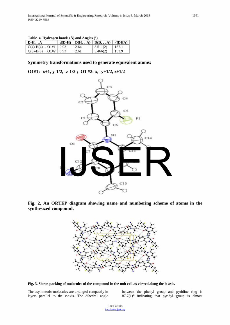
Fig. 3. Shows packing of molecules of the compound in the unit cell as viewed along the b-axis.
The asymmetric molecules are arranged compactly in layers parallel to the c-axis. The dihedral angle
between the phenyl group and pyridine ring is 87.7(1)° indicating that pyridyl group is almost
D-H. . .A d(D-H) D(H. . .A) D(D. . . A) <(DHA) C(4)-H(4). . .O1#1 0.93 2.64 3.511(2) 157.1 C(8)-H(8). . .O1#2 0.93 2.61 3.466(2) 153.9
IJSER

International Journal of Scientific & Engineering Research, Volume 6, Issue 3, March-2015 1552 ISSN 2229-5518
IJSER © 2015 http://www.ijser.org
perpendicular to the phenyl group making the way for its maximum interaction through weak hydrogen bonds. The dihedral angle could be a possible indication for the non-occurrence of either intra- or inter-hydrogen bonding between F(1) and O(1) atoms.
3.3. Antioxidant Property of the Compound
The anti-oxidant activity of the compound was estimated using thermostable radical scavenging assay method (Cottel et.al, 1996). In this assay, a mixture containing 200ml of DPPH (100µM in methanol) and the compound (20-100µg/ml) was incubated in dark for 20 minutes and observance of the mixture was measured at 517nm using spectrophotometer with suitable control according to the procedure. From the results obtained the percentage inhibition was calculated. The compound concentration required to reduce the initial DPPH radical by 50% was calculated using the following equation.
% of Inhibition = (AC – AT)*100 (1) AC where AC and AT stand for Absorbance of the Control (Ascorbic acid) and Absorbance of the test solutions (compound), respectively The antioxidant property of the compound is found to be very close to ascorbic acid taken as standard. The values are given in Tables (5 and 6). Table 5. IC50 Concentration of compounds taken in for DPPH assay
Sample Absorption At (100 μg/ml) IC50 (μg/ml) Ascorbic Acid 0.007 49.689(1)
Compound 0.006 48.180(1)
3.4. Hydrogen Peroxide Radical Scavenging Assay The hydrogen peroxide scavenging activity of the compound was determined by the modified method of (Dehpour et.al, 2009). Hydrogen peroxide solution (1%) was prepared in phosphate buffer (pH 7.4). The concentration with 0.1mg/ml was added toH2O2 solution and absorbance was measured at 230nm using UV-Spectrophotometer (Pc based UV-VIS Spectrophotometer UV-5704 SS) against the blank solution containing phosphate buffer without H2O2.
The percentage of H2O2 scavenging activity of the compound and the standard was calculated using equation (1).
Table 6. IC50 Concentration of compound. (Hydrogen Peroxide Scavenging Assay)
Sample Absorption at(100 μg/ml) IC50 (μg/ml) Ascorbic Acid 0.737 90.992(1) Compound 0.716 88.920(1)
Docking Analysis
In the present work, in silico docking of the synthesized compound with snake venom enzyme namely, Phospholipase (PLA2: PDB id:1FV0) (Fig.4) was performed using the molecular modeling software Maestro Schrodinger Software [17]. Induced Fit Docking (IFD) was carried out using Grid-based Ligand Docking with Energetics (GLIDE). IFD identifies the possible binding modes of ligand and associated conformational changes in the active site of the receptor molecule. In IFD, the first step is the protein and ligand preparation followed by energy minimization. Minimized conformation of protein and ligand was subjected to docking and the best Docked Poses were considered based on the Glide Energy (GE), Docking Score (DS), Hydrogen Bonds (HB) and Hydrophobic Interactions (HI). The present docking study of the compound has been taken up in order to know its inhibitory effect with the enzyme PLA2. The co-crystal ligand molecule aristolochic acid was re-docked using the method as described by Chandra et al [18] with the target and the best conformation was chosen as a template for comparison with the docked conformation of the synthesized compound. It is known that Phospholipase A2 (PLA2) hydrolyzes the fatty acid at the sn-2 position of membrane phospholipids resulting in the production of eicosanoids and related bioactive lipid mediators of inflammation. PLA2 enzyme was found to be the major component of the snake venom and chosen as the target for molecular docking studies to check the
DPPH RADICAL SCAVENGING ASSAY
% O
F IN
HIB
ITIO
N
20
30
40
50
60
70
80
90
CONCENTRATION (ug/ml)0 20 40 60 80 100 120
AscorbicAcidCompound
HYDROGEN PEROXIDE SCAVENGING ASSAY
% O
F IN
HIB
ITIO
N
10
20
30
40
50
60
CONCENTRATION (ug/ml)0 20 40 60 80 100 120
AscorbicAcidCompound
IJSER
International Journal of Scientific & Engineering Research, Volume 6, Issue 3, March-2015 1553 ISSN 2229-5518
IJSER © 2015 http://www.ijser.org
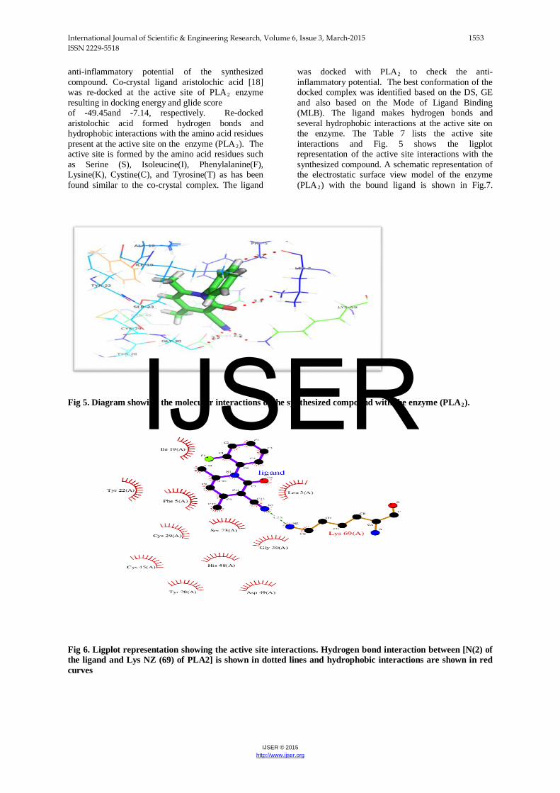
anti-inflammatory potential of the synthesized compound. Co-crystal ligand aristolochic acid [18] was re-docked at the active site of PLA2 enzyme resulting in docking energy and glide score of -49.45and -7.14, respectively. Re-docked aristolochic acid formed hydrogen bonds and hydrophobic interactions with the amino acid residues present at the active site on the enzyme (PLA2). The active site is formed by the amino acid residues such as Serine (S), Isoleucine(I), Phenylalanine(F), Lysine(K), Cystine(C), and Tyrosine(T) as has been found similar to the co-crystal complex. The ligand
was docked with PLA2 to check the anti-inflammatory potential. The best conformation of the docked complex was identified based on the DS, GE and also based on the Mode of Ligand Binding (MLB). The ligand makes hydrogen bonds and several hydrophobic interactions at the active site on the enzyme. The Table 7 lists the active site interactions and Fig. 5 shows the ligplot representation of the active site interactions with the synthesized compound. A schematic representation of the electrostatic surface view model of the enzyme (PLA2) with the bound ligand is shown in Fig.7.
Fig 5. Diagram showing the molecular interactions of the synthesized compound with the enzyme (PLA2). Fig 6. Ligplot representation showing the active site interactions. Hydrogen bond interaction between [N(2) of the ligand and Lys NZ (69) of PLA2] is shown in dotted lines and hydrophobic interactions are shown in red curves
IJSER

International Journal of Scientific & Engineering Research, Volume 6, Issue 3, March-2015 1554 ISSN 2229-5518
IJSER © 2015 http://www.ijser.org
Fig.7. Surface view model showing the bound ligand (shown as stick model) at the active site of the snake venom enzyme (PLA2). The Molecular Docking Analysis of the title compound as ligand at the active of the enzyme PLA2. (Fig.6) shows a preferred orientation of the compound with the enzyme and its stability through hydrogen bonding interaction at 3.25 Å along with hydrophobic interactions (Table 9) indicating that the compound may be considered as a good candidate for anti-inflammation activity. It is well known that the hydrogen bonding interaction [19] is one of the important parameter that is associated with biological activity such as the anti-inflammatory
activity [20]. The Table (7) also contains the docking score and glide energy score. The compound may be considered as a potential candidate for use as an anti-oxidant and anti-inflammatory agent with glide energy of -36.7134 [20, 21] with the hydrogen bond distance involving atoms (Lys A 69 NZ) and N(2) of the compound(Table 8).This has been supported by the data of anti-oxidant and DPPH assay. The interactions of the molecule with PLA2 enzyme are shown in Figs.4 and 6.
Table 7. Results of Molecular Docking Interactions of the compound
Entry ID
Docking score
Glide gscore
Glide Energy
XP GScore
XP HBond
XP Pose Rank
IFD Score
1 -6.26902 -6.269 -30.6092 -6.26902 -0.3500 5 -230.16
2 -5.72011 -5.7201 -32.0903 -5.72011 0.000 4 -229.98
3 -5.60604 -5.606 -33.0515 -5.60604 -0.3500 4 -229.83 4 -5.63882 -5.6388 -36.7134 -5.63882 -0.1134 3 -229.52 5 -5.10277 -5.1028 -32.0863 -5.10277 -0.5095 6 -229.09 6 -4.94928 -4.9493 -32.3803 -4.94928 -0.5426 4 -228.91 7 -4.78398 -4.784 -27.9839 -4.78398 -0.7000 4 -228.57
Table 8. Hydrogen bond interactionfrom Docking Study.
Donor (PLA2) Acceptor (compound) Distance (Å) LYS A 69 NZ N(2) 3.25
IJSER

International Journal of Scientific & Engineering Research, Volume 6, Issue 3, March-2015 1555 ISSN 2229-5518
IJSER © 2015 http://www.ijser.org
Table 9. Hydrophobic interactions due to Molecular Docking.
Atom 1 Atom 2 Distance (Å) C(13) LYS A 69 CE 3.85 C(12) CYS A 45 CB 3.75 C(12) CYS A 29 CA 3.73 C(5) SER A 23 CB 3.50 C(3) SER A 23 CB 3.76 C(14) TYR A 22 CB 3.54 C(14) TYR A 22 C 3.51 C(3) ILE A 19 CD1 3.77 C(1) ILE A 19 CD1 3.62 C(3) ILE A 19 CG1 3.48 C(1) ILE A 19 CG1 3.51 C(10) PHE A 5 CE2 3.62 C(9) PHE A 5 CE2 3.89 C(8) PHE A 5 CE2 3.67 C(4) PHE A 5 CD2 3.86
Conclusion and Future Work The molecule of the synthesized compound exhibits profound anti-oxidant property as has been observed from both DPPH and Scavenger Assay experiments. The molecular docking score study on the compound with PLA2 enzyme indicates that the compound is a good candidate as an anti-inflammatory agent based on the results of glide energy of the molecule which is -36.714 with glide score of -5.63. The molecules are staked along the longest c-axis held by both intra- and inter-hydrogen bonding interactions and thereby rendering stability to the crystalline lattice. The fluorine atom of the compound is not involved in hydrogen bonding. In future we are interested in studying the inhibitory action of several different thiophene derivatives with the enzyme and also compare with respect to those used to neutralize the PLA2 induced actions through synthetic herbal compounds and anti PLA2 rabbit antiserum in in vivo and in vitro models [21]. Acknowledgements
The authors thank Dr. Babu Varghese, Sophisticated Analytical Instrument Facility (SAIF), IIT Madras for solving the crystal structure and Dr..Gayathri, D for their kind help shown during the structure solution work. The authors also thank the Department of Bioinformatics Infrastructure Facility for Molecular Docking Studies at the University of Madras, Funded by Department of Biotechnology Government of India, New Delhi, for allowing us to make use of the facilities available over there.
REFERRENCES: .
[1] Victor B. Rybakov, Alexander A. Bush., Eugene V. Babaev and Leonid A.Alanov. . (2004) Acta Cryst). E60, o160.
[2] Choudhury. A.R.,Urs, U. K., Guru Row, T.N. and Nagarajan, K. [2002]. J. Mol. Struct.,605,71..
[3] Anilkumar, G.N.. Kokila,M.K., Puttaraja, Mohan,S. and Saravanan, J. (2007). Acta Cryst. E63, o2077.
[4] Subhadramma, S., Naveenhandra, and Saravanan, J. (2013). IJSER. 4, 1024.
[5] Ferreira, I. C. F., Calhelha,R.C., Estevinho,L.M., Queiroz M. J. R. (2004). Bioorg. Med. Chem. Lett. 14. 5831. [6] Shafeeque, S., Mohan, S. and Manjunatha, K.S. (1999). Indian J. Hetero. Chem. 8(4). 297. [7] Laddi, U. V., Talwar, M. B.,. Desai, S. R., Somannavar, Y. S., Bennur, R. S. and Bennur, S. C.( 1998); Indian Drugs; 35(8); 509. [8] Ferreira, C. F. R. Maria-Joao, R. P., Vilas-Boas, M., Estevinho, L. M., Begouin, A. and Gilbert, K. (2006). Bioorg. Med. Chem. Lett.2006; 16; 1384. [9] Gewald, K., Schinke, E. and Bottcher, H. (1966). Chem. Ber.; 99; 94. [10] George. H. Stout & Lyle H. Jensen X-ray structure determination II edition, A practical guide. A Wiley-inter science publication John Wiley& Sons, Chap 4. Pg 75-78, Chap 7. Pg. 181, Chap 10. Pg 245-246, Chap 11.Pg 249-278, Chap 16. Pg 344-378, Chap 17. Pg 378-390
[11] Vasu, Nirmala, K. A., Choudhury, A. R., Mohan, S.,
Saravanan, J. and Narasimhamurthy, T. (2003). Acta
Cryst. C59, o676..
[12] Vasu, Nirmala, K. A., Chopra, D., Mohan, S. and Saravanan, J. (2004). Acta Cryst. E60, o756.
[13] Sheldrick, G.M. (1997), SHELXL and SHELXL97, University of Gottingen Germany. [14]Computing molecular graphics ‘ORTEP 3’(FARRUGIA 1997) and MERCURY (BRUNO et.al, 2002)
IJSER

International Journal of Scientific & Engineering Research, Volume 6, Issue 3, March-2015 1556 ISSN 2229-5518
IJSER © 2015 http://www.ijser.org
[15] Watkin, D. M., Pearce, L. &Prout, C. K. (1993).CAMERON. Chemical Crystallography Laboratory, University of Oxford, England.
[16] Watkin, D. J., Prout, C.K and Pearce, L., J. (1996). CAMERON. Chemical Crystallography Laboratory, University of Oxford, England.
[17]. Schrödinger Suite 2009 Induced Fit Docking protocol; Glide version 5.0, Schrödinger, LLC,
[18] Chandra, V., Jasti, J., Kaur, P., Srinivasan, A., Betzel, C., Singh, T.P. Journal: (2002) Biochemistry 41: 10914-10919 [19] Susanta K. Nayak, Srijita Basu Mallik, Shankar PrasadKanaujia, Kana-garaj Sekar, K. R. Ranganathan, V.,Ananthalakshmi, G. Jeyaraman, S. S. Saralaya, K. Sundararaja Rao, K. Shridhara, K. Nagarajan and Tayur N. Guru Row CrystEngComm, 2013,15, 4871-4884 [20] Maria J.R. Yunta. American Journal of modelling and optimization, 2014, vol. 2, No.4, 84-102.
[21] Narendra Sharath Chandra JN, Ponnappa KC, Sadashiva CT, Priya BS, Nanda BL, Gowda TV, et.al.. Curr Top med chem 2007; 787-800.
IJSER