A Novel Role of Cytosolic Protein Synthesis Inhibition in ...
ARTICLE; AGRICULTURE AND ENVIRONMENTAL BIOTECHNOLOGY
Myconanoparticles: synthesis and their role in phytopathogens management
Mousa A. Alghuthaymia, Hassan Almoammarb, Mahindra Raic, Ernest Said-Galievd and Kamel A. Abd-Elsalame,f*
aBiology Department, Science and Humanities College, Shaqra University, Alquwayiyah, Saudi Arabia; bKing Abdulaziz City forScience and Technology (KACST), Saudi Arabia; cNanobiotechnology Laboratory, Department of Biotechnology, SGB AmravatiUniversity, Amravati-444-602, India; dA.N. Nesmeyanov Institute of Organoelement compounds (INEOS) of Russian Academyof Sciences, Moscow, Russia; ePlant Pathology Research Institute, Agricultural Research Center (ARC), Giza, Egypt; fUnit ofExcellence in Nano-Molecular Plant Pathology Research Institute, Giza, Egypt
(Received 22 July 2014; accepted 27 October 2014)
Nanotechnology can offer green and eco-friendly alternatives for plant disease management. Apart from being eco-friendly,fungi are used as bio-manufacturing units, which will provide an added benefit in being easy to use, as compared to othermicrobes. The non-pathogenic nature of some fungal species in combination with the simplicity of production and handlingwill improve the mass production of silver nanoparticles. Recently, a diverse range of fungi have been screened for theirability to create silver nanoparticles. Mycosynthesis of gold, silver, gold�silver alloy, selenium, tellurium, platinum,palladium, silica, titania, zirconia, quantum dots, usnic acid, magnetite, cadmium telluride and uraninite nanoparticles has alsobeen reported by various researchers. Nanotechnological application in plant pathology is still in the early stages. Forexample, nanofungicides, nanopesticides and nanoherbicides are being used extensively in agriculture practices. Remoteactivation and monitoring of intelligent nano-delivery systems can assist agricultural growers of the future to minimizefungicides and pesticides use. Nanoparticle-mediated gene transfer would be useful for improvement of crops resistant topathogens and pest. This review critically assesses the role of fungi in the synthesis of nanoparticles, the mechanism involvedin the synthesis, the effect of different factors on the reduction of metal ions in developing low-cost techniques for thesynthesis and recovery of nanoparticles. Moreover, the application of nanoparticles in plant disease control, antimicrobialmechanisms, and nanotoxicity on plant ecosystem and soil microbial communities has also been discussed in detail.
Keywords: disease management; antimicrobial action; phytotoxicity; nanophytopathology
Abbreviations
Ag NPs: silver nanoparticles
ATCC: American Type Culture Collection
CdTe: cadmium telluride
CNP: coated nanoparticles
EDX: energy dispersive X-ray
mm: millimetre
MNMs: manufactured nanomaterials
MNT: myconanotechnology
nm: nanometre
NPs: nanoparticles
NT: nanotechnology
QD: quantum dot
ROS: reactive oxygen species
Introduction
The word ‘nano’ is used to indicate one billionth of a
metre or 10¡9. The term nanotechnology (NT) was coined
by Professor Norio Taniguchi of Tokyo Science Univer-
sity in 1974 to illustrate precision manufacturing of mate-
rials at the nanometre level.[1] One nanometre (nm) is
one millionth of a millimetre (mm). To put the nanoscale
into context, a strand of DNA is 2.5 nm wide, a protein
molecule is 5nm, a red blood cell 7000 nm and a human
hair is 80,000 nm wide.
In green nanotechnology, for the synthesis of nanopar-
ticles (NPs) micro-organisms are used. It is well known
that many micro-organisms aggregate inorganic material
within or outside the cell to form NPs. While a large num-
ber of microbial species are capable of producing metal
NPs, the mechanism of NP biosynthesis is very important.
Microbial synthesis of NPs is a green chemistry approach
that interconnects nanotechnology and microbial biotech-
nology. Biosynthesis of gold, silver, gold�silver alloy,
selenium, tellurium, platinum, palladium, silica, titania,
zirconia, quantum dots (QDs), magnetite and uraninite
NPs by bacteria, actinomycetes, fungi, yeasts and viruses
have been reported.[2] Silver nanoparticles (Ag NPs)
have become one of the most commonly used nanomateri-
als in consumer products (104 out of 502 nanoproducts
surveyed).[3]
The exact mechanism by which Ag NPs destroy and
prevent fungal pathogen growth is not well understood.
*Corresponding author. Email: [email protected]
� 2015 The Author(s). Published by Taylor & Francis.
This is an Open Access article distributed under the terms of the Creative Commons Attribution License (http://creativecommons.org/licenses/by-nc/4.0/), which permits
unrestricted use, distribution, and reproduction in any medium, provided the original work is properly cited.
Biotechnology & Biotechnological Equipment, 2015
Vol. 29, No. 2, 221�236, http://dx.doi.org/10.1080/13102818.2015.1008194
There are many possible mechanisms discussed by
researchers but the exact mechanism has not been eluci-
dated. Bacteria are believed to use an enzyme to metabo-
lize oxygen to sustain life. Silver ions cripple the enzyme
and stop the metabolization of oxygen. This suffocates the
fungi and bacteria, resulting in death.[4] Fungal enzymes
interact with metal ions and reduce to form NPs. The kinet-
ics of the reaction has been studied using UV�Vis spec-
troscopy and was further characterized by X-ray diffraction
(XRD), energy dispersive X-ray (EDX) analysis and high-
resolution transmission electron microscopy (TEM).
Microbes may interact with nanomaterials or in certain
instances produce nanostructured materials.[5�10] This is
because of their excellent performance, selective adsorption
of metal ions, operation over a broad range of ecological
conditions (pH, ionic strength, temperature), low cost, free
availability, regeneration and high biosorption capacity and
the fact that large quantities can be obtained.[11]
The mycosynthesis of metal NPs, or myconano-
technology (MNT) [12,13] is the use of fungi in NT for
the synthesis of NPs. The capability of filamentous fungi
to grow on readily available and inexpensive substrates,
as well as their ability to produce a wide range of com-
mercially interesting metabolites have attracted consider-
able interest to exploit them as production micro-
organisms in biotechnology.[14,15] Nanodiagnostic meth-
ods include gene delivery, gene expression, gene sequenc-
ing, gene regulation, DNA targeting, DNA isolation,
DNA hybridization, fingerprints for DNA and RNA detec-
tion, cell probes, specific targeting, cell sorting and bioi-
maging, single-cell-based assay, tissue engineering,
proteomics and nanobiogenomics.[16] Preliminary studies
show the potential of nanomaterials in improving seed
germination and growth, plant protection, pathogen detec-
tion and pesticide/herbicide residue detection.[17]
This review on fungi and NT, or MNT, and its use to
control phytopathogens is important in sustainable agri-
culture. The nanotechnology is rapidly becoming a
majorly researched topic and has resulted in applications
being developed at a rapid rate; there is a need to find new
ways to produce NPs using quick, clean and inexpensive
methodology. Fungi are excellent candidates for research
in producing NPs and we provide a review of global
research efforts on the use of fungi in the biosynthesis of
NPs, and their use in disease detection and control.
Synthesis of myconanoparticles
The use of microbial cells for the synthesis of nanosized
material has emerged as a new approach for the synthesis
of metal NPs.[6] Several fungal strains have been used as
promising resources for nanoparticle fabrication, for
example Fusarium, Aspergillus, Verticillium and Pencil-
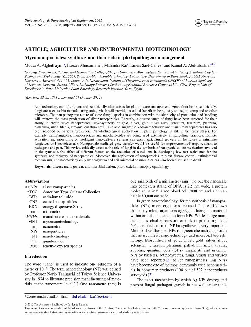
lium. Different fungal species are proficient candidates for
production of metal NPs both intra- and extracellulary
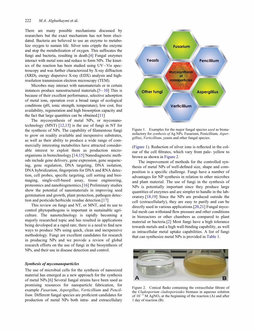
(Figure 1). Reduction of silver ions is reflected in the col-
our of the cell filtrates, which vary from pale- yellow to
brown as shown in Figure 2.
The improvement of methods for the controlled syn-
thesis of metal NPs of well-defined size, shape and com-
position is a specific challenge. Fungi have a number of
advantages for NP synthesis in relation to other microbes
and plant material. The use of fungi in the synthesis of
NPs is potentially important since they produce large
quantities of enzymes and are simpler to handle in the lab-
oratory.[18,19] Since the NPs are produced outside the
cell (extracellularly), they are easy to purify and can be
directly used in various applications.[20,21] Fungal myce-
lial mesh can withstand flow pressure and other conditions
in bioreactors or other chambers as compared to plant
material or bacteria.[2] Most fungi have a high tolerance
towards metals and a high wall-binding capability, as well
as intracellular metal uptake capabilities. A list of fungi
that can synthesize metal NPs is provided in Table 1.
Figure 1. Examples for the major fungal species used as biona-nofactory for synthesis of Ag NPs, Fusarium, Penicillium, Asper-gillius, Verticillium, yeasts and other fungal species.
Figure 2. Conical flasks containing the extracellular filtrate ofthe Cladosporium cladosporioides biomass in aqueous solutionof 10¡3 M AgNO3 at the beginning of the reaction (A) and after1 day of reaction (B).
222 M.A. Alghuthaymi et al.
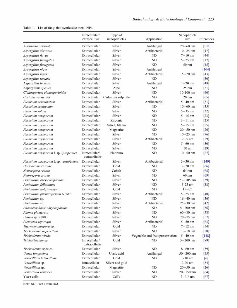
Table 1. List of fungi that synthesize metal NPs.
Intracellular/extracelluar
Type ofnanoparticles Application
Nanoparticlesize References
Alternaria alternata Extracellular Silver Antifungal 20�60 nm [103]
Aspergillus clavatus Extracellular Silver Antibacterial 10�25 nm [47]
Aspergillus flavus Extracellular Silver ND 7�10 nm [44]
Aspergillus fumigatus Extracellular Silver ND 5�25 nm [17]
Aspergillus fumigatus Extracellular Silver ND 50 nm [45]
Aspergillus niger Extracellular Silver Antifungal [104]
Aspergillus niger Extracellular Silver Antibacterial 15�20 nm [43]
Aspergillus tamarii Extracellular Silver ND [50]
Aspergillus terreus Extracellular Silver Antifungal 1�20 nm [48]
Aspergillius species Extracellular Zinc ND 25 nm [51]
Cladosporium cladosporioides Extracellular Silver ND 10-100 nm [60]
Coriolus versicolor Extracellular Cadmium sulphide ND 20 nm [65]
Fusarium acuminatum Extracellular Silver Antibacterial 5�40 nm [31]
Fusarium semitectum Extracellular Silver ND 10�60 nm [35]
Fusarium solani Extracellular Silver ND 5�35 nm [32]
Fusarium oxysporum Extracellular Silver ND 5�15 nm [23]
Fusarium oxysporum Extracellular Zirconia ND 3�11 nm [25]
Fusarium oxysporum Extracellular Silica, titania ND 5�15 nm [25]
Fusarium oxysporum Extracellular Magnetite ND 20�50 nm [26]
Fusarium oxysporum Extracellular Silver ND 10�25 nm [76]
Fusarium oxysporum Extracellular Silver Antibacterial 2�5 nm [29]
Fusarium oxysporum Extracellular Silver ND 5�60 nm [30]
Fusarium oxysporum Extracellular Silver ND 30 nm [29]
Fusarium oxysporum f. sp. lycopersici Intracellular/extracellular
Platinum ND 10�50 nm [27]
Fusarium oxysporum f. sp. vasinfectum Extracellular Silver Antibacterial 3�30 nm [149]
Hormoconis resinae Extracellular Gold ND 3�20 nm [66]
Neurospora crassa Extracellular Cobalt ND 64 nm [68]
Neurospora crassa Extracellular Silver ND 60 nm [69]
Penicillium brevicompactum Extracellular Silver ND 23�105 nm [39]
Penicillium fellutanum Extracellular Silver ND 5-25 nm [38]
Penicillium nalgiovense Extracellular Gold ND 15�25
Penicillium purpurogenum NPMF Extracellular Silver Antibacterial 5�25 nm [40]
Penicillium sp. Extracellular Silver ND 16�40 nm [36]
Penicillium sp. Extracellular Silver Antibacterial 25�30 nm [42]
Phaenerochaete chrysosporium Extracellular Silver ND 5�200 nm [56]
Phoma glomerata Extracellular Silver ND 60�80 nm [58]
Phoma sp.3.2883 Extracellular Silver ND 70�75 nm [57]
Pleurotus sajorcaju Extracellular Silver Antibacterial 5�50 nm [63]
Thermomonospora sp. Extracellular Gold ND 7�12 nm [54]
Trichoderma asperellum Extracellular Silver ND 13�18 nm [20]
Trichoderma viride Extracellular Silver Vegetable and fruit preservation 5�40 nm [148]
Trichothecium sp. Intracellular/extracellular
Gold ND 5�200 nm [89]
Trichoderma species Extracellular Silver ND 8�60 nm [59]
Usnea longissima Extracellular Usnic acid Antifungal 50�200 nm [55]
Verticillium luteoalbum Extracellular Gold ND >10 nm [6]
Verticillium sp. Intracellular Silver and gold 2-20 nm [54]
Verticillium sp. Extracellular Magnetite ND 20�50 nm [26]
Volvariella volvacea Extracellular Silver ND 20�150 nm [64]
Yeast cells Extracellular CdTe ND 2�3.6 nm [67]
Note: ND � not determined.
Biotechnology & Biotechnological Equipment 223
Biosynthesis of nanoparticles by Fusarium
Recently, the screening of different Fusarium sp. for selec-
tion of potential species has been made.[22] The authors
reported that Fusarium oxysporum synthesized the smallest
size of Ag NPs. It was found that F. oxysporum can extra-
cellularly reduce aqueous silver ions in water to generate
Ag NPs.[23�25] Zirconia NPs may be produced by chal-
lenging the fungus F. oxysporum with aqueous ZrF62¡
anions; extracellular protein-mediated hydrolysis of the
anionic complexes at room temperature results in the syn-
thesis of nanocrystalline zirconia.[26,27] F. oxysporum and
Verticillium sp. generated, in the presence of ferric and fer-
rous salts, magnetite NPs.[28] A strain of F. oxysporum f.
sp. lycopersici was screened and successfully produced
inter- and extracellular platinum NPs. Duran et al. and
Khosravi et al. [28,29] showed that Ag NPs could be syn-
thesized using F. oxysporum and the antibacterial proper-
ties of the biosynthesized Ag NPs when incorporated in
textile fabrics. A nitrate reductase-mediated technique was
used for the synthesis of Ag NPs using F. oxysporum.[30]
The extracellular production of metal NPs by numerous
strains of the fungus F. oxysporum was studied.[31] Simi-
larly, F. oxysporum strain 5115 was used for the fabrication
of Ag NPs. Mohammadian et al. [30] also evaluated the
biosynthesis of spherical and silver colloidal NPs using F.
oxysporum. Nanoparticles of 10�100 nm and hexagons,
pentagons, circles, squares and rectangles were produced
both intra- and extracellularly by F. oxysporum. The irregu-
lar-shaped NPs were obtained by F. oxysporum culture fil-
trate.[33] Ingle and co-workers [32,33] investigated the use
of F. acuminatum isolated from infected ginger, for the
synthesis of Ag NPs and evaluated its antimicrobial activity
against human pathogenic bacteria. The fungal mycelia
were challenged with aqueous silver nitrate with a final
concentration of 1 mmol/L. The colour of filtrate changed
from light-yellow to brown, which intensified after 2 h.
Detection and evaluation of the Ag NPs was carried out
using a UV�Vis spectrophotometer and TEM. The nano-
particles produced were between 5 and 40 nm in size and
spherical. Ingle et al. and Bawaskar et al. [32,34] further
investigated the synthesis of Ag NPs by Fusarium solani
and Fusarium culmorum, respectively. Highly stable and
crystalline Ag NPs were produced in a solution by treating
the filtrate of the fungus Fusarium semitectum with aque-
ous silver nitrate solution. The characterization of AgC
ions exposed to Fusarium isolates by UV�Vis and XRD
methodology confirmed a decrease of silver ions to Ag
NPs. The TEM images of the NPs suggest that they were
polydispersed and mostly spherical.[36]
Biosynthesis of nanoparticles by Penicillium
Nanoparticles produced by Penicillium possessed a nega-
tive zeta potential and were fairly stable at a pH value
above 8 due to electrostatic repulsion.[37] Penicillium sp.
could effectively myco-reduce and nucleate AuCl4 (-) ions,
and intracellular biosynthesis of size-controlled gold NPs
after exposure to HAuCl4 solution.[38] In vitro biosynthe-
sis of Ag NPs was achieved by Penicillium fellutanum
using AgNO3 as a substrate isolated from coastal mangrove
sediment.[39] An eco-friendly process for the synthesis of
nanomaterials using Penicillium brevicompactum WA
2315 and Penicillium purpurogenum NPMF has been
attempted, respectively.[40,41] The green synthesis of Ag
NPs by the cell-free filtrate of Penicillium nalgiovense
AJ15 was reported by Maliszewska et al.[41] The authors
claimed that Ag NPs synthesis by the P. nalgiovense AJ15
cell free filtrate is a non-enzymatic process and the proteins
containing cysteine play a significant role in the reducing
of silver ions. In another example, Singh et al. [42] reported
the synthesis of Ag NPs by an endophytic Penicillium sp.
isolated from healthy leaves of Curcuma longa (turmeric).
Biosynthesis of nanoparticles by Aspergillus
Biosynthesis of Ag NPs using Aspergillus niger isolated
from soil was reported by Kumar et al.[43] Cell filtrate of
A. niger was treated with 1 mmol/L silver nitrate and
placed on a rotary shaker at 120 rpm and 25 �C in the
dark. When treated with silver nitrate solution Aspergillus
flavus accumulated Ag NPs on the surface of its cell wall
after 72 h. The average size of the NPs was calculated as
8.92 § 1.61 nm. These Ag NPs are found to have a char-
acteristic absorption peak at 420 nm and emission peak at
553 nm.[45] Extracellular biosynthesis of Ag NPs using
Aspergillus fumigatus was investigated.[17,46] Silver
nanoparticles can be mycosynthesized extracellularly
using Aspergillus clavatus.[47,48] Silver nanoparticles
were synthesized using a reduction of aqueous AgC ion
with the culture supernatants of Aspergillus terreus. [49]
Mycosynthesized Ag NPs were polydispersed spherical
particles ranging in size between 1 and 20 nm and could
efficiently inhibit a variety of plant pathogenic fungi and
bacteria. Antibacterial action of Ag NPs against Escheri-
chia coli, Candida albicans and Pseudomonas fluores-
cence was revealed using a disc-diffusion technique.[48]
Similarly, the NPs showed antimicrobial activity against
fungal and bacterial strains.[50] An environmental
friendly process for the synthesis of Ag NPs using a fun-
gus Aspergillus tamarii has been investigated. The scan-
ning electron microscope (SEM) result showed the
distribution of spherical Ag NPs ranging from 25 to
50 nm.[51] Raliya and co-workers [51,52] reported the
synthesis of zinc, magnesium and titanium NPs by using
six Aspergillius species belonging to A. flavus, A. terreus,
Aspergillus tubingensis, A. niger, A. fumigatus and Asper-
gillus oryzae by employing various precursor salts of sul-
phates, nitrates, chlorides and oxides. The authors also
optimized the factors responsible for more production of
monodispersed Zn, Mg and Ti NPs.
224 M.A. Alghuthaymi et al.

Biosynthesis of nanoparticles by Verticillium
The theory of using biological entities as ‘reaction con-
tainers’ for the synthesis of NPs was discovered early.[53]
For instance, the exposure of the fungus, Verticillium sp.
to aqueous AuCl4 resulted in the reduction of the salt to
gold NPs with a diameter of 20 nm and either intracellular
or extracellular production.[54] The biosynthesis of inor-
ganic nanomaterials using fungi was achieved, with the
intracellular production of Ag NPs by Verticillium strains.
[55] Gericke and Pinches [6] screened diverse fungal spe-
cies for their capability to construct gold NPs; the most
promising results were obtained with cultures of Verticil-
lium luteoalbum. The rate of NP construction and the size
of the NPs could, to an extent, be manipulated by control-
ling parameters such as pH, temperature, gold concentra-
tion and exposure time to AuCl4.
Biosynthesis of nanoparticles by other fungi
The shape and size of biogenic NPs depends on the bio-
logical species involved, for instance, Colletotrichum sp.
produced essentially spherical NPs under identical condi-
tions.[5] The white rot fungus, Phanerochaete chrysospo-
rium formed stable Ag NPs when challenged with silver
nitrate in aqueous medium.[56] The coelomycetous
Phoma strain produced Ag NPs extracellularly ranging
from 60 to 80 nm when the fungal cell filtrate was
exposed to an aqueous silver nitrate solution at room tem-
perature.[57, 58] Silver nanoparticles were produced by
the biocontrol agent, Trichoderma asperellum, with a size
range of 13�18 nm with well-defined morphology and
being stable for several months.[21]
Five Trichoderma species belonging to T. asperellum,
T. harzianum, T. longibrachiatum, T. pseudokoningii and
T. virens were screened for the production of Ag NPs.
These NPs were found single or aggregated with round
and uniform shape and a size of 8�60 nm.[59] An extra-
cellular solution of Cladosporium cladosporioides was
used for the reduction of AgNO3 solution to Ag NP. TEM
analysis revealed the presence of polydispersed and spher-
ical-shaped particles.[60] Gade et al. [61] screened 18 dif-
ferent Phoma sp. for selection of potential species as a
novel synthesizer of Ag NPs. They also reported the for-
mation of silver rods by Phoma sorghina.
Biosynthesis of Ag NPs using Pleurotus sp. [62] and
Pleurotus sajorcaju [63] was reported. The authors also
reported antimicrobial activity of P. sajorcaju. An extra-
cellular synthesis method was developed for the prepara-
tion of Au, Ag and Au�Ag NPs in water, using an extract
from Volvariella volvacea, an industrial edible mush-
room, as reducing and protecting agents.[64] Gold NPs of
diverse sizes (20�150 nm) and shapes from triangular
nanoprisms to nearly spherical and hexagonal were
obtained by this new technique. Long-term studies were
carried out with the immobilized fungus Coriolus
versicolor in continuous column mode. The immobilized
fungus served a dual purpose of both bioremediating cad-
mium as well as synthesizing stable CdS NPs in aqueous
conditions.[65] Among the various fungi screened, Hor-
moconis resinae proved to be an excellent fungal source
for the extracellular synthesis of gold NPs with appreci-
able stability in solution.[66] A simple and proficient bio-
synthesis technique to prepare biocompatible cadmium
telluride (CdTe) QDs with tunable fluorescence emission
using yeast cells were evaluated.[67] The filamentous fun-
gus Neurospora crassa was found to be a potential biolog-
ical agent for the production of mono and bimetallic Au/
Ag NPs.[68,69] Gold NPs of 6�18 nm diameter were
mycosynthesized by treating the mycelia-free culture fil-
trate of the Nigrospora oryzae with gold chloride.[70]
The use of a biosource such as fungi that can catalyze spe-
cific reactions leading to inorganic NPs is a modern and
rational biosynthesis strategy that is an alternative to other
physical and chemical methods. In order for fungal syn-
thesis of NPs to become commercially practical, it is
essential to develop low-cost revival methods for separa-
tion of the particles from the fungal mat that can be used
routinely in manufacturing procedures.
Factors affecting fungal synthesis of metallic
nanoparticles
There is always a continuous interaction between fungus
and the environment in which they live. The environmen-
tal conditions exert an influence on growth and develop-
ment of organisms. The enzyme production by fungi is
influenced by the condition in which the organisms are
cultivated.[71] Therefore, optimization studies will not
only support good growth but also enhance product yield.
There are a few reports on the effects of culture condi-
tions on the biosynthesis of metal NPs.[31,46,52] Myco-
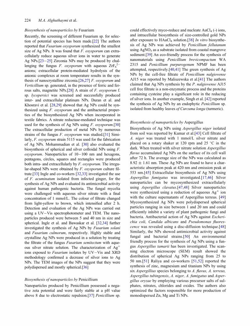
synthesis is directly affected by incubation conditions,
such as temperature, pH, incubation time, nature of the
parent compound or metal species (Figure 3)
[43,46,72,73], the biomass concentration of the fungal
species [43,74] and colloidal interaction conditions, that
control the size, shape, localization and dispersity of the
NPs formed. The factors such as temperature and pH can
be manipulated for initiating the geometry of Ag NPs.[75]
Raliya and Tarafdar [51] concluded that 0.1 mmol/L pre-
cursor salt concentration, 72 h of incubation at pH 5.5 and
temperature 28 �C resulted in larger NP yield.
Mechanism of nanoparticles formation
The actual mechanism of formation, for instance, of Ag
NPs, in all of these micro-organisms and plants, is still an
open question, even though much research has been
attempted to find different ways to investigate the possible
mechanisms.[13,75] A number of possible options exist,
Biotechnology & Biotechnological Equipment 225
which may explain the process of metal reduction in these
organisms. The extracellular synthesis of NPs using fungi
including three mechanisms: nitrate reductase action,
electron shuttle quinones or both. The nitrate reductase
assay was carried out by the reaction of nitrite with 2,3-
diaminophthalene.[75,76] Nitrate reductase was suggested
to initiate NP formation by many fungi including Penicil-
lium species, while several enzymes, a-NADPH-depen-dent reductases, nitrate-dependent reductases and an
extracellular shuttle quinone, were implicated in Ag NP
synthesis for F.oxysporum. Jain et al. [72] reported that
Ag NP synthesis for A. flavus occurs initially by a ‘33
kDa’ protein followed by a protein (cystein and free
amine groups), which stabilizes the NPs by forming a cap-
ping agent.[73] A number of researchers supported nitrate
reductase for extracellular synthesis of NPs.[33,44,77]
Fungal cell wall and cell wall sugars are likely to play
an important role in the absorption and reduction of metal
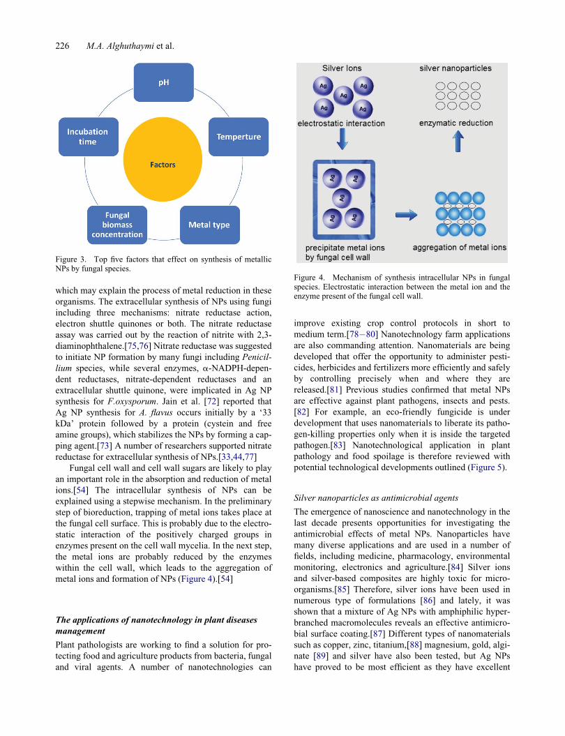
ions.[54] The intracellular synthesis of NPs can be
explained using a stepwise mechanism. In the preliminary
step of bioreduction, trapping of metal ions takes place at
the fungal cell surface. This is probably due to the electro-
static interaction of the positively charged groups in
enzymes present on the cell wall mycelia. In the next step,
the metal ions are probably reduced by the enzymes
within the cell wall, which leads to the aggregation of
metal ions and formation of NPs (Figure 4).[54]
The applications of nanotechnology in plant diseases
management
Plant pathologists are working to find a solution for pro-
tecting food and agriculture products from bacteria, fungal
and viral agents. A number of nanotechnologies can
improve existing crop control protocols in short to
medium term.[78�80] Nanotechnology farm applications
are also commanding attention. Nanomaterials are being
developed that offer the opportunity to administer pesti-
cides, herbicides and fertilizers more efficiently and safely
by controlling precisely when and where they are
released.[81] Previous studies confirmed that metal NPs
are effective against plant pathogens, insects and pests.
[82] For example, an eco-friendly fungicide is under
development that uses nanomaterials to liberate its patho-
gen-killing properties only when it is inside the targeted
pathogen.[83] Nanotechnological application in plant
pathology and food spoilage is therefore reviewed with
potential technological developments outlined (Figure 5).
Silver nanoparticles as antimicrobial agents
The emergence of nanoscience and nanotechnology in the
last decade presents opportunities for investigating the
antimicrobial effects of metal NPs. Nanoparticles have
many diverse applications and are used in a number of
fields, including medicine, pharmacology, environmental
monitoring, electronics and agriculture.[84] Silver ions
and silver-based composites are highly toxic for micro-
organisms.[85] Therefore, silver ions have been used in
numerous type of formulations [86] and lately, it was
shown that a mixture of Ag NPs with amphiphilic hyper-
branched macromolecules reveals an effective antimicro-
bial surface coating.[87] Different types of nanomaterials
such as copper, zinc, titanium,[88] magnesium, gold, algi-
nate [89] and silver have also been tested, but Ag NPs
have proved to be most efficient as they have excellent
Figure 3. Top five factors that effect on synthesis of metallicNPs by fungal species.
Figure 4. Mechanism of synthesis intracellular NPs in fungalspecies. Electrostatic interaction between the metal ion and theenzyme present of the fungal cell wall.
226 M.A. Alghuthaymi et al.

antimicrobial effectiveness against bacteria, viruses and
fungi.[90,91] The nanoparticles could be influenced
by the soil, but they can also change some soil characteris-
tics � mainly pollutants and pathogens.[92] Recently, the
in vitro activity of nanosilver against 18 plant pathogens
was demonstrated.[93]
Antimicrobial mechanisms of nano-metal toxicity. There
are five theories, which have been proposed about the
action mechanism [94]: (1) release of toxic ions (Cd2C,Zn2C, AgC) that can bind to sulphur-containing proteins;
this accumulation prevents the proteins from properly
functioning in the membrane and interfere in cell perme-
ability; (2) they can be genotoxic � toxic ions that can
destroy DNA which leads to cell death; (3) interruption of
electron transport, protein oxidation and membrane poten-
tial collapse due to its contact with CeO2 or nC60; (4)
generation of ROS (reactive oxygen species) � ROS-
mediated cellular damage, and different metal-catalyzed
oxidation reactions could underlie specific types of pro-
tein, membrane or DNA damage [95]; (5) interference
with nutrient uptake. These mechanisms may not operate
separately suggesting that more than one mechanism can
occur simultaneously. The multiple targets of action could
help NPs to fight effectively against different plant
pathogens.
Antifungal activity of nanoparticles
Control of food crop diseases is essential. Recently,
efforts have been made to develop harmless management
methods that pose fewer hazards to humans and animals,
and have focused on overcoming the lack of synthetic fun-
gicides.[96]
The antifungal effect of Ag NPs has received only
minor attention and with only a few published articles on
this topic.[97,98] There are some studies dealing more
specifically with their action against clinical isolates and
American Type Culture Collection (ATCC) strains of
Candida spp. and Trichophyton mentagrophytes are avail-
able.[48,99,100] The use of nanosized silver particles as
antimicrobial agents has become more widespread as
technological advances make their production more eco-
nomical. One of the probable applications in which silver
can be utilized is the management of plant diseases. Since
silver displays various modes of inhibitory action to plant
pathogens,[101] it may be used for controlling various
plant pathogens in a moderately safer way compared to
synthetic fungicides.[102] Ag-SiO2 NPs have a strong
antifungal effect against Botrytis cinerea.[102] The com-
bined effect of fluconazole and Ag NPs for their antifun-
gal activity was evaluated by Gajbhiye et al. [103] against
Phoma glomerata, Phoma herbarum, F.semitectum, Tri-
choderma sp. and C.albicans by disc diffusion technique.
Ag2S nanocrystals on amorphous silica particles show
antifungal activity against A. niger.[104] The potential
biocidal efficacy of ZnO and ZnTiO3 nanopowders
against the fungus A. niger was assessed.[105] ZnTiO3
nanopowder showed higher growth inhibition efficiency
than ZnO.[106]
Silver ions and NPs were evaluated to check the anti-
fungal action on Bipolaris sorokiniana and Magnaporthe
grisea. The in vitro and in vivo evaluations of both silver
ions and NPs decrease disease development of phytopath-
ogenic fungi.[107] Min et al. [100] evaluated the antifun-
gal effects of Ag NPs especially on sclerotium forming
phytopathogenic fungi. Pan�a�cek et al. [99] assayed the
fungistatic and fungicidal effects of the Ag NPs against
certain pathogenic yeasts such as C.albicans (I and II),
Candida tropicalis and Candida parapsilosis. The anti-
fungal activity of Ag NPs was evaluated against the
unidentified ambrosia fungus Raffaelea sp., which has
been responsible for the mortality of a large number of
oak trees in Korea.[107] The effect of Ag NPs on plant
Figure 5. Potential NTapplications in plant pathology.
Biotechnology & Biotechnological Equipment 227
pathogenic spores of F.culmorum was studied by Kaspro-
wicz et al.[108] Silver nanoparticles were also found to
exhibit antifungal activity against F.oxysporum.[109] Sil-
ver nanoparticles deeply decreased the number of germi-
nating fragments and sprout length relative to the control.
ZnO NPs inhibited the growth of B.cinerea by affecting
cellular functions, which caused deformation in fungal
hyphae. In addition, ZnO NPs inhibited the growth of
conidiophores and conidia of Penicillium expansum,
which finally led to the death of fungal mats.[110] The
effect of nano-silver liquid against the white rot of
the green onion caused by Sclerotium cepivorum was
evaluated.[111]
Silver nanoparticles may be less toxic to humans and
animals than synthetic fungicides. Moreover, the toxicity
that nanoparticles may cause in algae, plants and fungi,
may be coupled with some positive effects.[85] The anti-
fungal activity of the Ag NPs was evaluated on the phyto-
pathogen Colletotrichum gloeosporioides, which is
responsible for anthracnose in a wide range of fruit. The
growth of C. gloeosporioides in the presence of Ag NPs
was significantly decreased in a dose-dependent manner.
[112] A comparative study of elemental and nano-sulphur
has been conducted against facultative fungal food patho-
gen, A. niger. The results showed that nano-sulphur was
more efficient than its elemental form.[82] Different con-
centrations of Ag NPs were tested to determine the inhibi-
tory effect of fungal plant pathogens namely Alternaria
alternata, Sclerotinia sclerotiorum, Macrophomina pha-
seolina, Rhizoctonia solani, B.cinerea and Curvularia
lunata. Interestingly, 15 mg concentration of Ag NPs
showed excellent inhibitory activity against all the tested
pathogens.[113] Chitosan and Cu�chitosan NPs proved
their uniform size and stability, which may contribute to
their higher antifungal activity against A. alternata, M.
phaseolina and R. solani in in vitro studies. Cu�chitosan
NPs also showed maximum inhibition rate of spore germi-
nation of A. alternata. Compared to chitosan and Cu�chi-
tosan NPs, the chitosan�saponin NPs were found poor in
antifungal activity.[114]
Nanosized
An excellent protective effect on the causal organisms of
powdery mildew or downy mildew was reported when
aqueous silicate solution was used to treat diseased plants.
[115] The solution also promoted the physiological activ-
ity and growth of plants and induced disease and stress
resistance in plants.[116] Different concentrations of
nanosized silica-silver were evaluated for growth inhibi-
tion of phytopathogenic bacteria and fungi; it was found
that 100% growth inhibition of Pseudomonas syringae
and Xanthomonas campestris pv. vesicatoria occurred at
100 ppm. M.grisea, B.cinerea, C.gloeosporioides,
Pythium ultimum and R.solani showed 100% growth
inhibition at 10 ppm of the nanosized silica-silver.[102]
Nanosized silica-silver at 0.3 ppm also effectively con-
trolled powdery mildews of pumpkin in greenhouse and
in field assays. Erysiphe cichoracearum disappeared from
the infected leaves after three days. The antifungal effi-
cacy of colloidal nanosilver (1.5 nm average diameter)
solution, was evaluated against rose powdery mildew
caused by Sphaerotheca pannosa var. rosae.[117] Nano-
copper was reported to be highly effective in controlling
bacterial diseases viz. bacterial blight of rice (Xanthomo-
nas oryzae pv. oryzae) and leaf spot of mung (X. campest-
ris pv. phaseoli).[118]
Nano-delivery systems
Smart delivery systems for pesticides used in agriculture
can be achieved by NT with combination of the following
characteristics: time-controlled, spatially targeted, self-
regulated, remotely regulated, preprogrammed or multi-
functional characteristics to avoid biological barriers to
successful targeting.[119] Smart delivery systems also
can have the capability to examine the effects of the deliv-
ery of insecticides, fungicides, plants, insects, soils and
the environment.
Smart delivery system has a huge potential for
improving efficiency of fungicides in agriculture systems.
Development of these technologies in plant protection
would allow their use in crop protection.[119] The appli-
cation of smart delivery systems for improving treatment
of plant diseases with chemicals (fungicides, insecticides,
herbicides) could be immediate. However, the more com-
plex part is the translocation of the substances within the
plant and reaching the action point. If it is possible to
obtain the distribution of NPs through the plant vascular
system, and guide them to specific areas, they could be
used for phytosanitary treatments with a small amount of
active substance, which in turn could lead to reduced risks
for ecological pollution and the presence of chemicals in
the plant for further commercialization. For instance, NPs
could be designed to target specific plant pathogens such
as fungi, viruses, bacteria or parasitic plants.[78] An
essential class of NPs for application in food science are
nano-delivery systems.[120,121]
Nanofungicides, nanopesticides and nanoherbicides
are being used extensively in agriculture practices (e.g.
CruiserMaxx and Subdue MAXX) and industrial formula-
tions which contain 100�250 nm NPs are more soluble in
water, thus increasing their activity.[122] Other compa-
nies utilized nanoemulsions of nanoscale particles, which
could be either water or oil-based and contained uniform
suspensions of pesticide or herbicide NPs of
200�400 nm.[123] Similarly, cyclopropyl derivative of
cyclohexenone (Primo MAXX) has been developed as
plant growth regulator, but it helps the plant in withstand-
ing abiotic as well as biotic stresses including plant
228 M.A. Alghuthaymi et al.
pathogens.[118] ‘Nano Green’ a product prepared by mix-
ing several bio-based chemicals was reported to eliminate
M.grisea from infected rice plant.[118]
Since Ag and SiO2 are environmentally safe and even
beneficial to human health,[124] the charge of nanosized
silica-silver is much less in commercial fungicides; it is
believed that the formulation is very important in the man-
agement of various fungal plant diseases in eco-friendly
sustainable agriculture. It has also been successfully
applied as a thin film to boost cereal germination and
reduce fungal growth (NanoPool, http://www.nanopool.
eu/english/news.htm). Gonz�alez-Melendi et al. [125] has
focused on developing methods for controlled and tar-
geted release of substances in pathogen susceptible plant
organs.
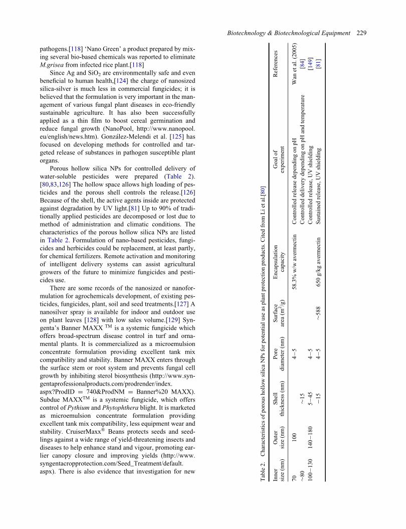
Porous hollow silica NPs for controlled delivery of
water-soluble pesticides were prepared (Table 2).
[80,83,126] The hollow space allows high loading of pes-
ticides and the porous shell controls the release.[126]
Because of the shell, the active agents inside are protected
against degradation by UV light.[81] Up to 90% of tradi-
tionally applied pesticides are decomposed or lost due to
method of administration and climatic conditions. The
characteristics of the porous hollow silica NPs are listed
in Table 2. Formulation of nano-based pesticides, fungi-
cides and herbicides could be replacement, at least partly,
for chemical fertilizers. Remote activation and monitoring
of intelligent delivery systems can assist agricultural
growers of the future to minimize fungicides and pesti-
cides use.
There are some records of the nanosized or nanofor-
mulation for agrochemicals development, of existing pes-
ticides, fungicides, plant, soil and seed treatments.[127] A
nanosilver spray is available for indoor and outdoor use
on plant leaves [128] with low sales volume.[129] Syn-
genta’s Banner MAXX TM is a systemic fungicide which
offers broad-spectrum disease control in turf and orna-
mental plants. It is commercialized as a microemulsion
concentrate formulation providing excellent tank mix
compatibility and stability. Banner MAXX enters through
the surface stem or root system and prevents fungal cell
growth by inhibiting sterol biosynthesis (http://www.syn-
gentaprofessionalproducts.com/prodrender/index.
aspx?ProdID D 740&ProdNM D Banner%20 MAXX).
Subdue MAXXTM is a systemic fungicide, which offers
control of Pythium and Phytophthera blight. It is marketed
as microemulsion concentrate formulation providing
excellent tank mix compatibility, less equipment wear and
stability. CruiserMaxx� Beans protects seeds and seed-
lings against a wide range of yield-threatening insects and
diseases to help enhance stand and vigour, promoting ear-
lier canopy closure and improving yields (http://www.
syngentacropprotection.com/Seed_Treatment/default.
aspx). There is also evidence that investigation for new
Table2.
Characteristicsofporoushollowsilica
NPsforpotentialuse
asplantprotectionproducts.Cited
from
Lietal.[80]
Inner
size
(nm)
Outer
size
(nm)
Shell
thickness(nm)
Pore
diameter
(nm)
Surface
area
(m2/g)
Encapsulation
capacity
Goalof
experim
ent
References
70
100
4�5
58.3%
w/w
avermectin
Controlled
releasedependingonpH
Wan
etal.(2005)
»80
»15
Controlled
deliverydependingonpHandtemperature
[84]
100�1
30
140�1
80
5�4
54�5
Controlled
release,UVshielding
[149]
¡15
4�5
»588
650g/kgavermectin
Sustained
release,UVshielding
[81]
Biotechnology & Biotechnological Equipment 229
formulations of plant protection products with quantita-
tively high application potential is continuing.
Improving plant resistance
Generally, plants are susceptible to many disease causing
agents like insect�pest, nematodes and other pathogens
and also drought which leads to the tremendous economic
loss. To avoid these losses, only alternative is to develop
resistant varieties of plants. Resistance in plants would
help in management of above-mentioned agents to over-
come the problem of economic loss.
Nanobiotechnology offers a novel set of procedures
using NPs, nanofibres and nanocapsules to multiply genes
and thus improve plant resistance.[129�131] The success-
ful insertion and integration of plasmid DNA in the plant
genome has been confirmed through gene expression.[132]
Nanoparticle-mediated plant transformations have the
potential to improve plants and disease resistance through
genetic modification.[129] Nanotechnology can specifically
target specific phytopathology problems in agriculture such
as in plant�pathogen interactions and provide new meth-
ods for crop disease management.[130] For example, intro-
duction of resistance genes in plant cells using
nanotechnological approaches may lead to development of
resistant varieties which will minimize expenses on agro-
chemicals required for disease control.[119]
Silver nanoparticles as nanopesticides
The rapid development in nanopesticide research over the
last two years have motivated a number of international
organizations to consider potential issues relating the use
of NT for crop protection.[133]
Silver has wide applications in metal or compound
form because it has antimicrobial activity against patho-
gens; however, it is nontoxic to humans.[134,135]
Recently, NT has increased the effectiveness of Ag NPs.
[117] The larger surface area-to-volume ratio of Ag NPs
increases their contact with microbes and their ability to
permeate.[117]
Micro-organisms have caused tremendous environ-
mental ecological changes. This is the result of entry of
new diseases into countries and has resulted in disease
and death of tree species (e.g. the USA and Europe). [119]
Agricultural crops and forestry must therefore be pro-
tected against the invasions of insect pests and fungal
pathogens. A mechanism for disease control is therefore
needed and the development of nanopesticides can help
control plant diseases.[119] The effect of Ag NPs on the
fungal phytopathogen Raffaelea sp. that causes oak wilt
was evaluated in vitro.[107] They reported that Ag NP
causes damage to fungal hyphae, interfered with their
microbial absorption, and increased inhibition of fungal
growth and conidial germination. Similarly Min et al. [99]
observed that Ag NPs remarkably inhibited the hyphal
growth of R.solani, S.sclerotiorum and S. minor in a dose-
dependent manner in vitro. Likewise, antifungal activity
of various forms of silver ions and NPs against B.sorokini-
ana and M.grisea were tested by Jo et al. [106] They
found that both silver ions and Ag NPs influence colony
formation of spores and disease progress of plant-patho-
genic fungi. These results suggest that Ag NPs may have
a huge potential for use as nanopesticides for the control
of phytopathogens.
Toxicity of silver nanoparticles
Nanoformulations are viewed to be safer and environment
friendly option for plant disease management, but high
toxicity of NPs inadvertently released in the environment
may pose greater threat to man and other organisms.[136]
The ecotoxicological effects of nanomaterials on
plants and soil micro-organisms have been widely investi-
gated. However, the nanotoxic effects of plant-soil inter-
active systems are still largely unknown.[137,138]
There are many gaps in our knowledge on the agric-
ecotoxicity of NPs and there are many unresolved prob-
lems and new challenges concerning the biological
effects.[139]
There is a need for phytotoxicity study on seed sys-
tems exposed to different concentrations of NPs to deter-
mine root length, germination effect, adsorption and
accumulation of NPs (uptake studies) into the plant sys-
tems.[140] When nanosized silica-silver particles were
applied in field condition to control powdery mildew dis-
eases of cucurbits, 100% control was achieved after three
weeks.[101] These NPs were found to be phytotoxic only
at a very high dose of 3200 ppm when tested in cucumber
and pansy plants. Similar study to deliver the NPs in the
targeted site of a diseased plant has been done by Corre-
dor et al.[141] The effect of NPs on different plant species
varies greatly, and both positive and negative effects have
been reported. Interestingly, NPs cause both positive
[142] and negative effects [143] on root elongation,
depending on the plant species (corn, cucumber, soybean,
cabbage, carrot and tomato). ZnO and TiO2 manufactured
nanomaterials (MNMs) impacted soil microbial commu-
nity diversity and biomass. Together, such reports imply
that cultivated soybean exposed to MNMs could be
impacted directly or through plant�microbe interactions,
including N2-fixing symbioses that is sensitive to some
metals.[144] The soil�plant interactive system might
help decrease the toxic effects of ZnO NPs on the rhizo-
bacteria population.[138] Some NPs have an influence on
bacterial growth and stress resistance, on plant sensitivity
to bacterial infection, and on the mechanisms of interac-
tion between plant and associated bacteria.[145]
In order to understand the possible benefits of apply-
ing NT to agriculture, the first step should be to analyse
230 M.A. Alghuthaymi et al.

penetration and transport of NPs in plants. Since nanoma-
terials are introduced into the soil as a result of human
activities, NPs can enter soil through atmospheric routes
and biosolids-amended agricultural soils.[146]
A potential transport pathway of NPs in the plant eco-
system was shown in Figure 6. The penetration and trans-
port of NPs inside whole plant was evaluated for the first
time by Gonz�alez-Melendi et al.[125] These results indi-
cate the possibility and potential of NPs in delivery of
substances inhibitory to various plant pathogens. Several
efforts are needed to clarify the interaction between nano-
materials, plants, phylloplane microflora, endophytes and
soil micro-organisms, both beneficial and pathogenic
effects on the plant health. Also, further work needs to be
performed in order to develop bio-indicators not only to
assess the effects of NPs on the agriculture environment
but also to recommend models for this assessment. In this
respect, some biological models developed to study mech-
anisms of interaction between different organisms could
be evaluated and proposed to study the effects of NPs on
biological systems such as those represented by plants and
associated microbes.[147] However, little data exist
regarding the toxicity of NPs on plant and soil microbial
community interactive systems.
Applications
(1) Nanosilver (Ag NP) materials have a wide range
of purposes including antimicrobial sterilization.
(2) Silver nanoparticles have been known to have
antifungal and bactericidal effects.
(3) Nanocapsules or emulsion for delivery of pesti-
cides, fungicides, fertilizers and other
agrochemicals.
(4) Nanoclays and nanofilms as barrier materials to
prevent fruit spoilage anti-post-harvest disease.
(5) Nanoparticles to selectively combine and elimi-
nate chemicals or pathogens from food.
Conclusions and future trends
Nanotechnology (NT) is gradually being incorporated into
the crop industry sector. There is an increasing interest in
the use of fungi for these processes, and fungi may
have the potential to present relatively rapid and environ-
mentally ‘clean’ nanobiofactories for metallic NPs.
Uncontrolled use of fungicides has caused many problems
such as adverse effects on human health, adverse effects
on pollinating insects and domestic animals, and entering
this material into the soil and water and its direct and indi-
rect effect on ecosystems. Intelligent use of chemicals on
the nanoscale can be a suitable solution for this problem.
These materials are used onto the part of the plant that
was attacked by disease or pest. Also, these carriers have
self-regulation, which means that the required amount of
medication can be delivered to the plant tissue. More
nanophytopathological studies on physiology of host and
pathogen, interaction, infection process and disease diag-
nosis will help in developing new disease management
strategy including nanopesticides that are less harmful to
the environment than conventional formulations.
Disclosure statement
No potential conflict of interest was reported by the authors.
Funding
The authors gratefully acknowledge Unit of Excellence in Nano-Molecular Plant Pathology Research (ARC), Egypt for fundingthis study. Also, this work was partially funded by the Scienceand Technology Development Fund (STDF), Egypt (STDF-STFprogram) [grant number 4552]. Mahendra Rai is thankful toDepartment of Science and Technology (Nanomission Pro-gramme), Government of India; to University Grants Commis-sion, New Delhi for financial assistance under UGC-SAPprogramme.
References
[1] Taniguchi N. On the basic concept of ‘nano-technology’.In: Proceedings of the international conference on pro-duction engineering Tokyo, Part II; 1974. p. 18�23.Tokyo: Japan Soc Precision Engineering.
Figure 6. A potential transport pathway of NPs in the plant eco-system, coated nanoparticles (CNP) and NPs.
Biotechnology & Biotechnological Equipment 231
[2] Narayanan KB, Sakthivel N. Biological synthesis ofmetal nanoparticles by microbes. Adv Colloid InterfaceSci. 2010;156:1�13.
[3] Maynard AD, Michelson, E. The Nanotechnology Con-sumer Product Inventory [Internet] Washington, DC:Woodrow Wilson International Center for Scholars; c2005.Available from: http://www.nanotechprojectorg/44S.
[4] Puebla RA, Dos Santos DS Jr, Aroca RF. Surface-enhanced Raman scattering for ultrasensitive chemicalanalysis of 1 and 2-naphthalenethiols. Analyst.2004;129:1251�1256.
[5] Shankar SS, Ahmad A, Pasricha R, Sastry M. Bioreduc-tion of chloroaurate ions by geranium leaves and its endo-phytic fungus yields gold nanoparticles of differentshapes. J Mater Chem. 2003;13:1822�1826.
[6] Gericke M, Pinches A. Biological synthesis of metalnanoparticles. Hydrometallurgy. 2006;83:132�140.
[7] He SY, Guo ZR, Zhang Y, Zhang S, Wang J, Gu N.Biosynthesis of gold nanoparticles using the bacteriaRhodopseudomonas capsulate. Mater Lett. 2007;61:3984�3987.
[8] Konishi Y, Ohno K, Saitoh N, Nomura T, Nagamine S,Hishida H, Takahashi Y, Uruga T. Bioreductive deposi-tion of platinum nanoparticles on the bacterium Shewa-nella algae. J Biotechnol. 2007;128:648�653.
[9] Moreau, JW, Weber PK, Martin MC, Gilbert B, HutcheonID, Banfield JF. Extracellular proteins limit the dispersalof biogenic nanoparticles. Science. 2007;316:1600�1603.
[10] Wang J, Chen C. Biosorbents for heavy metals removaland their future. Biotechnol Adv. 2009;27:195�226.
[11] Rai M, Yadav A, Gade A. Silver nanoparticles as a newgeneration of antimicrobials. Biotechnol Adv.2009;27:76�83.
[12] Rai M, Yadav P, Bridge P, Gade A. MycoNanotechnol-ogy (NT), a new and emerging science. In Rai Bridge,editor. Applied mycology. London, UK: CAB Interna-tional; 2009. p. 258�267.
[13] Meyer V. Genetic engineering of filamentous fungi �progress, obstacles and future trends. Biotechnol Adv.2008;26:177�185.
[14] Dhillon GS, Brar SK, Kaur S, Verma M. Green approachfor nanoparticle biosynthesis by fungi. Curr Trends Appl.2012;32:49�73.
[15] Sastry RK, Rashmi HB, Rao NH, Ilyas SM. Integratingnanotechnology (NT) into agri-food systems research inIndia: a conceptual framework. Technol Forecasting SocChange 2010;77:639�648.
[16] Khot LR, Sankaran S, Maja JM, Ehsani R, Schuster EW.Applications of nanomaterials in agricultural productionand crop protection: a review. Crop Prot. 2012;35:64�70.
[17] Bhainsa KC, D’Souza SF. Extracellular biosynthesis ofsilver nanoparticles using the fungus Aspergillus fumi-gates. Colloids Surfaces B. 2006;47:160�164.
[18] Mandal D, Bolander ME, Mukhopadhyay D, Sarkar G,Mukherjee P. The use of microorganisms for the forma-tion of metal nanoparticles and their application. ApplMicrobiol Biotechnol. 2006;69:485�492.
[19] Mohanpuria P, Rana NK, Yadav SK. Biosynthesis ofnanoparticles, technological concepts and future applica-tions. J Nanoparticle Res. 2007;7:9275�9280.
[20] Mukherjee P, Roy M, Mandal BP, Dey GK, MukherjeePK, Ghatak J, Tyagi AK, Kale SP. Green synthesis ofhighly stabilized nanocrystalline silver particles by a non-
pathogenic and agriculturally important fungus Tricho-derma asperellum. Nanotechnolology. 2008;19:075103.
[21] Gaikwad S, Birla SS, Ingle AP, Gade AK, Marcato PD,Rai MK, Duran D. Screening of different Fusariumspecies to select potential species for the synthesis of sil-ver nanoparticles. J Braz Chem Soc. 2013;24:1974�1982.
[22] Dias MA, Lacerda ICA, Pimentel PF, De Castro HF, RosaCA. Removal of heavy metals by an Aspergillus terreusstrain immobilized in a polyurethane matrix. Lett ApplMicrobiol. 2002;34:46�50.
[23] Ahmad A, Mukherjee P, Senapati S, Mandal D, Khan MI,Kumar R, Sastry M. Extracellular biosynthesis of silvernanoparticles using the fungus Fusarium oxysporum. Col-loid Surfaces B. 2003;28:313�318.
[24] Birla SS, Gaikwad SC, Gade AK, Rai MK. Rapid synthe-sis of silver nanoparticles from Fusarium oxysporum byoptimizing physicocultural conditions. Scientific World J.2013;2013:1�12.
[25] Bansal V, Rautaray D, Ahmad A, Sastry M. Biosynthesisof zirconia nanoparticles using the fungus Fusarium oxy-sporum. J Mater Chem. 2004;14:3303�3305.
[26] Bharde A, Rautaray D, Bansal V, Ahmad A, Sarkar I,Yusuf SM, Sanyal M, Sastry M. Extracellular biosynthe-sis of magnetite using fungi. Small. 2006;2:135�141.
[27] Riddin TL, Gericke M Whiteley CG. Analysis of theinter- and extracellular formation of platinum nanopar-ticles by Fusarium oxysporum f. sp. lycopersici usingresponse surface methodology. Nanotechnology.2006;17:3482�3489.
[28] Dur�an N, Marcato PD, Alves OL, Da Silva JPS, De SouzaGIH, Rodrigues, FA, Esposito E. Ecosystem protection byeffluent bioremediation, Silver nanoparticles impregna-tion in a textile fabrics process. J Nanoparticle Res.2010;12:285�292.
[29] Khosravi A, Shojaosadati SA. Evaluation of silver nano-particles produced by fungus Fusarium oxysporum. Int JNanotechnol. 2009;6:973�983.
[30] Mohammadian A, Shojaosadati, Rezaee MH. Fusariumoxysporum mediates photogeneration of silver nanopar-ticles. Sci Iran. 2007;14:323�326.
[31] Ingle A, Gade A, Pierrat S, Sonnichsen C, Rai M. Myco-synthesis of silver nanoparticles using the fungus Fusar-ium acuminatum and its activity against some humanpathogenic bacteria. Curr Nanosci. 2008;4:141�144.
[32] Ingle A, Gade A, Bawaskar M, Rai M. Fusarium solani, anovel biological agent for the extracellular synthesis ofsilver nanoparticles. J Nanoparticle Res. 2009;11:2079�2085.
[33] Deepa K, Panda T. Synthesis of gold nanoparticles fromdifferent cellular fractions of Fusarium oxysporum. JNanosci Nanotechnol. 2014;14:3455�3463.
[34] Bawaskar M, Gaikwad S, Ingle A, Rathod D, Gade A,Duran N, Marcato PD, Rai M. A new report on mycosyn-thesis of silver nanoparticles by Fusarium culmorum.Curr Nanosci. 2010;6:376�380.
[35] Basavaraja S, Balaji SD, Lagashetty A, Rajasab AH, Ven-kataraman A. Extracellular biosynthesis of silver nano-particles using the fungus Fusarium semitectum. Mat ResBull. 2008;43:1164�1170.
[36] Sadowski Z, Maliszewska IH, Grochowalska B, Polowc-zyk I, Ko�zlecki T. Synthesis of silver nanoparticles usingmicroorganisms. Mater Sci. 2008;26:219�224.
[37] Zhang X, He X, Wang K, Wang Y, Li H, Tan W. Biosyn-thesis of size-controlled gold nanoparticles using fungus,
232 M.A. Alghuthaymi et al.
Penicillium sp. J Nanosci Nanotechnol. 2009;10:5738�5744.
[38] Kathiresan K, Manivannan S, Nabeel MA, Dhivya B.Studies on silver nanoparticles synthesized by a marinefungus, Penicillium fellutanum isolated from coastal man-grove sediment. Colloids Surfaces B. 2009;7:133�137.
[39] Shaligram NS, Bule M, Bhambure R, Singhal RS, SinghSK, Szakacs G, Pandey A. Biosynthesis of silver nanopar-ticles using aqueous extract from the compactin produc-ing fungal strain. Process Biochem. 2009;44, 939�943.
[40] Nayak RR, Pradhan N, Behera D, Pradhan KM, Mishra S,Sukla LB, Mishra BK. Green synthesis of silver nanopar-ticle by Penicillium purpurogenum NPMF, the processand optimization. J Nanoparticle Res. 2010;13:3129�3137.
[41] Maliszewska I, Juraszek A, Bielska K. Green synthesisand characterization of silver nanoparticles using ascomy-cota fungi Penicillium nalgiovense AJ12. J Cluster Sci.2013;25:989�1004.
[42] Singh D, Rathod V, Ninganagouda S, Hiremath J, Singh,AK, Mathew J. Optimization and characterization of sil-ver nanoparticle by endophytic fungi Penicillium sp. iso-lated from Curcuma longa (turmeric) and applicationstudies against MDR E. coli and S. aureus. BioinorgChem Appl. 2014; doi:10.1155/2014/408021.
[43] Kumar R, Liu D, Zhang L. Advances in proteinous bio-materials. J Biobased Mater Bioenergy. 2008;2:1�24.
[44] Vigneshwaran N, Ashtaputre NM, Varadarajan PV,Nachane RP, Paralikar KM, Balasubramanya RH. Biolog-ical synthesis of silver nanoparticles using the fungusAspergillus flavus. Mater Lett. 2006;61:1413�1418.
[45] Navazi ZR, Pazouki M, Halek FS. Investigation of cul-ture conditions for biosynthesis of silver nanoparticlesusing Aspergillus fumigates. Iran J Biotechnol.2010;8:56�61.
[46] Saravanan M, Nanda A. Extracellular synthesis of silverbionanoparticles from Aspergillus clavatus and its antimi-crobial activity against MRSA and MRSE. Colloids Sur-faces B. 2010;77:214�218.
[47] Verma VC, Kharwar RN, Gange AC. Biosynthesis ofantimicrobial silver nanoparticles by the endophyticfungus Aspergillus clavatus. Nanomedicine. 2010;5:33�40.
[48] Li G, He D, Qian Y, Guan B, Gao S, Cui Y, Yokoyama K,Wang L. Fungus-mediated green synthesis of silver nano-particles using Aspergillus terreus. Int J Mol Sci.2012;13:466�476.
[49] Jaidev LR, Narasimha G. Fungal mediated biosynthesisof silver nanoparticles, characterization and antimicrobialactivity. Colloids Surfaces B. 2010;81:430�433.
[50] Kumar RR, Priyadharsani PK, Thamaraiselvi K. Myco-genic synthesis of silver nanoparticles by the Japaneseenvironmental isolate Aspergillus tamari. J NanoparticleRes. 2012;14:860�868.
[51] Raliya R, Tarafdar JC. 2014 Biosynthesis andcharacterization of zinc, magnesium and titanium nanopar-ticles: an eco-friendly approach. Int Nano Lett. 2014;93:3�10.
[52] Klaus T, Joerger R, Olsson E, Granqvist CG. Silver-basedcrystalline nanoparticles, microbially fabricated. ProcNatl Acad Sci USA. 1999;96: 13611�13614.
[53] Mukherjee P, Ahmad A, Mandal D, Senapati S, SainkarSR, Khan MI, Ramani R, Parischa R, Ajayakumar PV,Alam M, Sastry M, Kumar R. Bioreduction of AuCl4
¡
ions by the fungus, Verticillium sp. and surface trapping
of the gold nanoparticles formed. Angew Chem Int EdEngl. 2001;40(19):3585�3588.
[54] Sastry M, Ahmad A, Khan MI, Kumar R. Biosynthesis ofmetal nanoparticles using fungi and actinomycete. CurrSci. 2003;85:162�170.
[55] Shahi SK, Patra M. Biotechnological aspect for the syn-thesis of bioactive nanoparticle and their formulationactive against human pathogenic fungi. Rev Adv Mat Sc.2003;5:501�509.
[56] Vigneshwaran N, Kathe AA, Varadarajan PV, NachaneRP, Balasubramanya RH. Biomimetics of silver nanopar-ticles by white rot fungus, Phaenerochaete chrysospo-rium. Colloids Surfaces B. 2006;53:55�59.
[57] Chen JC, Lin ZH, Ma XX. Evidence of the production ofsilver nanoparticles via pretreatment of Phoma sp.3.2883with silver nitrate. Lett Appl Microbiol. 2003;37:105�108.
[58] Birla SS, Tiwari VV, Gade AK, Ingle AP, Yadav AP, RaiMK. Fabrication of silver nanoparticles by Phoma glom-erata and its combined effect against Escherichia coli,Pseudomonas aeruginosa and Staphylococcus aureus.Lett Appl Microbiol. 2009;48:173�179.
[59] Devi TP, Kulanthaivel S, Kamil D, Borah JL, Prabha-karan N, Srinivasa N. Biosynthesis of silver nanoparticlesfrom Trichoderma species. Indian J Exp Biol2013;51:543�547.
[60] Balaji DS, Basavaraja S, Deshpande R, Mahesh DB,Prabhakar BK, Venkataraman A. Extracellular biosynthe-sis of functionalized silver nanoparticles by strains ofCladosporium cladosporioides fungus. Colloids SurfacesB. 2009;68:88�92.
[61] Gade A, Gaikwad S, Duran N, Rai M. Screening of differentspecies of Phoma for synthesis of silver nanoparticles. Bio-technol Appl Biochem. 2013;60(5):482�493.
[62] Gade A, Rai M, Karwa A, Bonde P, Ingle A. Extracellularbiosynthesis of silver nanoparticles by Pleurotus species.Int J Med Mushroom Res. 2007;9(3�4):298�299.
[63] Nithya R, Ragunathan, R. Synthesis of silver nanoparticleusing Pleurotus sajor caju and its antimicrobial study.Digest J Nanomater Biostruct. 2009;4:623�629.
[64] Philip D. Biosynthesis of Au, Ag and Au�Ag nanopar-ticles using edible mushroom extract. SpectrochimicaActa Part A. 2009;73:374�381.
[65] Sanghi R, Verma PA. A facile green extracellular biosyn-thesis of CdS nanoparticles by immobilized fungus.Chem Eng J. 2009;155:886�891.
[66] Mishra AN, Bhadauria S, Gaur MS, Pasricha R. Extracel-lular microbial synthesis of gold nanoparticles using fun-gus Hormoconis resinae. JOM. 2010;62:45�48.
[67] Bao H, Hao N, Yang Y, Zhao D. Biosynthesis of biocom-patible cadmium telluride quantum dots using yeast cells.Nano Res. 2003;3:491�498.
[68] Rashmi K, Krishnaveni T, Ramanamurthy S, Mohan PM.Characterization of cobalt nanoparticle from a cobaltresistant strain of Neurospora crassa. In: InternationalSymposium of Research Students on Materials Scienceand Engineering; December 20�22. Chennai; 2004.
[69] Castro-Longoria E, Vilchis-Nestor AR, Avalos-Borja M.Biosynthesis of silver, gold and bimetallic nanoparticlesusing the filamentous fungus Neurospora crassa. ColloidsSurfaces B. 2011;83:42�48.
[70] Kar PK, Murmu S, Saha S, Tandon V, Acharya K. Anthel-mintic efficacy of gold nanoparticles derived from a phyto-pathogenic fungus, Nigrospora oryzae. PLoS ONE 2014;9(1):e84693.
Biotechnology & Biotechnological Equipment 233

[71] Singh D, Rathod V, Ninganagouda S, Herimath J, KulkarniP. Biosynthesis of silver nanoparticle by endophytic fungiPenicillium sp. isolated from Curcuma longa (turmeric)and its antibacterial activity against pathogenic gram nega-tive bacteria. J Pharm Res. 2013;7:448�453.
[72] Jain N, Bhargava A, Majumdar S, Tarafdar J, Panwar J.Extracellular biosynthesis and characterization ofsilver nanoparticles using Aspergillus flavus NJP08:a mechanism perspective. Nanoscale. 2011;3(2):635�641.
[73] Soni N, Prakash S. Factors affecting the geometry of sil-ver nanoparticles synthesis in Chrysosporium tropicumand Fusarium oxysporum. Am J Nanotechnol. 2011;2(1):112�121.
[74] Sunkar S, Nachiyar CV. Endophytic fungi mediatedextracellular silver nanoparticles as effective antibacterialagents. Int J Pharm Pharm Sci. 2013;5:95�100.
[75] Duran N, Marcato PD, Alves OL, DeSouza G, Esposito E.Mechanistic aspects of biosynthesis of silver nanopar-ticles by several Fusarium oxysporum strains. J Nanobio-technol. 2005;3:1�8.
[76] Kumar SA, Abyaneh, MK, Gosavi SW, Kulkarni SK, Pas-richa R, Ahmad A, Khan MI. Nitrate reductase-mediatedsynthesis of silver nanoparticles from AgNO3. BiotechnolLett. 2007;29:439�445.
[77] P�erez-de-Luque A, Rubiales D, Marquina CI, Ibarra MR,de la Fuente JM. Nanoparticles in agriculture, develop-ment of smart delivery systems for plant research Nano-Spain. Braga-Portugal, 14�18 April, 2008.
[78] Abd-Elsalam, KA. Nanoplatforms for plant pathogenicfungi management. Fungal Genomics Biol. 2012;2:e107.
[79] Kah M, Beulke S, Tiede K, Hofmann T. Nano-pesticides:state of knowledge, environmental fate and exposuremodelling. Crit Rev Environ Sci Technol.2013;43:1823�67.
[80] Li ZZ, Chen JF, Liu F, Liu AQ, Wang Q, Sun HY, WenLX. Study of UV-shielding properties of novel poroushollow silica nanoparticle carriers for avermectin. PestManag Sci. 2007;63:241�246.
[81] Rai M, Ingle A. Role of nanotechnology in agriculturewith special reference to management of insect pests.Appl Microbiol Biotechnol. 2012;94:287�293.
[82] Choudhury SR, Nair KK, Kumar R, Gogoi R, SrivastavaC, Gopal M, Subhramanyam BS, Devakumar C, Gos-wami A. Nanosulfur: a potent fungicide against foodpathogen, Aspergillus niger. AIP Conf Proc.2010;1276:154�157.
[83] Liu WT. Nanoparticles and their biological and environ-mental applications. J Biosci Bioeng. 2006;102:1�7.
[84] Navarro E, Baun A, Behra R, Hartmann NB, Filser J,Miao AJ, Quigg A, Santschi PH, Sigg L. Environmentalbehavior and ecotoxicity of engineered nanoparticles toalgae, plants, and fungi. Ecotoxicology. 2008;17:372�386.
[85] Sondi I, Salopek-Sondi B. Silver nanoparticles as antimi-crobial agent, a case study on E. coli as a model forGram-negative bacteria. J Colloid Interface Sci.2004;275:177�182.
[86] Aymonier C, Scholotterbeck U, LAntonietti P, ZachariasR, Thomann JC, Tiller Mecking S. Hybrids of silvernanoparticles with amphiphilic hyperbranched macromo-lecules exhibiting antimicrobial properties. Chem Com-mun. 2002;24:3018�3019.
[87] Retchkiman-Schabes PS, Canizal G, Becerra-Herrera R,Zorrilla C, Liu HB, Ascencio JA. Biosynthesis and
characterization of Ti/Ni bimetallic nanoparticles. OptMater. 2006;29:95�99.
[88] Gu H, Ho PL, Tong E, Wang L, Xu B. Presenting vanco-mycin on nanoparticles to enhance antimicrobial activi-ties. Nano Lett. 2003;3, 1261�1263.
[89] Ahmad Z, Pandey R, Sharma S, Khuller GK. Alginatenanoparticles as antituberculosis drug carriers, formula-tion development, pharmacokinetics and therapeuticpotential. Indian J Chest Dis Allied Sci.48;2005:171�176.
[90] Gong P, Li H, He X, Wang K, Hu J, Tan W. Preparationand antibacterial activity of Fe3O4 Ag nanoparticles.Nanotechnology. 2007;18:604�611.
[91] Lead JR, Wilkison KJ. Aquatic colloids and nanopar-ticles: current knowledge and future trends. EnvironChem. 2006;3:159�171.
[92] Kim SW, Kim KS, Lamsal K, Kim Y-J, Kim SB, Jung M,Sim SJ, Kim HS, Chang SJ, Kim JK, Lee YS. An in vitrostudy of the antifungal effect of silver nanoparticles onoak wilt pathogen Raffaelea sp. J Microbiol Biotechnol.2012;19:760�764.
[93] Prabhu S. Poulose EK. Silver nanoparticles: mechanismof antimicrobial action, synthesis, medical applications,and toxicity effects. Int Nano Lett. 2012;2:32.
[94] Lemire JA, Harrison JJ, Turner RJ. Antimicrobial activityof metals: mechanisms, molecular targets and applica-tions. Nat Rev Microbiol. 2013;11(6):371�384.
[95] Zeng F, Hou C, Wu SZ, Liu XX, Tong Z, Yu SN. Silvernanoparticles directly formed on natural macroporousmatrix and their anti-microbial activities. Nanotechnol-ogy. 2007;18:1�8.
[96] Falletta E, Bonini M, Fratini E, Lo Nostro A, PesaventoG, Becheri A. Clusters of poly (acrylates) and silver nano-particles: structure and applications for antimicrobial fab-rics. J Phys Chem C. 2008;112:11758�11766.
[97] Roe D, Karandikar B, Bonn-Savage N, Gibbins B, RoulletJB. Antimicrobial surface functionalization of plasticcatheters by silver nanoparticles. J Antimicrob Chemo-ther. 2008;61:869�876.
[98] Kim H, Kang H, Chu G, Byun H. Antifungal effectivenessof nanosilver colloid against rose powdery mildew ingreenhouses. Solid State Phenomenon 2008;135:15�18.
[99] Pan�a�cek A, Kol�a�r M, Ve�ce�rov�a R, Prucek R, Soukupov�aJ, Kry�stof V, Park HJ, Kim SH, Kim HJ, Choi SH.A new composition of nanosized silica-silver for controlof various plant diseases. Plant Pathol J. 2006;22:295�302.
[100] Min JS, Kim KS, Kim SW, Jung JH, Lamsal K, Kim SB,Jung M, Lee YS. Effects of colloidal silver nanoparticleson sclerotium-forming phytopathogenic fungi. PlantPathol J. 2009;25:376�380.
[101] Park H-J, Kim SH, Kim HJ, Choi S-H. A new composi-tion of nanosized silica-silver for control of various plantdiseases plant. Pathol J. 2006;22(3):295�302.
[102] Oh SD, Lee S, Choi SH, Lee IS, Lee YM, Chun JH, ParkHJ. Synthesis of Ag and Ag�SiO2 nanoparticles by y-irra-diation and their antibacterial and antifungal efficiencyagainst Salmonella enteric serovar Typhimurium and Botry-tis cinerea. Colloids Surfaces A. 2006;275:228�233.
[103] Gajbhiye M, Kesharwani J, Ingle A, Gade A, Rai M. Fun-gus mediated synthesis of silver nanoparticles and theiractivity against pathogenic fungi in combination with flu-conazole. Nanomedicine. 2009;5:382�386.
[104] Fateixa S, Neves MC, Almeida A, Oliveira J, Trindade T.Anti-fungal activity of SiO2/Ag2S nanocomposites
234 M.A. Alghuthaymi et al.

against Aspergillus niger. Colloids Surfaces B. 2009;74,304�308.
[105] Ruffolo, SA, La Russa, MF, Malagodi M, Oliviero RossiC, Palermo AM, Crisci GM. ZnO and ZnTiO3 nanopow-ders for antimicrobial stone coating. Appl Phys A.2010;100:829�834.
[106] Jo, YK, Kim BH, Jung G. Antifungal activity of silverions and nanoparticles on phytopathogenic fungi. PlantDis. 2009;93:1037�1043.
[107] Woo KS, Kim KS, Lamsal K, Kim YJ, Kim SB, Jung M,Sim SJ, Kim HS, Chang SJ, Kim JK, Lee YS. An in vitrostudy of the antifungal effect of silver nanoparticles onoak wilt pathogen Raffaelea sp. J Microbiol Biotechnol.2009;19:760�764.
[108] Kasprowicz MJ, Kozio» M, Gorczyca A. The effect of sil-ver nanoparticles on phytopathogenic spores of Fusariumculmorum. Can J Microbiol. 2010;56:247�253.
[109] Musarrat J, Dwivedi, S, Singh BR, Al-Khedhairy AA,Azam Naqvi A. A production of antimicrobial silver nano-particles in water extracts of the fungus Amylomyces rouxiistrain KSU-09. Biores Technol. 2010;101:8772�8776.
[110] He L, Liu, Y, Mustapha A, Lin M. Antifungal activityof zinc oxide nanoparticles against Botrytis cinereaand Penicillium expansum. Microb Res. 2010;166:207�215.
[111] Jung J-H, Kim S-W, Min J-S, Kim Y-J, Lamsal K, KimKS. The effect of nano-silver liquid against the white rotof the green onion caused by Sclerotium cepivorum.Mycobiology. 2010;38(1):39�45.
[112] Aguilar-M�endez MA, San Mart�ın-Mart�ınez E, Ortega-Arroyo L, Cobi�an-Portillo G, S�anchez-Esp�ındola E. Syn-thesis and characterization of silver nanoparticles: effecton phytopathogen Colletotrichum gloesporioides. J Nano-part Res. 2010;13:2525�2532.
[113] Krishnaraj C, Ramachandran R, Mohan K, KalaichelvanPT. Optimization for rapid synthesis of silver nanopar-ticles and its effect on phytopathogenic fungi. Spectrochi-mica Acta Part A. 2012;93:95�99.
[114] Saharan V, Mehrotra A, Khatik R, Rawal P, Sharma SS,Pal A. Synthesis of chitosan based nanoparticles and theirin vitro evaluation against phytopathogenic fungi. Int JBiol Macromol. 2013;62:677�683.
[115] Khaydarov RR, Khaydarov RA, Evgrafova S, Estrin Y.Using silver nanoparticles as an antimicrobial agent.NATO Sci Peace Security Ser A. 2011;169�177.
[116] Kanto T, Miyoshi A, Ogawa T, Maekawa K, Aino M.Suppressive effect of potassium silicate on powdery mil-dew of strawberry in hydroponics. J Gen Plant Pathol.2004;70:207�211.
[117] Kim KJ, Sung WS, Moon SK, Choi JS, Kim JG, Lee DG.Antifungal effect of silver nanoparticles on dermato-phytes. J Microbiol Biotechnol. 2008;18:1482�1484.
[118] Gogoi R, Dureja P, Singh PK. Nanoformulations: a saferand effective option for agrochemicals. Indian Farming.2009;59(8):7�12.
[119] Bouwmeester H, Dekkers S, Noordam MY, Hagens WI,Bulder AS, Heer Cde, Voorde SECGt, Wijnhoven SWP,Marvin HJP, Sips AJAM. Review of health safety aspectsof nanotechnologies in food production. Regul ToxicolPharmacol. 2009;53:52�62.
[120] Taylor TM, Davidson PM, Bruce BD, Weiss J. Liposomalnanocapsules in food science and agriculture. Crit RevFood Sci Nutr. 2005;45:587�605.
[121] Letchford K, Burt H. A review of the formation and clas-sification of amphiphilic block copolymer nanoparticulatestructures: micelles, nanospheres, nanocapsules and poly-mersomes. Eur J Pharm Biopharm. 2007;65:259�269.
[122] Owolade OF, Ogunleti DO, Adenekan MO. Titaniumdioxide affects disease development and yield of ediblecowpea. Electron J Environ Agric Food Chem. 2008;7(50):2942�2947.
[123] Rickman D, Luvall JC, Shaw J, Mask P, Kissel D, SullivanD. Precision agriculture, changing the face of farming.[Internet]. 1999. Available from: wwwghccmsfcnasagove/precisionag/.
[124] Yau CP, Wang L, Yu M, Zee SY, Yip WK. Differentialexpression of three genes encoding an ethylene receptorin rice during development, and in response to indole-3-acetic acid and silver ions. J Exp Bot. 2004;55:547�555.
[125] Gonz�alez-Melendi P, Fern�andez-Pacheco R, CoronadoMJ, Corredor E, Testillano PS, Risue~no MC, Marquina C,Ibarra MR, Rubiales D, P�erez-de-Luque A. Nanoparticlesas smart treatment-delivery systems in plants; assessmentof different techniques of microscopy for their visualiza-tion in plant tissues. Ann Bot. 2008;101:187�195.
[126] Wen LX, Li Z-Z, Zou H-K, Liu A-Q, Chen, J-F. Con-trolled release of avermectin from porous hollow silicananoparticles. Pest Manag Sci. 2005;61:583�590.
[127] FAO Crop Prospects and Food Situation. FAO GlobalCereal Supply and Demand Indicators. [Internet]. 2008.(No. 2, 2008 April). Available from: http://wwwfaoorg/docrep/010/ai465e/ai465305htm.
[128] NANOSYS GMBH Nano-Argentum 10 TechnischesMerkblatt. [Internet]. 2005. Available from: http://wwwnanosysch/tedablas/d/nanoargentum230905pdf.
[129] McKnight TE, Melechko AV, Griffin GD, Guillorn MA,Merkulov VI, Serna F, Hensley DK, Doktycz MJ, Low-ndes DH, Simpson ML. Intracellular integration of syn-thetic nanostructures with viable cells for controlledbiochemical manipulation. Nanotechnology.2003;14:551�556.
[130] Torney F, Trewyn BG, Lin SY, Wang K. Mesoporous sil-ica nanoparticles deliver DNA and chemicals into plants.Nat Nanotechnol. 2007;2:295�300.
[131] Rai M, Deshmukh S, Gade A, Elsalam K-A. Strategicnanoparticles-mediated gene transfer in plants and ani-mals � a novel approach. Curr Nano. 2012;8:170�179.
[132] Filipenko EA, Filipenko ML, Deineko EV, Shumnyi VK.Analysis of integration sites of T-DNA insertions in trans-genic tobacco plants. Cytol Genet. 2007;41:199�203.
[133] Kah M, Hofmann T. Nanopesticide research: current trendsand future priorities. Environ Int. 2014;63:224�235.
[134] Elchiguerra JL, Burt JL, Morones JR, Camacho- BragadoA, Gao X, Lara HH, Yacaman MJ. Interaction of silvernanoparticles with HIV-1. J Nanobiotechnol. 2005;3:1�10.
[135] Yeo SY, Lee HJ, Jeong SH. Preparation of nanocompo-site fibers for permanent antibacterial effect. J Mater Sci.2003;38:2143�2147.
[136] Banik S, Sharma P. Plant pathology in the era of nano-technology. Indian Phytopathol. 2011;64:120�127.
[137] Vittori-Antisari L, Carbone S, Fabrizi A, Gatti A, Via-nello G. Response of soil microbial biomass to CeO2
nanoparticles. EQA. 2011;7:135�150.[138] Lee S, Kim S, Kim S, Lee I. Effects of soil-plant interac-
tive system on response to exposure to ZnO nanoparticles.J Microbiol Biotechnol. 2012;22:1264�1270.
Biotechnology & Biotechnological Equipment 235
[139] Monica RC, Cremonini R. Nanoparticles and higherplants. Caryologia. 2009;62:161�165.
[140] Kumari M, Ernest V, Mukherjee A, Chandrasekaran N. Invivo nanotoxicity assays in plant models. Methods MolBiol. 2012;926:399�410.
[141] Corredor E, Testillano PS, Coronado M-J, Gonz�alez-Melendi P, Fern�andez-Pacheco R, Marquina C, IbarraMR, de la Fuente JM, Rubiales D, P�erez-de-Luque A,Risue~no MC. Nanoparticle penetration and transport inliving pumpkin plants: in situ subcellular identification.BMC Plant Biol. 2009;9:45.
[142] Khodakovskaya M, Dervishi E, Mahmood M, Xu Y, Li Z,Watanabe F, Biris AS. Carbon nanotubes are able to pene-trate plant seed coat and dramatically affect seed germina-tion and plant growth. ACS Nano. 2009;3:3221�3227.
[143] Lin D, Xing B. Phytotoxicity of nanoparticles: inhibitionof seed germination and root growth. Environ Pollut.2007;20:1�8.
[144] Ge Y, Schimel JP, Holden PA. Evidence for negativeeffects of TiO2 and ZnO nanoparticles on soil bacterialcommunities. Environ Sci Technol. 2011;45:1659�1664.
[145] Degrassi G, Bertani I, Devescovi G, Fabrizi A, Gatti A,Venturi V. Response of plant-bacteria interaction modelsto nanoparticles. EQA. 2012;8:39�50.
[146] Nowack B, Ranville JF, Diamond S, Gallego-Urrea JA,Metcalfe C, Rose J, Horne N, Koelmans AA, Klaine SJ.Potential scenarios for nanomaterial release and subse-quent alteration in the environment. Environ ToxicolChem. 2012;31:50�59.
[147] Thul ST, Sarangi BK, Pandey RA. Nanotechnology inagroecosystem: implications on plant productivity and itssoil environment. Expert Opin Environ Biol. 2013;2:2�7.
[148] Fayaz AM, Balaji K, Girilal M, Kalaichelvan PT, Venka-tesan R. Mycobased synthesis of silver nanoparticles andtheir incorporation into sodium alginate films for vegeta-ble and fruit preservation. Agric Food Chem.2009;57:6246�6252.
[149] Joshi P, Bonde S, Gaikwad S, Gade A, Abd-Elsalam KA,Rai M. Comparative studies on synthesis of silver nano-particles by Fusarium oxysporum and Macrophominaphaseolina and its efficacy against bacteria and Malasse-zia furfur. J Bionanosci. 2013;7:1�5.
236 M.A. Alghuthaymi et al.