Synthesis and Growth Mechanism of Iron Oxide Nanowhiskers
6
Published: February 10, 2011 r2011 American Chemical Society 1141 dx.doi.org/10.1021/nl200136j | Nano Lett. 2011, 11, 1141–1146 LETTER pubs.acs.org/NanoLett Synthesis and Growth Mechanism of Iron Oxide Nanowhiskers Soubantika Palchoudhury, † Wei An, † Yaolin Xu, † Ying Qin, ‡ Zhongtao Zhang, † Nitin Chopra, § Robert A. Holler, || C. Heath Turner,* ,† and Yuping Bao* ,† † Chemical and Biological Engineering, ‡ Alabama Institute for Manufacturing Excellence, § Metallurgical and Materials Engineering and Center for Materials for Information Technology, and ) Central Analytical Facility, The University of Alabama, Tuscaloosa, Alabama, 35487 United States b S Supporting Information ABSTRACT: Iron oxide nanowhiskers with dimensions of approximately 2 20 nm were successfully synthesized by selectively heating an iron oleate complex. Such nanostruc- tures resulted from the difference in the ligand coordination microenvironments of the Fe(III) oleate complex, according to our electronic structure calculations and thermogravi- metric analysis. A ligand-directed growth mechanism was subsequently proposed to rationalize the growth process. The formation of the nanowhiskers provides a unique example of shape-controlled nanostructures, offering additional insights into nanoparticle synthesis. KEYWORDS: Iron oxide nanowhiskers, ligand-directed growth, electronic structure calculation of complexes, iron oxide nanoclusters A nisotropic nanostructures have attracted much attention in various applications because of their unique electronic, magnetic, and optical properties. 1-3 In particular, the synthesis of one-dimensional (1D) metallic and semiconductor nano- structures has been well documented. 3-5 Most recently, ultrathin (∼2 nm) nanowires, 6 such as Au, 7-10 FePt, 11 and oxides, 12,13 have attracted much interest. In contrast, only few studies of 1D iron oxide magnetic nanoparticles have been reported (e.g., iron oxide nanobars 14 and nanowires 15 ). Spherical iron oxide nano- particles have been primarily explored in targeted drug delivery, localized therapy, or as contrast agents for magnetic resonance imaging (MRI). 16,17 A recent study of 1D iron oxide nanoworms showed long blood circulation time, enhanced retention at tumor sites, and improved targeting efficiency, 18 which suggests that anisotropic iron oxide nanoparticles could potentially lead to further advancement in biomedical applications. The synthetic approach to iron oxide spheres has been intensively focused on the thermal decomposition of iron(III) oleate complexes due to its great reproducibility and control of the physical parameters. 19 In this method, the Fe(III) oleate pre- cursor is typically heated up to over 300 °C, producing different- sized spherical nanoparticles with a narrow size distribution. Cubic and bipyramid-shaped particles were also reported using this method as a result of the selective absorption of impurity ions, such as Cl - , Na þ , or oleate. 20-26 So far, the formation of ultra- thin, 1D iron oxide nanoparticles has not been reported. Even though the decomposition of the iron oleate complex is widely used for the synthesis of iron oxide nanoparticles, few mechanistic studies are available to understand the growth process. Hyeon 27 proposed that the dissociation of the first oleate ligand at around 200-240 °C triggered the nucleation event, followed by nanoparticle growth through the decomposi- tion of the two remaining ligands above 300 °C. However, it has been rather difficult to conclusively confirm the dissociation process of the iron oleate complex. A recent density functional theory (DFT) electronic structure calculation of iron carboxylate complexes showed different dissociation temperatures of the three carboxylate ligands. 28 The first and the second ligands have similar dissociation temperatures, while the dissociation tem- perature of the third ligand was significantly higher. 28 The calculations further proposed the formation of an Fe-O bond between the third ligand and the iron center. Unfortunately, an understanding of the chemical microenvironments of these three ligands and their effects on the nanostructure formation is still lacking. In this Letter, we report the synthesis of ultrathin iron oxide nanowhiskers, based on an understanding of the precursor chemistry. Our DFT calculations of the Fe(III) oleate complex predict a large difference among the binding energies of the three ligands. Experimentally, such a difference was reflected by the distinct weight losses of the thermogravimetric analysis (TGA) plot of the Fe(III) oleate complex. The different bindings allowed us to selectively decompose the more weakly bound ligands at 150 °C, forming iron oxide nanoclusters through ligand-directed growth. This study provides a unique example of shape-controlled iron oxide nanoparticles, which demon- strates the importance of the chemical microenvironments, Received: November 19, 2010
Transcript of Synthesis and Growth Mechanism of Iron Oxide Nanowhiskers
Published: February 10, 2011
r 2011 American Chemical Society 1141 dx.doi.org/10.1021/nl200136j |Nano Lett. 2011, 11, 1141–1146
LETTER
pubs.acs.org/NanoLett
Synthesis and Growth Mechanism of Iron Oxide NanowhiskersSoubantika Palchoudhury,† Wei An,† Yaolin Xu,† Ying Qin,‡ Zhongtao Zhang,† Nitin Chopra,§
Robert A. Holler,|| C. Heath Turner,*,† and Yuping Bao*,†
†Chemical and Biological Engineering, ‡Alabama Institute for Manufacturing Excellence, §Metallurgical and Materials Engineering andCenter for Materials for Information Technology, and )Central Analytical Facility, The University of Alabama, Tuscaloosa,Alabama, 35487 United States
bS Supporting Information
ABSTRACT: Iron oxide nanowhiskers with dimensions ofapproximately 2 � 20 nm were successfully synthesized byselectively heating an iron oleate complex. Such nanostruc-tures resulted from the difference in the ligand coordinationmicroenvironments of the Fe(III) oleate complex, accordingto our electronic structure calculations and thermogravi-metric analysis. A ligand-directed growth mechanism wassubsequently proposed to rationalize the growth process.The formation of the nanowhiskers provides a unique example of shape-controlled nanostructures, offering additional insightsinto nanoparticle synthesis.
KEYWORDS: Iron oxide nanowhiskers, ligand-directed growth, electronic structure calculation of complexes, iron oxidenanoclusters
Anisotropic nanostructures have attracted much attention invarious applications because of their unique electronic,
magnetic, and optical properties.1-3 In particular, the synthesisof one-dimensional (1D) metallic and semiconductor nano-structures has been well documented.3-5 Most recently, ultrathin(∼2 nm) nanowires,6 such as Au,7-10 FePt,11 and oxides,12,13
have attracted much interest. In contrast, only few studies of 1Diron oxide magnetic nanoparticles have been reported (e.g., ironoxide nanobars14 and nanowires15). Spherical iron oxide nano-particles have been primarily explored in targeted drug delivery,localized therapy, or as contrast agents for magnetic resonanceimaging (MRI).16,17 A recent study of 1D iron oxide nanowormsshowed long blood circulation time, enhanced retention at tumorsites, and improved targeting efficiency,18 which suggests thatanisotropic iron oxide nanoparticles could potentially lead tofurther advancement in biomedical applications.
The synthetic approach to iron oxide spheres has beenintensively focused on the thermal decomposition of iron(III)oleate complexes due to its great reproducibility and control of thephysical parameters.19 In this method, the Fe(III) oleate pre-cursor is typically heated up to over 300 �C, producing different-sized spherical nanoparticles with a narrow size distribution.Cubic and bipyramid-shaped particles were also reported usingthis method as a result of the selective absorption of impurity ions,such as Cl-, Naþ, or oleate.20-26 So far, the formation of ultra-thin, 1D iron oxide nanoparticles has not been reported.
Even though the decomposition of the iron oleate complex iswidely used for the synthesis of iron oxide nanoparticles, fewmechanistic studies are available to understand the growthprocess. Hyeon27 proposed that the dissociation of the first
oleate ligand at around 200-240 �C triggered the nucleationevent, followed by nanoparticle growth through the decomposi-tion of the two remaining ligands above 300 �C. However, it hasbeen rather difficult to conclusively confirm the dissociationprocess of the iron oleate complex. A recent density functionaltheory (DFT) electronic structure calculation of iron carboxylatecomplexes showed different dissociation temperatures of thethree carboxylate ligands.28 The first and the second ligands havesimilar dissociation temperatures, while the dissociation tem-perature of the third ligand was significantly higher.28 Thecalculations further proposed the formation of an Fe-O bondbetween the third ligand and the iron center. Unfortunately, anunderstanding of the chemical microenvironments of these threeligands and their effects on the nanostructure formation is stilllacking.
In this Letter, we report the synthesis of ultrathin iron oxidenanowhiskers, based on an understanding of the precursorchemistry. Our DFT calculations of the Fe(III) oleate complexpredict a large difference among the binding energies of the threeligands. Experimentally, such a difference was reflected by thedistinct weight losses of the thermogravimetric analysis (TGA)plot of the Fe(III) oleate complex. The different bindingsallowed us to selectively decompose the more weakly boundligands at 150 �C, forming iron oxide nanoclusters throughligand-directed growth. This study provides a unique exampleof shape-controlled iron oxide nanoparticles, which demon-strates the importance of the chemical microenvironments,
Received: November 19, 2010

1142 dx.doi.org/10.1021/nl200136j |Nano Lett. 2011, 11, 1141–1146
Nano Letters LETTER
offering additional insight into nanoparticle synthesis mecha-nisms.
A typical synthesis of iron oxide nanowhiskers startswith the preparation of the iron oleate complex followedby selective decomposition at 150 �C. The ligand coordina-tion environments of the Fe(III) oleate complex were firstinvestigated using DFT electronic structure calculationsand TGA measurements. Figure 1a shows the optimizedgeometric structure of the Fe(III) oleate complex obtainedfrom our electronic structure calculations. This complexhas a FeO6 quasi-octahedral core structure with a none-quivalent arrangement of the three ligands: two symmetricbonds and one asymmetric bond. The calculated bindingenergies of two of those ligands are very similar (7.0 and10.5 eV) but the binding energy of the third one is muchhigher (39.2 eV). The lower binding energies are likelyassociated with the two symmetric ligands with a weakbinding to the Fe(III) center. These calculated bindingenergies agree well with previously reported simulations ofFe(III) carboxylate complexes, which showed two ligandswith low-dissociation temperatures and one with a muchhigher dissociation temperature.28
The thermal decomposition behavior of the Fe(III) oleatecomplex was experimentally studied using TGA measure-ments, which provide an indirect prediction of the ligandbinding strength of the complex. Figure 1b shows a TGA plotobtained at a constant heating rate of 5 �C/min. The initialweight loss before 100 �C was due to the evaporation of theadsorbed solvents from the synthesis of the iron oleatecomplex. The second distinct weight loss started at around150 �C and continued until 230 �C (Figure 1b, region a), andthis loss was attributed to the dissociation of the two sym-metric ligands with lower binding energies. The small percen-tage weight loss in the range of 230-295 �C is from thedecomposition of the third ligand (Figure 1b, region b).During these two steps of ligand decomposition, the weightlosses are mainly from the release of CO2 gas with a decom-position ratio close to 2:1. The decomposition ratio agreesvery well with our DFT calculations that two ligands havelower binding energies. The chemical reaction associated withthis process can be reasonably explained using the ketonicdecarboxylation reaction (eq 1), as suggested in many otherheating studies of iron carboxylate.29-31 The detailed reactionprocess is believed to occur through the decomposition andrecombination of several radical species (e.g., RCOO• andRC•O). The continuous weight loss between 295 and 345 �Cwas attributed to desorption of the decomposed ligands(Figure 1b, region c). Finally, vaporization of all organic
compounds resulted in further weight losses above 350 �C(Figure 1b, region d)
FeðOOCRÞ3 fΔRCOR þ RCOOFe-Oþ CO2 ð1Þ
To obtain further insight into the weight loss at 150 �C, TGAanalysis at a slower heating rate (1 �C/min) and an isothermalanalysis at 150 �C for 3.5 h were performed (SupportingInformation Figure S1). The TGA plot at a slower heating ratedemonstrated the same weight loss onset at around 150 �C, but itcontinued until 200 �C, suggesting a slow decomposition processof the two symmetric ligands. The isothermal analysis performedat 150 �C reached a weight loss of 9% after approximately 2.5 halso indicating a slow dissociation process of the two symmetricligands while heating. The weight loss was primarily due to therelease of CO2 gas from the ketonic decarboxylation reaction.The isothermal TGA analysis also suggested the high stability ofthe third ligand at 150 �C, where continuous weight loss was notobserved.
Our DFT calculations and TGA measurement suggest adifference in the ligand coordination environments within aFe(III) oleate complex, which has not previously studied.Accordingly, a synthesis was performed to selectively decomposethe more weakly bound ligands at 150 �C, a temperature thatwas previously believed not to facilitate nanoparticle forma-tion.19,25,32 Interestingly, iron oxide nanowhiskers with dimen-sions of approximately 2 � 20 nm were formed after 2.5 h ofheating (Figure 2a). A high-resolution transmission electronmicroscopy (TEM) image of a single nanowhisker showed clearlattice fringes, indicating the crystallinity of these nanostructures
Figure 2. Iron oxide nanowhiskers. (a) Bright-field TEM imageand HRTEM (inset), (b) electron diffraction pattern, (c) a Ramanspectrum, (d) Fe 2p core-level spectrum, and (e) room-temperatureM-H curve.
Figure 1. The iron oleate complex. (a) An optimized structure based onDFT calculations and (b) a TGA plot.

1143 dx.doi.org/10.1021/nl200136j |Nano Lett. 2011, 11, 1141–1146
Nano Letters LETTER
(Figure 2a, inset). The calculated interfringe distance of 0.298nm was close to the interfringe distance of the {220} plane of thecubic iron oxide spinel structure. The electron diffraction patterncollected on several nanowhiskers agreed well with the Fe2O3
crystal phase (Figure 2b). The observed diffraction dots ratherthan rings also indicated the crystallinity of these nanowhiskers.Unfortunately, the X-ray diffraction scan did not allow us toconfirm their crystal phases due to the significant size broadening(Supporting Information Figure S2).
To further confirm the crystal phase, a Raman spectrum ofthese nanowhiskers was collected using a Bruker Senterra system(Figure 2c). The absence of the major feature peak of Fe3O4 ataround 670 cm-1 suggests that these nanowhiskers are notmagnetite phase;33 in contrast, the main peaks of 725, 1295, 1430cm-1 can be readily assigned to the γ-Fe2O3 phase.
34 These ironoxide nanostructures are unlikely in theR-Fe2O3 phase, because theRaman peaks for R-Fe2O3 would be much sharper, especially in theregion of 200-400 cm-1.33 The broad peak around 1610 cm-1
corresponds to adsorbed moisture or -OH groups.Additionally, XPS analysis was performed to confirm the Fe
valence states of the nanowhiskers. This technique has beenutilized as an effective tool for differentiating magnetite (Fe3O4)from maghemite (γ-Fe2O3).
35 Figure 2d shows the core-levelXPS pattern of the nanowhiskers in the Fe 2p region. The twomajor peaks at 710.7 and 725.0 eV correspond to the 2p3/2 and2p1/2 core levels of iron oxide. Small satellite signals around718.0, 730.0, and 745.0 eV were an indicator of γ-Fe2O3 ratherthan Fe3O4, as suggested in ref 35. Independently, anotherreported XPS study of iron oxide nanoparticles utilized theshoulder peak between Fe 2p3/2 and Fe 2p1/2 to confirm thephase of γ-Fe2O3.
36 This shoulder peak can be identified in ourXPS pattern as well.
The magnetization versus applied field (M-H) curve of thesenanowhiskers showed mixed superparamagnetic and paramag-netic signals without saturation (Figure 2e). The observedmagnetic property is a result of the high surface-to-volume ratio
and surface iron-ligand complexation. Compared to spherical- orcubic-shaped nanoparticles, the thin nanowhiskers have muchhigher surface-to-volume ratios. Further, the high percentage ofsurface atoms was linked to oleate ligands through coordinationbonds, behaving as iron complexes. The surface effects generate amagnetic “dead layer” on the nanoparticle surfaces, which iscommonly observed in small magnetic nanoparticle systems.37-41
Theoretical simulations also suggested that the dead layer isaround 1 nm thick,42 and this effect could be significant in highsurface-to-volume ratio nanostructures, as observed in ournanowhisker system.
The growth process of the nanowhiskers was monitored bytaking samples out of the reaction solution at different timeintervals. These intermediate solutions were directly depositedon TEM grids without any wash or other treatments to capturethe intermediate nanostructural morphologies (Figure 3). At 0.5h, structures with no clear morphology on a dark backgroundwere observed (Figure 3a), and the nanowhiskers were notproduced. As time goes by, the paste morphology starts breakingdown, forming structures with 1D morphology at 1 h (FigureS3a, Supporting Information). At reaction time of 1.5 h, manynanowhiskers can be clearly observed (Figure S3b, SupportingInformation). After 2.5 h (the normal reaction time), iron oxidenanowhiskers were the primary product, as seen in Figure 2a.The whisker morphology remained after 6 h of heating(Figure 3b). Unfortunately, continuous heating (22 h) led tothe formation of assembled nanowhisker bundles or sheets(Figure 3c). Compared to the mixed paramagnetic and ferro-magnetic signals of the isolated nanowhiskers, the nanowhiskerbundles were ferromagnetic, as indicated by the open loop in theM-H curve (Figure 3d). But, the whisker morphology can still beclearly identified within the bundles, an indication of the nano-whisker stability at the reaction temperature.
The FTIR vibrational bands of carboxylic groups were utilizedto study the surface coordination environments of the precursorcomplex and the nanowhiskers (Figure 4). The frequencydifference, Δ, between the asymmetrical (νas) and symmetrical(νs) COO- vibration for a metal carboxylate complex is anindicator of the nature of the coordination bonds, includingmonodentate (Δ = 200-300 cm-1), bridging bidentate or ionicinteractions (Δ = 110-200 cm-1), and chelating bidentente (Δ<110 cm-1).43,44 The FTIR spectra of the iron oleate complexexhibited several characteristic IR bands of metal carboxylate,including 1710, 1580, 1418, and 1176 cm-1. The band at 1710cm-1 can be assigned to either free oleic acid45 or the asymme-trical unidentate carboxylate.47 The frequency difference of 162cm-1 between the two characteristic bands of the iron oleate
Figure 3. Time-dependent morphology evolution of nanowhiskers. (a)0.5 h, (b) 6 h, (c) 22 h, and (d) theM-H curve of assembled nanowhiskerbundles.
Figure 4. FTIR spectra of the iron oleate complex precursor (bottom)and iron oxide nanowhiskers synthesized with OA (top).
1144 dx.doi.org/10.1021/nl200136j |Nano Lett. 2011, 11, 1141–1146
Nano Letters LETTER
complex (1580 and 1418 cm-1) indicates the existence of abridging coordination bond. The bridging coordination modewas also reported in other iron carboxylate compounds.46 Theexperimentally estimated bridging bond mode can be under-stood by the binding energy differences of the bidentate bonds,according to the electronic structure calculations or the occur-rence of multiple Fe nuclei species in the precursor.
In contrast, the band at 1710 cm-1 for iron oxide nanowhis-kers disappeared, suggesting the remaining ligands were linked tothe nanoparticle surfaces. The frequency difference of 128 cm-1
also falls within the range of a bridging coordination bond,indicating little change in the ligand coordination environmentsduring the nanowhisker formation.
A temperature-dependent study was performed to further under-stand the formation process of these nanowhiskers, in particular therole of the third ligandwith stronger binding. A reaction conducted at100 �Cdid not producewhiskerlikemorphology, forming dark pastymaterials (Supporting Information Figure S4a); while nanowhiskerswere observed at 180 �C (Supporting Information Figure S4b).Interestingly, the reaction at 230 �C produced a mixture of nano-whiskers and small irregular nanoparticles (Supporting InformationFigure S4c). The formation of the small irregular nanoparticles islikely due to the further decomposition of the remaining ligand.Finally, spherical nanoparticles were observed for a reaction con-ducted above 300 �C (Supporting Information Figure S4d), ascommonly reported in the literature. These observations suggest thatthe remaining third ligand is critical for the nanowhisker formation,because the third ligand could start decomposing above 200 �Caccording to the TGA plot (Supporting Information Figure S1a).
To further investigate the role of the third ligand, the Fe(II)oleate and Fe(II)/Fe(III) oleate mixture were prepared in asimilar way but with inert gas protection. Subsequently, weperformed TGA analyses of these complexes at a heating rateof 5 �C/min and nanoparticle syntheses using them as precursors.
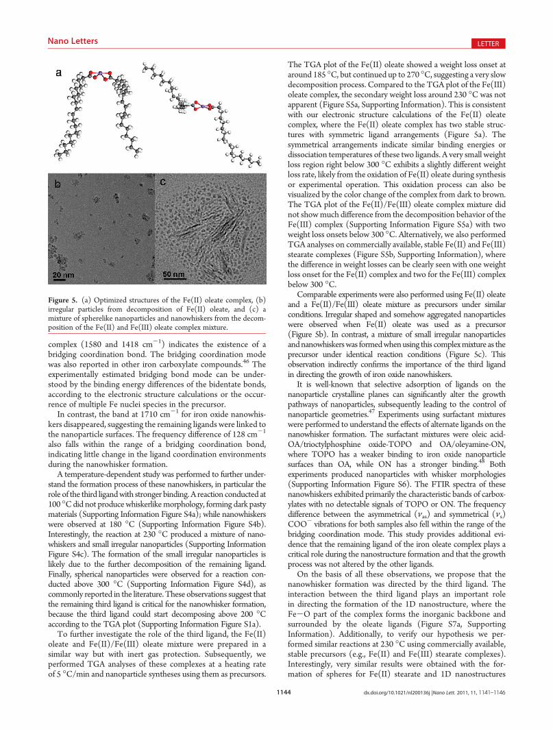
The TGA plot of the Fe(II) oleate showed a weight loss onset ataround 185 �C, but continued up to 270 �C, suggesting a very slowdecomposition process. Compared to the TGA plot of the Fe(III)oleate complex, the secondary weight loss around 230 �C was notapparent (Figure S5a, Supporting Information). This is consistentwith our electronic structure calculations of the Fe(II) oleatecomplex, where the Fe(II) oleate complex has two stable struc-tures with symmetric ligand arrangements (Figure 5a). Thesymmetrical arrangements indicate similar binding energies ordissociation temperatures of these two ligands. A very small weightloss region right below 300 �C exhibits a slightly different weightloss rate, likely from the oxidation of Fe(II) oleate during synthesisor experimental operation. This oxidation process can also bevisualized by the color change of the complex from dark to brown.The TGA plot of the Fe(II)/Fe(III) oleate complex mixture didnot showmuch difference from the decomposition behavior of theFe(III) complex (Supporting Information Figure S5a) with twoweight loss onsets below 300 �C. Alternatively, we also performedTGA analyses on commercially available, stable Fe(II) and Fe(III)stearate complexes (Figure S5b, Supporting Information), wherethe difference in weight losses can be clearly seen with one weightloss onset for the Fe(II) complex and two for the Fe(III) complexbelow 300 �C.
Comparable experiments were also performed using Fe(II) oleateand a Fe(II)/Fe(III) oleate mixture as precursors under similarconditions. Irregular shaped and somehow aggregated nanoparticleswere observed when Fe(II) oleate was used as a precursor(Figure 5b). In contrast, a mixture of small irregular nanoparticlesandnanowhiskerswas formedwhenusing this complexmixture as theprecursor under identical reaction conditions (Figure 5c). Thisobservation indirectly confirms the importance of the third ligandin directing the growth of iron oxide nanowhiskers.
It is well-known that selective adsorption of ligands on thenanoparticle crystalline planes can significantly alter the growthpathways of nanoparticles, subsequently leading to the control ofnanoparticle geometries.47 Experiments using surfactant mixtureswere performed to understand the effects of alternate ligands on thenanowhisker formation. The surfactant mixtures were oleic acid-OA/trioctylphosphine oxide-TOPO and OA/oleyamine-ON,where TOPO has a weaker binding to iron oxide nanoparticlesurfaces than OA, while ON has a stronger binding.48 Bothexperiments produced nanoparticles with whisker morphologies(Supporting Information Figure S6). The FTIR spectra of thesenanowhiskers exhibited primarily the characteristic bands of carbox-ylates with no detectable signals of TOPO or ON. The frequencydifference between the asymmetrical (νas) and symmetrical (νs)COO- vibrations for both samples also fell within the range of thebridging coordination mode. This study provides additional evi-dence that the remaining ligand of the iron oleate complex plays acritical role during the nanostructure formation and that the growthprocess was not altered by the other ligands.
On the basis of all these observations, we propose that thenanowhisker formation was directed by the third ligand. Theinteraction between the third ligand plays an important rolein directing the formation of the 1D nanostructure, where theFe-O part of the complex forms the inorganic backbone andsurrounded by the oleate ligands (Figure S7a, SupportingInformation). Additionally, to verify our hypothesis we per-formed similar reactions at 230 �C using commercially available,stable precursors (e.g., Fe(II) and Fe(III) stearate complexes).Interestingly, very similar results were obtained with the for-mation of spheres for Fe(II) stearate and 1D nanostructures
Figure 5. (a) Optimized structures of the Fe(II) oleate complex, (b)irregular particles from decomposition of Fe(II) oleate, and (c) amixture of spherelike nanoparticles and nanowhiskers from the decom-position of the Fe(II) and Fe(III) oleate complex mixture.

1145 dx.doi.org/10.1021/nl200136j |Nano Lett. 2011, 11, 1141–1146
Nano Letters LETTER
for Fe(III) stearate (Supporting Information Figure S7b,c).However, the presence of cavities within the 1D nanostructuresneeds further investigation.
A similar ligand-directed growth mechanism from single-ligandcomplexes has been reported for the formation of ultrathin goldnanowires.7-9 The fundamental building block of the nanowhiskersis hypothesized to be stable iron oxide nanoclusters with a ligandshell based on the diameter of the nanowhiskers. Iron oxidenanoclusters have been previously observed in many studies andexhibit magnetic transition characteristics of molecular magnets tobulk magnetism.49-51 It has been previously reported that ironcarboxylate complexes could self-assemble into crystalline films bydirect evaporation and without heat treatment.52
Accordingly, we performed electronic structure calculations on ahypothesized iron oxide nanocluster-oleate structure, Fe39O62-(HCOO)12 to understand its structural geometry and stability(Figure 6a). This structure has a 1.1 nm iron oxide nanoclustercenter with S6 symmetry and a ligand shell. The ligands weresimplified as HCOOH to reduce the workload from the longhydrocarbon chains (with an assumption that the chain lengthwouldonly mildly affect the nanocluster geometry, at most). In the ironoxide nanocluster center, the 39 Fe3þ cations occupy 8 tetrahedralsites and 7 octahedral sites in the inner core, and 12 pentahedral sites(blue) and 12 octahedral sites (blue) on the surface, forming 24 Fe-O coordinate bonds in a bridging mode with 12 HCOO- ligands.The bridgingmode coordination bonds are consistentwith the FTIRanalysis of the oleate coated nanowhiskers, yielding a RCOO- toFe2O3 ratio of 1:1.6. The bridging bonds of the relaxed structure(Figure 6a) can be clearly identified. After relaxation, the oleateligands were introduced to the nanocluster core, in order to presentthe configuration of the ligand shell complex (Figure 6b). It must berecognized that with a complex structure like Figure 6b, theoptimization likely only leads to a local minimum when consideringthe complete three-dimensional phase space. Thus, we refer to ourmodel as a “relaxed” representation of the cluster.
The ratio of the capping ligands to Fe2O3 can be experimen-tally determined using a TGA measurement. The weight lossstarted around 200 �C and gradually continued to 400 �C. Theearly weight loss can be ascribed to the decomposition of oleate,as suggested by the TGA plot of the oleate complex at a slowerheating rate (1 �C/min) (Figure S1a, Supporting Information).The weight loss above 300 �C is from desorption or vaporizationof oleate ligands. This thermal behavior is similar to that of oleicacid coated iron oxide nanoparticles.53 The final residue mass ofthe TGA measurements was about 62% after heating-up to over500 �C (Figure 6c). The mass percentage divided by themolecular weight of Fe2O3 and the oleate ligand led to amolecular ratio of RCOO- to Fe2O3 of about 1:2.8. The ratio
(1:1.6) of RCOO to Fe2O3 for the simulated nanocluster (1.1nm) was larger than that of the experimental estimation (1:2.8)because the size of the simulated nanocluster was smaller thanthe diameter of the nanowhiskers. With increasing cluster size,the ratio of RCOO to Fe2O3 will decrease because of thedecreasing surface atom percentage, which requires less ligandsto saturate the surface Fe sites. Our somewhat reduced simula-tion model was chosen for computational efficiency.
In summary, thin iron oxide nanowhiskers have been successfullysynthesized through selective decomposition of the iron oleatecomplex. The major discovery of this work is that the ligandcoordination microenvironments have been found to play animportant role in the nanowhisker formation, which has not beenstudied previously. The different ligand environments were probedby electronic structure calculations and TGA measurement. Subse-quently, a ligand-directed growth mechanism for the iron oxidenanowhisker formation was proposed and stable iron oxide nano-clusters were hypothesized to be the basic building blocks.Accordingly, we performed electronic structure calculations on ahypothesized nanocluster, which agrees well with our experimentalobservation. We expect that similar nanostructures can be synthe-sized for other metals once proper ligands and decompositiontemperatures are identified. The studies of Fe(II) and Fe(III)stearate provide a great support of our hypothesis. The formationof the nanowhiskers provides a unique shape-control example ofnanostructures based on a deep understanding of the precursorligand chemistry, offering additional insights into nanoparticlesynthesis. In particular, the effects of the ligand microenvironmentpresent another synthetic strategy for nanoparticle shape control.
’ASSOCIATED CONTENT
bS Supporting Information. Experimental details; TGAplots of isothermal and slower heating rate processes; X-ray dif-fraction pattern of iron oxide nanowhiskers; time and temperature-dependent study, TGA plots of Fe(II) oleate, Fe(II)/Fe(III)oleate mixture, Fe(II) stearate, and Fe(III) stearate; alternativeligand effects on the nanowhisker formation; and schematicdrawing of growth mechanism and experimental results usingFe(II) and Fe(III) stearate as precursors. This material isavailable free of charge via the Internet at http://pubs.acs.org.
’AUTHOR INFORMATION
Corresponding Author*(Y.B.) E-mail: [email protected]. Phone: (205) 348-9869. Fax:(205) 348-7558. (C.H.T.) E-mail: [email protected]. Phone:(205) 348-1733. Fax: (205) 348-7558.
Figure 6. (a) Relaxed structure of Fe39O62(HCOO)12, (b) structure of a simulated iron oxide nanocluster structure with oleate ligand shell, and (c) aTGA plot of iron oxide nanowhiskers.

1146 dx.doi.org/10.1021/nl200136j |Nano Lett. 2011, 11, 1141–1146
Nano Letters LETTER
’ACKNOWLEDGMENT
This work was supported in part by NSF-DMR 0907204 andan ORAU Junior Faculty Enhancement Award. We acknowledgethe UA Central Analytical Facility (CAF) and the BiologicalScience Department for the use of TEM. We thank the UA-MINT center for the use of the AGM. We thank NSF award(0925445) for the use of Raman Instrument. C.H.T. acknowl-edges support from an NSF CAREER Award (0747690). Super-computer resources were provided by the Alabama SupercomputerCenter and the NCSA TeraGrid. A portion of this research wasperformed using EMSL, a national scientific user facility sponsoredby the Department of Energy’s Office of Biological and Environ-mental Research and located at Pacific Northwest National Labora-tory. The authors also thank Dr. Steve Ritchie and Professor JinZhang for useful discussions.
’REFERENCES
(1) Cohen-Karni, T.; Qing, Q.; Li, Q.; Fang, Y.; Lieber, C. M. NanoLett. 2010, 10, 1098–1102.(2) Chen, J.; Wiley, B. J.; Xia, Y. Langmuir 2007, 23, 4120–4129.(3) Baker, J. L.; Widmer-Cooper, A.; Toney, M. F.; Geissler, P. L.;
Alivisatos, A. P. Nano Lett. 2010, 10, 195–201.(4) Xia, Y.; Xiong, Y.; Lim, B.; Skrabalak, S. E. Angew. Chem., Int. Ed.
2009, 48, 60–103.(5) Lee, E. P.; Peng, Z.; Cate, D. M.; Yang, H.; Campbell, C. T.; Xia,
Y. J. Am. Chem. Soc. 2007, 129, 10634–10635.(6) Cademartiri, L.; Ozin, G. A. Adv. Mater. 2009, 21, 1013–1020.(7) Li, Z.; Tao, J.; Lu, X.; Zhu, Y.; Xia, Y. Nano Lett. 2008, 8, 3052–
3055.(8) Wang, C.; Sun, S. Chem.--Asian J. 2009, 4, 1028–1034.(9) Poudyal, N.; Chaubey, G. S.; Nandwana, V.; Rong, C.; Yano, K.;
Liu, J. Nanotechnology 2008, 19, No. 355601-1-4.(10) Huo, Z.; Tsung, C.; Huang, W.; Zhang, X.; Yang, P. Nano Lett.
2008, 8, 2041–2044.(11) Chen, M.; Pica, T.; Jiang, Y.; Li, P.; Yano, K.; Liu, J.; Datye,
A. K.; Fan, H. J. Am. Chem. Soc. 2007, 129, 6348–6349.(12) Huo, Z.; Tsung, C.; Huang, W.; Fardy, M.; Yan, R.; Zhang, X.;
Li, Y.; Yang, P. Nano Lett. 2009, 9, 1260–1264.(13) Yu, T.; Joo, J.; Park, Y. I.; Hyeon, T. J. Am. Chem. Soc. 2006, 128,
1786–1787.(14) Lee, C. M.; Jeong, H. J.; Lim, S. T.; Sohn, M. H.; Kim, D. W.
ACS Appl. Mater. Interfaces 2010, 2, 756–759.(15) Wang, Y.; Yang, H. Chem. Eng. J. 2009, 147, 71–78.(16) Pankhurst, Q. A.; Connolly, J.; Jones, S. K.; Dobson, J. J. Phys.
D: Appl. Phys. 2003, 36, R167–R181.(17) Veiseh, O.; Gunn, J. W.; Zhang, M. Adv. Drug Delivery Rev.
2010, 62, 284–304.(18) Park, J. H.; von Maltzahn, G.; Zhang, L.; Schwartz, M. P.;
Ruoslahti, E.; Bhatia, S. N.; Sailor, M. J. Adv. Mater. 2008, 20, 1630–1635.(19) Park, J.; An, K. J.; Hwang, Y. S.; Park, J. G.; Noh, H. J.; Kim, J. Y.;
Park, J. H.; Hwang, N. M.; Hyeon, T. Nat. Mater. 2004, 3, 891–895.(20) Shavel, A.; Rodriguez-Gonzalez, B.; Spasova, M.; Farle, M.; Liz-
Marzan, L. M. Adv. Funct. Mater. 2007, 17, 3870–3876.(21) Shavel, A.; Rodriguez-Gonzalez, B.; Pacifico, J.; Spasova, M.;
Farle, M.; Liz-Marzan, L. M. Chem. Mater. 2009, 21, 1326–1332.(22) Shavel, A.; Liz-Marzan, L. M. Phys. Chem. Chem. Phys. 2009, 11,
3762–3766.(23) Hai, H.; Yang, H.; Kura, H.; Hasegawa, D.; Ogata, Y.; Takahashi,
M.; Ogawa, T. J. Colloid Interface Sci. 2010, 346 (1), 37–42.(24) Kovalenko, M. V.; Bodnarchuk, M. I.; Lechner, R. T.; Hesser,
G.; Schaffler, F.; Heiss, W. J. Am. Chem. Soc. 2007, 129, 6352–6353.(25) Xu, Z.; Shen, C.; Tian, Y.; Shi, X.; Gao, H. Nanoscale 2010, 2,
1027–1032.(26) Kim, D.; Park, J.; An, K.; Yang, N.; Park, J. G.; Hyeon, T. J. Am.
Chem. Soc. 2007, 129, 5812–5813.
(27) Kwon, S. G.; Piao, Y.; Park, J.; Angappane, S.; Jo, Y.; Hwang,N. M.; Park, J. G.; Hyeon, T. J. Am. Chem. Soc. 2007, 129, 12571–12584.
(28) Lopez-Cruz, A.; Lopez, G. E.Mol. Phys. 2009, 107, 1799–1804.(29) Garg, A. N.; Lanjewar, R. B. J. Radioanal. Nucl. Chem. 1995, 199,
443–452.(30) Davis, R.; Schltz, H. P. J. Org. Chem. 1962, 27, 854–857.(31) Ganguly, A.; Kundu, R.; Ramanujachary, K. V.; Lofland, S. E.;
Das, D.; Vasanthacharya, N. Y.; Ahmad, T.; Ganguli, A. K. J. Chem. Sci.2008, 120, 521–528.
(32) Roca, A. G.; Morales, M. P.; Serna, C. J. IEEE Trans. Magn.2006, 42, 3025–3029.
(33) Kwon, K. W.; Lee, B. H.; Shim, M. Chem. Mater. 2006, 18,6357–6363.
(34) Park, J. N.; Zhang, P.; Hu, Y.; McFarland, E. W.Nanotechnology2010, 21, 225708–1-8.
(35) Jia, C.; Sun, L.; Luo, F.; Han, X.; Heyderman, L. J.; Yan, Z.; Yan,C.; Zheng, K.; Zhang, Z.; Takano, M.; Hayashi, N.; Eltschka, M.; Klaui,M.; Rudiger, U.; Kasama, T.; Cervera-Gontard, L.; Dunin-Borkowski,R. E.; Tzvetkov, G.; Raabe, J. J. Am. Chem. Soc. 2008, 130, 16968–16977.
(36) Teng, X.; Yang, H. J. Mater. Chem. 2004, 14, 774–779.(37) Guardia, P.; Batlle-Brugal, B.; Roca, A. G.; Iglesias, O.; Morales,
M. P.; Serna, C. J.; Labarta, A.; Batlle, X. J. Magn.Magn.Mater. 2007, 316,E756–E759.
(38) Kachkachi, H.; Ezzir, A.; Nogues, M.; Tronc, E. Eur. Phys. J. B2000, 14, 681–689.
(39) Koseoglu, Y.; Kavas, H.; Aktas, B. Phys. Status Solidi A 2006,203, 1595–1601.
(40) Millan, A.; Urtizberea, A.; Silva, N. J. O.; Palacio, F.; Amaral,V. S.; Snoeck, E.; Serin, V. J. Magn. Magn. Mater. 2007, 312, L5–L9.
(41) Koseoglu, Y.; Kavas, H. J. Nanosci. Nanotechnol. 2008, 8 (2),584–590.
(42) Iglesias, O.; Labarta, A. Phys. Rev. B 2001, 63, 184416–11.(43) Nakamoto, K. Infrared and Raman Spectra of Inorganic and
Coordination Compounds, 4th ed.; John Wiley & Sons: New York, 1986.(44) Abrahamson, H. B.; Lukaski, H. C. J. Inorg. Biochem. 1994, 54,
115–130.(45) Lu, Y.; Miller, J. D. J. Colloid Interface Sci. 2002, 256, 41–52.(46) Culita, D. C.; Patron, L.; Teodorescu, V. S.; Balint, L. J. Alloys
Compd. 2007, 432, 211–216.(47) Hyeon, T. Chem. Commun. 2003, 927–934.(48) Palchoudhury, S.; Xu, Y.; An, W.; Turner, C. H.; Bao, Y. J. Appl.
Phys. 2010, 107, 09B311–09B313.(49) Gatteschi, D.; Caneschi, A.; Pardi, L.; Sessoli, R. Science 1994,
265, 1054–1058.(50) Canada-Vilalta, C.; O’Brien, T. A.; Pink, M.; Davidson, E. R.;
Christou, G. Inorg. Chem. 2003, 42, 7819–7829.(51) Christmas, C. A.; Tsai, H. L.; Pardi, L.; Kesselman, J. M.;
Gantzel, P. K.; Chadha, R. K.; Gatteschi, D.; Harvey, D. F.; Hendrickson,D. N. J. Am. Chem. Soc. 1993, 115, 12483–12490.
(52) Popescu, M.; Turta, C.; Meriacre, V.; Zubareva, V.; Gutberlet,T.; Bradaczek, H. Thin Solid Films 1996, 274, 143–146.
(53) Huang, J.; Parab, H. J.; Liu, R.; Lai, T.; Hsiao, M.; Chen, C.;Sheu, H.; Chen, J.; Tsai, D.; Hwu, Y. K. J. Phys. Chem. C 2008, 112,15684–15690.