SYNTHESIS AND CHARACTERIZATION OF CHITOSAN …docs.neu.edu.tr/library/6505887536.pdf · Chitosan is...
61
SYNTHESIS AND CHARACTERIZATION OF CHITOSAN MICROPARTICLE AND BLOOD COMPATIBILITY STUDIES A THESIS SUBMITTED TO THE GRADUATE SCHOOL OF APPLIED SCIENCES OF NEAR EAST UNIVERSITY By BINTA YAHAYA LAWAN In Partial Fulfilment of the Requirements for the Degree of Master of Science in Biomedical Engineering NICOSIA, 2017 BINTA YAHAYA LAWAN NEU,2017
Transcript of SYNTHESIS AND CHARACTERIZATION OF CHITOSAN …docs.neu.edu.tr/library/6505887536.pdf · Chitosan is...

SYNTHESIS AND CHARACTERIZATION OF
CHITOSAN MICROPARTICLE AND BLOOD
COMPATIBILITY STUDIES
A THESIS SUBMITTED TO THE GRADUATE
SCHOOL OF APPLIED SCIENCES
OF
NEAR EAST UNIVERSITY
By
BINTA YAHAYA LAWAN
In Partial Fulfilment of the Requirements for
the Degree of Master of Science
in
Biomedical Engineering
NICOSIA, 2017
B
INT
A Y
AH
AY
A L
AW
AN
NE
U,2
01
7


SYNTHESIS AND CHARACTERIZATION OF
CHITOSAN MICROPARTICLE AND BLOOD
COMPATIBILITY STUDIES
A THESIS SUBMITTED TO THE GRADUATE
SCHOOL OF APPLIED SCIENCES
OF
NEAR EAST UNIVERSITY
By
BINTA YAHAYA LAWAN
In Partial Fulfilment of the Requirements for
the Degree of Master of Science
in
Biomedical Engineering
NICOSIA, 2017

I hereby declare that all information in this document has been obtained and presented in
accordance with academic rules and ethical conduct. I also declare that, as required by these rules
and conduct, I have fully cited and referenced all materials and results that are not original to this
work.
Name, Last name:
Signature:
Date:

i
ACKNOWLEDGEMENTS
Firstly, I would like to thank God Almighty for the wonderful privilege of having our head of
department, Assoc. Prof. Dr. Terin Adali as supervisor and Assist. Prof. Dr. Urat Uncu Department
of Hematology laboratory who were of tremendous help, even with their busy schedule mapped out
time to see to the successful completion of this research work.
I am highly indebted to my Parents for their love, support and prayers. I will like to express my
gratitude to Musa Ibrahim and Adam Mustapha for their support at various stage of this work. I
would also like to express my special gratitude to Craig Adrian Little. My appreciation goes to my
colleague.
Lastly, I would like to thank all my friends for their time and support. They all can’t be mentioned
but I really do appreciate it from the deepest part of my heart.
ii
To my parents….

iii
ABSTRACT
The chitosan microparticles were synthesized by ionic gelation method. Their characterizations
were studied by SEM and XRD analysis. The blood compatibility studies were carried out in the
NEU Hospital. The prothrombine time (PT %), APTT and INR results were obtained by STAGO
STA compact Machine. Results showed that effective blood compatibility exhibiting higher
Prothrombine time. Results indicated that chitosan microparticles can be used as a blood compatible
material and may be able to prevent blood clotting.
Keywords: Chitosan; Microparticles; Ionic gelation method; Blood compatibility; Prothrombin
time

iv
ÖZET
Bu çalışmada, iyonik jelleşme metodu ile kitozan mikroparçacıkları (K-MP) sentezlendi. Kitozan
mikroparçacıklarının karakterizasyonu Taramalı Elektron Mikroskopu (TEM) ve X-Işın Analizi
(XIA) kullanılarak yapıldı. Mikro parçacıkların TEM analizleri sonucunda çubuk şeklinde
sentezlendiği gözlemlendi. X-Işın analiz sonuçları ise kristal yapısının varlığını kanıtladı. Kan
uyumluluğu analizleri, YDÜ Hastanesi klinik biyokimya laboratuvarında STAGO STA Compact
cihazı kullanılarak yapıldı. Yüksek Protrombin miktarlarının ölçülmesi, kitozan mikrokürelerinin
kan uyumluluğunun yüksekliğini etkin şekilde ıspatladı.
Sonuçlar, çubuk şeklindeki kitozan mikrokürelerin kan uyumluluğu gerekli olan biyomedikal cihaz
tasarımında kullanımını önerilmektedir.
Anahtar Kelimeler: Kitozan; Kan uyumluluğu; Mikroparçacık; Iyon jellesme metodu; Protrombin
zamani
v
TABLE OF CONTENTS
ACKNOWLEDGEMENTS ........................................................................................................ i
ABSTRACT .................................................................................................................................. iii
ÖZET ........................................................................................................................................... iv
TABLE OF CONTENTS ........................................................................................................... v
LIST OF TABLES ..................................................................................................................... vii
LIST OF FIGURES ................................................................................................................... viii
LIST OF ABBREVIATIONS .................................................................................................... ix
CHAPTER ONE: INTRODUCTION ....................................................................................... 1
1.1 General Introduction ........................................................................................................... 1
1.2 Chemical Structure of Chitosan .......................................................................................... 8
1.3 Chitosan Non-toxicity ........................................................................................................ 10
1.4 Properties and Characterization of Chitosan ...................................................................... 11
1.4.1 Hemocompatibility test of Chitosan ............................................................................ 12
1.4.2 Biodegradability .......................................................................................................... 12
1.4.3 Chitosan Mucoadhesitivity .......................................................................................... 13
1.4.4 Chitosan Cytotoxicity .................................................................................................. 14
1.5 Antimicrobial Activities ..................................................................................................... 14
1.6 Blood coagulation .............................................................................................................. 16
1.7 Aim of the Study ................................................................................................................ 18
1.8 Objectives of the Study ...................................................................................................... 18

vi
CHAPTER TWO: MATERIALS AND METHODS .............................................................. 20
2.1 Materials ............................................................................................................................. 20
2.2 Sample Collection .............................................................................................................. 20
2.2.1 Preparation of Chitosan Beads .................................................................................... 20
2.2.2 Preparation of Sodium Tripolypentaphosphate ........................................................... 23
2.3 Morphological Chitosan Characterisation .......................................................................... 23
2.4 Activated Partial Prothromboplastin Time (Aptt or Pt) ..................................................... 24
2.4.1 Partial Thromboplastin Time ....................................................................................... 24
2.5 Method ............................................................................................................................... 25
CHAPTER THREE: RESULTS AND DISCUSSIONS ......................................................... 26
CHAPTER FOUR: CONCLUSIONS AND RECOMMENDATION ................................... 35
4.1 Conclusion .......................................................................................................................... 35
4.2 Recommendations .............................................................................................................. 36
REFERENCES ........................................................................................................................... 37

vii
LIST OF TABLES
Table 3.1: Sample taken from five random patients without the addition of chitosan ............... 27
Table 3.2: Samples after addition of chitosan ............................................................................. 28
Table 3.3: Sample taken from five random patients without the addition of chitosan ............... 29
Table 3.4: Second run test with chitosan .................................................................................... 30
Table 3.5: Third run test without chitosan .................................................................................. 31
Table 3.6: Third run test with chitosan ....................................................................................... 32

viii
LIST OF FIGURES
Figure 1.1: Systematic representation of chitin and chitosan preparations ................................. 3
Figure 1.2: Showing extraction of chitin and chitosan ................................................................ 4
Figure 1.3: The basic structure of chitosan .................................................................................. 9
Figure 1.4: Chemical modification of chitin ............................................................................... 10
Figure 1.5: Systematic description of blood coagulation pathways. .......................................... 18
Figure 2.1: Preparation of TPP Solution in Biomedical Engineering Near East University ...... 21
Figure 2.2: Gel like chitosan ....................................................................................................... 22
Figure 2.3: Sample of dried chitosan particles at room temperature .......................................... 23
Figure 2.4: Chitosan particles viewed under electron microscope (SEM) ................................. 24
Figure 2.5: Schematic representation of APTT .......................................................................... 25

ix
LIST OF ABBREVIATIONS
APTT: Activated Partial Thrombin Time
PT: Prothrombin Time
TT: Thrombin Time
Pka: Professionally known as (acid dissociation)
Kj: Kilojoules
Mol: Mole
TPP: Sodium Tripolypentaphosphate
DD: Degree of N-deacetylation
%: Percentage

i

1
CHAPTER ONE
INTRODUCTION
1.1 General Introduction
Chitosan is known as a versatile biopolymer (scaffolds, particles, films, membranes and gels). It
is a composed randomly distributed N – acetyl – D – glucosamine (acetylated unit) and β – (1 – 4)
– linked D- glucosamine (deacetylated unit) which is a linear polysaccharide and is composed of
2 – amino – 2 – deoxy – D – glucopyranose and N – acetyl – 2 – amino – 2 – deoxy – D –
glucopyranose obtained by chitin which is thermochemically deacetylated in the presence of alkali.
The shell waste of crabs, lobsters, krills, and shrimps are the main commercial sources of chitin
(Dash et al., 2011). β – Chitin is found in squid pens and α – chitin is found in fungal and yeast
cells and these are the forms in which chitin occurs.
The degree of N-deacetylation (DD) is the difference between chitin and chitosan and this is known
as deacetylated units, 2 – amino – 2- deoxy – D – glucopyranose which is defined generally as the
molar ratio. Fully or partially deacetylated chitin compounds when bought together is known
collectively as chitosan (Tikhonov et al., 2006). Depending on their DD and MM parameters that
can influence its biological and chemical properties significantly, chitosan can be found in batches
in great numbers. For instance, chitosan solution viscosity (Kofuji et al., 2005) and chitosan
solubility (Schiffman and Schauer, 2007) increases when DD increases and as for polymer
crystallinity (Aranaz et al., 2009), when DD decreases it also decreases. Due to the quaternisation
of the amine group (pKa 6.3), in dilute acidic solutions (pH < 6.0) chitosan is readily soluble: as
there is an increase in the pH above 6, the polymer becomes insoluble and chitosan’s amines are
deprotonated.
With a unique structure, chitosan is a cationic polyelectrolyte from the chemical point of view.
Two types of functional groups are exhibited (hydroxyl and amino) (Agrawal et al., 2009). The
non-toxicity and its biocompatibility has already been proven (Chatelet et al., 2001; Rao and
Sharma, 1997; Thanou et al., 2001), thereby primarily, the use of chitosan as biomaterial is
sustained. According to Rao and Sharma (1997) sited that chitosan is a safety material based on

2
the acute toxicity test results. Substances such as sodium hydroxide which is an alkaline solution,
is used to produce chitosan by treating crustacean shells and shrimps.
In the medical field, Chitosan has been used extensively. It has been widely employed in different
forms and for a great number of applications which ranges from industrial areas (e.g dye removal,
adsorption of metal ions) to biomedical field (e.g tissue engineering, drug delivery) (Dash et al.,
2011; Hritcu et al., 2012; Muzzarelli, 2008; Muzzarelli et al., 2005; Muzzarelli et al., 2012). It is
either fully deacetylated or partially chitin. Chitosan is a fully biocompatible natural polymer and
also biodegradable. It can be used as an antifungal agent, as an adhesive and as an antibacterial
agent.
Due to its biocompatible properties, it has been extensively investigated as a potential drug carrier.
Some studies suggested that, in order to increase their bioavailability and reduce their impact on
the body, nanoparticles made of other materials should be coated with chitosan. In order to obtain
a different physio-mechanical properties, the molecular weight and the degree of deacetylation of
chitosan can be modified. ~5.3 x 10 Daltons is the average viscosity molecular weight of chitosan.
Nitrogen (7.97%), Carbon (44.11%), and hydrogen (6.84%) are the elemental composition of the
chitosan polymer.
Chitosan has some possible biomedical uses and quiet a number of commercial uses also. In
medicine, it can be used as a delivery agent (drug delivery into the skin), as an antibacterial agent
and in bandages for stopping or reducing of blood. Chitosan biological properties however such
as anticoagulant activity (Yang et al., 2008), proliferate and cell adhesion (Chatelet et al., 2001),
biocompatibility (Schiffman et al., 1999), and biodegradability (Zhang et al., 2001), are influenced
greatly by its parameters. Due to highly non-toxicity for biomedical applications, chitosan of a
lower DD is recommended. Imposed by a lot of aspects, biodistribution is an important aspect that
concerns the biocompatibility of chitosan, ranging from the formulation type used administration
route and polymer characteristics (Schiffman et al., 1999).
3
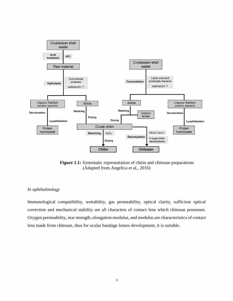
Figure 1.1: Systematic representation of chitin and chitosan preparations
(Adapted from Angelica et al., 2016)
In ophthalmology
Immunological compatibility, wettability, gas permeability, optical clarity, sufficient optical
correction and mechanical stability are all characters of contact lens which chitosan possesses.
Oxygen permeability, tear strength, elongation modulus, and modulus are characteristics of contact
lens made from chitosan, thus for ocular bandage lenses development, it is suitable.

4
Figure 1.2: Showing extraction of chitin and chitosan
(Adapted from Angelica et al., 2016)

5
Functionalized-chitosan based system in biomedical application
Nature has been great source of Chitosan, a polysaccharide derived from a chitin, which is the
second most abundant polysaccharide in the world after cellulose (Chen et al., 2011). Chitosan
presents unique properties such as biocompatibility, biodegradability and its non-toxic nature
which confers it wider application in biomedical and pharmaceutical areas such as drug and gene
delivery, tissue engineering, wound healing, central nervous system treatment, Cardiovascular
applications, antibacterial and microbial activities, and theragnosis (Chen et al., 2011; Balan and
Verestiuc, 2014; Ibrahim and El-Zairy, 2015).
Gene and drug delivery
In recent years the use of chitosan as a vehicle of drug delivery has been increasing area of interest
to ensure effectiveness and patient compliance. Balan and Verestiuc (2014) underline the newest
directions in the field of functionalized carriers based on chitosan as parenteral delivery systems
that could overcome the current limitations of clinical use of drug delivery system. Similarly, study
conducted by Zhang et al. (2008) has revealed the use of chitosan as a carrier of drug system. In
the study, a comprehensive evaluation of pharmacokinetics, tissue distributions, metabolism,
excretion, acute toxicity, safety and cell viability of N-octyl-O-sulfate chitosan based nanoparticles
was conducted, which showed a streaking result of potential use of this new system of drug carrier.
The positive charge on chitosan and negative charge on the cell membrane allow the interaction
that leads to reorganization and an opening of the tight junction proteins, explaining the permeation
enhancing property of this polysaccharide.
Recent studies revealed that chitosan and its derivatives exhibited antitumor activity in both in
vitro and in vivo models. Tumor-targeting property of cisplatin-loaded glycol chitosan
nanoparticles was demonstrated by Kim et al. (2008). Similarly, Zhu et al. (2011) also showed the
ability of chitosan to serve as drug loading vehicle in a modified chitosan with folate. This model
could be used for tumor targeting carrier for hydrophobic therapeutics or diagnostic agents. The
chemical properties of chitosan allow modification with various side chains to suite the desired
target. For instance, Sahul et al. (2011) prepared hydrophobically modified carboxylmethyl
chitosan nanoparticles for targeted delivery of paclitaxel with excellent non-cytotoxic property
compared with native drug. The main merit of use of chitosan in drug delivery apart from

6
biocompatibility is they are not hazardous and less expensive.
Tissue engineering
Tissue engineering is a multifaceted technology that involves the use of a combination of cells,
engineering materials, and suitable biochemical factors to improve or replace biological functions.
This includes a wide range of applications such as repair or replacement of part or whole tissues
(Cheung et al., 2015). Chitosan and its derivatives have been explicatively explored as a supporting
material used for tissue engineering process (Balan and Verestiuc, 2014). It shared great similarity
with gylycosaminoglycans, the components of the liver extra cellular matrix, thus chitosan used
as a promising scaffold for hepatocytes cultures. For instance, in a study by Cheung et al. (2015)
Chitosan-â-tricalcium phosphate composite exhibited histocompatibility with Beagle
mesenchymal stem cells and was devoid of an effect on cellular growth and proliferation. It
manifested efficacy in enhancing osteogenesis and vascularization and repair of bone defects in
conjunction with mesenchymal stem cells. In other study, chitosan/collagen/heparin matrix with
excellent blood compatibility and good hepatocyte compatibility has been suggested as matrix for
implantable bioartificial liver (Wang et al., 2005). This reveals the recent application of chitosan-
based biomaterial in tissue and cell repair and engineering.
In fluorescent
Fluorescent chitosan nanoparticles are formed by the interaction between chitosan nanoparticles
amino groups by which Rhodamine dye get attached to the chitosan nanoparticles. In the near
future for the application of cell imaging, prepared fluorescent chitosan nanoprobe could be used.
In addition, to improve the chemical and mechanical properties of chitosan, it can be modified by
chemical and physical methods.
Healing of wound
The basic features of chitosan and its derivatives in wound healing are its biodegradability,
biocompatibility and antimicrobial activity which confer it as excellent biomaterials for wound
healing. It has been found that, chitosan accelerate the effect of wound healing where regenerated
chitin fibers, films and sponges exhibit an increase in wound healing by over 30 percent (Dutta et
al., 2004). Thus, the complex process of wound healing which involves inflammation, granulation
7
tissue formation, extracellular matrix formation, angionesis and remodeling can be stimulated by
chitosan and its derivatives. Cheng et al. (2011) proposed that incorporation of growth factors;
antibiotics and antibacterial increase the efficacy of chitosan as excellent biomaterials.
The adhesive-based wound dressing could be applied in surgery to enhance wound healing. The
chitosan adhesive shows strong sealing strength as well as not requiring sutures or staples. It can
effectively stop bleeding from blood vessels along with air leakage from the lung (Ishihara et al.,
2006). Many clinical studies showed a significant result in chitosan in would healing by patients
undergoing plastic surgeries, skin grafting and endoscopic sinus surgery (Biagini et al., 1991;
Stone et al., 2000; Azad et al., 2004; Valentine et al., 2010). Similarly, due to their versatility,
chitosan and chitin are used as diluents in the pharmaceutical industries.
Treatment Cardiovascular Diseases
Ability of chitosan in supporting endothelial cells and to enhance angiogenesis, functionalized
chitosan- based formulation has been investigated as cardiovascular scaffolds that able to deliver
regenerative cells or bioactive compounds in the treatment (Balan and Verestiuc, 2014). Study by
Lu et al. (2009) proved the effectiveness of chitosan-collagen cross linked as a cardiovascular
supportive material both in vitro and in vivo tests. Similarly, remarkable results showed that
chitosan hydrogel could improve myocardial infarction microenvironment, enhance stem cell
engraftment, homing in ischemic heart and stimulate heart repair (Balan and Verestiuc, 2014). This
agreed with other studies that proved chitosan as excellent biomaterial and could be employed in
heart disease treatments (Meng et al., 2009; Pok et al., 2013; Cheung et al., 2015).
Central nervous system treatment
The structural barrier such as blood brain barrier that separates blood and cerebral parenchyma
limits the drugs such as anti-Alzheimer, antibiotics, antineoplastic agents and other neuroleptic
drugs penetrate deeply into the system. It has been proposed that if this barrier can be effectively
been crossed would increase brain absorption of the drugs (Hombach et al., 2009). A preliminary
in vitro study using mouse brain endothelial cell showed an efficient uptake of drugs particles
across the blood brain barrier. This promising result could be used in employing chitosan coated
with nanoparticles or chitosan-based polymers as brain targeting agent.
8
Nutrition and Food
Lactose intolerance effect occurs in many humans and animals. The whey production showed
considerable increase by small supplement of chitosan as feed, which improved change in
intestinal microflora as well as in chickens, 2-0.5% • chitosan supplement improved weight gain
Artificial Skin
Chitosan is used for the treatment of fibroplasias and healing of wounds caused by subcutaneous
tissue and scapel insertions in skin especially when caused by fire (this is used in situations such
as extensive skin loss by patients)
Capturing of water engineering metal from waste water
In heavy metal ions chitosan has a natural selectivity and in acidic medium, N-benzylsulfonate
derivates of chitosan were used as sorbents for the metal ions, removal of NI2+, Hg2+, Zn2+, and
Cu2+ by the adsorption parameters of chitosan has been enhanced, in which on the outer surface of
chitosan, metal ions are preferably absorbed.
In textile dye (color removal)
Chitosan has an extremely high affinity and as well have dye of many classes which includes
napthal dyes, acid, vat, sulfur, direct, reactive, and disperse and also has a unique molecular
structure. Loading thermodynamics enhances adsorption rate and adsorption of dye is significantly
caused by the effect of pH, and temperature which shows the favored reaction. In addition, it can
also be employed in Photography.
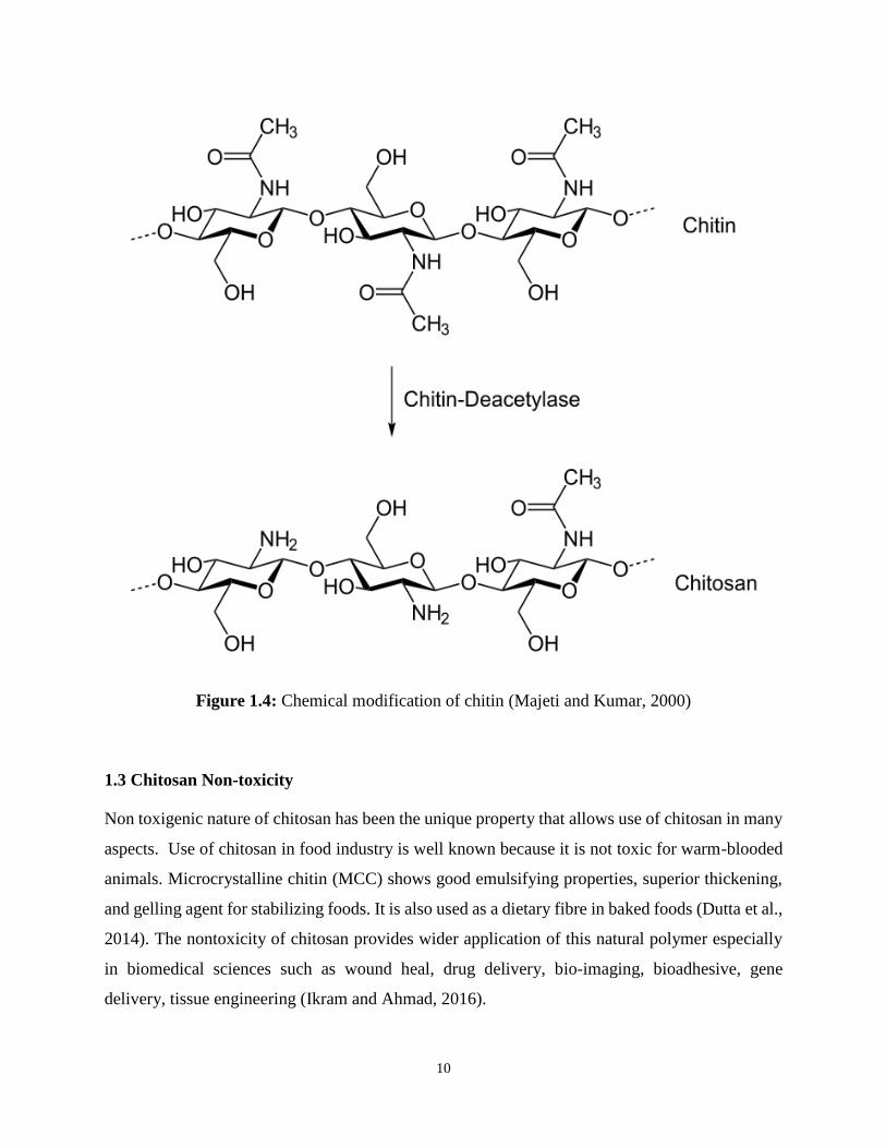
1.2 Chemical Structure of Chitosan
The chemical structure of chitosan has been proposed to have affords from deacylation of chitin
and this provides sides for chemical modification (Berger et. al., 2004; Mourya et al., 2010). Based
on the source, chitin occurs in two allomorphic forms named α and β, and the third allormorphic
structure γ have also been suggested (Averous and Pollet, 2012). The basic difference between the
above chemical forms is the presence of tight inter-sheet hydrogen bond.

9
Figure 1.3: The basic structure of chitosan (Majeti and Kumar, 2000)
10
Figure 1.4: Chemical modification of chitin (Majeti and Kumar, 2000)
1.3 Chitosan Non-toxicity
Non toxigenic nature of chitosan has been the unique property that allows use of chitosan in many
aspects. Use of chitosan in food industry is well known because it is not toxic for warm-blooded
animals. Microcrystalline chitin (MCC) shows good emulsifying properties, superior thickening,
and gelling agent for stabilizing foods. It is also used as a dietary fibre in baked foods (Dutta et al.,
2014). The nontoxicity of chitosan provides wider application of this natural polymer especially
in biomedical sciences such as wound heal, drug delivery, bio-imaging, bioadhesive, gene
delivery, tissue engineering (Ikram and Ahmad, 2016).

11
Its ability to biodegrade into non-toxic residues and the rate of its degradation being highly related
to the molecular mass of the polymer and its deacetylation degree and has proved to some extent
biocompatibility with physiological medium. All these singular properties make chitosan an
outstanding candidate for biomedical applications (Croisier and Jerome, 2013).
1.4 Properties and Characterization of Chitosan
Deacetylation of chitin is a commercial way of the production of chitosan, which in fungi cell wall
and crustaceans (such as shrimps and crabs) is the structural element in the exoskeleton. NMR
Spectroscopy is used determining the degree of deacetylation, and the commercial chitosan with
deacetylation has a range of about 60-100%. Averagely, about 3800 to 20,000 Dalton in the
molecular weight of chitosan produced commercially.
Deacetylation of chitin using water as a solvent and as a reagent using sodium hydroxide in excess
is one common method for chitosan synthesis. There are two stages in which first order kinetic
control reaction takes place. The second step is lower than the first in which the first is the
activation energy 25-120oc at 48.76kj/mol is its estimated value (Hammou et al., 2013).
The pKa of chitosan amino group has a value of approximately 6.5, which can lead to a protonation
to neutral in acidic solution with a density charge negatively charged surfaces (Dong et al., 2013;
Chanoong et al., 2015). Chitosan is biodegradable and biocompatible which across the epithelial
surfaces enhances the transportation of polar drugs, for biomedical applications, there are available
quantities of purified chitosan.
Trimethylchitosan which is a derivative of chitosan have been used in the delivery of gene which
are nonviral. Quaternised chitosan or trimethylchitosan with increased degree of trimethylation
increasing the cytotoxicity has been shown to transfect breast cancer cells, approximately
trimethylation at about 50%, in gene delivery, the derivative is the most efficient. Oligomeric
derivative of 3-6kDa have good gene delivery properties and are relatively nontoxic (Kean et al.,
2005). Antitumor activity and immunoadjuvant, fungistatic, central nervous system, osteogenesis
(El-Kamel et al., 2007; Roldo et al., 2004), hemostatic, anticholesteremic (Dash et al., 2011;
Muzzarelli and Muzzarelli, 2005; Muzzarelli et al., 2012), promoter of wound healing (Fischer et
al., 2007; Fischer et al., 2005; Madhumathi et al., 2010; Sudheesh et al., 2010), and spermicidal

12
are biological properties of chitosan and a biomaterial sustain their use,
1.4.1 Hemocompatibility test of Chitosan
Due to establishing a possible standard for blood material interactions, Hemocompatibility has
been studied greatly. Blood compatibility assessment is taken into account due to the fact that there
many mechanisms in which blood can respond. Classified into five categories are blood
compatibility which are hematology, platelets functions, complement system, blood coagulation,
and thrombosis. One of the tests used to evaluate medical device or hemocompatibility of a
biomaterial is blood coagulation which is the most used test. Activation of clotting factor cascade
is a phenomenon which gives rise to blood coagulation. Activated partial thromboplastin time
(APTT) is measured so as to give the estimated value of common coagulation pathway and
intrinsic pathway. Prothrombin time (PT) test evaluates coagulation cascade activation of common
pathway and extrinsic pathway while fibrin polymerization or thrombin activity is examined by
thrombin time (TT). Adsorbed proteins also governs blood compatibility on material surface which
is followed by blood exposure (Gorbet et al., 2004). Plasma components and the quantification of
cellular of the blood is known as hematology. Hemolysis is the mechanism through which
hemoglobin is evaluated. In estimating blood biocompatibility of materials, the reliable and simple
method which can be used is the in vitro hemolysis. When it is less than 5%, hemolysis index is
regarded as safe (Zhou et al., 2011).
1.4.2 Biodegradability
The successful use of a material in biological application which is an essentially important
requirement in chitosan is biodegradability. Chitosan is composed to both enzymatic degradation
and chemical degradation in the human body. Colonic bacterial enzymes (Zhang and Neau, 2001),
rat cecal, pectinase isozyme (Kittur et al., 2005), leucine amino-peptidase (Rao and Sharma, 1997),
and lysozyme (Kean and Thanou, 2009) are enzymes that are able to degrade this polymer when
in vitro degradation that test of chitosan was carried out. Eight human chitinases have been
identified in recent studies of which (enzymatic activity was revealed in three of them)
(Funkhouser and Aronson, 2007) but so far on its derivatives or regarding the effects of these

13
enzymes on chitosan, there are no literature reports yet.
1.4.3 Chitosan Mucoadhesitivity
Several studies have been conducted to examine the mucoadhesive properties of chitosan (Dhawan
et al., 2004; Bizhan et al., 2008; Khameneh et al., 2014). He et al. (1998) evaluated the
mucoadhesive properties of chitosan and chitosan microspheres by studying the interaction
between mucin and chitosan in aqueous solution by turbidimetric measurements and the
measurement of mucin adsorbed on the microspheres.
A strong interaction between chitosan microspheres and mucin was detected. This reveals another
important feature of chitosan in absorption of substances using the available charges on the surface,
thus it can be used for developing drug delivery systems because of its excellent mucoadhesive
properties. Properties such as primary amino groups, electrostatic attraction, hydrogen bonding
and hydrophobicity are believe to be responsible for the mucoadhesivity of chitosan (Saqias et al.,
2008). In answering why chitosan is mucoadhesive, the authors revealed that change in these
properties may influence its mucoadhesivity, for instance, reducing the number of amino groups
through their half acetylation results in expansion of chitosan's pH-solubility window up to pH 7.4
but also reduces its capacity to aggregate mucin. Khobragade and Puranik (2015) clearly stated the
mucoadhesive property of chitosan and its derivates. The authors described Chitosan structure to
possess cationic group, due to this group it has mucoadhesive property.
Chitosan mainly combine with anionic group of mucus i.e sialic acid and sulfonic group
substituent. Hence ionic interaction is take place in between cationic primary amino acid group of
chitosan with anionic sialic acid group of mucus, mucoadhesion can be achieved. However, the
hydrophobic interactions might contribute to its mucoadhesive properties. In comparison with
various anionic polymeric excipients such as carbomer, polycarbophil, and hyaluronic acid,
however, its mucoadhesive properties are weak. The authors also suggest way to achieve better
mucoadhesive properties; polymer should have cohesive properties as that of adhesive. If not then
polymer fails to achieve mucoadhesion.
Chitosan has weak cohesive properties but can be improved by formation of complexes with
multivalent anionic drug or other anionic excipients. In addition, chitosan possesses excellent

14
mucoadhesive properties in its swollen state. It can easily adhere to hard and soft tissues. Clinical
tests conducted with the use of materials containing chitosan and its derivatives revealed that they
do not cause any inflammatory or allergic reactions in the human body. Chitosan is well tolerated
by living tissues, including connective, muscle, epithelial, and nervous (Nawrotek et al., 2015).
1.4.4 Chitosan Cytotoxicity
Chitosan and its derivatives have been extensively applied in medical and pharmaceutical fields
as promising drug delivery systems. Hence, the safety of this important polymer to cells needs to
be studied. Studies on cytotoxicity of chitosans have been previously reported on cultured cells,
for example, A549 human alveolar epithelial cells, others used zebra fish model and antitumor
effect of paclitaxel-bound nanoparticles in gastric cancer cells (Zaki et al., 2015).
Sabudin et al. (2012) studied the cytotoxicity of silver nanoparticles incorporated in chitosan using
Normal Human Dermal Fibroblasts (NHDFs). The result showed chitosan considerable cell
cytotoxicity, mainly for water-soluble chitosan paste samples and concluded that the toxicity of
the silver nanoparticles is still within the acceptable range for human use. Similarly, different
degrees of de-acetylation of chitosan on the cytotoxicity were investigated by (Grobler et al.,
2016), where 3T3 fibroblast cells as well as two different human tooth pulp fibroblast cell-lines
were used. The study revealed that different cell lines react differently towards different degrees
of de-acetylation. The relative survival rates of different cell lines changed at different degrees of
de-acetylation. Thus, the cytotoxicity of chitosan is within the limit or showed no toxicity to cells.
1.5 Antimicrobial Activities
The antimicrobial activity of chitosan have been examined against wide range of microorganisms
like algae, bacteria, yeasts and fungi in experiments involving in vivo and in vitro interactions
with chitosan in different forms such solutions, films and composites (Goy et al., 2009).
Historically, early research describing the antimicrobial potential of chitin, chitosan, and their
derivatives dated from the 1980-1990s (Goy et al., 2016). Generally, in these studies the chitosan
is considered to be a bacteriocidal (kills the live bacteria or some fraction therein) or bacteriostatic
(hinders the growth of bacteria but does not imply whether or not bacteria are killed), often with
15
no distinction between activities (Goy et al., 2009; Goy et al., 2016). However, recent study by
Benhabiles et al. (2012) indicated the tendency to characterize chitosan as bacteriostatic rather
than bactericida, although the exact mechanism is not fully understood and several other factors
may contribute to the antibacterial action.
Models have been suggested to elucidate the exact mechanisms of action of chitosan and its
derivatives against microorganisms. Goy et al. (2009) proposed the most acceptable being the
interaction between positively charged chitin/chitosan molecules and negatively charged microbial
cell membranes. In this model the interaction is mediated by the electrostatic forces between the
protonated NH3+ groups and the negative residues presumably by competing with Ca2+ for
electronegative sites on the membrane surface. This interaction results in double actions i) by
promoting changes in the properties of membrane wall permeability, thus provoke internal osmotic
imbalances and consequently inhibit the growth of microorganisms and ii) by the hydrolysis of the
peptidoglycans in the microorganism wall, leading to the leakage of intracellular electrolytes such
as potassium ions and other low molecular weight proteinaceous constituents (e.g. proteins, nucleic
acids, glucose, and lactate dehydrogenase). Thus affect the integrity of the cell wall and osmotic
pressure. This mechanism if fully understood would help greatly in controlling some resistant
organism and reduce exposure to the use of conventional antibiotics.
Regarding the spectrum of activity, it is somewhat controversial but it is suggested by some authors
that chitosan generally showed stronger effects for gram-positive bacteria (e.g. Listeria
monocytogenes, Bacillus megaterium, B. cereus, Staphylococcus aureus, Lactobacillus plantarum,
L. brevis, L. bulgaris, etc.) than for gram-negative bacteria (e.g. E. coli, Pseudomonas fluorescens,
Salmonella typhymurium, Vibrio parahaemolyticus, etc.) (Goy et al., 2009). Similarly, Manhwan
et al. (2014) indicated the potential use of chitosan as an antibacterial agent. Their studies found
that the chitosan reduced pathogenic Escherichia coli O157:H7 shedding in cattle by disrupting
bacterial cell membrane. Many studies about the antimicrobial characteristics of films made of
chitosan and its derivatives have been reported (Xie et al., 2002; Peng et al., 2005; Martins et al.,
2014; Sun et al., 2014.). The use of chitosan and its derivatives as a potential antimicrobial agents
with wide application on many pathogenic organisms.
16
1.6 Blood coagulation
Blood coagulation is a complex process that involves activity of red blood cells, white blood cells
and plasma. The blood platelets play vital role in blood coagulation. Reduction of protein
adsorption is achieved when there is improvement of blood compatibility on a biomaterial surface
(Terin and Murat, 2015). The mechanism of blood coagulation is complicated with various factors
which are derived from blood proteins (Natalya et al., 2002). Heemskerk et al. (2002) describe the
platelet activation and coagulation as complementary, and mutually dependent processes in
haemostasis and thrombosis. The authors describe the interaction with many coagulation factors
(FI-FXIII), while the coagulation product thrombin as a potent platelet-activating agonist.
There exist two pathways acting as an extracellular signaling cascade; extrinsic and intrinsic
factors (Figure 5). Several studies reviewed this important phenomenon of great physiological
relevance (Patill, 2002; Natalya et al., 2002; Chu, 2010).
Patill (2002) summarized the mechanisms of both pathways as presented below.
1. Extrinsic Mechanism (Factors involved – III-VII-X-V) for formation of prothrombin
activator
i. It begins with trauma to blood vessel or tissues outside the blood vessel. It releases
tissue factor and Tissue phospholipids and clotting process starts.
ii. The tissue factor complexes with blood clotting factor VII and activates it.
iii. Activated factor VII in presence of ca++ and tissue phospholipids acts on factor –X
and activates it.
iv. Activated factor X acts on Factor V and activates it.
v. Activated F-X complexes with tissue phospholipids, Factor-V, ca++ and forms a
complex called prothrombin activator.
vi. Prothrombin activator converts prothrombin in to thrombin under influence of ca++
vii. Thrombin acts on fibrinogen and converts it in to fibrin monomers
viii. Fibrin monomers polymerize with other fibrin monomers and form long fibrin threads
that form reticulum of the clot.
ix. At first clot is weak but later on with the help of active fibrin stabilizing factor (F- X
III) clot becomes strong.

17
x. WBCs and RBCs get trapped in to reticulum of the clot
xi. Clots adhere to the damaged surface of the blood vessel and thereby prevents the blood
loss.
xii. Clot retraction Following clot formation, the volume of the clot decreases , this is called
as clot retraction platelets are necessary for clot retraction contain contractile protein
Thrombosthenin, which contracts and reduces the volume of the clot. Following this a
clear fluid is separated out called as serum.
2. Intrinsic Mechanism begins with injury to blood itself and continues through following
steps (F-III, F-XII-F-XI-F-IX-FVIII-F-X-F-V)
i. Trauma to blood alters two important clotting factors in the blood
ii. Factor XII and Platelet Phospholipids i.e. F- III
iii. When F-XII comes in contact with collagen outside the blood vessel, it gets activated
and acts as an enzyme for activation of F-XI
iv. Damaged platelets adhere to the wet surface of blood vessel and release platelet
phospholipids i.e. F- III.
v. Activated factor XII acts enzymatically on F-XI i.e. Plasma Thromboplastin
Antecedent (PTA –Factor) and activates it.
vi. Activated factor XI acts enzymatically on F- IX i.e. Christmas factor and activates it
(ca++ are necessary)
vii. Factor IX activates F-VIII (Anti Haemophilic Factor)
viii. Activated F- IX, F-VIII and platelet phospholipids, activate factor-X.
ix. Activated Factor X acts enzymatically on Factor V (Proaccelarin) and activates it,
(ca++ are nessessory).
x. Activated F-V, activated X, Platelet phospholipids and ca++ form a complex called
prothrombin activator Prothrombin activator converts prothrombin in to thrombin
under influence of ca++
xi. Thrombin acts on fibrinogen and converts it in to fibrin monomers
xii. Fibrin monomers polymerize with other fibrin monomers and form long fibrin threads
that form reticulum of the clot. 12)At first clot is weak but later on with the help of
active fibrin stabilizing factor ( F- X III ) clot becomes strong.
xiii. WBCs and RBCs get trapped in to reticulum of the clot

18
xiv. Clots adhere to the damaged surface of the blood vessel.
Figure 1.5: Systematic description of blood coagulation pathways
(Adapted from Chu, 2010)
1.7 Aim of the Study
The aim of this thesis is to determine the blood compatibility of chitosan microparticles. This is
because chitosan is widely investigated for applications in regenerative medicine and in drug
delivery Systems. It is a natural origin of biocompatible polymer.

19
1.8 Objectives of the Study
The objectives of this research work are:
1. Improve Blood Compatibility (Nano/Microparticles)
2. Determining the sensitivity of Chitosan in the Blood
3. Determining the biomedical material versatilely when in contact with bloodstream
4. Chitosan based formulations of Commercialization and clinical application of
hemocompatible

20
CHAPTER TWO
MATERIALS AND METHODS
This work was conducted at the Near East University at the hematology unit, from September to
October 2016. The chemicals used in this study are of high standard and purity. All glass wares
which were used were properly sterilized by washing and dried in a hot air oven. TPP was
purchased from SIGMA-ALDRICH. During the whole experimental process distilled water was
used.
2.1 Materials
Materials used are as follow
Acetic acid
Chitosan
Stago US (STAcompact hemostasis system equipment machine) connected to a computer to
enable the reading of results
Ethanol
Blood samples
TPP (Sodium Tripolypentaphosphate)
2.2 Sample Collection
Sodium tripolypentaphosphate (TPP) and chitosan were prepared in the laboratory. The brand is
Sigma-Aldrich. The viscosity was about 200cps, the deacetylation degree was ˃ 85% with a
molecular weight of about 190,000-375,000 as indicated on the container.
2.2.1 Preparation of Chitosan Beads
Two grams of Chitosan powder was dissolved in 1.50 ml of acetic acid. The chitosan was weighed
by using the sensitive balance machine. After weighing the chitosan was then put in a beaker and
1.50 ml of acetic acid was added. The mixture was stirred with a magnetic bar in the solution. The

21
solution was then placed on the magnetic stirrer at a temperature of about 600c and at set speed of
40 rpm. With the use of a syringe needle (small syringe needle in order to create nano particles)
the chitosan was dropped gently into the TPP solution which instantaneously formed gelled
spheres.
Figure 2.1: Preparation of TPP Solution in Biomedical Engineering
Near East University

22
Below is a sample of gel formed chitosan before drying.
Figure 2.2: Gel like chitosan

23
Figure 2.3: Sample of dried chitosan particles at room temperature
2.2.2 Preparation of Sodium Tripolypentaphosphate
In the preparation of TPP aqueous solution 0.1% (w/v), the powder was dissolved in distilled water.
The droplet of the chitosan stood in the solution over-night. The chitosan droplet was then filtered
using filtering paper was let too air dry under room temperature.
2.3 Morphological Chitosan Characterisation
The morphologies of the dried beads, the cross-sectional and surface were examined using Field
Emission Microscopy (FEM) and Scanning Electron Microscopy (SEM).The air dried chitosan
was grinded and stored in the refrigerator in test tubes Tao et al. (2013). The chitosan were
sterilized.
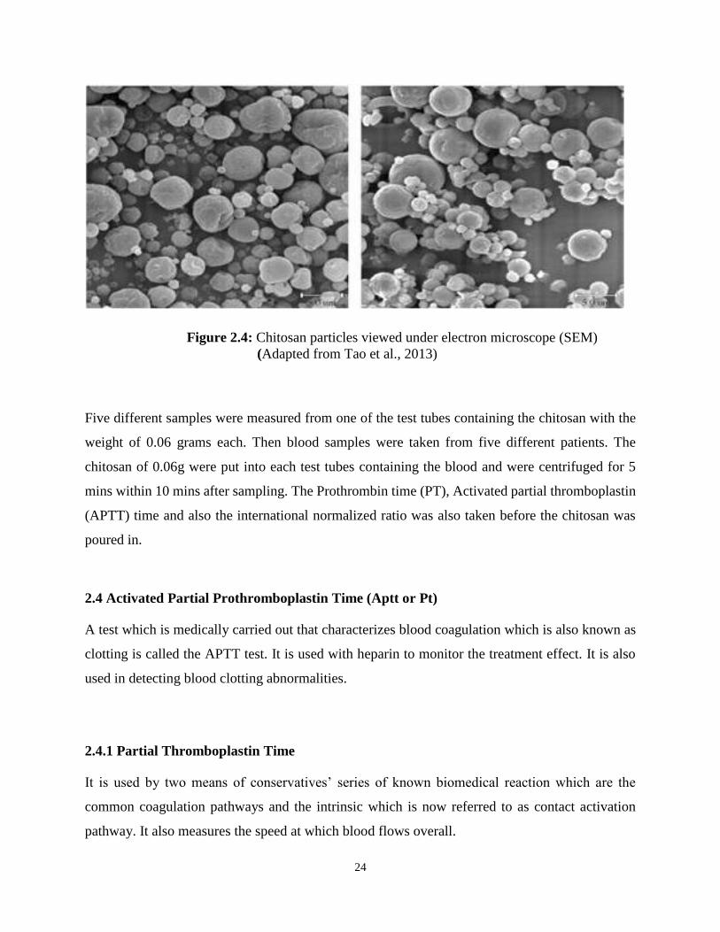
24
Figure 2.4: Chitosan particles viewed under electron microscope (SEM)
(Adapted from Tao et al., 2013)
Five different samples were measured from one of the test tubes containing the chitosan with the
weight of 0.06 grams each. Then blood samples were taken from five different patients. The
chitosan of 0.06g were put into each test tubes containing the blood and were centrifuged for 5
mins within 10 mins after sampling. The Prothrombin time (PT), Activated partial thromboplastin
(APTT) time and also the international normalized ratio was also taken before the chitosan was
poured in.
2.4 Activated Partial Prothromboplastin Time (Aptt or Pt)
A test which is medically carried out that characterizes blood coagulation which is also known as
clotting is called the APTT test. It is used with heparin to monitor the treatment effect. It is also
used in detecting blood clotting abnormalities.
2.4.1 Partial Thromboplastin Time
It is used by two means of conservatives’ series of known biomedical reaction which are the
common coagulation pathways and the intrinsic which is now referred to as contact activation
pathway. It also measures the speed at which blood flows overall.

25
In determining how quickly blood clotting takes place, it is used in conjunction with another
measure which is known as the (PT) prothrombin time. The prothrombin time, with the aid of
extrinsic pathway measures the speed of clotting. Tissue factor pathway is also known as extrinsic
pathway.
2.5 Method
On an automated instrument at 37oC partial thromboplastin time is analyzed. From the mixture
due to the absence of tissue factor, the test is termed “Partial”. In the test tube containing citrate or
oxalate, which binds calcium in a sample (they are molecules which act as an anticoagulant) blood
is drawn into the test tube. The blood is mixed gently, then to separate plasma from blood cells it
is centrifuged (the most commonly used method for blood plasma is Partial thromboplastin test).
Placed into a measuring test tube is a sample of the plasma extracted from the test tube. Then
mixed into the plasma sample is an excess of calcium. Finally, an activator is added such as (ellagic
acid, kaolin, silica, and celite) in order to activate the intrinsic pathway of coagulation.
Figure 2.5: Schematic representation of Activated Partial Thrombin Time
(Adapted from Perry and Todd, 2013)

26
CHAPTER THREE
RESULTS AND DISCUSSIONS
In vitro blood compatibility tests of chitosan as a biomaterial were performed using blood clotting
test and scanning electronic microscope. Blood compatibility and biodegradability of chitosan
were tested in some studies (Lee et al., 1995; Yang and Lin 2002; Lee et al., 2004; Yong et al.,
2007).In all the studies, a chemical modification of chitosan were made, this is attributed to the
striking characteristic of chitosan as a good biomaterial that allows chemical changes in its
structure and molecule.
In this study five (5) samples were taken randomly from healthy individuals and analyzed without
addition of chitosan and the result is presented on table 1. In the initial samples, the PT showed a
general pattern of coagulation of 12 to 21 seconds, this connotes a significance record of the
samples. World health organization recommend standardization of anticoagulants monitoring
because different laboratories used indices in expressing result of these tests.
The clotting cascade which comprised of Prothrombin (PT), which evaluate the integrity of
extrinsic factors as well as factors common both systems and partial thromboplastin Time (PTT),
which determines the integrity of intrinsic factors and the common pathways. Similarly, in all the
tests run for the samples without adding chitosan showed similar responds tables 1, 3 and 5.
Activated partial Thromboplastin Time (APTT) test a good indicator of monitoring coagulation
disorders and to monitor patients taking an anticoagulants drugs also investigated in this study.
Strong positive charge in chitosan play vital role in interaction with other substances. Interestingly,
the charges improve the crossing-linking of chitosan with other organic compounds. Fernandes et
al. (2011) describe that the activity of blood could be influence by environmental factors and the
electrical charges. The authors suggested that intermolecular forces are involved in interaction of
blood with any substance which would determine its compatibility. This suggests that the cross-
linking of chitosan with blood is determined by the conformity of the charges present and improve
the biological activities.
27
The size of chitosan also play important in binding capacity to substance as evidenced in study
conducted by (Tao et al., 2013). This is also evidenced by this study, where blood became
compatible with nanoparticles of chitosan in record time and believed to be contributed by the size
of chitosan. The interaction of chitosan and its derivatives has received attention of recent, for
instance, the formation of an ethylenic double bond in the chitosan–glutaraldehyde interaction (Li
et al., 2013).
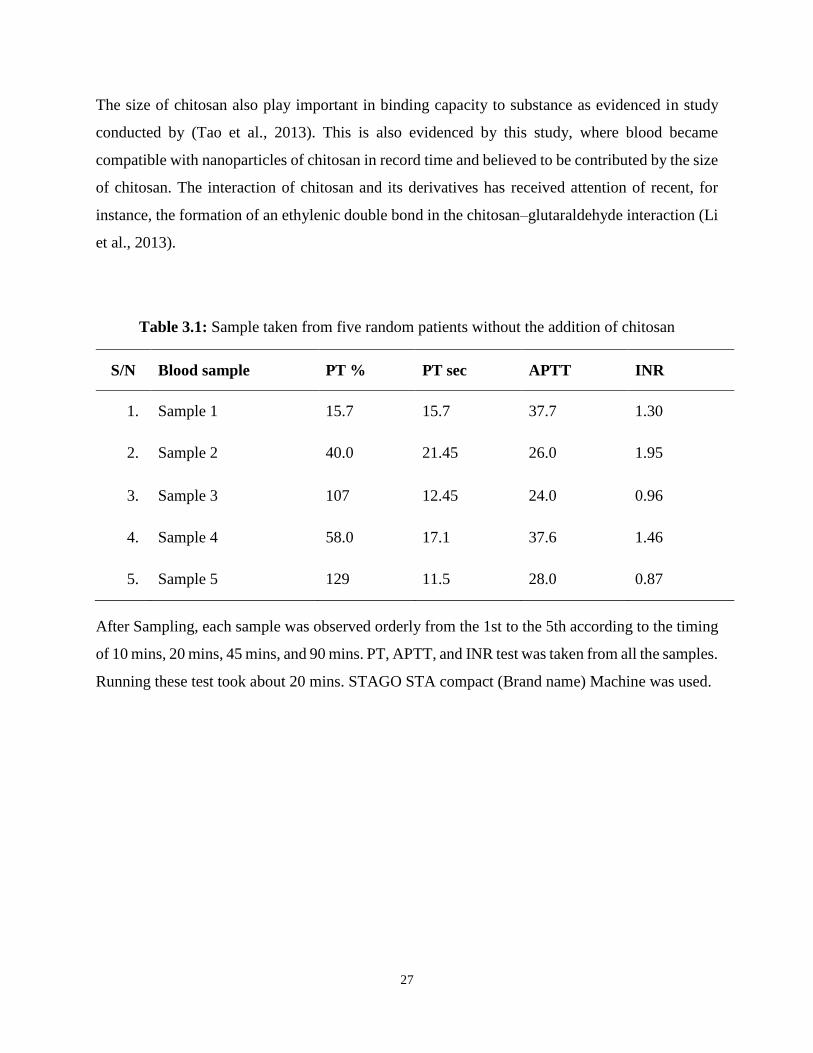
Table 3.1: Sample taken from five random patients without the addition of chitosan
S/N Blood sample PT % PT sec APTT INR
1. Sample 1 15.7 15.7 37.7 1.30
2. Sample 2 40.0 21.45 26.0 1.95
3. Sample 3 107 12.45 24.0 0.96
4. Sample 4 58.0 17.1 37.6 1.46
5. Sample 5 129 11.5 28.0 0.87
After Sampling, each sample was observed orderly from the 1st to the 5th according to the timing
of 10 mins, 20 mins, 45 mins, and 90 mins. PT, APTT, and INR test was taken from all the samples.
Running these test took about 20 mins. STAGO STA compact (Brand name) Machine was used.

28
Table 3.2: Samples after addition of chitosan
Samples ∆t (Minutes) PT% PTsec APTT INR
1. 10 7 86.2 39.8 11.93
20 5 120.1 96.5 18.37
45 5 120.1 58.1 18.37
90 5 120.1 67.2 18.36
2. 10 7 85.9 167.9 11.88
20 5 120.1 207.2 18.37
45 5 120.1 300.1 18.37
90 5 120.1 300.1 18.36
3. 10 18 39.3 300.1 4.30
20 No plasma No plasma No plasma No plasma
45 - - - -
90 - - - -
4. 10 5 120.1 300.1 18.37
20 10 65.3 71.9 8.32
45 5 120.1 134.8 18.73
90 5 120.1 140.2 18.81

29
The second experiment was carried out by recording the normal values of each blood samples
before the test was carried out. The table below shows the value of the blood values.
Table 3.3: Sample taken from five random patients without the addition of chitosan
S/N Blood sample PT % PT sec APTT INR
1. Sample 1 88 13.6 40.6 1.08
2. Sample 2 37 21.8 49.7 2.12
3. Sample 3 63 16.2 40.4 1.36
4. Sample 4 37 22.6 38.0 2.09
5. Sample 5 91 13.4 37.7 1.06
After Sampling, each sample was observed orderly from the 1st to the 5th according to the timing
of 10 mins, 20 mins, 45 mins, and 90 mins. PT, APTT, and INR test was taken from all the samples.

30
Table 3.4: Second run test with chitosan
Samples ∆t Minutes PT% PTsec APTT INR
1. 10 83 14.0 47.9 1.12
20 72 1.12 51.8 1.19
45 68 15.5 54.6 1.28
90 63 16.6 59.1 1.36
2. 10 36 23.3 44.6 2.18
20 35 23.3 45.9 2.23
45 31 25.6 46.1 2.46
90 30 26.2 48.1 2.54
3. 10 59 16.9 37.9 2.28
20 58 17.0 38.9 2.29
45 52 18.2 41.1 2.58
90 51 18.4 43.7 2.47
4. 10 34 24.1 41.6 2.28
20 34 24.2 44.1 2.29
45 30 26.2 51.7 2.58
90 31 25.7 54.4 2.47
5. 10 82 14.1 35.3 1.13
20 83 14.0 37.1 1.12
45 74 14.9 37.8 1.12
90 67 15.7 41.6 1.30

31
The third experiment was carried out by recording the normal values of each blood samples before
the test was carried out. The table below shows the value of the blood values.
Table 3.5: Third run test without chitosan
S/N Blood sample PT % PT sec APTT INR
6. Sample 1 105 12.5 30 0.97
7. Sample 2 62 16.4 31.9 1.38
8. Sample 3 105 12.5 23 0.97
9. Sample 4 88 13.6 31 1.08
10. Sample 5 87 13.7 32 1.09

32
Table 3.6: Third run test with chitosan
Samples ∆t (Minutes) PT% PTsec APTT INR
1. 10 76 14.6 59.0 1.60
20 61 16.5 102 1.39
45 58 17 98.2 1.45
90 57 17.2 94.5 1.47
2. 10 51 18.4 42.3 1.60
20 49 18.8 52.3 1.65
45 48 19 51.5 1.67
90 44 20.3 59.7 1.82
3. 10 109 12.3 31.0 0.95
20 97 13.0 34.2 1.02
45 94 13.2 35.0 1.04
90 78 14.4 36.3 1.17
4. 10 32 24.9 67.0 2.38
20 26 29.1 113.8 2.91
45 23 31.8 75.3 3.26
90 25 29.9 300.1 3.01
5. 10 98 12.9 37.4 1.09
20 92 13.3 32.5 1.05
45 95 13.1 44.9 13.1
90 83 14.0 44.8 14.0

33
Studies searching for a compatible biomaterial with living tissues has been a hallmark in
bioengineering and biomedical science. In this study, chitosan exhibited a striking ability of
dissolving in blood in record time and interestingly no coagulation observed, thus a great
advantage in employed in treatment of wounds. Natalya et al. (2002) and Chu (2010) studied blood
compatibility of modified chitosan, the results showed ability of the biomaterial to completely
dissolve in the blood, and this is in agreement with study where the chitosan became compatible
within the blood materials.
Mixture of foreign substances with blood may clot the blood in vitro and may provoke strong
immune responds in vivo, thus inflammatory reactions and other immune disorders can occur.
Thus, with the results shown in this study, the chitosan will not be considered as a foreign
substance by the body immune system, which blood contains many immune cells. Tables 2, 4 and
6, showed results of intrinsic and extrinsic factors, PT and APTT respectively.
Though there are some variability among the different samples which could be attributed
individual’s blood nature and might involve deficiency of clotting factors like vitamin K. In
addition, time play an important role in the compatibility process, in overall, it indicated that
increase in time may enhance total dissolving of the chitosan in all the indicators (PT and APTT).
This study significantly found that chitosan can be compatible in living tissue, thus can be employ
in bioengineering. The slight increase in both PT and APTT in this work is attributed to time factor
in both tests and can play role in its biological activity. Previously, other studies also confirmed
biocompatibility of chitosan and its derivatives with tissue. Romani et al. (2013) recognized that
the effect of surface materials on erythrocytes aggregation and platelet adhesion and activation as
the major parameter in hemocompatibility studies. This is also in agreement with the present
studies and other studies (Chen et al., 2009; Xiong et al., 2011; Balan and Verestiuc, 2014). Based
on the parameters used to study blood compatibility of chitosan as biomaterial indicated a
promising application in bioengineering due its versatility.
Chitosan often not used as single component but mixed with other polymers to enhance
compatibility or increase lifetime. Crosslink was often obtained using well-known glutaraldehyde,
or m- and p-phthalaldehydes (Rinaudo, 2010). In the study, genepin used as a natural cross-linker
by an amide linkage to allow convenient biomedical applications application. In preparation of

34
chitosan nanoparticles different cross-linkers are used based on their molecular weight and their
toxicity. Banerjee et al. (2002) described preparation of chitosan nanoparticles cross-liked using
reverse micelles as the media and their physicochemical characterization as well as biodistribution.
The authors employed the used of glutaraldehyde and degree of the cross-linking was observed
and suggested for a particular degree of cross-linking, the particle size has been determined at
infinite dilution of the particle solution. Sodium TPP is considered a good cross link agent for it
non toxicity by some researchers (K’deeva et al., 2009; Ponnuraj et al., 2015).
The reaction of cross-linking is to obtain different heterogenous biomolecules for different
applications. Owing its nature and structure, chitosan medication by bifunctional cross-linking can
be achieved to enhanced activity. The properties of polymers can be modified by using several
cross-linking methods (physical and chemical methods). Sionkowska et al. (2014) differentiate
between physical and chemical cross-linking, in which physical cross-linking methods, one can
use UV radiation, temperature, and gamma radiation and it considered cheap and easy to perform.
However, the main disadvantage of this method is the difficulty in controlling the cross-linking
process, which means that it is very hard to obtain desirable degrees of cross-linking. In the other
hand, Chemical cross-linking methods are based on the chemical reactions which take place after
adding the chemical compound as a cross-linking agent to the polymer. Chemicals such as
glutaraldehyde or formaldehyde are used as cross-linking agents. But the authors suggested that
such chemicals increase the toxicity and make unpleasant for biomaterials for contact with living
bodies, hence natural cross-linking agents such as tannic acid are preferred.

35
CHAPTER FOUR
CONCLUSIONS AND RECOMMENDATION
4.1 Conclusion
Due to its remarkable combination of mechanical, biological and physicochemical properties that
highlights its applications extensively, for life science chitosan is a very interesting biomaterial. In
different therapeutic systems it can be manufactures and it is readily available: membrane,
scaffolds, hydrogels, or nano/microparticles. In biomedical field, chitosan exhibits suitable
properties. In blood stream its use is limited, since blood coagulation and surface induced
thrombosis is promoted. Strong positive charge in chitosan play vital role in interaction with other
substances. Interestingly, the charges improve the crossing-linking of chitosan with other organic
compounds.
The reaction of cross-linking is to obtain different heterogenous biomolecules for different
applications. Owing its nature and structure, chitosan medication by bifunctional cross-linking can
be achieved to enhanced activity. The properties of polymers can be modified by using several
cross-linking methods (physical and chemical methods). It showed that the cross-linking of
chitosan with blood is determined by the conformity of the charges present and improves the
biological activities. The size of chitosan also plays an important role in binding capacity to
substance. Chitosan blood interaction have been studied over the years and several strategies are
proposed to modulate the interactions.
The result of this work showed that when chitosan was mixed with blood, there was no sign or
form of blood clotting. With the result, there was biocompatibility. The result showed that chitosan
can be used as a safe biomaterial. Some of these proposed strategies are the association of
hemocompatible compounds with chitosan while some are based on the chemical modifications of
biopolymer.
So far there has been a lot of progress and researches pertaining higher hemocompatibility and a
great number of chitosan have been developed and also in a rapid state. Nevertheless when it comes
to the contact with blood, chitosan based system behavior is not quite know. Blood interaction of

36
biomaterial surfaces are becoming more complex as new therapeutic systems are emerging. For
successful recognition of hemocompatibility materials (serves as a derivative for chitosan) and
chitosan additional studies are required because most of these formulations are still at the lower
level (Laboratory level).
4.2 Recommendations
Series of blood compatibility test was carried out in these studies that evaluated phenomenas such
as hematology and thrombosis. Working with chitosan and blood (hematology) which could lead
to a giant breakthrough in this research; the world of microparticles with excellent numerous
biomaterials will help gain more awareness and acceptance of biomedical engineering and as well
as supporting needy individuals or individual in need will tend to benefit good and effective health
care.

37
REFERENCES
Agrawal, P., Strijkers, G. J., Nicolay, K. (2009). Chitosan-based systems for molecular imaging.
Advanced Drug Delivery Rev. 62, 42-58.
Angelica, M. D., Tiozon, R. N., Espana, R. C. N. (2016). Chitosan from portunus pelagicus in the
synthesis of reduced gold nanoparticles as potential carrier for the delivery of
erythropoietin. Doi:https://doi.org/10.1101/044875.
Aranazi, Menginbar, M., Harris, R., Panos, Miralles, B., Acosta N. (2009). Functional
characterization of chitin and chitosan. Curr Chem Biol. 3, 203-30.
Averous, L., & Pollet, E. (2002). Biodegradable polymer environmental silicate nano-
biocompasite. Doi. 10.100.7.978-14471-41008-2.
Balan, V., & Verestiuc, L. (2014). Strategies to improve chitosan hemocompatibility: A review.
European polymer journal. 53, 171-188.
Barnejee, T., Mitra, S., Singh, A. K., Sharma, R. K., & Maitra, A. (2002). Preparation,
characterization and biodistribution of ultrafine chitosan nanoparticles. International
journal of pharmaceautics. 243 (1-2), pp. 93-05.
Berger, J., Reist, M., Mayer, J. M., Felt, O., Peppa, N. A., Gurney, K. (2004). Structure and
interaction in covently and ironically cross-linked chitosan hydrogel for biomedical
application. European journal of pharmaceutical and biopharmaceutics. 57, 19-34.
Biagini, G., Bertani, A., Muzzarelli, R., Damadei, A., DiBenedetto, G., Bellogolli, A., Riccotti,
G., Zucchini, & Rizzoli, C. (1991). Wound management with N-carboxylbentyl chitosan.
Biomaterials. 12, 281-286.

38
Bizham, M. K., Sajadi, S. A., & Jaafari, M. R. (2008). Preparations, characterization and
mucoadhesive properties of chitosan-coated microspheres encapsulated with cyclosporins
A. Journal of drug delivery. 34(5), 21-36.
Chatelet, C., Damour, O., Domard, A. (2001). Influence of the degree of acelation on some
biological properties of chitosan films. Biomaterials. 22, 261-8.
Chen, H., Tian, X., & Zou, H. (2009). Preparation and blood compatibility of new silicon-chitosan
hybrid biomaterials. Artificial cell, blood substituent and biotechnology. 26 (4), 431-436.
Chen, M. C., Mi, F. L., Liao, Z. X., & Sung, H. W. (2011a). Chitosan: Its application in drug
eluting devices. Advances in polymer science. 342, 185-230.
Chen M. C, Mi, F. L, Liaon, Z. X, & Sung H. W. (2011b) Chitosan: Its application in drug eluting
devices. Advances in polymer science. 243, 185-230.
Cheung, R. C. F., Bun, T. N. G., Wong, J. H., & Chen, W. Y. (2015). Chitosan update on potential
Biomedical and pharmaceutical applications. Marine drugs. 13, 5156-5186.
Croisier, F., & Jerome, C. (2013). Chitosan based biomaterials for Tissue engineering. European
Polymer Journal. 49, (4), 780-792.
Dash, M., Chiellini, F., Ottenbrite, R. M., Chiellini, E. (2011). Chitosan-A versatile semi-synthetic
polymer in biomedical applications. Program Polymer Science. 36,981-1014.
Dhawan, S., Singla, A. K., & Sinha, R. (2004). Evaluation of mucoadhesive properties of chitosan
microspheres prepared by different methods. APPS, Pharmaceutical Science Technology.
5(4), 1-7.
Dutta, K. P., Dutta, J., & Tripathi, V. S. (2014). Chitin and chitosan chemistry, properties and
application. Journal of scientific and industrial Research. 63, 20-31.
39
Dutta, P. K., Dutta, D., & Tripathin, N. (2004). Chitin and chitosan: chemistry, properties and
applications. Journal of scientific and industrial research. 63, 20-31.
El-kamel, A. H., Ashiri, L. Y., & Alsarra, I. A. (2007). Micrometrical metronidazole benzoate film
as local mucoadhesive delivery system for treatment of periodontal diseases. AAPS
Pharmaceutical Science Technology. 8, E75/1.E75/11.
Fernandes, H. P., Cesar, C. L., & Castro L. B. (2011). Electrical properties of the red blood cell
membrane and immunohematological investigation. Rev Bras Hematol. 33 (4), pp. 297-
301.
Fisher, T. H., Bode, A. P., Demcheva, M., & Vournakis, J. N. (2007). Hemostatic properties of
glucosamine-based materials. J. Biomed Mater Res Part A. 80,167-74.
Fisher, T. H., Thatte, H. S., Nicolas, T. C., Bener-Neal, D. E., Bellinger, A. D., & Vournakis, J. N.
(2005). Synthetic platelet integrin signaling and factor XII activation in poly-N-acetyl
glucosamine fiver-mediated hemostasis. Biomaterials. 26, 5433-43.
Funkhouser, J. D., & Aronson Jr, N. N. (2007). Chitinase family GH 18: evolutionary insights
from the genomic history of a diverse protein family. BMC Biol. 7, 96.
Gorbet, M. B., & Sefton, M. V. (2004). Biomaterial-associated thrombosis: role of coagulation
factors, complements, platelets and leukocytes. Biomaterials. 25, 5681-703.
Goy, R. C., Douglas, D. B., & Assis, B. G. (2009). A review pf the antimicrobial activity of
chitosan. Ciencia e Technogia. 19, 241-247.
Goy, R. C., Sinara, T. B., Odilio, T. B., & Assis, BG. (2006). Evaluation of the activity of chitosan
and its quaternized derivatives on E. coli and S. aureus growth. Revita Brasileira de
farmacognosia. 26, 122-127.

40
He, P., Davis, S. S., & Illum, L. (1998). In vitro evaluation of the mucoadhesive properties of
chitosan microsphere. International journal of pharmaceutics. 166 (1), 75-88.
Heemskerk, J. M. N., Bevers, E. M., & Lindhout, T. (2002). Platelet activation and blood
coagulation. Thromb Heamostatis. 88, 186-93
Hombach, J., & Bernkop-schnurch, A. (2009). Chitosan solutions and particles: evaluation of their
permeation enhancing potential on MDCK cells used as blood brain barrier model.
International Journal of pharmacology. 376, 104-109.
Hritcu, D., Humelnicu, D., Dodi, G., & Popa, M. I. (2012). Magnetic chitosan composite particles:
evaluation of thorium and uranyl ion adsorption from aqueous solutions. Carbohydrate
polymer. 87. 1158-91.
Ibrahim, H. M., & El-Zairy, E. M. R. (2015). Chitosan as a Biomaterial- structure, properties and
electrospun nanofibres. Concepts, compounds and alternative antibacterial. 3, 87-98.
Ikram, S., & Ahmad, S. (2016). Chitosan based scaffolds and their applications in wound healing.
Achievements in the life science. 10 (1), 27-37.
Ishihara, M., Obara, K., Morimoto, Y., Fujita, M., Masuoka, K., Kanatari, Y., Takase, B., & Hotari
H. (2006). Chitosan hydrogel as a drug delivery carrier to control angiogenesis. Journal
of artificial organs. 9, 8-16.
Kean, T., & Thanou, M. (2009). Chitin and chitoson –sources, production and medical
applications. London, Kentus books. 327-61.
Khamenah, B., Mahdi, M. N., & Tataghodi, M. (2014). In vivo evaluation of mucoadhesive
properties of nanoliposomal formulation upon coating with trimethylchitosan polymer.
Nanomedicine Journal. 1 (3), 147-154.

41
Khobragade, P. K., & Puranik, P. (2015). Chitosan: A mucoadhesive polymer. World journal of
pharmacy and pharmaceutical science. 4 (4), 1829-1847
Kil’deeva, R. K., Perminov, P. A., Vladimirov, L. B., Novikov, V. V., & Mikhailov, S. N. (2009).
On the Mechanism of the Reaction of Glutaraldehyde with Chitosan. Russian Journal of
Bioorganic Chemistry. 35 3), pp. 360–369.
Kittur, F. S., Vishu, Kumar, A. B., Varadaraj, M. C., & Tharanathan, R. N. (2005).
Chitoologosaccharides-preparation with the aid of pectinase isozyme from Aspergillus
niger and their antibacterial activity. Carbohydr Res. 340, 1239-45.
Kofuji, K., Qian, C-J., Nishimura, M., Sugiyami, N., Murata, Y., & Kawashima, S. (2005).
Relationship between physiochemical characteristics and functional properties of
chitosan. Eur Polymer J. 41, 2784-91.
Lee, D. W., Powers, K., & Baney, R. (2004). Physiochemical properties and blood compatibility
of acylated chitosan nanoparticles. Carbohydrate. Polymers. 58(4), 371-377.
Lee, K. Y., Ha, W. S., & Park, W. H. (1995). Blood compatibility and biodegradability of partially
N-acylated chitosan derivatives. Biomaterials. 16 (16), 1211-1216.
Lien, C., Molnar, E., Tomna, P., Tsibouklis, J., Pilkington, G., & Gorecki, D. (2012). In vitro
assessment of alkylglyceryl-functionalized chitosan nanoparticles as a permeating vectors
for the blood brain barrier. Biomacromolecules. 13, 1067-1073.
Li, B., Shan, C. L., Zhou, Q., Fang, Y., Wang, Y. L., Xu, F., Han, L. R., Ibrahim, M., Guo, L. B,
Xie, G. L & Sun, G. C. (2013). Synthesis, Characterization, and Antibacterial Activity of
Cross-Linked Chitosan-Glutaraldehyde. Marine drugs. 11, 1534-1552
42
Lu, W. N, Lu, SH., Wang, H. B., Li, D. X., Duan, C. M, & Liu, Z. Q. (2009). Functional
improvement of infarcted heart by co-injection of embryonic stem cells with temperature-
responsive chitosan hydrogel. Tissue engineering Apart A. 15, 1437-1447.
Madhumathi, K., Sudheesh, Kumar, P. T., Abhilash, S., Sreeja, V., & Tamura. (2010).
Development of novel chitin/nanosilver composite scaffolds for wound dressing
applications. J Mater Science Mater Med. 21, 807-13.
Majeti, N. V., & Kumar, R. (2000). A review of chitin and chitosan application. Reactive and
functional polymer. 46 (1), 1-27.
Manhwan, S. J., Yeo, W. S., Galvao, K. N., & Jeong, K. C. (2014). Underlying mechanism of
antimicrobial activity of chitosan microparticle and implication for the treatment of
infectious diseases. PLOS one. 9 (3), 92723-doi.10.137.
Martins, F. A., Facchi, S. P., Fallmann, H. D., Pereira, A. B., Rubira, A. F, & Muriz, E. C. (2014).
Antimicrobial activity of chitosan derivatives containing N-Quaternized moieties in its
backbond: A review. International journal of Molecular science. 15 (11), 20800-20832.
Meng, S., Liu, Z., Shen, L., Guo, Z., Chou, L. L., & Zhong, W. (2009). The effect of layer-by-
layer chitosan –heparin coating on the endothelialization and coagulation properties of a
coronary stent system. Biomaterials. 30, 2276-2283.
Mounrya, V. K, Inamda, N. N, & Tiwari, A. (2010) Carboxymethyl chitosan and its application.
Advanced materials letters. 1(1), 11-33
Muzzarelli, R. A. A., Boudrant, J., Meyer, D., Manno, N., De Marchis, M., & Paoletti, M. G.
(2012). Current views on fungal chitin/chitosan, human chitinases, food preservation,
glucans, pectins, and inulin: a tribute to Henri Braconnot, precursor of the carbohydrates
polymers science on the bicentennial. Carbohydrate polyp. 87, 995-1012.
43
Muzzarelli, R. A. A, & Muzzarelli C. (2005). Chitosan chemistry: relevance to the biomedical
sciences. Advanced polymer Sci. 186, 151-209.
Muzzarelli, R. A. A. (2008). Chitin nanostructure in living organisms. In: Gupta SN, Briggs D,
editors. Chitin in the fossil record. New York: Springer.
Natalya, M. A., Koulauskaia, D. V., Shima, M., & Saenko, E. L. (2002). Intrinsic pathway of blood
coagulation contributes to thrombogenicity of atherosclerotic plaque. Blood. 99 (12),
4475-4485.
Nawrotek, K., Modrzejewska, Z., Paluch, D., Zarzycki R., & Rusak, A. (2015). Cytotoxicity of
chitosan based thermosensitives hydrogels intended for nervous tissue engineering.
Progress on chemistry and approach of chitin and its derivatives. Voume XX, 1-14.
Peng, Y., Han, B., Liu, W., Xu, X. (2005). Preparation and antimicrobial activity of hydroxypropyl
chitosan. Carbohydrates.340, 1846–1851.
Perry, D. &Todd, T. (2013). A practical guide to laboratory haemostasis. www.practical-
haemostasis.com
Pok, S., Myers, J. D., Madihally, S. V., & Jacot, J. G. (2013). A multilayered scaffold of a chitosan
and gelatin hydrogel supported by a PCL Core cardiac tissue engineering. Acta
Biomaterials. 9, 5630-5642.
Ponnuraj, R. K. J., Gopalakrishnan S., Senthilnathan, K., Meganathan, V., & Saravanan, P. (2015).
Formulation and Characterization of Epigallocatechin Gallate Nanoparticles. Indo
American Journal of Pharmaceutical Research. 5(1), pp. 387-399.
Raldo, M., Hornof, M., Caliceti, P., & Bernkop-Schnurch, A. (1992). Mucuadhesive thiolated
chitosan as platforms for oral controlled drug delivery: synthesis and in vitro evaluation.
Eur J Pharm Biopharm. 57, 115-21.

44
Renault, F., Sancy, B., Badot, P. M., & Crini, G. (2009). Chitosan for coagulation/flocculation
processes-an eco-friendly approach. Eur Polym J. 1337-48.
Rinaudo, M. (2010). New way to crosslink chitosan in aqueous solution. European polymer journal
46 (7), pp. 1537-1544.
Roa, S. B., & Sharma, C. P. (1997). Use of chitosan as a biomaterial: studies on its safety and
hemostatic potential. Journal of mater Res. 34, 21-8.
Romani, A. A., Luingi, I., Riccardi, R. F, Pipitano, S., Morganti, M., Boroni, M. C, Borghetti, A.
F, & Bettini, R. (2013). In vitro blood compatibility of novel hydrophillic chitosan films
for vessels regeneration and repair. Advances in biomaterials science and biomedical
application. 5772, 156-172.
Sabudin, S., Derman, M. A., Zainol, I., & Noorsalk, K. (2012). In vitro cytotoxicity and cell
seeding studies of a chitosan-silver composite for potential wound management
application. Journal of engineering science. 8, 29-37.
Sahul, S. K., Maiti, S., Ghosh, S. K., & Pramanik, P. (2011). Hydrophobically modified
carboymethyl chitosan nanoparticles targeted delivery of paclitaxel. Journal of drug
target. 19, 104-113.
Saqias, A. Williams, A. C, & Khutoryanskiy, V. V. (2008). Why chitosan mucoadhesive?
Biomolecules. 9 (7), 1837-1842.
Schiffman, J. D., & Schauer, C. L. (2007). Cross-linking chitosan nanofibres. Biomolecules. 8,
594-601.
Schipper, N. G., Varum, K. M., Stenberg, P., Ocklind, G., Lennenas, H., & Artursson, P. (1999).
Chitosans as absorption enhancers of poorly absorbable drugs. Eur J Pharm Sci. 8, 335-
43.

45
Sionkowska, A., Kaczmarek, B., & Lewandowska, K. (2014). Characterisation of chitosan after
cross-linking by tannic acid. Progress on Chemistry and Application of Chitin and Its
Derivatives, 16, pp. 135-139.
Stone, C. A., Wright, H., Devaraj, V. S., Clarke, T., & Powell, R. (2004). Chitosan membranes as
a wound-healing dressing, characterization and clinical application. Journal of Biomedical
material. 69, 216-222.
Sudhess, Kumar, P. T., Abhilash, S., Manzoor, K., Nair, S. V., Tanura, H., & Jayakumar, R.
(2010). Preparation and characterization of novel β-chitin/nanosilver composite scaffolds
for wound dressing applications. Carbohydr Polym, 80, 761-7.
Sun, X., Wang, Z., Kadouh, H., & Zhou, K. (2014). The antimicrobial, mechanical, physical and
structural properties of chitosan–gallic acid films. Food Science Technology. 57:83–89.
Tao, Y., Zhang, H. L., Yu, Y. M., Wan, S., & Su, Z. Q. (2013). Preparation of Chitosan and Water-
Soluble Chitosan Microspheres via Spray-Drying Method to Lower Blood Lipids in Rats
Fed with High-Fat Diets. International Journal of Molecular science. 14, 4174-4184.
Terin, A., & Murat, U. (2015). Silk fibroin as a non-thrombogenic biomaterial. International
journal of biological macromolecules. www.elsevier.com/locate/ijbiomac
Thanous, M., Verhoef, J. C., & Junginger, H. E. (2001). Oral drug absorption enhancement by
chitosan and its derivatives. Advanced drug Delivery Rev. 52, 117-26.
Tikhonov, V. E., Stepnova, E. A., Babak, V. G, Yamskov, I. A., Palma-Guerrero, J., & Johnson,
H. B. (2006). Bactericidal and antifungal activities of a low molecular weight chitosan
and its N-/2(3)-(dodec-2-enyl) succinonyl/-derivatives. Carbohydr Polym. 64, 66-72.
46
Valentine, R., Athanasiadis, T., Marattis, S., Hanton, L., & Robinson, S. (2010). The efficacy of
novel chitosan gel on hemostasis and wound healing after endoscopic sinus surgery.
American Journal of Rhinological Allergy. 24, 70-75.
Wang, X., Yan, Y., Lin, F., Xion Z., Wu R., & Zhang R. (2005). Preparation and characterization
of a collagen/chitosan/heparin matrix for an implantable bioartificial liver. Journal of
biomaterial science. 16, 1063-1080.
Xie, W., Xu, P., Wang, W., & Liu, Q. (2002). Preparation and antibacterial activity of a water-
soluble chitosan derivative. Carbohydrates. 50, 35-40.
Xiong, W. Y., Yi, Y., Wang, H., Liu, J. H., & Ying, G. Q. (2011). Selective carboxypropionylation
of chitosan: synthesis, characterization, blood compatibility and degradation.
Carbohydrates research. 346 (10), 1217-1217.
Yang, J., Tian, F., Wang, Z., Wang, Q., Zeng, Y. J, & Chen, S. Q. (2008). Effects of chitosan
molecular weight and deacetylation degree on hemostasis. J Biomed Mater Res B Applied
Biomaterial. 84, 131-7.
Yang, M. C., & Kin, W. C. (2002). Surface modification and blood compatibility of polycylonitrile
membrane with immobolized chitosan-heparin conjugate. Journal of polymer research. 9
(3), 201-206.
Yang, Y., Zhou, Y., Chou, H., Wang, S., & Yu, J. (2007). Blood compatibility and mechanical
properties of oxidized chitosan films. Journal of applied polymer science. Doi. 106; 372-
377
Yi, H., Wu, L. Q, Bentley, W. E., Ghodssi, R., Rubloff, G. W, & Culver, J. N. (2005).
Biofabrication with chitosan. Biomacromolecules. 6, 2881-94.

47
Yong, D. H., Kohle, H., & Kauss, H. (1982). Plant physiology. 70, 1449-1454. Zaki, SO, Ibrahim,
M. N., and Katas, H. (2015) Particle size effects concentrate-dependant cytotoxicity of
chitosan nanoparticles towards mouse hematopoietic stem cells. Journal of
Nanotechnology. 2, 1-5.
Zhang, H., & Neau, H. (2001). In vitro degradation of chitosan by a commercial enzymes
preparation: effect of molecular weight and degree of deacylation. Biomaterials.22, 1653-
8.
Zhang, C., Qu, G., Sun, Y., Yang, T., Yoa, Z., & Shen, W. (2008). Biological evaluation of N-
Octyl-O-sulfate chitosan as a new nano-carrier of intravenous drugs. European Journal of
pharmacological science. 33, 415-423.
Zhou, H. Y., Zhang, Y. P., Zhang, W. F, & Chen, X. G. (2011). Biocompatibility and characteristic
of injectable chitosan-based thermosensitive hydrogel for drug delivery. Carbohydr
Polym. 83, 1643-51.
Zhu, H., Liu, F., Guo, J., Xue, J., Qian, Z., & Gu, Y. (2011). Folate-modified chitosan micelles
with enhanced tumor targeting evaluated by near infrared imaging system. Carbohydrate
polymer. 86, 1118-1129.