Membrane Membrane receptors. Single-pass membrane receptors membrane.
Upload
andrew-marshCategory
view
213download
0
Synthesis and characterisation of new ditopic receptorsfor guanosine
Andrew Marsh,* Nathaniel W. Alcock, William Errington and Rajeeve Sagar
Department of Chemistry, University of Warwick, Coventry CV4 7AL, UK
Received 15 July 2002; revised 7 April 2003; accepted 1 May 2003
Abstract—Novel ditopic receptors for guanosine have been prepared and characterised. Their association constants with a lipophilicguanosine derivative are somewhat smaller than the expected values for a guanosine–cytidine base pair, but remain in a useful range forbuilding supramolecular structures. q 2003 Elsevier Science Ltd. All rights reserved.
1. Introduction
The construction of supramolecular complexes1 based uponthe use of hydrogen bonding is a popular and successfulstrategy.2 The recognition code embedded in nucleobases3
makes them attractive sub-units for the preparation of manysupramolecular aggregates and there are now a number ofexamples where nucleobases have been used to makediscrete structures4 and two-dimensional arrays.5 We havebeen preparing a modular construction set based around theguanosine–cytidine (G·C) base pair because it formspredictable complexes with a viable association constantin non-polar environments. The G·C base pair has found lessapplication in supramolecular chemistry than the adenine–thymine (A·T) pair, in part due to the notoriously lowsolubility and self-aggregation of guanosine based systems.6
Despite these inherent problems the G·C base pair has foundsome application in supramolecular chemistry including thesynthesis of a rigid guanosine derived molecule which self-associates through a novel four-point hydrogen bondinginteraction,7 an ‘artificial dinucleotide duplex’,8 a self-assembled electron transfer pair9 and a tape-like aggre-gate.10 We now wish to disclose our own work on a relatedsystem. We have designed and synthesised a ditopicmolecular receptor based around cytosine linked throughan acetylenic axle (Fig. 1). A similar strategy has recentlybeen used to prepare nucleobase ‘ferroreceptors’11 allowinghydrogen bonded complexes to be built up around the rigidacetylenic axle. The synthesis exploits well-precedentedSonogashira coupling reactions12 allowing ready access toboth mono- and bis-alkynyl receptors 1 and 2, respectively.Through the use of 1H NMR titration we have determinedassociation constants for the interaction of lipophilic
guanosine derivatives with the ditopic receptors 1, 2 andthe corresponding half-molecule.
2. Design
The receptors were designed to have two hydrogen bondingsites linked through a conjugated bridge. This potentiallyallows electronic communication between hydrogen bond-ing sites, as well as providing functionality which may findapplication in the arena of molecular electronics. The ribosehydroxyl functions are ideal sites for attachment oftriisopropylsilyl solubilising groups13 to allow investi-gations into supramolecular structure to be carried out innon-polar solvent.14
3. Synthesis
The route used for the synthesis of receptors 1 and 2 is
0040–4020/03/$ - see front matter q 2003 Elsevier Science Ltd. All rights reserved.doi:10.1016/S0040-4020(03)00708-7
Tetrahedron 59 (2003) 5595–5601
Figure 1.
* Corresponding author. Tel.: þ44-24-7652-4565; fax: þ44-24-7652-4112;e-mail: [email protected]
Keywords: guanosine; Sonogashira coupling; supramolecular; cytidine.

shown in Scheme 1. Protection of the hydroxyls in 5-iodo-20-deoxycytidine 3 using triisopropylsilyl chloride gavematerial 4 which is soluble in non-polar solvent.Sonogashira coupling of this intermediate with trimethyl-silyl acetylene under conditions described for relatedsystems15,16 proceeded in good yield to give intermediate5 which was deprotected under basic conditions to giveterminal acetylene 6. Purification by flash chromatographyrequired dry loading of the compound onto silica gel prior toloading onto the column, otherwise blockage of the columnresulted. The synthesis of the mono-alkynyl receptor 1 wasthen accomplished through a second Sonogashira couplingof 6 with protected 5-iodo 20-deoxycytosine 4.
It was found that on certain occasions an appreciableamount of self-coupling of 6 also occurred to give the di-alkynyl receptor 2. At first this was believed to be due toingress of oxygen, although the presence of residualcopper(II) in the CuI may itself encourage self-couplingof acetylenes after the fashion of an Eglinton–Hay coupling(vide infra). Acetylene 6 was crystalline and slow growthfrom isopropanol gave crystals whose molecular structurewas solved by X-ray crystallography (Fig. 2). Interestinglythis cytosine derivative showed no intermolecular hydrogenbonding contacts other than to solvent. This was somewhatsurprising given cytidine’s ability to self-associate into adimer in the solid state17 and solution14 and this observationmay be due to a preferential packing interaction of thetriisopropylsilyl groups with one another.
Although crystals of receptor 1 of a suitable size for analysisby X-ray crystallography were grown from alcoholic
solvents, none diffracted well enough to allow high qualitydata to be collected.
The longer dialkynyl receptor 2 was better prepared by oneof two methods. The first method used was a classicalEglinton–Hay18 coupling under atmospheric oxidationconditions. The second was a more recent method involvingthe presence of catalytic amounts of Pd(PPh3)2Cl2 and CuIusing iodine as the co-oxidant to regenerate the active Pd(II)catalyst.19 This gave a good yield of receptor 2 without theneed to remove large amounts of copper impurities(Scheme 2).
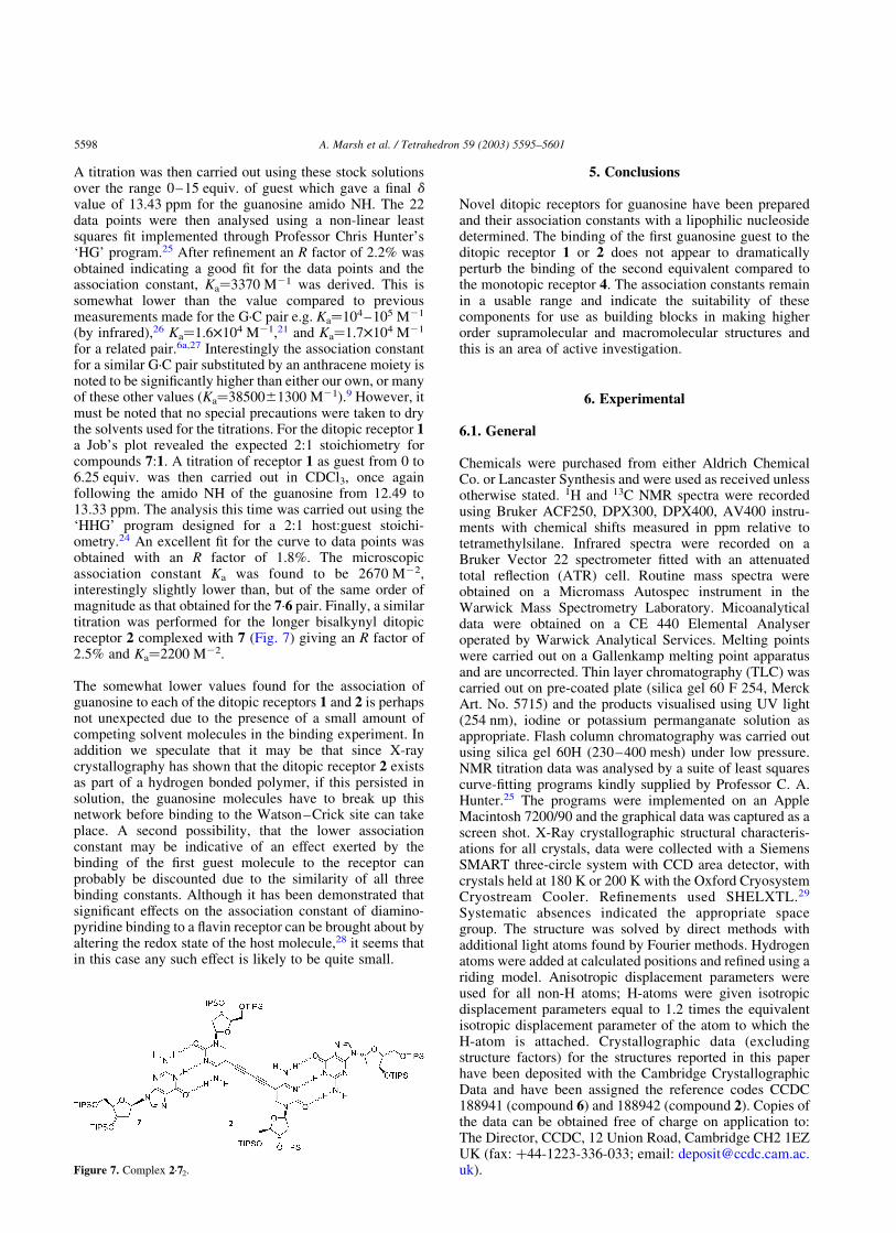
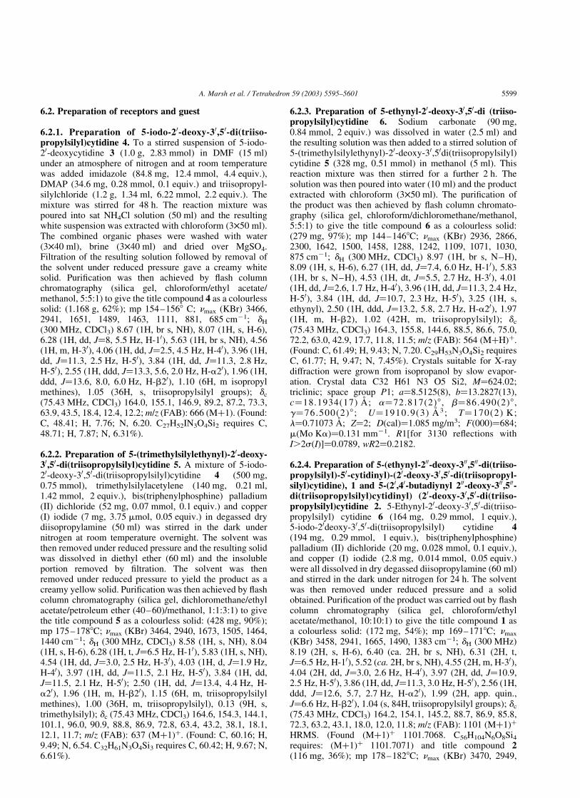
Crystals of 2 grown from isopropanol were of a sufficientquality to allow the molecular structure to be solved byX-ray diffraction (Fig. 3). In contrast to the half-molecule 6,contacts between the pyrimidine amino groups (N-9 andN-14) were evident in the form of classical cytidinedimers17 (Fig. 4) leading to a helical supramolecular ribbonwith a narrow central channel (Fig. 5).
In addition there is another hydrogen bond contact evidentbetween the pyrimidine carbonyl and the amino groupsleading to a secondary supramolecular structure (notshown). The triisopropyl groups were sufficientlydisordered to preclude refinement of the alkyl side-chains,
Figure 2. Molecular structure of 6 (X-ray: H’s not shown).
Scheme 2. Reagents and conditions: (i) Pd(PPh3)2Cl2, CuI, I2, diiso-propylamine, rt (78%).
Scheme 1. Reagents and conditions: (i) triisopropylsilyl chloride,imidazole, DMAP, DMF (62%); (ii) trimethylsilyl acetylene, Pd(PPh3)2-
Cl2, CuI, diisopropylamine, dark, rt (90%); (iii) Na2CO3, H2O–MeOH, 2 h,rt (97%); (iv) 4, Pd(PPh3)2Cl2, CuI, diisopropylamine, dark, rt (1: 54%; 2:36%).
A. Marsh et al. / Tetrahedron 59 (2003) 5595–56015596
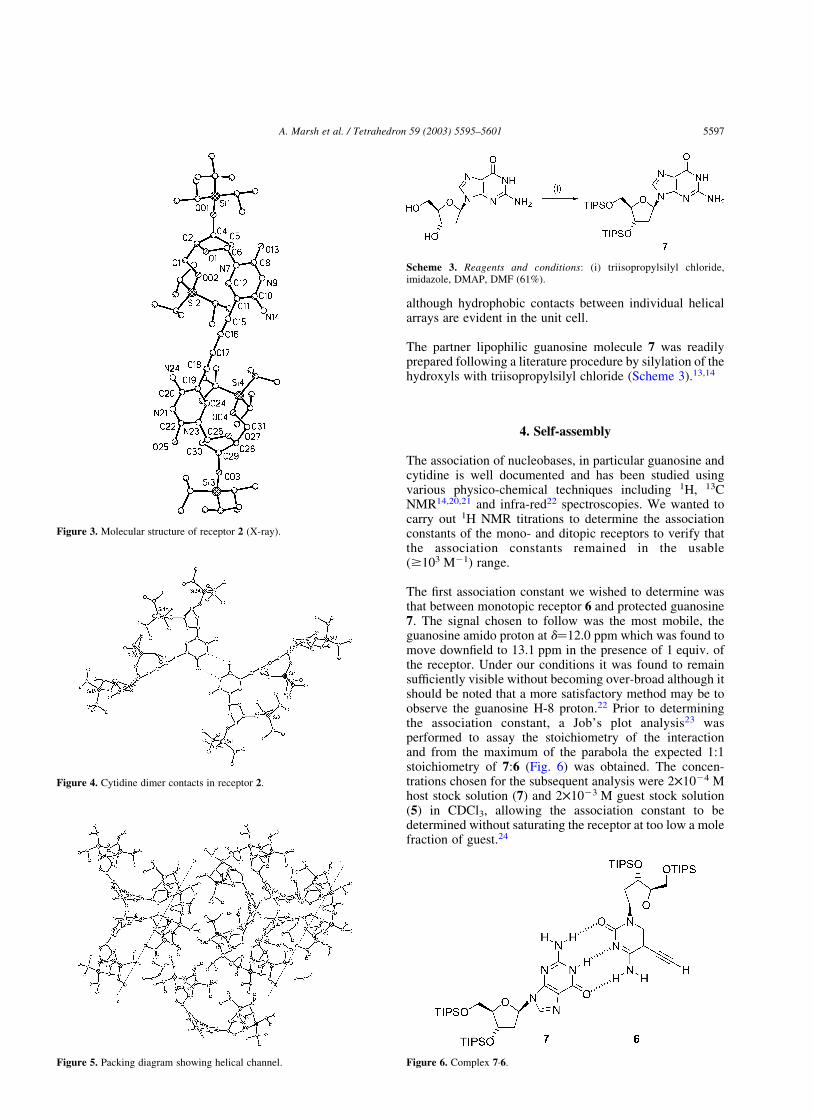
although hydrophobic contacts between individual helicalarrays are evident in the unit cell.
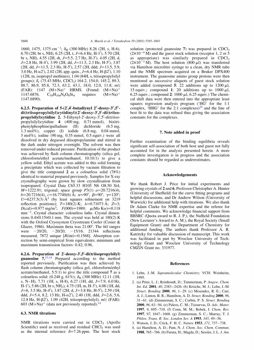
The partner lipophilic guanosine molecule 7 was readilyprepared following a literature procedure by silylation of thehydroxyls with triisopropylsilyl chloride (Scheme 3).13,14
4. Self-assembly
The association of nucleobases, in particular guanosine andcytidine is well documented and has been studied usingvarious physico-chemical techniques including 1H, 13CNMR14,20,21 and infra-red22 spectroscopies. We wanted tocarry out 1H NMR titrations to determine the associationconstants of the mono- and ditopic receptors to verify thatthe association constants remained in the usable($103 M21) range.
The first association constant we wished to determine wasthat between monotopic receptor 6 and protected guanosine7. The signal chosen to follow was the most mobile, theguanosine amido proton at d¼12.0 ppm which was found tomove downfield to 13.1 ppm in the presence of 1 equiv. ofthe receptor. Under our conditions it was found to remainsufficiently visible without becoming over-broad although itshould be noted that a more satisfactory method may be toobserve the guanosine H-8 proton.22 Prior to determiningthe association constant, a Job’s plot analysis23 wasperformed to assay the stoichiometry of the interactionand from the maximum of the parabola the expected 1:1stoichiometry of 7:6 (Fig. 6) was obtained. The concen-trations chosen for the subsequent analysis were 2£1024 Mhost stock solution (7) and 2£1023 M guest stock solution(5) in CDCl3, allowing the association constant to bedetermined without saturating the receptor at too low a molefraction of guest.24
Figure 3. Molecular structure of receptor 2 (X-ray).
Figure 4. Cytidine dimer contacts in receptor 2.
Figure 5. Packing diagram showing helical channel.
Scheme 3. Reagents and conditions: (i) triisopropylsilyl chloride,imidazole, DMAP, DMF (61%).
Figure 6. Complex 7·6.
A. Marsh et al. / Tetrahedron 59 (2003) 5595–5601 5597
A titration was then carried out using these stock solutionsover the range 0–15 equiv. of guest which gave a final dvalue of 13.43 ppm for the guanosine amido NH. The 22data points were then analysed using a non-linear leastsquares fit implemented through Professor Chris Hunter’s‘HG’ program.25 After refinement an R factor of 2.2% wasobtained indicating a good fit for the data points and theassociation constant, Ka¼3370 M21 was derived. This issomewhat lower than the value compared to previousmeasurements made for the G·C pair e.g. Ka¼104–105 M21
(by infrared),26 Ka¼1.6£104 M21,21 and Ka¼1.7£104 M21
for a related pair.6a,27 Interestingly the association constantfor a similar G·C pair substituted by an anthracene moiety isnoted to be significantly higher than either our own, or manyof these other values (Ka¼38500^1300 M21).9 However, itmust be noted that no special precautions were taken to drythe solvents used for the titrations. For the ditopic receptor 1a Job’s plot revealed the expected 2:1 stoichiometry forcompounds 7:1. A titration of receptor 1 as guest from 0 to6.25 equiv. was then carried out in CDCl3, once againfollowing the amido NH of the guanosine from 12.49 to13.33 ppm. The analysis this time was carried out using the‘HHG’ program designed for a 2:1 host:guest stoichi-ometry.24 An excellent fit for the curve to data points wasobtained with an R factor of 1.8%. The microscopicassociation constant Ka was found to be 2670 M22,interestingly slightly lower than, but of the same order ofmagnitude as that obtained for the 7·6 pair. Finally, a similartitration was performed for the longer bisalkynyl ditopicreceptor 2 complexed with 7 (Fig. 7) giving an R factor of2.5% and Ka¼2200 M22.
The somewhat lower values found for the association ofguanosine to each of the ditopic receptors 1 and 2 is perhapsnot unexpected due to the presence of a small amount ofcompeting solvent molecules in the binding experiment. Inaddition we speculate that it may be that since X-raycrystallography has shown that the ditopic receptor 2 existsas part of a hydrogen bonded polymer, if this persisted insolution, the guanosine molecules have to break up thisnetwork before binding to the Watson–Crick site can takeplace. A second possibility, that the lower associationconstant may be indicative of an effect exerted by thebinding of the first guest molecule to the receptor canprobably be discounted due to the similarity of all threebinding constants. Although it has been demonstrated thatsignificant effects on the association constant of diamino-pyridine binding to a flavin receptor can be brought about byaltering the redox state of the host molecule,28 it seems thatin this case any such effect is likely to be quite small.
5. Conclusions
Novel ditopic receptors for guanosine have been preparedand their association constants with a lipophilic nucleosidedetermined. The binding of the first guanosine guest to theditopic receptor 1 or 2 does not appear to dramaticallyperturb the binding of the second equivalent compared tothe monotopic receptor 4. The association constants remainin a usable range and indicate the suitability of thesecomponents for use as building blocks in making higherorder supramolecular and macromolecular structures andthis is an area of active investigation.
6. Experimental
6.1. General
Chemicals were purchased from either Aldrich ChemicalCo. or Lancaster Synthesis and were used as received unlessotherwise stated. 1H and 13C NMR spectra were recordedusing Bruker ACF250, DPX300, DPX400, AV400 instru-ments with chemical shifts measured in ppm relative totetramethylsilane. Infrared spectra were recorded on aBruker Vector 22 spectrometer fitted with an attenuatedtotal reflection (ATR) cell. Routine mass spectra wereobtained on a Micromass Autospec instrument in theWarwick Mass Spectrometry Laboratory. Micoanalyticaldata were obtained on a CE 440 Elemental Analyseroperated by Warwick Analytical Services. Melting pointswere carried out on a Gallenkamp melting point apparatusand are uncorrected. Thin layer chromatography (TLC) wascarried out on pre-coated plate (silica gel 60 F 254, MerckArt. No. 5715) and the products visualised using UV light(254 nm), iodine or potassium permanganate solution asappropriate. Flash column chromatography was carried outusing silica gel 60H (230–400 mesh) under low pressure.NMR titration data was analysed by a suite of least squarescurve-fitting programs kindly supplied by Professor C. A.Hunter.25 The programs were implemented on an AppleMacintosh 7200/90 and the graphical data was captured as ascreen shot. X-Ray crystallographic structural characteris-ations for all crystals, data were collected with a SiemensSMART three-circle system with CCD area detector, withcrystals held at 180 K or 200 K with the Oxford CryosystemCryostream Cooler. Refinements used SHELXTL.29
Systematic absences indicated the appropriate spacegroup. The structure was solved by direct methods withadditional light atoms found by Fourier methods. Hydrogenatoms were added at calculated positions and refined using ariding model. Anisotropic displacement parameters wereused for all non-H atoms; H-atoms were given isotropicdisplacement parameters equal to 1.2 times the equivalentisotropic displacement parameter of the atom to which theH-atom is attached. Crystallographic data (excludingstructure factors) for the structures reported in this paperhave been deposited with the Cambridge CrystallographicData and have been assigned the reference codes CCDC188941 (compound 6) and 188942 (compound 2). Copies ofthe data can be obtained free of charge on application to:The Director, CCDC, 12 Union Road, Cambridge CH2 1EZUK (fax: þ44-1223-336-033; email: [email protected]).Figure 7. Complex 2·72.
A. Marsh et al. / Tetrahedron 59 (2003) 5595–56015598
6.2. Preparation of receptors and guest
6.2.1. Preparation of 5-iodo-20-deoxy-30,50-di(triiso-propylsilyl)cytidine 4. To a stirred suspension of 5-iodo-20-deoxycytidine 3 (1.0 g, 2.83 mmol) in DMF (15 ml)under an atmosphere of nitrogen and at room temperaturewas added imidazole (84.8 mg, 12.4 mmol, 4.4 equiv.),DMAP (34.6 mg, 0.28 mmol, 0.1 equiv.) and triisopropyl-silylchloride (1.2 g, 1.34 ml, 6.22 mmol, 2.2 equiv.). Themixture was stirred for 48 h. The reaction mixture waspoured into sat NH4Cl solution (50 ml) and the resultingwhite suspension was extracted with chloroform (3£50 ml).The combined organic phases were washed with water(3£40 ml), brine (3£40 ml) and dried over MgSO4.Filtration of the resulting solution followed by removal ofthe solvent under reduced pressure gave a creamy whitesolid. Purification was then achieved by flash columnchromatography (silica gel, chloroform/ethyl acetate/methanol, 5:5:1) to give the title compound 4 as a colourlesssolid: (1.168 g, 62%); mp 154–1568 C; nmax (KBr) 3466,2941, 1651, 1489, 1463, 1111, 881, 685 cm21; dH
(300 MHz, CDCl3) 8.67 (1H, br s, NH), 8.07 (1H, s, H-6),6.28 (1H, dd, J¼8, 5.5 Hz, H-10), 5.63 (1H, br s, NH), 4.56(1H, m, H-30), 4.06 (1H, dd, J¼2.5, 4.5 Hz, H-40), 3.96 (1H,dd, J¼11.3, 2.5 Hz, H-50), 3.84 (1H, dd, J¼11.3, 2.8 Hz,H-50), 2.55 (1H, ddd, J¼13.3, 5.6, 2.0 Hz, H-a20), 1.96 (1H,ddd, J¼13.6, 8.0, 6.0 Hz, H-b20), 1.10 (6H, m isopropylmethines), 1.05 (36H, s, triisopropylsilyl groups); dc
(75.43 MHz, CDCl3) 164.0, 155.1, 146.9, 89.2, 87.2, 73.3,63.9, 43.5, 18.4, 12.4, 12.2; m/z (FAB): 666 (Mþ1). (Found:C, 48.41; H, 7.76; N, 6.20. C27H52IN3O4Si2 requires C,48.71; H, 7.87; N, 6.31%).
6.2.2. Preparation of 5-(trimethylsilylethynyl)-20-deoxy-30,50-di(triisopropylsilyl)cytidine 5. A mixture of 5-iodo-20-deoxy-30,50-di(triisopropylsilyl)cytidine 4 (500 mg,0.75 mmol), trimethylsilylacetylene (140 mg, 0.21 ml,1.42 mmol, 2 equiv.), bis(triphenylphosphine) palladium(II) dichloride (52 mg, 0.07 mmol, 0.1 equiv.) and copper(I) iodide (7 mg, 3.75 mmol, 0.05 equiv.) in degassed drydiisopropylamine (50 ml) was stirred in the dark undernitrogen at room temperature overnight. The solvent wasthen removed under reduced pressure and the resulting solidwas dissolved in diethyl ether (60 ml) and the insolubleportion removed by filtration. The solvent was thenremoved under reduced pressure to yield the product as acreamy yellow solid. Purification was then achieved by flashcolumn chromatography (silica gel, dichloromethane/ethylacetate/petroleum ether (40–60)/methanol, 1:1:3:1) to givethe title compound 5 as a colourless solid: (428 mg, 90%);mp 175–1788C; nmax (KBr) 3464, 2940, 1673, 1505, 1464,1440 cm21; dH (300 MHz, CDCl3) 8.58 (1H, s, NH), 8.04(1H, s, H-6), 6.28 (1H, t, J¼6.5 Hz, H-10), 5.83 (1H, s, NH),4.54 (1H, dd, J¼3.0, 2.5 Hz, H-30), 4.03 (1H, d, J¼1.9 Hz,H-40), 3.97 (1H, dd, J¼11.5, 2.1 Hz, H-50), 3.84 (1H, dd,J¼11.5, 2.1 Hz, H-50); 2.50 (1H, dd, J¼13.4, 4.4 Hz, H-a20), 1.96 (1H, m, H-b20), 1.15 (6H, m, triisopropylsilylmethines), 1.00 (36H, m, triisopropylsilyl), 0.13 (9H, s,trimethylsilyl); dc (75.43 MHz, CDCl3) 164.6, 154.3, 144.1,101.1, 96.0, 90.9, 88.8, 86.9, 72.8, 63.4, 43.2, 38.1, 18.1,12.1, 11.7; m/z (FAB): 637 (Mþ1)þ. (Found: C, 60.16; H,9.49; N, 6.54. C32H61N3O4Si3 requires C, 60.42; H, 9.67; N,6.61%).
6.2.3. Preparation of 5-ethynyl-20-deoxy-30,50-di (triiso-propylsilyl)cytidine 6. Sodium carbonate (90 mg,0.84 mmol, 2 equiv.) was dissolved in water (2.5 ml) andthe resulting solution was then added to a stirred solution of5-(trimethylsilylethynyl)-20-deoxy-30,50di(triisopropylsilyl)cytidine 5 (328 mg, 0.51 mmol) in methanol (5 ml). Thisreaction mixture was then stirred for a further 2 h. Thesolution was then poured into water (10 ml) and the productextracted with chloroform (3£50 ml). The purification ofthe product was then achieved by flash column chromato-graphy (silica gel, chloroform/dichloromethane/methanol,5:5:1) to give the title compound 6 as a colourless solid:(279 mg, 97%); mp 144–1468C; nmax (KBr) 2936, 2866,2300, 1642, 1500, 1458, 1288, 1242, 1109, 1071, 1030,875 cm21; dH (300 MHz, CDCl3) 8.97 (1H, br s, N–H),8.09 (1H, s, H-6), 6.27 (1H, dd, J¼7.4, 6.0 Hz, H-10), 5.83(1H, br s, N–H), 4.53 (1H, dt, J¼5.5, 2.7 Hz, H-30), 4.01(1H, dd, J¼2.6, 1.7 Hz, H-40), 3.96 (1H, dd, J¼11.3, 2.4 Hz,H-50), 3.84 (1H, dd, J¼10.7, 2.3 Hz, H-50), 3.25 (1H, s,ethynyl), 2.50 (1H, ddd, J¼13.2, 5.8, 2.7 Hz, H-a20), 1.97(1H, m, H-b2), 1.02 (42H, m, triisopropylsilyl); dc
(75.43 MHz, CDCl3) 164.3, 155.8, 144.6, 88.5, 86.6, 75.0,72.2, 63.0, 42.9, 17.7, 11.8, 11.5; m/z (FAB): 564 (MþH)þ.(Found: C, 61.49; H, 9.43; N, 7.20. C29H53N3O4Si2 requiresC, 61.77; H, 9.47; N, 7.45%). Crystals suitable for X-raydiffraction were grown from isopropanol by slow evapor-ation. Crystal data C32 H61 N3 O5 Si2, M¼624.02;triclinic; space group P1; a¼8.5125(8), b¼13.2827(13),c¼18.1934(17) A; a¼72.817(2)8 , b¼86.490(2)8 ,g¼76.500(2)8 ; U¼1910.9(3) A3; T¼170(2) K;l¼0.71073 A; Z¼2; D(cal)¼1.085 mg/m3; F(000)¼684;m(Mo Ka)¼0.131 mm21. R1[for 3130 reflections withI.2s(I)]¼0.0789, wR2¼0.2182.
6.2.4. Preparation of 5-(ethynyl-200-deoxy-300,500-di(triiso-propylsilyl)-50-cytidinyl)-(20-deoxy-30,50-di(triisopropyl-silyl)cytidine), 1 and 5-(20,40-butadiynyl 200-deoxy-300,500-di(triisopropylsilyl)cytidinyl) (20-deoxy-30,50-di(triiso-propylsilyl)cytidine 2. 5-Ethynyl-20-deoxy-30,50-di(triiso-propylsilyl) cytidine 6 (164 mg, 0.29 mmol, 1 equiv.),5-iodo-20deoxy-30,50-di(triisopropylsilyl) cytidine 4(194 mg, 0.29 mmol, 1 equiv.), bis(triphenylphosphine)palladium (II) dichloride (20 mg, 0.028 mmol, 0.1 equiv.),and copper (I) iodide (2.8 mg, 0.014 mmol, 0.05 equiv.)were all dissolved in dry degassed diisopropylamine (60 ml)and stirred in the dark under nitrogen for 24 h. The solventwas then removed under reduced pressure and a solidobtained. Purification of the product was carried out by flashcolumn chromatography (silica gel, chloroform/ethylacetate/methanol, 10:10:1) to give the title compound 1 asa colourless solid: (172 mg, 54%); mp 169–1718C; nmax
(KBr) 3458, 2941, 1665, 1490, 1383 cm21; dH (300 MHz)8.19 (2H, s, H-6), 6.40 (ca. 2H, br s, NH), 6.31 (2H, t,J¼6.5 Hz, H-10), 5.52 (ca. 2H, br s, NH), 4.55 (2H, m, H-30),4.04 (2H, dd, J¼3.0, 2.6 Hz, H-40), 3.97 (2H, dd, J¼10.9,2.5 Hz, H-50), 3.86 (1H, dd, J¼11.3, 3.0 Hz, H-50), 2.56 (1H,ddd, J¼12.6, 5.7, 2.7 Hz, H-a20), 1.99 (2H, app. quin.,J¼6.6 Hz, H-b20), 1.04 (s, 84H, triisopropylsilyl groups); dc
(75.43 MHz, CDCl3) 164.2, 154.1, 145.2, 88.7, 86.9, 85.8,72.3, 63.2, 43.1, 18.0, 12.0, 11.8; m/z (FAB): 1101 (Mþ1)þ
HRMS. (Found (Mþ1)þ 1101.7068. C56H104N6O8Si4requires: (Mþ1)þ 1101.7071) and title compound 2(116 mg, 36%); mp 178–1828C; nmax (KBr) 3470, 2949,
A. Marsh et al. / Tetrahedron 59 (2003) 5595–5601 5599
1660, 1475, 1375 cm21; dH (300 MHz) 8.26 (2H, s, H-6),6.70 (2H, br s, NH), 6.25 (2H, t, J¼6.4 Hz, H-10), 5.70 (2H,br s, NH), 4.55 (2H, dt, J¼5.5, 2.7 Hz, H-30), 4.05 (2H, d,J¼2.8 Hz, H-40), 3.99 (2H, dd, J¼11.5, 2.3 Hz, H-50), 3.87(2H, dd, J¼11.5, 2.3 Hz, H-50), 2.57 (2H, ddd, J¼13.5, 5.9,3.0 Hz, H-a20), 2.02 (2H, app. quin., J¼6.4 Hz, H-b20), 1.10(12H, m, isopropyl methines), 1.04 (84H, s, triisopropylsilylgroups); dc (75.43 MHz, CDCl3) 164.2, 154.0, 145.2, 89.3,88.7, 86.9, 85.8, 72.3, 63.2, 43.1, 18.0, 12.0, 11.8; m/z(FAB): 1147 (MþNa)þ HRMS. (Found: (MþNa)þ
1147.6876. C58H104N6O8Si4 requires (MþNa)þ
1147.6890).
6.2.5. Preparation of 5-(20,40-butadiynyl 200-deoxy-300,500-di(triisopropylsilyl)cytidinyl)(20-deoxy-30,50-di(triiso-propylsilyl)cytidine 2. 5-Ethynyl-20-deoxy-30,50-di(triiso-propylsilyl)cytidine 4 (400 mg, 0.71 mmol), bis(tri-phenylphosphine)palladium (II) dichloride (6.5 mg,1.3 mol%), copper (I) iodide (6.8 mg, 0.04 mmol,5 mol%), iodine (90 mg, 0.35 mmol, 0.5 equiv.) were alldissolved in dry degassed diisopropylamine and stirred inthe dark under nitrogen overnight. The solvent was thenremoved under reduced pressure. Purification of the productwas achieved by flash column chromatography (silica gel,chloroform/ethyl acetate/methanol, 10:10:1) to give ayellow solid. Ethyl acetate was added to this solid forminga precipitate which was collected by vacuum filtration togive the title compound 2 as a colourless solid (78%)identical to material prepared previously. Samples for X-raycrystallography were grown by slow crystallisation fromisopropanol. Crystal Data C65.33 H105 N6 O8.50 Si4,M¼1222.91; trigonal; space group P3(1) a¼20.7216(4),b¼20.7216(4), c¼16.7740(6) A; a¼908, b¼908, g¼1208;U¼6237.5(3) A3 (by least squares refinement on 3219reflection positions); T¼180(2) K; l¼0.71073 A; Z¼3;D(cal)¼0.977 mg/m3; F(000)¼1989; m(Mo Ka)¼0.118mm21. Crystal character: colourless laths. Crystal dimen-sions 0.4£0.15£0.1 mm. The crystal was held at 180(2) Kwith the Oxford Cryosystem Cryostream Cooler (Cosier &Glazer, 1986). Maximum theta was 21.008. The hkl rangeswere 220/20, 220/20, 215/16. 21344 reflectionsmeasured, 7872 unique [R(int)¼0.1568]. Absorption cor-rection by semi-empirical from equivalents; minimum andmaximum transmission factors: 0.42; 0.96.
6.2.6. Preparation of 20-deoxy-30,50-di(triisopropylsilyl)guanosine 7.13,14 Prepared according to the methodreported previously. Purification was then achieved byflash column chromatography (silica gel, chloroform/ethylacetate/methanol, 5:5:1) to give the title compound 7 as acolourless solid: (0.248 g, 61%); dH (300 MHz) 12.11 (1H,s, N–H), 7.71 (1H, s, H-8), 6.27 (1H, dd, J¼7.9, 6.0 Hz,H-10), 5.86 (2H, br s, NH2), 4.75 (1H, m, H-30), 4.08 (1H, dd,J¼6, 3.5 Hz, H-40), 3.87 (2H, d, J¼3.8 Hz, H-50), 2.59 (1H,ddd, J¼5.4, 8.2, 13 Hz, H-a20), 2.40 (1H, ddd, J¼2.6, 5.6,12.8 Hz, H-b20), 1.09 (42H, triisopropylsilyl); m/z (FAB):603 (MþNa)þ (data not previously reported).14
6.3. NMR titrations
NMR titrations were carried out in CDCl3 (ApolloScientific) used as received and residual CHCl3 was usedas the internal reference d¼7.26 ppm. The host stock
solution (protected guanosine 7) was prepared in CDCl3(2£1024 M) and the guest stock solution (receptor 1, 2 or 5as appropriate) was similarly prepared in CDCl3
(2£1023 M). The host solution (800 ml) was transferredvia Hamilton microlitre syringe to a clean, dry NMR tubeand the NMR spectrum acquired on a Bruker DPX400instrument. The guanosine amino group protons were thenmonitored as successive aliquots of guest stock solutionwere added (compound 5: 22 additions up to 1200 ml,15 equiv.; compound 1: 20 additions up to 1000 ml,6.25 equiv.; compound 2: 1000 ml, 6.25 equiv.) The chemi-cal shift data were then entered into the appropriate leastsquares regression analysis program (‘HG’ for the 1:1complex, ‘HHG’ for the 2:1 complexes)25 and the line ofbest fit to the data was refined thus giving the associationconstants for the complexes.
7. Note added in proof
Further examination of the binding equilibria revealssignificant self-association of both host and guest not fullyaccounted for in the analysis presented herein. A morecomplete investigation is in progress and the associationconstants should be regarded as underestimates.
Acknowledgements
We thank Robert J. Price for initial experiments andgrowing crystals of 2 and 6, Professor Christopher A. Hunter(University of Sheffield) for the curve fitting programs andhelpful discussions, and Dr Andrew Wilson (University ofWarwick) for additional help with titrations. We also thankDr Adam Clarke for NMR expertise and the referee forhelpful comments. We acknowledge financial support fromBBSRC (Quota award to R. J. P.), the Nuffield Foundation(New Lecturer’s Award to A. M.), the Royal Society (SmallEquipment Grant) and the Department of Chemistry foradditional funding. The authors thank Professor A. R.Katritzky for valuable discussion of manuscript. This workwas facilitated in part by Wroclaw University of Tech-nology Grant and Wroclaw University of TechnologyCMZiN Grant no. 331977.
References
1. Lehn, J.-M. Supramolecular Chemistry; VCH: Weinheim,
1995.
2. (a) Prins, L. J.; Reinhoudt, D.; Timmerman, P. Angew. Chem.
Int. Ed. 2001, 40, 2383–2426. (b) Krische, M. J.; Lehn, J.-M.
Struct. Bonding 2000, 96, 1–29. (c) Mesendez, R. E.; Carr,
A. J.; Linton, B. R.; Hamilton, A. D. Struct. Bonding 2000, 96,
31–61. (d) Zimmerman, S. C.; Corbin, P. S. Struct. Bonding
2000, 96, 63–94. (e) Paleos, C. M.; Tsiourvas, D. Adv. Mater.
1997, 9, 695–710. (f) Conn, M. M.; Rebek, J. Chem. Rev.
1997, 97, 1647–1668. (g) Zimmerman, S. C.; Murray, T. J.
Philos. Trans. R. Soc. London Ser. A 1993, 345, 49–56.
3. Watson, J. D.; Crick, F. H. C. Nature 1953, 171, 737–738.
4. (a) Hamilton, A. D.; Pant, N. J. Chem. Soc. Chem. Commun.
1988, 765–766. (b) Furuta, H.; Magda, D.; Sessler, J. L. J. Am.
A. Marsh et al. / Tetrahedron 59 (2003) 5595–56015600
Chem. Soc. 1991, 113, 978–985. (c) Furuta, H.; Furuta, K.;
Sessler, J. L. J. Am. Chem. Soc. 1991, 113, 4706–4707.
(d) Schall, O. F.; Gokel, G. W. J. Am. Chem. Soc. 1994, 116,
6089–6100. (e) Davis, J. T.; Tirumala, J. R.; Jenssen, E.;
Radler, E.; Fabris, D. J. Org. Chem. 1995, 60, 4167–4176. (f)
Park, T. K.; Schroeder, J.; Rebek, J. Tetrahedron 1991, 47,
2507–2518.
5. (a) Kitano, H.; Ringsdorf, H. Bull. Chem. Soc. Jpn 1985, 58,
2826–2828. (b) Sasaki, D. Y.; Kurihara, K.; Kunitake, T.
J. Am. Chem. Soc. 1992, 114, 10994–10995.
6. Guanosine self-association: (a) Kyogoku, Y.; Lord, R. C.;
Rich, A. Biochim. Biophys. Acta 1969, 179, 10–17. (b) Sessler,
J. L.; Magda, D.; Furuta, H. J. Org. Chem. 1992, 57, 818–826,
and see Ref. 14.
7. Sessler, J. L.; Wang, R. Z. Angew. Chem. Int. Ed. 1998, 37,
1726–1729.
8. Sessler, J. L.; Wang, R. Z. J. Org. Chem. 1998, 63,
4079–4091.
9. Sessler, J. L.; Sathiosatham, M.; Brown, C. T.; Rhodes, T. A.;
Wiederrecht, G. J. Am. Chem. Soc. 2001, 123, 3655–3660.
10. Araki, K.; Takasawa, R.; Yoshikawa, I. Chem. Commun. 2001,
1826–1827.
11. (a) Inouye, M.; Konishi, T.; Isagawa, K. J. Am. Chem. Soc.
1993, 115, 8091–8095. (b) Inoye, M.; Hyodo, Y.; Nakazumi,
H. J. Org. Chem. 1999, 64, 2704–2710.
12. Sonogashira, K. Comprehensive Organic Synthesis; Trost,
B. M., Fleming, I., Eds.; Pergamon: New York, 1991; Vol. 3,
pp 521–549.
13. Ogilvie, K. K.; Thompson, E. A.; Quilliam, M. A.; Westmore,
J. B. Tetrahedron Lett. 1974, 33, 2865–2868.
14. Williams, L. D.; Chawla, B.; Shaw, B. R. Biopolymers 1987,
26, 591–603.
15. (a) Robins, M. J.; Barr, P. J. Tetrahedron Lett. 1981, 22,
421–424. (b) Robins, M. J.; Barr, P. J. J.Org. Chem. 1983, 48,
1854–1862. (c) De Clercq, E.; Descamps, J.; Balzarini, J.;
Giziewicz, J.; Barr, P. J.; Robins, M. J. J. Med. Chem. 1983,
26, 661–666.
16. (a) Hobbs, F. W. J. Org. Chem. 1989, 54, 3420–3422.
(b) Robins, M. J.; Vinayak, R. S.; Wood, S. G. Tetrahedron
Lett. 1990, 26, 3731–3734.
17. Mathews, F. S.; Rich, A. Nature 1964, 201, 179–180.
18. (a) Behr, O. M.; Eglinton, G.; Galbraith, A. R.; Raphael, R. A.
J. Chem. Soc. 1960, 3614–3625. (b) Hay, A. S. J. Org. Chem.
1962, 27, 3320–3321. for a review of acetylenic couplings
see: (c) Siemsen, P.; Livingston, R. C.; Diederich, F. Angew.
Chem. Int. Ed. 2000, 39, 2633–2657.
19. Liu, Q.; Burton, D. J. Tetrahedron Lett. 1997, 38, 4371–4374.
20. Williams, N. G.; Williams, L. D.; Shaw, B. R. J. Am. Chem.
Soc. 1989, 111, 7205–7209.
21. Sartorius, J.; Schneider, H.-J. Chem. Eur. J. 1996, 2, 1446.
22. Kyogoku, Y.; Lord, R. C.; Rich, A. Science 1966, 154,
518–520.
23. Connors, K. A. Binding Constants; Wiley: Chichester, 1987;
pp 24–28.
24. (a) Wilcox, C. S. In Frontiers in Supramolecular Chemistry
and Photochemistry; Schneider, H. J., Durr, H., Eds.; VCH:
Weinheim, 1991; pp 123 – 143. (b) Schneider, H. J.;
Yatsimirsky, A. Principles and Methods in Supramolecular
Chemistry; Wiley: Chichester, 2000.
25. Bisson, A. P.; Hunter, C. A.; Morales, J. C.; Young, K. Chem.
Eur. J. 1998, 4, 845–851.
26. Jorgensen, W. L.; Pranata, J. J. Am. Chem. Soc. 1990, 112,
2008–2010.
27. Kelly, T. R.; Zhao, C.; Bridges, G. J. J. Am. Chem. Soc. 1989,
111, 3744–3745.
28. Niemz, A.; Rotello, V. Acc. Chem. Res. 1999, 32, 44–52.
29. Sheldrick, G. M. SHELXTL Ver. 5.1: Bruker Analytical X-ray
Systems; Madison: Wisconsin, USA, 1997.
A. Marsh et al. / Tetrahedron 59 (2003) 5595–5601 5601