SYNTHESIS AND CHARACTERISATION OF 1,3- …eprints.utar.edu.my/709/1/CE-2012-1000867.pdf · ii...
121
SYNTHESIS AND CHARACTERISATION OF 1,3- DIHYDROXYXANTHONE DERIVATIVES AND THEIR ANTIOXIDANT ACTIVITIES LAI CHOOI KUAN BACHELOR OF SCIENCE (HONS.) CHEMISTRY FACULTY OF SCIENCE UNIVERSITI TUNKU ABDUL RAHMAN OCTOBER 2012 LAI CHOOI KUAN B. Sc. (Hons.) Chemistry 2012
-
Upload
trinhkhanh -
Category
Documents
-
view
227 -
download
0
Transcript of SYNTHESIS AND CHARACTERISATION OF 1,3- …eprints.utar.edu.my/709/1/CE-2012-1000867.pdf · ii...

SYNTHESIS AND CHARACTERISATION OF 1,3-
DIHYDROXYXANTHONE DERIVATIVES
AND THEIR ANTIOXIDANT ACTIVITIES
LAI CHOOI KUAN
BACHELOR OF SCIENCE (HONS.) CHEMISTRY
FACULTY OF SCIENCE
UNIVERSITI TUNKU ABDUL RAHMAN
OCTOBER 2012
LA
I CH
OO
I KU
AN
B. S
c. (Hon
s.) Ch
emistr
y
2012

SYNTHESIS AND CHARACTERISATION OF 1,3-
DIHYDROXYXANTHONE DERIVATIVES
AND THEIR ANTIOXIDANT ACTIVITIES
By
LAI CHOOI KUAN
A Project Report Submitted to the Department of Chemical Science,
Faculty of Science,
Universiti Tunku Abdul Rahman
in Partial Fulfillment of the Requirement for the
Degree of Bachelor of Science (Hons.) Chemistry
October 2012

ii
ABSTRACT
SYNTHESIS AND CHARACTERISATION OF 1,3-
DIHYDROXYXANTHONE DERIVATIVES
AND THEIR ANTIOXIDANT ACTIVITIES
Lai Chooi Kuan
In this study, a xanthonic block 1,3-dihydroxyxanthone (39) and three new
prenylated xanthone derivatives, namely 1-hydroxy-2,4-bis(3-methyl-but-2-enyl)-
3-(3-methyl-but-2-enyloxy)-xanthen-9-one (41), 1-hydroxy-2,4,4-tris(3-methyl-
but-2-enyl)-4H-xanthen-3,9-dione (42), and 1,3-dihydroxy-2,4-bis(3-methyl-but-
2-enyl)-xanthen-9-one (43) were successfully synthesized. These pure compounds
were isolated by using column chromatography and their structures were
elucidated through 1H-NMR,
13C-NMR and UV-Vis analyses, and were further
confirmed on the basis of 2D-NMR including HMQC and HMBC analyses.
Among these compounds, compound (42) was found to bear a novel skeleton of
xanthen-3,9-dione.
The xanthonic block was synthesized using salicylic acid and phloroglucinol in
the presence of Eaton’s reagent and was subsequently used as starting material for

iii
prenylation with two different bases, potassium carbonate and potassium
hydroxide in aqueous medium.
The DPPH radical scavenging assay was carried out to study the antioxidant
activities of the isolated compounds, and the results indicated compound 43 to
exhibit significant antioxidant activity, in which the activity was found to be
weaker as compared to the reference compounds, ascorbic acid and kaempferol.
On the other hand, the xanthonic block (39) and other prenylated derivatives, 41
and 42 showed no significant antioxidant activities.

iv
ABSTRAK
Dalam projek ini, satu blok xanthone iaitu 1,3-dihidroksixanthone (39) dan tiga
prenilasi xanthone, iaitu 1-hidroksi-2,4-bis(3-metil-but-2-enil)-3-(metil-but-2-
eniloksi)-xanthen-9-one (41), 1-hidroksi-2,4,4-tris(3-metil-but-2-enil)-4H-
xanthen-3,9-dione (42), dan 1,3-dihidroksi-2,4-bis(3-metil-but-2-enil)- xanthen-9-
one (43) telah berjaya dihasilkan dan dikenalpastikan. Hasil sintesis ini telah
dipisah melalui kromatografi kolom dan identiti sebatian-sebatian tersebut telah
dikenalpasti melalui kaedah spektroskopi seperti UV-Vis, IR, 1D- dan 2D-NMR.
Blok xanthone telah disintesis menggunakan asid salisilik dan phloroglucinol.
Hasil sintesis tersebut digunakan sebagai bahan permulaan untuk prenilasi dalam
larutan akueus, dengan menggunakan dua jenis alkali yang berbeza, iaitu
potassium hidroksida dan potassium karbonat.
Semua xanthone yang dihasilkan telah diuji aktiviti antioksidan masing-masing
dengan menggunakan kaedah DPPH dan didapati hanya 1,3-dihidroksi-2,4-bis(3-
metil-but-2-enil)-xanthen-9-one (43) menunjukkan aktiviti yang nyata. Manakala
blok xanthone (39) dan prenilasi xanthone yang lain, 41 dan 43 tidak memberikan
aktiviti antioksidan yang efektif.

v
ACKNOWLEDGEMENT
First of all, I gratefully express deep appreciation to my supervisor, Dr. Lim Chan
Kiang, who has shown his kind and invaluable interaction, support, and advice for
my academic success.
Special thanks to my seniors Koo Hooi Tee and Lim Cheng Hoe, for their
unconditional guidance and help.
I would like to thank my group mates and lab mates, for their assistance and
companionship in the laboratory. It has been a pleasant experience to work with
all of them.
Finally, I most deeply acknowledge my families and friends for their unceasing
encouragement, support, and patience throughout the course of this project.
vi
DECLARATION
I hereby declare that the project report is based on my original work except for
quotations and citations which have been duly acknowledged. I also declare that it
has not been previously or concurrently submitted for any other degree at UTAR
or other institutions.
(LAI CHOOI KUAN)

vii
APPROVAL SHEET
This project report entitled “SYNTHESIS AND CHARACTERISATION OF
1,3-DIHYDROXYXANTHONE DERIVATIVES AND THEIR
ANTIOXIDANT ACTIVITIES” was prepared by LAI CHOOI KUAN and
submitted in partial fulfillment of the requirements for the degree of Bachelor of
Science (Hons.) in Chemistry at Universiti Tunku Abdul Rahman.
Approved by:
Date:
(Dr. Lim Chan Kiang)
Supervisor
Department of Chemical Science
Faculty of Science
Universiti Tunku Abdul Rahman

viii
FACULTY OF SCIENCE
UNIVERSITI TUNKU ABDUL RAHMAN
Date:
PERMISSION SHEET
It is hereby certified that LAI CHOOI KUAN (ID No: 10ADB00867) has
completed this report entitled “SYNTHESIS AND CHARACTERISATION
OF 1,3-DIHYDROXYXANTHONE DERIVATIVES AND THEIR
ANTIOXIDANT ACTIVITIES” supervised by Dr. Lim Chan Kiang from the
Department of Chemical Science, Faculty of Science.
I hereby give permission to my supervisor to write and prepare manuscript of
these research findings for publishing in any form, if I do not prepare it within six
(6) month time from this date, provided that my name is included as one of the
authors for this article. The arrangement of the name depends on my supervisor.
Yours truly,
(LAI CHOOI KUAN)

ix
TABLE OF CONTENTS
Page
ABSTRACT ii
ABSTRAK iv
ACKNOWLEDGEMENT v
DECLARATION vi
APPROVAL SHEET vii
PERMISSION SHEET viii
LIST OF TABLES xii
LIST OF FIGURES xiii
LIST OF ABBREVATIONS xvii
CHAPTER
1 INTRODUCTION 1
1.1. Background 1
1.2. Chemistry and Classification of Xanthones
1.2.1. Chemistry
1.2.2. Classification of Xanthones
2
2
3
1.3. Occurrence and Natural Distribution of Xanthones 3
1.4. Synthetic Xanthones 4
1.5. Prenylated Xanthones 5
1.6. Bioactivities of Xanthones
1.7. Antioxidant Activities of Xanthones
1.8. Objectives
6
6
8
2 LITERATURE REVIEW 9
2.1. Synthesis Approaches of Xanthones 9

x
2.2. Biosynthesis 10
2.2.1. Acetate Polymalonic Route 10
2.2.2. Mixed Shikimate Acetate Pathway 11
2.3. Chemical Synthesis 13
2.3.1. The Grover, Shah and Shah (GSS) Reaction 13
2.3.2. Modifications to the Grover, Shah and Shah Reaction
2.3.3. Asahina-Tanase Method
2.3.4. New Approaches to the Synthesis of Xanthonic
Tricyclic System
14
15
16
2.4. Synthesis of Prenylated Xanthones 18
2.4.1. O- and C-Prenylated Xanthones 18
2.5 DPPH Scavenging Activity 20
3 MATERIALS AND METHODS 21
3.1. Chemicals 21
3.2. Methodology 25
3.2.1. Synthesis of the Xanthonic Block,
1,3-Dihydroxyxanthone
25
3.2.2. Prenylation of 1,3-Dihydroxyxanthone in Potassium
Carbonate Solution
26
3.2.3. Prenylation of 1,3-Dihydroxyxanthone in Potassium
Hydroxide Solution
27
3.2.4. Column Chromatography 28
3.2.5. Thin Layer Chromatography (TLC) 29
3.3. Instruments 30
3.3.1. Nuclear Magnetic Resonance (NMR) 30
3.3.2. Ultraviolet-Visible (UV-Vis) Spectroscopy 31
3.3.3. Melting Point Apparatus 31
3.4. Antioxidant Assay 32
3.5. Calculation 34

xi
3.5.1. Percentage Yield of Xanthones 34
3.5.2. Inhibition Rate 34
4 RESULTS AND DISCUSSION 35
4.1. Synthesis of 1,3-Dihydroxyxanthone
4.1.1. Proposed Mechanism for Synthesis of
1,3-Dihydroxyxanthone
4.1.2 Structural Elucidation of 1,3-Dihydroxyxanthone
35
36
37
4.2. Prenylation of 1,3-Dihydroxyxanthone 45
4.2.1. Proposed Mechanism for Synthesis of 1-Hydroxy-2,4-
bis(3-methyl-but-2-enyl)-3-(3-methyl-but-2-enyloxy)-
xanthen-9-one
49
4.2.2. Structural Elucidation of 1-Hydroxy-2,4-bis(3-methyl-
but-2-enyl)-3-(3-methyl-but-2-enyloxy)-xanthen-9-one
50
4.2.3. Proposed Mechanism for Synthesis of 1-Hydroxy-
2,4,4-tris(3-methyl-but-2-enyl)-4H-xanthen-3,9-dione
59
4.2.4. Structural Elucidation of 1-Hydroxy-2,4,4-tris(3-
methyl-but-2-enyl)-4H-xanthen-3,9-dione
4.2.5. Proposed Mechanism for Synthesis of 1,3-Diydroxy-
2,4-bis(3-methyl-but-2-enyl)-xanthen-9-one
4.2.6. Structural Elucidation of 1,3-Diydroxy-2,4-bis(3-
methyl-but-2-enyl)-xanthen-9-one
60
69
70
4.3. Antioxidant Assay 79
5 CONCLUSIONS 84
5.1. Conclusions 84
5.2. Future Studies 85
REFERENCES
APPENDICES
86
95

xii
LIST OF TABLES
Table Page
3.1. Chemicals used in synthesis of 1,3-dihydroxyxanthone 21
3.2. Chemicals used for the prenylation of 1,3-dihydroxyxanthone 22
3.3. Solvents and materials used in purification of synthesized
compounds
23
3.4. Deuterated solvents and materials used in chemical analysis 24
3.5. List of materials and reagents used in antioxidant assay 24
4.1. Summary of physical data of 1,3-dihydroxyxanthone 35
4.2. Summary of NMR data of 1,3-dihydroxyxanthone 39
4.3. Summary of physical data of prenylated xanthones 48
4.4. Summary of NMR data of 1-hydroxy-2,4-bis(3-methyl-but-2-
eyl)-3-(3-methyl-but-2-enyloxy)-xanthen-9-one
52
4.5. Summary of NMR data of 1-hydroxy-2,4,4-tris(3-methyl-but-2-
eyl)-4H-xanthen-3,9-dione
62
4.6. Summary of NMR data of 1,3-dihydroxy-2,4-bis(3-methyl-but-
2-eyl)-xanthen-9-one
72
4.7. Free radical scavenging activity of the test compounds 80

xiii
LIST OF FIGURES
Figure Page
1.1. The basic skeletal structure of xanthones 2
2.1. Biosynthesis of xanthone through acetate polymalonic route 11
2.2. Biosynthesis of xanthone through mixed shikimate acetate
pathway
12
2.3. Synthesis of xanthone by the Grover, Shah and Shah method 14
2.4. Synthesis of xanthone by the modified Grover, Shah and Shah
method
15
2.5. Synthesis of xanthone by the modified Asahina-Tanase method 16
2.6. Synthesis of xanthone by heterogeneous catalyst reaction 17
2.7. Synthesis of xanthone by CAN-mediated oxidation reaction 17
2.8. O-prenylation with prenyl bromide in potassium carbonate 18
2.9. C-prenylation with prenyl bromide in potassium hydroxide 19
2.10. Synthesis of prenylated xanthones with MAOS 19
2.11. Principle of antioxidant (DPPH) assay 20
3.1. Synthesis of 1,3-dihydroxyxanthone 25
3.2. Prenylation of 1,3-dihydroxyxanthone in potassium carbonate
solution
26
3.3. Prenylation of 1,3-dihydroxyxanthone in potassium hydroxide
solution
27
3.4. Developed thin layer chromatography (TLC) plate 29
3.5. DPPH antioxidant assay using 96-well plate 33
4.1. Synthesis of 1,3-dihydroxyxanthone 36
4.2. Proposed mechanism for synthesis of 1,3-dihydroxyxanthone 36
4.3. Structure of 1,-3-dihydroxyxanthone (39) 37
4.4. 1H-NMR spectrum of 1,3-dihydroxyxanthone 40
4.5. 13
C-NMR spectrum of 1,3-dihydroxyxanthone 41

xiv
4.6. HMQC spectrum of 1,3-dihydroxyxanthone 42
4.7. HMBC spectrum of 1,3-dihydroxyxanthone 43
4.8. UV-Vis spectrum of 1,3-dihydroxyxanthone 44
4.9. Prenylation of 1,3-dihydroxyxanthone in potassium carbonate
solution
46
4.10. Prenylation of 1,3-dihydroxyxanthone in potassium hydroxide
solution
47
4.11. Proposed mechanism for synthesis of 1-hydroxy-2,4-bis(3-
methyl-but-2-enyl)-3-(3-methyl-but-2-enyloxy)-xanthen-9-one
49
4.12. Structure of 1-hydroxy-2,4-bis(3-methyl-but-2-enyl)-3-(3-
methyl-but-2-enyloxy)-xanthen-9-one
50
4.13. 1H-NMR spectrum of 1-hydroxy-2,4-bis(3-methyl-but-2-enyl)-
3-(3-methyl-but-2-enyloxy)-xanthen-9-one
54
4.14. 13
C-NMR spectrum of 1-hydroxy-2,4-bis(3-methyl-but-2-enyl)-
3-(3-methyl-but-2-enyloxy)-xanthen-9-one
55
4.15. HMQC spectrum of 1-hydroxy-2,4-bis(3-methyl-but-2-enyl)-3-
(3-methyl-but-2-enyloxy)-xanthen-9-one
56
4.16. HMBC spectrum of 1-hydroxy-2,4-bis(3-methyl-but-2-enyl)-3-
(3-methyl-but-2-enyloxy)-xanthen-9-one
57
4.17. UV-Vis spectrum of 1-hydroxy-2,4-bis(3-methyl-but-2-enyl)-3-
(3-methyl-but-2-enyloxy)-xanthen-9-one
58
4.18. Proposed mechanism for the synthesis of 1-hydroxy-2,4,4-
tris(3-methyl-but-2-enyl)-4H-xanthene-3,9-dione
59
4.19. Structure of 1-hydroxy-2,4,4-tris(3-methyl-but-2-enyl)-4H-
xanthene-3,9-dione
60
4.20. 1H-NMR spectrum of 1-hydroxy-2,4,4-tris(3-methyl-but-2-
enyl)-4H-xanthene-3,9-dione
64
4.21. 13
C-NMR spectrum of 1-hydroxy-2,4,4-tris(3-methyl-but-2-
enyl)-4H-xanthene-3,9-dione
65
4.22. HMQC spectrum of 1-hydroxy-2,4,4-tris(3-methyl-but-2-enyl)-
4H-xanthene-3,9-dione
66

xv
4.23. HMBC spectrum of 1-hydroxy-2,4,4-tris(3-methyl-but-2-enyl)-
4H-xanthene-3,9-dione
67
4.24. UV-Vis spectrum of 1-hydroxy-2,4,4-tris(3-methyl-but-2-enyl)-
4H-xanthene-3,9-dione
68
4.25. Proposed mechanism for the synthesis of 1,3-dihydroxy-2,4-
bis(3-methyl-but-2-enyl)-xanthen-9-one
69
4.26. Structure of 1,3-dihydroxy-2,4-bis(3-methyl-but-2-enyl)-
xanthen-9-one
70
4.27. 1H NMR spectrum 1,3-dihydroxy-2,4-bis(3-methyl-but-2-enyl)-
xanthen-9-one
74
4.28. 13
C NMR spectrum 1,3-dihydroxy-2,4-bis(3-methyl-but-2-
enyl)-xanthen-9-one
75
4.29. HMQC spectrum 1,3-dihydroxy-2,4-bis(3-methyl-but-2-enyl)-
xanthen-9-one
76
4.30. HMBC spectrum 1,3-dihydroxy-2,4-bis(3-methyl-but-2-enyl)-
xanthen-9-one
77
4.31. UV-Vis spectrum 1,3-dihydroxy-2,4-bis(3-methyl-but-2-enyl)-
xanthen-9-one
78
4.32. Graph of inhibition rate (%) vs. concentration (µg/mL) of
ascorbic acid
81
4.33. Graph of inhibition rate (%) vs. concentration (µg/mL) of
kaempferol
81
4.34. Graph of inhibition rate (%) vs. concentration (µg/mL) of 1,3-
dihydroxyxanthone (39)
82
4.35. Graph of inhibition rate (%) vs. concentration (µg/mL) of 1-
hydroxy-2,4-bis(3-methyl-but-2-enyl)-3-(3-methyl-but-2-
enyloxy)-xanthen-9-one (41)
82
4.36. Graph of inhibition rate (%) vs. concentration (µg/mL) of 1-
hydroxy-2,4,4-tris(3-methyl-but-2-enyl)-4H-xanthene-3,9-dione
(42)
83

xvi
4.37. Graph of inhibition rate (%) vs. concentration (µg/mL) of 1,3-
dihydroxy-2,4-bis(3-methyl-but-2-enyl)-xanthen-9-one (43)
83

xvii
LIST OF ABBREVIATIONS
α Alpha
δ Chemical shift in ppm
oC Degree Celsius
1H Proton
13C Carbon-13
cm Centimetre
μL Microlitre
μm Micromitre
% Percent sign
λmax Wavelength maxima in nm
A0 Absorbance of negative control in DPPH assay
A1 Absorbance of test compound in DPPH assay
Ace Acetone
AlCl3 Aluminium chloride
Aq. Aqueous
C Carbon
C-prenylated Carboprenylated
CAN Ceric ammonium nitrate
CH3SO3H Methanesulfonic acid
CO2 Carbon dioxide

xviii
CoA Coenzyme A
Cu Copper
dd Doublet of doublet
d Doublet
DCM Dichloromethane
DMF Dimethylformamide
DNA Deoxyribonucleic acid
DPPH 2,2-diphenyl-1-picrylhydrazyl
EA Ethyl acetate
et. al. et alii (and others)
FeCl3 Iron (III) chloride
g Gram
G. mangostana Garcinia mangostana
GSS Grover, Shah and Shah
h Hour
H Hydrogen
H2O Water
HBF4 Fluoroboric acid
HCl Hydrochloric acid
HEX Hexane
HMBC Heteronuclear Multiple Bond Coherence
HMQC Heteronuclear Multiple Quantum Coherence
HSCoA Coenzyme A

xix
IC50 50% Inhibitory Concentration
IR Infrared
J Coupling constant in Hz
KBr Potassium bromide
K2CO3 Potassium carbonate
KOH Potassium hydroxide
MAOS Microwave-assisted organic synthesis
Me Methyl
mg Milligram
MHz Megahertz
ml Millilitre
min Minute
mmol Milimole
mol Mole
MW Microwave
MS Mass Spectrometry
n-Buli n-Butyllithium
NH4Cl Ammonium chloride
nm nanometer
NMR Nuclear Magnetic Resonance
2D-NMR Two dimensional Nuclear Magnetic Resonance
NO2 Nitogen dioxide
o Ortho

xx
O-prenylated Oxyprenylated
P2O5 Phosphorus pentoxide
PPA Polyphosphoric acid
ppm Parts per million
POCl3 Phosphorus oxychloride
r.t. Room temperature
Rf Retention factor
ROS Reactive oxygen species
s Singlet
SAR Structure-activity relationship
SiO2 Silicon dioxide
t Triplet
td Triplet of doublet
TiCl4 Titanium tetrachloride
TLC
TMS
Thin Layer Chromatography
Tetramethylsilane
UV-Vis Ultraviolet-Visible
W Watt
ZnCl2 Zinc chloride

CHAPTER 1
INTRODUCTION
1.1 Background
Mangosteen (Garcinia mangostana Linn.) is a tropical fruit belonging to the
Guttiferae family and because of its popularity mangosteen is considered
‘‘queen of the tropical fruit’’ (Zarena & Sankar, 2011). Mangostin (now
designated α-mangostin) from the fruit hulls of G. mangostana appears to be
the first biologically significant chemical constituent to be isolated from
Garcinia plant. This pioneering work was published in 1855 by Dr W. Schmid,
a German chemist. Because of the bright yellow colour of mangostin, he
coined the word xanthone, based on the Greek word xanthos for yellow, to
name the new chemical class he had discovered during its structural
investigation and reported mangostin as the first naturally occurring xanthone
derivative. Since then active investigations into the isolation, characterization,
and biological effects of a wide variety of novel chemical constituents from
Garcinia species have continued to-date unabated.

2
1.2 Chemistry and Classification of Xanthones
1.2.1 Chemistry
Chemically xanthones (9H-xanthen-9-ones) (1) are heterocyclic compound
with the dibenzo-γ-pyrone framework and it is symmetric (Bennett & Lee,
1989). The tricyclic aromatic system makes the xanthone molecule very stable
and allows it to be extremely versatile (Zarena & Zankar, 2009).
Looking at Figure 1.1, certain features are evident: (a) two benzene rings are
fused to a pyran-4-one ring, (b) the benzene rings are identical, thus imparting
symmetry elements to the structure, (c) C-1 (or C-8) and C-4 (or C-5) are
acidic sites and (d) either or both rings are susceptible to electrophilic
substitution, facilitated and directed by the pyran oxygen on the one hand,
while hindered by the carbonyl group, on the other hand. Manipulation allows
varying substitution patterns to be accessible. The advantage of this approach
is to provide simpler, shorter and more efficient (in certain cases) access to
derivatives of xanthone (Odrowaz-Sypniewski, Tsoungas, Varvounis,
Cordopatis, 2009).
Figure 1.1: The basic skeletal structure of xanthones

3
1.2.2 Classification of Xanthones
Xanthones have been classified in five froups: (a) simple oxygenated
xanthones, (b) xanthone glycosides, (c) prenylated xanthones, (d)
xanthonolignoids and (e) miscellaneous xanthones (Marzano, Caffieri, Fossa,
& Bordin, 1997).
1.3 Occurrence and Natural Distribution of Xanthones
Xanthones are secondary metabolites found in some higher plant families,
fungi and lichens. They occur in eight families, namely Gentianaceae,
Guttiferae, Polygalaceae, Leguminosae, Lythraceae, Moraceae, Loganiaceae,
and Rhamnaceae. Extensive studies on xanthones have been made in the
Gentianaceae and Guttiferae families (Hostettmann & Hostettmann, 1989).
Garcinia L. (Guttiferae family) is a large genus of polygamous trees or shrubs,
distributed in tropical Asia, Africa, and Polynesia. It is indigenous to Malaysia
and cultivated in the west coast of India and Ceylon. The fruit is the
mangosteen, rated one of the most delectable of the tropics and pulp gives the
fruit its reputation as one of the finest and most delicious of fruits. The fruit is
a rounded berry 5 to 7 centimeters in diameter, smooth, and dark purple. The
rind is firm, spongy, thick, and full of yellow, resinous juice.

4
The pericarp of mangosteen-fruit has been used as a medicinal agent by
Southeast Asians for centuries in the treatment of skin infections and wounds
(Mahabusarakam, Wiriyachtra, & Taylor, 1987), amoebic dysentery (Garnett
& Sturton, 1932; Chopra, Nayar, & Chopra, 1956), etc. In Ayurvedic medicine,
the pericarp of mangosteen-fruit has been widely used against inflammation
and diarrhea (Balasubramanian & Rajagopalan, 1988), and cholera and
dysentery (Sen et al., 1980).
1.4 Synthetic Xanthones
The type and position of the substituents of natural xanthones are limited
where the biological activities of this class of compounds are associated with
their tricyclic scaffold and depending on the nature and position of the
different substituents (Souza & Pinto, 2005; Mandal, Das & Joshi, 1992).
The limitation of natural xanthones can be overcome through chemical
synthesis to extend the possibilities of having different nature and positions of
the substituents on the xanthone core. This allows scientists to rationalize and
characterize the structure features that are important to their bioactivity (Pedro,
Cerqueira, Sousa, Nascimento, & Pinto, 2002).

5
New xanthones and its derivatives are produced through biosynthesis and
chemical synthesis. Biosynthesis involves enzymatic reactions in living
organisms to produce various xanthone’s derivatives from precursor units,
while chemical synthesis involves catalytic reactions carried out in laboratory.
In chemical synthesis, reaction of benzoic acid derivative with
polyhydroxybenzene happens with cyclization to enable both the benzene
rings to fuse together forming a tricyclic structure of xanthones.
1.5 Prenylated Xanthones
Prenylated xanthones are the major group of naturally occurring xanthones.
Prenylated xanthones, including furan and pyran derivatives, have been
reported to mediate several interesting biological activities, concerning a large
variety of targets with therapeutic value (Pinto, Sousa, & Nascimento, 2005;
Pinto & Castanheiro, 2008).
Although the oxygenation pattern of these derivatives can play an important
role in their biological activity, the presence of the prenyl side chains seems
also to be associated with the enhanced interaction with biological membranes
and with target proteins when compared with their non-prenylated analogs
(Epifano, Genovese, Menghini, & Curini 2007). For this reason, a series of
prenylated xanthone derivatives have been synthesized and evaluated for their

6
antioxidant activities in this study. The synthesis of the prenylated xanthone
derivatives is normally carried out by introduction of the prenyl side chain to
the hydroxyxanthone nucleus, in a rather vigorous condition.
1.6 Bioactivities of Xanthones
Xanthones have been reported to inhibit lipid peroxidation, antioxidant
activity, neuroprotective properties (Mahabusarakam, Proudfoot, Taylor, &
Croft, 2000; Yu, Zhao, Yang, Zhao, & Jiang, 2007; Weecharangsan et. al.,
2006), and also inhibit prostaglandin E2 synthesis (Nakatani et. al., 2002), and
HIV-1 protease (Chen, Wan, & Loh, 1996). Furthermore, studies had shown
that xanthones exhibit a wide range of microbial and other pharmacological
activities, e.g., cytotoxic, anti-inflammatory, antimicrobial, antifungal,
xanthine oxidase and monoamine oxidase inhibitory activity (Kosem, Han, &
Moongkarndia, 2007; Nkengfack, Kounga, Fomum, Meyer, & Bodo, 2002).
1.7 Antioxidant Activities of Xanthones
Reactive oxygen species (ROS) are resulting in oxidation of various cell
constituents as DNA, lipid, and proteins and consequently cause oxidative
damage to cellular substance leading to cell death (Boonstra and Post, 2004).
The oxidative damage of DNA induced by ROS leads to certain cancers, and

7
ROS may also play a role in cell cycle progression. ROS is implicated in
numerous pathological events including metabolic disorders, cellular aging,
reperfusion damage of DNA, inflammation, atherosclerosis and carcinogenesis
(Robak, Shridi, Wolbis & Krolikowska, 1988).
Antioxidants may prevent these degenerative processes by various
mechanisms including scavenging of free radicals. There are numerous reports
about the reduction of the incidence of degenerative diseases due to the
consumption of fruits and vegetables (Gordon, 1996; Feskanich et. al., 2000;
Joshipura et. al., 2001; Gardner, White, McPhail, & Duthie, 2000). These
positive bioactivities are considered mainly to be due to the presence of
various antioxidants in fruits and vegetables. Most of the antioxidant activity
in foods is thought to be due to vitamins C and E, polyphenols, and
carotenoids (Gardner et. al., 2000; Klimczak, Maleecka, Szlachta, &
Gliszczyńska-Świgło, 2007).
Xanthones have been extensively exploited both because of their wide-ranging
pharmacological properties and also because they serve as important units for
donating electrons (Boots, Haenen, den Hartog, & Bast, 2002). Their potent
antioxidant activity plays a preventive role against disease by removing the
reactive oxygen species (ROS) which cause destructive and irreversible
damage to the components of a cell (Lopaczyski & Zeisel, 2001).
8
1.8 Objectives
The objectives of this study are:
- To synthesize xanthonic block and its prenylated derivatives.
- To purify the synthetic compounds through various chromatographic
methods.
- To identify and characterize the pure synthetic compounds through
1D- & 2D-NMR and UV-Vis spectroscopic analyses.
- To evaluate antioxidant activities of the synthetic compounds.

9
CHAPTER 2
LITERATURE REVIEW
2.1 Synthesis Approaches of Xanthones
Xanthones, a particular class of plant phytochemicals from mangosteen, are
highly biological active natural products. In the field of medicinal chemistry,
the groups of compounds that can bind to different classes of receptors have
attracted much attention (Bringmann, Ochse, Schupp, & Tasler, 2001). The
main objectives of xanthone syntheses are not only for the development of
more diverse and complex bioactive compounds for biological activity and
structure-activity relationship (SAR) studies but also for other applications in
medicinal chemistry, such as preparation of fluorescent probes, due to the
photochemical properties of xanthones (Sousa & Pinto, 2005).
The traditional extraction methods used to obtain natural products have several
drawbacks; they are time consuming, laborious, have low selectivity and/or
low extraction yields. Moreover, these traditional techniques employ large
amounts of toxic solvents.

10
On the other hand, the biosynthetic pathways are a limiting factor for the
structural variation of natural-occuring xanthones, the synthesis of new
derivatives can help rationalize the relation of structural features versus
activity (Sousa & Pinto, 2005). Due to important biological applications of
xanthones, some synthetic strategies leading to more complex derivatives have
been widely explored in the past years (Sousa & Pinto, 2005).
2.2 Biosynthesis
The biosynthetic pathway of xanthones in plants has been abundantly studied
by many authors in vivo (Fujita & Inoue, 1980) and in vitro with markers
(Gröger et al., 1968; Gupta & Lewis, 1971). They attempted to inter-relate the
observed oxygen patterns of natural xanthones and correlate them with the
recognised oxygen patterns. They had proposed two different processes that
are involved in their biosynthesis: acetate polymalonic route (Figure 2.1) and
mixed shikimate acetate pathway (Figure 2.2).
2.2.1 Acetate Polymalonic Route
It has been shown for some xanthones in lower plants (micro-organisms and
lichens) that their synthesis was totally acetate-derived from seven acetate
units (McMaster, Scott, Trippett, 1960; Birch, Hlubucek, Simpson &

11
Westerman, 1976). The biosynthetic mechanism of ravelenin from
Helminthosporium ravenelii proposed by Birch et. al. in 1976 gives an
illustration of the acetate polymalonic route. Benzophenone is involved as an
intermediate (Figure 2.1).
Figure 2.1: Biosynthesis of xanthone through acetate polymalonic route
2.2.2 Mixed Shikimate Acetate Pathway
Biosynthetically, the xanthone backbone is assumed to derive from common
benzophenone intermediates. Their oxygenation patterns indicate a mixed
shikimate (formation of C ring)-acetate (formation of A ring) pathway
(Dewick, 1998, Herrmann & Weaver, 1999, Gottlieb, 1968). The proposed
biosynthesis is exemplified with the synthesis of maclurin (12) and 1,3,5,6-
Ravenelin
+

12
tetrahydroxyxanthone (13) in Figure 2.2 (Locksley, Moore, & Scheinmann,
1967, Carpenter, Locksley, & Scheinmann, 1969, Bennett & Lee, 1988,
Bennett , Lee, & Nagaratnam, 1990). Shikimic acid (8), derived from
shikimic pathway, can be converted into protocatechuic acid (9) after
oxidation, dehydration, and enolization. Reaction of protocatechuic acid (9)
with coenzyme A (HSCoA) can produce an activated ester 10 that can further
react with three units of malonyl-coenzyme A to yield intermediate 11. A
Dieckmann condensation gives rise to benzophenones, such as maclurin (12).
Depending upon the benzophenone produced, this is a branch point in the
biogenesis of other benzophenone-type natural products. It is generally
accepted that xanthones such as 1,3,5,6-tetrahydroxyxanthone (13) are formed
by means of phenolic coupling of the benzophenone precursors (Bennet & Lee,
1988, Bennett et. al., 1990). Cinnamic acid, benzoic acid, m-hydroxybenzoic
acid, malonic acid, and 4’-deoxymaclurin as the intermediate benzophenone
were found to be efficient precursors.
Figure 2.2: Biosynthesis of xanthone through mixed shikimate acetate pathway
NaDP+ [O]
dehydration
enolization HSCoA
Dieckmann
condensation
3 x malonyl-CoA
shikimic acid (8) protocatechuic acid (9) (10)
1,3,5,6-tetrahydroxyxanthone (13) maclurin (12) (11)

13
2.3 Chemical Synthesis
Some xanthones are from natural origins, they have limited type and position
of substituents imposed by the biosynthetic pathways. Synthesis of new
compounds enables enlargement of the possibilities of having different natures
and positions of substituents on the xanthone nucleus. This will allow us to
have different structures with a variety of biological activities.
2.3.1 The Grover, Shah and Shah (GSS) Reaction
Distillation of a mixture of a phenol, a phenolic acid, and acetic anhydride is
the earliest and simplest method for the synthesis of hydroxyxanthones
(Michael, 1883; Kostanecki & Nessler, 1891; Lund, Robertson, & Whalley,
1953), but yields are often poor, experimental conditions are rather drastic,
and there is a possibility of decarboxylation, autocondensation, and other side
reactions (Lespegnol, Bertrand, & Dupas, 1939)
However in 1955, Grover, Shah and Shah developed a general method for the
synthesis of xanthones that still enjoys great popularity today. The Grover,
Shah and Shah reaction offers a convenient method for preparing
hydroxyxanthones due usually to the accessibility of the starting materials
(Sousa & Pinto, 2005). It requires a salicylic acid derivative such as (14) and
an activated polyphenol for example (15) that are heated together with zinc

14
chloride in phosphoryl chloride as solvent. The Grover, Shah and Shah (GSS)
method can afford the xanthone skeleton (17) directly only if the
benzophenone intermediate (16) carries another phenol substituent in an
alternate site (*) for cyclization (Figure 2.3).
Figure 2.3: Synthesis of xanthone by the Grover, Shah and Shah method
2.3.2 Modifications to the Grover, Shah and Shah Reaction
Recent modifications to the Grover, Shah, and Shah (GSS) reaction have been
reported. Better results were obtained using a mixture of phosphorus
pentoxide-methanesulfonic acid (Eaton’s reagent) instead of phosphorus
oxychloride-zinc chloride as catalyst (Pillai et. al., 1986; Moreau et. al., 2002).
The former acylation catalyst was found to be an excellent condensing agent
between 3-methylsalicylic acid (18) and phloroglucinol (19), providing high
2,4-dihydroxybenzoic acid (14) phloroglucinol (15)
ZnCl2, POCl3
+
benzophenone intermediate (16)
1,3,6-trihydroxyxanthone (17)

15
yields (90–95%) of the xanthone (20) and no detectable amounts of the
possible benzophenone (21) (Figure 2.4).
Figure 2.4: Synthesis of xanthone by the modified Grover, Shah and Shah
method
2.3.3 Asahina-Tanase Method
This is a useful method for the synthesis of some methoxylated xanthones or
xanthones with acid-sensitive substituents (Granoth & Pownall, 1975). Later,
Vitale et al. (Vitale, Romanelli, Autinio, & Pomilio, 1994) modified the
procedure as shown in Figure 2.5.
+ P2O5, CH3SO3H
3-methylsalicylic acid (18) phloroglucinol (19)
benzophenone intermediate (21)
1,3-dihydroxy-5-metylxanthone (20)

16
Figure 2.5: Synthesis of xanthone by the modified Asahina-Tanase
method
2.3.4 New Approaches to the Synthesis of the Xanthonic Tricyclic System
Different modes to construct the xanthone core have emerged in the last few
years. The strategies of these methods were to provide an advantage for
building highly polyoxygenated xanthones with regioselectivity (Sousa &
Pinto, 2005).
Multi-component reactions give rise to production of complicated molecules
in only one process. It is a very fast, efficient, and time-saving manner. Zhang
and co-workers have reported that using HBF4/SiO2 as an efficient, green, and
inexpensive catalytic system has synthesized xanthen-11-one derivatives via a
one-pot three-component reaction. The reactions proceeded rapidly at 80oC
under solvent-free conditions (Figure 2.6) (Zhang, Wang, and Ren, 2009).

17
Figure 2.6: Synthesis of xanthone by heterogeneous catalyst reaction
In recent years, the development of sustainable, environmentally friendly, and
low-cost C−C bond-forming methods attracted much attention in synthesizing
xanthones. Effective and economical catalysts such as ceric ammonium nitrate
(CAN), FeCl3, AlCl3, Cu, and TiCl4 were used. In these catalyzing reactions,
most of the metal catalysts could be recovered and reutilized (Yang, Ma, Wei,
Han, & Gao, 2012). For example, the reaction of (2,4,5-trimethoxyphenyl) (2-
hydroxyphenyl) methanone with CAN furnishing the xanthone, 2,3-
dimethoxy-9H-xanthen-9-one (Figure 2.7) (Johnson et al., 2010).
Figure 2.7: Synthesis of xanthone by CAN-mediated oxidation reaction

18
2.4 Synthesis of Prenylated Xanthones
The synthetic approach used to synthesize prenylated xanthones involved the
nucleophilic substitution of xanthonic building blocks (Pinto & Castanheiro,
2009). The two major prenylated xanthones are O-prenylated (oxyprenylated)
and C-prenylated (carboprenylated) xanthones.
2.4.1 O-and C-Prenylated Xanthones
O-alkylation of a hydroxyxanthone with prenyl bromide in alkaline medium,
usually K2CO3, affords prenyloxy xanthones (Patel & Trivedi, 1988) (Figure
2.8). On the other hand, C-prenylation may occur when the reaction is
performed in aqueous potassium hydroxide solution (Oger et. al., 2003;
Helesbeux et. al., 2004) (Figure 2.9).
Figure 2.8: O-prenylation with prenyl bromide in potassium carbonate

19
Figure 2.9: C-prenylation with prenyl bromide in potassium hydroxide
In order to optimize the synthetic process to obtain biologically active
prenylated xanthones, microwave-assisted organic synthesis (MAOS), a
methodology not only to dramatically accelerate many organic reactions, but
also improve yields and selectivity has been recently used. Castanheiro et al.
reported that, the usage of microwave (MW) irradiation had led to increased
yields and in a remarkably shorter reaction time when compared to the
conventional heating in the classical synthesis (Castanheiro et. al., 2009)
(Figure 2.10).
Figure 2.10: Synthesis of prenylated xanthones with MAOS

20
2.5 DPPH Scavenging Activity
DPPH (2,2-diphenyl-1-picrylhydrazyl) assay is one of the popular method to
test the antioxidant activity or hydrogen donating ability of compounds by
spectrophotometric method. The scavenging potential is compared with
known antioxidants, such as ascorbic acid and kaempferol. The assay is based
on one electron reduction of DPPH, in which because of the odd electron,
DPPH gives strong absorption maxima at 517 nm by visible spectroscopy. As
the odd electron of the radical becomes paired off in the presence of a
hydrogen donor, i.e., a free radical scavenging antioxidant to form DPPH-H,
the absorption intensity is decreased, and the colour changes from violet to
yellow. The antioxidants are believed to donate hydrogen from the phenolic
hydroxyl groups and break the free radical chain of oxidation forming a stable
end product, which does not initiate or propagate further oxidation (Sherwin,
1978). The DPPH radicals get stabilized by accepting the hydrogen donated by
the hydroxyl groups present on the phenolic compounds.
Figure 2.11: Principle of antioxidant (DPPH) assay

21
CHAPTER 3
MATERIALS AND METHODS
3.1 Chemicals
The chemicals used in the synthesis of 1,3-dihydroxyxanthone are listed in
Table 3.1:
Table 3.1: Chemicals used in synthesis 1,3-dihydroxyxanthone
Chemical reagents Molecular
formula
Molecular
weight,
(g mol-1
)
Source, Country
Salicylic acid
(2-hydroxybenzoic acid)
C7H6O3 138.12 Acros Organics,
Belgium
Phloroglucinol
(benzene-1,3,5-triol)
C6H6O3 236.11 Sigma-Aldrich,
USA
Eaton’s reagent P2O5/MeSO3H - Acros Organics,
Belgium

22
The chemicals used for the prenylation of 1,3-dihydroxyxanthone are listed in
Table 3.2.
Table 3.2: Chemical used for the prenylation of 1,3-dihydroxyxanthone
Chemical reagents Molecular
formula
Molecular
weight,
Mw (g mol-1
)
Source, Country
Acetone CH3COCH3 58.08 QREC, Malaysia
Ethyl Acetate CH3COOC2H5 88.11 LAB-SCAN,
Ireland
Hydrochloric acid
(37%)
HCl 36.46 Fisher Scientific,
UK
Potassium carbonate K2CO3 138.21 John Kollin
Corporation,
USA
Potassium hydroxide KOH 56.11 John Kollin
Corporation,
USA
Prenyl bromide (3,3-
dimethylallyl bromide)
C5H9Br 149.09 Sigma-Aldrich,
USA

23
The solvents used in purification by using column chromatography are listed
in Table 3.3.
Table 3.3: Solvents and materials used in purification of synthesized
compounds
Solvents/Materials Molecular
formula
Density, ρ (g ml-1
) Source,
Country
Acetone CH3COCH3 0.791 QREC,
Malaysia
Dichloromethane CH2Cl2 1.325 Fisher
Scientific, UK
Ethyl Acetate CH3COOC2H5 0.902 Lab-Scan,
Ireland
n-Hexane CH3(CH2)4CH3 0.659 Merck,
Germany
Methanol CH3OH 0.791 Mallinckrodt
Chemicals,
Phillipsburg
Sodium sulphate
anhydrous
Na2SO4 - John Kollin
Corporation,
USA
Silica gel (60 Å) - - a) Silicycle,
Canada
b) Merck,
Germany

24
Deuterated solvents used in NMR and materials used in chemical analyses are
listed in Table 3.4.
Table 3.4: Deuterated solvents and materials used in chemical analyses
Deuterated solvents/ Materials Source, Country
Acetone-d6 Acros Organis, Belgium
Deuterated chloroform (CDCl3) Acros Organis, Belgium
Methanol-d4 Acros Organis, Belgium
TLC silica gel 60 F254 Merck, Germany
Chemical reagents and materials used in antioxidant assay are listed in Table
3.5.
Table 3.5: List of materials and reagents used in antioxidant assay
Reagents/Materials Source, Country
96-well plate Techno Plastic Products AG ,
Switzerland
Ascorbic acid Sigma-Aldrich, USA
Kaempferol Sigma-Aldrich, USA
DPPH (2,2-diphenyl-1-picrylhydrazyl) Sigma-Aldrich, USA

25
3.2 Methodology
3.2.1 Synthesis of the Xanthonic Block, 1,3-Dihydroxyxanthone
50 mmol (6.91 g) of salicylic acid and 50 mmol (6.31 g) of phloroglucinol
were mixed in a 250 ml flat-bottomed flask. 120 ml of Eaton’s reagent was
then added slowly into the mixture followed by refluxing the mixture for 30
minutes at 80oC with constant stirring. The reaction was monitored by TLC
(thin layered chromatography). After completion of the reaction, the mixture
was cooled to room temperature and poured onto crushed ice, filtered using
Buchner filtration and extracted using ethyl acetate. After that, the upper
organic layer was collected and the solvent was removed under reduced
pressure and afforded the crude product that was subsequently purified by
column chromatography.
Figure 3.1: Synthesis of 1,3-dihydroxyxanthone

26
3.2.2 Prenylation of 1,3-Dihydroxyxanthone in Potassium Carbonate
Solution
1.83 g (8 mmol) of 1,3-dihydroxyxanthone and 24.15 g (175 mmol) of
potassium carbonate in 100 mL of distilled water were mixed and stirred in a
250 ml flat-bottomed flask for 10 minutes at room temperature. After that,
8.94 g (60 mmol) of prenyl bromide in 9 ml of acetone was injected via
syringe into the reaction mixture. The mixture was then stirred for 19 hours at
room temperature. After that, the reaction mixture was acidified by 100 mL of
10% HCl followed by extracting with 50 mL of ethyl acetate thrice. The upper
organic layer was collected, treated with anhydrous sodium sulphate and the
solvent was removed using rotary evaporator. The crude product was then
subjected to column chromatography with gradient elution.
Figure 3.2: Prenylation of 1,3-dihydroxyxanthone in potassium carbonate
solution

27
3.2.3 Prenylation of 1,3-Dihydroxyxanthone in Potassium Hydroxide
Solution
0.91 g (4 mmol) of 1,3-dihydroxyxanthone and 1.68 g (30 mmol) of potassium
hydroxide in 60 mL of distilled water were mixed and stirred in a 250 ml flat-
bottomed flask for 10 minutes at room temperature. After that, 2.98 g (20
mmol) of prenyl bromide in 3 ml of acetone was injected into the reaction
mixture via syringe. The mixture was then stirred for 19 hours at room
temperature. After that, the reaction mixture was acidified by 100 mL of 10%
HCl. The mixture was then extracted with 50 mL of ethyl acetate thrice. The
upper organic layer was collected, treated with anhydrous sodium sulphate and
the solvent was removed using rotary evaporator. The crude product was then
subjected to column chromatography with gradient elution.
Figure 3.3: Prenylation of 1,3-dihydroxyxanthone in potassium hydroxide
solution

28
3.2.4 Column Chromatography
In column chromatography, the stationary phase, also known as the adsorbent
was prepared by mixing Silicycle or Merck silica gel (40-63 µm) and hexane,
to form a slurry. The slurry was introduced into a vertical glass column and
was allowed to settle down by eluting with the non-polar solvent, hexane.
The packed column was eluted with suitable amount of hexane to compact the
silica gel before the sample was introduced into the column. The sample was
prepared by using dry packing method in which the sample was dissolved in a
least amount of solvent and were mixed evenly with dry silica gel drop by
drop. The mixture was left to dry in order to coat onto the silica gel. The
sample was then introduced into the packed column followed by gradient
elution in increasing polarity to give separation effect to the sample. The
fractions collected from column chromatography were analyzed using thin
layer chromatography (TLC).

29
3.2.5 Thin Layer Chromatography (TLC)
Thin layer chromatography (TLC) is a simple and inexpensive
chromatographic method used to separate compounds based on difference in
their polarities. This method was performed on a sheet of aluminium plate
coated with Merck brand silica gel 60 F254 as adsorbent. A thin capillary tube
was dipped into the sample solution and was spotted onto the baseline drawn
on the plate. The plate was then put into the developing chamber saturated
with the mobile phase. The plate was removed from the chamber when the
solvent reached the solvent front. The spots developed were visualized under
ultra-violet lamp with both short (254 nm) and long (365 nm) wavelengths.
The retention factor, Rf value of each spot was obtained according to the
equation below:
Rf =
Figure 3.4: Developed thin layer chromatography (TLC) plate
baseline

30
3.3 Instruments
3.3.1 Nuclear Magnetic Resonance (NMR)
Nuclear Magnetic Resonance (NMR) spectroscopy is applicable to any
nucleus possessing spin and is used to determine the number and type of
chemical entities in a molecule. JEOL JNM-ECX 400 MHz spectrometer was
used in this project. The 1H-NMR and
13C-NMR were devoted to structure
elucidation of xanthone derivatives based on chemical shifts, which were
estimating from electronic and anisotropic effect. On the other hand, the 2D-
NMR, HMQC (Heteronuclear Multiple Quantum Coherence) and HMBC
(Heteronuclear Multiple Bond Coherence) were used to solve more complex
structures, by observing cross-signals for C, H spin pairs connected by two- or
three-bond coupling (2JC-H or
3JC-H) . Tetramethylsilane (TMS) was used as the
internal standard and reference.
NMR samples were prepared by dissolving samples in a small amount of
deuterated-solvent that filled the NMR tube up to a height of approximately 4
cm. The NMR tube was labelled and the cap was sealed with parafilm to
prevent evaporation.

31
3.3.2 Ultraviolet-Visible (UV-Vis) Spectroscopy
Ultraviolet-visible spectroscopy utilizes the light in the visible and adjacent
(near-UV and near-IR) ranges to provide qualitative information for highly
conjugated organic compounds. In this project, Perkin-Elmer Lambda
(25/35/45) UV-Vis spectrophotometer was used for sample analysis. A small
amount of sample was dissolved by absolute ethanol in a quartz cuvette. The
absorption maxima were measured in the range of 190 nm to 400 nm.
3.3.3 Melting Point Apparatus
Melting point of a compound is the temperature at which the material changes
from solid to a liquid state. Pure crystalline substances have clear and sharp
defined melting point, while impurities enlarge the melting range of a
substance. In this project, melting point determination of samples was carried
out by using Barnstead Electrothermal 9100 melting point apparatus to
determine the purity of a sample. The fine solid sample was introduced into a
haematocrit capillary before it was inserted into the instrument. The range of
temperature at which the solid started to melt and completely melted was
recorded.

32
3.4 Antioxidant Assay
The antioxidant activities of the synthesized compounds were tested using
DPPH (2,2-Diphenyl-1-picrylhydrazyl) free radical scavenging assay. Firstly,
all the samples and standard compounds were dissolved in methanol for
preparation of master stocks at a concentration of 1 mg/mL. Then, the master
stocks were sonicated for 5 minutes for thorough dissolution. Next, the DPPH
powder was dissolved in methanol to get a concentration of 4 mg/mL. The
solution was then sonicated and stored in dark at 4oC.
After that, the samples and standard compounds were diluted with methanol
into a series of concentrations of 240, 120, 60, 30, 15, 7.5, and 3.75 µg/mL.
They were then transferred into a 96-well plate and added with 5 µL of DPPH
solution. A DPPH methanolic solution was taken as the negative control. All
tests were run in triplicate and averaged.
Immediately after the addition of reagents, the plate was wrapped with
aluminium foil to avoid evaporation and stored in dark at room temperature
for 30 minutes. After that, the absorbance of the mixture in each well was
measured at 520 nm using a microplate reader (Model 680, Bio-Rad
Laboratories, Hercules, CA, USA) and results were interpreted by the
Microplate Manager®, Version 5.2.1 software. Inhibition rates of the

33
compounds were calculated, and the resulting data were presented as a plot of
inhibition rate against concentration of the samples.
Figure 3.5: DPPH antioxidant assay using 96-well plate

34
3.5 Calculation
3.5.1 Percentage Yield of Xanthones
The percentage yield of each synthesized xanthone was calculated by using
the equation below:
Percentage yield =
x 100%
3.5.2 Inhibition Rate
Inhibition rates of the test compounds were calculated by using the formula
below:
Inhibition rate (%) =
× 100%
Where Ao = Absorbance of the negative control (blank)
A1 = Absorbance of the test compound
The inhibition rate was plotted against the sample concentration to obtain IC50,
defined as the concentration of sample necessary to cause 50% inhibition to
the DPPH radical scavenging activity.

35
CHAPTER 4
RESULTS AND DISCUSSION
4.1 Synthesis of 1,3-Dihydroxyxanthone
Reaction of 50 mmol of salicylic acid and 50 mmol of phloglucinol with
Eaton’s reagent as catalyst and coupling agent has afforded the xanthonic
block, 1,3-dihydroxyxanthone (39) which has a molecular formula of
C13H8O4. A moderate yield of 66% was obtained. The compound appeared as
a yellowish-brown solid with melting range of 229-230oC. The spot of the
compound gave Rf value of 0.74 on a TLC plate running with mobile phase of
hexane: ethyl acetate: dichloromethane in 1:1:3 ratio.
Table 4.1: Summary of physical data of 1,3-dihydroxyxanthone
Molecular formula C13H8O4
Molecular weight, g mol-1
228.20
Physical appearance Yellowish-brown solid
Mass obtained, g 7.5273
Melting point, oC 229-230
Percentage yield, % 66.0
Rf value 0.74
(TLC solvent system of Hex: EA: DCM = 1:1:3)

36
Figure 4.1: Synthesis of 1,3-dihydroxyxanthone
4.1.1 Proposed Mechanism for Synthesis of 1,3-Dihydroxyxanthone
Figure 4.2 Proposed mechanism for synthesis of 1,3-dihydroxyxanthone

37
4.1.2 Structural Elucidation of 1,3-Dihydroxyxanthone
Figure 4.3 Structure of 1,3-dihydroxyxanthone (39)
The structure of 1,3-dihydroxyxanthone (39) was established by 1D-NMR,
2D-NMR and UV techniques. A total of six proton signals between δ 6-9 were
observed in 1H NMR spectrum (Figure 4.4). They were assigned to the six
aromatic protons in the structure. The peri-hydroxyl group showed
intramolecular hydrogen bonding with the carbonyl group and gave a
downfield signal at δ 12.88 and this confirmed the presence of hydroxyl
substitution at position 1.
The 13
C NMR spectrum (Figure 4.5) revealed a total of thirteen carbon signals
indicating the presence of thirteen aromatic carbons in the structure. The
highly deshielded carbonyl carbon gave a downfield signal at δ 180.5. The
four carbon signals having relatively higher shift at δ 165.8, 163.9, 158.0 and
156.0 were assigned to the oxygenated aromatic carbons C3, C1, C4a and

38
C10a, respectively. On the other hand, signals at δ 135.4, 125.5, 124.2, 117.1,
98.2 and 94.1 were assigned to the six methine carbons, C6, C8, C7, C5, C2
and C4, respectively.
The structure of the compound was further deduced from the 2D-NMR spectra
in which the correlations between carbons and hydrogens were shown in Table
4.2. In the HMBC spectrum (Figure 4.7), long range correlations from 1-OH,
H2 and H4 to C9a, H5 and H7 to C8a, and from H6 and H8 to C10a were
observed. The correlation data was found to be in correspondence to the
structure assigned.
The UV spectrum (Figure 4.8) of 39 gave λmax at 213, 235 and 306 nm
resembling that the compound was highly conjugated.

39
1,3-Dihydroxyxanthone (39)
Molecular formula: C13H8O4
Molecular mass: 228.20 gmol-1
Table 4.2: Summary of NMR data of 1,3-dihydroxyxanthone
Position δH (ppm) δC
(ppm)
HMBC
2J
3J
1 - 163.9 - -
2 6.24 (1H, d, J = 1.8 Hz) 98.2 C1 C9a, C4
3 - 165.8 - -
4 6.40 (1H, d, J = 1.8 Hz) 94.1 C3, C4a C2, C9a
4a - 158.0 - -
5 7.49 (1H, d, J = 8.0 Hz) 117.1 C10a C7, C8a
6 7.80 (1H, td, J = 8.0, 1.2 Hz) 135.4 - C8, C10a
7 7.42 (1H, t, J = 8.0 Hz) 124.2 - C5, C8a
8 8.15 (1H, dd, J = 8.0, 1.2 Hz) 125.5 - C6, C9, C10a
8a - 120.4 - -
9 - 180.5 - -
9a - 103.0 - -
10a - 156.0 - -
1-OH 12.88 (OH, s) - C1 C2, C9a
3-OH 9.88 (OH, s) - - -

40
1,3-Dihydroxyxanthone (39)
Molecular formula: C13H8O4
Molecular mass: 228.20 gmol-1
Figure 4.4: 1H NMR spectrum of 1,3-dihydroxyxanthone (400 MHz,
acetone-d6)
1-OH 3-OH H8 H2 H4 H7 H5 H6

41
1,3-Dihydroxyxanthone (39)
Molecular formula: C13H8O4
Molecular mass: 228.20 gmol-1
Figure 4.5: 13
C NMR spectrum of 1,3-dihydroxyxanthone (400 MHz,
acetone-d6)
C9 C3 C1 C4a
C10a
C6 C8 C7 C8a C5
C9a
C2 C4

42
1,3-Dihydroxyxanthone (39)
Molecular formula: C13H8O4
Molecular mass: 228.20 gmol-1
Figure 4.6: HMQC spectrum of 1,3-dihydroxyxanthone (400 MHz,
acetone-d6)
H2 H4 H8 H6 H5
H7
C4 C2
C5
C6
C7 C8

43
1,3-Dihydroxyxanthone (39)
Molecular formula: C13H8O4
Molecular mass: 228.20 gmol-1
Figure 4.7: HMBC spectrum of 1,3-dihydroxyxanthone (400 MHz,
acetone-d6)
C4 C2
C6
C1
1-OH H8 H4 H2
C8
H6
C9a
C8a C5
H5 H7
C7
C4a
C3
C10a
C9

44
1,3-Dihydroxyxanthone (39)
Molecular formula: C13H8O4
Molecular mass: 228.20 gmol-1
Figure 4.8: UV-Vis spectrum of 1,3-dihydroxyxanthone
190.0 200 210 220 230 240 250 260 270 280 290 300 310 320 330 340 350 360 370 380 390 400.0
0.000
0.05
0.10
0.15
0.20
0.25
0.30
0.35
0.40
0.45
0.50
0.55
0.60
0.65
0.70
0.75
0.80
0.85
0.90
0.95
1.000
nm
A
305.53,0.30017
235.50,0.63442
212.94,0.44437
203.00,0.26233
196.14,0.27628
193.94,0.25426

45
4.2 Prenylation of 1,3-Dihydroxyxanthone
The synthetic methods employed for the prenylated compounds are depicted in
figures 4.9 and 4.10. Introduction of prenyl group was conducted in two
different basic media, potassium carbonate and potassium hydroxide.
Consequently, the triprenylated xanthone, 42 appeared as a product for both
syntheses. Using potassium carbonate for prenylation gave the carbo-
prenylated xanthone 42 as the major product in 8.5% yield and a minor carbo-
and oxy-prenylated product 41 (1.2%). On the contrary, synthesis using
potassium hydroxide afforded selectively carbo-prenylated xanthones 42 and
43. With this method, the yield of the prenylated xanthone 42 was increased,
to 9.3%. On the other hand, the biprenylated product 43 has a higher yield of
18.3%.
Compound 41 with the molecular formula of C28H32O4 was found to be yellow
powder and has melted between 95-96oC. It has a Rf value of 0.87 when
developed on a TLC plate eluted with a mobile phase of 85% hexane: 15%
ethyl acetate. On the other hand, compound 42, obtained as orange crystals
was established to have a molecular formula C28H32O4 with a melting range of
110-112oC. The spot of 42 gave Rf value of 0.25 on a TLC plate eluted with a
solvent mixture of 90% hexane: 10% ethyl acetate. Compound 43 was
obtained as yellow powder having the molecular formula C23H24O4 and melted
between 131-133oC. It gave Rf value of 0.53 when developed on a TLC plate
eluted with a solvent mixture of 50% dichloromethane: 50% hexane. The

46
physical data of these three prenylated xanthones were summarized in table
4.3.
Figure 4.9: Prenylation of 1,3-dihydroxyxanthone in potassium carbonate
solution

47
Figure 4.10: Prenylation of 1,3-dihydroxyxanthone in potassium
hydroxide solution

48
Table 4.3: Summary of physical data of prenylated xanthones
Compound 41
42 43
Molecular
formula
C28H32O4 C28H32O4 C23H24O4
Molecular
weight,
g mol-1
432.56 432.56 364.44
Physical
appearance
Yellow powder Orange crystals Yellow powder
Mass
obtained, g
0.0417 0.2947 (synthesis
with K2CO3 as
base)
0.1605 (synthesis
with KOH as base)
0.2662
Melting
point, oC
95-96 110-112 131-133
Percentage
yield, %
1.21 8.52 (synthesis
with K2CO3 as
base)
9.28 (synthesis
with KOH as base)
18.26
Rf value 0.87
(TLC solvent
system
= 85% Hex: 15%
EA)
0.25
(TLC solvent
system
= 90% Hex: 10%
EA)
0.53
(TLC solvent
system
= 50% DCM: 50%
Hex)

49
4.2.1 Proposed Mechanism for Synthesis of 1-Hydroxy-2,4-bis(3-methyl-
but-2-enyl)-3-(3-methyl-but-2-enyloxy)-xanthen-9-one
Figure 4.11: Proposed mechanism for synthesis of 1-hydroxy-2,4-bis(3-
methyl-but-2-enyl)-3-(3-methyl-but-2-enyloxy)-xanthen-9-one

50
4.2.2 Structural Elucidation of 1-Hydroxy-2,4-bis(3-methyl-but-2-enyl)-3-
(3-methyl-but-2-enyloxy)-xanthen-9-one
Figure 4.12: Structure of 1-hydroxy-2,4-bis(3-methyl-but-2-enyl)-3-(3-
methyl-but-2-enyloxy)-xanthen-9-one (41)
The presence of the C5-prenyl substituents on compound 41 can be inferred
from the 1H and
13C NMR chemical shifts. The
1H NMR spectrum (Figure
4.13) revealed a downfield singlet at δ 12.95, suggesting the presence of
chelated hydroxyl group at position 1. In comparison with 1H-NMR spectrum
(Figure 4.4) of compound 39, the absence of proton signals at positions 2 and
4 and hydroxyl signal at position 3 indicating the substitution of three prenyl
groups onto the xanthonic block. On top of that, the spectrum exhibited three
doublets at δ 4.41, 3.55 and 3.43, each integrated for two protons, indicating
the presence of additional three methylene groups. Furthermore, the methyl
protons H14, H15, H19, H20, H24, H25 have contributed to six singlets in the

51
region of δ 1.68 to 1.88. Based on the evidence above, compound 41 was
derived to be a xanthone with three prenyl groups.
The 13
C NMR spectrum (Figure 4.14) showed a total of 28 resonance signals,
in which the signals lying above δ 150 were assigned to the oxygenated sp2
hybridized carbons, C1, C3, C4a and C10a in the structure. The carbonyl
carbon C9 was observed at δ 181.2 in the downfield region.
2D-NMR data collected is essentially needed to prove the structure and the
data is found to be in agreement with the proposed structure. In the HMBC
spectrum (Figure 4.16), correlations were observed from H11 to C1, C2 and
C3, and from H21 to C3, C4, C4a. These correlations were used to assign the
positions of two carbo-prenylated units and with that compound 41 was
determined to be 1-hydroxy-2,4-bis(3-methyl-but-2-enyl)-3-(3-methyl-but-2-
enyloxy)-xanthen-9-one.
The UV-Vis spectrum (Figure 4.17) has shown that the compound was highly
conjugated with absorption maxima detected at 213.05, 236.04, 261.35 and
304.21 nm.

52
1-hydroxy-2,4-bis(3-methyl-but-2-enyl)-3-(3-methyl-but-2-enyloxy)-xanthen-
9-one (41)
Molecular formula: C28H32O4
Molecular mass: 432.56 gmol-1
Table 4.4: Summary of NMR data of 1-hydroxy-2,4-bis(3-methyl-but-2-
enyl)-3-(3-methyl-but-2-enyloxy)-xanthen-9-one
Position δH (ppm) δC
(ppm)
HMBC
2J
3J
1 - 159.1 - -
2 - 117.6 - -
3 - 162.9 - -
4 - 113.5 - -
4a - 153.2 - -
5 7.45 (1H, d, J = 8.0 Hz) 117.8 C10a C7, C8a
6 7.70 (1H, td, J = 8.0, 1.2 Hz) 135.1 - C8, C10a
7 7.35 (1H, t, J = 8.0 Hz) 123.9 - C5, C8a
8 8.25 (1H, dd, J = 8.0, 1.2 Hz) 126.0 - C6, C10a
8a - 120.5 - -
9 - 181.9 - -
9a - 106.2 - -
10a - 156.2 - -

53
11 3.43 (2H, d, J = 6.7 Hz) 22.9 C2, C12 C1, C3, C13
12 5.26 (1H, t, J = 6.7 Hz) 122.8 - C14, C15
13 - 132.2 - -
14 1.79 (3H, s) 18.1 C13 C12, C15
15 1.69 (3H, s) 25.8 C13 C12, C14
16 4.41 (2H, d, J = 6.7 Hz) 71.9 - C17, C18
17 5.59 (1H, t, J = 6.7 Hz) 120.2 - C19, C20
18 - 138.1 - -
19 1.68 (3H, s) 18.2 C18 C17, C20
20 1.81 (3H, s) 26.0 C18 C17, C19
21 3.55 (2H, d, J = 6.7 Hz) 23.1 C4, C22 C3, C4a, C23
22 5.23 (1H, t, J = 6.7 Hz) 123.0 - C24, C25
23 - 131.8 - -
24 1.88 (3H, s) 18.2 C23 C22, C25
25 1.69 (3H, s) 25.8 C23 C22, C24
1-OH 12.95 (OH, s) - C1 C2, C9a

54
1-hydroxy-2,4-bis(3-methyl-but-2-enyl)-3-(3-methyl-but-2-enyloxy)-xanthen-9-one (41)
Molecular formula: C28H32O4
Molecular mass: 432.56 gmol-1
Figure 4.13: 1H NMR spectrum of 1-hydroxy-2,4-bis(3-methyl-but-2-
enyl)-3-(3-methyl-but-2-enyloxy)-xanthen-9-one
(400 MHz, CDCl3)
1-OH
H8 H6 H5 H7
H17
H12
H22 H16 H11 H21
H15
H19
H25
H14 H20
H24

55
1-hydroxy-2,4-bis(3-methyl-but-2-enyl)-3-(3-methyl-but-2-enyloxy)-xanthen-
9-one (41)
Molecular formula: C28H32O4
Molecular mass: 432.56 gmol-1
Figure 4.14: 13
C NMR spectrum of 1-hydroxy-2,4-bis(3-methyl-but-2- enyl)-3-(3-methyl-but-2-enyloxy)-xanthen-9-one (400 MHz, CDCl3)
C9
C3 C1 C10a
C4a
C18 C6
C13
C23
C8a
C17
C5 C2
C4
C9a
C16
C15
C20
C25
C11
C21 C14
C19
C24
C8
C7 C12
C22

56
1-hydroxy-2,4-bis(3-methyl-but-2-enyl)-3-(3-methyl-but-2-enyloxy)-xanthen-
9-one (41)
Molecular formula: C28H32O4
Molecular mass: 432.56 gmol-1
Figure 4.15: HMQC spectrum of 1-hydroxy-2,4-bis(3-methyl-but-2-enyl)-
3-(3-methyl-but-2-enyloxy)-xanthen-9-one (400 MHz, CDCl3)
H8 H6 H5 H7
H17
H12 & H22
H16 H21
H11
C11 & C21
C14, C19 & C24
C15, C20 & C25
H15, H19 & H25 H24
H20 & H14
C16
C8 C7
C12 & C22
C17 C5
C6

57
1-hydroxy-2,4-bis(3-methyl-but-2-enyl)-3-(3-methyl-but-2-enyloxy)-xanthen-
9-one (41)
Molecular formula: C28H32O4
Molecular mass: 432.56 gmol-1
Figure 4.16: HMBC spectrum of 1-hydroxy-2,4-bis(3-methyl-but-2-enyl)-
3-(3-methyl-but-2-enyloxy)-xanthen-9-one (400 MHz, CDCl3)
H8 H6 H5 H7
H17
H12 & H22
H16 H21
H11
C14, C19 & C24
C15, C20 & C25
H15, H19 & H25 H24
H20 & H14
C7
C17 & C8a
C12 & C22
C2 & C5
C4 C9a
C8
C18 C6 C13 & C23
1-OH
C4a C10a
C1 C3

58
1-hydroxy-2,4-bis(3-methyl-but-2-enyl)-3-(3-methyl-but-2-enyloxy)-xanthen-
9-one (41)
Molecular formula: C28H32O4
Molecular mass: 432.56 gmol-1
Figure 4.17: UV-Vis spectrum of 1-hydroxy-2,4-bis(3-methyl-but-2-enyl)-
3-(3-methyl-but-2-enyloxy)-xanthen-9-one
190.0 200 210 220 230 240 250 260 270 280 290 300 310 320 330 340 350 360 370 380 390 400.0
0.00
0.1
0.2
0.3
0.4
0.5
0.6
0.7
0.8
0.9
1.0
1.1
1.2
1.3
1.4
1.5
1.6
1.7
1.8
1.9
2.00
nm
A
371.40,0.14224
304.21,0.46245
261.35,0.94597236.04,0.94577
213.05,0.96882
204.04,0.47993
194.92,0.39031

59
4.2.3 Proposed Mechanism for Synthesis of 1-Hydroxy-2,4,4-tris(3-
methyl-but-2-enyl)-4H-xanthen-3,9-dione
Figure 4.18: Proposed mechanism for synthesis of 1-hydroxy-2,4,4-tris(3-
methyl-but-2-enyl)-4H-xanthen-3,9-dione

60
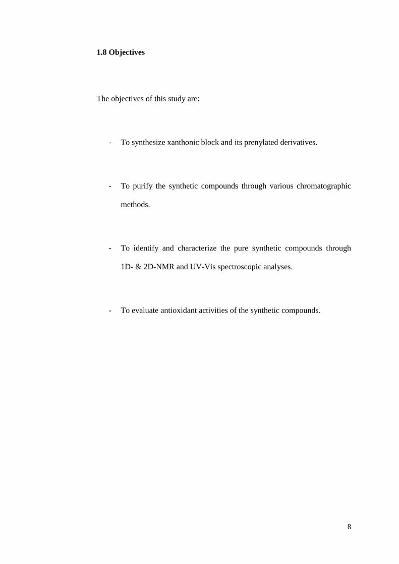
4.2.4 Structural Elucidation of 1-Hydroxy-2,4,4-tris(3-methyl-but-2-enyl)-
4H-xanthen-3,9-dione
Figure 4.19: Structure of 1-hydroxy-2,4,4-tris(3-methyl-but-2-enyl)-4H-
xanthen-3,9-dione (42)
By referring to the 1H NMR spectrum (Figure 4.20), the disappearance of
proton signals at C2 and C4, in contrast to that of compound 39, had proven
the existence of prenyl groups at these positions. The presence of a pair of
doublet of doublets around δ 2.8 suggested the existence of magnetically
equivalent methylene protons of the geminal diprenyl chains attached to the
same carbon. On the other hand, the presence of doublet at δ 3.10 was due to
the methylene protons of the third prenyl chain substituted in xanthone ring A.
The respective positions of the prenyl groups were determined on the basis of
HMBC correlation (Table 4.4). The prenyl methylene proton, H11 showed

61
linkages with carbons C1, C2 and C3 indicating the location of prenyl group at
carbon C2. The 3J correlations of H16 and H16’ to both C3 and C4a
confirmed the substitution of the diprenyl chains at C4.
The two carbonyl groups in the structure were confirmed by the observation of
two downfield signals at δ 195.2 and 179.3 in the 13
C NMR spectrum (Figure
4.21). The characteristic signal of the sp3 hybridised ring carbon C4 was
observed at δ 57.8. The methyl carbons of the prenyl groups gave five signals
at δ 18.1 (C19), 18.2 (C14), 19.1 (C19’), 26.1 (C20 and C20’), and 26.2 (C15).
Meanwhile the protonated olefinic carbons showed signals at δ 122.5 (C12)
and 117.6 (C17 and C17’).
The structure of compound 42, therefore can be proposed as 1-hydroxy-2,4,4-
tris(3-methyl-but-2-enyl)-4H-xanthen-3,9-dione. This compound was proven
to have a novel skeleton due to the distorted aromatic ring caused by the sp3
hybridised carbon C4.
The absorption maxima at 261.91, 230.73, 294.35 and 322.88 nm, displayed in
the UV-Vis spectrum (Figure 4.24), confirmed that compound 42 is highly
conjugated.

62
1-hydroxy-2,4,4-tris(3-methyl-but-2-enyl)-4H-xanthen-3,9-dione (42)
Molecular formula: C28H32O4
Molecular mass: 432.56 gmol-1
Table 4.5: Summary of NMR data of 1-hydroxy-2,4,4-tris(3-methyl-but-2-
enyl)-4H-xanthen-3,9-dione
Position δH (ppm) δC
(ppm)
HMBC
2J
3J
1 - 164.9 - -
2 - 114.8 - -
3 - 195.2 - -
4 - 57.8 - -
4a - 175.5 - -
5 7.56 (1H, d, J = 8.0 Hz) 118.3 C10a C7, C8a
6 7.80 (1H, td, J = 8.0, 1.2
Hz)
135.8 - C8, C10a
7 7.52 (1H, t, J = 8.0 Hz) 126.7 C8 C5, C8a
8 8.25 (1H, dd, J = 8.0, 1.2
Hz)
126.4 - C9, C10a

63
8a - - -
9 - 179.3 - -
9a - 110.9 - -
10a - 155.5 - -
11 3.10 (2H, d, J = 8.0 Hz) 21.2 C2, C12 C1, C3, C13
12 5.10 (1H, t, J = 8.0 Hz ) 122.5 - C14, C15
13 - 131.9 - -
14 1.74 (3H, s) 18.2 - C15
15 1.65 (3H, s) 26.2 - C14
16 2.75 (1H, dd, J = 13.4,
6.7 Hz)
2.88 (1H, dd, J = 13.4,
6.7 Hz)
39.3 C4, C17 C3, C4a, C18
17 4.71, (1H, t, J = 6.7 Hz) 117.6 C16 C19, C20
18 - 135.7 - -
19 1.40 (3H, s) 18.1 - C20
20 1.44 (3H, s) 26.1 - C19
16’ 2.88 (1H, dd, J = 13.4,
6.7 Hz)
2.75 (1H, dd, J = 13.4,
6.7 Hz)
39.3 C4, C17’ C3, C4a, C18’
17’ 4.71, (1H, t, J = 6.7 Hz) 117.6 C16’ C19’, C20’
18’ - 135.7 - -
19’ 1.40 (3H, s) 19.1 - C20’
20’ 1.44 (3H, s) 26.1 - C19’
1-OH 12.87 (OH, s) - C1 C2, C9a
64
1-hydroxy-2,4,4-tris(3-methyl-but-2-enyl)-4H-xanthen-3,9-dione (42)
Molecular formula: C28H32O4
Molecular mass: 432.56 gmol-1
Figure 4.20: 1H NMR spectrum of 1-hydroxy-2,4,4-tris(3-methyl-but-2-
enyl)-4H-xanthen-3,9-dione (400 MHz, CDCl3)
1-OH
H5
H7
H8 H6 H12
7
H17
H17’
H16
H16’ H11
7
H14
H15
H19
H20
H19’
H20’

65
1-hydroxy-2,4,4-tris(3-methyl-but-2-enyl)-4H-xanthen-3,9-dione (42)
Molecular formula: C28H32O4
Molecular mass: 432.56 gmol-1
Figure 4.21: 13
C NMR spectrum of 1-hydroxy-2,4,4-tris(3-methyl-but-2-
enyl)-4H-xanthen-3,9-dione (400 MHz, CDCl3)
C3 C9 C4a
C1
C10a
C6
C18
C18’ C5
C8a
C12
C7
C8
C13
C17
C17’
C9a
C2
C4
C16
C16’
C20
C20’
C19
C19’
C14 C15
C11

66
1-hydroxy-2,4,4-tris(3-methyl-but-2-enyl)-4H-xanthen-3,9-dione (42)
Molecular formula: C28H32O4
Molecular mass: 432.56 gmol-1
Figure 4.22: HMQC spectrum of 1-hydroxy-2,4,4-tris(3-methyl-but-2-
enyl)-4H-xanthen-3,9-dione (400 MHz, CDCl3)
H8 H6 H5 & H7
H12
H17 & H17’
H11 H16
H16’
H14 H15
H19 & H19’ H20 & H20’
C11
C14, C19
& C19’
C15, C20
& C20’
C16 & C16’
C17 & C17’
C12 C5
C7 & C8
C6

67
1-hydroxy-2,4,4-tris(3-methyl-but-2-enyl)-4H-xanthen-3,9-dione (42)
Molecular formula: C28H32O4
Molecular mass: 432.56 gmol-1
Figure 4.23: HMBC spectrum of 1-hydroxy-2,4,4-tris(3-methyl-but-2-
enyl)-4H-xanthen-3,9-dione (400 MHz, CDCl3)
1-OH
H6 H8
H5 & H7
H12
H17 & H17’
H11 H16
H16’
H14
H15 H19 H19’ H20
H20’
C14, C19 & C19’
C15, C20 & C20’
C16 & C16’
C4
C13
C17 & C17’
C7 & C8
C9a
C3
C9 C4a
C1 C10a
C6, C18 & C18’
C2
C5
C8a & C12

68
1-hydroxy-2,4,4-tris(3-methyl-but-2-enyl)-4H-xanthen-3,9-dione (42)
Molecular formula: C28H32O4
Molecular mass: 432.56 gmol-1
Figure 4.24: UV-Vis spectrum of 1-hydroxy-2,4,4-tris(3-methyl-but-2-
enyl)-4H-xanthen-3,9-dione
190.0 200 210 220 230 240 250 260 270 280 290 300 310 320 330 340 350 360 370 380 390 400.0
0.00
0.1
0.2
0.3
0.4
0.5
0.6
0.7
0.8
0.9
1.0
1.1
1.2
1.3
1.4
1.5
1.6
1.7
1.8
1.9
2.00
nm
A
322.88,0.89336
294.35,0.82547
230.73,1.3719216.91,1.3819
200.96,0.48113
195.33,0.42968

69
4.2.5 Proposed Mechanism for Synthesis of 1,3-Dihydroxy-2,4-bis(3-
methyl-but-2-enyl)-xanthen-9-one
Figure 4.25: Proposed mechanism for the synthesis of 1,3-dihydroxy-2,4-
bis(3-methyl-but-2-enyl)-xanthen-9-one

70
4.2.6 Structural Elucidation of 1,3-Dihydroxy-2,4-bis(3-methyl-but-2-
enyl)-xanthen-9-one
Figure 4.26: Structure of 1,3-dihydroxy-2,4-bis(3-methyl-but-2-enyl)-
xanthen-9-one (43)
In the 1H NMR spectrum of 43 (Figure 4.27), a downfield singlet for chelated
hydroxyl group was displayed at δ 13.18, indicating that the peri-hydroxyl
group was not reacted during the prenylation. Instead, the disappearance of
proton signals at C2 and C4, in compared to that of 39, revealed the existence
of carbo-prenylation at these two positions. The two doublets at δ 3.44 and
3.52 assigning to two groups of methylene protons suggested that the
compound was diprenylated.

71
The 13
C NMR spectrum (Figure 4.28) showed three methyl carbon signals of
prenyl groups at δ 18.1 (C14 and C19), 25.9 (C15) and 26.0 (C20). On the
other hand, the methylene carbon signals of the prenyl groups were shown at δ
21.7 (C11) and 21.9 (C16).
The observed key HMBC correlations for the structure assignment are shown
in Table 4.5. Linkages of proton H11 with carbons C1 and C3 indicated that
the prenyl group was located at C2, whereas the correlation between proton
H16 and carbons C3 and C4a established that another prenyl group was
attached to carbon C4. Thereby, the complete structure of compound 43 was
concluded as 1,3-dihydroxy-2,4-bis(3-methyl-but-2-enyl)-xanthen-9-one.
The UV-Vis spectrum (Figure 4.31) displayed absorption bands at 216.94,
238.06, 259.52 and 316.61 nm. This confirmed that the compound 43 was
highly conjugated.

72
1,3-dihydroxy-2,4-bis(3-methyl-but-2-enyl)-xanthen-9-one (43)
Molecular formula: C23H24O4
Molecular mass: 364.44 gmol-1
Table 4.6: Summary of NMR data of 1,3-dihydroxy-2,4-bis(3-methyl-but-
2-enyl)-xanthen-9-one
Position δH (ppm) δC
(ppm)
HMBC
2J
3J
1 - 158.6 - -
2 - 109.0 - -
3 - 161.0 - -
4 - 105.5 - -
4a - 152.9 - -
5 7.39 (1H, d, J = 8.0 Hz) 117.6 C10a C7, C8a
6 7.65 (1H, td, J = 8.0, 1.2 Hz) 134.7 C5 C8, C10a
7 7.30 (1H, t, J = 8.0 Hz) 123.8 - C5, C8a
8 8.20 (1H, dd, J = 8.0, 1.2
Hz)
125.9 - C6, C9, C10a

73
8a - 120.4 - -
9 - 181.2 - -
9a - 103.5 - -
10a - 156.0 - -
11 3.44 (2H, d, J = 6.7 Hz) 21.7 C2, C12 C1, C3, C13
12 5.29 (1H, t, J = 6.7 Hz) 121.6 C11 C14, C15
13 - 135.5 - -
14 1.85 (3H, s) 18.1 C13 C12, C15
15 1.73 (3H, s) 25.9 C13 C12, C14
16 3.52 (2H, d, J = 6.7 Hz) 21.9 C4, C17 C3, C4a, C18
17 5.26 (1H, t, J = 6.7 Hz) 121.9 C16 C19, C20
18 - 133.8 - -
19 1.88 (3H, s) 18.1 C18 C17, C20
20 1.77 (3H, s) 26.0 C18 C17, C19
1-OH 13.18 (OH, s) - C1 C2, C9a
3-OH 6.49 (OH, s) - C3 C2, C4

74
1,3-dihydroxy-2,4-bis(3-methyl-but-2-enyl)-xanthen-9-one (43)
Molecular formula: C23H24O4
Molecular mass: 364.44 gmol-1
Figure 4.27: 1H NMR spectrum of 1,3-dihydroxy-2,4-bis(3-methyl-but-2-
enyl)-xanthen-9-one (400 MHz, CDCl3)
1-OH H6
H8
H11 H16
H12
H17
3-OH H5
H7
H14
H15
H19
H20

75
1,3-dihydroxy-2,4-bis(3-methyl-but-2-enyl)-xanthen-9-one (43)
Molecular formula: C23H24O4
Molecular mass: 364.44 gmol-1
Figure 4.28: 13
C NMR spectrum of 1,3-dihydroxy-2,4-bis(3-methyl-but-2-
enyl)-xanthen-9-one (400 MHz, CDCl3)
C9
C3
C10a
C1
C4a
C6 C18
C13
C8 C7
C17
C12
C5
C8a
C4
C2
C9a
C14
C19
C15
C20
C11
C16

76
1,3-dihydroxy-2,4-bis(3-methyl-but-2-enyl)-xanthen-9-one (43)
Molecular formula: C23H24O4
Molecular mass: 364.44 gmol-1
Figure 4.29: HMQC spectrum of 1,3-dihydroxy-2,4-bis(3-methyl-but-2-
enyl)-xanthen-9-one (400 MHz, CDCl3)
H8 H6
H5 H7 H12 H17 H11 &
H16
H14 H15 H19 H20
C14 & C19
C15 & C20
C11 & C16
C5
C6
C12
& C17
C7 C8
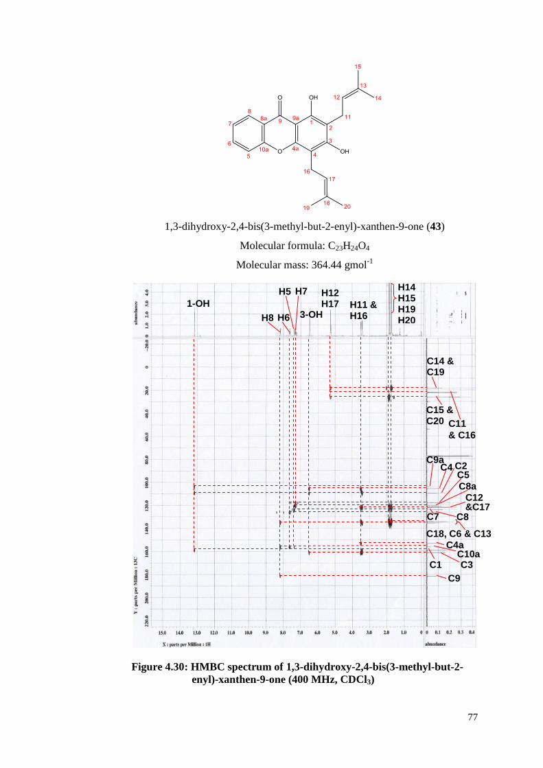
77
1,3-dihydroxy-2,4-bis(3-methyl-but-2-enyl)-xanthen-9-one (43)
Molecular formula: C23H24O4
Molecular mass: 364.44 gmol-1
Figure 4.30: HMBC spectrum of 1,3-dihydroxy-2,4-bis(3-methyl-but-2-
enyl)-xanthen-9-one (400 MHz, CDCl3)
1-OH
H8 H6
H5 H7 H12 H17 H11 &
H16
H14 H15 H19 H20
C14 & C19
C15 & C20 C11
& C16
C4 C2 C5
C8a C12 &C17
C7 C8
C18, C6 & C13 C4a
C10a C3 C1
C9
C9a
3-OH

78
1,3-dihydroxy-2,4-bis(3-methyl-but-2-enyl)-xanthen-9-one (43)
Molecular formula: C23H24O4
Molecular mass: 364.44 gmol-1
Figure 4.31: UV-Vis spectrum of 1,3-dihydroxy-2,4-bis(3-methyl-but-2-
enyl)-xanthen-9-one
190.0 200 210 220 230 240 250 260 270 280 290 300 310 320 330 340 350 360 370 380 390 400.0
0.000
0.02
0.04
0.06
0.08
0.10
0.12
0.14
0.16
0.18
0.20
0.22
0.24
0.26
0.28
0.30
0.32
0.34
0.36
0.38
0.40
0.42
0.44
0.46
0.48
0.500
nm
A
316.61,0.18731
259.52,0.28394
238.06,0.38978
216.94,0.38494
196.00,0.16394

79
4.3 Antioxidant Assay
Phenolic compounds, such as benzoic acid derivatives have been extensively
exploited because of their multiple biological activities, including antioxidant
effects. Since they contain at least one hydroxyl-substituted aromatic ring
system, they can form chelate complexes with metal ions and are easily
oxidized, as well as serving important units for donating electrons (Rice-Evans,
Miller & Paganga, 1997). Research has shown that the majority of phenolic
compounds in the fruit hull of G. mangostana belong to the xanthone family,
may help to offset chronic diseases related with ROS.
The isolated xanthones were screened in view of their antioxidant activities.
They were examined with DPPH (1,1-diphenyl-2-dipicrylhydrazyl), of which
DPPH is widely used for assessing the ability of polyphenols to transfer labile
H-atoms to radicals. The IC50 values (effective concentration leading to a 50%
loss of DPPH radical activity) were obtained by linear regression analysis of
the dose response curves, which were plots of percentage radical scavenging
versus concentration depicted in Figures 4.32, 4.33, 4.34, 4.35, 4.36 and 4.37.
The dihydroxyl substitutions at C1 and C3 of compound 43 was consistent
with its elicited antioxidant effect (IC50= 85 µg/mL). Although the DPPH
radical scavenging ability of 43 was weaker than that of ascorbic acid (IC50= 5

80
µg/mL) and kaempferol (IC50= 10 µg/mL), it was evident that 43 did show
proton-donating ability on DPPH radical to form DPPHH molecules.
Unexpectedly, compound 39 with two hydroxyl group as well did not show
noticeable antioxidant activity.
Meanwhile compounds 41 and 42 did not show significant antioxidant
activities although they both possess a hydroxyl group at C1. From the result it
was suggested that the chelating effect between the hydroxyl group and the
adjacent carbonyl group has weakened the radical scavenging activities of the
compounds. The electron-withdrawing induction effect of the carbonyl group
has retarded the homolytic cleavage of the O-H bond.
Table 4.7: Free radical scavenging activities of the test compounds
Compound IC50 (µg/mL)
1,3-dihydroxyxanthone (39) > 200
1-hydroxy-2,4-bis(3-methyl-but-2-enyl)-3-(3-
methyl-but-2-enyloxy)-xanthen-9-one (41)
> 200
1-hydroxy-2,4,4-tris(3-methyl-but-2-enyl)-4H-
xanthen-3,9-dione (42)
>200
1,3-dihydroxy-2,4-bis(3-methyl-but-2-enyl)-
xanthen-9-one (43)
85
Ascorbic acid (Vitamin C) 5
Kaempferol 10

81
Figure 4.32: Graph of inhibition rate (%) vs. concentration (µg/mL) of
ascorbic acid
Figure 4.33: Graph of inhibition rate (%) vs. concentration (µg/mL) of
kaempferol
0
10
20
30
40
50
60
70
80
90
100
0 50 100 150 200 250
Inh
ibit
ion
Ra
te (
%)
Concentration (µg/mL)
Graph of Inhibition Rate (%) vs. Concentration (µg/mL)
5
IC50
0
10
20
30
40
50
60
70
80
90
100
0 50 100 150 200 250
Inh
ibit
ion
Rate
(%
)
Concentration (µg/mL)
Graph of Inhibition Rate (%) vs. Concentration (µg/mL)
IC50
10

82
Figure 4.34: Graph of inhibition rate (%) vs. concentration (µg/mL) of
1,3-dihydroxyxanthone (39)
Figure 4.35: Graph of inhibition rate (%) vs. concentration (µg/mL) of 1-
hydroxy-2,4-bis(3-methyl-but-2-enyl)-3-(3-methyl-but-2-
enyloxy)-xanthen-9-one (41)
0
5
10
15
20
25
30
35
0 50 100 150 200 250
Inh
ibit
ion
Ra
te (
%)
Concentration (µg/mL)
Graph of Inhibition Rate (%) vs. Concentration (µg/mL)
0
2
4
6
8
10
12
14
16
18
20
0 50 100 150 200 250
Inh
ibit
ion
Rate
(%
)
Concentration (µg/mL)
Graph of Inhibition Rate (%) vs. Concentration (µg/mL)

83
Figure 4.36: Graph of inhibition rate (%) vs. concentration (µg/mL) of 1-
hydroxy-2,4,4-tris(3-methyl-but-2-enyl)-4H-xanthen-3,9-
dione (42)
Figure 4.37: Graph of inhibition rate (%) vs. concentration (µg/mL) of
1,3-dihydroxy-2,4-bis(3-methyl-but-2-enyl)-xanthen-9-one (43)
0
5
10
15
20
25
30
35
40
45
50
0 50 100 150 200 250
Inh
ibit
ion
Ra
te (
%)
Concentration (µg/mL)
Graph of Inhibition Rate (%) vs. Concentration (µg/mL)
0
10
20
30
40
50
60
70
0 50 100 150 200 250
Inh
ibit
ion
Ra
te (
%)
Concentration (µg/mL)
Graph of Inhibition Rate (%) vs. Concentration (µg/mL)
85
IC50

84
CHAPTER 5
CONCLUSIONS
5.1 Conclusions
In this study, a xanthonic block 1,3-dihydroxyxanthone (39) and three new
prenylated xanthone derivatives, namely 1-hydroxy-2,4-bis(3-methyl-but-2-
enyl)-3-(3-methyl-but-2-enyloxy)-xanthen-9-one (41), 1-hydroxy-2,4,4-tris(3-
methyl-but-2-enyl)-4H-xanthen-3,9-dione (42), and 1,3-dihydroxy-2,4-bis(3-
methyl-but-2-enyl)-xanthen-9-one (43) were successfully synthesized. The
structures of these pure compounds were elucidated through 1H-NMR,
13C-
NMR and UV-Vis analyses, and were further confirmed on the basis of 2D-
NMR including HMQC and HMBC analyses. Among these compounds,
compound 42 was found to bear a novel skeleton of xanthen-3,9-dione.
The DPPH radical scavenging assay was carried out to study the antioxidant
activities of the isolated compounds, and the results indicated compound 43 to
exhibit significant antioxidant activity at an IC50 value of 85 µg/mL. This
activity was found to be weak as compared to the reference compounds,
ascorbic acid and kaempferol with their IC50 values of 5 µg/mL and 10 µg/mL,
respectively.

85
5.2 Future Studies
The prenylated products suffer a low yield, syntheses which are able to give
higher yield can be persued. One example is the application of microwave-
assisted organic synthesis (MAOS) which has proven to increase the yield and
has remarkable short reaction time. In addition, a combination of a microwave
irradiation with the Montmorillonite K10 clay has the possibility to vary the
conditions such as the type of clay, the absence and presence of solvent and
the temperature. Besides being environmental friendly, this methodology has
proven to give a much higher yield when compare to the classical method in
which zinc chloride and high temperature are used.
Secondly, the synthesized compounds did not show significant antioxidant
activities. It is suggested that a further study on other potential biological
activities such as cytotoxic, anti-malarial and anti-bacterial to be carried out.
Besides the gravity column chromatography, other more advanced
chromatographic methods such as flash column chromatography, centrifugal
chromatography and high performance liquid chromatography are
recommended to improve separation to the crude product and to fasten the
purification work.

86
REFERENCES
Balasubramanian, K. & Rajagopalan, K. (1988). Novel xanthones from
Garcinia mangostana, structures of BR-xanthone-A and BR-xanthone-B.
Phytochemistry, 27 (5), 1552–1554.
Bennett, G. J. & Lee, H. H. (1988). The biosynthesis of mangostin: the origin
of the xanthone skeleton. Journal of the Chemical Society, Chemical
Communications, pp. 619–620.
Bennett, G. J., Lee, H. H., & Nagaratnam, P. D. (1990). Biosynthesis of
Mangostin. Part 1: The origin of the xanthone skeleton. Journal of Chemistry
Society, Perkin Transactions, 1(10), 2671-2676.
Bennett, G.J. & Lee, H. H. (1989). Xanthones from Guttiferae. Phytochemistry,
28(4), 967-988.
Birch, A. J., Baldas, J., Hlubucek, J. R., Simpson, T. J., & Westerman, P. W.
(1976). Acetate Polymalonic Route of Biosynthesis Xanthones. Journal of
Chemistry Society, Perkin Transactions, 8, pp. 898-904.
Boonstra, J., & Post, J. A. (2004). Molecular events associated with reactive
oxygen species and cell cycle progression in mammalian cells. Gene, 337, pp.
1-13.
Boots, A. W., Haenen, G. R., den Hartog, G. J., & Bast, A. (2002). Oxidative
damage shifts from lipid peroxidation to thiol arylation by catechol-containing
antioxidants. Biochimica et Biophysica Acta, 1583(3), 279-284.

87
Brigmann, G., Ochse, M.,Schupp, O., & Tasler, S. (2001). Progress in the
Chemistry of Organic Natural Products. Wien, Germany: Springer.
Cardona, M. L., Fernadez M. I., Pedro J. R., & Serrano A. (1990). Xanthones
from Hypericum reflexum. Pytochemistry, 29(9), 3003-3006.
Carpenter I., Locksley H. D., & Scheinmann F (1969). Xanthones in higher
plants: Biogenetic proposals and a chemotaxonomic survey. Phytochemistry,
8(10), 2013-2025.
Castanheiro, R. A. P., Pinto, M. M. M., Cravo, S. M. M., Pinto, D. C. G. A.,
Silva, A. M. S., & Kijjoa, A. (2009). Improved methodologies for synthesis of
prenylated xanthones by microwave irradiation and combination of
heterogeneous catalysis (K10 clay) with microwave irradiation. Tetrahedron,
65(19), 3848-3857.
Castanheiro, R. A. P., Pinto, M. M. M., Silva, A. M. S., Cravo, S. M. M.,
Gales, L., Damas, A. M., Pedro, M. M., Nazareth, N., Nascimento, M. S. J.,
Eaton, G. (2007). Dihydroxyxanthones prenylated derivatives: synthesis,
structure elucidation, and growth inhibitory activity on human tumor cell lines
with improvement of selectivity for MCF-7. Bioorganic & Medical Chemistry,
15(18), 6080-6088.
Chen, S. X., Wan, M., & Loh, B. N. (1996). Active constituents against HIV-1
protease from Garcinia mangostana. Planta Medica, 62(4), 381–382.
Chopra, R. N., Nayar, S. L., & Chopra, I. C. (1956). Glossary of indian
medicinal plants (pp. 123). New Delhi, India: The National Institute of
Science Communication and Information Resources.
Dewick, P. M. (1998). The biosynthesis of shikimate metabolites. Natural
Product Report, 15(1), 17-58.

88
Epifano, F., Genovese, S., Menghini, L., & Curini, M. (2007). Chemistry and
pharmacology of oxyprenylated secondary plant metabolites. Phytochemistry,
68(7), 939-953.
Feskanich, D., Ziegler, R. G., Michaud, D. S., Giovannucci, E. L., Speizer, F.
E., Willett, W. C., & Colditz, G. A. (2000). Prospective study of fruit and
vegetable consumption and risk of lung cancer among men and women.
Journal of the National Cancer Institute, 92(22), 1812 1823.
Fujita, M., & Inoue, T. (1980). Biosynthesis of mangiferin in Anemarrhena
asphodeloides Bunge. I. The origin of the xanthone nucleus. Chemical &
Pharmaceutical Bulletin, 28(8), 2476–2481.
Gardner, P. T., White, T. A. C., McPhail, D. B., & Duthie, G. G. (2000).
Relative contributions of vitamin C, carotenoids and phenolics to the
antioxidant potential of fruit juices. Food Chemistry, 68(4), 471–474.
Garnett, M., & Sturton, S. D. (1932). Garcinia Mangostana in the treatment of
amoebic dysentery. Chinese Medical Journal, pp. 969–973.
Gordon, M. H. (1996). Dietary antioxidants in disease prevention. Natural
Product Reports, 13(4), 265–273.
Gottlieb, O.R. (1968). Biogeneticproposals regarding aucuparins and
xanthones. Phytochemistry, 7(3), 411-421
Granoth, I., Pownall, H. J. (1975). Synthesis and substituent effects in the
nuclear magnetic resonance and mass spectra of dimethyl- and
dihaloxanthones. The Journal of Organic Chemistry, 40(14), 2088-2091.
Gröger, D., Erge, D., Franck, B., Ohnsorge, U., Flasch, H., & Huper, F. (1968).
Mycotoxin from mold fungi. Chemische Berichte, 101, pp. 1970-1978.

89
Grover, P. K., Shah, G. D., & Shah, R. C. (1955). Xanthones. Part IV. A new
synthesis of hydroxyxanthones and hydroxybenzophenones. Journal of the
Chemical Society, pp. 3982-3985.
Gupta, P. & Lewis, J. R. (1971). Xanthones in Cell Suspension Cultures of
Two Centaurium Species. Journal of the Chemical Society, 4, pp. 629-631.
Helesbeux, J. J., Duval, O., Dartiguelongue, C., Séraphin, D., Oger, J. M., &
Richomme, P. (2004). Synthesis of 2-hydroxy-3-methylbut-3-enyl substituted
coumarins and xanthones as natural products. Tetrahedron, 60(10), 2293-2300.
Herrmann, K. M., & Weaver, L. M. (1999). The Shikimate Pathway. Annual
Review of Plant Physiology and Plant Molecular Biology, 50(1), 473-503.
Hostettmann, K., & Hostettmann, M. (1989). Xanthones. In Dey P. M. &
Harborne J. B. (Eds.), Methods in Plant Biochemistry (Vol. 1). London, UK:
Academic Press.
Johnson, M. M., Naidoo, J. M., Fernandes, M. A., Mmutlane, E. M., van
Otterlo, W. A., & de Koning, C. B. (2010). CAN-mediated oxidations for the
synthesis of xanthones and related products. The Journal of Organic
Chemistry, 75(24), 8701-8704.
Joshipura, K. J., Hu, F. B., Manson, J. E., Stampfer, M. J., Rimm, E. B.,
Speizer, F. E., Colditz, G., Ascherio, A., Rosner, B., Spiegelman, D., Willett,
W. C. (2001). The effect of fruit and vegetable intake on risk for coronary
heart disease. Annals of Internal Medicine, 134(12), 1106–1114.
Khan, M. T. H. & Ather, A. (2006). Xanthones as therapeutic agents:
chemistry and pharmacology. In Lead Molecules from Natural Products,
Volume 2: Discovery and New Trends (p. 273). Oxford, UK: Elsevier.

90
Kinghorn, A. D., Falk, H., & Kobayashi, J. (Eds.). (2011). Progress in the
Chemistry of Organic Natural Products (Vol. 94). New York: Springer.
Klimczak, I., Maleecka, M., Szlachta, M., & Gliszczyńska-Świgło, A. (2007).
Effect of storage on the content of polyphenols, vitamin C and the antioxidant
activity of orange juices. Journal of Food Composition and Analysis, 20(3-4),
313–322.
Kosem, N., Han, Y. H., & Moongkarndia, P. (2007). Antioxidant and
cytoprotective activities of methanolic extract from Garcinia mangostana
hulls. Science Asia, 33, pp. 283–292.
Kostanecki, St. V. & Nessler, B. (1891). Synthesen von Oxyxanthonen. Ber.
Dtsch. Chem. Ges., 24(1), 1894–1897.
Lespegnol, Bertrand, & Dupas (1939). Bulletin de la Société Chimique de
France, 6, pp. 1925.
Locksley, H. D., Moore, I., & Scheinmann, F. (1967). Extractives from
guttiferae—VI : The significance of maclurin in xanthone biosynthesis.
Tetrahedron, pp. 2229-2234.
Lopaczyski, W., & Zeisel, S. H. (2001). Nutrition Research, 21(1-2), 295-307.
Lund, N. A., Robertson, A., & Whalley, W. B. (1953). The chemistry of fungi.
Part XXI. Asperxanthone and a preliminary examination of aspergillin.
Journal of the Chemical Society, pp. 2434-2439.

91
McMaster, W. J., Scott, A. T., & Trippett, S. (1960). Biosynthetic Pathway of
Xanthones: Acetate Polymalonic Route. Journal of the Chemical Society,
5(894). 4628-4631.
Mahabusarakam, W., Proudfoot, J., Taylor, W., & Croft, K. (2000). Inhibition
of lipoprotein oxidation by prenylated xanthones derived from mangostin.
Free Radical Research, 33(5), 643–659.
Mahabusarakam, W., Wiriyachtra, P., & Taylor, W. (1987). Chemical
constituents of Garcinia mangostana. Journal of Natural Products, 50(3), 474–
478.
Mandal S., Das P. C., & Joshi P. C. (1992). Xanthones from Natural
Resources. Journal of Indian Chemical Society, 69, pp. 611-636.
Marzano, C., Caffieri, S., Fossa, P., & Bordin, F. (1997). Activity of 3
carbethoxyangelicin photolysis products. Journal of Photochemistry and
Photobiology B: Biology, 38(2-3), 189-195.
Michael, A. A. (1883). Journal of the American Chemical Society, 5, pp. 81.
Moreau, S., Varache-Lembege, M., Larrouture, S., Fall, D., Neveu, A.,
Deffieux, G., Vercauteren, J., Nuhrich, A. (2002). (2-Arylhydrazonomethyl)
substituted xanthones as antimycotics: synthesis and fungistatic activity
against Candida species. European Journal of Medicinal Chemistry, 37(3),
237-253.
Nakatani, K., Atsumi, M., Arakawa, T., Oosawa, K., Shimura, S., Nakahata,
N., & Ohizumi, Y. (2002). Inhibitions of histamine release and prostaglandin
E2 synthesis by mangosteen, a Thai medicinal plant. Biological &
Pharmaceutical Bulletin, 25(9), 1137–1141.

92
Nkengfack, E. A., Kounga P. M., Fomum T. Z., Meyer M., & Bodo B. (2002).
Globulixanthones A & B, two new cytotoxic xanthones with isoprenoid
groups from the root bark of Symphonia globulifera. Journal of Natural
Products, 65(5), 734–736.
Odrowaz-Sypniewski, M. R., Tsoungas, P. G., Varvounis, G., & Cordopatis, P.
(2009). Xanthone in synthesis: a reactivity profile via directed lithiation of its
dimethyl ketal. Tetrahedron Letters, 50(44), 5981–5983.
Oger, J. M., Morel, C., Helesbeux, J. J., Litaudon, M., Séraphin, D.,
Dartiguelongue, C., Larcher, G., Richomme, P., Duval, O (2003). First 2-
hydroxy-3-methylbut-3-enyl substituted xanthones isolated from plants:
structure elucidation, synthesis and antifungal activity. Natural Product
Research, 17(3), 195-199.
Patel, G. N., & Trivedi, K. N. (1988). Abnormal Claisen Rearrangement.
Journal of Indian Chemical Society, 65, pp. 192-193.
Pedro, M., Cerqueira, F., Sousa, M. E., Nascimento, M. S., & Pinto, M. (2002).
Xanthones as inhibitors of growth of human cancer cell lines and their effects
on the proliferation of human lymphocytes in vitro. Bioorganic & Medicinal
Chemistry, 10(12), 3725-3730.
Pillai, R. K. M., Naiksatam, P., Johnson, F., Rajagopalan, R., Watts, P.C.,
Cricchio, R., & Borras, S. (1986). Thermorubin II: 1,3-dihydroxy-9H-
xanthones and 1,3-dihydroxy-9H-xanthenes. New methods of synthesis. The
Journal of Organic Chemistry, 51(5), 717-723.
Pinto, M. M. M. & Castanheiro, R. A. P. (2009). Synthesis of Prenylated
Xanthones: An Overview. Current Organic Chemistry, 13(12), 1215-1240.
Pinto, M. M. M., Sousa, M. E., & Nascimento, M. S. J. (2005). Xanthone
Derivatives: New Insights in Biological Activities. Current Medical Chemistry
12(21), 2517-2538.

93
Pinto, M., & Castanheiro, R. (2008). Chapter 11: Natural Prenylated
Xanthones: Chemistry and Biological Activities in Natural Products. In
Brahmachari, G., (Ed.), Chemistry, Biochemistry and Pharmacology (pp 520 -
676). New Delhi, India: Narosa Publishing House.
Poupon, E. & Nay, B. (2011). Polyprenylated Xanthones. In Biomimetic
Organic Synthesis (p. 54). Weinheim, Germany: Wiley-VCH.
Rice-Evans, C. A., Miller, N. J., & Paganga, G. (1997) Antioxidant properties
of phenolic compounds. Trends in Plant Science, 2(4), 152- 9.
Richardson, P. M. (1983). The taxonomic significance of C-
glycosylxanthones in flowering plants. Biochemical Systematics and
Ecology,11(4), 371-375.
Robak, J., Shridi, F., Wolbis, M., & Krolikowska, M. (1988). Screening of the
influence of flavonoids on lipoxygenase and cycloxygenase activities as well
as on nonenzymic lipid peroxidation. Polish Journal of Pharmacology &
Pharmacy, 40(5), 451- 458.
Robert, J. C. (1961). Naturally occurring xanthones. Chemical Reviews, 61(6),
591-605.
Sen, A. K., Uusvuori, R., Hase, T. A., Benerji, N., Sarkar, K. K., & Mazumder,
P.C. (1980). A xanthone from Garcinia mangostana. Phytochemistry 19(10),
2223–2225.
Sherwin, E. R. (1978). Oxidation and antioxidants in fat and oil processing.
Journal of the American Oil Chemists’ Society, 55(11), 809-814.
Sousa, M. E. & Pinto, M. M. M. (2005). Synthesis of Xanthones: An
Overview. Current Medical Chemistry, 12(21), 2447-2479.

94
Vitale, A. A., Romanelli, G. P., Autinio J. C., & Pomilio A. B. (1994). On the
substitution-recation of 1-bromo-2-aryloxyethanes with sodium-cyanide.
Journal of Chemical Research, 82(1), 19-23.
Weecharangsan, W., Opanasopit, P., Sukma, M., Ngawhirunpat, T.,
Sotanaphun, U., & Siripong, P. (2006). Antioxidative and neuroprotective
activities of extracts from the fruit hull of mangosteen (Garcinia mangostana
Linn.). Medicanal Principles and Practice, 15(4), 281–287.
Yang, C. H., Ma, L., Wei, Z. P., Han, F., & Gao J. (2012). Advances in
Isolation and Synthesis of Xanthone Derivatives. Chinese Herbal Medicines,
4(2), 87-102.
Yu, L., Zhao, M., Yang, B., Zhao, Q., & Jiang, Y. (2007). Phenolics from hull
of Garcinia mangostana fruit and their antioxidant activities. Food Chemistry,
104(1), 176–181.
Zarena, A. S. & Sankar, K. U. (2009). Supercritical carbon dioxide extraction
of xanthones with antioxidant activity from Garcinia mangostana:
Characterization by HPLC/LC–ESI-MS. The Journal of Supercritical Fluids,
49 (3), 330–337.
Zarena, A. S. & Sankar, K. U. (2011). Xanthones enriched extracts from
mangosteen pericarp obtained by supercritical carbon dioxide process.
Separation and Purification Technology, 80 (1), 172–178.
Zhang, Z. H., & Wang, H. J., & Ren X. Q., (2009). A facile and efficient
method for synthesis of xanthone derivatives catalyzed by HBF4/SiO2 under
solvent-free conditions. Monatshefte für Chemie, 140(12), 1481-1483.

95
APPENDICES
APPENDIX A
The following table summarizes the results of inhibition rates vary with
concentrations for ascorbic acid in DPPH radical scavenging activity.
Concentration
(µg/mL)
Absorbance Inhibition
Rate (%)
1 2 3 Mean1
240 0.0676 0.0669 0.0680 0.0675±0.0006 89.97
120 0.0685 0.0695 0.0695 0.0692±0.0006 89.72
60 0.0744 0.0636 0.0690 0.0690±0.0054 89.75
30 0.0672 0.0685 0.0662 0.0673±0.0012 90.00
15 0.0679 0.0683 0.0677 0.0680±0.0003 89.89
7.5 0.0660 0.1646 0.1410 0.1239±0.0515 81.59
3.75 0.3523 0.5684 0.5121 0.4776±0.1121 29.02
0 0.6803 0.6673 0.6711 0.6729±0.0067 0
1 Each value was obtained by calculating the average of three experiments ±
standard deviation.

96
APPENDIX B
The following table summarizes the results of inhibition rates vary with
concentrations for kaempferol in DPPH radical scavenging activity.
Concentration
(µg/mL)
Absorbance Inhibition
Rate (%)
1 2 3 Mean1
240 0.0721 0.0700 0.0698 0.0706±0.0013 90.13
120 0.0664 0.0667 0.0666 0.0666±0.0002 90.69
60 0.0764 0.0697 0.0680 0.0714±0.0044 90.02
30 0.0701 0.0687 0.0759 0.0716±0.0038 89.99
15 0.0807 0.2414 0.1781 0.1667±0.0810 76.69
7.5 0.3999 0.4737 0.4035 0.4257±0.0416 40.48
3.75 0.5513 0.2857 0.4305 0.4225±0.1330 40.93
0 0.6297 0.8326 0.6833 0.7152±0.1051 0
1 Each value was obtained by calculating the average of three experiments ±
standard deviation.

97
APPENDIX C
The following table summarizes the results of inhibition rates vary with
concentrations for 1,3-dihydroxyxanthone (39) in DPPH radical scavenging
activity.
Concentration
(µg/mL)
Absorbance Inhibition
Rate (%)
1 2 3 Mean1
240 1.1282 1.1941 0.8737 1.0653±0.1692 33.77
120 1.0056 1.2151 1.0877 1.1028±0.1056 31.43
60 1.0589 1.7135 0.8964 1.2229±0.4325 23.97
30 1.2808 1.6022 0.8763 1.2531±0.3637 22.09
15 1.3086 1.2985 1.2496 1.2856±0.0316 20.07
7.5 1.1218 1.6164 1.2774 1.3385±0.2529 16.78
3.75 1.3498 1.5549 1.1693 1.3580±0.1929 15.57
0 1.9581 1.8702 0.9968 1.6084±0.5315 0
1 Each value was obtained by calculating the average of three experiments ±
standard deviation.

98
APPENDIX D
The following table summarizes the results of inhibition rates vary with
concentrations for 1-hydroxy-2,4-bis(3-methyl-but-2-enyl)-3-(3-methyl-but-2-
enyloxy)-xanthen-9-one (41) in DPPH radical scavenging activity.
Concentration
(µg/mL)
Absorbance Inhibition
Rate (%)
1 2 3 Mean1
240 0.8715 0.8061 0.7539 0.8105±0.0589 17.70
120 0.7862 0.8897 0.8091 0.8283±0.0544 15.89
60 0.7971 0.9006 0.8186 0.8388±0.0546 14.83
30 0.8604 0.8303 0.8637 0.8515±0.0184 13.54
15 0.8393 0.8547 0.9401 0.8780±0.0543 10.84
7.5 0.9046 0.9865 0.9400 0.9437±0.0411 4.17
3.75 0.9125 0.9836 1.0393 0.9785±0.0636 0.64
0 1.0775 0.9614 0.9154 0.9848±0.0835 0
1 Each value was obtained by calculating the average of three experiments ±
standard deviation.

99
APPENDIX E
The following table summarizes the results of inhibition rates vary with
concentrations for 1-hydroxy-2,4,4-tris(3-methyl-but-2-enyl)-4H-xanthen-3,9-
dione (42) in DPPH radical scavenging activity.
Concentration
(µg/mL)
Absorbance Inhibition
Rate (%)
1 2 3 Mean1
240 0.6359 0.5750 0.5216 0.5775±0.0572 47.74
120 0.6216 0.7902 0.7422 0.7180±0.0869 35.03
60 0.8469 0.9111 0.8790 0.8630±0.0321 21.91
30 0.9188 0.8785 0.8833 0.8935±0.0220 19.15
15 0.9488 0.9476 1.0049 0.9671±0.0327 12.49
7.5 0.9858 0.9990 0.9725 0.9858±0.0133 10.80
3.75 1.0753 1.0922 1.0104 1.0593±0.0432 4.14
0 1.1190 1.1578 1.0384 1.1051±0.0609 0
1 Each value was obtained by calculating the average of three experiments ±
standard deviation.

100
APPENDIX F
The following table summarizes the results of inhibition rates vary with
concentrations for 1,3-dihydroxy-2,4-bis(3-methyl-but-2-enyl)-xanthen-9-one
(43) in DPPH radical scavenging activity.
Concentration
(µg/mL)
Absorbance Inhibition
Rate (%)
1 2 3 Mean1
240 0.5175 0.4313 0.4422 0.4637±0.0469 58.33
120 0.5035 0.3992 0.4963 0.4663±0.0583 58.09
60 0.6352 0.7138 0.6916 0.6802±0.0405 38.87
30 0.8851 0.8034 0.8465 0.8450±0.0409 24.06
15 0.9447 0.9750 0.9401 0.9533±0.0190 14.33
7.5 1.0787 1.0595 1.0185 1.0522±0.0308 5.44
3.75 1.1549 1.0770 1.0837 1.1052±0.0432 0.67
0 1.0534 1.1847 1.1001 1.1127±0.0666 0
1 Each value was obtained by calculating the average of three experiments ±
standard deviation.