Synovial Plicae of the Knee - UCSD Musculoskeletal Radiology
181
Synovial Plicae of the Knee James Koenig MD January 6, 2011
Transcript of Synovial Plicae of the Knee - UCSD Musculoskeletal Radiology

Synovial Plicae of the Knee
James Koenig MD
January 6, 2011
Outline
• Briefly discuss the history of plica research
• Discuss the embryology of the synovial plicae of the knee
• Discuss the anatomy, incidence, and classification systems for all four synovial plicae
• Show examples of plicae on multiple imaging modalities
• Discuss the various plica syndromes
Definitions
• What is a plica?– Definition: a fold or ridge
• In the body:– A plica is a fold of synovial tissue found in the
lining of a joint1
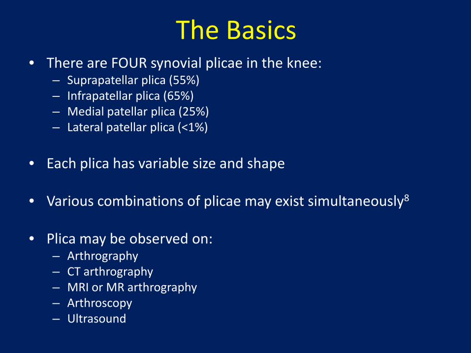
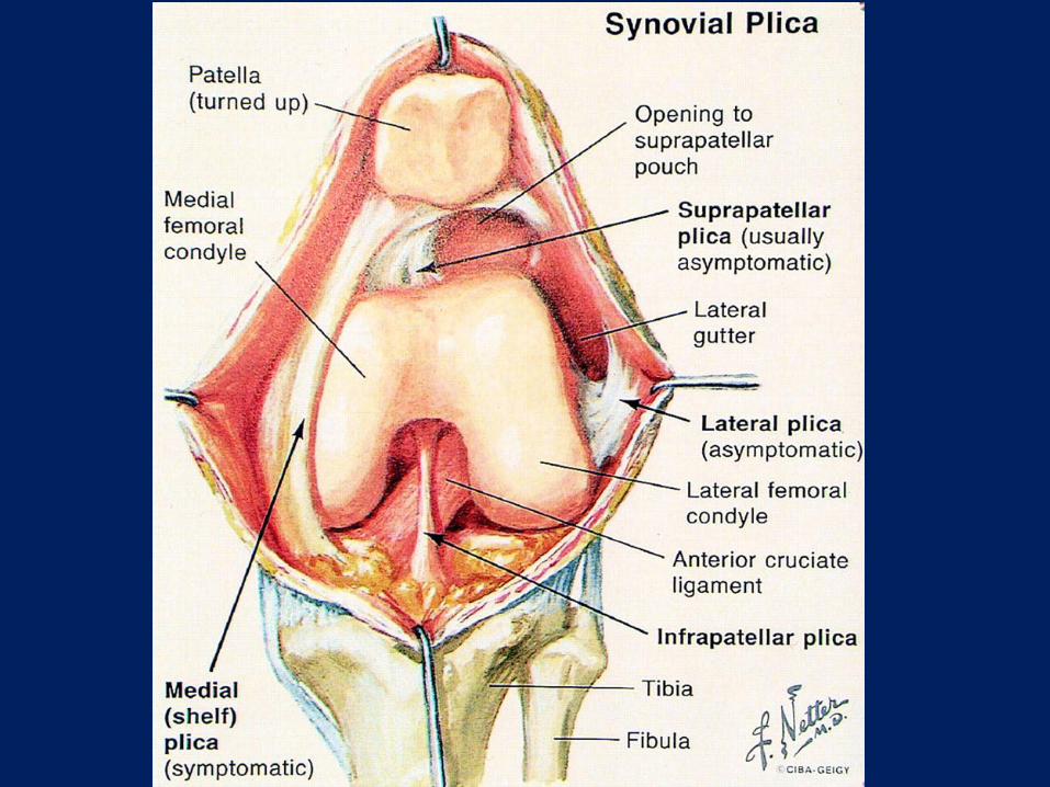
The Basics• There are FOUR synovial plicae in the knee:
– Suprapatellar plica (55%)– Infrapatellar plica (65%)– Medial patellar plica (25%)– Lateral patellar plica (<1%)
• Each plica has variable size and shape
• Various combinations of plicae may exist simultaneously8
• Plica may be observed on:– Arthrography– CT arthrography– MRI or MR arthrography– Arthroscopy– Ultrasound
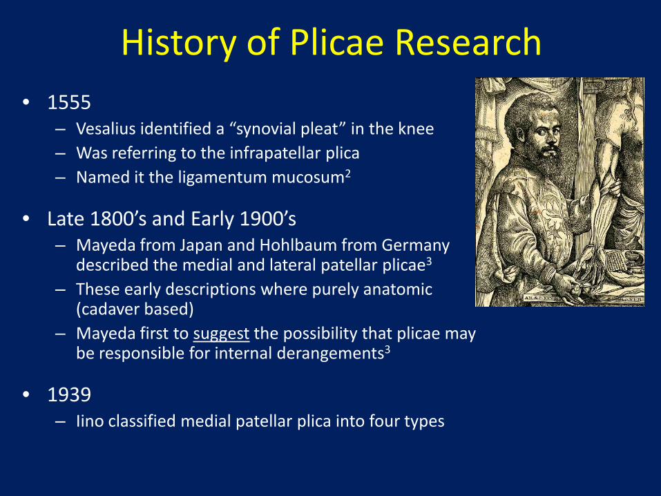
History of Plicae Research• 1555
– Vesalius identified a “synovial pleat” in the knee– Was referring to the infrapatellar plica – Named it the ligamentum mucosum2
• Late 1800’s and Early 1900’s– Mayeda from Japan and Hohlbaum from Germany
described the medial and lateral patellar plicae3
– These early descriptions where purely anatomic (cadaver based)
– Mayeda first to suggest the possibility that plicae may be responsible for internal derangements3
• 1939– Iino classified medial patellar plica into four types

History of Plicae Research• 1950
– Pipkin– First article linking clinical signs to the
presence of a plica– He described two cases of post-traumatic,
symptomatic suprapatellar plicae treated by surgical excision and with subsequent healing
• 1960’s– Interest in orthopedic community grew with
the advent of arthroscopy– Early focus was on the suprapatellar plica
and it was felt to be the most common cause of symptoms
– Later authors realize the medial plica is more often involved in producing clinical symptoms
How do plicae form?
• An understanding of knee embryology is key to understanding the existence of plicae.
• Knee joint forms between the 7th to 10th week of fetal development.
• No consensus about the way the knee joint cavity forms6
• Widely believed that the knee is originally composed of three separate compartments:– Medial and lateral synovial compartments– Suprapatellar bursa
• Incomplete resorption of the septa between the compartments leads to the formation of synovial plica– Explains the existence of the suprapatellar and infrapatellar plicae– However, no coronally orientated septum exists in the fetus
• How does the medial and lateral plica form?
How do plicae form?
• 1990 Ogata and Uhthoff6
– Most complete study of the embryology of the human knee
– Studied 116 knees from 112 embryos and fetuses ranging from 6-20 weeks gestational age
– Purpose:• To elucidate the process of formation of the knee joint cavity
• Determine the incidence of synovial plica during the fetal stage
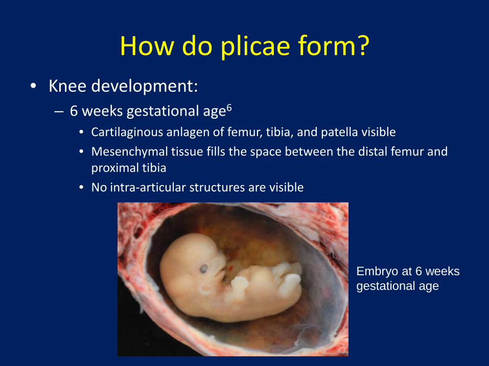
How do plicae form?• Knee development:
– 6 weeks gestational age6
• Cartilaginous anlagen of femur, tibia, and patella visible
• Mesenchymal tissue fills the space between the distal femur and proximal tibia
• No intra-articular structures are visible
Embryo at 6 weeksgestational age
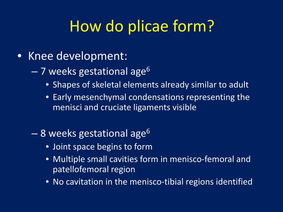
How do plicae form?
• Knee development:– 7 weeks gestational age6
• Shapes of skeletal elements already similar to adult• Early mesenchymal condensations representing the
menisci and cruciate ligaments visible
– 8 weeks gestational age6
• Joint space begins to form• Multiple small cavities form in menisco-femoral and
patellofemoral region• No cavitation in the menisco-tibial regions identified
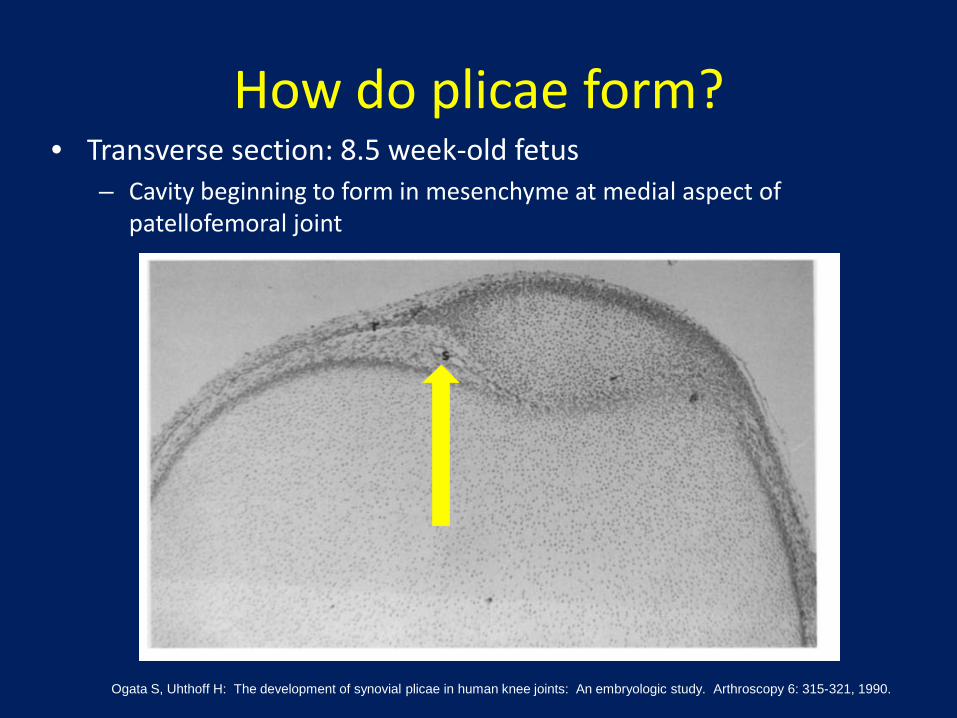
How do plicae form?• Transverse section: 8.5 week-old fetus
– Cavity beginning to form in mesenchyme at medial aspect of patellofemoral joint
Ogata S, Uhthoff H: The development of synovial plicae in human knee joints: An embryologic study. Arthroscopy 6: 315-321, 1990.
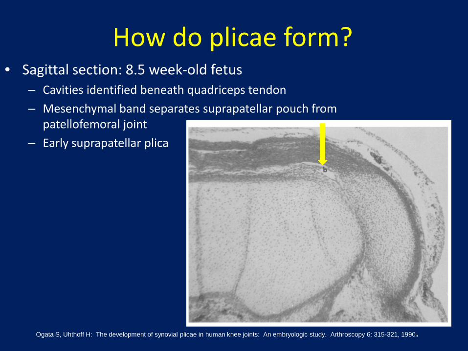
How do plicae form?• Sagittal section: 8.5 week-old fetus
– Cavities identified beneath quadriceps tendon
– Mesenchymal band separates suprapatellar pouch from patellofemoral joint
– Early suprapatellar plica
Ogata S, Uhthoff H: The development of synovial plicae in human knee joints: An embryologic study. Arthroscopy 6: 315-321, 1990.
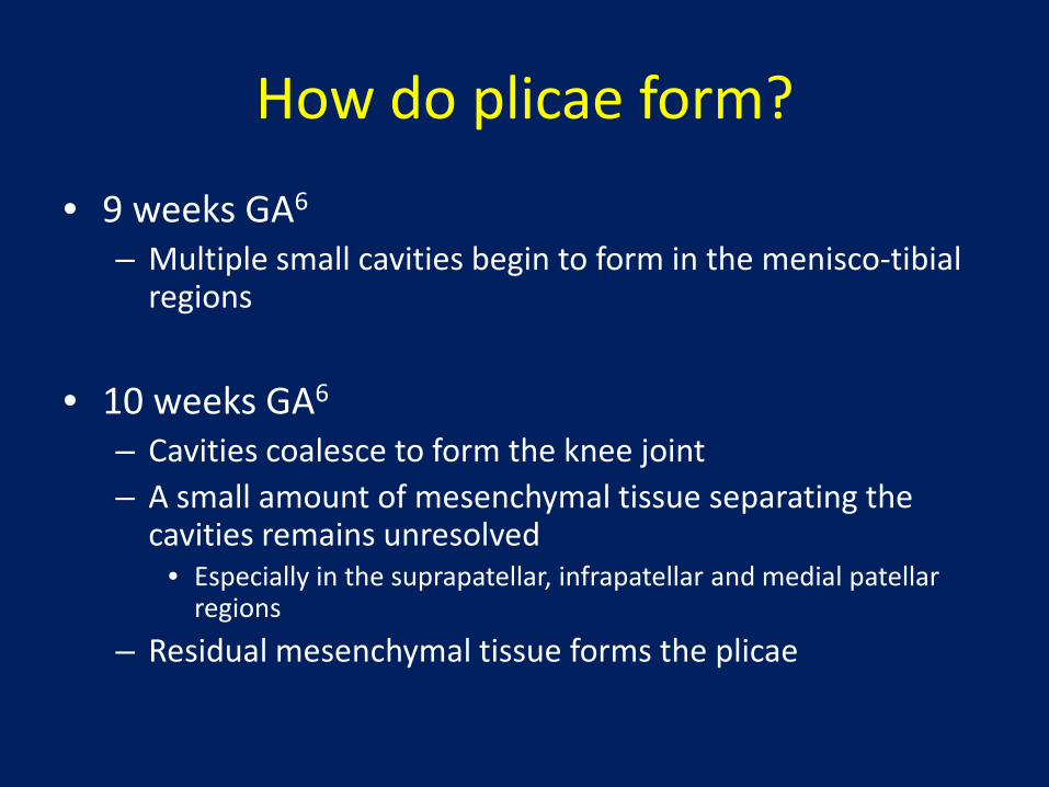
How do plicae form?
• 9 weeks GA6
– Multiple small cavities begin to form in the menisco-tibial regions
• 10 weeks GA6
– Cavities coalesce to form the knee joint– A small amount of mesenchymal tissue separating the
cavities remains unresolved • Especially in the suprapatellar, infrapatellar and medial patellar
regions
– Residual mesenchymal tissue forms the plicae
How do plicae form?
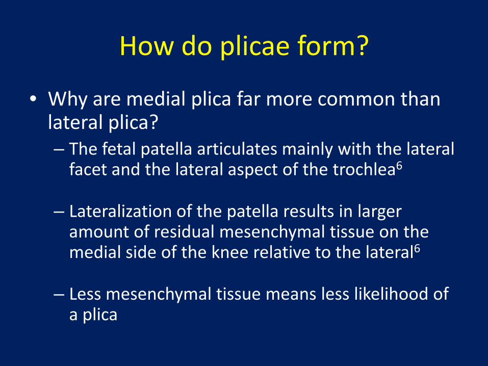
• Why are medial plica far more common than lateral plica?– The fetal patella articulates mainly with the lateral
facet and the lateral aspect of the trochlea6
– Lateralization of the patella results in larger amount of residual mesenchymal tissue on the medial side of the knee relative to the lateral6
– Less mesenchymal tissue means less likelihood of a plica
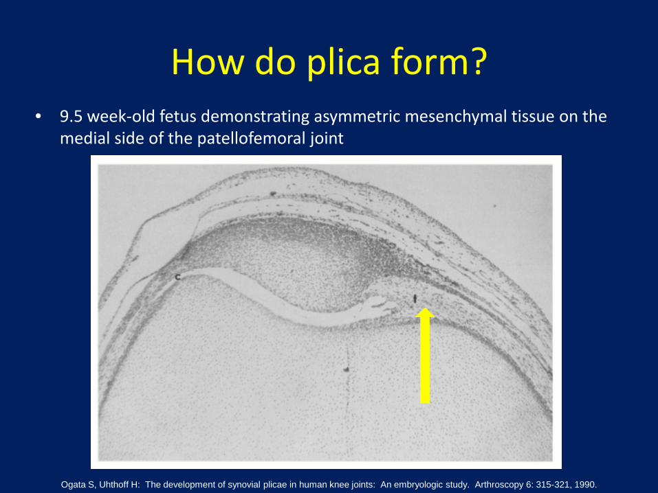
How do plica form?• 9.5 week-old fetus demonstrating asymmetric mesenchymal tissue on the
medial side of the patellofemoral joint
Ogata S, Uhthoff H: The development of synovial plicae in human knee joints: An embryologic study. Arthroscopy 6: 315-321, 1990.
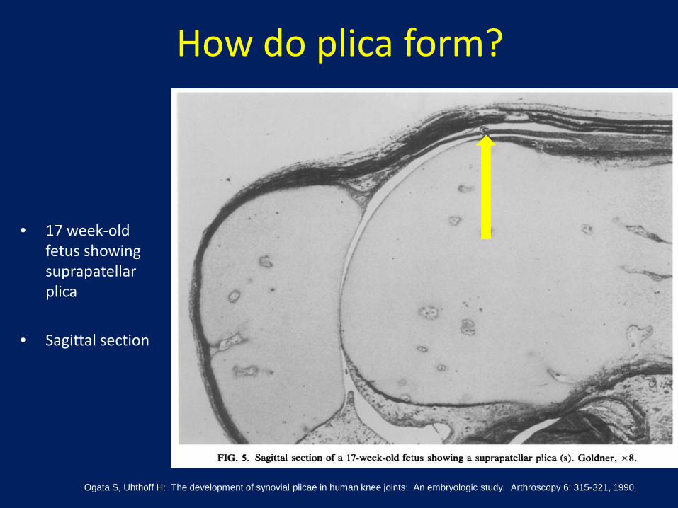
How do plica form?
• 17 week-old fetus showing suprapatellar plica
• Sagittal section
Ogata S, Uhthoff H: The development of synovial plicae in human knee joints: An embryologic study. Arthroscopy 6: 315-321, 1990.
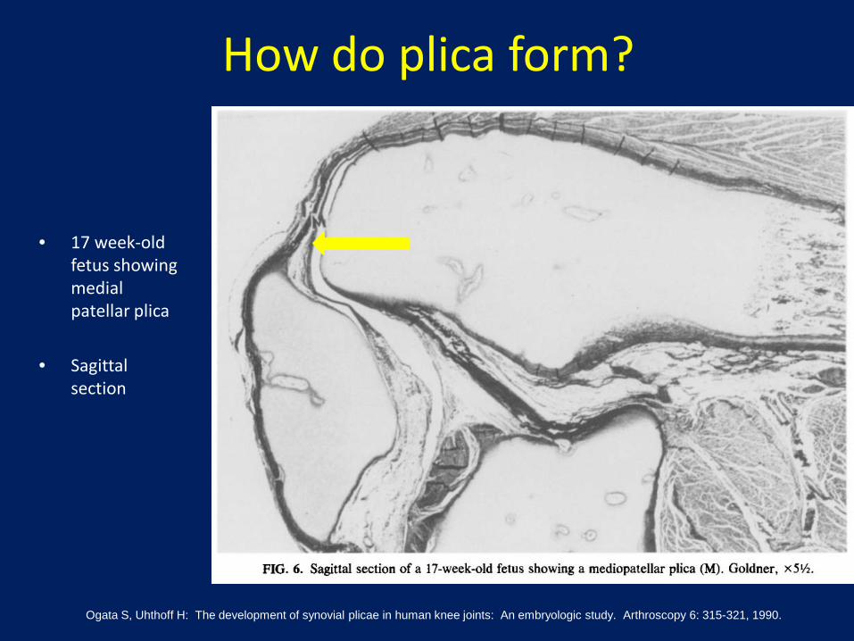
How do plica form?
• 17 week-old fetus showing medial patellar plica
• Sagittal section
Ogata S, Uhthoff H: The development of synovial plicae in human knee joints: An embryologic study. Arthroscopy 6: 315-321, 1990.
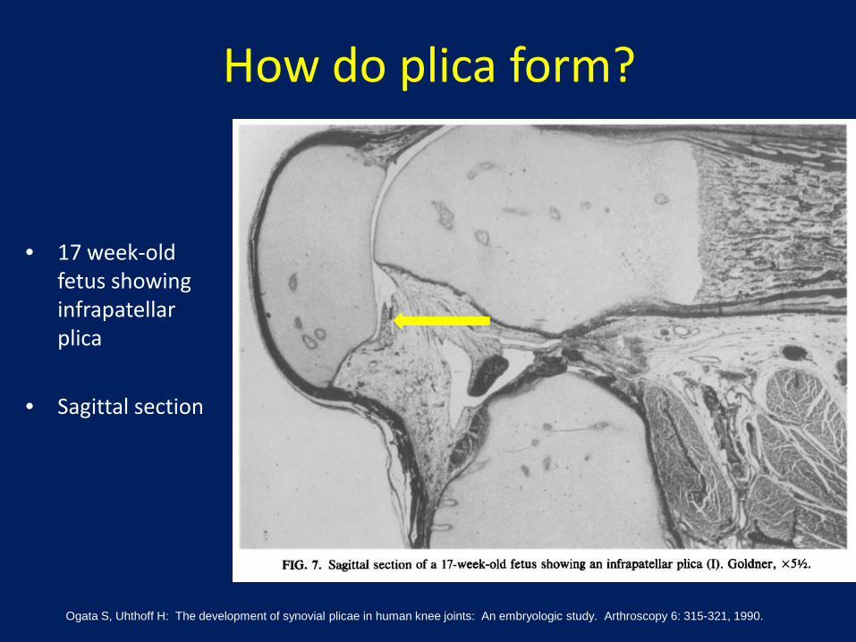
How do plica form?
• 17 week-old fetus showing infrapatellar plica
• Sagittal section
Ogata S, Uhthoff H: The development of synovial plicae in human knee joints: An embryologic study. Arthroscopy 6: 315-321, 1990.
How do plica form?
• Conclusions6:– Suprapatellar plica
• Residual septum between the suprapatellar bursa and patellofemoral cavity
– Infrapatellar plica• Residual septum between the medial and lateral femorotibial
cavitations
– Medial and lateral plica• Not a remnant of a septum• Remnant mesenchymal tissue• Lateralization of the patella during fetal period results in far more
medial plica
Plicae• The following will be discussed for each plica:
– Anatomy of each plica
– Incidence of the plica• Incidences stated in the literature are highly variable
and depend on if study was cadaveric or arthroscopic
– Classification system(s) that exist
– Examples of plica on various imaging modalities will be shown

Suprapatellar Plica

Suprapatellar plica
• Synonyms in the literature2:– Superior plica
– Plica synovialis suprapatellaris
– Superomedial plica
– Medial suprapatellar plica
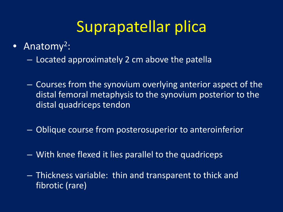
Suprapatellar plica• Anatomy2:
– Located approximately 2 cm above the patella
– Courses from the synovium overlying anterior aspect of the distal femoral metaphysis to the synovium posterior to the distal quadriceps tendon
– Oblique course from posterosuperior to anteroinferior
– With knee flexed it lies parallel to the quadriceps
– Thickness variable: thin and transparent to thick and fibrotic (rare)
Suprapatellar plica
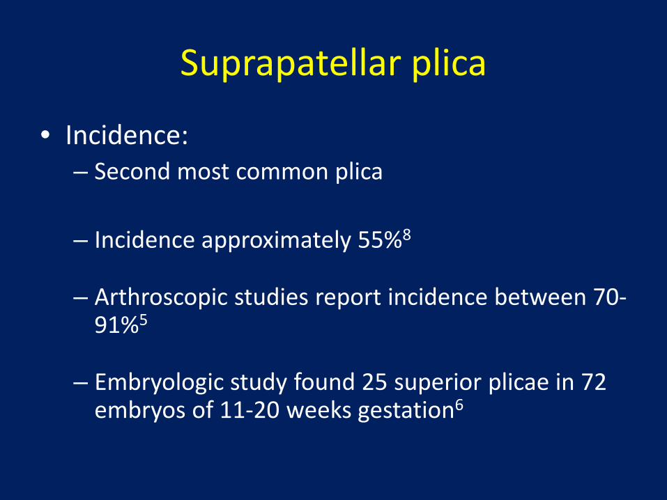
• Incidence:– Second most common plica
– Incidence approximately 55%8
– Arthroscopic studies report incidence between 70-91%5
– Embryologic study found 25 superior plicae in 72 embryos of 11-20 weeks gestation6
Suprapatellar Plica
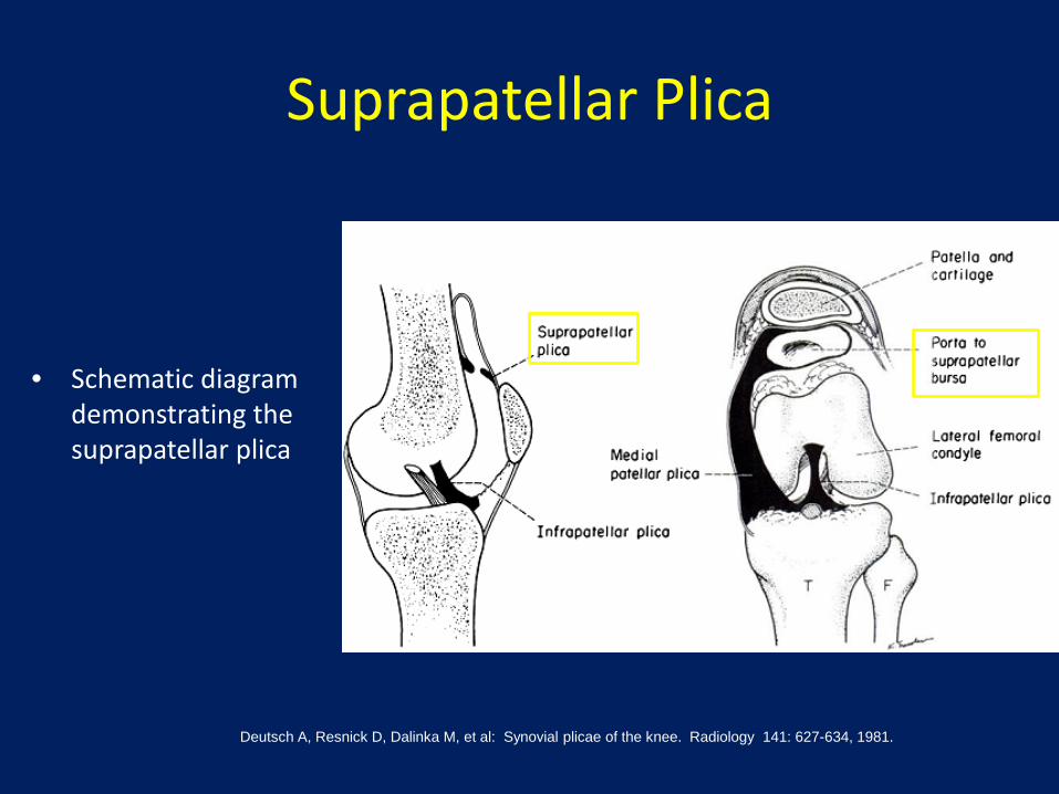
• Schematic diagram demonstrating the suprapatellar plica
Deutsch A, Resnick D, Dalinka M, et al: Synovial plicae of the knee. Radiology 141: 627-634, 1981.
Suprapatellar plica
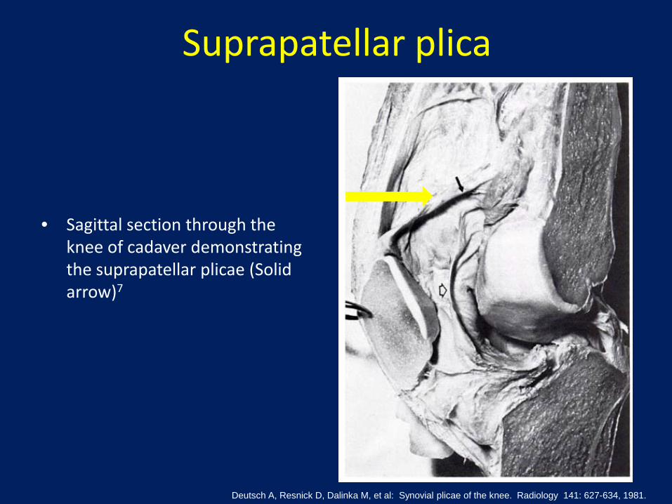
• Sagittal section through the knee of cadaver demonstrating the suprapatellar plicae (Solid arrow)7
Deutsch A, Resnick D, Dalinka M, et al: Synovial plicae of the knee. Radiology 141: 627-634, 1981.
Suprapatellar Plica
Deutsch A, Resnick D, Dalinka M, et al: Synovial plicae of the knee. Radiology 141: 627-634, 1981.
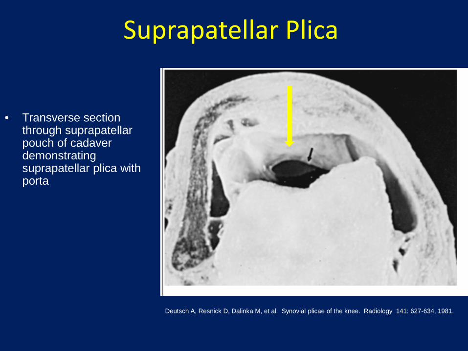
• Transverse section through suprapatellar pouch of cadaver demonstrating suprapatellar plica with porta
Suprapatellar Plica
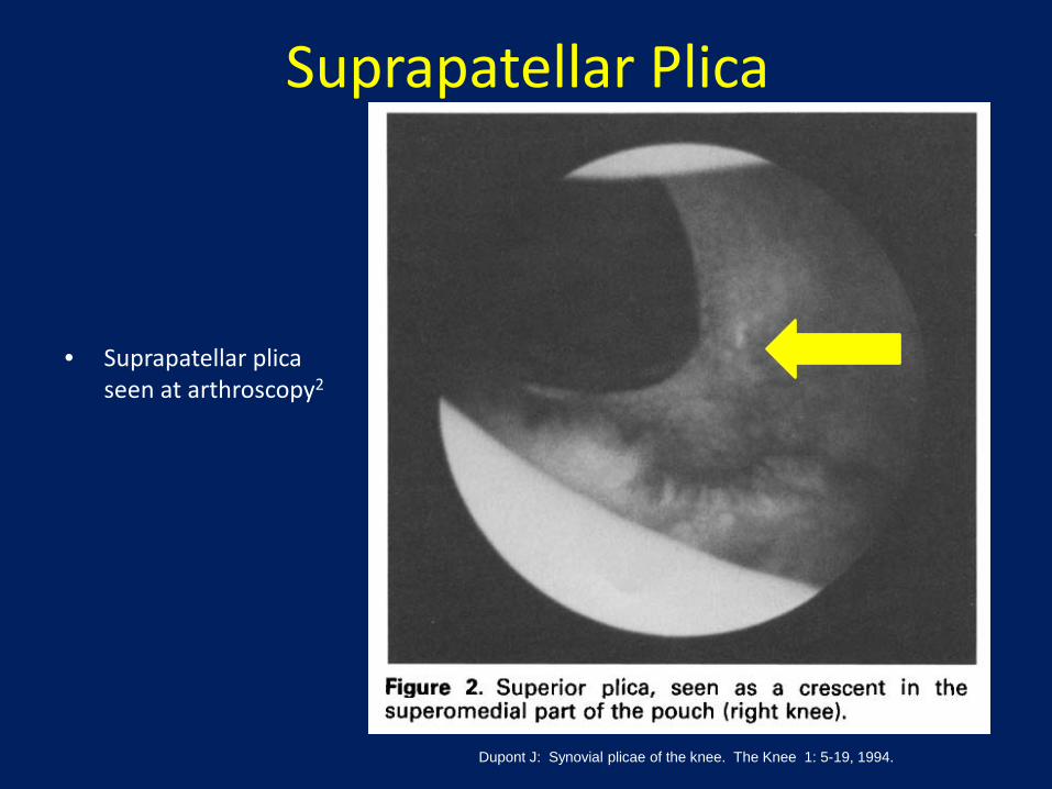
• Suprapatellar plica seen at arthroscopy2
Dupont J: Synovial plicae of the knee. The Knee 1: 5-19, 1994.
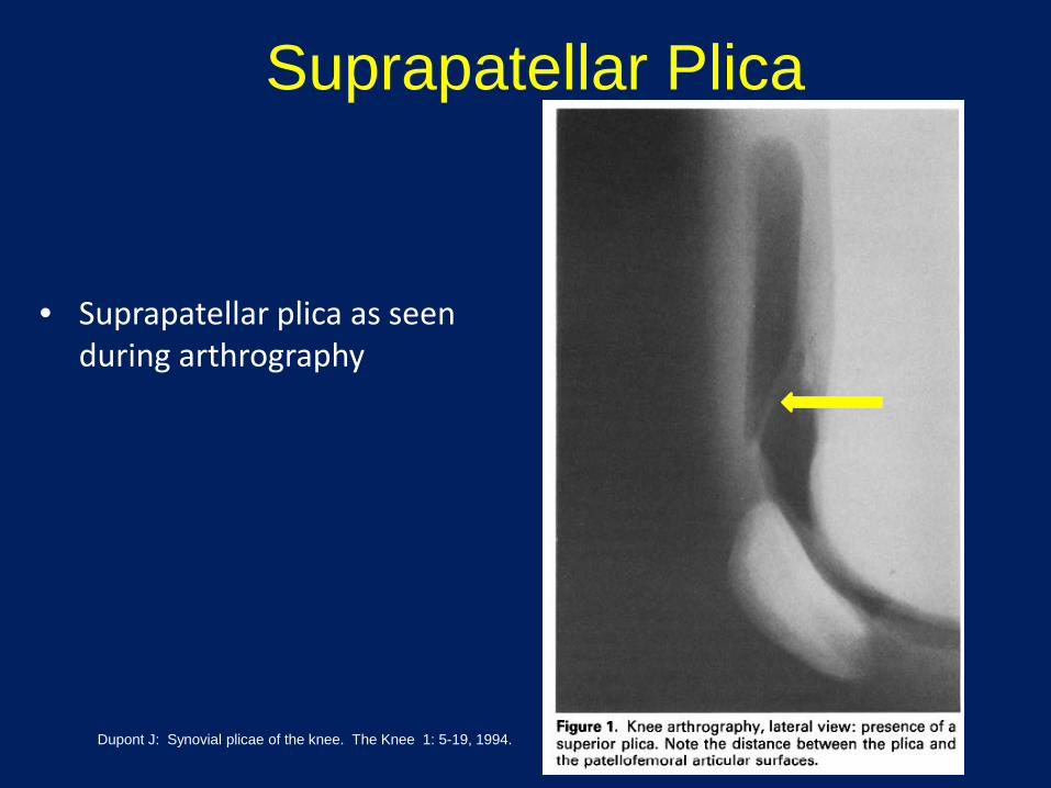
• Suprapatellar plica as seen during arthrography
Dupont J: Synovial plicae of the knee. The Knee 1: 5-19, 1994.
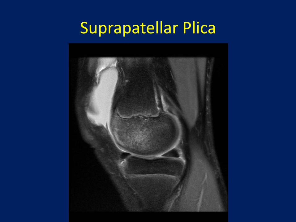
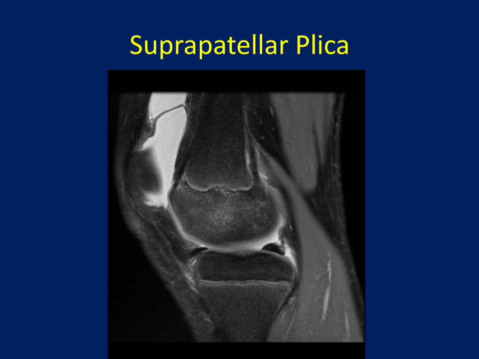
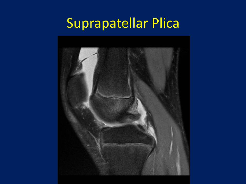
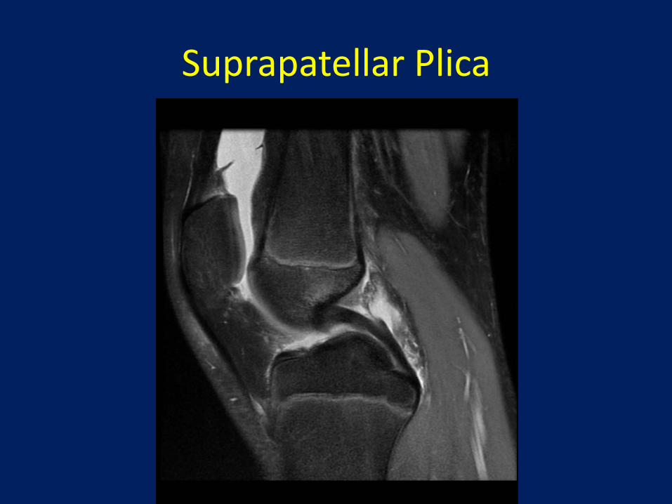
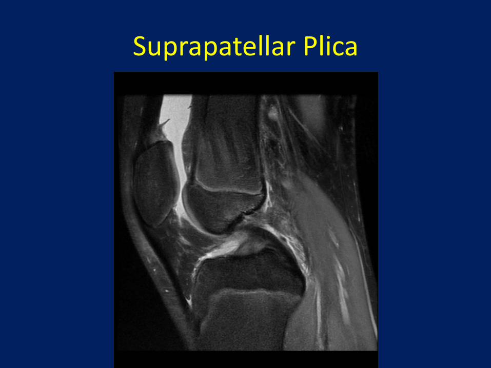
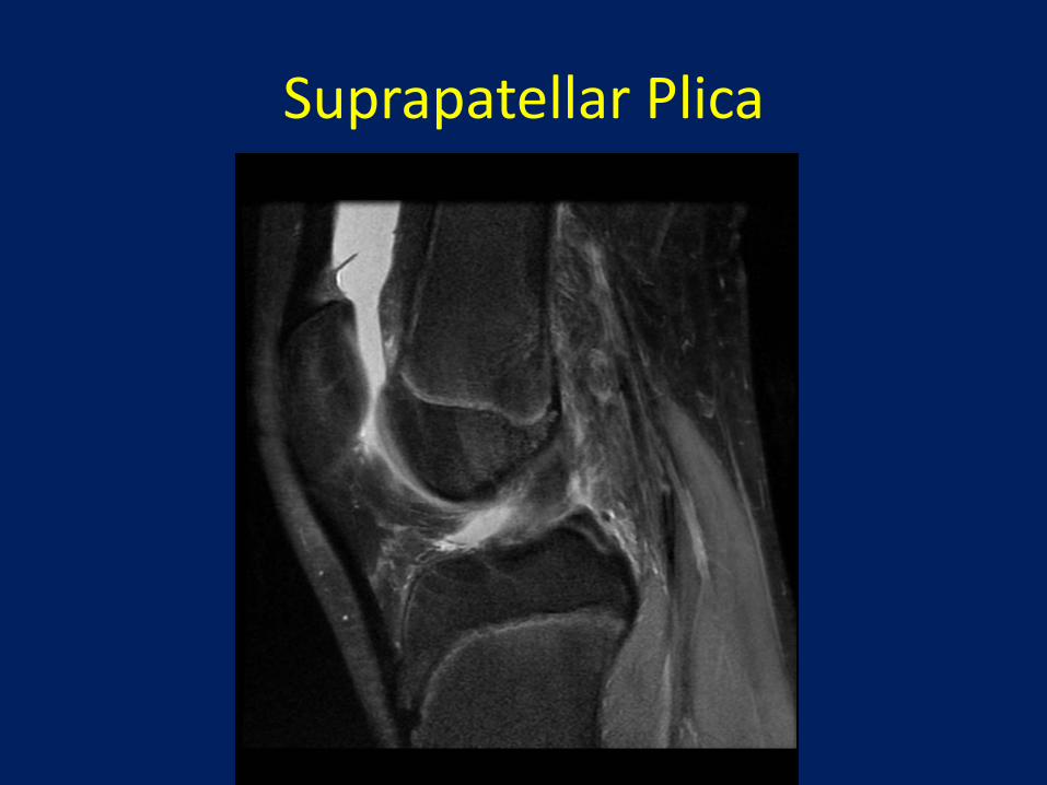
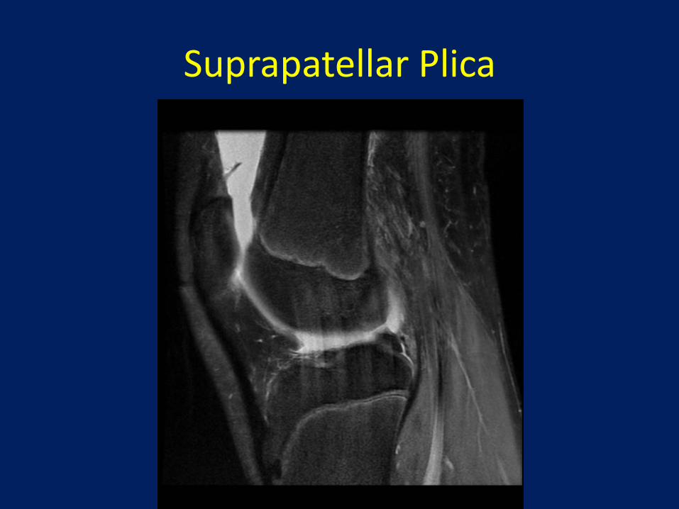
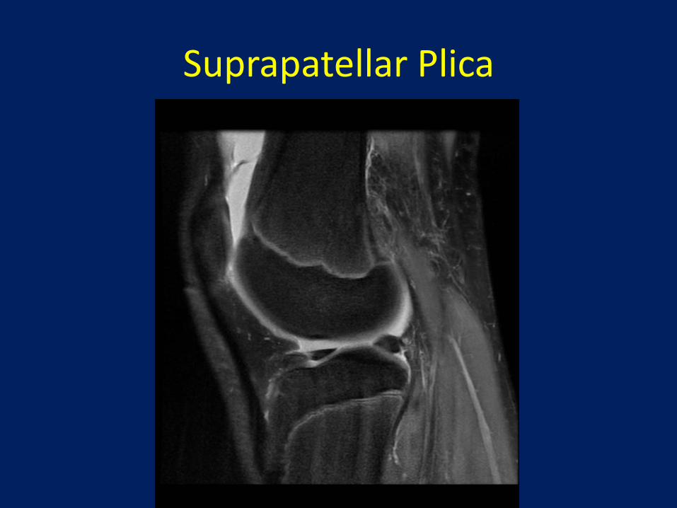
Suprapatellar Plica
Suprapatellar Plica
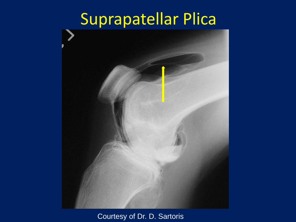
Courtesy of Dr. D. Sartoris
Suprapatellar Plica
Suprapatellar Plica
Suprapatellar Plica
Suprapatellar Plica
Suprapatellar Plica
Suprapatellar Plica
Suprapatellar Plica

Suprapatellar Plica
Suprapatellar Plica
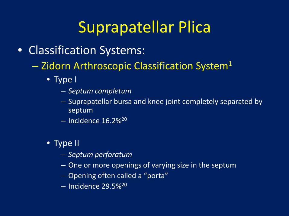
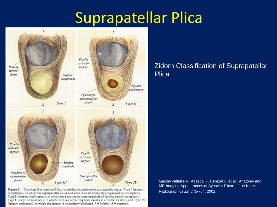
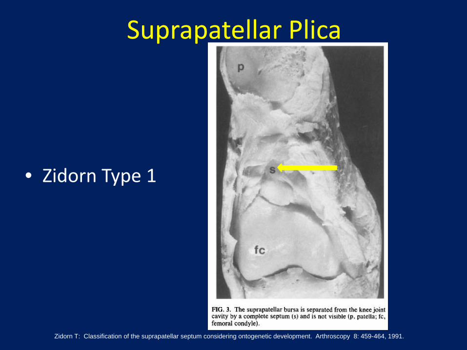
Suprapatellar Plica• Classification Systems:
– Zidorn Arthroscopic Classification System1
• Type I– Septum completum– Suprapatellar bursa and knee joint completely separated by
septum– Incidence 16.2%20
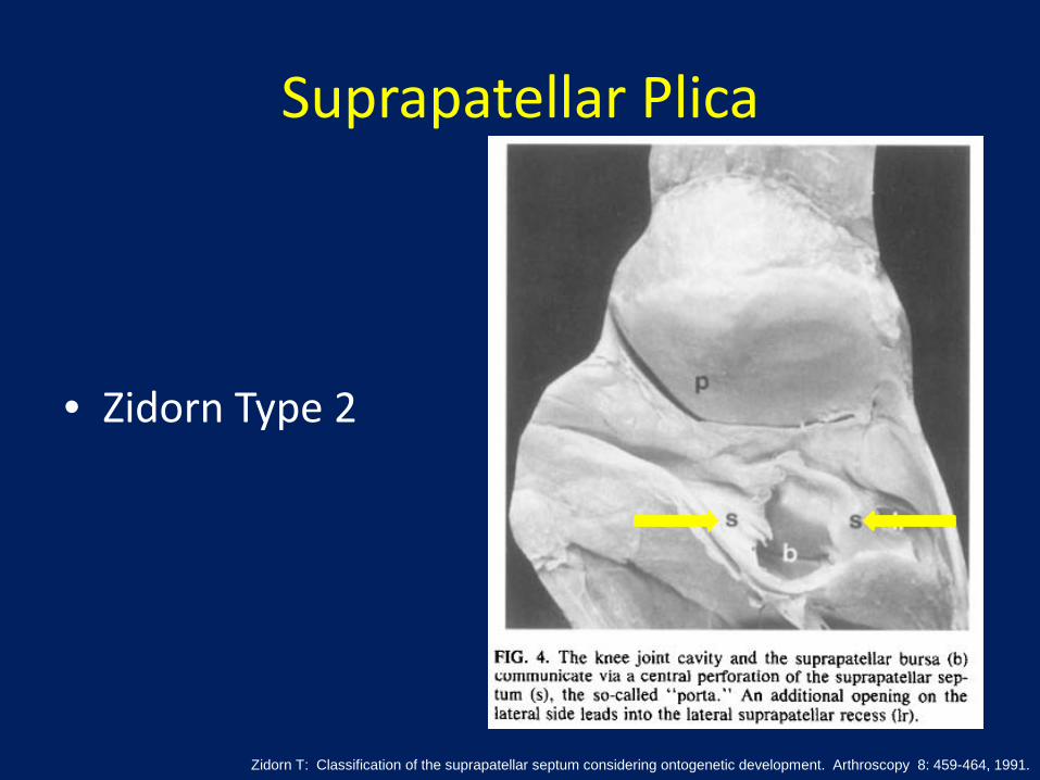
• Type II– Septum perforatum– One or more openings of varying size in the septum– Opening often called a “porta”– Incidence 29.5%20
Suprapatellar Plica
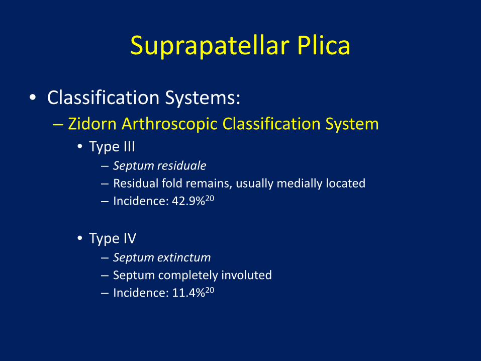
• Classification Systems:– Zidorn Arthroscopic Classification System
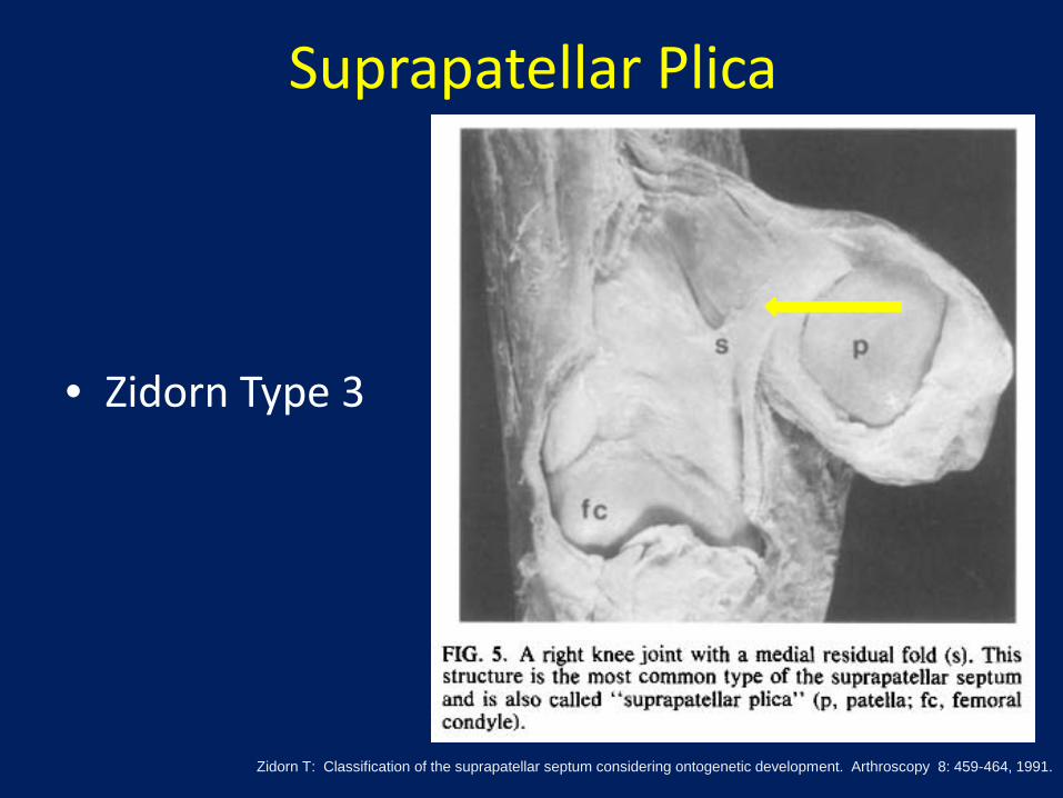
• Type III– Septum residuale– Residual fold remains, usually medially located– Incidence: 42.9%20
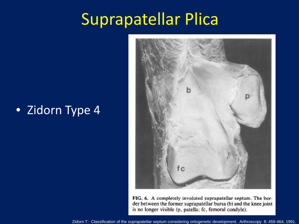
• Type IV– Septum extinctum– Septum completely involuted– Incidence: 11.4%20
Suprapatellar Plica
Garcia-Valtuille R, Abascal F, Cerezal L, et al: Anatomy and MR Imaging Appearances of Synovial Plicae of the Knee. Radiographics 22: 775-784, 2002.
Zidorn Classification of SuprapatellarPlica
Suprapatellar Plica
• Zidorn Type 1
Zidorn T: Classification of the suprapatellar septum considering ontogenetic development. Arthroscopy 8: 459-464, 1991.
Suprapatellar Plica
• Zidorn Type 2
Zidorn T: Classification of the suprapatellar septum considering ontogenetic development. Arthroscopy 8: 459-464, 1991.
Suprapatellar Plica
• Zidorn Type 3
Zidorn T: Classification of the suprapatellar septum considering ontogenetic development. Arthroscopy 8: 459-464, 1991.
Suprapatellar Plica
• Zidorn Type 4
Zidorn T: Classification of the suprapatellar septum considering ontogenetic development. Arthroscopy 8: 459-464, 1991.
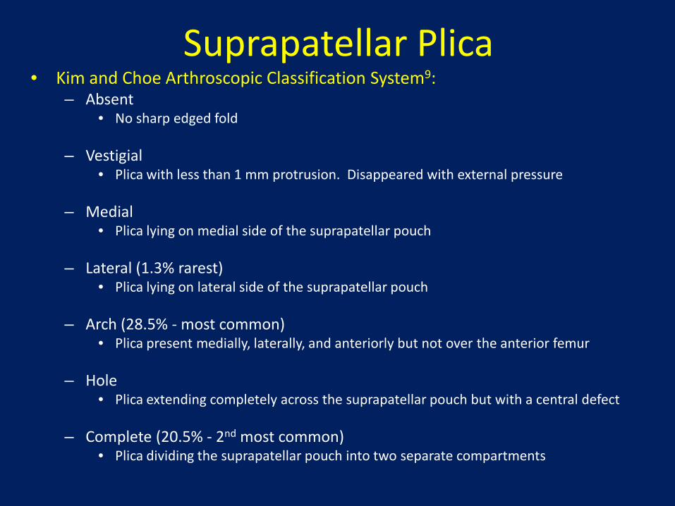
Suprapatellar Plica• Kim and Choe Arthroscopic Classification System9:
– Absent• No sharp edged fold
– Vestigial• Plica with less than 1 mm protrusion. Disappeared with external pressure
– Medial• Plica lying on medial side of the suprapatellar pouch
– Lateral (1.3% rarest)• Plica lying on lateral side of the suprapatellar pouch
– Arch (28.5% - most common)• Plica present medially, laterally, and anteriorly but not over the anterior femur
– Hole• Plica extending completely across the suprapatellar pouch but with a central defect
– Complete (20.5% - 2nd most common)• Plica dividing the suprapatellar pouch into two separate compartments
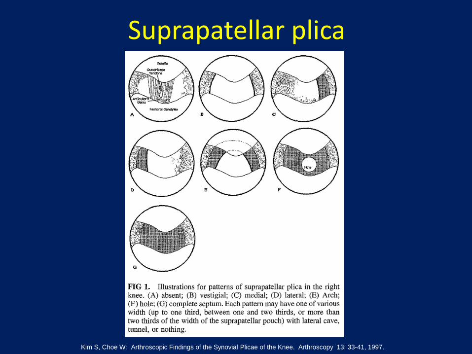
Suprapatellar plica
Kim S, Choe W: Arthroscopic Findings of the Synovial Plicae of the Knee. Arthroscopy 13: 33-41, 1997.

Infrapatellar Plica
Infrapatellar Plica
• Synonyms in the literature2
– Ligamentum mucosum
– Inferior plica
– Plica synovialis patellaris
– Plica synovialis patellae
– Infrapatellar fold
– Infrapatellar septum
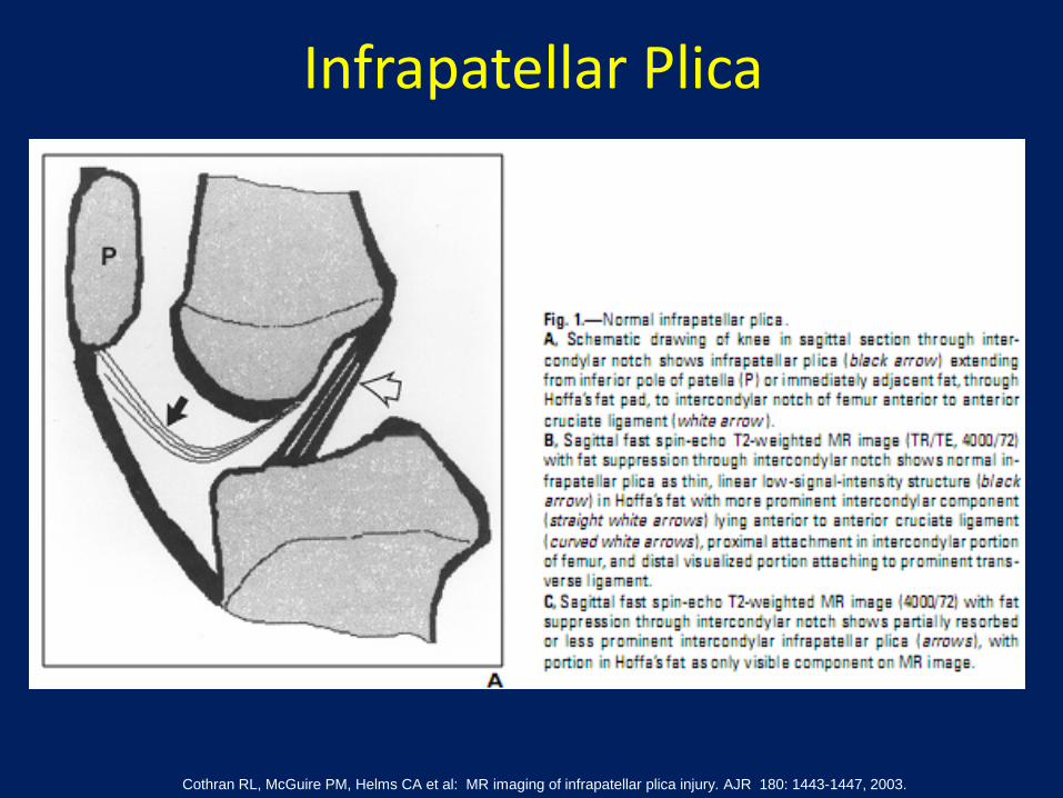
Infrapatellar Plica• Anatomy
– Very commonly seen on arthroscopy; much less commonly seen on MR
– Intercondylar notch
– Anterior to the anterior cruciate ligament• May be separate, fenestrated or continuous with the anterior cruciate
ligament
– Thin proximally and attaches to the very anterior aspect of the intercondylar notch
– It widens as it descends anteriorly and inferiorly through Hoffa’s fat pad to attach to the inferoposterior aspect of the patella.
– After attaching to the inferior aspect of Hoffa’s fat pad it gives off two alar folds that extend laterally to cover the fat pad21
Infrapatellar Plica• Anatomy cont’d:
– The plica then thins and runs through the Hoffa’s fat pad and often attaches to the inferior aspect of the patella29
– Fibous septa within Hoffa’s fat pad may connect the plica to the patellar tendon29
– Purpose of infrapatellar plica:28
• Prevents excessive anterior displacement of Hoffa’s pad in the setting of a large joint effusion
• Alar folds help to maintain the medial and lateral aspects of the Hoffa’s fat pad in the presence of a large joint effusion
– May be extremely thin and thread-like to very thick (as large as the ACL itself)
• May be mistaken for the ACL in ACL-deficient knees
Infrapatellar Plica
• Incidence– Most common plica seen at arthroscopy1
• Seen in the vast majority of arthroscopies
• Often must be cut to visualize the anterior cruciate ligament
– Overall incidence at arthroscopy 86%5
• Seen on nearly all the time
Infrapatellar Plica
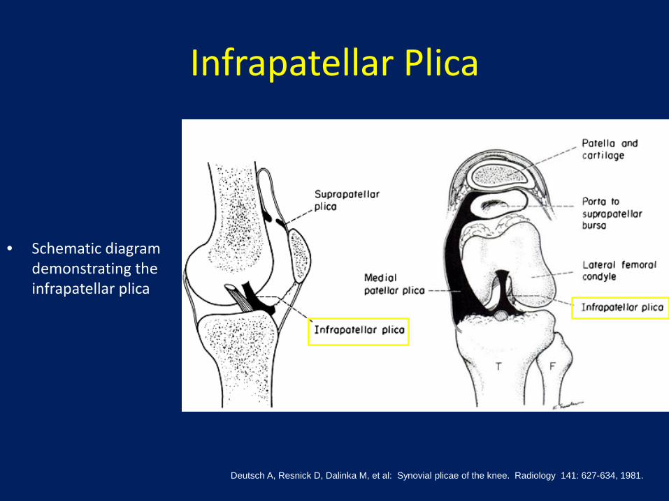
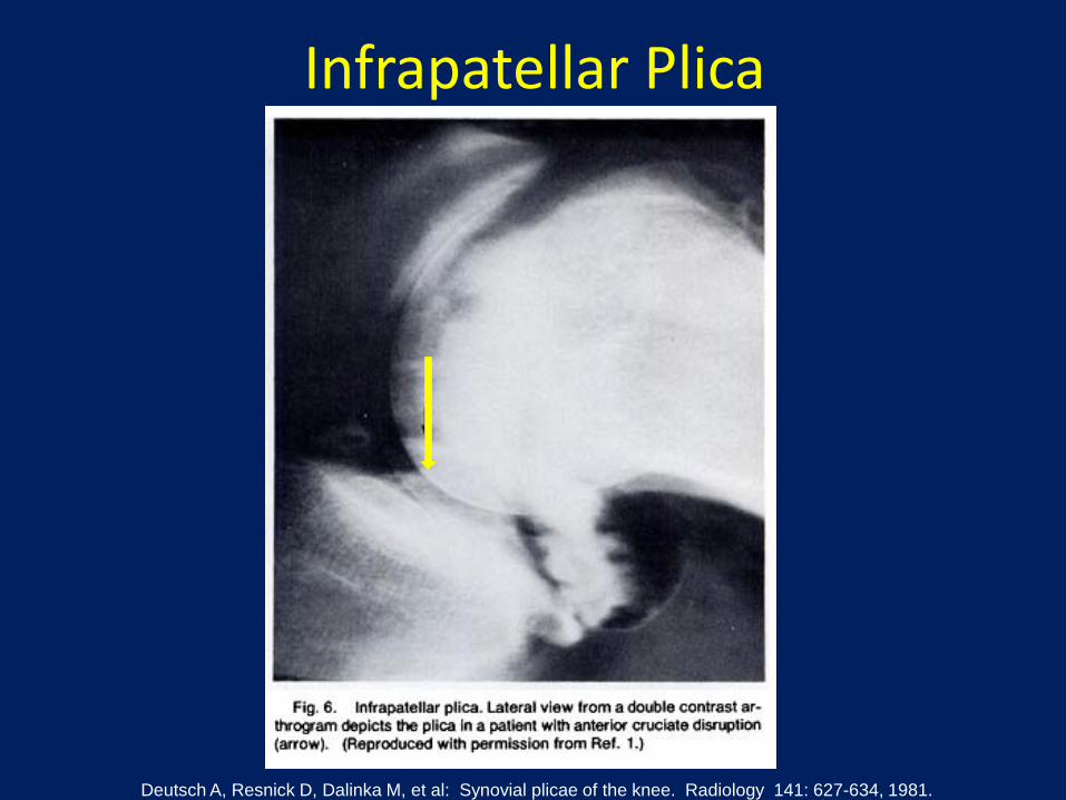
• Schematic diagram demonstrating the infrapatellar plica
Deutsch A, Resnick D, Dalinka M, et al: Synovial plicae of the knee. Radiology 141: 627-634, 1981.
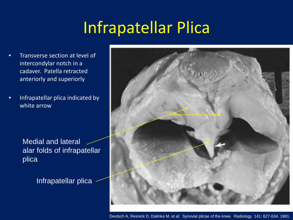
Infrapatellar Plica• Transverse section at level of
intercondylar notch in a cadaver. Patella retracted anteriorly and superiorly
• Infrapatellar plica indicated by white arrow
Deutsch A, Resnick D, Dalinka M, et al: Synovial plicae of the knee. Radiology 141: 627-634, 1981.
Infrapatellar plica
Medial and lateralalar folds of infrapatellarplica
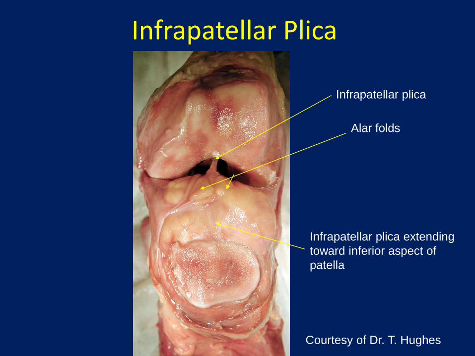
Infrapatellar Plica
Courtesy of Dr. T. Hughes
Infrapatellar plica
Alar folds
Infrapatellar plica extendingtoward inferior aspect of patella
Infrapatellar Plica
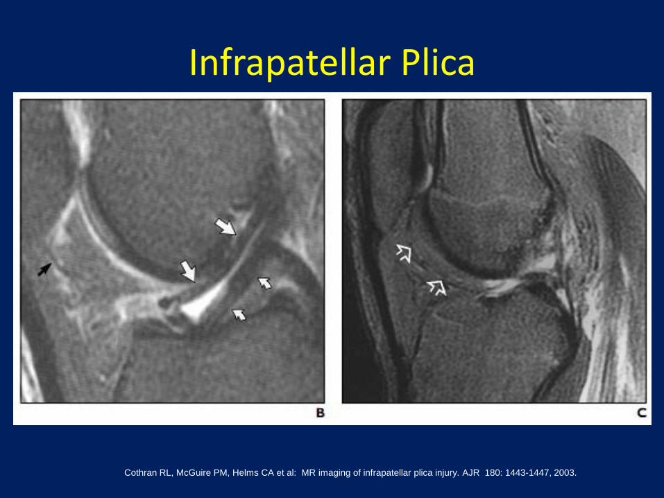
Cothran RL, McGuire PM, Helms CA et al: MR imaging of infrapatellar plica injury. AJR 180: 1443-1447, 2003.
Infrapatellar Plica
Deutsch A, Resnick D, Dalinka M, et al: Synovial plicae of the knee. Radiology 141: 627-634, 1981.
Infrapatellar Plica
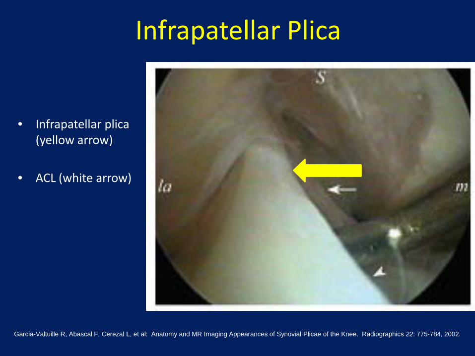
• Infrapatellar plica (yellow arrow)
• ACL (white arrow)
Garcia-Valtuille R, Abascal F, Cerezal L, et al: Anatomy and MR Imaging Appearances of Synovial Plicae of the Knee. Radiographics 22: 775-784, 2002.
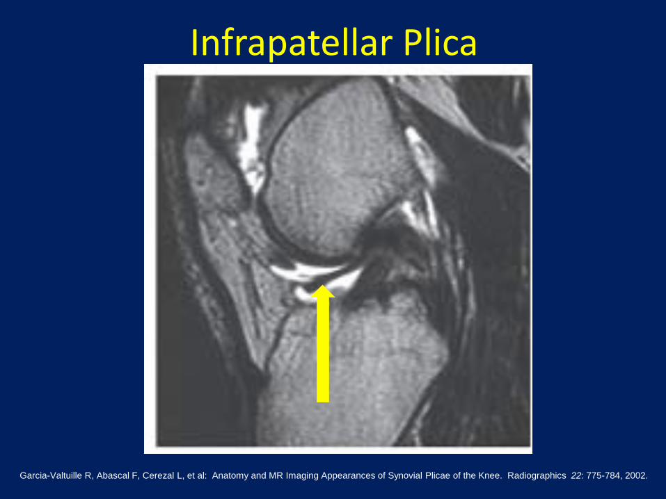
Infrapatellar Plica
Garcia-Valtuille R, Abascal F, Cerezal L, et al: Anatomy and MR Imaging Appearances of Synovial Plicae of the Knee. Radiographics 22: 775-784, 2002.
Infrapatellar Plica
Cothran RL, McGuire PM, Helms CA et al: MR imaging of infrapatellar plica injury. AJR 180: 1443-1447, 2003.
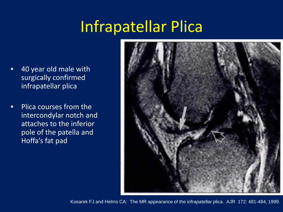
Infrapatellar Plica
• 40 year old male with surgically confirmed infrapatellar plica
• Plica courses from the intercondylar notch and attaches to the inferior pole of the patella and Hoffa’s fat pad
Kosarek FJ and Helms CA: The MR appearance of the infrapatellar plica. AJR 172: 481-484, 1999.
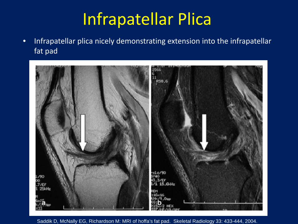
Infrapatellar Plica• Infrapatellar plica nicely demonstrating extension into the infrapatellar
fat pad
Saddik D, McNally EG, Richardson M: MRI of hoffa’s fat pad. Skeletal Radiology 33: 433-444, 2004.
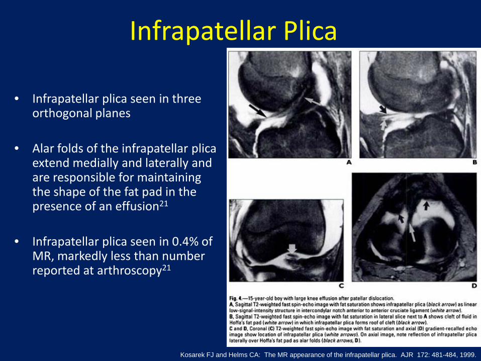
Infrapatellar Plica
• Infrapatellar plica seen in three orthogonal planes
• Alar folds of the infrapatellar plica extend medially and laterally and are responsible for maintaining the shape of the fat pad in the presence of an effusion21
• Infrapatellar plica seen in 0.4% of MR, markedly less than number reported at arthroscopy21
Kosarek FJ and Helms CA: The MR appearance of the infrapatellar plica. AJR 172: 481-484, 1999.
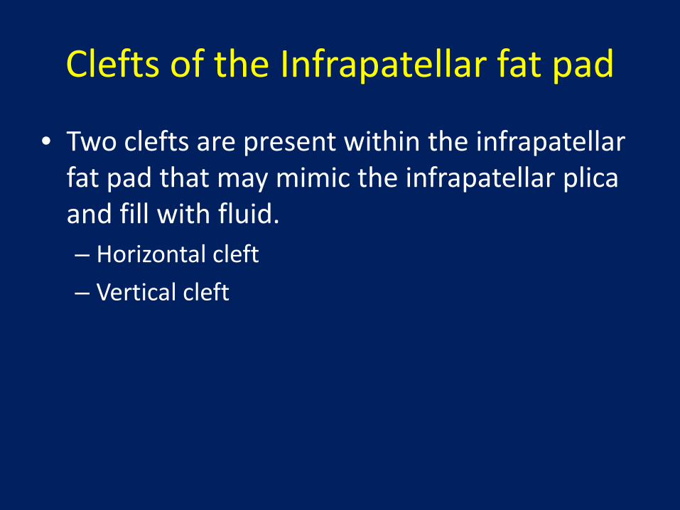
Clefts of the Infrapatellar fat pad
• Two clefts are present within the infrapatellar fat pad that may mimic the infrapatellar plica and fill with fluid.– Horizontal cleft
– Vertical cleft
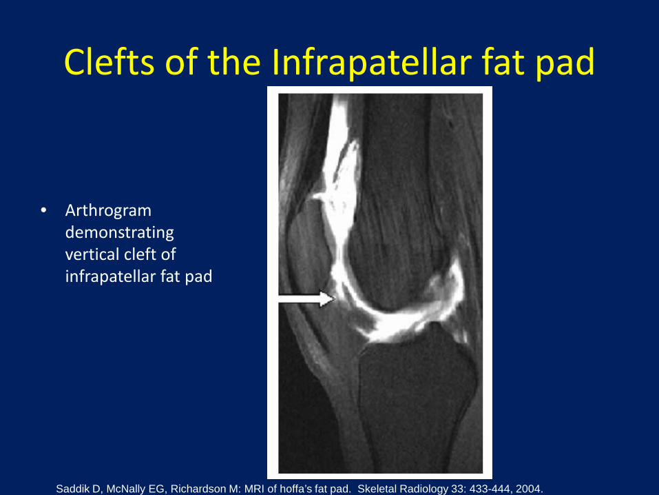
Clefts of the Infrapatellar fat pad
• Arthrogram demonstrating vertical cleft of infrapatellar fat pad
Saddik D, McNally EG, Richardson M: MRI of hoffa’s fat pad. Skeletal Radiology 33: 433-444, 2004.
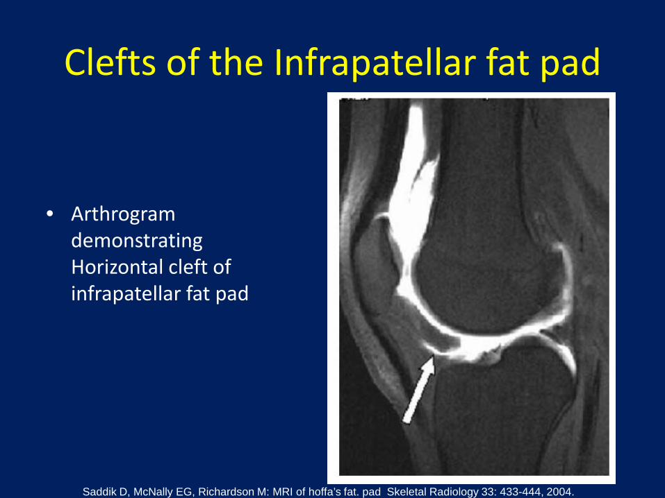
Clefts of the Infrapatellar fat pad
• Arthrogram demonstrating Horizontal cleft of infrapatellar fat pad
Saddik D, McNally EG, Richardson M: MRI of hoffa’s fat. pad Skeletal Radiology 33: 433-444, 2004.
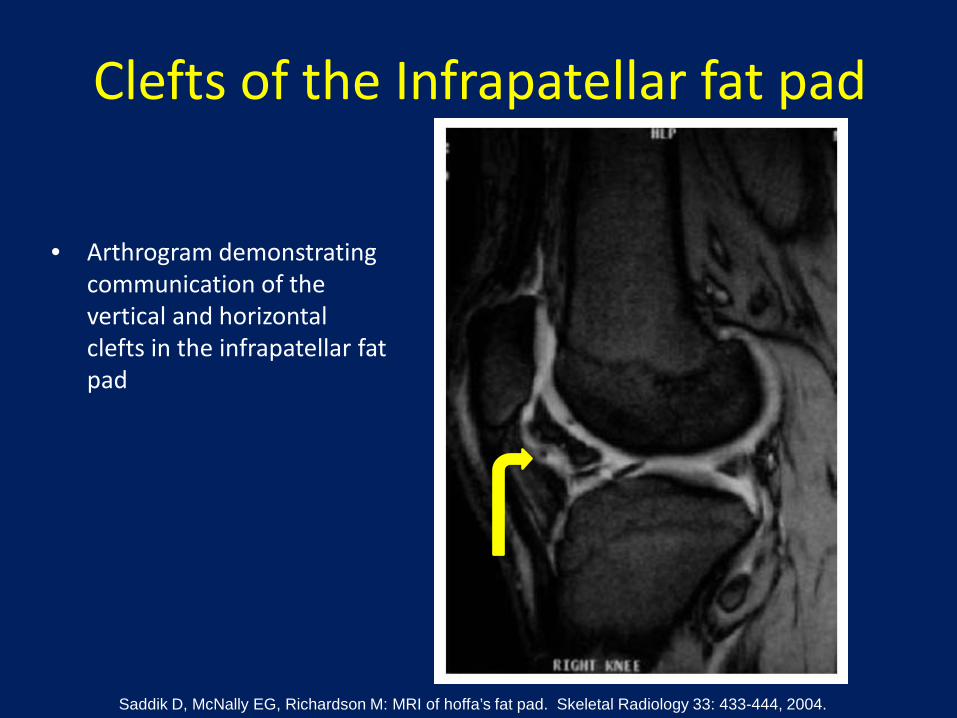
Clefts of the Infrapatellar fat pad
• Arthrogram demonstrating communication of the vertical and horizontal clefts in the infrapatellar fat pad
Saddik D, McNally EG, Richardson M: MRI of hoffa’s fat pad. Skeletal Radiology 33: 433-444, 2004.
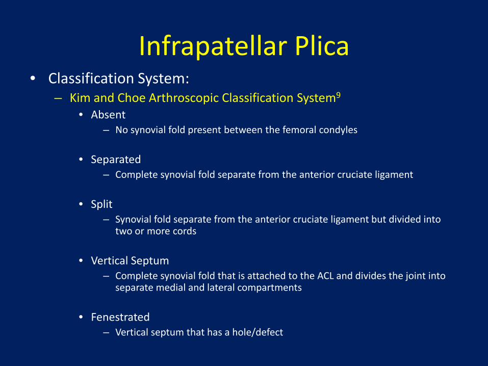
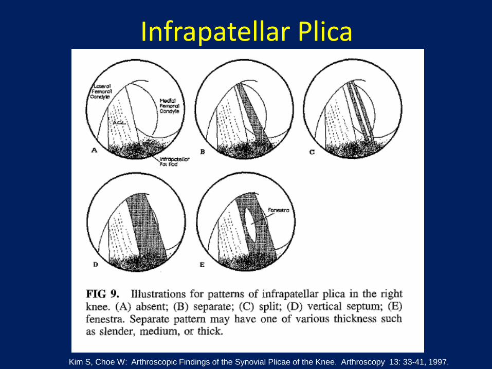
Infrapatellar Plica• Classification System:
– Kim and Choe Arthroscopic Classification System9
• Absent– No synovial fold present between the femoral condyles
• Separated– Complete synovial fold separate from the anterior cruciate ligament
• Split– Synovial fold separate from the anterior cruciate ligament but divided into
two or more cords
• Vertical Septum– Complete synovial fold that is attached to the ACL and divides the joint into
separate medial and lateral compartments
• Fenestrated– Vertical septum that has a hole/defect
Infrapatellar Plica
Kim S, Choe W: Arthroscopic Findings of the Synovial Plicae of the Knee. Arthroscopy 13: 33-41, 1997.
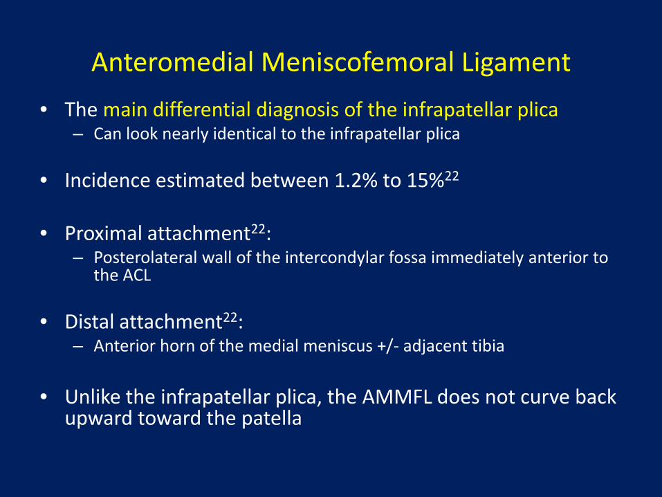
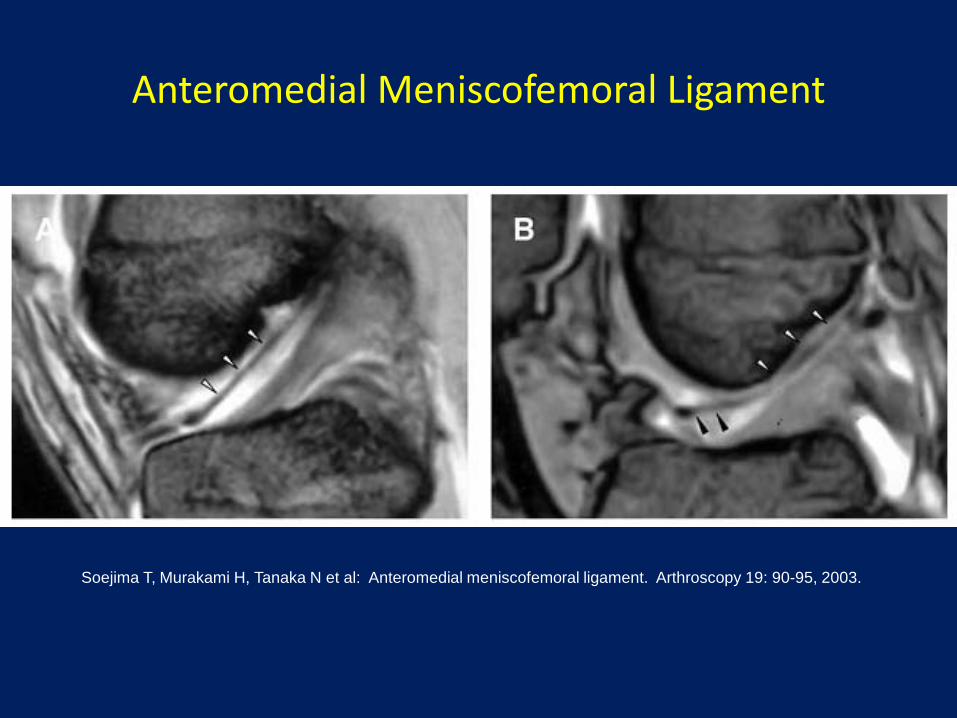
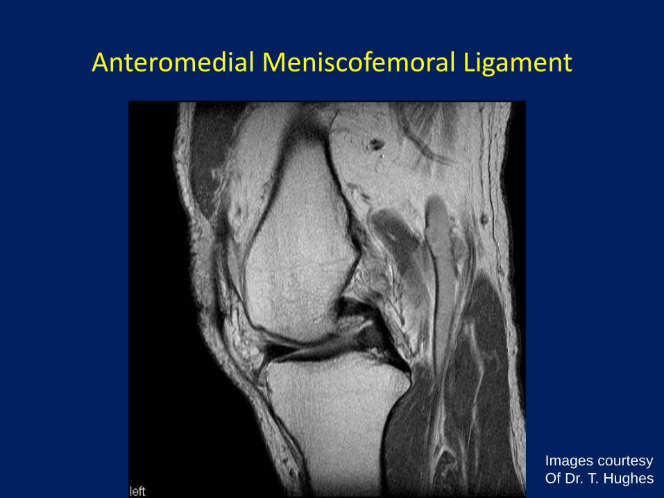
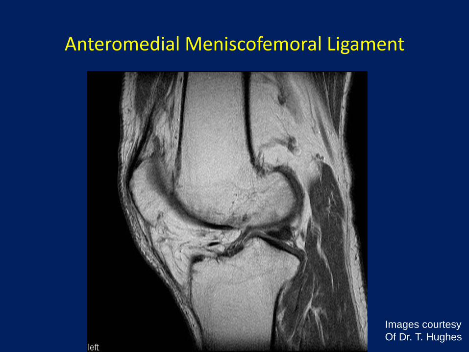
Anteromedial Meniscofemoral Ligament
• The main differential diagnosis of the infrapatellar plica– Can look nearly identical to the infrapatellar plica
• Incidence estimated between 1.2% to 15%22
• Proximal attachment22:– Posterolateral wall of the intercondylar fossa immediately anterior to
the ACL
• Distal attachment22:– Anterior horn of the medial meniscus +/- adjacent tibia
• Unlike the infrapatellar plica, the AMMFL does not curve back upward toward the patella
Anteromedial Meniscofemoral Ligament
Soejima T, Murakami H, Tanaka N et al: Anteromedial meniscofemoral ligament. Arthroscopy 19: 90-95, 2003.
Anteromedial Meniscofemoral Ligament
Images courtesy Of Dr. T. Hughes
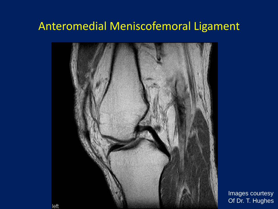
Anteromedial Meniscofemoral Ligament
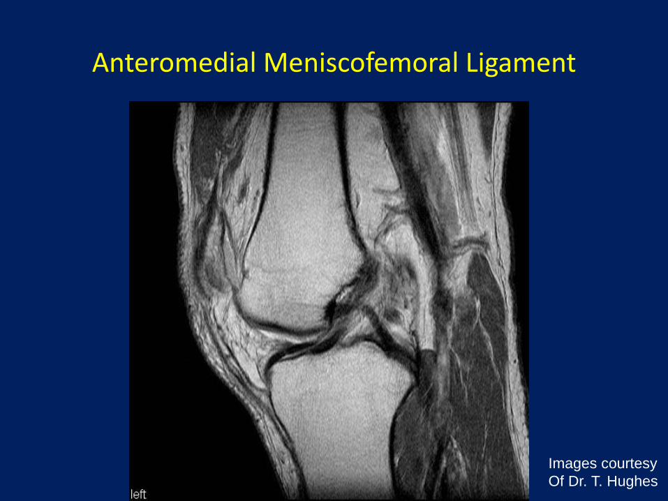
Images courtesy Of Dr. T. Hughes
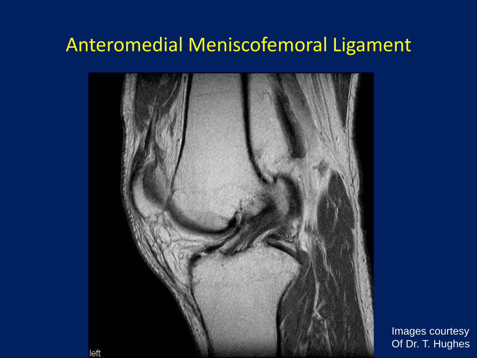
Anteromedial Meniscofemoral Ligament
Images courtesy Of Dr. T. Hughes
Anteromedial Meniscofemoral Ligament
Images courtesy Of Dr. T. Hughes
Anteromedial Meniscofemoral Ligament
Images courtesy Of Dr. T. Hughes
Anteromedial Meniscofemoral Ligament
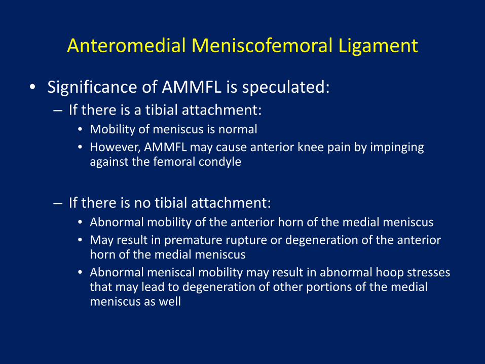
• Significance of AMMFL is speculated:– If there is a tibial attachment:
• Mobility of meniscus is normal• However, AMMFL may cause anterior knee pain by impinging
against the femoral condyle
– If there is no tibial attachment:• Abnormal mobility of the anterior horn of the medial meniscus• May result in premature rupture or degeneration of the anterior
horn of the medial meniscus• Abnormal meniscal mobility may result in abnormal hoop stresses
that may lead to degeneration of other portions of the medial meniscus as well

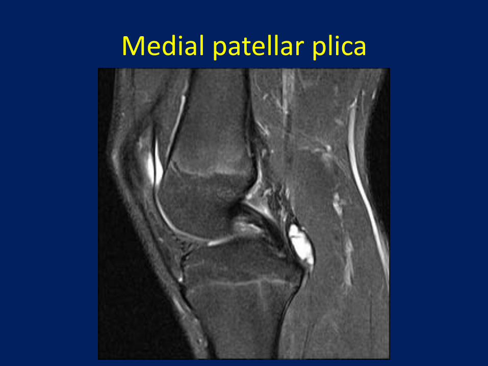
Medial patellar plica
Medial patellar plica• Synonyms in the literature2
– Medial plica– Plica synovialis patellaris/mediopatellaris– Shelf, Synovial shelf, Medial shelf– Medial intra-articular band– Iino’s band– Synovial chorda– Medial pleat– Intra-articular medial band, ledge, wedge, cleat– Alar ligament– Plica alaris elongata– Semilunar fold– Mediopatellar pseudo-meniscus– Patellar meniscus– AND MORE!!!
Medial patellar plica
• Anatomy– Originates from the medial wall of the knee joint, runs
obliquely downward, and attaches to the synovium covering the infrapatellar fat pad
– Superiorly may attach to the suprapatellar plica
– Free border may extend over the medial facet of the trochlea or under the medial facet of the patella
Medial patellar plica
• Incidence:– Third most common plica
– Arthroscopic incidence ranges from 55-72%5
Medial patellar plica• Five structures present within the medial gutter11:
– Superomedial plica• Portion of the suprapatellar plica• Transverse orientation and lies well above the patella
– Alar fold or ligament/Plica Alaris Elongata • Longitudinal fold of synovium just medial to the patella• Simply folding of the medial retinaculum when relaxed• Inconstant, wide, and extremely soft• Triangular shape in cross-section• Usually not seen on arthroscopy but seen commonly on MR imaging
– Medial patellar plica• Located one fingerbreadth from the patella and alar fold• Thinner than alar fold
– Anteromedial fringe of synovium• Covers the anterior horn of the medial meniscus
– Transverse arcuate folds• Folds of synovium located at the bottom of the medial gutter
Medial patellar plica
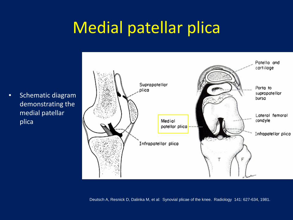
• Schematic diagram demonstrating the medial patellar plica
Deutsch A, Resnick D, Dalinka M, et al: Synovial plicae of the knee. Radiology 141: 627-634, 1981.
Medial patellar plica
Deutsch A, Resnick D, Dalinka M, et al: Synovial plicae of the knee. Radiology 141: 627-634, 1981.
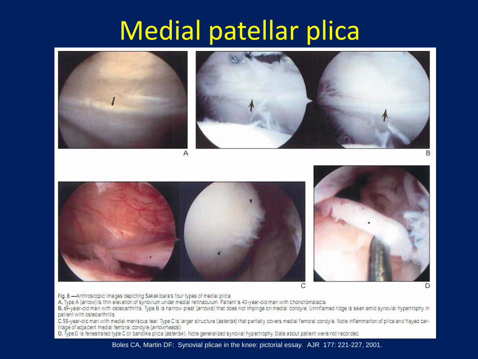
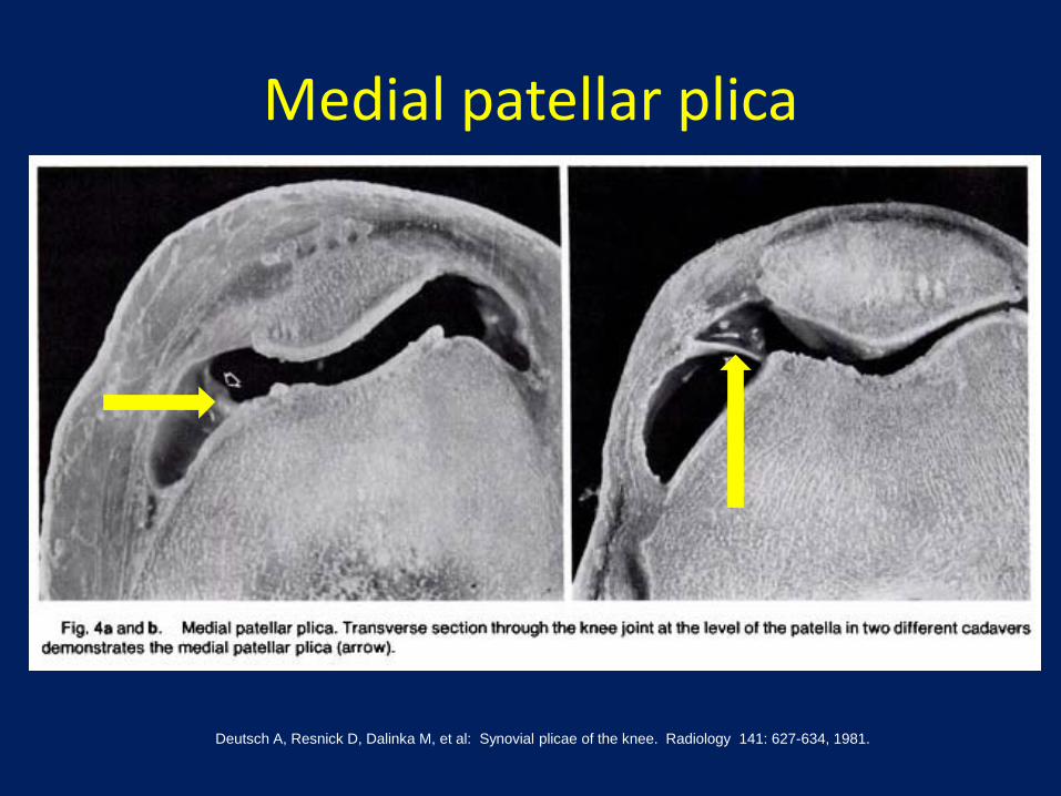
Medial patellar plica
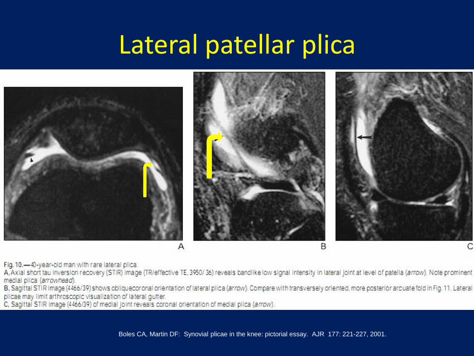
Boles CA, Martin DF: Synovial plicae in the knee: pictorial essay. AJR 177: 221-227, 2001.
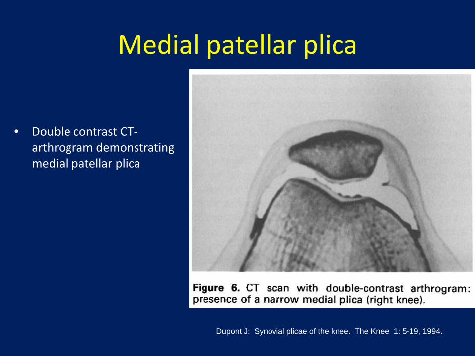
Medial patellar plica
• Double contrast CT-arthrogram demonstrating medial patellar plica
Dupont J: Synovial plicae of the knee. The Knee 1: 5-19, 1994.
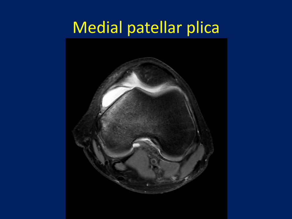
Medial patellar plica
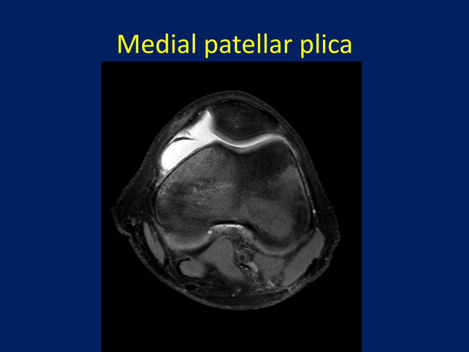
Medial patellar plica
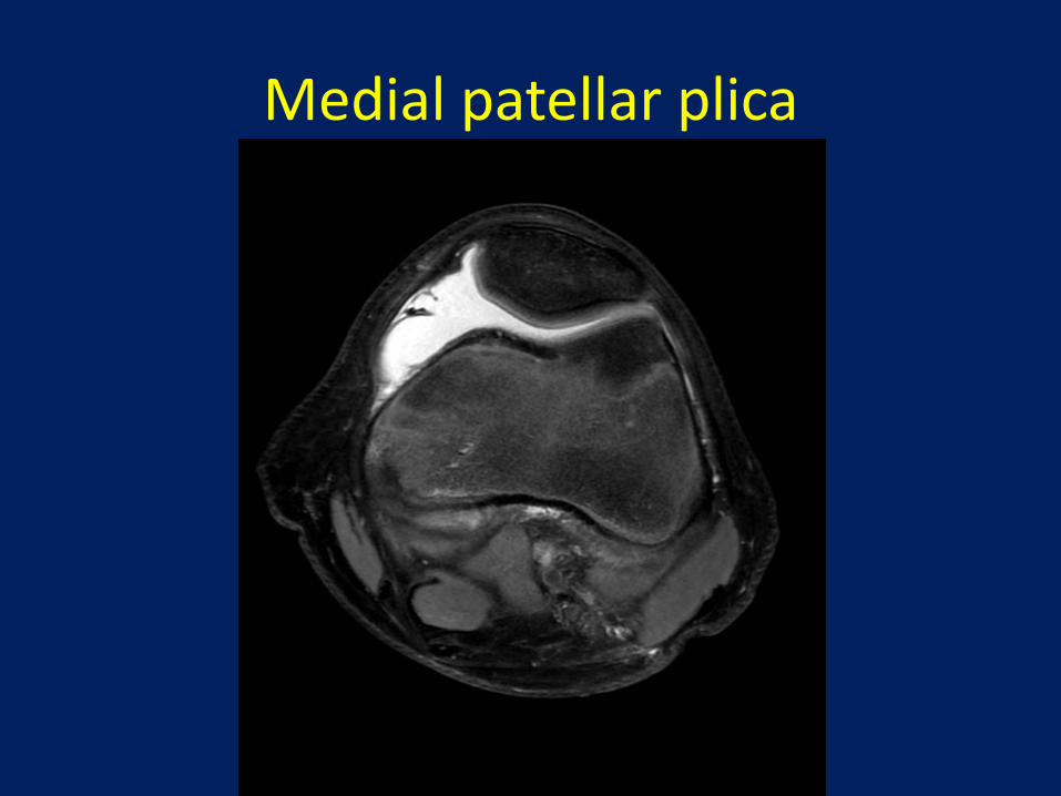
Medial patellar plica
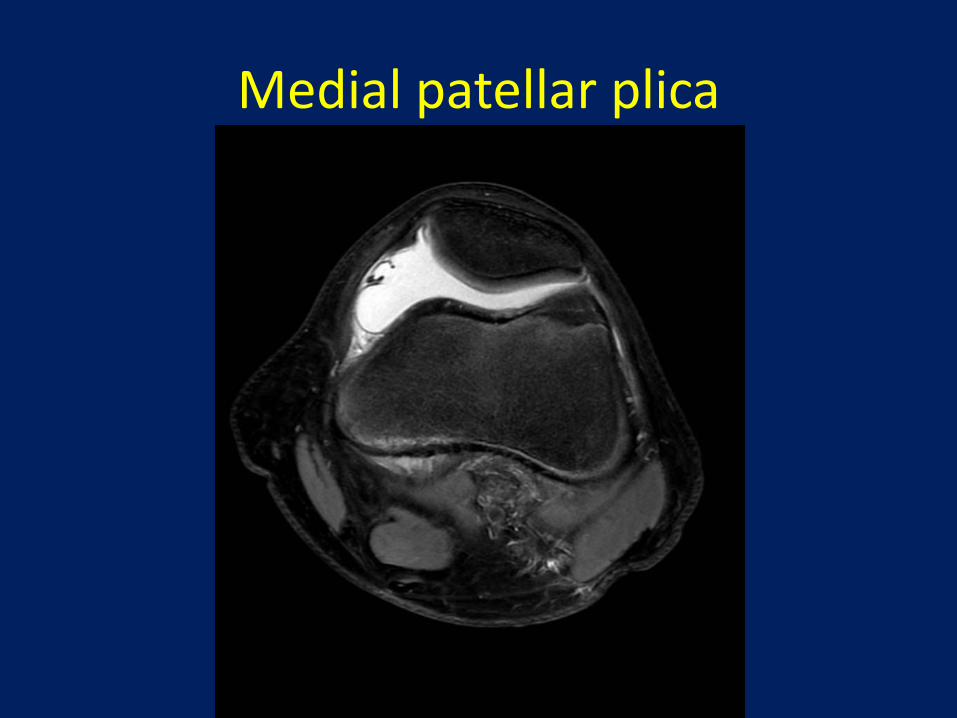
Medial patellar plica
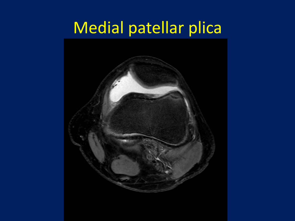
Medial patellar plica
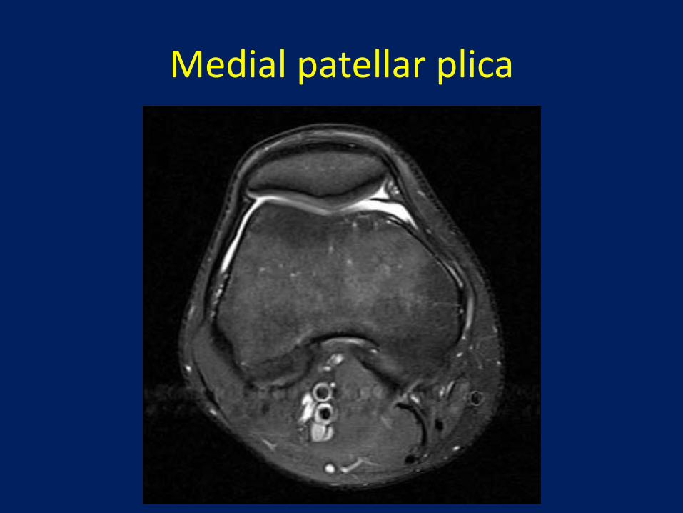
Medial patellar plica
Medial patellar plica
Medial patellar plica
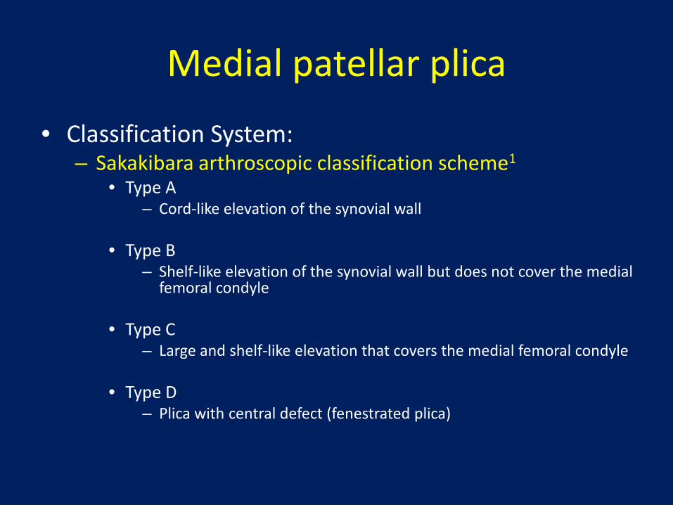
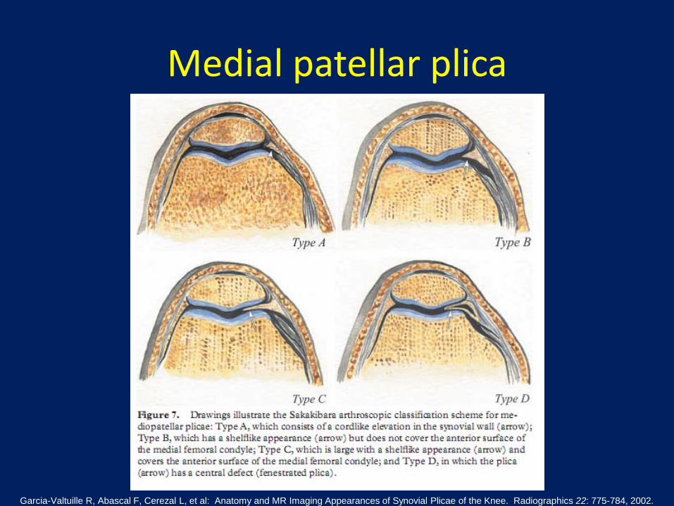
• Classification System:– Sakakibara arthroscopic classification scheme1
• Type A– Cord-like elevation of the synovial wall
• Type B– Shelf-like elevation of the synovial wall but does not cover the medial
femoral condyle
• Type C– Large and shelf-like elevation that covers the medial femoral condyle
• Type D– Plica with central defect (fenestrated plica)
Medial patellar plica
Garcia-Valtuille R, Abascal F, Cerezal L, et al: Anatomy and MR Imaging Appearances of Synovial Plicae of the Knee. Radiographics 22: 775-784, 2002.
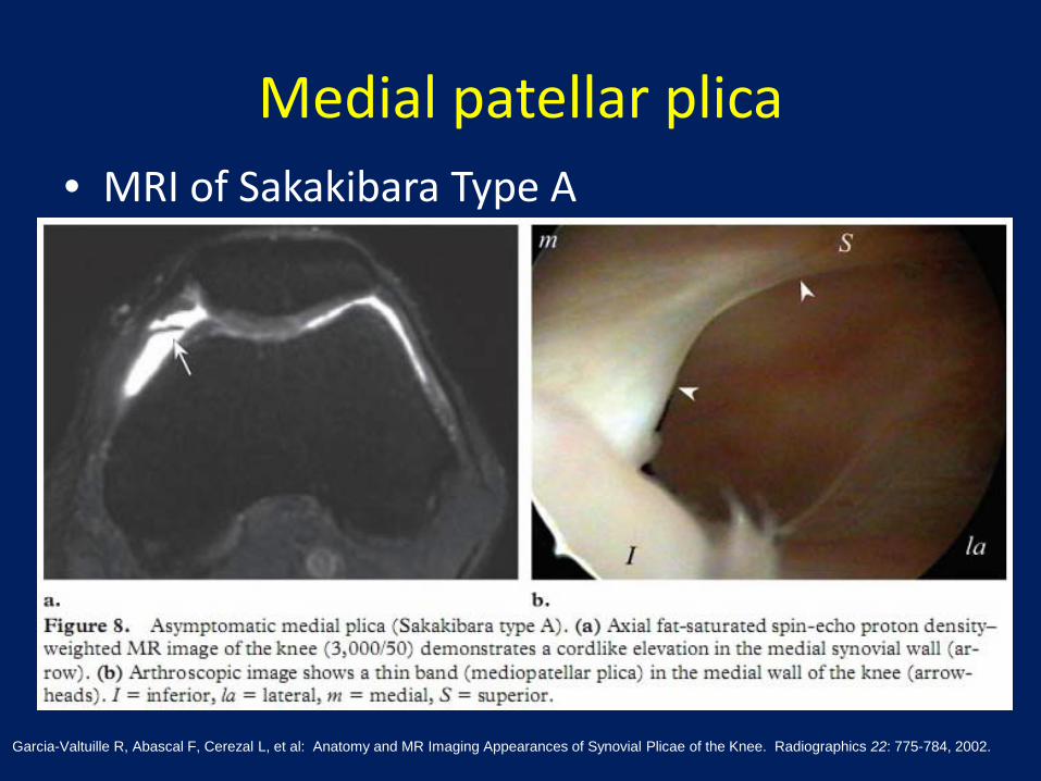
Medial patellar plica• MRI of Sakakibara Type A
Garcia-Valtuille R, Abascal F, Cerezal L, et al: Anatomy and MR Imaging Appearances of Synovial Plicae of the Knee. Radiographics 22: 775-784, 2002.
Medial patellar plica
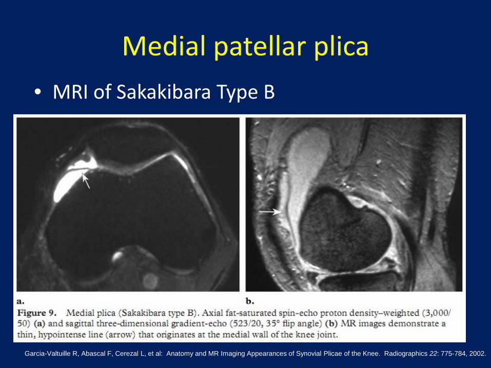
• MRI of Sakakibara Type B
Garcia-Valtuille R, Abascal F, Cerezal L, et al: Anatomy and MR Imaging Appearances of Synovial Plicae of the Knee. Radiographics 22: 775-784, 2002.
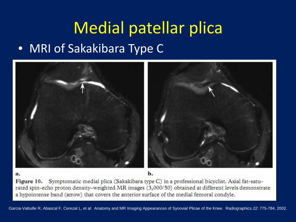
Medial patellar plica• MRI of Sakakibara Type C
Garcia-Valtuille R, Abascal F, Cerezal L, et al: Anatomy and MR Imaging Appearances of Synovial Plicae of the Knee. Radiographics 22: 775-784, 2002.
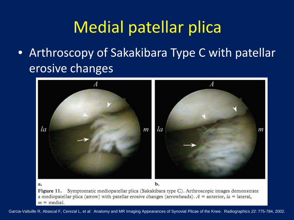
Medial patellar plica• Arthroscopy of Sakakibara Type C with patellar
erosive changes
Garcia-Valtuille R, Abascal F, Cerezal L, et al: Anatomy and MR Imaging Appearances of Synovial Plicae of the Knee. Radiographics 22: 775-784, 2002.

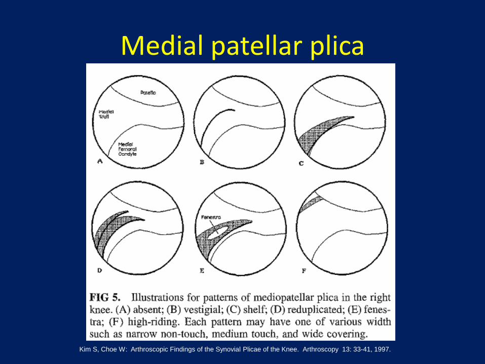
Medial patellar plica• Kim and Choe Arthroscopic Classification System9:
– Absent• No synovial shelf on the medial wall
– Vestigial• Less than 1 mm of synovial elevation that disappears with external pressure
– Shelf• A complete fold with sharp free margin
– Reduplicated• Two or more shelves running parallel to the knee. They may be differing sizes.
– Fenestra• The shelf contains a central defect
– High-riding• A shelf-like structure running anterior to the posterior part of the patella, in a
position where it could not touch the femur
Medial patellar plica
Kim S, Choe W: Arthroscopic Findings of the Synovial Plicae of the Knee. Arthroscopy 13: 33-41, 1997.
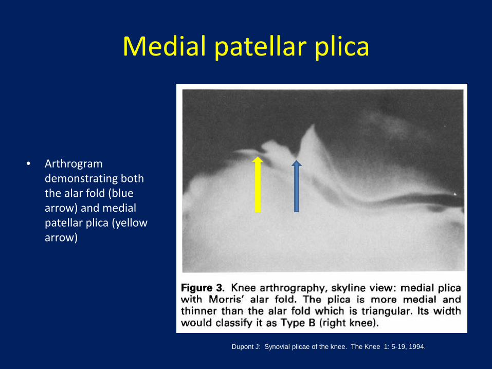
Medial patellar plica
• Arthrogram demonstrating both the alar fold (blue arrow) and medial patellar plica (yellow arrow)
Dupont J: Synovial plicae of the knee. The Knee 1: 5-19, 1994.
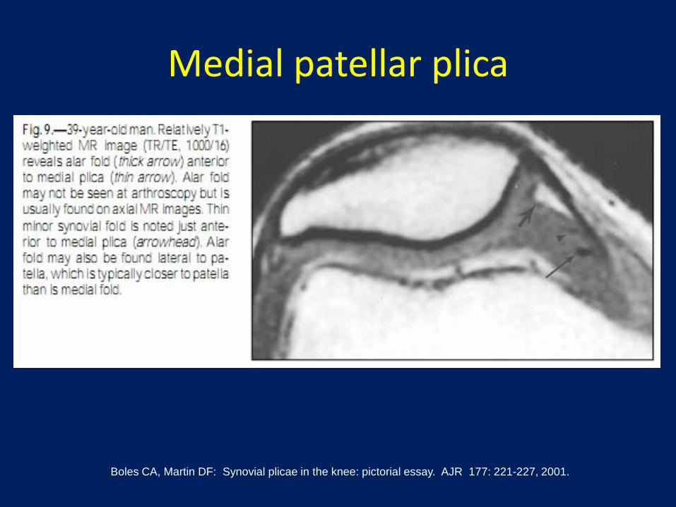
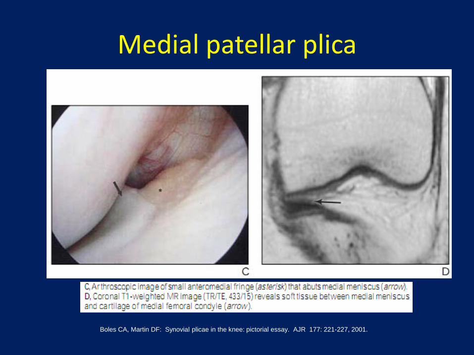
Medial patellar plica
Boles CA, Martin DF: Synovial plicae in the knee: pictorial essay. AJR 177: 221-227, 2001.
Medial patellar plica
Boles CA, Martin DF: Synovial plicae in the knee: pictorial essay. AJR 177: 221-227, 2001.

Lateral patellar plica
Lateral patellar plica
• Anatomy:– The equivalent of the medial patellar plica except
on the lateral side (mirror image of the medial plica)2
– Longitudinal, very thin, and located 1-2 cm lateral to the patella2
Lateral patellar plica
• Incidence:– By far the rarest plica
– Arthroscopic studies indicate an incidence of 0.7 – 1.0%2,5
• Commonly used arthroscopic approaches may miss this plica so the incidence may be underestimated (requires medial approach)
– Ogata and Uhthoff noted one in a 9-week embryo
Lateral patellar plica• Four structures of the lateral gutter:
– Superolateral plica• Lateral portion of the suprapatellar plica• Transverse orientation and lies well above the patella
– Alar fold or ligament/Plica Alaris Elongata • Longitudinal fold of synovium just lateral to the patella• Simply folding of the lateral retinaculum when relaxed• Inconstant, wide, and extremely soft• Triangular shape in cross-section• Usually not seen on arthroscopy but seen commonly on MR imaging
– Lateral patellar plica• Located one fingerbreadth from the patella and alar fold• Thinner than alar fold
– Transverse arcuate folds• Folds of synovium located at the bottom of the lateral gutter
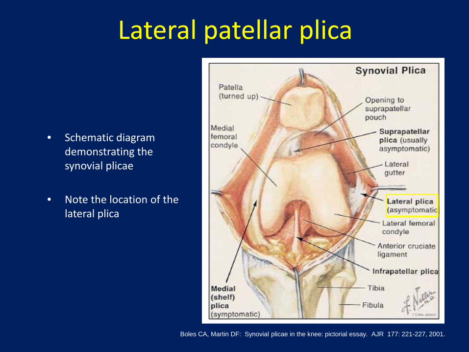
Lateral patellar plica
• Schematic diagram demonstrating the synovial plicae
• Note the location of the lateral plica
Boles CA, Martin DF: Synovial plicae in the knee: pictorial essay. AJR 177: 221-227, 2001.
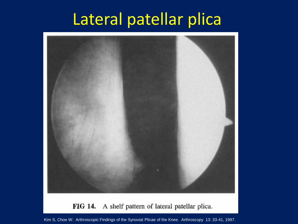
Lateral patellar plica
Kim S, Choe W: Arthroscopic Findings of the Synovial Plicae of the Knee. Arthroscopy 13: 33-41, 1997.
Lateral patellar plica
Boles CA, Martin DF: Synovial plicae in the knee: pictorial essay. AJR 177: 221-227, 2001.
Lateral patellar plica
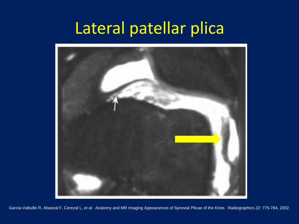
Garcia-Valtuille R, Abascal F, Cerezal L, et al: Anatomy and MR Imaging Appearances of Synovial Plicae of the Knee. Radiographics 22: 775-784, 2002.

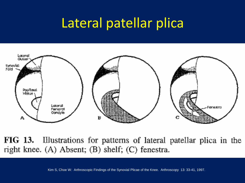
Lateral patellar plica
• Kim and Choe Arthroscopic Classification System9:– Absent
• No plica present
– Shelf• Complete shelf present with sharp free edge
– Fenestra• Shelf with central defect
Lateral patellar plica
Kim S, Choe W: Arthroscopic Findings of the Synovial Plicae of the Knee. Arthroscopy 13: 33-41, 1997.
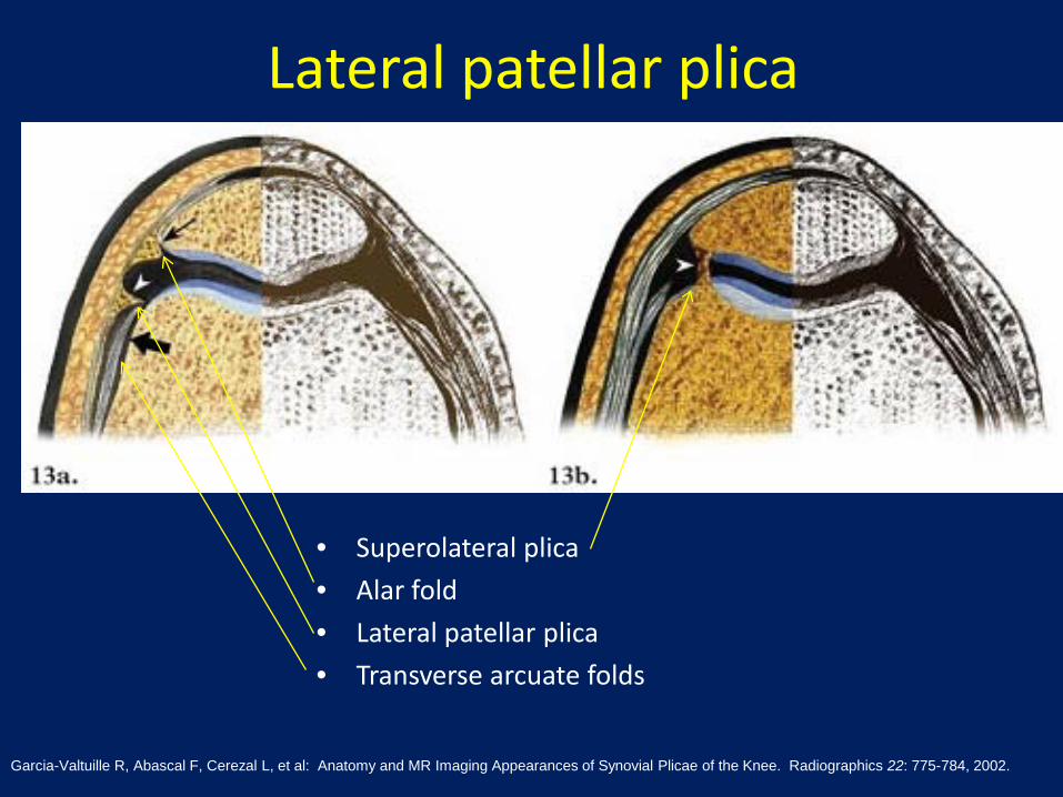
Lateral patellar plica
• Superolateral plica
• Alar fold
• Lateral patellar plica
• Transverse arcuate folds
Garcia-Valtuille R, Abascal F, Cerezal L, et al: Anatomy and MR Imaging Appearances of Synovial Plicae of the Knee. Radiographics 22: 775-784, 2002.
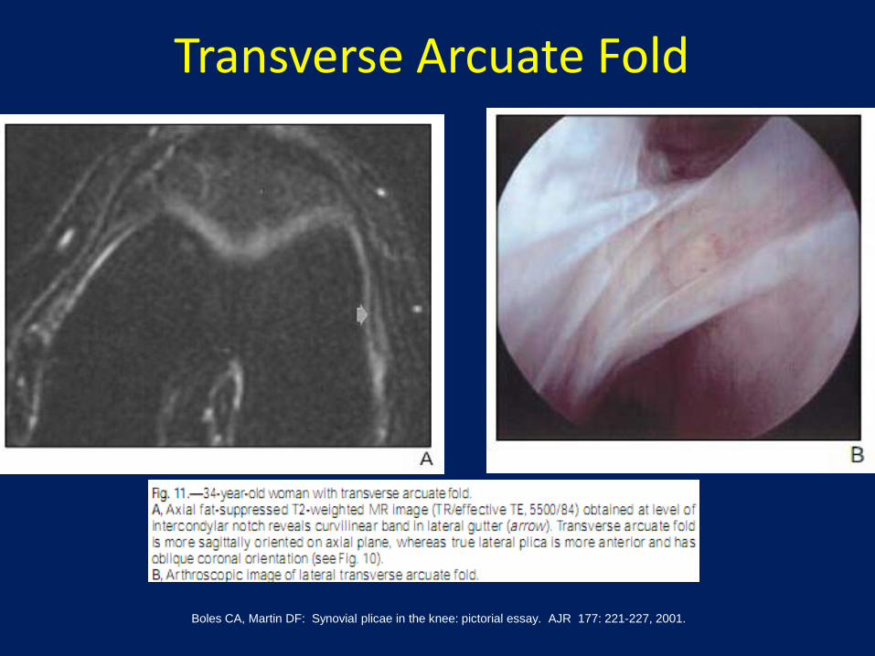
Transverse Arcuate Fold
Boles CA, Martin DF: Synovial plicae in the knee: pictorial essay. AJR 177: 221-227, 2001.
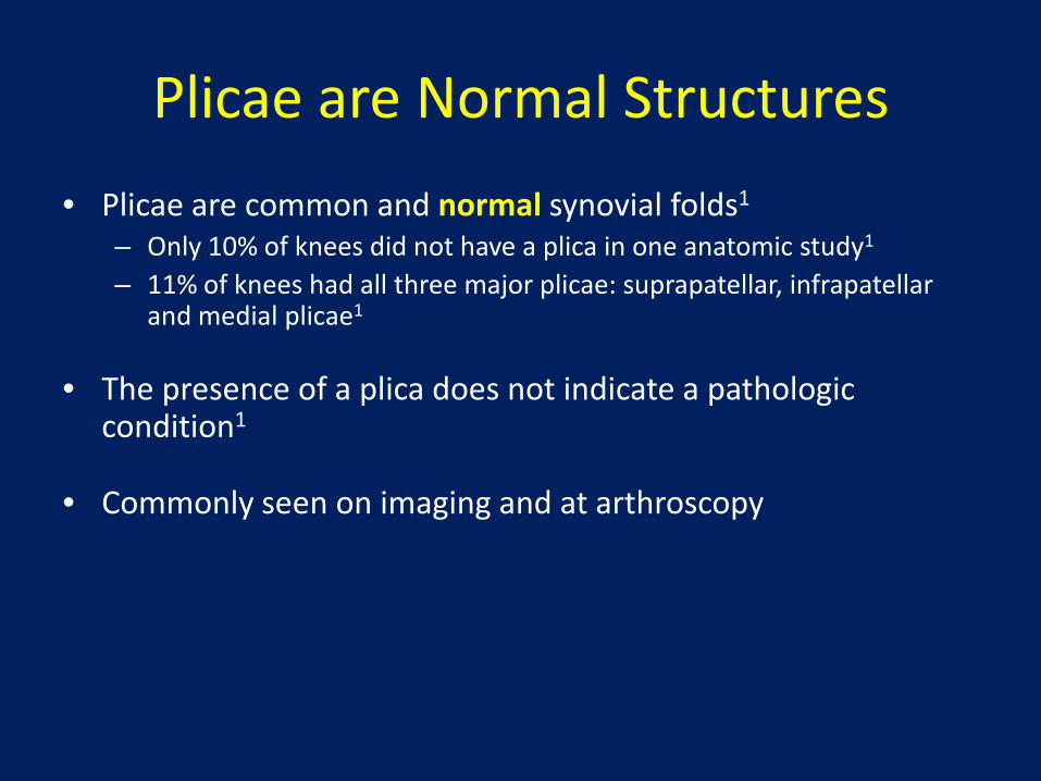
Plicae are Normal Structures
• Plicae are common and normal synovial folds1
– Only 10% of knees did not have a plica in one anatomic study1
– 11% of knees had all three major plicae: suprapatellar, infrapatellar and medial plicae1
• The presence of a plica does not indicate a pathologic condition1
• Commonly seen on imaging and at arthroscopy
Plicae are Normal Structures
• Proposed physiologic purposes of plicae8:– Limit friction
– Increase the area covered by the synovial membrane
– May help spread synovial fluid over the articular surfaces of the knee aiding in joint lubrication
When good plica go bad: “Plica Syndromes”
• Plica can lead to clinical symptoms that can mimic internal derangements of the knee13
– “Plica Syndromes”
• Two general mechanisms cause plica syndromes:– Inflammatory process or trauma can thicken and stiffen a plica
• Normal elastic tissues replaced by fibrous elements• Plica becomes inelastic and tight resulting in:
– Mechanical synovitis– Softening and erosion of adjacent articular cartilage
– Complete failure of resorption leaving a complete septum and compartmentalization a joint
• Suprapatellar and infrapatellar plica

Pathologic Plica: Suprapatellar Plica
Plica Syndrome: Suprapatellar Plica
• How does the suprapatellar plica cause symptoms?– Joint Compartmentalization
• By far the most common cause of symptoms• Suprapatellar plica with a small porta may close and function
as a complete plica if it pathologically thickens
– Impingement on medial femoral condyle with possible adjacent cartilage damage
Plica Syndrome: Suprapatellar Plica
• Joint Compartmentalization
– 1996 Trout et al.13
• Report on five patients with abnormal suprapatellar plica that led to compartmentalization of the suprapatellar pouch
• Led to distension of the suprapatellar pouch and manifested as a soft-tissue mass clinically
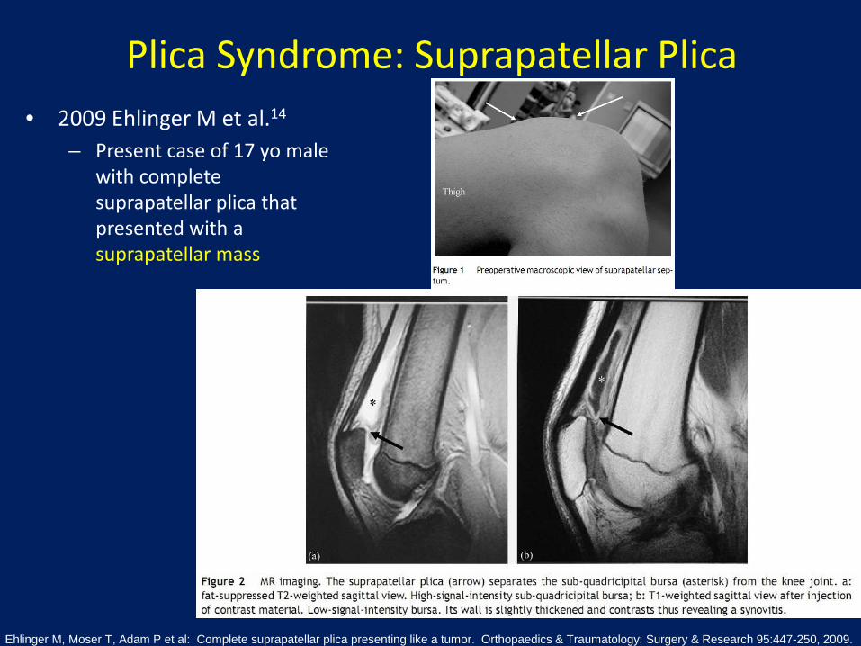
Plica Syndrome: Suprapatellar Plica• 2009 Ehlinger M et al.14
– Present case of 17 yo male with complete suprapatellar plica that presented with a suprapatellar mass
Ehlinger M, Moser T, Adam P et al: Complete suprapatellar plica presenting like a tumor. Orthopaedics & Traumatology: Surgery & Research 95:447-250, 2009.
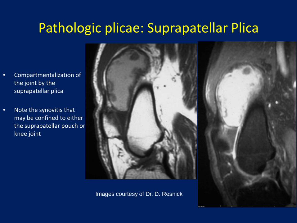
Pathologic plicae: Suprapatellar Plica
• Compartmentalization of the joint by the suprapatellar plica
• Note the synovitis that may be confined to either the suprapatellar pouch or knee joint
Images courtesy of Dr. D. Resnick
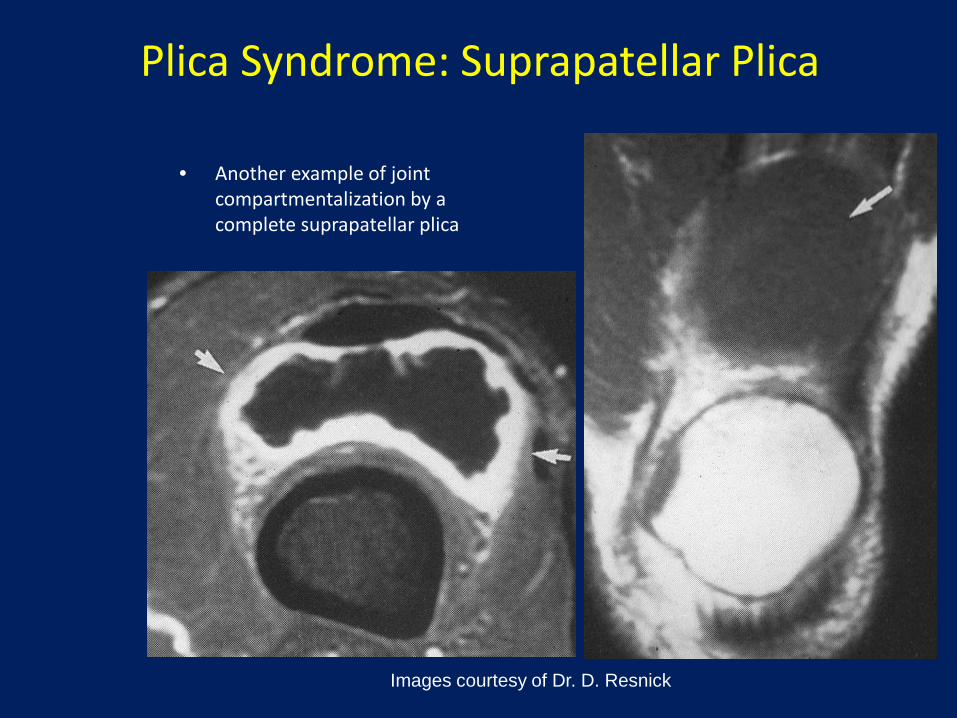
Plica Syndrome: Suprapatellar Plica
• Another example of joint compartmentalization by a complete suprapatellar plica
Images courtesy of Dr. D. Resnick
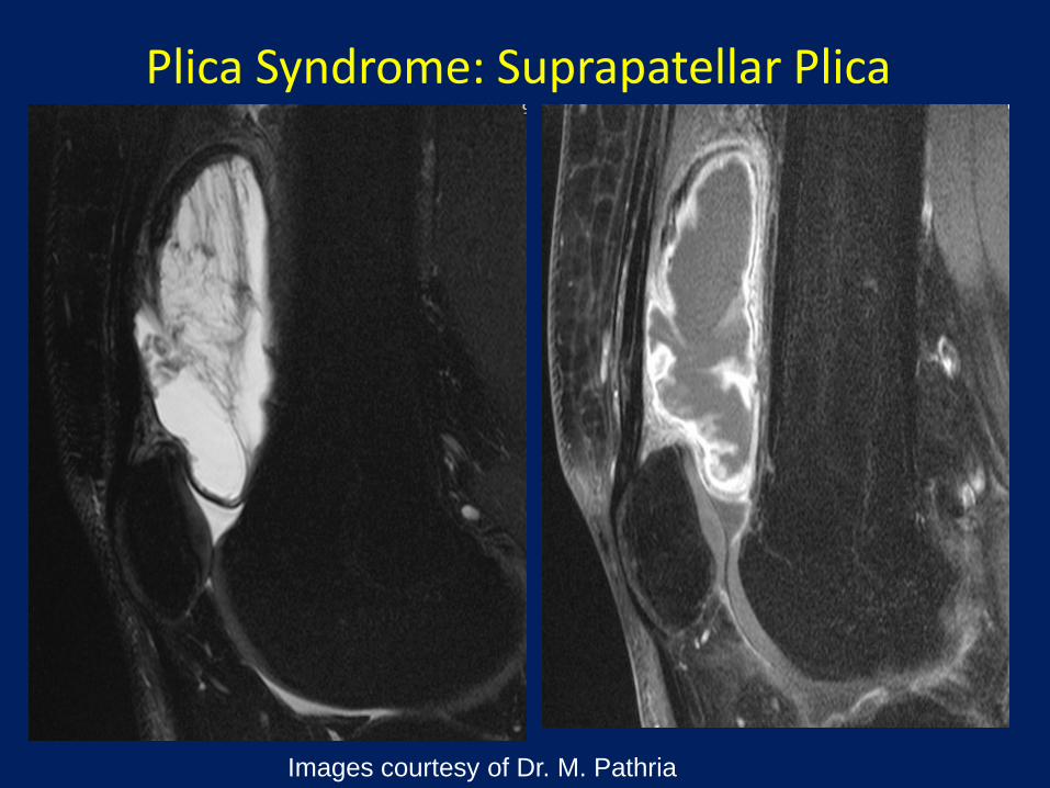
Plica Syndrome: Suprapatellar Plica
Images courtesy of Dr. M. Pathria
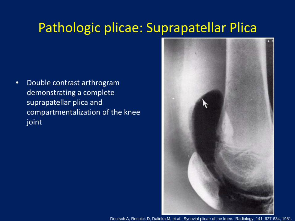
Pathologic plicae: Suprapatellar Plica
• Double contrast arthrogram demonstrating a complete suprapatellar plica and compartmentalization of the knee joint
Deutsch A, Resnick D, Dalinka M, et al: Synovial plicae of the knee. Radiology 141: 627-634, 1981.
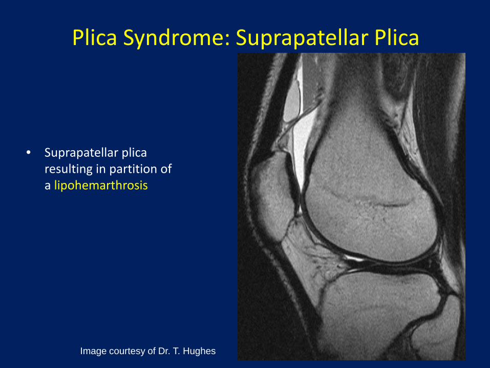
Plica Syndrome: Suprapatellar Plica
• Suprapatellar plica resulting in partition of a lipohemarthrosis
Image courtesy of Dr. T. Hughes
Pathologic plicae: Suprapatellar Plica
• Documentation of a complete suprapatellar plica is very important– Will change surgical approach
– Cases of osteocartilaginous bodies in a separate suprapatellar bursa that could not be located after infrapatellar incision8
– Diffuse synovial processes (ex. PVNS) may only affect the sequestered suprapatellar pouch8
– Acute suppurative bursitis – rupture of the plica may unnecessarily contaminate the joint8
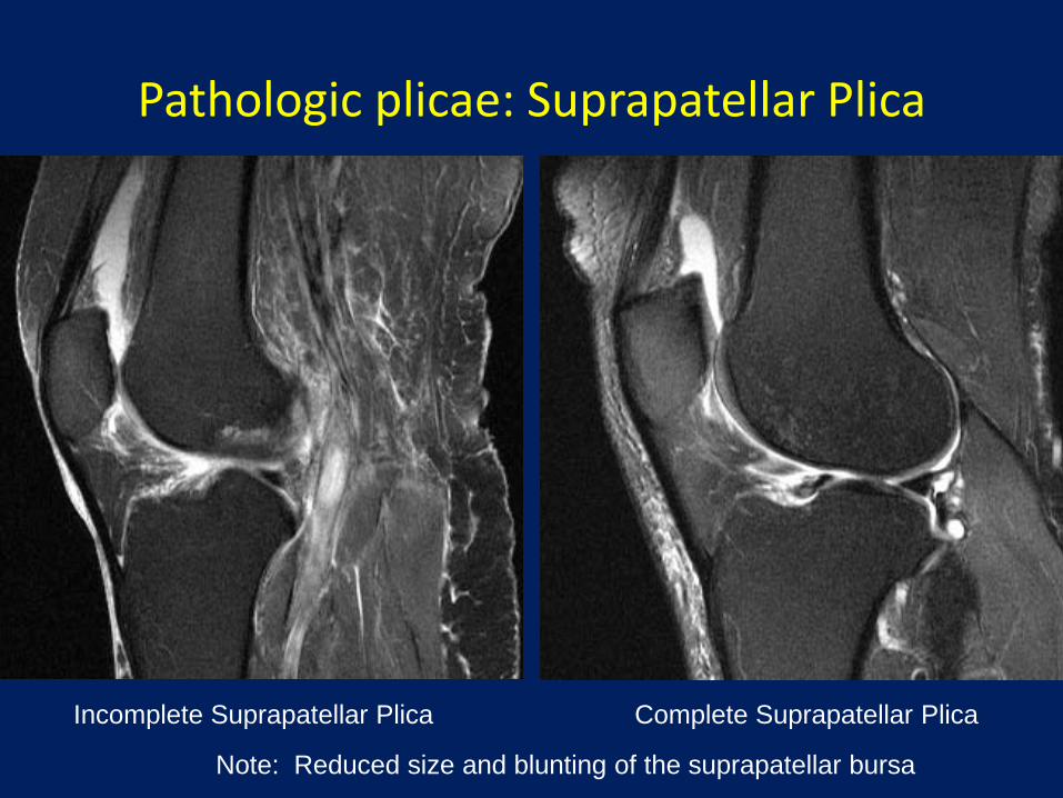
Pathologic plicae: Suprapatellar Plica
Incomplete Suprapatellar Plica Complete Suprapatellar Plica
Note: Reduced size and blunting of the suprapatellar bursa
Plica Syndrome: Suprapatellar Plica
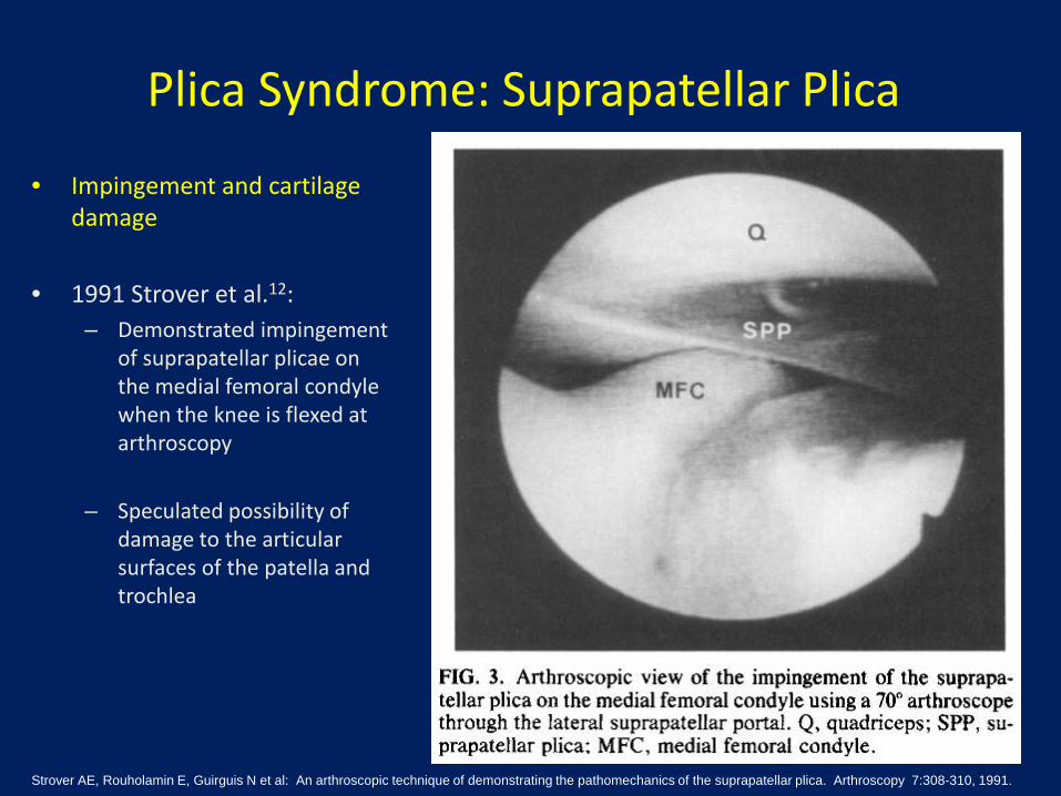
• Impingement and cartilage damage
• 1991 Strover et al.12:– Demonstrated impingement
of suprapatellar plicae on the medial femoral condyle when the knee is flexed at arthroscopy
– Speculated possibility of damage to the articular surfaces of the patella and trochlea
Strover AE, Rouholamin E, Guirguis N et al: An arthroscopic technique of demonstrating the pathomechanics of the suprapatellar plica. Arthroscopy 7:308-310, 1991.
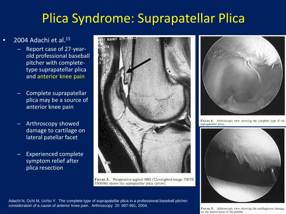
Plica Syndrome: Suprapatellar Plica• 2004 Adachi et al.15
– Report case of 27-year-old professional baseball pitcher with complete-type suprapatellar plica and anterior knee pain
– Complete suprapatellar plica may be a source of anterior knee pain
– Arthroscopy showed damage to cartilage on lateral patellar facet
– Experienced complete symptom relief after plica resection
Adachi N, Ochi M, Uchio Y: The complete type of suprapatellar plica in a professional baseball pitcher: consideration of a cause of anterior knee pain. Arthroscopy 20: 987-991, 2004.
Plica Syndrome: Infrapatellar Plica
Plica Syndrome: Infrapatellar plica
• Very rarely a cause of symptoms
• Etiologies of Infrapatellar Plica Syndrome:– May become thick and fibrotic resulting in impingement in the
intercondylar notch
– Can rupture and mimic an ACL tear and result in a hemarthrosis
– May form a complete septum and result in compartmentalization of the medial and lateral knee compartments
• Similar compartmentalization of the joint has been seen in hereditary onycho-osteoarthrodysplasia (nail-patella syndrome)
– Can limit mobility of the arthroscope and make arthroscopy more difficult
Plica Syndrome: Infrapatellar plica
• 1996 Kim and Choe16
– Two cases of flexion contractures of the knees where a vertical septum type infrapatellar plica was demonstrated
– Plica were thickened, fibrotic, and lost their normal elasticity
– Resection of the plica resulted in complete symptom resolution
Plica Syndrome: Infrapatellar plica
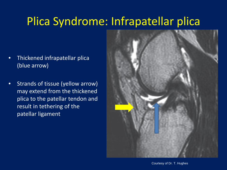
• Thickened infrapatellar plica (blue arrow)
• Strands of tissue (yellow arrow) may extend from the thickened plica to the patellar tendon and result in tethering of the patellar ligament
Courtesy of Dr. T. Hughes
Plica Syndrome: Infrapatellar plica
• Ruptured infrapatellar plica may result in a post-traumatic hemarthrosis
• 1995 Kohn et al.18
– Anatomic and clinical study in the orthopedic literature
– Describes torn infrapatellar plica as only abnormality in 3/57 patients with posttraumatic hemarthrosis and a clinically stable joint
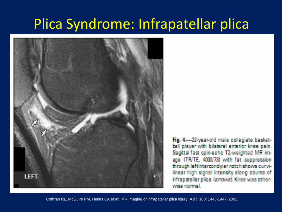
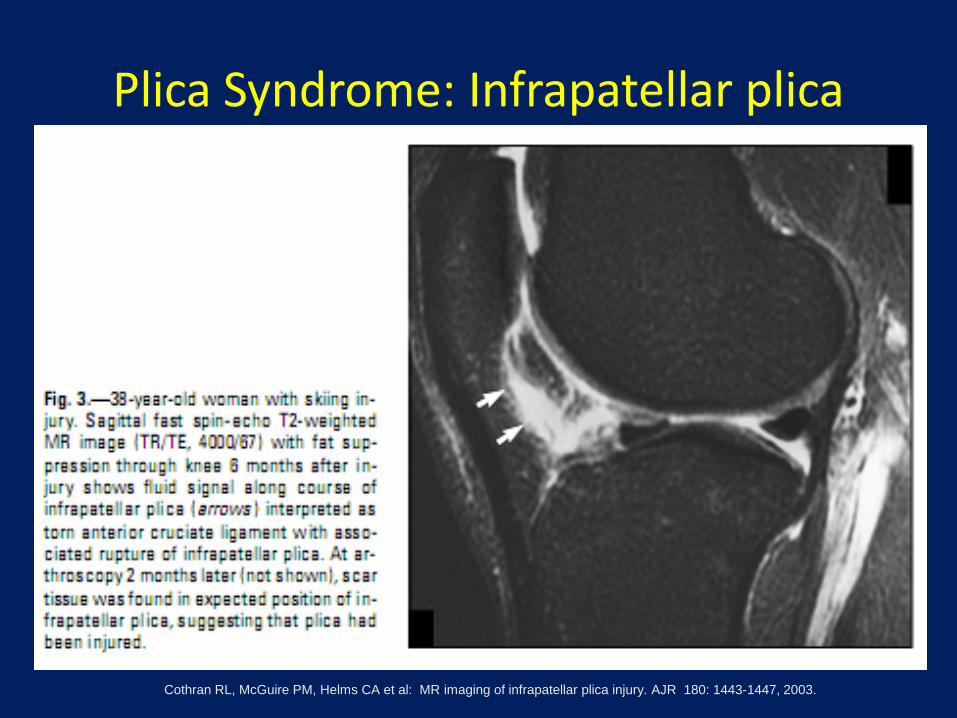
Plica Syndrome: Infrapatellar plica• 2003 Cothran et al.17
– Often we see increased signal along the course of the infrapatellar plica
– Abnormal increased signal along the infrapatellar plicae at MR imaging may indicate direct trauma to the plica or inflammation related to Hoffa’s disease
– Isolated injury to plica is uncommon and therefore they recommend exclusion of other causes of internal derangements prior to attributing symptoms to an injured plica
– Most of the examples cited in the article were diagnosed retrospectively
• Caution is warranted in diagnosing infrapatellar plica pathology8:– Abnormal signal along the course of the plica is frequent
• There are normal recesses in Hoffa’s fat pad that may collect fluid
Plica Syndrome: Infrapatellar plica
Cothran RL, McGuire PM, Helms CA et al: MR imaging of infrapatellar plica injury. AJR 180: 1443-1447, 2003.
Plica Syndrome: Infrapatellar plica
Cothran RL, McGuire PM, Helms CA et al: MR imaging of infrapatellar plica injury. AJR 180: 1443-1447, 2003.
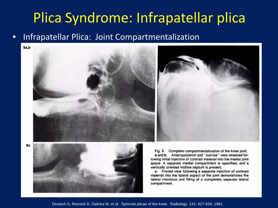
Plica Syndrome: Infrapatellar plica• Infrapatellar Plica: Joint Compartmentalization
Deutsch A, Resnick D, Dalinka M, et al: Synovial plicae of the knee. Radiology 141: 627-634, 1981.

Plica Syndrome: Medial patellar plica
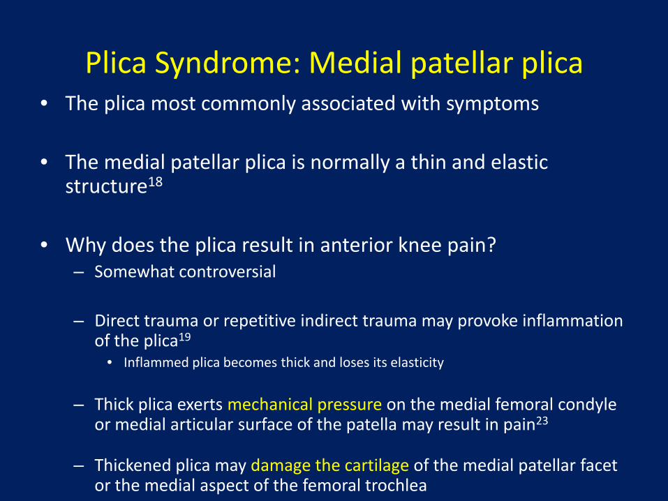
Plica Syndrome: Medial patellar plica• The plica most commonly associated with symptoms
• The medial patellar plica is normally a thin and elastic structure18
• Why does the plica result in anterior knee pain?– Somewhat controversial
– Direct trauma or repetitive indirect trauma may provoke inflammation of the plica19
• Inflammed plica becomes thick and loses its elasticity
– Thick plica exerts mechanical pressure on the medial femoral condyle or medial articular surface of the patella may result in pain23
– Thickened plica may damage the cartilage of the medial patellar facet or the medial aspect of the femoral trochlea
Plica Syndrome: Medial patellar plica
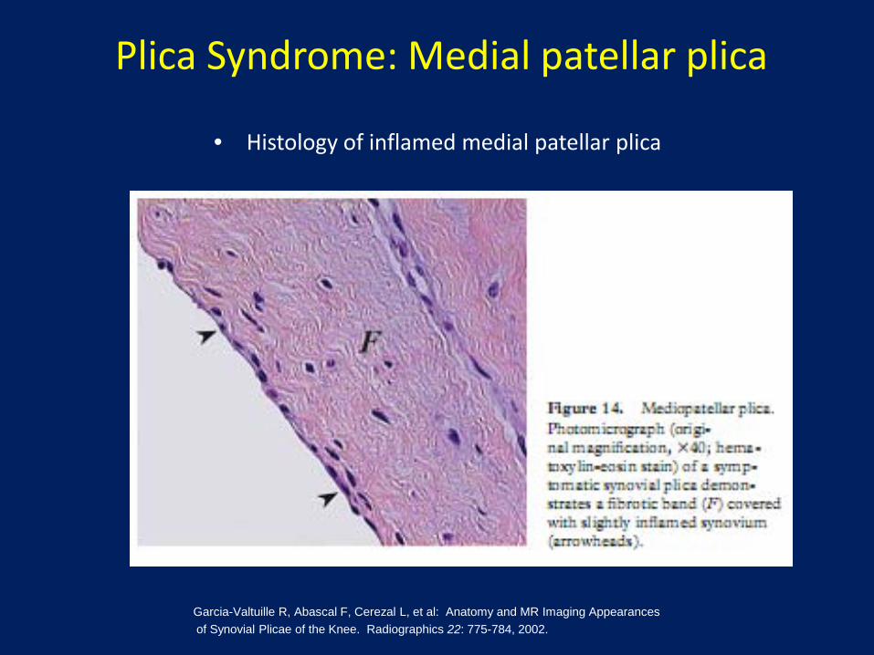
• Histology of inflamed medial patellar plica
Garcia-Valtuille R, Abascal F, Cerezal L, et al: Anatomy and MR Imaging Appearancesof Synovial Plicae of the Knee. Radiographics 22: 775-784, 2002.
Plica Syndrome: Medial patellar plica
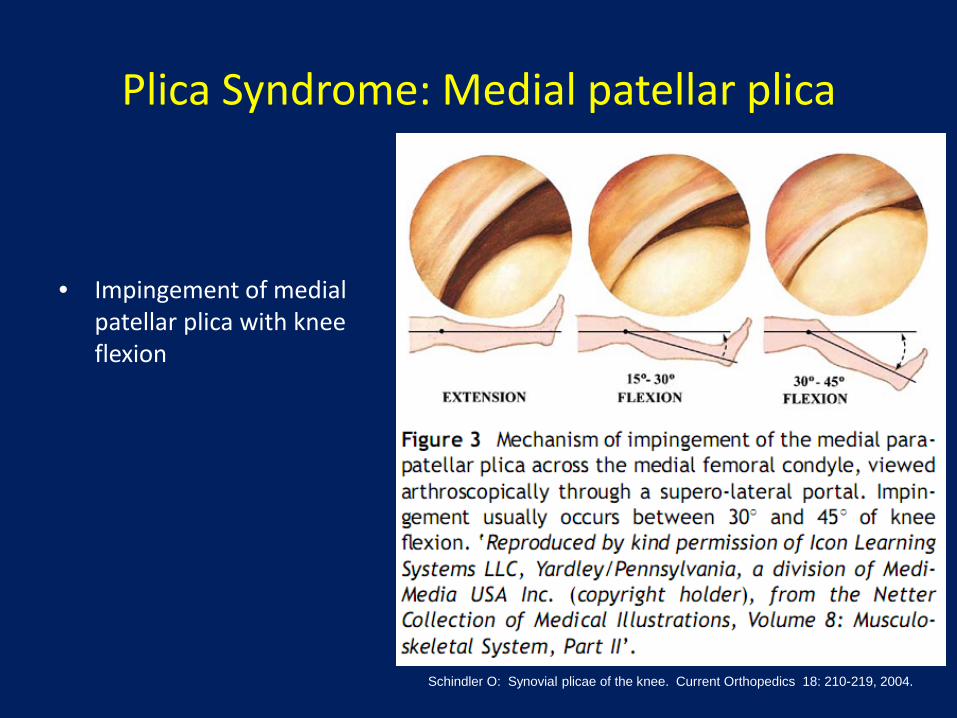
• Impingement of medial patellar plica with knee flexion
Schindler O: Synovial plicae of the knee. Current Orthopedics 18: 210-219, 2004.
Plica Syndrome: Medial patellar plica
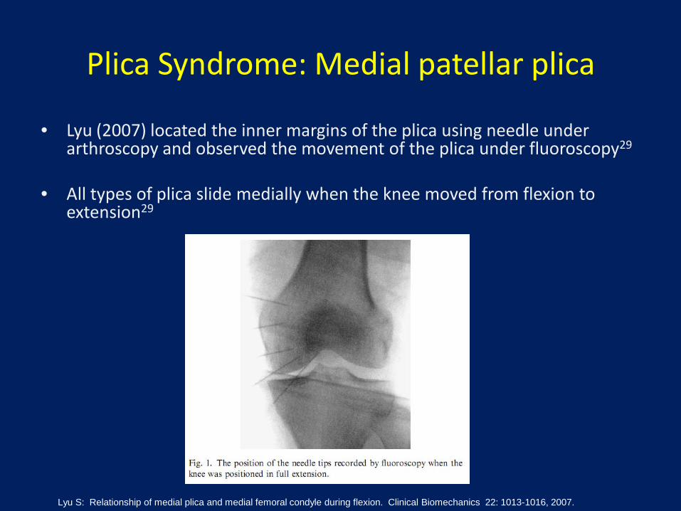
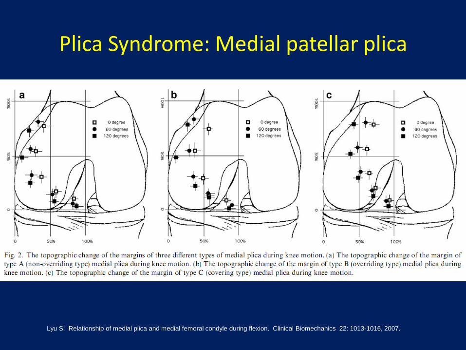
• Lyu (2007) located the inner margins of the plica using needle under arthroscopy and observed the movement of the plica under fluoroscopy29
• All types of plica slide medially when the knee moved from flexion to extension29
Lyu S: Relationship of medial plica and medial femoral condyle during flexion. Clinical Biomechanics 22: 1013-1016, 2007.
Plica Syndrome: Medial patellar plica
Lyu S: Relationship of medial plica and medial femoral condyle during flexion. Clinical Biomechanics 22: 1013-1016, 2007.
Plica Syndrome: Medial patellar plica
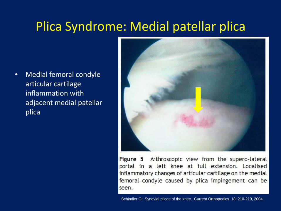
• Medial femoral condyle articular cartilage inflammation with adjacent medial patellar plica
Schindler O: Synovial plicae of the knee. Current Orthopedics 18: 210-219, 2004.
Plica Syndrome: Medial patellar plica
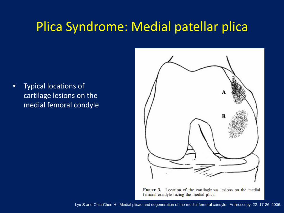
• Typical locations of cartilage lesions on the medial femoral condyle
Lyu S and Chia-Chen H: Medial plicae and degeneration of the medial femoral condyle. Arthroscopy 22: 17-26, 2006.
Plica Syndrome: Medial patellar plica
• 2004 Farkas et al.23
– The criteria for a “symptomatic plica” is not uniform and often the diagnosis is only made after removal of the plica causes a reduction in the patient’s symptoms
– Even plica that are not in direct contact with the articular surface may be a source of pain
– Discovered a significantly increased number of neuroelements in symptomatic plica compared to asymptomatic plica
• The more neuroelements the greater the pain sensitivity of the structure
– Found that the number of neuroelements was significantly higher in patients that had previous trauma
• Trauma may induce neuroelement formation and “sensitize” the plica
Plica Syndrome: Medial patellar plica• Clinical history of medial plica syndrome18:
– Usually teenagers to young adults
– Dull, achy, pain that increases with activity• Worse when going up or down stairs, squatting or bending, rising from
chair after sitting
– Pain localizes to proximomedial aspect of the knee
– Some will give history of recent trauma
– 50% give history of repetitive strenuous activity
– Plica may catch and product a “pseudo-locking” event
– Symptoms mimic a torn medial meniscus, arthritis, OCD, or other internal derangement
Plica Syndrome: Medial patellar plica
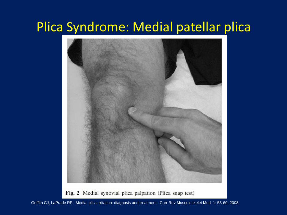
• Diagnosis:– Physical examination18:
• Palpation between medial border of patella and adductor tubercle reproduces patients symptoms
– Plica is rolled under the examiner’s fingers– Ribbon-like fold is palpated
• Many other provocative tests have been developed
Plica Syndrome: Medial patellar plica
Griffith CJ, LaPrade RF: Medial plica irritation: diagnosis and treatment. Curr Rev Musculoskelet Med 1: 53-60, 2008.
Plica Syndrome: Medial patellar plica
• Diagnosis:– Imaging:
• The main purpose of imaging is to rule out other sources of pathology not to diagnose plica syndrome18
• Radiographs of the knee are obtained to rule out osseous sources of pain (OCD, OA, fractures, etc.)18
Plica Syndrome: Medial patellar plica
• Diagnosis:– Imaging
• MRI– MR findings are non-specific18
– No correlation between MR findings and arthroscopic classification systems19
– No correlation between MR findings and probability of resection at time of arthroscopy19
– No significant correlation between preoperative MR imaging and arthroscopic findings19
Plica Syndrome: Medial patellar plica• Diagnosis:
– Imaging:• MRI
– A normal MR does not rule out the presence of a medial plica19
» Sensitivity lower if minimal joint fluid is present
– Most authors agree that wider plica are more often symptomatic and demonstrate pathologic changes at histology1
» Most asymptomatic plica are 1-2 mm in thickness» Most symptomatic plica are over 2 mm and may even
reach 1 cm in size» However, no absolute thickness can distinguish them8
– MR may show chondral abnormalities on the medial patellar facet or medial femoral condyle associated with the plica
– MR is excellent at ruling out other internal derangements that may cause the patient’s symptoms
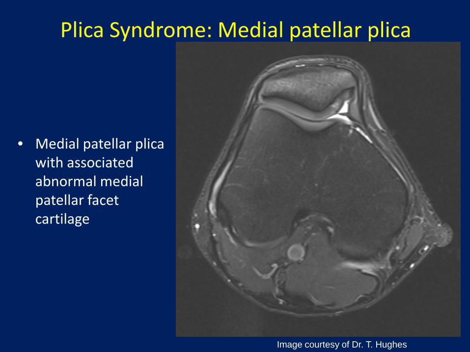
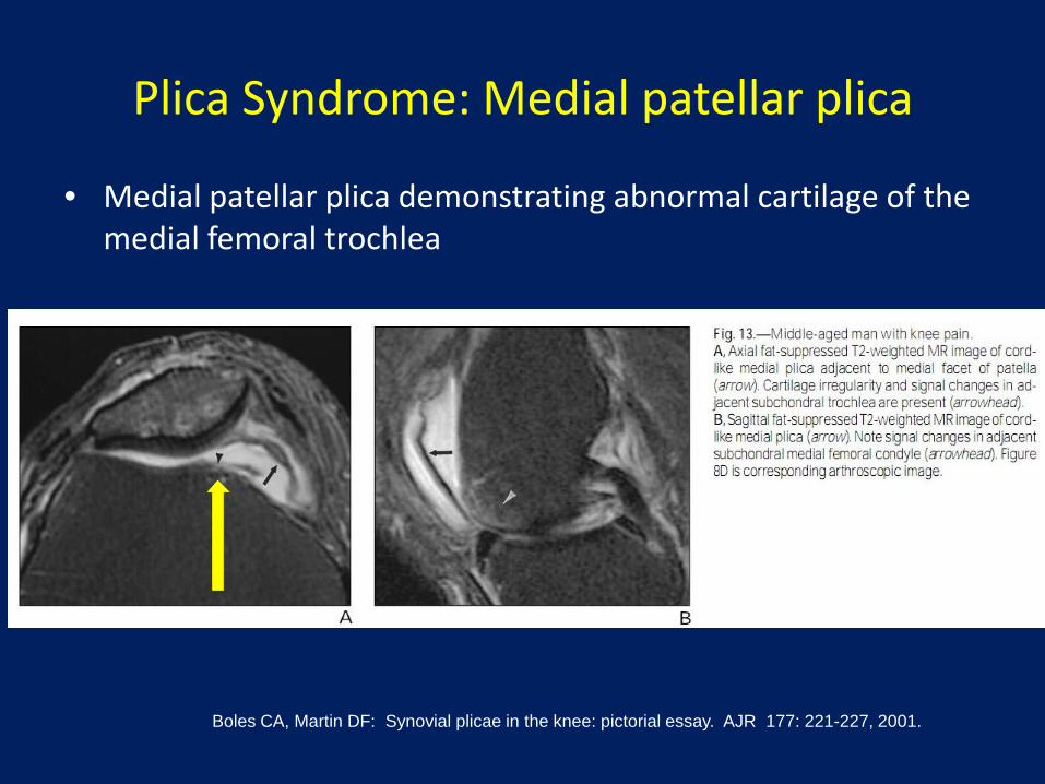
Plica Syndrome: Medial patellar plica
• Medial patellar plica with associated abnormal medial patellar facet cartilage
Image courtesy of Dr. T. Hughes
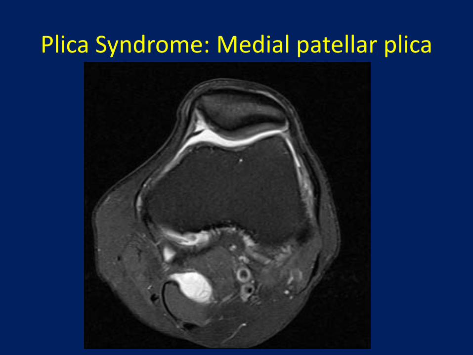
Plica Syndrome: Medial patellar plica
Plica Syndrome: Medial patellar plica
• Medial patellar plica demonstrating abnormal cartilage of the medial femoral trochlea
Boles CA, Martin DF: Synovial plicae in the knee: pictorial essay. AJR 177: 221-227, 2001.
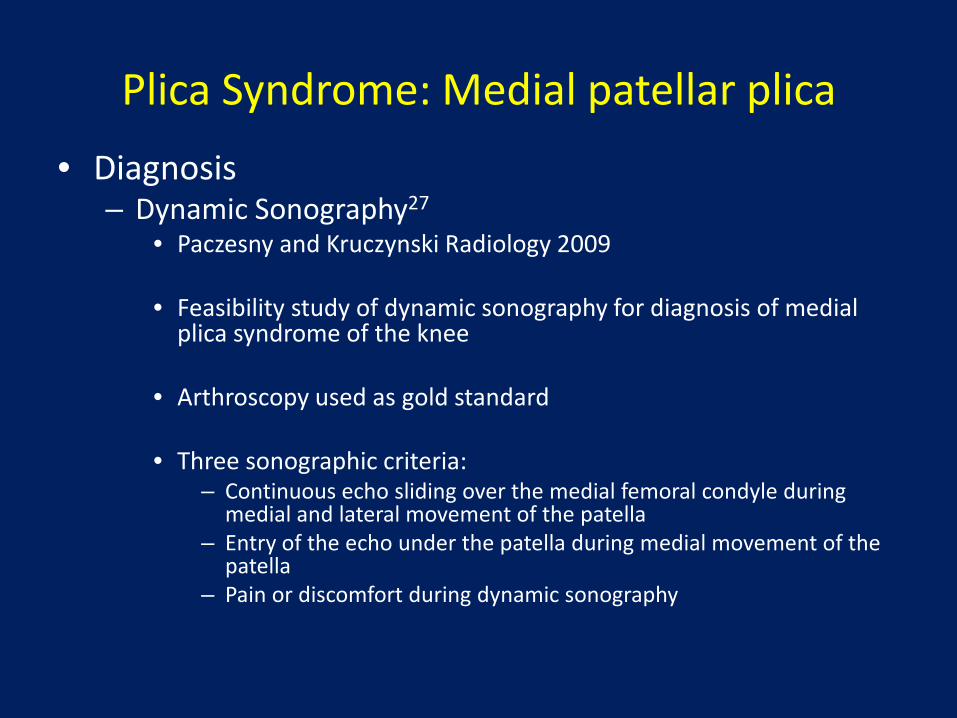
Plica Syndrome: Medial patellar plica
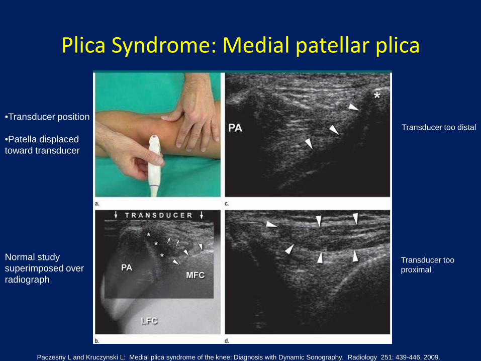
• Diagnosis– Dynamic Sonography27
• Paczesny and Kruczynski Radiology 2009
• Feasibility study of dynamic sonography for diagnosis of medial plica syndrome of the knee
• Arthroscopy used as gold standard
• Three sonographic criteria:– Continuous echo sliding over the medial femoral condyle during
medial and lateral movement of the patella– Entry of the echo under the patella during medial movement of the
patella– Pain or discomfort during dynamic sonography
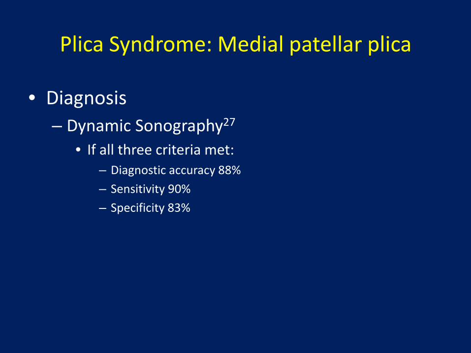
Plica Syndrome: Medial patellar plica
• Diagnosis– Dynamic Sonography27
• If all three criteria met:– Diagnostic accuracy 88%
– Sensitivity 90%
– Specificity 83%
Plica Syndrome: Medial patellar plica
Transducer too distal
Transducer too proximal
•Transducer position
•Patella displacedtoward transducer
Normal study superimposed overradiograph
Paczesny L and Kruczynski L: Medial plica syndrome of the knee: Diagnosis with Dynamic Sonography. Radiology 251: 439-446, 2009.
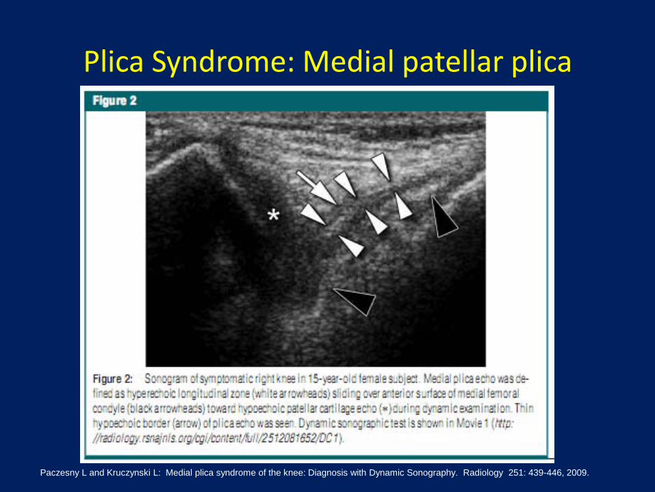
Plica Syndrome: Medial patellar plica
Paczesny L and Kruczynski L: Medial plica syndrome of the knee: Diagnosis with Dynamic Sonography. Radiology 251: 439-446, 2009.
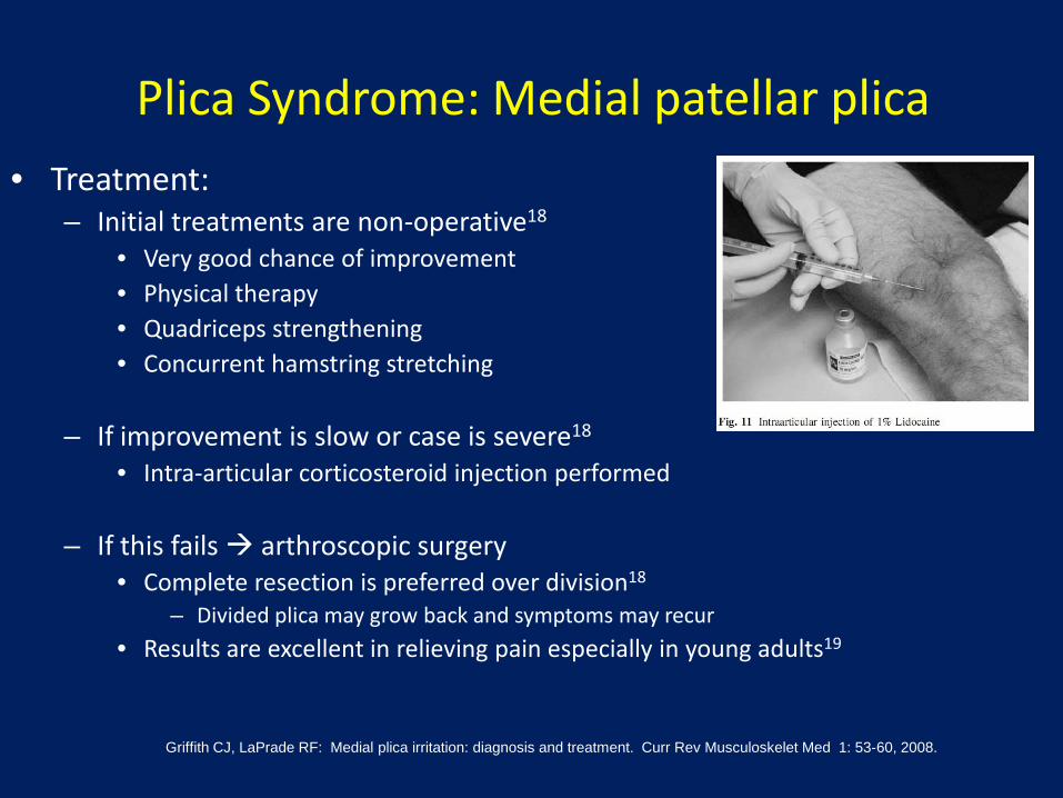
Plica Syndrome: Medial patellar plica• Treatment:
– Initial treatments are non-operative18
• Very good chance of improvement• Physical therapy• Quadriceps strengthening• Concurrent hamstring stretching
– If improvement is slow or case is severe18
• Intra-articular corticosteroid injection performed
– If this fails arthroscopic surgery• Complete resection is preferred over division18
– Divided plica may grow back and symptoms may recur
• Results are excellent in relieving pain especially in young adults19
Griffith CJ, LaPrade RF: Medial plica irritation: diagnosis and treatment. Curr Rev Musculoskelet Med 1: 53-60, 2008.

Medial Plica Resection
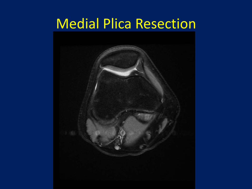
Medial Plica Resection
Plica Syndrome: Medial patellar plica
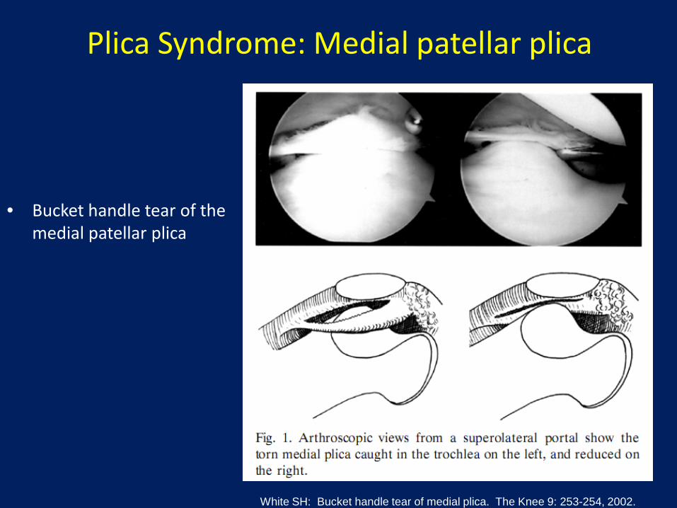
• Bucket handle tear of the medial patellar plica
White SH: Bucket handle tear of medial plica. The Knee 9: 253-254, 2002.

Plica Syndrome: Lateral patellar plica
Plica Syndrome: Lateral patellar plica
• Essentially same entity as medial plica syndrome except occurring on the lateral side of the knee
• Etiology, clinical presentation, and physical examination identical
• Role of imaging is the same
• Entity is rarer due to much reduced prevalence of the lateral plica
Plica Syndrome: Lateral patellar plica
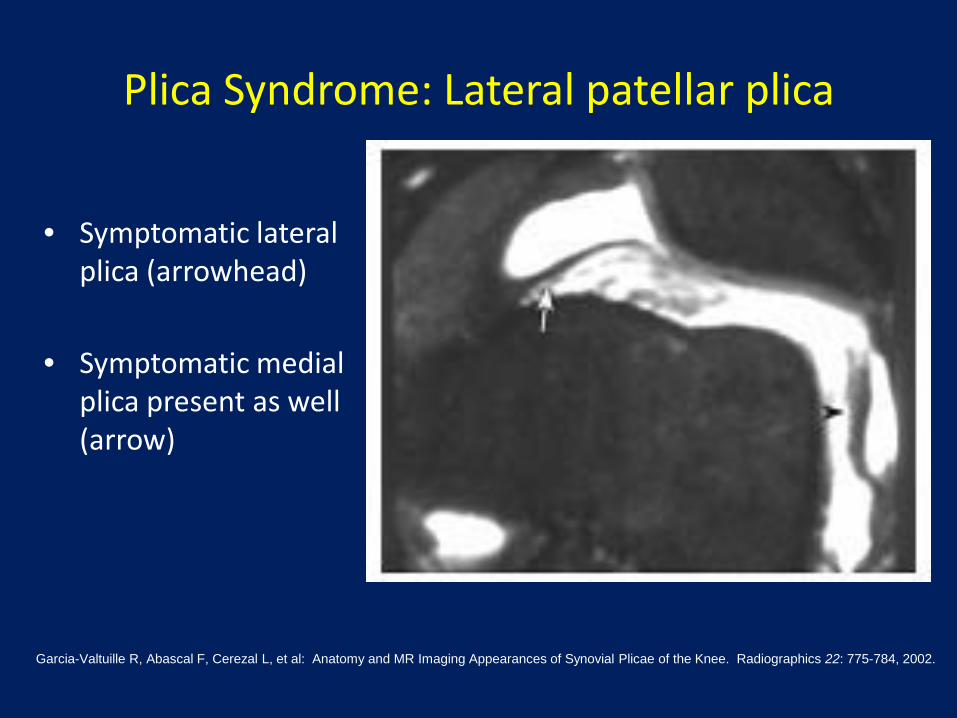
• Symptomatic lateral plica (arrowhead)
• Symptomatic medial plica present as well (arrow)
Garcia-Valtuille R, Abascal F, Cerezal L, et al: Anatomy and MR Imaging Appearances of Synovial Plicae of the Knee. Radiographics 22: 775-784, 2002.
In Review
• The suprapatellar and infrapatellar plicae are remnants of the septa that form during knee development
• The medial patellar and lateral patellar plica are formed from residual mesenchymal tissue– The medial plica is much more common than the lateral
plica because of lateralization of the patella in the embryo

In Review
• Many arthroscopic classifications for the plicae exist.– Imaging has not been shown to reliably classify
plica
• Plicae are normal structures and there presence alone does not indicate a pathologic condition
In Review
• Plica syndromes occur for two main reasons:– Inflammatory processes or trauma may thicken and stiffen
a plica• May result in adjacent chondral injuries• Plica itself may become a source of pain• May mimic internal derangement of the knee
– Failure of resorption of plica can lead to joint compartmentalization
• Suprapatellar and infrapatellar plica
• Imaging cannot reliably diagnose medial patellar plica syndrome
In Review• It is important for radiologists to:
– State if a plica is demonstrated on imaging
– Give the thickness and length of the plica
– Document any associated chondral abnormalities
– Document the presence of a complete plica• Suprapatellar or infrapatellar plica
– Rule out other mimics of plica syndromes• Meniscal tears, chondral injuries etc.

References• 1. Garcia-Valtuille R, Abascal F, Cerezal L, et al: Anatomy and MR Imaging Appearances of
Synovial Plicae of the Knee. Radiographics 22: 775-784, 2002.
• 2. Dupont J: Synovical plicae of the knee: Controversies and Review. Clin Sports Med 16: 87-122, 1997.
• 3. Schindler O: Synovial plicae of the knee. Current Orthopedics 18: 210-219, 2004.
• 4. Dorfmann H, Orengo P, Amarenco G, et al: Pathologie des replis synoviaux du genou: interet de l’arthroscopie. Rev Rhum 49: 67-73, 1982.
• 5. Kent M, Khanduja V: Synovial plicae around the knee. The Knee 17: 97-102. 2010.
• 6. Ogata S, Uhthoff H: The development of synovial plicae in human knee joints: An embryologic study. Arthroscopy 6: 315-321, 1990.
• 7. Deutsch A, Resnick D, Dalinka M, et al: Synovial plicae of the knee. Radiology 141: 627-634, 1981.

References• 8. Resnick D, Heung SK, Pretterklieber ML: Internal Derangements of Joints, 2nd Edition,
Volume 2. Saunders Elsevier 2007. Pg. 1582-1586.
• 9. Kim S, Choe W: Arthroscopic Findings of the Synovial Plicae of the Knee. Arthroscopy 13: 33-41, 1997.
• 10. Boles CA, Martin DF: Synovial plicae in the knee: pictorial essay. AJR 177: 221-227, 2001.
• 11. Dupont J: Synovial plicae of the knee. The Knee 1: 5-19, 1994.
• 12. Strover AE, Rouholamin E, Guirguis N et al: An arthroscopic technique of demonstrating the pathomechanics of the suprapatellar plica. Arthroscopy 7:308-310, 1991.
• 13. Trout TE, Bock H, Resnick D: Suprapatellar plicae of the knee presenting as a soft-tissue mass report of five patients. Clinical imaging 20:55-59, 1996.
• 14. Ehlinger M, Moser T, Adam P et al: Complete suprapatellar plica presenting like a tumor. Orthopaedics & Traumatology: Surgery & Research 95:447-250, 2009.
References• 15. Adachi N, Ochi M, Uchio Y: The complete type of suprapatellar plica in a professional
baseball pitcher: consideration of a cause of anterior knee pain. Arthroscopy 20: 987-991, 2004.
• 16. Kim S, Choe, W: Pathological infrapatellar plica: A report of two cases and literature review. Arthroscopy 12: 236-239, 1996.
• 17. Cothran RL, McGuire PM, Helms CA et al: MR imaging of infrapatellar plica injury. AJR 180: 1443-1447, 2003.
• 18. Griffith CJ, LaPrade RF: Medial plica irritation: diagnosis and treatment. Curr Rev Musculoskelet Med 1: 53-60, 2008.
• 19. Weckstrom M, Niva MH, Lamminen A et al: Arthroscopic resection of medial plica in the knee in young adults. The Knee: 17: 103-107, 2010.
• 20. Zidorn T: Classification of the suprapatellar septum considering ontogenetic development. Arthroscopy 8: 459-464, 1991.
• 21. Kosarek FJ and Helms CA: The MR appearance of the infrapatellar plica. AJR 172: 481-484, 1999.

References• 22. Soejima T, Murakami H, Tanaka N et al: Anteromedial meniscofemoral ligament.
Arthroscopy 19: 90-95, 2003.
• 23. Farkas C, Hargitai Z, Gaspar L et al: Histological changes in the symptomatic mediopatellar plica. The Knee 11: 103-108, 2004.
• 24. Lyu S: Relationship of medial plica and medial femoral condyle during flexion. Clinical Biomechanics 22: 1013-1016, 2007.
• 25. Lyu S and Chia-Chen H: Medial plicae and degeneration of the medial femoral condyle. Arthroscopy 22: 17-26, 2006.
• 26. White SH: Bucket handle tear of medial plica. The Knee 9: 253-254, 2002.
• 27. Paczesny L and Kruczynski L: Medial plica syndrome of the knee: Diagnosis with Dynamic Sonography. Radiology 251: 439-446, 2009.
• 28. Gallagher J, Tierney P, Murray P et al: The infrapatellar fat pad: anatomy and clinical considerations. Knee Surg Sports Traumatol Arthrosc 13: 268-272.

References
• 29. Saddik D, McNally EG, Richardson M: MRI of hoffa’s fat pad. Skeletal Radiology 33: 433-444, 2004.
• 30. Patel SJ, Kaplan PA, Dussault RG, Kahler, DM: Anatomy and clinical significance of the horizontal cleft in the infrapatellar fat pad of the knee: MR imaaging. AJR 170: 1551-1555, 1998.