Syndrome of Mental Physical Retardation, Speech Disorders ... · JournalofMedicalGenetics (1972)....
4
Journal of Medical Genetics (1972). 9, 60. A Syndrome of Mental and Physical Retardation, Speech Disorders, and Peculiar Facies in Two Sisters OSVALDO MUTCHINICK From the Medical Genetics Center, Combate de los Pozos 2193, Buenos Aires, Argentina Two sisters, born to consanguineous parents, presumed to be affected with a new syndrome of mental retardation and multiple congenital ano- malies are reported. Case Reports Case 1 (IV.1) (see Fig. 1). This girl was born on 21 September 1961. Her mother was 22 and her father 24 years old at the time of birth. The only known unusual event during pregnancy was a large umbilical hernia and an abdominal x-ray was taken at about 30 weeks of gestation. Labour was induced by amniotomy and oxygen was given during labour because of a tetanic con- traction. Delivery was uneventful but the newborn was anoxic and required active resuscitation. The birth weight is unknown. She remained in the nursery for a week because of feeding difficulties and occasional vomit- ing of blood. Both physical and motor developments were slow from birth and during the first 3 months of life she had fre- quent crises of irritability and hypertonicity. She first raised her head at 4 months, sat at 6 months, and started walking at 30 months. At 18 months she began to speak isolated words and was able to construct short sentences at 48 months; there was then evidence of a speech im- pairment which persists to the present time. She had had no other serious illness. Physical examination. She was first examined by the author at 71 years. The findings at that time were as follows: weight 17-5 kg; height 110 cm; vertex-pubis 57 cm; head circumference 45 cm; chest circumference 57 cm. All these measurements, except for the chest circumference, were below the 3rd centile for age and sex. The significant positive physical findings were: microbrachycephaly with narrow sloping forehead; marked hypertelorism, small palpebral fissures (ble- pharophimosis) with anti-mongoloid slant; long curly eyelashes, light blue iris, severe photophobia, hyper- metropia, and normal fundi. The nose was broad and straight with a lack of depression at the bridge. The mouth was wide-tented, with outward and downward slant of the corners, the upper lip covering the visible mucosal portion of the lower lip. She had a high Received 17 May 1971. arched palate and dental anomalies such as malocclu- sion, misplaced teeth slanting inwards at 450, and irregu- lar edges of the incissors. There was moderate prognathism. Her ears were very large and flared with a lack of differentiation of the antihelix branches. Her chest was pigeon shaped with anterior sternocostal pro- trusion. A systolic murmur was heard best over the pulmonic area and the second heart sound was split. Scanty, thin, and blonde pubic hair was present. Her skin was white, with 15 moles, none larger than 2 mm in diameter, distributed on the chest and abdomen. Hair was thin, blonde, rather scarce, and normally implanted. She had hyperconvex thumb nails and bilateral clino- dactyly of the 5th finger. She had severe mental retardation (IQ = 42) with speech disorders: dyslalia, anomia, and agrammatism. She also had a broad stand- ing base and a spastic gait. The following studies were within normal limits: haemogram, serum calcium, phosphate, uric acid, pro- tein-bound iodine, carbohydrate and protein electro- phoresis, and lipidogram. Studies of urine calcium, carbohydrate, amino-acid chromatography, and 17- ketosteroids were also normal. The qualitative urinary tests for phenylpyruvic acid, ketoacids, cysteine, homocysteine, tyrosine, hydroxyphenylpyruvic acid, and mucopolysacharides were all negative. The only abnormal result was an increased 17-ketogenico-steroid excretion of 12-6 mg in 24 hours. Radiological findings. The skull x-ray showed thin frontal diploe, small posterior fossae, orbital hyper- teleorism, and low posterior part of the mandibular horizontal branch. The chest x-ray showed premature sternal fusion, sternocostal forward protrusion, and cuboid shaped dorsolumbar vertebral bodies. X-ray of the pelvis showed open ischiopubic synchondrosis. The x-ray of the limbs showed a small carpus and normal bone age; generalized osteoporosis was present. An IVP showed a moderate bilateral calyceal dilation with dilated right ureter; the left ureter could not be seen well. ECG demonstrated a complete right bundle branch block and findings compatible with interauricu- lar septal defect plus valvular pulmonic stenosis. EEG showed a defective central bioelectric organization. Case 2 (IV.3) (see Fig. 1). This girl was born on 12 June 1965 after an uncomplicated pregnancy of 40 weeks. She was exposed to x-rays during the 5th 60 on June 14, 2020 by guest. Protected by copyright. http://jmg.bmj.com/ J Med Genet: first published as 10.1136/jmg.9.1.60 on 1 March 1972. Downloaded from
Transcript of Syndrome of Mental Physical Retardation, Speech Disorders ... · JournalofMedicalGenetics (1972)....

Journal of Medical Genetics (1972). 9, 60.
A Syndrome of Mental and Physical Retardation,Speech Disorders, and Peculiar Facies in Two Sisters
OSVALDO MUTCHINICK
From the Medical Genetics Center, Combate de los Pozos 2193, Buenos Aires, Argentina
Two sisters, born to consanguineous parents,presumed to be affected with a new syndrome ofmental retardation and multiple congenital ano-malies are reported.
Case ReportsCase 1 (IV.1) (see Fig. 1). This girl was born on 21
September 1961. Her mother was 22 and her father 24years old at the time of birth. The only known unusualevent during pregnancy was a large umbilical herniaand an abdominal x-ray was taken at about 30 weeks ofgestation. Labour was induced by amniotomy andoxygen was given during labour because of a tetanic con-traction. Delivery was uneventful but the newborn wasanoxic and required active resuscitation. The birthweight is unknown. She remained in the nursery for aweek because of feeding difficulties and occasional vomit-ing of blood.Both physical and motor developments were slow from
birth and during the first 3 months of life she had fre-quent crises of irritability and hypertonicity. She firstraised her head at 4 months, sat at 6 months, and startedwalking at 30 months. At 18 months she began to speakisolated words and was able to construct short sentencesat 48 months; there was then evidence of a speech im-pairment which persists to the present time. She hadhad no other serious illness.
Physical examination. She was first examined by theauthor at 71 years. The findings at that time were asfollows: weight 17-5 kg; height 110 cm; vertex-pubis57 cm; head circumference 45 cm; chest circumference57 cm. All these measurements, except for the chestcircumference, were below the 3rd centile for age and sex.The significant positive physical findings were:
microbrachycephaly with narrow sloping forehead;marked hypertelorism, small palpebral fissures (ble-pharophimosis) with anti-mongoloid slant; long curlyeyelashes, light blue iris, severe photophobia, hyper-metropia, and normal fundi. The nose was broad andstraight with a lack of depression at the bridge. Themouth was wide-tented, with outward and downwardslant of the corners, the upper lip covering the visiblemucosal portion of the lower lip. She had a high
Received 17 May 1971.
arched palate and dental anomalies such as malocclu-sion, misplaced teeth slanting inwards at 450, and irregu-lar edges of the incissors. There was moderateprognathism. Her ears were very large and flared witha lack of differentiation of the antihelix branches. Herchest was pigeon shaped with anterior sternocostal pro-trusion. A systolic murmur was heard best over thepulmonic area and the second heart sound was split.Scanty, thin, and blonde pubic hair was present. Herskin was white, with 15 moles, none larger than 2 mm indiameter, distributed on the chest and abdomen. Hairwas thin, blonde, rather scarce, and normally implanted.She had hyperconvex thumb nails and bilateral clino-dactyly of the 5th finger. She had severe mentalretardation (IQ= 42) with speech disorders: dyslalia,anomia, and agrammatism. She also had a broad stand-ing base and a spastic gait.The following studies were within normal limits:
haemogram, serum calcium, phosphate, uric acid, pro-tein-bound iodine, carbohydrate and protein electro-phoresis, and lipidogram. Studies of urine calcium,carbohydrate, amino-acid chromatography, and 17-ketosteroids were also normal. The qualitative urinarytests for phenylpyruvic acid, ketoacids, cysteine,homocysteine, tyrosine, hydroxyphenylpyruvic acid, andmucopolysacharides were all negative. The onlyabnormal result was an increased 17-ketogenico-steroidexcretion of 12-6 mg in 24 hours.
Radiological findings. The skull x-ray showed thinfrontal diploe, small posterior fossae, orbital hyper-teleorism, and low posterior part of the mandibularhorizontal branch. The chest x-ray showed prematuresternal fusion, sternocostal forward protrusion, andcuboid shaped dorsolumbar vertebral bodies. X-ray ofthe pelvis showed open ischiopubic synchondrosis.The x-ray of the limbs showed a small carpus and normalbone age; generalized osteoporosis was present. AnIVP showed a moderate bilateral calyceal dilation withdilated right ureter; the left ureter could not be seenwell. ECG demonstrated a complete right bundlebranch block and findings compatible with interauricu-lar septal defect plus valvular pulmonic stenosis. EEGshowed a defective central bioelectric organization.Case 2 (IV.3) (see Fig. 1). This girl was born on 12
June 1965 after an uncomplicated pregnancy of 40weeks. She was exposed to x-rays during the 5th
60
on June 14, 2020 by guest. Protected by copyright.
http://jmg.bm
j.com/
J Med G
enet: first published as 10.1136/jmg.9.1.60 on 1 M
arch 1972. Dow
nloaded from

A Syndrome ofMental and Physical Retardation, Speech Disorders, and Peculiar Facies in Two Sisters 61
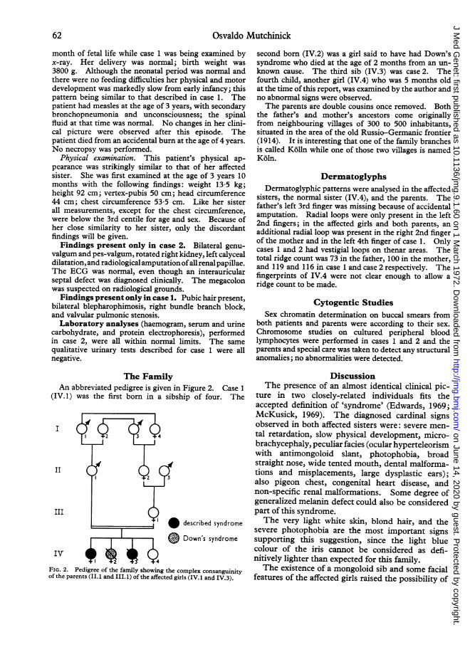
FIG. 1. The two affected sisters. 1-3: case 1 (IV.1) at 71 years. 4-6: case 2 (IV.3) at 31 years.
on June 14, 2020 by guest. Protected by copyright.
http://jmg.bm
j.com/
J Med G
enet: first published as 10.1136/jmg.9.1.60 on 1 M
arch 1972. Dow
nloaded from
Osvaldo Mutchinick
month of fetal life while case 1 was being examined byx-ray. Her delivery was normal; birth weight was3800 g. Although the neonatal period was normal andthere were no feeding difficulties her physical and motordevelopment was markedly slow from early infancy; thispattern being similar to that described in case 1. Thepatient had measles at the age of 3 years, with secondarybronchopneumonia and unconsciousness; the spinalfluid at that time was normal. No changes in her clini-cal picture were observed after this episode. Thepatient died from an accidental burn at the age of 4 years.No necropsy was performed.
Physical examination. This patient's physical ap-pearance was strikingly similar to that of her affectedsister. She was first examined at the age of 3 years 10months with the following findings: weight 13-5 kg;height 92 cm; vertex-pubis 50 cm; head circumference44 cm; chest circumference 53-5 cm. Like her sisterall measurements, except for the chest circumference,were below the 3rd centile for age and sex. Because ofher close similarity to her sister, only the discordantfindings will be given.
Findings present only in case 2. Bilateral genu-valgum and pes-valgum, rotated right kidney, left calycealdilatation, and radiological amputation ofall renal papillae.The ECG was normal, even though an interauricularseptal defect was diagnosed clinically. The megacolonwas suspected on radiological grounds.Findings present only in case 1. Pubic hair present,
bilateral blepharophimosis, right bundle branch block,and valvular pulmonic stenosis.Laboratory analyses (haemogram, serum and urine
carbohydrate, and protein electrophoresis), performedin case 2, were all within normal limits. The samequalitative urinary tests described for case 1 were allnegative.
The FamilyAn abbreviated pedigree is given in Figure 2. Case 1
(IV.1) was the first born in a sibship of four. The
I
II
III
* described syndrome
0 Down's syndrome
IV YPyFIG. 2. Pedigree of the family showing the complex consanguinityof the parents (II. 1 and III. 1) of the affected girls (IV. 1 and IV.3).
second born (IV.2) was a girl said to have had Down'ssyndrome who died at the age of 2 months from an un-known cause. The third sib (IV.3) was case 2. Thefourth child, another girl (IV.4) who was 5 months oldat the time of this report, was examined by the author andno abnormal signs were observed.The parents are double cousins once removed. Both
the father's and mother's ancestors come originallyfrom neighbouring villages of 300 to 500 inhabitants,situated in the area of the old Russio-Germanic frontier(1914). It is interesting that one of the family branchesis called K6lln while one of those two villages is namedK6ln.
DermatoglyphsDermatoglyphic patterns were analysed in the affected
sisters, the normal sister (IV.4), and the parents. Thefather's left 3rd finger was missing because of accidentalamputation. Radial loops were only present in the left2nd fingers; in the affected girls and both parents, anadditional radial loop was present in the right 2nd fingerof the mother and in the left 4th finger of case 1. Onlycases 1 and 2 had vestigial loops on thenar areas. Thetotal ridge count was 73 in the father, 100 in the mother,and 119 and 116 in case 1 and case 2 respectively. Thefingerprints of IV.4 were not clear enough to allow aridge count to be made.
Cytogentic StudiesSex chromatin determination on buccal smears from
both patients and parents were according to their sex.Chromosome studies on cultured peripheral bloodlymphocytes were performed in cases 1 and 2 and theparents and special care was taken to detect any structuralanomalies; no abnormalities were detected.
DiscussionThe presence of an almost identical clinical pic-
ture in two closely-related individuals fits theaccepted definition of 'syndrome' (Edwards, 1969;McKusick, 1969). The diagnosed cardinal signsobserved in both affected sisters were: severe men-tal retardation, slow physical development, micro-brachycephaly, peculiar facies (ocular hyperteleorismwith antimongoloid slant, photophobia, broadstraight nose, wide tented mouth, dental malforma-tions and misplacements, large dysplastic ears);also pigeon chest, congenital heart disease, andnon-specific renal malformations. Some degree ofgeneralized melanin defect could also be consideredpart of this syndrome.The very light white skin, blond hair, and the
severe photophobia are the most important signssupporting this suggestion, since the light bluecolour of the iris cannot be considered as defi-nitively lighter than expected for this family.The existence of a mongoloid sib and some facial
features of the affected girls raised the possibility of
62
on June 14, 2020 by guest. Protected by copyright.
http://jmg.bm
j.com/
J Med G
enet: first published as 10.1136/jmg.9.1.60 on 1 M
arch 1972. Dow
nloaded from
A Syndrome of Mental and Physical Retardation, Speech Disorders, and Peculiar Facies in Two Sisters 63
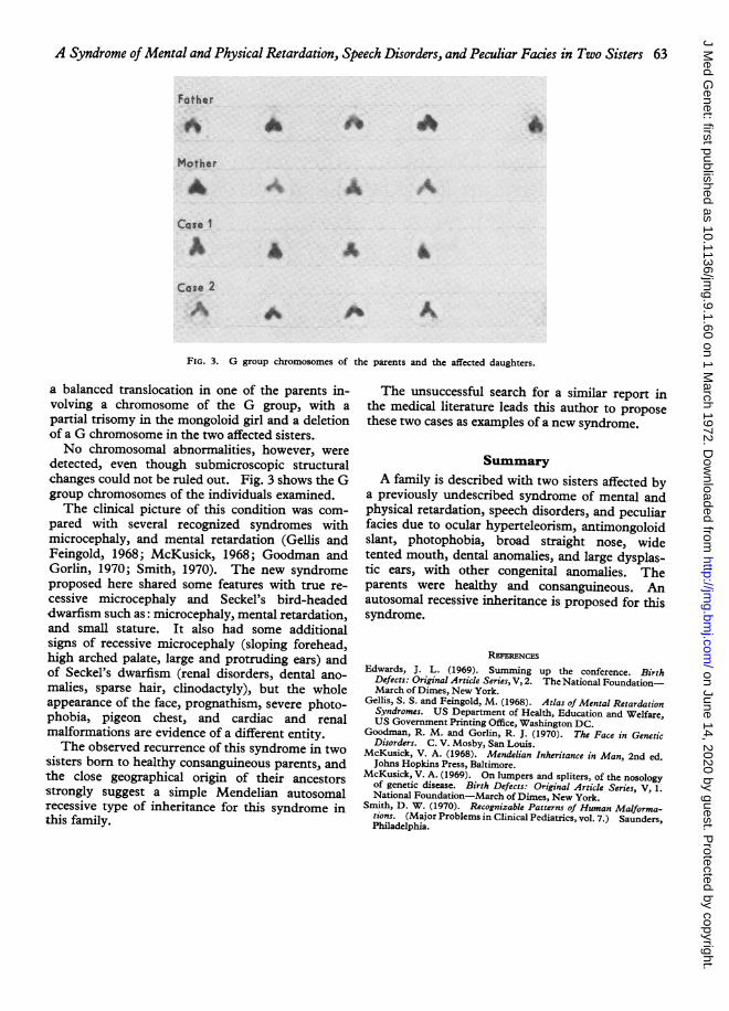
FIG. 3. G group chromosomes of the parents and the affected daughters.
a balanced translocation in one of the parents in-volving a chromosome of the G group, with apartial trisomy in the mongoloid girl and a deletionof a G chromosome in the two affected sisters.No chromosomal abnormalities, however, were
detected, even though submicroscopic structuralchanges could not be ruled out. Fig. 3 shows the Ggroup chromosomes of the individuals examined.The clinical picture of this condition was com-
pared with several recognized syndromes withmicrocephaly, and mental retardation (Gellis andFeingold, 1968; McKusick, 1968; Goodman andGorlin, 1970; Smith, 1970). The new syndromeproposed here shared some features with true re-cessive microcephaly and Seckel's bird-headeddwarfism such as: microcephaly, mental retardation,and small stature. It also had some additionalsigns of recessive microcephaly (sloping forehead,high arched palate, large and protruding ears) andof Seckel's dwarfism (renal disorders, dental ano-malies, sparse hair, clinodactyly), but the wholeappearance of the face, prognathism, severe photo-phobia, pigeon chest, and cardiac and renalmalformations are evidence of a different entity.The observed recurrence of this syndrome in two
sisters born to healthy consanguineous parents, andthe close geographical origin of their ancestorsstrongly suggest a simple Mendelian autosomalrecessive type of inheritance for this syndrome inthis family.
The unsuccessful search for a similar report inthe medical literature leads this author to proposethese two cases as examples of a new syndrome.
SummaryA family is described with two sisters affected by
a previously undescribed syndrome of mental andphysical retardation, speech disorders, and peculiarfacies due to ocular hyperteleorism, antimongoloidslant, photophobia, broad straight nose, widetented mouth, dental anomalies, and large dysplas-tic ears, with other congenital anomalies. Theparents were healthy and consanguineous. Anautosomal recessive inheritance is proposed for thissyndrome.
RssE'ENCESEdwards, J. L. (1969). Summing up the conference. Birth
Defects: Original Article Series, V, 2. The National Foundation-March of Dimes, New York.
Gellis, S. S. and Feingold, M. (1968). Atlas of Mental RetardationSyndromes. US Department of Health, Education and Welfare,US Government Printing Office, Washington DC.
Goodman, R. M. and Gorlin, R. J. (1970). The Face in GeneticDisorders. C. V. Mosby, San Louis.
McKusick, V. A. (1968). Mendelian Inheritance in Man, 2nd ed.Johns Hopkins Press, Baltimore.
McKusick, V. A. (1969). On lumpers and spliters, of the nosologyof genetic disease. Birth Defects: Original Article Series, V, 1.National Foundation-March of Dimes, New York.
Smith, D. W. (1970). Recognizable Patterns of Human Malforma-tions. (Major Problems in Clinical Pediatrics, vol. 7.) Saunders,Philadelphia.
on June 14, 2020 by guest. Protected by copyright.
http://jmg.bm
j.com/
J Med G
enet: first published as 10.1136/jmg.9.1.60 on 1 M
arch 1972. Dow
nloaded from