Syndrome Arthrogryposis (SAP) cattle; … · becomes highly concentrated in end-plate rich regions...
10
The Syndrome of Arthrogryposis and Palatoschisis (SAP) in Charolais cattle; Abnormal motor innervation and defect in the focalization of 16 S acetylcholinesterase in the end- plates rich regions of the muscle. (1) F. RIEGER, Martine PINÇON-RAYMOND, P. DREYFUS Monique GUITTARD M. FARDEAU Groupe de Biologie et Pathologie Neuromusculaives I.N.S.E.R.M. U 153 . 17 , rue du Fey-à-Moulin, q $ oog Paris, France Summary The Syndrome of Arthrogryposis and Palatoschisis SAP is an hereditary disease in the Charolais cattle bread. We studied the neurobiological and neuropathological correlates of this disease in SAP calves, immediately after birth. We find that nerve-muscle interaction is abnormal; the focal accumulation of 1 6S AChE in end-plate rich regions is not observed as in control calf, and the motor innervation, evidenced by silver nitrate impregnation techniques, shows abnormal features, as frequent preterminal branching and ultraterminal sprouting. Introduction The Syndrome of Arthrogryposis and Palatoschisis (SAP) in Charolais cattle has been first described by L AUVERGNE and B UN ( 19 6 7 ). It is an hereditary disease, carried by an autosomal recessive gene with incomplete penetrance (see I a EFO R T et al. 1977 and also L AUV E R G N E and F AU C O rr, 197 6, and LAUVERGNE et al. 1979 for a review). Its clinical expression is rather variable in severity, but is always characterized by fixture of joints (arthrogryposis) and severity of the disease is the consequence of different movement disabilities of the limbs, due to variable angles of fixture and also aberrant insertion of muscles or even lack of a given muscle. In man, arthrogryposis has been described as a congenital rigidity of joints in newborns, in the last decades of the nineteenth century. It is now considered as a symptom, (z) Part of this work has been presented at the Meeting of the International Charolais Association, Sept. 6-7, 197 9 , Vichy (France) by F. R IEGER .
Transcript of Syndrome Arthrogryposis (SAP) cattle; … · becomes highly concentrated in end-plate rich regions...

The Syndrome of Arthrogryposis and Palatoschisis (SAP)in Charolais cattle; Abnormal motor innervation and defectin the focalization of 16 S acetylcholinesterase in the end-plates rich regions of the muscle. (1)
F. RIEGER, Martine PINÇON-RAYMOND, P. DREYFUS
Monique GUITTARD M. FARDEAU
Groupe de Biologie et Pathologie Neuromusculaives I.N.S.E.R.M. U 153.17, rue du Fey-à-Moulin, q$oog Paris, France
Summary
The Syndrome of Arthrogryposis and Palatoschisis SAP is an hereditary disease in theCharolais cattle bread. We studied the neurobiological and neuropathological correlates ofthis disease in SAP calves, immediately after birth. We find that nerve-muscle interaction isabnormal; the focal accumulation of 16S AChE in end-plate rich regions is not observed as incontrol calf, and the motor innervation, evidenced by silver nitrate impregnation techniques,shows abnormal features, as frequent preterminal branching and ultraterminal sprouting.
Introduction
The Syndrome of Arthrogryposis and Palatoschisis (SAP) in Charolais cattlehas been first described by LAUVERGNE and BUN (1967). It is an hereditarydisease, carried by an autosomal recessive gene with incomplete penetrance (seeIaEFORT et al. 1977 and also LAUVERGNE and FAUCOrr, 1976, and LAUVERGNEet al. 1979 for a review).
Its clinical expression is rather variable in severity, but is always characterizedby fixture of joints (arthrogryposis) and severity of the disease is the consequenceof different movement disabilities of the limbs, due to variable angles of fixtureand also aberrant insertion of muscles or even lack of a given muscle. In man,arthrogryposis has been described as a congenital rigidity of joints in newborns,in the last decades of the nineteenth century. It is now considered as a symptom,
(z) Part of this work has been presented at the Meeting of the International Charolais Association,Sept. 6-7, 1979, Vichy (France) by F. RIEGER.
or a syndrome, and not a definite disease, which may be due to different mecha-nisms, spinal radicular, or myogenic. In some cases, nerve and muscle biopsiesperformed in this syndrome revealed the features of a neurogenic process (BYERSand BANKER, Ig6I; HOOSHMAUD et al. 1971, YUILL and LYNCH, 1974). A fewcases exhibited morphological abnormalities suggesting a myopathic process(BANKER et al. ig57). In many cases the muscle is surprisingly normal in biopsies,suggesting an early abnormality in the embryonic muscle development (DuBo-WITZ, Ig78). In all conditions, the joint and soft tissue changes are evidencedas secondary to the absence of a normal muscle activity in the limbs in utero.
In the search of primary causes of abnormal embryonic early differentiation,developmental genetics offers animal models of considerable value. GLUECKSOHN-W AELSCH has developed the study of embryonic differentiation by identifyinglethal genes in the mouse (GLUECKSOHN-WAELSCH, 1963), leading either to atotal, foetally lethal, arrest of the development of the mouse embryo (with rapidregression in uteyo) or to early defects, only lethal at birth. To the later case
belongs one fascinating mutation for developmental neuroscientists: musculardysgenesis (mdg) (GI,U!CKSOHN-WA!I,SCH, 1963). Muscular dysgenesis is cha-racterized by death at birth, due to a total lack of muscle contractile activityand a delay (or arrest) in foetal differentiation (PAi, Zg65a, b; BANKER, 1977).Mdg embryos also show early nerve defects, with profuse collateral and ultra-terminal sprouting (RIEGER and PINqON-RAYMOND, ig8o).
One of the main characteristic of the mdg embryo occurs to be a pronouncedarthrogrypotic feature (often associated with cleft palate). Such a genetic modelfor the study of the consequences of early impairment of muscle activity in utero,has prompted us to set suitable neurobiological techniques for the study of mouseneonate nerve and muscle. These methods and the general neurobiologicalstrategy is well adapted to study SAP neonate calves.
This preliminary report presents two abnormal, pathological features ofSAP muscle and nerve. The first one is related to a molecular aspect of nerve-muscle interaction: one of the molecular forms of acetylcholinesterase (16s)becomes highly concentrated in end-plate rich regions of mouse or rat muscles,during embryonic and especially post-natal development (HALL, 1973; VIGNYet al. Ig76; KOENIG and RIEGER, 1980; RIEGER et al. ig8ob), most probably undernerve influence. This is not realized in SAP calf muscles, where there is, appa-rently, no such focalization. The second one is an abnormal cytological aspectof the motor innervation: the motor innervation clearly presents frequent preter-minal axonal branching and ultraterminal sprouting from nerve terminals.
Material and methods
We have studied two SAP calves and one control animal. The SAP calveswere transported alive from the region of Nevers, to our laboratory still alive(immediately after birth). The normal control animal was obtained after caesa-rian of a sacrified brucellic cow 2-3 weeks before giving birth and the calf immedia-tely brought on ice to our laboratory. The SAP animals were sacrified by blee-ding, after Ketalar general anesthesia and dissected for anatomical observation.Sternomastoid, biceps and diaphragm were dissected out and immediately pro-cessed further for analysis of the molecular forms of acetylcholinesterase or
metallic impregnation of the motor innervation.
- Analysis of the molecular forms of acetylcholinesterase (AChE) in end-
plate rich and end-plate free regions : we performed, on the fresh muscles, a cyto-chemical reaction following a modified Koelle method at pH 7, to visually iden-tify and dissect out end-plate rich or free regions of the muscles to be studied
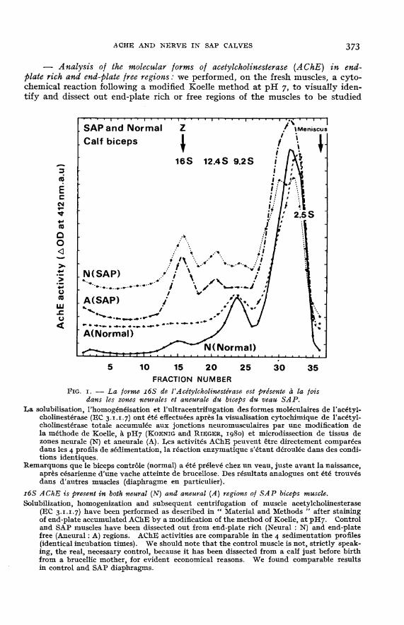
La solubilisation, 1’homogdn6isation et 1’ultracentrifugation des formes moléculaires de I’ac6tyl-cholinesterase (EC 3.1.1.7) ont été effectu6es apr6s la visualisation cytochimique de 1’acetyl-cholinesterase totale accumul6e aux jonctions neuromusculaires par une modification dela méthode de Koelle, b, pH7 (KOENIG and RIEGER, ig8o) et microdissection de tissus dezones neurale (N) et aneurale (A). Les activites AChE peuvent 6tre directement compar6esdans les 4 profils de sedimentation, la reaction enzymatique s’étant déroulée dans des condi-tions identiques.
Remarquons que le biceps contr6le (normal) a été prélevé chez un veau, juste avant la naissance,après césarienne d’une vache atteinte de brucellose. Des résultats analogues ont été trouv6sdans d’autres muscles (diaphragme en particulier).
z6S AChE is present in both neural (N) and aneuyal (A) regions of SAP biceps muscle.Solubilization, homogenization and subsequent centrifugation of muscle acetylcholinesterase
(EC 3.1.1.7) have been performed as described in &dquo; Material and Methods &dquo; after stainingof end-plate accumulated AChE by a modification of the method of Koelle, at pH7. Controland SAP muscles have been dissected out from end-plate rich (Neural : N) and end-platefree (Aneural : A) regions. AChE activities are comparable in the 4 sedimentation profiles(identical incubation times). We should note that the control muscle is not, strictly speak-ing, the real, necessary control, because it has been dissected from a calf just before birthfrom a brucellic mother, for evident economical reasons. We found comparable resultsin control and SAP diaphragms.
(KOENIG and RIEGER, ig8o). The dissected tissues (after washing of the reactionmixture in a Krebs-Ringer solution) were homogenized (in a i to 10 weight tovolume proportion) solution containing I M Nacl, 0.001 M EGTA, i p. 100 triton X100 and o.oi M Tris pH 7.2 (standard medium) or in a standard medium withan antiprotease cocktail (0.25 mg bacitracin, 0.1 mM benzethonium chloride,0.2 mg jml benzamidine, 0.02 mg jml pepstatin). No difference was found betweenthese two media, suggesting no particular proteolytic instability of the homo-genate. AChE activity was measured by the method of ELLMAN et al. (ig6i).Continuous sucrose gradient centrifugation was performed by loading 75 ¡LI ali-quots of the supernatants of low speed centrifugation (SORVALL RC2; 20 00o gfor 15 mn) on top of preformed 12 ml 5. 20 p. 100 sucrose gradients, made withthe standard medium and runned in a SW 4r BECKMAN rotor, in a L8 BECKMANultracentrifuge for 15 hours at 4 °C and 38 00o rpm. 35 to ¢o fractions werecollected, assayed for AChE activity and sedimentation coefficients were esti-mated by comparison with alcohol dehydrogenase (ADH: 4. S,,, S20,w) and p-galac-tosidase (Z : 16 S20, Szo,w).- Neurofibrillary staining: We used a modification of the original method of
BI!I,CHOWSKY-GROS: after fixation of the muscles in a mixture of Arseniateanhydride at saturation (I vol.) g5°, Ethanol (i vol.) and 20 p. 100 neutral formalin
(i vol.) for I hour, the tissues were kept 48 hours in 20 p. 100 neutral formalinand washed for 30 min. in bidistilled water. 50 !,m frozen sections were madestained in 20 p. 100 silver nitrate during 10 to 60 min. After a rapid wash in20 p. 100 neutral formalin, a brown staining is obtained by plunging the sectionsinto ammoniacal silver nitrate, immediately followed by immersion in 20 p. 100
pure ammonia for 30 min. After several washes in bisdistilled water. the sectionsare mounted in aqueous glycerol.A modification of the method of BARKER and IP (1963) was sometimes used
to specifically stain the neurofibrillary network in teased preparations.
Results
Solubilization of acetylcholinesterase in the presence of highthionic strengthbuffer (I M NaCl) and detergent (i p. 100 Triton X 100) is effective, Yielding90-95 P. 100 of the total tissue acetylcholinesterase. There is no loss of activityafter incubation of the tissues in the KOELLE and FRIEDENWALD histochemical

mixture for 5 to 15 min. (end-plates are made visible after only a few minutes,as white spots under binocular observation) and, as already evidenced for mouseor rat tissues, the sedimentation profiles remain unchanged after incubation,washing and subsequent homogenization and centrifugation compared to un-treated (unrevealed fresh) tissues.
Figure i shows that in biceps (and we found same results for diaphragm)control muscle does not possess any i6S AChE contribution to total activityin end-plate free regions, when end-plate rich regions contains 2 to 5 p. 100 ofi6S AChE. This shows that focalization and concentration of i6S AChE is
already realized in end-plate regions in calf muscle at birth. This is not at allthe case for any of the SAP muscles analyzed. 16S AChE can be found (in bicepsas well as in diaphragm) in both regions, approximatively in equivalent propor-tion and its level is proportionnaly higher in SAP muscle than in control muscle.BWr,cHOwsxy silver nitrate impregnation technique allow to visualize for
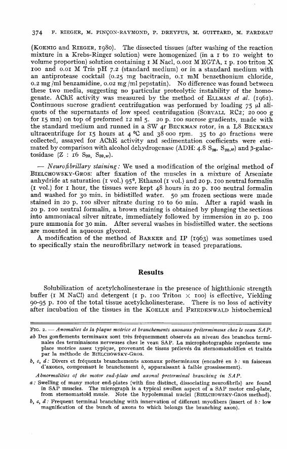
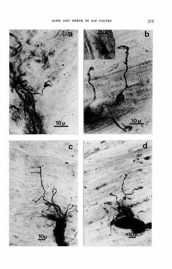
optic microscopy the pattern of motor innervation of the muscles. Figure 2 showstwo abnormal aspects of the terminal arborization: very frequent swellings withsmall excrescences of the SAP motor end-plate and especially abnormally frequentpreterminal branching of the axons.
Figure 3 gives some evidence of some ultra-terminal sprouting, from smallsprouts slightly extending the motor end-plate region to long sprouts runningalong the length of the muscle fiber. These aspects have been confirmed byusing the technique of BARKER and Ip (which is more specific for neurofibrillarystructures and stain less other structures like myelin or nuclei).
Discussion
The origin, or primary cause, of the SAP mutation is unknown and is expectedto be extremely difficult to establish. Only a multidisciplinary approach has achance to find the first expression of the mutation at its early, probable, timeof appearance. The anatomical aberrations (lack of a whole muscle), suggestabnormalities of somites differentiation and migration. It will be certainly diffi-cult to find at so early times of embryogenesis the defective element (precursor ofmuscle or neuronal cells) which has to be identified and isolated in order to studythe cellular or molecular expression of the SAP gene deficiency.
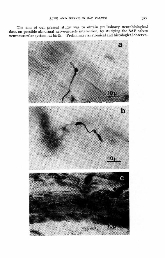
The aim of our present study was to obtain preliminary neurobiologicaldata on possible abnormal nerve-muscle interaction, by studying the SAP calvesneuromuscular system, at birth. Preliminary anatomical and histological observa-

tions on muscle do not show any evident fiber type modification, atrophy ordelay in their cyto-enzymologic differentiation. However a differentiation pro-cessus, which is believed to be under neural control the concentration and focaliza-tion of i6S AChE seems to be ineffective in SAP calf muscle. The real meaningof this observation is difficult to assess, because the mechanisms underlying theaccumulation of AChE in end-plate regions, though under intensive studies, arenot yet understood (RIEGER et al., ig8oa; KOENIG and RIEGER, 1980; RIEGERet al., ig8oa). But a comparison with the results obtained with mdg mice couldshed some light on certain aspects of SAP mutation. In mdg mouse musclethe 16S AChE is not present. This is because of the total lack of muscle activity(which is permissive for the biosynthesis of 16S (RIEGER et al. zg8oa). In SAPcalf muscles 16S AChE is present, even in higher amounts than in control muscle.This suggests that muscle activity in itself is not involved in the pathologic process.The non focal accumulation of this r6S means, on the other hand, that some kind ofnerve-muscle uncoupling is involved in the disease as expressed in the neonates.
Such an abnormality in the focal accumulation of i6S AChE may have a poten-tial value for the recognition of SAP gene heterozygote carriers: a partial expressionof this abnormal feature could be searched for, as in + /mdg heterozygote adultmice, which show appreciable differences in AChE forms relative content inskeletal muscles.
This nerve-muscle abnormal interaction receives a cytological illustrationfrom the observation of the terminal arborization of the motor innervation.We have observed systematic aspects of ultraterminal sprouting from abnormallylooking end-plates and, often, preterminal branching of axons which very rarelyoccurs in normal cattle (SwATr,ANn, 1973). Increased terminal branching is
usually interpreted either 1° as a change in the normal ratio of motor neuronsto myofibers, often accompanied by denervation-reinnervation (after a loss ofmotor neurons or myofiber hyperplasia) or 2° as suggestive of multiple innerva-tion of the myofibers. The first possibility is usually accompanied by histo-chemical fiber type grouping, which has not been observed in preliminary experi-ments. The second possible explanation can only be evidenced by single fiberisolation techniques, to obtain evidence of several foci of AChE or ACh Receptorsaccumulations at nerve-muscle contacts. We found, in whole SAP diaphragmmuscles, several AChE stained, end-plates like rich bands per muscle fasciculus,observation may be related to the occurrence of multiple innervation. In brief,the SAP mutation is mainly characterized by spectacular muscle impairmentaffecting muscles and joints; the neurobiological correlates of this arthrogrypoticcondition are suggestive of defects of the nerve-muscle interactions (with the lackof effective focal accumulation of 16S AChE in the region of the neuromuscularjunctions) and primary and /or secondary nerve changes and modifications, withpossible multiple innervation of myofibers.
Requ pour publication en février ig8o.
Acknowledgements
We are especially grateful to Mr A. FAUCON (Deputy Director ITEB) and Dr J. J. I,nu-VERGNE for discussions and continuous interest and help in the course of this work. We thankthe Service of Veterinary Medicine of Nevers (and especially Dr BORDAS) for providing the SAPcalves. We also thank Mrs A. ROUCHE and Mr P. BIOCHE and especially for expert technicalassistance. This work has been partially supported by funds from INSERM (ATP 78.79.110-23.AT 54 77 86) CNRS (ATP 3927) and Muscular Dystrophy Association of America.
Résumé
Le syndrome d’Arthrogrypose et de Palatoschisis (SAP) du bétail Charolaisinnervation motrice anormale et défaut dans la focalisation de l’acetylcholinestérase 16S
dans les régions riches en terminaisons nerveuses
Le syndrome d’Arthrogrypose et de Palatoschisis (SAP) réalise une atteinte neuromuscu-laire héréditaire congénitale de la race bovine Charolaise. Une étude neurobiologique et neuro-pathologique préliminaire de veaux SAP nouveau-nés a montré que l’interaction nerf-muscleest établie de façon anormale; il n’y a pas d’accumulation préférentielle de la forme 1 65 de l’acétyl-cholinesterase dans les zones riches en terminaisons nerveuses. L’innervation motrice, aprèsimprégnation métallique, présente des caractéristiques anormales : branchements préterminauxaxonaux fréquents et bourgeonnements nerveux ultraterminaux.
References
BANKER B. Q., VICTOR M., ADAMS R. D., 1957. Arthrogryposis multiplex congenita due tocongenital muscular dystrophy. Brain, 80, 319-333.
BANKER B. Q., 1977. Muscular dysgenesis in the mouse (mdg ¡mdg). I Ultrastructural studyof skeletal and cardiac muscle. J. Neuropath. Exp. Neuvol., 36, 100-127. ,
BARGETON $., NEZIE!LOF Cl., GuRnrT Ph., JOB J. C., 1961. E!tude anatomique d’un cas d’arthro-grypose multiple congenitale et familiale. Rev. Neuvol., 104, 479-489.
BARKER D., Ip M. C., 1963. A silver method for demonstrating the innervation of mammalianmuscles in teased preparations. J. Physiol. (Loud), 169, 73 p.BYERS R. K., BANKER B. Q., 1961. Infantile muscular atrophy. Avch. Neurol. (Chic), 5, i4o-r64.DUBOWITZ V., 1978. In Muscle disorders in child hood. W. B. Saunders Ltd, London, Phila-
delphia, Toronto.ELLMAN G. I,., CouxTrrEy K. D., ANDRES V., FEATHERSTONE A. M., i96i. A new and rapid
colorimetric determination of acetylcholinesterase activity. Biochem. Pharmacol., 7,89-95.GLUECKSOHN-WAELSCH S., 1963. Lethal genes and analysis of differentiation. Science, 142,1269-1276.
HALL Z., 1973. Multiple forms of acetylcholinesterase and their distribution in end-plate andnon end-plate regions of rat diaphragm muscle. J. Neurobiol., 4, 343-362.
HOOSHMAUD H., MARTINEZ J., ROSENBLUM L, 1971. Arthrogryposis multiplex congenita;simultaneous involvement of peripheral nerve and skeletal muscle. Avch. Neurol., 24, 561-572.KOENIG J., RIEGER F., 1980. Biochemical analysis of the multiple molecular forms of acetyl-
cholinesterase in rat and mouse muscles, after pH cytochemical staining. A new tool formuscle microdissection and analysis (in preparation).LAUVERGNE J. J., BLIN P. C., 1967. Fissure palatine associée à l’ankylose des membres dans la
race Chavolaise. Ann. Zootech., 16, 291-300.LAUVERGNE J. J., FAUCON A., 1976. Le syndrome d’arthrogrypose et de palatoschisis (SAP)en race bovine Charolaise. Bibliographie annotée : 1967-1975, Ann. Genet. Sgl. anim., 8, $I-’7o.
LAUVERGNE J. J., HOWELL W. E., RIEGER F., FAUCON A., 1979. A review of literature on thesyndrome of arthrogryposis and palatoschisis (SAP) in Chavolais cattle 1976-1979, Ann.Génét. sa. anim., 11, 271-28o.
I,ExoxT G., LAUVERGNE J. J., FABREGUE P., 1977. Frequence et p6n6trance du gene respon-sable du Syndrome d’arthrogrypose et de Palatoschisis dans le bétail charolais en France.Ann. Gén. Sel. Anim., 9, 283-300.PAï A., 1965. Developmental genetics of a lethal mutation (mdg) in the mouse. I genetic ana-
lysis and gross morphology. Develop. Biol., 11, 8z-9z.PAI A., i965b. Developmental genetics of a lethal mutation (mdg). II Developmental analysis.
Develop. Biol., 11, 93-109.
RIEGER P., PINION-RAYMOND M., 1979. Neurobiological aspects of Muscular dysgenesis(mdg /mdg) in the mouse: profuse and abnormal motor innervation, in vivo, and polydistri-bution of focal accumulations of acetylcholinesterase (In preparation).RIEGER F., ViGrnt M., KOENIG J., ig8oa. Spontaneous contractile activity and the presence
of the i6S form of acetylcholinesterase in rat muscle cells in culture. Reversible suppressiveaction of tetrodotoxin. Develop. Biol. (in press).
RIEGER P., TRAN I,. Y., GOUDOU D., KOENIG J., ig8ob. Extrajunctional 16S acetylcholines-terase in rat and mouse muscles during post-natal development (in preparation).
RUSSELL R. G., OTBRUEr.o F. T., SMITH D. I,. T., 1979. The innervation of developing musclein bovine arthrogryposis. An electron microscopy study. IVth Int. Congr. Neuromusc. DiseasesSept. 78, Montreal, Abstr.
SWATLAND H. J., 1973. Innervation of genetically enlarged muscles from double muscledcattle. J. Anim. Science, 36, 355-361.
ViGNY M., KOENIG J., RIEGER F., 1976. The motor end-plate specific form of acetylcholines-terase: appearance during embryogenesis and reinnervation of rat muscle. J. Neuvochem.,27, 1347-1353.YUILL G. M., LYNCH P. G., 1974. Congenital non-progressive peripheral neuropathy with
arthrogryposis multiplex. J. Neurol. Neurosurg. Psych., 37, 316-323